Unlocking Microbial Worlds: A Comprehensive Guide to Illumina Sequencing for Ecology Research
This article provides a comprehensive overview of Illumina next-generation sequencing (NGS) and its transformative role in microbial ecology.
Unlocking Microbial Worlds: A Comprehensive Guide to Illumina Sequencing for Ecology Research
Abstract
This article provides a comprehensive overview of Illumina next-generation sequencing (NGS) and its transformative role in microbial ecology. It covers foundational principles, from the historical context of microbial ecology to the specific advantages of Illumina platforms for characterizing unculturable microbes and complex communities. Detailed methodological workflows are presented, including sample preparation, 16S rRNA amplicon sequencing, and whole-genome sequencing for various applications like outbreak monitoring and host-pathogen interaction studies. The guide also addresses common troubleshooting and optimization strategies for sampling, DNA extraction, and data analysis, alongside a comparative analysis of Illumina sequencing against other methodological approaches. Finally, it explores the future implications of these technologies for environmental sustainability, restoration ecology, and biomedical research, highlighting the paradigm shift towards understanding and managing microbial communities for ecosystem health.
The Microbial Ecology Revolution: From Leeuwenhoek to Next-Gen Sequencing
Defining Microbial Ecology and Its Impact on Human and Environmental Health
Microbial ecology is the scientific discipline dedicated to the study of the relationships and interactions within microbial communities, as well as the interactions between these microbes and their environment, within a defined space [1]. We live in a microbial world where microorganisms are fundamental to virtually every ecosystem on Earth, influencing processes from the human gut to global biochemical cycles [2] [3]. The field examines microbes—including bacteria, archaea, fungi, protists, and viruses—not as isolated entities but as dynamic communities that form complex, interactive networks [2] [3].
The core concepts of microbial ecology are essential for understanding life on Earth. All macroorganisms, including humans, have co-evolved with microbial communities, which are now understood to be critical to their hosts' fitness and phenotypic expression [2]. The holobiont concept, first proposed by Lynn Margulis, describes the entity formed by a host and its symbionts, while the hologenome refers to the sum of the genetic information of both the host and its symbiotic microorganisms [2]. This framework recognizes the holobiont as a complex unit and an essential entity in biological evolution, with the microbiome being transmitted between generations [2].
Microbial ecology has expanded our understanding of life by revealing that humans and other animals are supraorganisms, composed of both their own cells and a vast number of microbial cells that perform essential functions [2]. These microbiomes provide vital ecosystem services that benefit health through homeostasis, and their disruption, known as dysbiosis, can have significant consequences for disease development [2].
The Human and Environmental Microbiome
The Human as a Supraorganism
The human body is colonized by a diverse array of microbial communities, with the number of microbial cells exceeding human cells by at least tenfold [4]. These microbial cells are not merely passengers; they are integral to human physiology, metabolism, nutrition, and immunity [2]. The collective genomes of this host-associated microbial life are called the microbiome, while the term microbiota refers to the microbial cells themselves [2] [1].
Humans acquire their initial microbiota during birth. Seminal work by Dominguez-Bello demonstrated that vaginally delivered babies acquire bacteria primarily from the mother's vagina (mostly Lactobacilli), while babies born via C-section acquire microbes mainly from the skin [2]. This initial microbial exposure is crucial for priming the neonatal immune system. Postnatal development of the microbiome continues with feeding; breast milk provides Lactobacilli and Bifidobacterium, which are influential in shaping the immune system, while formula feeding alters the baby's microbiota [2]. The introduction of solid food around six months introduces new bacterial diversity that steadily increases until about three years of age [2].
The composition of the human microbiota varies significantly across different body sites, each maintaining a careful balance between host response and microbial colonizers [2]. The following table summarizes the key microbial niches in the human body.
Table 1: Microbial Niches in the Human Body
| Body Site | Dominant Microbial Phyla/Genera | Key Functions & Notes |
|---|---|---|
| Gut | Firmicutes, Bacteroidetes; Bacteroides, Bifidobacterium, Prevotella, Ruminococcus, Faecalibacterium, Akkermansia [2] [4] | Development of immunity, physiology, nutrition, resistance to pathogens; highest bacterial diversity [2]. |
| Oral Cavity | Streptococcus, Staphylococcus, Actinomyces, Veillonella, Fusobacterium, Porphyromonas [2] | - |
| Skin | Corynebacteriaceae, Propionibacteriaceae, Staphylococcaceae; Propionibacterium spp. in sebaceous areas [2] [4] | - |
| Respiratory Tract | Corynebacterium, Cutibacterium, Streptococcus, Dolosigranulum [4] | - |
| Vagina | Often dominated by Lactobacillus (varies by ethnicity) [2] | Lactic acid production maintains low pH; glycogen-rich environment [2]. |
The Environmental Microbiome and Its Connection to Human Health
Environmental microbiomes are vastly more diverse than human-associated microbiomes [4]. These microbial communities are critical for global biochemical cycles, including nitrogen, phosphorus, and carbon cycles, through processes like nitrogen fixation, mineralization, nitrification, and denitrification [4].
There is an intrinsic and dynamic link between environmental and human microbiomes. Humans have evolved over millions of years in direct and intimate contact with environmental microbes, and human physiology is now intrinsically linked to them [5]. However, modern urbanization has reduced exposure to diverse environmental microbiota, which may contribute to a hidden disease burden, including immune dysregulation [5]. This has led to recommendations for urban planning to incorporate large public green spaces, which can function as ecosystems that deliver diverse aerobiomes, potentially improving public health by re-introducing health-giving microbial exposures [5].
The interaction between environmental and human microbiomes is a critical area of research. While pathogenic microbes have received more attention due to their immediate health effects, beneficial environmental microbes may act as modulators of the human microbiome [4]. Conservation and thoughtful design of our environments are thus crucial for maintaining the microbial diversity essential for human health.
Microbial Ecology in Health and Disease
Homeostasis and Dysbiosis
A healthy, balanced microbiome is in a state of homeostasis and supplies essential ecosystem services that benefit the host [2]. Beneficial microbes protect against pathogen colonization through various mechanisms, including resource competition, production of anti-microbial compounds, and modulation of the host immune system [4]. For example, gut bacteria like Bifidobacterium and Lactobacillus stimulate the immune system and provide protection against gastrointestinal infections, while Faecalibacterium has a protective role in inflammatory bowel disease and colorectal cancer [4].
The loss of the indigenous microbiota or a shift in its composition leads to dysbiosis, an imbalance that can have significant disease consequences [2]. Dysbiosis can be triggered by various factors, including antibiotic use, diet changes, and other ecological pressures [1]. When a person takes antibiotics, for instance, the drugs kill both pathogenic germs and beneficial microbes, resulting in an unbalanced microbiome [1]. This creates an opportunity for pathogens, including antimicrobial-resistant ones, to dominate, increasing the risk of infection [1].
Microbial Ecology in Disease Pathogenesis
Dysbiosis and specific microbial actors are implicated in a wide range of diseases. The link between microbes and cancer was highlighted decades ago when Helicobacter pylori was identified as a Group 1 carcinogen for gastric cancer [2]. More recently, Fusobacterium nucleatum, an opportunistic oral commensal, has been found to be dominant in the colons of patients with colorectal cancer [2]. This bacterium produces the virulence factor FadA, which increases colonic epithelial cell permeability [2].
The process of how colonization can lead to infection is clearly demonstrated in healthcare settings [1]. A patient colonized with an antimicrobial-resistant pathogen (e.g., on the skin or in the gut) may not initially have an infection. However, when the patient's microbiome is disrupted by antibiotics, the resistant pathogen is not killed and can outcompete the beneficial germs, becoming dominant. Subsequently, this dominant pathogen can invade the body, causing a life-threatening infection that is difficult to treat [1].
Table 2: Microbial Ecology Concepts in Health and Disease
| Concept | Definition | Impact on Health |
|---|---|---|
| Homeostasis | The careful balance between the host response and its colonizing microbiota [2]. | Maintains health through development of immunity, physiology, and nutrition [2]. |
| Dysbiosis | The loss of the indigenous microbiota or a shift to an imbalanced state [2]. | Contributes to many pathologies, including infectious diseases, inflammatory conditions, and cancer [2]. |
| Colonization | When a germ is found on or in the body but does not cause symptoms or disease [1]. | Can represent a reservoir for potential future infection, especially if the microbiome is disrupted [1]. |
| Dominance | When a particular microbe makes up a large portion (>30%) of a microbial community [1]. | Increased portion of a pathogen is associated with higher risk for development of infection, sepsis, or other adverse outcomes [1]. |
Fundamentals of Illumina Sequencing in Microbial Ecology
From Sanger to Next-Generation Sequencing (NGS)
The field of microbial ecology was revolutionized by the advent of DNA sequencing technologies. Early studies relied on Sanger sequencing, which, while highly accurate, was labor-intensive, time-consuming, and low-throughput [6]. The development of Next-Generation Sequencing (NGS) technologies, such as those developed by Illumina, enabled massive parallel analysis, reducing the cost and time required for sequencing while dramatically increasing throughput [6].
Illumina's NGS platforms use sequencing by synthesis (SBS) technology, which relies on DNA polymerase and the detection of fluorescent signals as nucleotides are incorporated into the nascent DNA strand [6]. This allows for millions of DNA fragments to be sequenced simultaneously, making it possible to profile complex microbial communities from environmental or human samples in unprecedented detail [7].
Key Sequencing Approaches for Microbial Ecology
Two primary NGS approaches are used in microbial ecology studies: amplicon sequencing and shotgun metagenomics.
- 16S rRNA Amplicon Sequencing: This targeted approach sequences a specific hypervariable region of the bacterial 16S ribosomal RNA gene, which serves as a phylogenetic marker [8]. It is a cost-effective method for determining the taxonomic composition of a bacterial community and comparing diversity (alpha and beta diversity) across samples [8]. However, it primarily profiles bacteria and archaea, offers limited functional insight, and is susceptible to amplification biases [8].
- Shotgun Metagenomic Sequencing: This untargeted approach sequences all the DNA fragments in a sample [8]. It allows for the simultaneous assessment of taxonomic composition (potentially at the strain level) and the functional genetic potential of the entire community, including viruses and eukaryotes [8]. Its disadvantages include higher cost, greater computational demands, and the fact that it reveals functional potential rather than actual activity [8].
The following diagram illustrates the typical workflow for an Illumina-based microbiome study, from sample collection to biological insight.
Diagram 1: Workflow for an Illumina-based microbiome study. The process begins with sample collection and proceeds through DNA extraction, library preparation, sequencing on an Illumina platform, bioinformatic analysis, and finally, biological interpretation.
The Critical Importance of Strain-Level Resolution
It has become increasingly apparent that microbial functions, including those relevant to human health, are often strain-specific [8]. A strain is defined as germs with very similar genetics and one or more genetic traits that make them different from other strains [1]. For example, while some strains of Escherichia coli are harmless gut commensals, others are enterohemorrhagic pathogens or probiotics [8].
Strain-level analysis is crucial for translational applications but presents challenges. Traditional 16S amplicon sequencing often clusters sequences into operational taxonomic units (OTUs) at a 97% similarity threshold, which typically groups organisms at the genus or species level, obscuring strain-level differences [8]. While newer algorithms can resolve finer differences, shotgun metagenomics is better suited for strain-level identification. This can be achieved by calling single nucleotide variants (SNVs) or by identifying the presence or absence of variable genomic elements, such as genes from the pangenome [8]. Achieving sufficient sequencing depth for strain-level resolution via SNV calling requires deep coverage, often 10x or more, which can be computationally intensive and expensive for complex communities [8].
Advanced Methodologies and Experimental Design
Multi-Omics Integration
To move beyond a catalog of "who is there" and "what they could do," microbial ecology increasingly relies on multi-omics approaches that integrate various data types to understand what microbes are actually doing in their environment [8].
- Metatranscriptomics: Sequences all the RNA in a community, revealing which genes are actively being transcribed under specific conditions [8]. This provides a dynamic view of community activity but requires careful sample preservation and is sensitive to the exact timing of collection.
- Metaproteomics: Identifies and quantifies the proteins present in a community, linking genetic potential to functional molecules [8].
- Metabolomics: Profiles the small-molecule metabolites, which represent the end products of microbial activity and can directly influence the host [8].
Integrating these data layers with metagenomics provides a powerful, holistic view of microbial community structure and function. However, this integration requires sophisticated computational tools and careful experimental design to ensure that samples for different 'omics' layers are collected and processed in a parallel and comparable manner [8].
Quantitative Fundamentals and Experimental Robustness
A critical challenge in microbiome science is ensuring that analyses reflect the underlying biology rather than technical artifacts. Amplicon sequencing data are compositional, meaning they convey relative abundance (proportions) rather than absolute abundance [9]. This property has major implications for data analysis, as an increase in the relative abundance of one taxon necessarily leads to a decrease in others.
Robust experimental design for microbial ecology must account for several key factors:
- Library Size Normalization: Samples have different numbers of sequence reads (library sizes), which must be normalized before comparison. Common methods include rarefying (subsampling), proportions, and variance-stabilizing transformations [9].
- Confounding Factors: The microbiome is highly sensitive to environmental exposures like diet, medications, and host genetics. These covariates must be measured and accounted for in epidemiological studies [8].
- Replication and Power: Studies must be sufficiently powered to detect effects, which requires adequate sample sizes and technical replication to account for variability in sample processing and sequencing [8].
The following table outlines key reagents and materials used in Illumina-based microbiome studies.
Table 3: Research Reagent Solutions for Illumina-Based Microbial Ecology
| Reagent/Material | Function | Application in Workflow |
|---|---|---|
| Preservation Kits | Stabilizes microbial community DNA/RNA at the moment of collection to prevent shifts. | Sample Collection [8] |
| DNA Extraction Kits | Lyses microbial cells and purifies genomic DNA from complex sample matrices (e.g., stool, soil). | DNA Extraction [7] |
| PCR Enzymes & Primers | For 16S studies: Amplifies target hypervariable regions. For shotgun: Amplifies library fragments. | Library Preparation [7] |
| Illumina Library Prep Kits | Prepares DNA fragments for sequencing by adding flow-cell binding adapters and sample indices. | Library Preparation [7] |
| Illumina Sequencing Kits | Contains enzymes, buffers, and fluorescently labeled nucleotides for Sequencing-by-Synthesis. | Illumina Sequencing [7] |
| Bioinformatic Pipelines | Software for quality control, read assembly, taxonomic assignment, and functional profiling. | Bioinformatic Analysis [8] |
Experimentally Capturing Microbial Interactions
Microbial interactions are context-dependent and can result in a range of ecological outcomes, including mutualism, commensalism, competition, and exploitation [3]. Mapping these interactions experimentally is a non-trivial task. A variety of innovative culture systems have been developed to capture these different dimensions, moving beyond traditional liquid co-cultures [3].
These systems are often combined with various methods to measure microbial fitness and growth, including optical density, quantitative PCR (qPCR) with specific primers, amplicon sequencing combined with optical density, and plate counts [3]. The choice of experimental system determines which attributes of an interaction can be captured, such as whether it is bidirectional, contact-dependent, involves volatile compounds, or incorporates ecological feedback and dynamics [3]. High-throughput versions of these assays are essential for systematically mapping interaction networks and understanding community architecture [3]. The diagram below illustrates the process of moving from sample collection to the mapping of microbial interaction networks.
Diagram 2: Workflow for mapping microbial interaction networks. This involves collecting samples, cultivating microbial communities in high-throughput systems, characterizing phenotypic outcomes, constructing an interaction matrix from the data, and finally generating a network map of the interactions.
The study of microbial ecology, supercharged by Illumina and other NGS technologies, has fundamentally altered our understanding of human and environmental health. We now recognize that health is not merely the absence of pathogens but is deeply dependent on the stability and function of our associated microbial ecosystems. The implications for drug development and clinical practice are profound.
Future directions in the field include the development of microbiome therapeutics. Strategies like fecal microbiota transplantation (FMT) and live biotherapeutic products (e.g., Rebyota and VOWST) are already approved for recurrent Clostridioides difficile infection and are known to reduce the number of antimicrobial-resistant pathogens in treated patients [1]. Other emerging strategies include the use of bacteriophages (viruses that infect bacteria) for precise pathogen reduction and the application of probiotic consortia designed to restore a healthy microbial community [1].
Furthermore, the concept of One Health—which recognizes the close interdependence between the health of humans, animals, plants, and the environment—is now a key element of global health initiatives [6]. Understanding the structure and function of microbial communities across these ecosystems is essential for addressing global challenges such as antimicrobial resistance, zoonotic diseases, and climate change [6]. As sequencing technologies continue to evolve, becoming more accurate, affordable, and portable, they will further empower researchers to decipher the complex rules of microbial ecology and leverage this knowledge to improve health outcomes across the planet.
The field of microbial ecology has undergone a revolutionary transformation with the advent of genomic technologies, moving from traditional culture-dependent methods to sophisticated culture-independent approaches centered on next-generation sequencing (NGS). This evolution has fundamentally reshaped our understanding of microbial communities, revealing unprecedented diversity and functional capabilities that were previously inaccessible through conventional techniques [10]. For decades, culture-dependent methods served as the cornerstone of microbiology, relying on the growth of microorganisms in laboratory conditions using various nutrient media to isolate and identify individual microbial species [10]. While these techniques enabled detailed study of microbial physiology and biochemistry, they suffered from a fundamental limitation now known as the "great plate count anomaly"—the observation that only a small fraction (typically <1%) of microbial diversity in most environments can be cultivated under laboratory conditions [10].
The development of culture-independent methods, particularly those leveraging Illumina sequencing technology, has enabled researchers to analyze microbial communities without cultivation, providing a more comprehensive view of microbial ecosystems in their natural habitats [10]. This transition represents more than just a technical improvement—it constitutes a fundamental paradigm shift in how we investigate, understand, and utilize the microbial world. By allowing direct analysis of genetic material from environmental samples, culture-independent NGS approaches have uncovered vast reservoirs of previously unknown microbial diversity and enabled new discoveries across diverse fields including human health, environmental science, and biotechnology [11] [12].
The Era of Culture-Dependent Methods
Principles and Techniques
Culture-dependent methods encompass traditional microbiological techniques that rely on growing microorganisms in artificial laboratory environments. These approaches use various selective and non-selective nutrient media to either stimulate the growth of microbial populations as a whole or select for particular types of microorganisms [10]. Common non-selective media include R2A agar, tryptic soy broth, and plate count agar, which support the growth of diverse aerobic microbes [13]. Selective media such as cetrimide for Pseudomonas species, MacConkey agar for Gram-negative bacteria, and BYCE agar for Legionella species enable targeted isolation of specific microbial groups [13].
In field settings, practical tools like dip slides for aerobic microbes and Biological Activity Reaction Tests (BARTs) provide convenient alternatives to laboratory plating [13]. BARTs employ selective media in specialized tubes to encourage growth of various microbial types when inoculated with water samples. The visual reaction patterns and timing of microbial growth in these systems help identify and quantify microorganisms present in industrial water systems and other environments [13].
Applications and Limitations
Culture-dependent techniques have proven invaluable for numerous applications in microbial research and diagnostics. These methods allow for isolation and preservation of pure microbial strains essential for detailed physiological studies and biotechnological applications [10]. They enable comprehensive assessment of microbial growth characteristics, nutrient requirements, and metabolic capabilities, and facilitate antimicrobial susceptibility testing crucial for clinical microbiology [10]. Furthermore, culture-based approaches provide opportunities for discovery of novel bioactive compounds including antibiotics and antifungals, and enable genetic manipulation and strain improvement for industrial applications [10].
Despite these advantages, culture-dependent methods face significant limitations that restrict their effectiveness for comprehensive microbial community analysis:
- Limited representation: They detect only cultivable microorganisms, missing the vast majority (>99%) of microbial diversity in most environments [10]
- Selection bias: They preferentially select for fast-growing or easily cultivable microorganisms, potentially misrepresenting true community structure [10]
- Artificial conditions: Laboratory media cannot replicate complex environmental conditions, potentially altering microbial behavior and interactions [10]
- Time-intensive nature: These methods require specialized media, long incubation times, and rigorous sterile techniques, making them unsuitable for large-scale ecological studies [10]
Table 1: Advantages and Limitations of Culture-Dependent Methods
| Advantages | Limitations |
|---|---|
| Enables detailed physiological study of isolated strains | Only detects <1% of total microbial diversity |
| Allows antimicrobial susceptibility testing | Introduces bias toward fast-growing organisms |
| Supports discovery of novel bioactive compounds | Cannot replicate complex environmental conditions |
| Facilitates genetic manipulation | Time-consuming and labor-intensive |
| Provides pure cultures for biotechnological applications | May alter native microbial behavior and interactions |
The Rise of Culture-Independent NGS
Historical Development of Sequencing Technology
The transition from culture-dependent to culture-independent methods represents one of the most significant advancements in modern microbiology. This shift began with the development of first-generation sequencing methods, notably the chain-termination technique developed by Frederick Sanger in 1977 [11]. The commercial launch of the Applied Biosystems ABI 370 automated sequencer in 1987 marked a critical milestone, significantly increasing the speed and accuracy of DNA sequencing through fluorescently labeled dideoxynucleotides and capillary electrophoresis [11].
The true revolution began with the emergence of next-generation sequencing (NGS) technologies in the mid-2000s, which enabled massively parallel sequencing of millions to billions of DNA fragments simultaneously [11] [14]. A pivotal development occurred in the mid-1990s at Cambridge University, where scientists Shankar Balasubramanian and David Klenerman pioneered the concept of sequencing by synthesis (SBS) while using fluorescently labeled nucleotides to observe polymerase activity at the single molecule level [15]. Their creative discussions during the summer of 1997 led to breakthroughs in using clonal arrays and massively parallel sequencing of short reads using solid phase sequencing with reversible terminators—concepts that became the foundation for SBS technology [15].
The commercialization journey began with the formation of Solexa in 1998, which secured initial seed funding and established corporate facilities by 2000 [15]. Critical technological integration occurred in 2004 when Solexa acquired molecular clustering technology from Manteia, enabling amplification of single DNA molecules into clusters that enhanced sequencing fidelity and accuracy while generating stronger signals for detection [15]. In 2005, the company sequenced the complete genome of bacteriophage phiX-174—the same genome Sanger had first sequenced using his method—generating over 3 million bases from a single run and demonstrating the unprecedented power of SBS technology [15]. Following a reverse merger with Lynx Therapeutics that same year, Solexa launched the Genome Analyzer in 2006, giving scientists the power to sequence 1 gigabase (Gb) of data in a single run [15]. The acquisition of Solexa by Illumina in early 2007 accelerated the commercialization and refinement of NGS technology, leading to the powerful sequencing platforms widely used today [15].
Fundamental Principles of NGS
Next-generation sequencing represents a fundamental departure from both traditional Sanger sequencing and culture-dependent methods. The basic NGS process involves fragmenting DNA or RNA into multiple pieces, adding adapters, sequencing the libraries, and reassembling them to form a genomic sequence [14]. While conceptually similar to capillary electrophoresis in its reconstruction approach, the critical difference lies in NGS's ability to sequence millions to billions of fragments in a massively parallel fashion, dramatically improving speed and accuracy while reducing costs [14].
The core NGS workflow consists of four essential steps:
- Nucleic Acid Extraction: Isolation of DNA or RNA from samples through cell lysis and purification from other cellular components [14]
- Library Preparation: Fragmentation of purified DNA or RNA samples into smaller pieces, followed by addition of specialized adapters to fragment ends—a crucial process for enabling efficient amplification and sequencing [14]
- Sequencing: Placement of flow cells into sequencing systems where sequencing-by-synthesis occurs; Illumina's proven SBS chemistry detects single bases as they are incorporated into growing DNA strands [14]
- Data Analysis: Conversion of raw sequence data into biological insights through connected data platforms with integrated secondary and tertiary analysis [14]
Table 2: Evolution of Key DNA Sequencing Technologies
| Sequencing Technology | Sequencing Principle | Amplification Method | Read Length (bp) | Key Limitations |
|---|---|---|---|---|
| Sanger Sequencing | Chain termination | PCR | 400-900 | Low throughput, high cost per base |
| 454 Pyrosequencing | Sequencing by synthesis | Emulsion PCR | 400-1000 | Homopolymer errors |
| Ion Torrent | Sequencing by synthesis (H+ detection) | Emulsion PCR | 200-400 | Homopolymer signal degradation |
| Illumina | Sequencing by synthesis (reversible terminators) | Bridge PCR | 36-300 | Signal crowding at high densities |
| SOLiD | Sequencing by ligation | Emulsion PCR | 75 | Substitution errors, short reads |
| PacBio SMRT | Real-time sequencing | Without amplification | 10,000-25,000+ | Higher cost per sample |
| Oxford Nanopore | Electrical signal detection | Without amplification | 10,000-30,000+ | Higher error rate (~15%) |
NGS Workflows and Methodologies in Microbial Ecology
Sample Processing and Sequencing Approaches
The application of NGS in microbial ecology primarily utilizes two complementary approaches: marker gene studies and whole-genome shotgun (WGS) metagenomics [12]. Each method offers distinct advantages and addresses different research questions, with the choice depending on study objectives, sample type, and available resources.
Marker gene analysis focuses on sequencing specific phylogenetic marker genes to reveal the diversity and composition of taxonomic groups present in environmental samples [12]. The most commonly used marker genes include:
- 16S rRNA gene: For analyzing bacteria and archaea [12]
- Internal Transcribed Spacer (ITS) region: For characterizing fungal communities [12]
- 18S rRNA gene: For profiling microbial eukaryotes [12]
This approach involves targeted amplification of hypervariable regions of these marker genes, which provides taxonomic signals for classifying microorganisms. The V4 region of the 16S rRNA gene is frequently targeted using primers such as 515F (GTGYCAGCMGCCGCGGTAA) and 806R (GGACTACNVGGGTWTCTAAT) [13]. After amplification, the resulting libraries are sequenced, typically using Illumina MiSeq or similar platforms with paired-end sequencing (e.g., 2 × 250 bp or 2 × 300 bp) to obtain sufficient overlap for constructing high-quality consensus sequences [13] [12].
In contrast, WGS metagenomics takes an untargeted approach by sequencing all genomic material present in a sample, enabling simultaneous analysis of biodiversity and functional potential of microbial communities [12]. This method sequences the entire DNA content without prior amplification of specific genes, allowing identification of all domains of life—including bacteria, archaea, eukaryotes, viruses, and plasmids—along with their genomic content [12]. WGS metagenomics provides several advantages over marker gene approaches, including identification of organisms at species and strain levels, recovery of whole-genome sequences through metagenome-assembled genomes (MAGs), and characterization of functional genes and metabolic pathways [12].
Bioinformatics Analysis Pipelines
The massive datasets generated by NGS platforms require sophisticated bioinformatics processing to extract biologically meaningful information. While specific tools and workflows vary depending on the sequencing approach and research questions, several common steps form the foundation of most analysis pipelines.
For marker gene studies, the bioinformatics workflow typically includes:
- Quality Filtering and Preprocessing: Removal of low-quality sequences, sequencing adapters, and short reads using tools like Trimmomatic, PRINSEQ, or FASTX-Toolkit [12]
- Paired-end Read Joining: Merging of forward and reverse reads using programs such as PEAR or fastq-join to create longer, higher-quality consensus sequences [12]
- Clustering into Operational Taxonomic Units (OTUs): Grouping sequences based on similarity thresholds (typically 97%) using algorithms like UPARSE to define taxonomic units [13]
- Taxonomic Classification: Assignment of taxonomic identities using reference databases such as SILVA, Greengenes, or the Ribosomal Database Project (RDP) [13] [16]
- Diversity and Statistical Analysis: Calculation of alpha and beta diversity metrics and statistical testing using packages like vegan, picante, and ggplot2 in R [13]
For WGS metagenomics, the analysis pipeline involves additional complex steps:
- Quality Control: Similar initial quality filtering as marker gene analysis, with exploratory quality assessment using FASTQC or SeqKit [12]
- Sequence Assembly: Reconstruction of longer contiguous sequences (contigs) from short reads using assemblers designed for complex metagenomic data [12]
- Binning and Genome Reconstruction: Grouping contigs into metagenome-assembled genomes (MAGs) based on sequence composition and abundance patterns [12]
- Gene Prediction and Annotation: Identification of coding sequences and functional annotation using databases like KEGG, COG, and eggNOG [12]
- Taxonomic and Functional Profiling: Characterization of community composition and metabolic potential through alignment to reference databases or de novo analysis [12]
Table 3: Essential Research Reagents and Tools for NGS Microbial Ecology
| Category | Specific Tools/Reagents | Function/Application |
|---|---|---|
| DNA Extraction | Qiagen DNeasy Blood & Tissue Kit, MO-BIO UltraClean Fecal DNA Kit | Isolation of high-quality genomic DNA from various sample types |
| PCR Amplification | 16S rRNA primers (515F/806R), Taq polymerase, dNTPs | Target amplification for marker gene studies |
| Library Preparation | Illumina Nextera XT, TruSeq DNA PCR-Free, adapter sequences | Preparation of DNA fragments for sequencing |
| Sequencing Platforms | Illumina MiSeq, NovaSeq, PacBio Sequel, Oxford Nanopore | High-throughput DNA sequencing |
| Quality Control | Agilent Bioanalyzer, Qubit Fluorometer, FASTQC | Assessment of DNA quality and sequence data |
| Sequence Processing | Trimmomatic, PEAR, USEARCH, MOTHUR | Quality filtering, read joining, and preprocessing |
| Taxonomic Classification | SILVA database, Greengenes, RDP Classifier | Taxonomic assignment of sequence data |
| Functional Analysis | KEGG, COG, eggNOG, MetaCyc | Functional annotation of genes and pathways |
Comparative Analysis: Culture-Dependent vs. Culture-Independent Methods
Technical Comparisons and Complementary Applications
Direct comparisons between culture-dependent and culture-independent methods reveal both stark contrasts and complementary strengths. A landmark 2014 study examining bronchoalveolar lavage (BAL) fluid specimens from lung transplant recipients provided compelling evidence for the superior detection sensitivity of NGS approaches [16]. The researchers found that bacteria were identified in 44 of 46 (95.7%) BAL fluid specimens by culture-independent pyrosequencing, significantly more than the number detected by conventional culture (37 of 46, 80.4%) or reported as pathogens (18 of 46, 39.1%) [16]. This study also established important correlations between culture results and culture-independent indices, finding that culture growth above 10^4 CFU/ml was significantly associated with increased bacterial DNA burden, decreased community diversity, and increased relative abundance of specific pathogens like Pseudomonas aeruginosa [16].
Similarly, a 2024 study comparing microbial populations in industrial water samples using both BARTs (culture-dependent) and NGS (culture-independent) demonstrated that while overall agreement existed between the methods, in some cases the most abundant taxa found in water samples differed significantly from those detected in BARTs [13]. This highlights how growth-based methods may select for certain microorganisms based on their adaptability to laboratory conditions rather than their actual abundance in the original environment.
The integration of both approaches often yields the most comprehensive understanding of microbial systems. Culture-dependent methods remain invaluable for obtaining pure isolates necessary for detailed physiological studies, antibiotic susceptibility testing, and biotechnological applications [10]. Meanwhile, culture-independent NGS approaches provide unprecedented insights into total microbial diversity, community dynamics, and functional potential [10]. This complementary relationship is particularly powerful when NGS guides targeted cultivation efforts by revealing which microorganisms warrant isolation attempts based on their abundance and potential ecological significance.
Applications in Water Quality and Clinical Diagnostics
The transition from culture-dependent to culture-independent methods has produced particularly significant impacts in water quality assessment and clinical diagnostics. In water quality research, the decades-old "gold standard" of culture-based enumeration of fecal indicator bacteria (FIB)—including total coliforms, Escherichia coli, and Enterococci—is being complemented and in some cases replaced by molecular approaches [17]. While FIB cultivation has proven useful for assessing microbial water safety, these proxies are imperfect as they may originate from non-human sources and their predictive power for pathogen presence can be compromised by environmental interactions [17].
NGS-based methods have enabled development of more specific microbial source tracking approaches using human-associated genetic markers from genera like Bacteroides [17]. Beyond source tracking, metagenomic analyses of waterborne microbial communities provide insights into processes affecting water quality, including algal blooms, contaminant biodegradation, and dissemination of antibiotic resistance genes [17]. The U.S. Environmental Protection Agency has recognized this paradigm shift by approving DNA-based methods for quantification of the fecal indicator Enterococcus [17].
In clinical diagnostics, NGS has revolutionized pathogen identification and outbreak investigation. Metagenomic sequencing (mNGS) enables agnostic analysis of all nucleic acids in clinical samples, allowing detection of rare, atypical, or unexpected pathogens without prior knowledge of their presence [18]. This approach proved crucial during the COVID-19 pandemic, when RNA-based mNGS of a respiratory sample from a patient in Wuhan enabled identification of the novel coronavirus SARS-CoV-2 [18]. Beyond pathogen discovery, NGS has become indispensable for tracking viral variants, investigating foodborne illness outbreaks, and assessing antimicrobial resistance [18]. The U.S. Food and Drug Administration's GenomeTrakr Network exemplifies how WGS data from foodborne pathogens is being aggregated and shared across public health laboratories to enable real-time comparison and analysis, leading to numerous public health interventions including food recalls and outbreak investigations [18].
The evolution from culture-dependent methods to culture-independent NGS represents one of the most transformative developments in modern microbiology. This transition has fundamentally expanded our understanding of microbial diversity, revealing that the microbial world is vastly more complex and diverse than previously imagined from culture-based studies alone. The continued advancement of NGS technologies—characterized by decreasing costs, increasing throughput, and improving accuracy—promises to further democratize access to genomic tools and expand their applications across diverse fields [11] [14].
Looking ahead, several trends are likely to shape the future of microbial ecology research. The integration of multiple sequencing technologies—combining the high accuracy of Illumina short-read sequencing with the long-read capabilities of PacBio and Oxford Nanopore platforms—will enable more comprehensive genome reconstruction from complex samples [12]. The development of single-cell genomics approaches will allow characterization of microbial functionality at the individual cell level, providing insights into heterogeneity within microbial populations [10]. Advancements in meta-omics integration—combining metagenomics with metatranscriptomics, metaproteomics, and metabolomics—will enable more complete understanding of microbial community functions and activities in situ [10].
Furthermore, the ongoing reduction in sequencing costs—exemplified by Illumina's achievement of the $200 human genome—will make NGS increasingly accessible for routine monitoring and diagnostic applications [19]. This accessibility, combined with improvements in bioinformatics tools and computational methods, will support the development of more sophisticated models predicting microbial community dynamics and their impacts on human health and ecosystem functioning.
In conclusion, while culture-dependent methods retain important roles in microbiology for obtaining isolates and conducting functional studies, culture-independent NGS approaches have irrevocably transformed our ability to characterize microbial communities in their natural complexity. The continued evolution of these technologies, framed within the context of Illumina sequencing advancements, promises to further unravel the profound influence of microorganisms on human health, ecosystem functioning, and biotechnological innovation. As these tools become increasingly integrated into research and applied settings, they will undoubtedly continue to reveal new dimensions of microbial life and enable novel approaches to addressing some of humanity's most pressing challenges.
Next-generation sequencing (NGS) has fundamentally transformed microbial ecology research by providing unprecedented insights into complex microbial communities. Among the various NGS platforms, Illumina sequencing technology stands out for its high accuracy, scalability, and throughput. This technical guide details the core principles of Illumina sequencing, focusing on its proprietary sequencing by synthesis (SBS) chemistry and massively parallel sequencing approach. We examine the complete NGS workflow from sample preparation to data analysis, highlight key methodological considerations for microbial studies, and explore applications in environmental microbiology, food safety, and ecosystem restoration. The comprehensive overview provided herein serves as a foundational resource for researchers and drug development professionals seeking to leverage genomic insights in their microbial investigations.
Next-generation sequencing (NGS) represents a revolutionary approach to genetic analysis that enables rapid, high-throughput sequencing of DNA and RNA fragments. Unlike traditional Sanger sequencing, NGS processes millions to billions of DNA fragments simultaneously in a massively parallel fashion, dramatically reducing costs and time requirements while expanding the scale of genomic studies [14] [20]. This technological advancement has proven particularly transformative in microbial ecology, where researchers routinely characterize complex microbial communities, track pathogen evolution, and investigate microbial functions in environmental systems.
Illumina's NGS technology has emerged as a widely adopted platform for microbial investigations due to its exceptional data accuracy, broad dynamic range, and application flexibility [20]. The technology can be applied to entire genomes, targeted regions of interest, or transcriptomes, allowing researchers to address diverse biological questions about microbial identity, function, and activity [21]. In restoration ecology and environmental monitoring, NGS enables deep characterization of microbial communities that drive critical ecosystem processes, providing insights that were previously inaccessible with culture-based methods [22].
Fundamental Principles of Illumina Sequencing
Sequencing by Synthesis (SBS) Chemistry
The core technology underlying Illumina sequencing platforms is sequencing by synthesis (SBS), a sophisticated biochemical process that tracks nucleotide incorporation in real-time as DNA chains are synthesized. This method employs fluorescently-labeled reversible terminators that enable single-base resolution during sequencing [23]. In each cycle, all four deoxynucleotide triphosphates (dNTPs) compete for incorporation, minimizing sequence context bias and ensuring highly accurate base calling.
The SBS process operates through a repeating cycle of nucleotide incorporation, imaging, and cleavage. Specifically, each dNTP contains a fluorescent label and a reversible terminator that halts further extension after incorporation. Following each nucleotide addition, the flow cell is imaged to identify the incorporated base based on its fluorescent signal. The terminator and fluorescent label are then cleaved, allowing the next cycle to begin [24] [23]. This cyclical process repeats for a predetermined number of cycles, generating sequence reads of specific lengths tailored to application requirements.
Recent advancements in SBS chemistry, particularly the development of XLEAP-SBS, have delivered significant improvements in sequencing speed, fidelity, and robustness. This enhanced chemistry features up to 2× faster incorporation speed and up to 3× greater accuracy compared to standard Illumina SBS chemistry [23], enabling researchers to obtain higher quality data in less time for their microbial ecology studies.
Massive Parallelization
A defining characteristic of Illumina NGS technology is its massive parallel sequencing capability. While conventional Sanger sequencing processes individual DNA fragments sequentially, Illumina platforms simultaneously sequence millions to billions of DNA fragments [14] [20]. This parallel processing capability enables extraordinary throughput, allowing researchers to sequence entire microbial genomes in a single run or to multiplex hundreds of samples in targeted sequencing approaches.
The scale of parallelization in Illumina systems is made possible by the flow cell, a glass surface containing immobilized oligonucleotides that serve as anchors for DNA fragment attachment. Each fragment is amplified and sequenced in situ, forming distinct clusters that can be individually detected during the imaging process [24]. The massive parallelism of Illumina sequencing provides the depth of coverage necessary for detecting rare microbial variants in complex environmental samples and for assembling complete genomes from metagenomic data.
Key Technological Differentiators
Several technological innovations distinguish Illumina sequencing from other NGS platforms. The reversible terminator chemistry fundamentally eliminates errors associated with homopolymer sequences (strings of identical nucleotides), a common challenge in alternative sequencing technologies [23]. Furthermore, the natural competition between all four reversible terminator-bound dNTPs during each sequencing cycle minimizes incorporation bias, ensuring balanced representation of all nucleotide sequences.
Illumina sequencing supports both single-read and paired-end library approaches. Paired-end sequencing, which sequences both ends of each DNA fragment, provides significant advantages for microbial genome assembly, structural variant detection, and gene expression analysis through improved mappability and resolution [23]. The combination of short inserts with longer reads increases the ability to fully characterize microbial genomes, including repetitive regions that are challenging for alternative technologies.
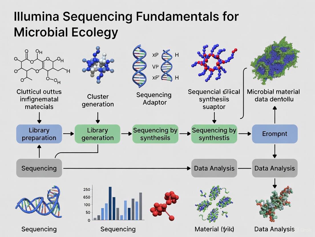
The Illumina NGS Workflow
The standard Illumina NGS workflow comprises four integrated steps: nucleic acid extraction, library preparation, sequencing, and data analysis [25] [14]. Each step requires careful execution and quality control to ensure optimal results for microbial ecology studies.
Nucleic Acid Extraction
The initial step in any NGS workflow involves isolating genetic material from microbial samples, which may include pure cultures, complex environmental samples, or clinical isolates. The quality of extracted nucleic acids critically impacts downstream sequencing results, making this a crucial stage for obtaining reliable data [24].
Key considerations for nucleic acid extraction include:
- Yield: Sufficient quantities of DNA or RNA must be obtained for library preparation, typically ranging from nanograms to micrograms depending on the application. For samples with limited material, such as single cells or low-biomass environments, whole genome amplification (WGA) or whole transcriptome amplification (WTA) may be employed [24].
- Purity: Isolated nucleic acids must be free of contaminants that could inhibit enzymatic reactions during library preparation, including phenol, ethanol, heparin, or humic acids from environmental samples [24].
- Quality: DNA integrity and RNA preservation are essential for generating representative sequencing libraries. Microbial DNA should be of high molecular weight and intact, while RNA should show minimal degradation [24].
Quality assessment typically involves UV spectrophotometry (A260/A280 and A260/A230 ratios), fluorometric quantification, and gel-based or microfluidic electrophoresis to evaluate fragment size distribution [24]. For RNA sequencing, the RNA Integrity Number (RIN) provides a quantitative measure of RNA quality [24].
Library Preparation
Library preparation converts extracted nucleic acids into sequenceable fragments compatible with Illumina platforms. This process involves several key steps that vary depending on the specific application but generally include [24]:
- Fragmentation: DNA or cDNA is fragmented into appropriate sizes for sequencing, typically ranging from 200-500 bp, though longer inserts are possible with certain applications.
- Adapter Ligation: Platform-specific oligonucleotide adapters (P5 and P7) are ligated to fragment ends, enabling hybridization to flow cell oligonucleotides and serving as priming sites for sequencing and amplification.
- Size Selection: Libraries may undergo size selection to remove fragments outside the desired size range, improving sequencing efficiency and data quality.
- Library Amplification: PCR amplification may be performed to enrich for adapter-ligated fragments, particularly when working with limited starting material.
- Library Quantification: Final libraries are quantified using fluorometric methods or quantitative PCR to ensure optimal loading concentrations for sequencing.
For microbial ecology studies, additional steps such as target enrichment may be incorporated to focus sequencing on specific genomic regions of interest, such as marker genes (e.g., 16S rRNA for bacteria) or virulence factors [21].
Clonal Amplification and Sequencing
Prior to sequencing, library fragments undergo clonal amplification to create sufficient copies for detection. In Illumina systems, this occurs on the flow cell surface through either bridge amplification or exclusion amplification (ExAmp) chemistry [24].
In bridge amplification, each library fragment anneals to complementary oligonucleotides on the flow cell and undergoes repeated rounds of amplification, forming clusters of identical DNA molecules. Each cluster originates from a single library fragment and generates sufficient signal intensity for base detection during sequencing [24]. The ExAmp chemistry, used with patterned flow cells, enables instantaneous amplification of individual fragments while preventing cross-contamination between sites [24].
Following cluster generation, sequencing proceeds using the SBS chemistry described previously. The flow cell is loaded into an Illumina sequencer, where cycles of nucleotide incorporation, imaging, and cleavage generate sequence reads of predetermined length [24]. Modern Illumina platforms offer run times ranging from several hours to days, depending on the instrument type and read length requirements.
Data Analysis
The final NGS workflow stage converts raw sequencing data into biologically meaningful information through bioinformatics analysis. This process typically involves three stages [24]:
- Primary Analysis: Base calling, demultiplexing of indexed samples, and quality control assessment.
- Secondary Analysis: Read alignment to reference genomes or de novo assembly, variant calling, and quantitative analysis.
- Tertiary Analysis: Biological interpretation, including pathway analysis, variant annotation, and comparative genomics.
For microbial ecology applications, specialized bioinformatics tools are employed for tasks such as taxonomic classification, functional annotation, metagenomic assembly, and phylogenetic analysis [26]. Illumina offers integrated data analysis solutions through its DRAGEN Bio-IT Platform, which provides highly accurate, rapid secondary analysis, and BaseSpace Sequence Hub, a cloud computing environment with specialized applications for microbial genomics [26].
NGS Workflow Visualization
Figure 1: Comprehensive NGS Workflow for Microbial Ecology. The diagram illustrates the sequential steps in Illumina next-generation sequencing, highlighting critical quality control checkpoints that ensure data reliability for microbial community analysis.
Key NGS Platforms and Their Applications in Microbial Ecology
Table 1: Comparison of NGS Technologies for Food Science Applications (Adapted from [27])
| NGS Technology | Principle | Advantages | Disadvantages | Microbial Ecology Applications |
|---|---|---|---|---|
| Illumina | Sequencing by synthesis | High throughput and accuracy | Short reads, high initial investment | Metagenetics for evaluating environmental quality; Whole Genome Sequencing of environmental pathogens; Metatranscriptomics for microbial function |
| Ion Torrent | Sequencing by synthesis, detection of H+ ions | Small sample size needed, fast sequencing | Short reads, relatively higher error rate | Metagenetics for aquatic systems; Investigation of microbial communities in specialized habitats |
| PacBio | Single-molecule real-time (SMRT) sequencing | Long reads, high accuracy, minimal bias | High initial investment, large sequencer size | Complete genome sequencing of unculturable microbes; Analysis of complex microbial communities |
| Nanopore | Nanopore electrical signal sequencing | Long reads, portability, easy to use | Relatively high error rates | In-field identification of environmental pathogens; Real-time antimicrobial resistance gene monitoring; Spoilage microorganism detection |
Essential Research Reagent Solutions
Table 2: Key Research Reagents and Their Functions in NGS Workflows
| Reagent Category | Specific Examples | Function in NGS Workflow |
|---|---|---|
| Nucleic Acid Extraction Kits | DNA/RNA isolation kits for various sample types | Lyses cells and purifies genetic material from environmental samples, ensuring yield, purity, and quality needed for library preparation [24] |
| Library Preparation Kits | Illumina DNA Prep, Nextera XT | Fragments nucleic acids and attaches platform-specific adapters, converting samples into sequenceable libraries [25] |
| Sequence Adapters | P5 and P7 oligos | Oligonucleotides with sequences complementary to flow cell primers, enabling fragment attachment and cluster generation [24] |
| Target Enrichment Panels | Microbial pathogen panels, 16S rRNA panels | Captures genomic regions of interest through hybridization, allowing focused sequencing on specific genes or microbial groups [21] |
| Quality Control Reagents | Qubit dsDNA HS Assay, Bioanalyzer DNA chips | Quantifies and qualifies nucleic acids and prepared libraries at critical workflow stages to ensure optimal sequencing performance [24] |
Applications in Microbial Ecology and Environmental Research
Illumina NGS technologies have enabled significant advances in microbial ecology by providing culture-independent methods for characterizing complex microbial communities. Several key applications demonstrate the transformative impact of these technologies:
Environmental Monitoring and Ecosystem Assessment
In environmental microbiology, NGS enables comprehensive analysis of microbial communities driving critical ecosystem processes. Researchers can track microbial population dynamics in response to environmental changes, monitor ecosystem health through bioindicator taxa, and characterize novel microorganisms without culturing requirements [22]. The deep sequencing capability of Illumina platforms allows detection of rare taxa that may serve as early warning indicators of environmental stress or ecosystem disruption.
Restoration ecology particularly benefits from NGS approaches, as microbial communities provide sensitive metrics of ecosystem recovery. By comparing microbial composition and functional potential between disturbed and reference sites, researchers can assess restoration progress and identify missing functional groups that may require intervention [22]. The ability to sequence entire microbial communities rather than relying on culturable representatives has revealed the astonishing diversity of microbial life and its importance in ecosystem functioning.
Food Safety and Quality Management
NGS has revolutionized food microbiology by enabling precise tracking of foodborne pathogens, monitoring microbial communities during food production, and authenticating food products. Whole Genome Sequencing (WGS) of foodborne pathogens allows for high-resolution strain typing and source tracking during outbreak investigations, significantly improving public health responses [27].
In fermented food production, NGS tools monitor starter culture performance, track microbial succession during fermentation, and identify spoilage organisms. Metatranscriptomic approaches reveal gene expression patterns underlying flavor development and quality attributes, enabling optimization of fermentation processes [27]. The application of NGS in food authentication helps detect fraud and verify product provenance, supporting food safety and regulatory compliance.
Microbial Source Tracking and Antibiotic Resistance Surveillance
NGS technologies provide powerful approaches for tracking microbial contamination in environmental samples and monitoring the spread of antibiotic resistance genes. Metagenomic sequencing can identify sources of fecal pollution in water systems by characterizing the full microbial community composition, offering advantages over traditional marker-based methods [27].
The expanding capability to sequence complex microbial communities directly from environmental samples without culturing has revealed extensive reservoirs of antibiotic resistance genes in natural environments. This information is critical for understanding the emergence and dissemination of antibiotic resistance and developing mitigation strategies [27] [22].
Methodological Considerations for Microbial Ecology Studies
Experimental Design and Sampling Strategies
Robust experimental design is particularly important in microbial ecology studies due to the inherent complexity and variability of microbial communities. Appropriate sampling strategies must account for spatial and temporal heterogeneity in microbial distributions [22]. Sample replication is essential for distinguishing biological patterns from technical variability, though the high cost of NGS has sometimes led to inadequate replication in previous studies [22].
Modern approaches recommend collecting sufficient replicates to capture system variability while considering sequencing depth requirements for detecting rare taxa. For composite sampling strategies, researchers should maintain individual samples for variability assessment before pooling [22]. Metadata collection encompassing environmental parameters (temperature, pH, nutrient levels, etc.) is crucial for interpreting sequencing results and identifying environmental drivers of microbial community structure.
Technical Considerations and Quality Control
Successful NGS experiments in microbial ecology require careful attention to technical details throughout the workflow. Sample collection and storage conditions must preserve nucleic acid integrity and represent in vivo microbial communities. Snap freezing, rapid drying, or chemical preservatives may be employed to prevent nucleic acid degradation or microbial growth after sampling [27].
Library preparation methods must be selected based on application requirements, with consideration for potential biases introduced during amplification or adapter ligation. For quantitative applications like metatranscriptomics, methods preserving original abundance relationships are essential [27]. Sequencing depth must be sufficient for the research question, with deeper sequencing required for detecting rare community members or for de novo genome assembly.
Bioinformatic analysis requires appropriate parameter settings and algorithm selection for specific data types and research questions. Validation of bioinformatic pipelines with mock microbial communities of known composition helps identify technical biases and optimize analytical approaches [22].
Illumina next-generation sequencing has fundamentally transformed microbial ecology research by providing powerful tools for characterizing microbial communities with unprecedented depth and resolution. The core technology, based on sequencing by synthesis with reversible terminators, enables highly accurate, massive parallel sequencing of DNA fragments, making it possible to address complex biological questions about microbial diversity, function, and dynamics.
The standardized NGS workflow—encompassing nucleic acid extraction, library preparation, sequencing, and bioinformatic analysis—provides a robust framework for investigating microbial systems across diverse environments, from natural ecosystems to engineered systems. As sequencing costs continue to decline and analytical methods improve, NGS technologies will undoubtedly yield further insights into microbial ecology, enabling more precise monitoring of environmental systems, enhanced tracking of microbial contaminants, and improved understanding of ecosystem functioning.
For researchers in microbial ecology and environmental science, understanding the fundamental principles of Illumina sequencing technologies provides a foundation for selecting appropriate experimental approaches, interpreting sequencing data, and leveraging genomic insights to address pressing challenges in environmental sustainability, public health, and ecosystem management.
Why Illumina? Key Advantages for Microbial Community Analysis
Within the field of microbial ecology, the accurate characterization of diverse microbial communities is fundamental to advancing our understanding of environmental and human health. Next-generation sequencing (NGS) technologies have revolutionized this field, with Illumina sequencing emerging as a predominant platform for 16S rRNA gene-based surveys. This whitepaper details the core technical advantages of Illumina technology, framing it as an essential tool for researchers. We examine its high data accuracy, cost-effective throughput, and robust, standardized bioinformatics pipelines that together provide a reliable foundation for broad-scale microbial community profiling, while also acknowledging its limitations relative to emerging long-read platforms.
The 16S ribosomal RNA (rRNA) gene is a cornerstone for microbial ecology research, containing conserved regions that serve as universal primer-binding sites and hypervariable regions (V1–V9) that provide taxonomic specificity for bacterial classification [28]. The choice of sequencing platform is a critical methodological decision that directly influences the resolution, accuracy, and scope of microbial community data.
Illumina sequencing has established itself as a benchmark for high-accuracy, short-read sequencing. It typically targets specific hypervariable regions, such as V3-V4, generating millions of short, paired-end reads (~300 bp) with an exceptionally low error rate of less than 0.1% [28]. This whitepaper explores the fundamental technical strengths of Illumina sequencing that make it particularly suited for microbial community analysis, especially within large-scale or reproducibility-focused studies.
Core Technical Advantages of Illumina Sequencing
A comparative analysis of sequencing technologies reveals a clear set of advantages for Illumina in specific application contexts.
Superior Sequencing Accuracy and Data Fidelity
Illumina's core strength lies in its high base-call accuracy. This technology produces sequences with an error rate of <0.1%, a figure that is crucial for distinguishing between closely related microbial taxa and for detecting rare taxa within a community [28]. This high fidelity minimizes false positives in variant calling and provides a reliable foundation for quantitative analyses. In contrast, while improving, Oxford Nanopore Technologies (ONT) has historically exhibited higher error rates in the range of 5–15% [28] [29].
High-Throughput and Cost-Efficiency for Large-Scale Studies
Illumina platforms provide an unparalleled combination of high throughput and cost-efficiency, making them ideal for population-level studies and projects with large sample sizes. One study noted an average of 30,184 ± 1,146 reads per sample for Illumina, which, while lower in count than ONT in some direct comparisons, represents a highly cost-effective and accurate data generation method [30]. This scalability enables researchers to achieve the statistical power necessary for robust ecological inference.
High-Resolution Taxonomic Classification
The high accuracy of Illumina sequencing enables reliable taxonomic classification down to the genus level. A comparative study on gut microbiota demonstrated that Illumina successfully classified 80% of sequences to the genus level [30]. While this was lower than ONT's 91%, it remains a robust performance for many ecological questions focused on community shifts at the genus level or above. The high throughput ensures sufficient sequence depth to capture a broad range of taxa, with studies indicating that Illumina can capture greater species richness compared to some long-read platforms in certain sample types [28] [29].
Table 1: Comparative Performance of Sequencing Platforms for 16S rRNA Analysis
| Feature | Illumina (e.g., NextSeq, MiSeq) | Oxford Nanopore (e.g., MinION) | PacBio HiFi |
|---|---|---|---|
| Read Length | Short (~300 bp, targets hypervariable regions) | Long (~1,500 bp, full-length 16S) | Long (~1,453 bp, full-length 16S) |
| Error Rate | Very Low (<0.1%) | Historically Higher (5-15%) | Very Low (~Q27) |
| Typical Output/Throughput | High (e.g., 30,184 ± 1,146 reads/sample [30]) | Variable, can be very high (e.g., 630,029 ± 92,449 reads/sample [30]) | Moderate (e.g., 41,326 ± 6,174 reads/sample [30]) |
| Primary Strength | High accuracy, cost-effectiveness, large-scale studies | Species-level resolution, real-time portability | High accuracy long reads for species-level resolution |
| Optimal Use Case | Genus-level profiling, broad microbial surveys, large cohort studies | Applications requiring species/strain resolution in the field or lab | Applications demanding high accuracy and species-level resolution |
Established and Standardized Bioinformatics Ecosystems
Illumina data is supported by a mature and robust bioinformatics ecosystem, which simplifies data analysis and ensures reproducibility. Workflows like nf-core/ampliseq provide standardized, containerized pipelines that handle data from quality control (using tools like FastQC and MultiQC) through primer trimming (Cutadapt), error correction, and Amplicon Sequence Variant (ASV) generation using DADA2 [28]. This well-supported computational environment lowers the barrier to entry and facilitates comparative meta-analyses across different studies.
Detailed Experimental Protocol: Illumina 16S rRNA Gene Sequencing
The following section outlines a standard laboratory protocol for preparing 16S rRNA gene sequencing libraries for the Illumina platform, as cited in recent literature [28].
Sample Collection and DNA Extraction
- Sample Collection: Respiratory samples (e.g., from ventilator-associated pneumonia patients) are collected and immediately stored at -80°C to preserve microbial DNA integrity.
- DNA Extraction: Genomic DNA is extracted using a commercial kit (e.g., Sputum DNA Isolation Kit, Norgen Biotek). The protocol follows the manufacturer's instructions, with potential modifications to optimize DNA yield and purity for challenging sample types.
- Quality Control: The concentration and purity of the extracted DNA are assessed using a spectrophotometer (e.g., Nanodrop 2000) and a fluorometer (e.g., Qubit 4).
Library Preparation for Illumina Sequencing
This protocol uses the QIAseq 16S/ITS Region Panel (Qiagen) [28].
- First-Stage PCR (Target Amplification): The V3-V4 hypervariable region of the 16S rRNA gene is amplified using region-specific primers.
- Amplification Program:
- Denaturation: 95°C for 5 minutes.
- 20 cycles of:
- Denaturation: 95°C for 30 seconds.
- Annealing: 60°C for 30 seconds.
- Extension: 72°C for 30 seconds.
- Final Elongation: 72°C for 5 minutes.
- Amplification Program:
- Second-Stage PCR (Index Attachment): A second, limited-cycle PCR is performed to attach unique dual indices and Illumina adapter sequences (e.g., from the QIAseq 16S/ITS Index Kit). This enables sample multiplexing.
- Library Validation and Pooling: The final PCR products are validated (e.g., using an Agilent Bioanalyzer) and pooled in equimolar ratios to create the final sequencing library.
Sequencing
- The pooled library is loaded onto an Illumina sequencing platform, such as the NextSeq, for paired-end sequencing (2 × 300 bp) according to the manufacturer's specifications.
The following workflow diagram summarizes this standardized experimental process.
The Scientist's Toolkit: Essential Research Reagents and Materials
The following table details key reagents and kits used in a standard Illumina 16S rRNA sequencing workflow, as featured in the cited experimental protocol [28].
Table 2: Key Research Reagent Solutions for Illumina 16S rRNA Sequencing
| Item | Function / Application | Example Product (from cited protocol) |
|---|---|---|
| DNA Extraction Kit | Isolation of high-quality genomic DNA from complex biological samples. | Sputum DNA Isolation Kit (Norgen Biotek) [28] |
| 16S Amplicon Panel | Provides primers and master mix for targeted amplification of specific 16S rRNA hypervariable regions. | QIAseq 16S/ITS Region Panel (Qiagen) [28] |
| Indexing Kit | Provides unique nucleotide barcodes for multiplexing multiple samples in a single sequencing run. | QIAseq 16S/ITS Index Kit (Qiagen) [28] |
| Library Quantification | Accurate quantification of DNA library concentration prior to sequencing. | Qubit dsDNA HS Assay Kit (Thermo Fisher) [31] |
| Sequencing Platform | High-throughput instrument for generating short-read sequence data. | Illumina NextSeq [28] |
Limitations and Complementary Technologies
A complete understanding of Illumina's role requires acknowledging its limitations. The primary constraint is the short read length, which often prevents reliable classification down to the species level due to insufficient informational content in the short, targeted hypervariable regions [28] [30]. In contrast, long-read platforms like Oxford Nanopore and PacBio sequence the entire ~1,500 bp 16S rRNA gene, providing higher taxonomic resolution and enabling better differentiation of closely related species [28] [31].
Furthermore, PCR amplification steps, common to most amplicon sequencing approaches including Illumina, can introduce amplification biases, preferentially amplifying certain templates and potentially distorting true quantitative relationships [32] [33].
Consequently, while Illumina is ideal for broad microbial surveys and genus-level profiling, studies requiring species- or strain-level resolution may benefit from a hybrid sequencing approach, leveraging the strengths of both short- and long-read technologies [28] [29] [33].
Illumina sequencing remains a powerful and preferred platform for microbial community analysis, particularly for studies where high accuracy, cost-effective throughput, and reproducible genus-level classification are the primary objectives. Its well-established protocols and mature bioinformatics ecosystem make it a reliable and accessible choice for large-scale microbial surveys in both environmental and clinical settings. As the field advances, the strategic combination of Illumina's broad profiling capabilities with the high resolution of long-read technologies promises to further deepen our understanding of complex microbial ecosystems.
In the field of microbial ecology, precise terminology is foundational for interpreting data and communicating findings. The advent of next-generation sequencing (NGS), particularly platforms developed by Illumina, has revolutionized our capacity to study complex microbial communities [34] [35]. This technical guide defines four core terms—Microbiota, Microbiome, Metagenomics, and Operational Taxonomic Units (OTUs)—within the context of Illumina sequencing. A clear grasp of these concepts is essential for researchers and drug development professionals designing and interpreting microbial ecology studies.
Defining the Core Concepts
The following table provides concise definitions and key characteristics of the core terminology.
Table 1: Core Terminology in Microbial Ecology Research
| Term | Definition | Key Characteristics |
|---|---|---|
| Microbiota | The assemblage of living microorganisms present in a defined environment [36]. | • Refers to the microorganisms themselves (bacteria, archaea, fungi, viruses) [36].• Often used to describe taxonomic composition (e.g., phylum, genus) [34]. |
| Microbiome | The entire ecological niche of a microbiota, including the microorganisms, their genomes, and the surrounding environmental conditions [36]. | • A broader term that encompasses the microbiota [36].• Includes the collective genomic material of the microbiota (the metagenome) and microbial functions and activities [34] [36]. |
| Metagenomics | The direct genetic analysis of genomes contained within an environmental sample, bypassing the need for cultivation [34] [37]. | • A sequencing-based approach to study the microbiome [34].• Shotgun metagenomics sequences all DNA in a sample, enabling taxonomic and functional profiling [34] [38].• 16S rRNA gene sequencing (metataxonomics) targets a specific marker gene for taxonomic profiling [38]. |
| Operational Taxonomic Unit (OTU) | A cluster of similar DNA sequences (e.g., 16S rRNA reads) used to classify and quantify microbial taxa in a sample [39] [37]. | • An operational proxy for a microbial species or genus, typically defined by a sequence similarity threshold (e.g., 97%) [39].• Reduces dataset complexity by grouping sequences into biologically relevant units for diversity analysis [39]. |
The Technological Driver: Illumina Sequencing in Microbial Ecology
The progress in microbiome research is intrinsically linked to advancements in NGS technology. Illumina's next-generation sequencing platforms provide the high-throughput, scalable, and cost-effective data generation required to dissect complex microbial communities [35].
The paradigm shift from studying single microorganisms in isolation to analyzing entire communities was enabled by massively parallel sequencing [34]. This technology allows for the simultaneous detection, quantification, and characterization of thousands of microbial taxa and their genes from a single sample [34]. For microbiome research, two primary metagenomic sequencing strategies are employed: 16S rRNA gene sequencing and shotgun metagenomic sequencing [38].
Table 2: Comparison of Primary Metagenomic Sequencing Approaches
| Feature | 16S rRNA Gene Sequencing (Metataxonomics) | Whole Genome Shotgun Sequencing (Metagenomics) |
|---|---|---|
| Target | Amplified hypervariable regions of the 16S rRNA gene [38]. | All genomic DNA in a sample, fragmented randomly [34] [38]. |
| Primary Output | Operational Taxonomic Units (OTUs) or Amplicon Sequence Variants (ASVs) [38]. | Sequencing reads from all genomic material present. |
| Analysis Focus | Taxonomic identification and profiling of the bacterial and archaeal community [38]. | Comprehensive taxonomic profiling and functional potential analysis (gene content) [34] [38]. |
| Resolution | Limited to genus or species level; can miss strains [39] [38]. | Higher resolution, capable of reaching species and strain level, and identifying less abundant taxa [38]. |
| Functional Insight | Indirect, inferred from identified taxa. | Direct, via analysis of sequenced genes and metabolic pathways [34]. |
| Required Sequencing Depth | Lower (e.g., tens of thousands of reads per sample) [38]. | Significantly higher (e.g., millions of reads per sample) for adequate coverage [38]. |
| Key Limitation | Primer bias can affect taxonomic representation; limited functional data [38]. | Higher cost and computational demand; requires complex bioinformatic analysis [34] [38]. |
The relationship between these core concepts and the sequencing workflow can be visualized as a logical pathway from sample to insight.
Diagram 1: From Sample to Microbiome Insight. This workflow illustrates how Illumina sequencing of a sample generates data that, through bioinformatic analysis, answers the fundamental questions of "what's there?" (Microbiota/OTUs) and "what does it do?" (Microbiome).
Operational Taxonomic Units (OTUs): The Unit of Diversity
Definition and Purpose
An Operational Taxonomic Unit (OTU) is a cluster of DNA sequences, grouped based on their similarity, which serves as a proxy for a microbial taxon (e.g., species or genus) in a marker-gene analysis [39]. The standard practice is to cluster 16S rRNA gene sequences that share ≥97% nucleotide identity into a single OTU, approximating a bacterial species [39]. This clustering reduces the complexity of millions of sequencing reads into manageable units for ecological analysis, accounting for both real biological variation and potential sequencing errors [39].
Methodologies for OTU Picking
There are several computational approaches for clustering sequences into OTUs, each with strengths and weaknesses [37].
Table 3: Primary Methodologies for OTU Picking
| Method Type | Principle | Advantages | Disadvantages |
|---|---|---|---|
| De Novo Clustering | Groups all sequences against each other without a reference, based on pairwise sequence distances [37]. | • Does not require a reference database.• Can detect novel, uncharacterized taxa [37]. | • Computationally intensive.• Results can be specific to a single study. |
| Closed-Reference Clustering | Compares each sequence to an annotated reference database; sequences matching the same reference are grouped [37]. | • Fast and computationally efficient.• Provides consistent OTUs across studies. | • Fails to classify sequences not in the database, losing novel diversity [37]. |
| Open-Reference Clustering | A hybrid approach: first uses closed-reference clustering, then clusters unassigned sequences de novo [37]. | • Combines speed with the ability to capture novel taxa. | • Uses two different OTU definitions, which can complicate analysis [37]. |
The choice of method impacts the biological interpretation. De novo clustering is often considered the most powerful for capturing comprehensive diversity within a dataset, making it a preferred choice for many researchers [37].
Experimental Protocols and Considerations
A Protocol for Comparative Metagenomic Analysis
A detailed methodology from a cited study comparing 16S and shotgun sequencing [38] is summarized below.
Objective: To compare the reliability of 16S rRNA gene sequencing and shotgun metagenomic sequencing for taxonomic profiling of the gut microbiota.
Experimental Workflow:
- Sample Collection & DNA Extraction: Gut content samples (e.g., from chicken crop and caeca) were collected at different time points. Total genomic DNA was extracted from all samples.
- Library Preparation & Sequencing:
- 16S rRNA Protocol: The hypervariable regions of the 16S rRNA gene were amplified using specific primers. The resulting amplicons were sequenced on an Illumina platform.
- Shotgun Metagenomic Protocol: Total genomic DNA was mechanically sheared into short fragments without target-specific amplification. Standard Illumina sequencing libraries were prepared and sequenced.
- Bioinformatic Analysis:
- 16S Data Processing: Sequences were quality-filtered and then clustered into OTUs using a de novo clustering method (e.g., in QIIME or mothur) at a 97% identity threshold. Taxonomy was assigned by comparing representative sequences from each OTU to a reference database (e.g., Greengenes or SILVA) [39] [38].
- Shotgun Data Processing: Reads were quality-filtered and then analyzed using two approaches:
- Taxonomic Profiling: Reads were directly classified against a microbial genome database to estimate taxonomic abundance.
- De novo Assembly: Reads were assembled into longer contigs, which were then binned and annotated to reconstruct metagenome-assembled genomes (MAGs).
- Comparative Statistics: The relative abundance distributions, alpha-diversity, and beta-diversity metrics were compared. A differential abundance analysis (e.g., using DESeq2) was performed to test the ability of each method to distinguish between experimental conditions (e.g., gut compartment, sampling time) [38].
Key Findings from the Comparative Study
The comparative study highlighted critical considerations for experimental design [38]:
- Shotgun sequencing identified a statistically significant higher number of taxa, particularly less abundant genera, compared to 16S sequencing when a sufficient depth of sequencing (>500,000 reads per sample) was achieved.
- The less abundant genera detected exclusively by shotgun sequencing were biologically meaningful and able to discriminate between experimental conditions as effectively as the more abundant genera.
- 16S sequencing suffered from detection limitations, leading to the partial or total absence of some genera in its profile, which in turn caused discordance in differential abundance results compared to shotgun sequencing.
The Scientist's Toolkit: Essential Reagents and Materials
Successful metagenomic research relies on a suite of wet-lab and computational tools. The following table details key solutions used in typical workflows.
Table 4: Key Research Reagent Solutions and Materials for Metagenomics
| Item | Function in Workflow |
|---|---|
| Illumina DNA Prep Kit | A standardized library preparation kit for preparing genomic DNA for shotgun metagenomic sequencing on Illumina platforms. |
| 16S rRNA Primers | Specific oligonucleotide pairs (e.g., targeting the V4 region) used to amplify the 16S rRNA gene for metataxonomic studies. |
| NovaSeq & MiSeq i100 Reagents | Flow cells and sequencing reagents for Illumina's sequencing instruments. The MiSeq i100 series is designed for simplicity in lower-throughput runs. |
| QIIME 2 | A powerful, extensible, and decentralized bioinformatics platform for analyzing and integrating microbiome data from 16S and shotgun sequencing. |
| MetaPhlAn | A computational tool for profiling the taxonomic composition of microbial communities from shotgun metagenomic data. |
| Kraken2 | A system for assigning taxonomic labels to metagenomic DNA sequences, using exact k-mer matches to a reference database. |
The precise interpretation of microbiota, microbiome, metagenomics, and OTUs is fundamental for microbial ecology. These concepts are interconnected: Illumina NGS enables metagenomic sequencing, which allows researchers to define OTUs and characterize the collective genome of a microbiota to understand the functional potential of the microbiome. As sequencing technologies continue to evolve, becoming more accessible and powerful, these core terms will remain the essential vocabulary for unlocking the profound influence of microbes on human health, disease, and the global ecosystem.
From Sample to Sequence: Practical Illumina Workflows for Ecological Insights
Next-Generation Sequencing (NGS) has revolutionized microbial ecology research, enabling comprehensive analysis of microbial communities directly from environmental samples without the need for cultivation. Illumina sequencing technologies provide the high accuracy and throughput required to decode complex microbial ecosystems, from soil and water to the human microbiome. This technical guide details the core steps of the Illumina NGS workflow, framed within the context of microbial ecology research, to empower scientists and drug development professionals in their genomic discoveries.
The Fundamental NGS Workflow: A Four-Step Process
The Illumina next-generation sequencing workflow transforms raw biological samples into actionable genomic data through a structured series of laboratory and computational steps. This process remains consistent across various applications but requires specific considerations for microbial ecology studies [25] [24].
Detailed Workflow Breakdown: From Sample to Sequence
Step 1: Nucleic Acid Extraction and Quality Control
The workflow begins with the isolation of genetic material from microbial samples, which may include environmental samples, bulk tissue, individual cells, or biofluids [25]. For microbial ecology studies, this step is critical as sample types vary widely from soil and water to host-associated environments.
Key Considerations for Microbial Ecology:
- Yield: Sufficient nucleic acid must be obtained for subsequent library preparation, typically nanograms to micrograms of DNA or RNA [24].
- Purity: Isolated DNA or RNA should be free of inhibitors that can interfere with downstream enzymatic reactions. Common contaminants include phenol, ethanol, heparin, or humic acids from environmental samples [24].
- Quality: Nucleic acid integrity is essential. For DNA, high molecular weight and intact strands are ideal, while for RNA, degradation should be minimized [24].
Quality Assessment Methods:
- UV spectrophotometry for purity assessment (A260/A280 and A260/A230 ratios)
- Fluorometric methods for accurate nucleic acid quantitation [25]
- Gel-based or microfluidic electrophoresis for determining fragment size distribution [24]
- RNA Integrity Number (RIN) for assessing RNA quality [24]
For low-biomass microbial samples, such as those from extreme environments or host-associated niches with high host DNA contamination, whole-genome amplification (WGA) or whole-transcriptome amplification (WTA) may be necessary to obtain sufficient material for sequencing [24] [12].
Step 2: Library Preparation - Bridging Biology to Sequencing Technology
Library preparation converts isolated nucleic acids into a format compatible with Illumina sequencing systems. This crucial step fragments the genetic material and adds platform-specific adapters [25] [40].
Core Library Preparation Steps:
- Nucleic Acid Fragmentation: Sample DNA or RNA is fragmented into appropriate sizes for massively parallel sequencing [24].
- Adapter Ligation: Illumina-specific adapter sequences (P5 and P7) are attached to fragment ends, enabling binding to the flow cell and initiating sequencing [24]. Adapters may also include barcodes for sample multiplexing and unique molecular identifiers to distinguish biological duplicates from technical artifacts [40].
- Library Quantification: Prepared libraries are quantified via fluorometric spectroscopy or real-time PCR to ensure optimal loading concentration on the sequencer [24].
Microbial Ecology-Specific Library Strategies:
- Targeted Amplicon Sequencing: Amplifies specific marker genes like bacterial 16S rRNA, fungal ITS, or eukaryotic 18S rRNA to profile community membership [40] [12]. This approach minimizes host-derived sequences in host-associated samples and has well-established bioinformatics pipelines [40].
- Shotgun Metagenomic Sequencing: Sequences all DNA in a sample without targeting specific regions, enabling analysis of both community composition and functional potential [40] [12]. This method provides a more comprehensive view of microbial ecosystems but may generate substantial host sequence data in host-associated samples [12].
- Metatranscriptomics: Sequences RNA to profile actively expressed genes and understand microbial community function in response to environmental conditions [40].
Table 1: Library Preparation Strategies for Microbial Ecology
| Approach | Target | Key Applications | Advantages | Limitations |
|---|---|---|---|---|
| 16S/ITS Amplicon | 16S rRNA (bacteria/archaea) or ITS (fungal) regions | Community profiling, diversity analysis | Cost-effective, well-established bioinformatics, minimizes host background | Limited functional information, PCR bias [12] |
| Shotgun Metagenomics | All genomic DNA | Community composition & functional potential, genome assembly | Provides functional insights, strain-level resolution | Higher cost, computationally intensive, host DNA contamination issues [12] |
| Metatranscriptomics | RNA transcripts | Gene expression, active metabolic pathways | Reveals active functions, dynamic responses | RNA instability, rRNA depletion needed [40] |
Step 3: Sequencing - SBS Chemistry and Platform Selection
Illumina sequencing employs proven Sequencing by Synthesis (SBS) technology, which detects single bases as they are incorporated into growing DNA strands [25]. The process involves:
- Cluster Generation: DNA fragments undergo clonal amplification on a flow cell through bridge amplification, creating clusters of identical molecules [24].
- Cyclic Reversible Termination: Each sequencing cycle incorporates a single fluorescently-labeled dNTP with a reversible terminator. After imaging to identify the incorporated base, the terminator is cleaved to enable the next incorporation cycle [24].
Sequencing Platform Selection for Microbial Ecology: Illumina offers a range of sequencing platforms with varying throughput and capabilities. Selection depends on project scale, required read length, and application focus.
Table 2: Illumina Sequencing Platform Comparison for Microbial Ecology Applications
| Platform | Max Output | Run Time | Max Read Length | Key Microbial Ecology Applications |
|---|---|---|---|---|
| MiSeq i100 | 30 Gb | ~4-24 hours | 2 × 500 bp | Targeted gene sequencing (e.g., 16S), small genome sequencing [41] |
| NextSeq 1000/2000 | 540 Gb | ~8-44 hours | 2 × 300 bp | Shotgun metagenomics, transcriptome sequencing, exome sequencing [41] |
| NovaSeq X Series | 8-16 Tb | ~17-48 hours | 2 × 150 bp | Large-scale WGS, population-level metagenomics [41] |
For microbial ecology, the MiSeq system is commonly used for 16S rRNA amplicon sequencing due to its longer read lengths, while the NextSeq and NovaSeq platforms are preferred for shotgun metagenomics requiring higher throughput [41] [12].
Step 4: Data Analysis - From Raw Sequences to Biological Insights
Bioinformatics processing converts raw sequencing data into meaningful biological insights through multiple computational stages [24]. For microbial ecology, analysis strategies differ significantly between targeted amplicon and shotgun metagenomic approaches.
Data Analysis Workflow:
Primary Analysis:
Secondary Analysis:
- For targeted amplicon data: Clustering into Operational Taxonomic Units (OTUs) or Amplicon Sequence Variants (ASVs), taxonomic classification against reference databases [12]
- For shotgun metagenomics: Assembly into contigs, binning to group contigs into Metagenome-Assembled Genomes (MAGs), taxonomic profiling, and gene prediction [12]
Tertiary Analysis:
- Diversity assessments (alpha and beta diversity)
- Differential abundance testing
- Functional annotation and pathway analysis
- Comparative genomics and phylogenetic analysis [24]
Microbial Ecology Applications and Method Selection
In microbial ecology, the choice between targeted amplicon sequencing and whole-genome shotgun (WGS) metagenomics depends on research questions, sample type, and resources [12].
Key Advantages of Each Approach:
Targeted Amplicon Sequencing:
- More cost-effective for large sample sets
- Simplified data analysis and established pipelines
- Higher sensitivity for low-biomass samples
- Reduced host background in host-associated samples [12]
Shotgun Metagenomics:
- Provides strain-level resolution
- Reveals functional potential through gene content analysis
- Enables reconstruction of metagenome-assembled genomes (MAGs)
- Avoids PCR amplification biases associated with primer selection [12]
Emerging Technologies and Future Directions
Illumina continues to advance NGS technologies with innovations that will further transform microbial ecology research:
Constellation Mapped Read Technology: Estimated availability in the first half of 2026, this innovation eliminates traditional library preparation by enabling direct loading of long, unfragmented DNA onto flow cells. It provides long-range genomic information while maintaining short-read accuracy, potentially revolutionizing the analysis of complex microbial communities [42] [43].
5-Base Solution for Methylation Studies: This end-to-end workflow enables simultaneous detection of genetic variants and methylation patterns in a single assay, using novel chemistry that converts 5-methylcytosine to thymine. This technology provides dual genomic and epigenomic insights relevant for understanding microbial epigenetic regulation [42] [43].
Spatial Transcriptomics: Expected in the first half of 2026, this technology will enable spatial mapping of gene expression patterns, potentially applicable to structured microbial communities like biofilms and microbial mats [43].
The Scientist's Toolkit: Essential Research Reagent Solutions
Table 3: Key Research Reagent Solutions for Microbial NGS Workflows
| Reagent/Solution | Function | Application in Microbial Ecology |
|---|---|---|
| Host Depletion Kits (e.g., Zymo HostZERO) | Reduces host nucleic acid background | Enhances microbial sequence recovery in host-associated samples (e.g., gut, skin) [40] |
| rRNA Depletion Kits (e.g., Zymo RiboFree) | Removes ribosomal RNA | Improves mRNA sequencing efficiency in metatranscriptomics [40] |
| Whole Genome Amplification Kits | Amplifies minimal DNA inputs | Enables sequencing from low-biomass environments [24] |
| Standardized Commercial Kits (e.g., Zymo Quick 16S) | Provides validated protocols | Ensures reproducibility and cross-study comparability [40] |
| DNA/RNA Extraction Kits | Isolates nucleic acids from diverse sample types | Optimized for challenging environmental samples (soil, sediment) |
The Illumina end-to-end NGS workflow provides microbial ecologists with powerful tools to explore and understand complex microbial communities. From nucleic acid extraction through data analysis, each step requires careful consideration of methods and reagents tailored to specific research questions and sample types. As innovations like constellation technology and 5-base sequencing emerge, they will further enhance our ability to decode microbial ecosystems with unprecedented resolution and efficiency, driving discoveries in environmental science, medicine, and biotechnology.
The accurate characterization of microbial communities in diverse environments is foundational to advancing microbial ecology research. Within the context of Illumina sequencing, the initial step of sample collection and preservation is arguably the most critical, as it fundamentally determines the quality and reliability of all subsequent genomic data. The challenges of microbiome sampling vary significantly across environments—soil, water, and host-associated niches—each presenting unique matrix effects, biomass yields, and contamination risks. Soil presents exceptional spatial heterogeneity and chemical complexity, aquatic environments often feature low microbial density, and host-associated sites can be particularly vulnerable to contamination from the host or surrounding tissues. This guide details evidence-based sampling strategies tailored to these diverse environments, providing a structured framework to ensure sample integrity from the field to the sequencer, thereby laying the groundwork for robust and reproducible Illumina sequencing outcomes.
Environment-Specific Sampling Strategies
The physical and biological characteristics of each sampling environment demand tailored approaches to preserve microbial community structure and minimize bias. Key parameters and methodologies for soil, water, and host-associated environments are detailed below.
Soil Microbiome Sampling
Soil is a spatially and temporally heterogeneous environment, requiring careful strategy to obtain representative samples.
- Spatial Considerations: Sample depth significantly influences parameters like moisture and carbon-nitrogen ratio. The rhizosphere (the soil region influenced by plant roots) creates a unique environment rich in metabolites and requires separate sampling from bulk soil [44]. Even at the microscale, uneven distribution of nutrients and organisms complicates analysis, as average properties can be misleading for understanding microscale interactions [44].
- Collection Technique: Consistent coring devices (e.g., stainless steel soil corers) should be used to obtain uniform samples. For bulk soil, samples are typically sieved (e.g., through a 2-mm sieve) to remove larger particles, rocks, and leaves [44]. To capture microbial diversity adequately, composite samples created from multiple corings within a defined area are recommended over single cores.
- Handling and Preservation: Immediate flash-freezing in liquid nitrogen and storage at -80°C is ideal for preserving nucleic acid integrity. As an alternative, placement in nucleic acid stabilization buffers (e.g., RNAlater) is effective, especially for RNA analyses. Transport to the laboratory on dry ice is essential for preserving the in-situ state of the microbial community [44].
Water Microbiome Sampling
Water samples, particularly from freshwater ecosystems like rivers, are critical for monitoring biodiversity and public health risks, including antibiotic resistance genes [45].
- Collection Methodology: For water depths exceeding 0.5 meters, samples should be collected from the centroid of flow at approximately 0.3 meters below the surface to avoid surface scum. In shallower sites (≤0.5 m depth), samples are taken at approximately one-third of the total water depth from the surface [45]. Sterile bottles must be submerged carefully to minimize surface contamination and turbulence.
- Filtration and Concentration: Microbial biomass in water is often low and must be concentrated. A standard method involves filtering a known volume of water (e.g., 100 mL to several liters, depending on turbidity) through 0.2 μm pore-size membrane filters. These filters are then aseptically placed in sterile petri dishes and stored at -80°C until DNA extraction [45].
- Transport: Samples should be transported to the laboratory in ice-filled coolers on the day of collection to slow microbial metabolic activity and preserve community structure [45].
Host-Associated Microbiome Sampling
Host-associated environments, including certain human tissues, often constitute low-biomass systems where contamination is a paramount concern [46].
- Minimizing Contamination: Sampling procedures must account for all possible contamination sources, including the host's skin, the operator, and the sampling equipment. Using single-use, DNA-free collection swabs and vessels is ideal. Where reusability is necessary, equipment should be decontaminated with 80% ethanol followed by a nucleic acid degrading solution (e.g., bleach, UV-C light) [46].
- Use of Personal Protective Equipment (PPE): Operators should wear extensive PPE—including gloves, goggles, coveralls, and masks—to limit the introduction of human-associated contaminants from aerosol droplets, skin, or clothing [46].
- Sampling Controls: The inclusion of controls is critical for identifying contaminants. These may include empty collection vessels, swabs exposed to the air in the sampling environment, swabs of PPE, or aliquots of the preservation solution. These controls must be processed alongside actual samples through all downstream steps [46].
Table 1: Key Considerations for Sampling Different Environments
| Environment | Primary Challenge | Recommended Sampling Method | Immediate Preservation Method |
|---|---|---|---|
| Soil | High spatial heterogeneity & complexity | Composite coring; sieving to 2 mm; separate rhizosphere sampling | Flash-freezing (-80°C) or nucleic acid stabilization buffer |
| Water | Low microbial biomass; seasonal flux | Depth-stratified collection; filtration through 0.2 μm membranes | Freeze membrane filter at -80°C |
| Host-Associated | Extremely low biomass; high contamination risk | Single-use DNA-free tools; stringent PPE | Flash-freezing (-80°C); immediate immersion in lysis buffer |
Contamination Control in Low-Biomass Environments
In low-biomass environments (e.g., certain human tissues, treated drinking water, hyper-arid soils, the atmosphere), the inevitable introduction of contaminant DNA from reagents, kits, or the laboratory environment can constitute a significant portion of the sequenced DNA, leading to spurious results [46]. Therefore, a contamination-conscious workflow is non-negotiable.
- Decontamination of Equipment and Reagents: Beyond using single-use consumables, all reusable tools and surfaces should be decontaminated. A two-step process is recommended: treatment with 80% ethanol to kill contaminating organisms, followed by a nucleic acid degrading solution (e.g., sodium hypochlorite, UV-C light, hydrogen peroxide) to remove residual DNA. It is critical to note that sterility is not synonymous with being DNA-free; autoclaving or ethanol treatment alone may not remove persistent extracellular DNA [46].
- Inclusion of Comprehensive Controls: Multiple types of negative controls are essential.
- Field Controls: Include samples of the sterile collection vessels, sampling fluids (e.g., drilling fluid), and swabs of the air or PPE.
- Extraction Blanks: Reagents taken through the DNA extraction process without any sample.
- PCR Blanks: Sterile water included in the amplification step. These controls allow for the bioinformatic identification and subsequent subtraction of contaminating sequences from the dataset [46].
- DNA Extraction and Kit Considerations: Commercial DNA extraction kits are commonly used, but different kits have varying efficiencies and biases depending on the sample type and target species [44]. It is vital to select a kit validated for the specific sample matrix (e.g., soil, water) and to use the same kit and protocol consistently within a study to ensure comparability. Kits designed for low-biomass inputs can also improve yield.
The following workflow outlines the critical steps for contamination-aware sampling, applicable across diverse environments, with special emphasis on procedures for low-biomass contexts.
From Sample to Sequence: Method Selection for Illumina Platforms
Once high-quality, preserved samples are obtained, selecting the appropriate Illumina sequencing method is the next critical decision. The choice depends on the research question, whether it requires broad taxonomic profiling or deep functional insights.
- 16S/ITS Amplicon Sequencing: This targeted approach amplifies and sequences specific phylogenetic marker genes (16S rRNA for bacteria/archaea, ITS for fungi). It is a cost-effective, less computationally demanding method for comparing microbial community structure and phylogeny across a large number of samples [47] [48]. Its primary limitation is the lack of functional resolution and potential for primer bias [45] [48].
- Shotgun Metagenomic Sequencing: This method sequences all the DNA in a sample, enabling not only taxonomic profiling at a higher resolution, potentially down to the species or strain level, but also functional characterization of the community. It allows for the detection of antibiotic resistance genes (ARGs), virulence factors (VFs), and other functional traits, and facilitates the reconstruction of metagenome-assembled genomes (MAGs) [49] [45] [48].
- Hybrid Capture Enrichment: A targeted method that uses biotinylated probes to enrich for specific genomic regions of interest (e.g., for broad pathogen surveillance or antimicrobial resistance profiling) from complex samples before sequencing. It is ideal for detecting low-abundance targets and requires a prior knowledge of target sequences for probe design [50].
Table 2: Comparison of Common Illumina Sequencing Methods for Microbial Ecology
| Method | Primary Application | Key Advantage | Key Limitation | Example Workflow/Kit |
|---|---|---|---|---|
| 16S/ITS Amplicon | Taxonomic profiling & community ecology | Cost-effective; simplified data analysis; high sensitivity for rare taxa | Limited functional insight; primer bias; lower taxonomic resolution | 16S Metagenomic Sequencing Library Prep [47] |
| Shotgun Metagenomic | Functional potential & higher-res taxonomy | Functional gene discovery; strain-level profiling; MAG generation | Higher cost; computationally intensive; host DNA can dominate | Illumina DNA Prep; Nextera XT [45] |
| Targeted Hybrid Capture | Detection & characterization of specific targets (e.g., pathogens, AMR) | High sensitivity for known targets in complex samples | Requires pre-defined targets; more complex workflow | Respiratory Pathogen ID/AMR Panel [50] |
The decision-making process for selecting the optimal sequencing method can be visualized as a flowchart based on research goals and practical constraints.
The Scientist's Toolkit: Essential Reagents and Controls
A successful sampling-to-sequencing project relies on a suite of trusted reagents and meticulously planned controls. The following table details key components of this toolkit.
Table 3: Research Reagent Solutions and Essential Materials
| Item Category | Specific Examples | Function & Importance |
|---|---|---|
| DNA Extraction Kits | DNeasy PowerSoil Pro Kit (Qiagen), ZymoBIOMICS DNA Miniprep Kit (Zymo Research), FastDNA SPIN Kit (MP Biomedicals) | Standardized, optimized protocols for lysing diverse microbial cells and purifying DNA from complex matrices like soil; critical for yield and reproducibility [45] [48] [44]. |
| Negative Controls | Field Blanks, Extraction Blanks, PCR Blanks (e.g., sterile water, empty collection vessels) | Identify contamination introduced during sampling, DNA extraction, and library preparation; essential for data decontamination, especially in low-biomass studies [46] [45]. |
| Positive Controls | ZymoBIOMICS Microbial Community Standard (Zymo Research) | Verify the entire workflow (extraction to sequencing) is functioning correctly; allows for benchmarking of taxonomic and functional assignments [45]. |
| Sample Stabilizers | RNAlater, DNA/RNA Shield | Immediately stabilize nucleic acids at the point of collection, preventing degradation and preserving the in-situ microbial profile, particularly important during transport [44]. |
| Library Prep Kits | Illumina DNA Prep, Nextera XT DNA Library Prep Kit | Prepare purified DNA for sequencing on Illumina platforms by fragmenting, adding adapters, and amplifying the library in a single, streamlined workflow [50] [48]. |
The path to meaningful insights in microbial ecology begins long before samples are loaded onto an Illumina sequencer. Robust and environment-specific sampling strategies, rigorous contamination control, and informed selection of downstream sequencing methods are the fundamental pillars supporting data quality and biological validity. As sequencing technologies continue to evolve, embracing these standardized, meticulous practices in sample acquisition and handling will ensure that the resulting data accurately reflects the complex microbial worlds we seek to understand, thereby strengthening the conclusions drawn from powerful Illumina sequencing platforms.
DNA Extraction and Library Preparation for Microbial Communities
The study of microbial communities through next-generation sequencing (NGS) has revolutionized fields ranging from microbial ecology to clinical diagnostics. The accuracy and reliability of these studies fundamentally depend on two critical wet laboratory procedures: the extraction of microbial DNA and the subsequent preparation of sequencing libraries. Within the context of Illumina sequencing, these steps must be optimized to provide a true representation of microbial community structure, which is often complex and comprised of taxa with varying cell wall characteristics. This technical guide details the core principles, methodologies, and quantitative comparisons of DNA extraction and library preparation techniques, providing a foundational resource for researchers employing Illumina sequencing in microbial ecology.
DNA Extraction Techniques for Complex Microbial Communities
The initial step in any microbiome study involves the liberation and purification of genomic DNA from a complex mixture of microbial cells and environmental or host-derived material. The chosen DNA extraction method directly influences DNA yield, purity, fragment length, and most critically, the relative representation of different microbial taxa [51].
Key Considerations in DNA Extraction
The physical and chemical structure of microbial cells presents the primary challenge for DNA extraction. Gram-positive bacteria, with their thick peptidoglycan layer, require more rigorous lysis conditions compared to Gram-negative bacteria [51]. An extraction protocol that is too gentle may therefore underrepresent Gram-positive taxa, while an overly harsh protocol may shear DNA excessively, compromising its utility for long-read sequencing or certain library prep methods. Furthermore, samples from environments like soil or gut content contain inhibitors that can co-purify with DNA and interfere with downstream enzymatic steps in library preparation.
Comparison of DNA Extraction Methodologies
Researchers typically employ commercial kits or custom-developed protocols. A comparative study evaluated three commercial kits and one custom protocol using a defined microbial community standard (ZymoBIOMICS Gut Microbiome Standard) to assess performance [51].
Table 1: Comparison of DNA Extraction Method Performance
| Method Type | Example Kits/Protocols | Performance Characteristics | Suitability for Sequencing |
|---|---|---|---|
| Commercial Kit | PureLin Microbiome DNA Purification Kit | Superior recovery of DNA from Gram-positive bacteria [51]. | Ideal for general community profiling. |
| Commercial Kit | Wizard Kit | Yielded high molecular weight (HMW) DNA [51]. | Suitable for long-read Oxford Nanopore sequencing [51]. |
| Custom Protocol | Lopukhin Federal Research Center Protocol | Optimized for HMW DNA recovery; performance comparable to best commercial kits for Gram-positive bacteria [51]. | Optimal for long-read sequencing technologies [51]. |
The findings indicate that a customized DNA extraction protocol can be optimized to outperform or match commercial kits for specific applications, such as the recovery of HMW DNA for long-read sequencing [51]. This highlights the importance of validating extraction methods against a known standard for any given sample type.
Contamination and Quality Control
A critical, often overlooked aspect of DNA extraction is the problem of contaminating DNA present in extraction reagents themselves. These "kitomes" can vary significantly between different brands and even between different manufacturing lots of the same brand [52]. Such contamination can lead to false positives and severely confound the interpretation of samples with low microbial biomass.
To mitigate this, it is essential to:
- Include Extraction Blanks: Process molecular-grade water through the entire extraction and library prep workflow as a negative control in every run [52].
- Profile Background Microbiota: Use these blanks to identify contaminant species specific to the reagents and laboratory environment.
- Employ Bioinformatics Decontamination: Utilize tools like Decontam [52] to statistically identify and remove contaminant sequences from the dataset based on their prevalence in negative controls versus true samples.
Library Preparation for Illumina Sequencing
Once high-quality DNA is extracted, it must be converted into a format compatible with the Illumina sequencing platform. This process, known as library preparation, involves fragmenting the DNA and adding platform-specific adapter sequences.
Illumina Library Prep Technologies
Two primary technologies are used in Illumina library preparation kits: adapter ligation and tagmentation [53]. Adapter ligation, a established method, involves mechanically shearing DNA and then ligating adapters to the fragment ends. In contrast, tagmentation is an innovative technology that uses a transposase enzyme to simultaneously fragment the DNA and insert the adapter sequences in a single step, significantly reducing hands-on time and workflow complexity [53]. This "on-bead fragmentation" is a feature of kits like the Illumina DNA Prep and does not require subsequent library quantification [53].
The Illumina Microbial Amplicon Prep (IMAP) Workflow
For targeted studies of microbial communities, amplicon sequencing is a widely used approach. The Illumina Microbial Amplicon Prep (IMAP) kit is a flexible solution for this application [54]. This kit enables a multiplexed, PCR-based workflow for various targets, including the 16S rRNA gene for bacterial identification and fungal ITS regions [54].
Table 2: Key Specifications of the Illumina Microbial Amplicon Prep (IMAP)
| Parameter | Specification |
|---|---|
| Assay Time | < 9 hours [54] |
| Hands-on Time | ~3 hours for 48 samples [54] |
| Input Quantity | Varies depending on sample source [54] |
| Nucleic Acid Type | DNA or RNA [54] |
| Mechanism of Action | Multiplex PCR [54] |
The IMAP workflow is compatible with custom, published, or commercially available primer sets, allowing researchers to target specific genomic regions for infectious disease surveillance, antimicrobial resistance marker analysis, or broader microbial ecology studies [54]. Analysis can be streamlined using the DRAGEN Targeted Microbial App on Basespace Sequence Hub [54].
Comparative Analysis of Sequencing Approaches
The choice between different sequencing approaches, primarily 16S rRNA amplicon sequencing and shotgun metagenomic sequencing, has profound implications for the resolution and scope of a microbial community study.
16S rRNA Amplicon vs. Shotgun Metagenomic Sequencing
16S rRNA amplicon sequencing targets a specific, phylogenetically informative gene. While cost-effective and excellent for genus-level classification, it has limitations. High homology between the 16S rRNA genes of closely related species can lead to misclassification, preventing reliable species-level identification [51]. Furthermore, as it is based on PCR amplification, it is subject to amplification biases.
In contrast, shotgun metagenomic sequencing fragments and sequences all the DNA in a sample. This provides the most accurate representation of the reference community composition [51]. It allows for species-level and sometimes strain-level resolution, and simultaneously enables the functional characterization of the community by revealing the presence of metabolic genes and pathways.
Platform-Specific Biases: Illumina vs. Oxford Nanopore
Even within the same broad sequencing approach, the choice of platform can influence results. A 2025 comparative analysis of Illumina NextSeq and Oxford Nanopore Technologies (ONT) for 16S rRNA profiling of respiratory communities revealed distinct platform-specific biases [28].
The study found that while Illumina captured greater species richness, ONT, with its full-length 16S reads, provided improved resolution for dominant bacterial species [28]. Differential abundance analysis showed that ONT overrepresented certain taxa (e.g., Enterococcus, Klebsiella) while underrepresenting others (e.g., Prevotella, Bacteroides) compared to Illumina [28]. This underscores that platform selection should align with study objectives: Illumina is ideal for broad microbial surveys where high accuracy is paramount, whereas ONT excels in applications requiring species-level resolution and real-time data generation [28].
The Scientist's Toolkit: Essential Reagents and Materials
Successful execution of microbial community sequencing relies on a suite of specialized reagents and tools. The following table details key solutions used in the featured experiments and broader field.
Table 3: Research Reagent Solutions for Microbial Community Sequencing
| Item | Function | Example Products / Protocols |
|---|---|---|
| DNA Extraction Kits | Lyses microbial cells and purifies genomic DNA, critical for yield and taxonomic representation [51]. | PureLin Microbiome DNA Purification Kit; Wizard Kit; ZymoBIOMICS DNA Miniprep Kit [51] [52]. |
| Metagenomic Standard | Validates extraction and sequencing workflow accuracy by providing a known community composition [51]. | ZymoBIOMICS Gut Microbiome Standard; ZymoBIOMICS Spike-in Control I [51] [52]. |
| Library Prep Kits | Fragments DNA and adds Illumina-compatible adapter sequences for sequencing [54] [53]. | Illumina Microbial Amplicon Prep (IMAP); Illumina DNA Prep [54] [53]. |
| Negative Control | Identifies contaminating DNA from reagents and laboratory environment [52]. | Molecular-grade water (e.g., Sigma-Aldrich W4502) processed as an extraction blank [52]. |
| Bioinformatics Tools | Identifies and removes contaminant sequences from final datasets computationally [52]. | Decontam; microDecon; SourceTracker [52]. |
DNA extraction and library preparation are foundational processes that determine the success of any Illumina-based microbial ecology study. The evidence demonstrates that there is no single "best" method; rather, the optimal approach depends on the specific research question. Researchers must consider the trade-offs between 16S amplicon and shotgun metagenomic sequencing, as well as the biases introduced by different DNA extraction techniques. By rigorously validating their wet-lab methods using defined standards, diligently including controls to account for contamination, and aligning their library prep strategy with the desired taxonomic resolution, scientists can ensure the generation of robust, reliable, and meaningful data on microbial communities.
16S ribosomal RNA (rRNA) amplicon sequencing represents a powerful and widely adopted method for characterizing the composition and dynamics of microbial communities in diverse environments. By targeting the evolutionarily conserved 16S rRNA gene, researchers can identify and quantify bacterial and archaeal populations directly from environmental samples, bypassing the need for cultivation. This technical guide explores the fundamental principles, methodologies, and applications of 16S rRNA amplicon sequencing, situating it within the broader context of Illumina next-generation sequencing (NGS) platforms. The document provides a comprehensive overview of experimental workflows, from primer selection and library preparation to bioinformatic analysis and functional prediction, serving as an essential resource for researchers and drug development professionals engaged in microbial ecology research.
The 16S rRNA gene is a cornerstone of microbial phylogenetics and ecology. Its utility stems from its universal presence in all prokaryotes (Bacteria and Archaea), containing a mosaic of highly conserved regions, useful for primer binding, and hypervariable regions (V1-V9), which provide the taxonomic resolution necessary to distinguish between different microbial taxa [55] [56]. Sequencing of 16S rRNA genes amplified directly from environmental DNA allows for the description of microbial diversity in natural ecosystems without the bias and limitations associated with cultivation techniques [56]. This culture-independent approach has revealed an unprecedented level of microbial diversity in habitats ranging from the human gut to hypersaline environments [55] [56].
When framed within the capabilities of Illumina sequencing technology, 16S rRNA amplicon sequencing becomes a highly accessible, high-throughput, and cost-effective tool. Illumina's platform enables the simultaneous sequencing of millions of 16S rRNA amplicons from hundreds of samples in a single run, making it ideal for large-scale comparative studies. The resulting data provides a fingerprint of the microbial community, answering the fundamental ecological question, "Who is there?" [57]. While metagenomic shotgun sequencing can offer deeper functional insights, 16S amplicon sequencing remains a popular choice for taxonomic profiling due to its lower cost, simpler data analysis, and the extensive curated databases available for classification [57] [58].
Core Workflow and Experimental Protocol
The standard workflow for 16S rRNA amplicon sequencing involves a series of critical steps, from sample preservation to sequencing, each requiring careful execution to ensure reliable and reproducible results.
Sample Collection and Nucleic Acid Extraction
The initial step involves collecting environmental samples (e.g., digestate, water, soil) and preserving them immediately to stabilize the microbial community and prevent RNA degradation. For instance, samples can be transported on ice and stored at -80°C prior to processing [55]. A key methodological consideration is the co-extraction of DNA and RNA from the same sample to avoid biases from variable cell lysis efficiency. The simultaneous extraction of DNA and RNA allows researchers to profile both the total (DNA) and the potentially active (RNA) microbial communities, providing a more nuanced understanding of community dynamics [55]. Commercial kits, such as the RNA PowerSoil Total RNA Isolation Kit with a DNA Elution Accessory Kit, are commonly used. For RNA work, samples must be flash-frozen in liquid nitrogen to prevent degradation, and extracted RNA must be treated with DNase to remove residual DNA before being converted to complementary DNA (cDNA) for sequencing [55].
PCR Amplification and Library Preparation
Following nucleic acid extraction, the 16S rRNA gene (or its transcript) is amplified using polymerase chain reaction (PCR) with primers designed to target specific hypervariable regions.
Table 1: Commonly Targeted 16S rRNA Gene Hypervariable Regions and Primer Sequences
| Target Group | Target Region | Forward Primer (5'-3') | Reverse Primer (5'-3') | Notes |
|---|---|---|---|---|
| Bacteria | V3-V4 | 341F [55] | 785R [55] | Often used with an additional wobble base in the reverse primer for improved universality. |
| Archaea | Nested PCR | 340F [55] (1st PCR) | 1000R [55] (1st PCR) | A nested approach with a second PCR using universal primers (e.g., 341F, 806R) improves specificity and yield for archaea. |
| 341F [55] (2nd PCR) | 806R [55] (2nd PCR) |
The PCR products are then purified, and Illumina-compatible sequencing adapters are ligated to create the final library. Libraries are pooled and size-selected before loading onto an Illumina flow cell for sequencing on platforms such as the MiSeq or NovaSeq [55].
Sequencing and Data Processing
Sequencing generates millions of paired-end reads. The raw data must undergo a rigorous preprocessing pipeline to ensure quality:
- Trimming and Quality Control: Reads are trimmed and quality-filtered using tools like Sickle, which removes reads with an average quality score below a set threshold (e.g., 20) [55].
- Error Correction and Paired-end Assembly: Tools like BayesHammer and PANDAseq are used for error correction and assembling paired-end reads, significantly reducing substitution error rates [55].
- OTU Clustering and Chimera Removal: The UPARSE pipeline is commonly employed to cluster high-quality sequences into Operational Taxonomic Units (OTUs) at a defined sequence similarity threshold (typically 97%). A critical step is chimera filtering to remove artificial sequences generated during PCR, which can be done de novo and reference-based against databases like the "gold" database [55].
- Taxonomic Classification: Representative sequences from each OTU are classified taxonomically using reference databases such as the Ribosomal Database Project (RDP) with the RDP Classifier [55].
Data Analysis and Interpretation
Once taxonomic data is obtained, it can be analyzed to answer biological questions using statistical programming environments like R.
Normalization and Data Structure
Because sequencing depth (the total number of reads per sample) can vary significantly, count data must be normalized before comparison. Common methods include rarefying (randomly subsampling to an equal number of reads per sample) or using median scaling [59]. The processed data—comprising an OTU count table, a taxonomy table, and sample metadata—is often integrated into a dedicated data object using packages like phyloseq in R, which facilitates streamlined analysis and visualization [59].
Diversity Analysis
Microbial diversity is assessed through two primary metrics:
- Alpha Diversity: Measures the diversity within a single sample (e.g., richness, Shannon index). Comparisons of alpha diversity can reveal if certain conditions support more or less diverse communities [59].
- Beta Diversity: Measures the differences in community composition between samples. Analysis often involves calculating distance matrices (e.g., Bray-Curtis dissimilarity) and visualizing results using Principal Coordinates Analysis (PCoA) plots to identify sample clustering based on experimental factors [59].
Distinguishing Total and Active Communities
A significant advantage of a combined DNA and RNA (cDNA) approach is the ability to differentiate between the total present community and the transcriptionally active fraction. Research on anaerobic digesters has shown a significantly higher diversity of archaea on the DNA level compared to the RNA level, suggesting only a subset of the total community is active at any time. Beta diversity analysis also reveals significant differences in community composition between DNA and RNA for both bacteria and archaea, implying that activity is not uniform across all taxa [55]. This combined approach is crucial for accurately estimating community stability and function.
Functional Prediction
While 16S rRNA data itself does not directly reveal metabolic function, tools like Tax4Fun can predict functional profiles by mapping OTUs to reference genomes and inferring associated KEGG orthologs [55]. This provides a hypothetical functional landscape of the microbial community based on its taxonomic composition.
Table 2: Key 16S rRNA Sequencing Analysis Metrics and Their Interpretation
| Analysis Type | Key Metric | Biological Interpretation | Common Tools/Methods |
|---|---|---|---|
| Alpha Diversity | Observed OTUs, Shannon Index | Species richness and evenness within a sample; higher values indicate greater diversity. | phyloseq, metaseqR [59] |
| Beta Diversity | Bray-Curtis Dissimilarity, Weighted Unifrac | Compositional differences between microbial communities; lower dissimilarity between samples from the same group indicates a consistent microbiome. | phyloseq distance functions [59] |
| Taxonomic Composition | Relative Abundance | Proportional representation of bacterial phyla, families, or genera in a community. | Stacked bar charts, heatmaps [59] |
| Community Activity | DNA vs. RNA Profile Ratio | Identifies which taxa are potentially active (high RNA:DNA ratio) versus merely present. | Differential abundance analysis [55] |
The Scientist's Toolkit: Essential Reagents and Materials
Successful 16S rRNA amplicon sequencing relies on a suite of specialized reagents and kits.
Table 3: Key Research Reagent Solutions for 16S rRNA Amplicon Sequencing
| Reagent/Material | Function | Example Product |
|---|---|---|
| Nucleic Acid Co-Extraction Kit | Simultaneously extracts total DNA and RNA from complex environmental samples, minimizing lysis bias. | RNA PowerSoil Total RNA Isolation Kit with DNA Elution Accessory Kit [55] |
| DNase I Treatment Kit | Removes residual genomic DNA from RNA extracts to ensure subsequent sequencing targets only rRNA transcripts. | DNase I Kit for Purified RNA in Solution [55] |
| cDNA Synthesis Kit | Converts purified RNA into stable complementary DNA (cDNA) for PCR amplification. | qScriber cDNA Synthesis Kit [55] |
| High-Fidelity DNA Polymerase | Amplifies 16S rRNA genes from DNA or cDNA with low error rates, crucial for accurate sequence data. | Not specified in results, but essential for PCR. |
| Indexed PCR Primers | Oligonucleotides designed to target specific hypervariable regions of the 16S gene, containing unique barcodes to multiplex samples. | Primers 341F, 785R for bacteria [55] |
| Library Preparation Kit | Prepares amplified DNA fragments for Illumina sequencing by adding required flow cell adapters and indexes. | Ovation Rapid DR Multiplex System [55] |
| Sequence Classification Database | Reference database of curated 16S sequences used for taxonomic assignment of OTUs. | Ribosomal Database Project (RDP) [55] |
16S rRNA amplicon sequencing, particularly when leveraged on Illumina NGS platforms, is an indispensable method for profiling bacterial and archaeal diversity across a vast range of environments. The technique provides a robust, high-throughput means to decipher complex microbial communities. As the field advances, the integration of DNA and RNA-based analyses, coupled with sophisticated bioinformatic tools and functional prediction algorithms, continues to enhance our understanding of microbial ecology, from fundamental biogeochemical processes to human health and disease. This guide provides the foundational framework for researchers to design, execute, and interpret 16S rRNA sequencing studies effectively.
Shotgun metagenomics represents a transformative, culture-independent method that allows for the direct genetic analysis of all microorganisms within an environmental sample [12]. By sequencing the total DNA extracted from a sample, this approach provides unparalleled access to the genomic content of entire microbial communities, including bacteria, archaea, eukaryotes, and viruses [12]. Unlike targeted marker gene approaches (such as 16S rRNA sequencing), shotgun metagenomics enables researchers to simultaneously assess both the taxonomic composition and the functional potential of microbial ecosystems [12]. This comprehensive capability has revolutionized microbial ecology by revealing previously unknown microbial diversity and functional capabilities, providing insights into the complex relationships between microbial communities and their environments, and uncovering novel biological pathways with potential applications across medicine, biotechnology, and environmental science [60].
The fundamental advantage of shotgun metagenomics lies in its ability to bypass the limitations of traditional cultivation methods, thereby providing access to the vast majority of microorganisms that cannot be grown in laboratory settings [61]. When coupled with high-throughput sequencing technologies, particularly Illumina platforms, shotgun metagenomics allows researchers to generate massive amounts of genomic data from complex microbial communities [12] [14]. This technological synergy has opened new frontiers in microbial ecology, enabling the study of community structure, functional dynamics, and ecological relationships at an unprecedented resolution and scale [12].
Shotgun Metagenomics Versus Marker Gene Approaches
Understanding the distinctions between shotgun metagenomics and marker gene approaches is crucial for selecting the appropriate methodology for microbial community analysis. The table below summarizes the key differences between these two fundamental techniques.
Table 1: Comparison between shotgun metagenomics and marker gene approaches
| Feature | Shotgun Metagenomics | Marker Gene Approaches |
|---|---|---|
| Target | All genomic DNA in a sample [12] | Specific gene regions (e.g., 16S, 18S, ITS) [12] |
| Information Obtained | Taxonomic composition & functional potential [12] | Primarily taxonomic composition [12] |
| Taxonomic Resolution | Species and strain level [12] [62] | Usually genus level, sometimes species [12] |
| PCR Amplification Bias | Generally less affected [12] | Affected by primer choice and PCR conditions [12] |
| Host DNA Contamination | Challenging for low-biomass samples [12] | More suitable for low-biomass samples [12] |
| Cost and Data Complexity | Higher cost, complex analysis [12] [61] | Lower cost, simpler analysis [12] |
| Functional Insights | Direct assessment of genes and pathways [12] [60] | Indirect inference based on taxonomy [12] |
| Genome Recovery | Enables recovery of Metagenome-Assembled Genomes (MAGs) [62] | Not applicable |
The choice between these approaches depends heavily on the research questions and resources. Marker gene sequencing is faster, simpler to analyze, and less expensive, making it advantageous for large-scale biodiversity studies or projects with numerous samples [12]. Conversely, WGS metagenomics is the method of choice when the objective extends beyond cataloging community members to understanding their functional capabilities, interactions, and genetic potential [12] [60].
The Shotgun Metagenomics Workflow: From Sampling to Data Analysis
A successful shotgun metagenomics study requires careful execution of a multi-stage process, with each step critically influencing the final outcome. The workflow can be divided into three main phases: wet-lab procedures, sequencing, and bioinformatics analysis.
Sample Collection, Processing, and DNA Extraction
Sample processing constitutes the first and most crucial step in any metagenomics project [63]. The primary goal is to obtain sufficient amounts of high-quality DNA that accurately represents the entire microbial community present in the original sample [63]. The specific protocols vary significantly depending on the sample type (e.g., soil, water, human gut, host-associated). For host-associated samples, fractionation or selective lysis may be necessary to minimize co-extraction of host DNA, which could overwhelm the microbial signal during sequencing [63]. The DNA extraction method itself can introduce bias; direct lysis within the sample matrix versus indirect lysis after cell separation can yield different representations of microbial diversity, DNA yield, and sequence fragment length [63]. For low-biomass samples, Multiple Displacement Amplification (MDA) may be required to generate sufficient DNA for library preparation, though this method can introduce artifacts such as chimeras and sequence bias [63].
Library Preparation and Sequencing
Following DNA extraction, the next steps involve library preparation and sequencing. Library preparation includes fragmenting the DNA into smaller pieces, adding platform-specific adapters, and sometimes amplifying the library to ensure sufficient quantity for sequencing [14]. The choice of sequencing platform involves trade-offs between read length, accuracy, throughput, and cost.
Table 2: Comparison of sequencing technologies for shotgun metagenomics
| Sequencing Platform | Read Length | Key Features | Error Rate | Suitability for Metagenomics |
|---|---|---|---|---|
| Illumina [12] | Short (150-300 bp) | High throughput, high accuracy, low cost | 0.1-1% [61] | Excellent for most applications, high accuracy enables precise taxonomic and functional assignment [12] |
| Pacific Biosciences (PacBio) [60] | Long (1-10 kb) | Long reads help resolve complex genomic regions | Low [60] | Superior for assembling complete genomes from complex communities [60] |
| Oxford Nanopore [12] [60] | Long (1-100 kb) | Real-time sequencing, very long reads | High [12] | Useful for hybrid assembly approaches, portable sequencing [12] |
Illumina sequencing has become the dominant platform for shotgun metagenomics due to its very high outputs, high accuracy, and wide availability [12] [61]. The basic principle of Illumina sequencing involves sequencing-by-synthesis (SBS) with reversible terminators, allowing for massive parallel sequencing of hundreds of millions of clusters simultaneously [14].
Bioinformatics Analysis: From Raw Data to Biological Insights
The analysis of shotgun metagenomic data involves multiple computational steps to transform raw sequencing reads into meaningful biological information. The following workflow diagram outlines the key stages in this process:
Key Bioinformatics Steps:
Quality Control and Host Removal: Raw sequencing reads first undergo quality control to remove low-quality sequences, adapters, and contaminants using tools like FastQC, fastp, or Trimmomatic [12] [62]. For host-associated samples, tools like Bowtie2 or KneadData are used to identify and remove reads that map to the host reference genome [62].
Read-Based Analysis: Clean reads can be directly analyzed without assembly. For taxonomic profiling, tools like Kraken2 or MetaPhlAn compare reads against reference databases to identify which microorganisms are present and their relative abundances [62]. For functional profiling, tools like HUMAnN3 determine the presence and abundance of metabolic pathways and gene families [62].
Assembly-Based Analysis: To recover genomes and access more comprehensive genetic information, clean reads are assembled into longer sequences called contigs using assemblers like MEGAHIT or MetaSPAdes [62]. These contigs are then grouped into bins representing individual populations (binning), often using tools like MetaWRAP, to obtain Metagenome-Assembled Genomes (MAGs) [62]. MAGs provide high-resolution insights into the genomic features of specific, often uncultured, organisms within the community [62]. Finally, gene prediction and functional annotation are performed using databases such as KEGG, eggNOG, and MetaCyc to interpret the biological functions encoded in the metagenome [62] [61].
Essential Tools and Databases for Shotgun Metagenomics
The field of shotgun metagenomics relies on a diverse and constantly evolving collection of bioinformatics tools and databases. The table below summarizes key resources that form the foundation of a standard analysis pipeline.
Table 3: Key software and databases for shotgun metagenomics analysis
| Analysis Step | Tool/Database | Primary Function |
|---|---|---|
| Quality Control | FastQC [62], fastp [62], Trimmomatic [12] [62] | Assess and improve read quality, remove adapters |
| Host Removal | KneadData [62], Bowtie2 [62] | Identify and remove host-derived sequences |
| Taxonomic Profiling | Kraken2 [62], MetaPhlAn [62], Bracken [62] | Classify reads and estimate taxonomic abundance |
| Functional Profiling | HUMAnN3 [62] | Quantify gene families and metabolic pathways |
| Assembly | MEGAHIT [62], metaSPAdes [62] | De novo assembly of reads into contigs |
| Binning & MAGs | MetaWRAP [62] | Recover Metagenome-Assembled Genomes (MAGs) |
| Gene Annotation | Prodigal [62] | Predict protein-coding genes |
| Functional Databases | KEGG [62] [61], eggNOG [62] [61], MetaCyc [62] | Annotate gene function and metabolic pathways |
| Taxonomic Databases | GTDB, SILVA [61] | Reference databases for taxonomic classification |
Integrated pipelines like EasyMetagenome have been developed to streamline the entire process, from raw data preprocessing to publication-ready visualizations, by bundling many of these tools into a unified, user-friendly framework [62]. This is particularly valuable for ensuring reproducibility and standardization across studies [62].
Applications and Case Studies in Microbial Ecology
Shotgun metagenomics has been instrumental in advancing our understanding of microbial communities across diverse environments. Its application extends beyond basic ecology to address pressing issues in environmental and human health.
Environmental Monitoring and Ecosystem Health
Shotgun metagenomics provides powerful tools for assessing ecosystem health and monitoring environmental changes. A study of the marine protected area (MPA) in Sulaibikhat Bay in the Arabian Gulf utilized shotgun metagenomics to characterize the microbial communities under varying anthropogenic pressures [64]. The research revealed significantly higher microbial diversity within the MPA compared to adjacent waters, with environmental parameters like phosphate, nitrogen, and salinity being key drivers of the community structure [64]. This study demonstrates how metagenomic data can serve as a sensitive indicator of environmental conditions and the ecological impact of human activities.
Soil Microbiome and Agricultural Sustainability
In agriculture, soil health is paramount. Shotgun metagenomics has been used to investigate the relationship between soil management practices and the functional potential of the soil microbiome. For instance, research has shown that multi-species cover cropping can significantly enhance microbial abundance and diversity compared to single-species cover crops [60]. This shift in the community structure, detectable through metagenomic analysis, is linked to improved nutrient cycling and soil health, ultimately contributing to higher crop yields [60].
Pathogen Surveillance and Antimicrobial Resistance
Another critical application is the monitoring of pathogens and antibiotic resistance genes (ARGs) in the environment. Shotgun metagenomics enables the simultaneous detection of a wide spectrum of microbial contaminants and resistance markers without prior knowledge of what is present [60]. For example, the technique has been employed to identify diverse microbial taxa and potential pathogens in urban air samples, providing insights into public health risks associated with air quality [60]. The ability to comprehensively profile resistance genes using databases like CARD helps in tracking the dissemination of antimicrobial resistance [61].
Shotgun metagenomics has fundamentally changed our approach to studying microbial ecosystems, providing a powerful lens through which we can observe the vast diversity and functional capacity of microbial life without the need for cultivation [12] [61]. As the field continues to evolve, several trends are shaping its future. The integration of long-read sequencing technologies from PacBio and Oxford Nanopore is improving the ability to reconstruct complete genomes from complex metagenomes [12] [60]. Furthermore, the combination of metagenomic data with other 'omics' data types, such as metatranscriptomics and metaproteomics, is providing a more dynamic view of microbial community activity rather than just functional potential [63].
The development of more sophisticated bioinformatics tools and standardized, user-friendly pipelines like EasyMetagenome is making this powerful technology more accessible to a broader range of researchers [62]. However, challenges remain, including the management and interpretation of the enormous datasets generated, the need for continued expansion and curation of reference databases, and the development of computational methods that can accurately reveal the intricate interactions within microbial communities [61]. Despite these challenges, shotgun metagenomics remains an indispensable tool for microbial ecologists. As sequencing costs continue to decrease and analytical methods improve, its application will undoubtedly expand, deepening our understanding of the microbial world and its profound influence on our planet's health and our own.
Next-generation sequencing (NGS) technologies, particularly those developed by Illumina, are fundamentally reshaping the landscape of microbial ecology research. By providing high-resolution insights into microbial communities and pathogen genomes, these tools are pivotal for advancing public health responses. This technical guide delves into the application of Illumina sequencing in two critical areas: tracking infectious disease outbreaks and understanding antimicrobial resistance (AMR), core pillars of modern microbial science.
The rapid and precise identification of infectious agents is paramount for clinical care and public health. While traditional methods like culture and targeted molecular assays remain useful, they are often limited by turnaround time, sensitivity, and the need for prior knowledge about the pathogen [65]. Genomic sequencing has emerged as a powerful alternative, enabling:
- Culture-independent, hypothesis-free pathogen detection from clinical and environmental samples [65].
- High-resolution phylogenetic analysis for uncovering cryptic transmission pathways during outbreaks, surpassing conventional typing methods [65].
- Prediction of antimicrobial resistance (AMR) by directly identifying resistance genes and markers from genomic data, facilitating evidence-based antimicrobial stewardship [65].
The integration of pathogen genomic data with epidemiological metadata and electronic health records supports a shift toward precision medicine in infectious disease management, improving risk stratification and personalized therapy options [65].
Sequencing Technologies and Methodological Approaches
Selecting the appropriate sequencing platform and assay is a critical first step in designing a genomic surveillance study. The choice depends on the specific research question, balancing factors like required resolution, turnaround time, and cost.
Sequencing Platform Selection
The table below summarizes the primary sequencing technologies used in infectious disease research.
Table 1: Comparison of Sequencing Platforms for Pathogen Analysis
| Technology | Platform Examples | Typical Read Length | Per-Base Accuracy | Strengths | Limitations | Ideal Applications |
|---|---|---|---|---|---|---|
| Short-read Sequencing | Illumina MiSeq, NextSeq, NovaSeq | 50–300 bp (paired-end) | >99.9% [65] | High accuracy, cost-effective, standardized pipelines [65] | Fragmented assemblies in repetitive regions; poor plasmid/structural resolution [65] | Routine bacterial WGS, viral surveillance, variant calling [65] |
| Long-read Sequencing (Nanopore) | ONT MinION, GridION, PromethION | 1 kb to several Mb | ~90–99% (raw) [65] | Real-time sequencing, portable, long contigs [65] | Lower per-base accuracy; error-prone homopolymers [65] | Rapid pathogen ID, outbreak response, metagenomics [65] |
| Long-read Sequencing (PacBio HiFi) | Sequel IIe, Revio | 15–20 kb (up to 50 kb) | >99.9% [65] | Highly accurate long reads, excellent assembly quality [65] | Higher cost per run; longer prep workflows [65] | Complete assemblies, plasmid and resistance island mapping [65] |
| Targeted Sequencing | Ion Torrent, AmpliSeq panels | 200–600 bp | ~98–99% [65] | Fast turnaround, focused panels, high sensitivity for known loci [65] | Requires prior knowledge; limited to predefined targets [65] | AMR gene panels, viral genotyping, clinical diagnostics [65] |
Targeted vs. Untargeted (Metagenomic) Approaches
Two primary methodological paradigms are employed in sequencing-based surveillance:
- Targeted Sequencing: This approach uses predefined primers or probes to capture specific genomic regions of interest. It is ideal for the sensitive detection of known pathogens or specific AMR genes and is widely used in focused public health surveillance programs [65].
- Shotgun Metagenomic Sequencing (mNGS): This is a culture-independent, hypothesis-free method that sequences all nucleic acids in a sample. It is powerful for broad pathogen discovery, especially in cases of unexplained illness or for characterizing complex microbial communities, such as the human microbiome [58] [65]. A study presented at ASM Microbe 2025, for instance, evaluated the performance of mNGS and targeted NGS (tNGS) for diagnosing pulmonary infections in HIV-positive patients [66].
In practice, many laboratories implement hybrid workflows, combining rapid targeted assays for common pathogens with metagenomic sequencing for complex or unresolved cases to maximize clinical yield and cost-effectiveness [65].
Experimental Protocols for Key Applications
This section outlines detailed methodologies for core applications in outbreak tracking and AMR surveillance.
Protocol for Genomic Epidemiology and Outbreak Investigation
Objective: To reconstruct pathogen transmission chains and identify the source of an outbreak using whole-genome sequencing.
Workflow:
dot code block for Figure 1: Pathogen WGS Outbreak Workflow (Width: 760px)
Sample Collection & Nucleic Acid Extraction:
- Collect bacterial isolates from clinical specimens (e.g., blood, urine) from suspected cases during an outbreak. Adhere to strict biosafety protocols.
- Culture isolates to obtain pure growth.
- Extract high-quality genomic DNA using standardized commercial kits. Quantify DNA using fluorometric methods to ensure sufficient input for library preparation.
Library Preparation & Sequencing:
- Prepare sequencing libraries using Illumina DNA library prep kits (e.g., Nextera XT, Nextera Flex). This process involves DNA fragmentation, adapter ligation, and PCR amplification.
- Perform quality control on the final libraries using capillary electrophoresis (e.g., Bioanalyzer, Fragment Analyzer).
- Sequence the libraries on an appropriate Illumina platform (e.g., MiSeq or NextSeq) to achieve sufficient coverage (e.g., >50x) for high-confidence variant calling [65].
Bioinformatic Analysis & Phylogenetics:
- Quality Control & Trimming: Use tools like FastQC to assess raw read quality and Trimmomatic or FastP to remove adapter sequences and low-quality bases.
- Genome Assembly & Annotation: Perform de novo assembly using assemblers like SPAdes or Shovill. Annotate genomes using pipelines like Prokka or public databases (NCBI, PATRIC).
- Variant Calling & Phylogenetics: Map reads to a reference genome (e.g., BWA, Bowtie2) and call variants (e.g., with GATK, Snippy). Construct a phylogenetic tree from the core genome alignment (e.g., using SNPhyl or IQ-TREE) to visualize the genetic relatedness between isolates [65].
Data Integration & Interpretation:
- Integrate the phylogenetic tree with epidemiological metadata (e.g., patient location, timing of admission, ward movement) using visualization tools (e.g., Microreact).
- A tight genetic cluster (e.g., 0-5 single nucleotide polymorphisms (SNPs)) between patient isolates strongly suggests recent direct transmission, guiding infection control teams to investigate common sources or contacts [65].
Protocol for Metagenomic Detection of AMR Genes
Objective: To comprehensively profile the entire repertoire of antimicrobial resistance genes (the "resistome") in a complex sample without the need for culture.
Workflow:
dot code block for Figure 2: Metagenomic AMR Profiling Workflow (Width: 760px)
Sample Collection & Nucleic Acid Extraction:
- Collect complex samples like stool, sputum, or wastewater. Immediate freezing at -80°C or preservation in stabilizing buffers is recommended.
- Extract total nucleic acid (DNA and/or RNA) using kits designed for metagenomics, which maximize lysis efficiency across diverse microbial taxa. For RNA viruses, include a reverse transcription step.
Metagenomic Library Preparation & Sequencing:
- Prepare libraries from the extracted DNA/RNA using Illumina's shotgun metagenomic library prep kits. For low-biomass samples, consider employing enrichment strategies such as probe-based hybridization or on-device enrichment to increase pathogen signal [65].
- Sequence on high-throughput Illumina platforms (e.g., NextSeq 1000/2000, NovaSeq X) to generate sufficient data depth for detecting low-abundance resistance genes [58].
Bioinformatic Resistome Profiling:
- After standard QC and trimming, perform resistome analysis. This can be done via:
- Read-based mapping: Directly align sequencing reads to curated AMR gene databases using tools like KMA, SRST2, or BWA. This is faster and allows for quantification of gene abundance.
- Assembly-based analysis: Assemble reads into contigs first, then identify AMR genes on the contigs using BLAST or Diamond against databases.
- Common databases include the Comprehensive Antibiotic Resistance Database (CARD), ResFinder, and AMRFinder [65].
- Advanced analysis can involve examining the genomic context of AMR genes (e.g., proximity to mobile genetic elements like plasmids) using tools like mlplasmids or MOB-suite, which is crucial for understanding horizontal gene transfer potential [65].
- After standard QC and trimming, perform resistome analysis. This can be done via:
The Scientist's Toolkit: Essential Research Reagents and Materials
Successful implementation of the protocols above requires a suite of trusted reagents and tools. The following table details key solutions for NGS-based microbial research.
Table 2: Essential Research Reagent Solutions for Microbial NGS
| Product / Solution Category | Specific Examples | Function & Application |
|---|---|---|
| Sequencing Platforms | Illumina MiSeq, NextSeq, NovaSeq X Series | Benchtop to production-scale systems for short-read sequencing; provides high accuracy for WGS and metagenomics [65]. |
| Library Preparation Kits | Illumina DNA Prep | Streamlined, high-throughput library preparation for whole-genome sequencing of bacterial isolates [65]. |
| Metagenomic Kits | Illumina Nextera XT / Flex | Tagmentation-based library prep for shotgun metagenomic sequencing from complex samples [65]. |
| Bioinformatic Software & Databases | Illumina DRAGEN Bio-IT Platform, CARD, ResFinder | Accelerated secondary analysis (e.g., mapping, variant calling); curated databases for accurate AMR gene annotation [65]. |
| Targeted Panels | AmpliSeq Panels (e.g., for AMR genes) | Focused, highly sensitive detection of predefined pathogens or resistance loci for rapid diagnostics and surveillance [65]. |
Data Presentation and Quantitative Analysis
Effective communication of genomic surveillance data relies on clear summarization and visualization to guide public health action.
Summarizing Quantitative Surveillance Data
Quantitative data from surveillance studies should be summarized to show central tendencies and variation within and between groups. For instance, when comparing AMR gene abundance between sample types, data should be summarized for each group, and the difference between means should be calculated [67].
Table 3: Example Summary of Quantitative Surveillance Data
| Group | Mean (AMR Gene Count) | Standard Deviation | Sample Size (n) |
|---|---|---|---|
| Hospital Wastewater | 45.2 | 12.5 | 15 |
| Community Wastewater | 28.7 | 9.8 | 15 |
| Difference | 16.5 | - | - |
Data Visualization for Comparison
Appropriate graphs are essential for comparing quantitative data between groups [67].
- Boxplots: Best for displaying the distribution of data (medians, quartiles, outliers) across multiple groups, such as comparing the genetic diversity (SNP counts) of pathogen strains from different outbreak clusters [67].
- 2-D Dot Charts: Useful for small to moderate amounts of data, showing individual data points for groups, for example, to display the number of resistance genes detected per individual patient sample in a cohort study [67].
Illumina sequencing technologies provide an powerful and versatile foundation for modern microbial ecology and public health research. The ability to conduct high-resolution genomic surveillance and perform culture-independent resistome profiling has transformed our capacity to track disease outbreaks with precision and manage the growing threat of antimicrobial resistance. As these technologies continue to evolve, becoming faster and more integrated into routine workflows, they promise to further solidify the role of genomics in achieving precision public health and effective global health security.
Host-microbe interactions represent a fundamental frontier in microbial ecology, influencing processes ranging from metabolic exchange to immune regulation in diverse ecosystems. The intricate relationships between eukaryotic hosts and their associated microbiomes—comprising bacteria, archaea, fungi, and viruses—contribute significantly to ecosystem functioning and host fitness [68]. These complex interactions form a unit of selection during evolution, a concept known as the holobiont or metaorganism [68]. The emergence of next-generation sequencing (NGS) technologies, particularly Illumina platforms, has revolutionized our capacity to decipher these relationships at unprecedented resolution and scale, enabling researchers to move from descriptive studies to mechanistic understanding of host-microbe dynamics.
The complexity of host-microbe ecosystems presents unique challenges that NGS methodologies are uniquely positioned to address. Microbial communities exhibit high dimensionality, with more features than samples, combined with substantial data volume, inherent complexity, sparsity (high number of zeros), and compositional nature [69]. Illumina sequencing provides the robust, high-throughput framework necessary to navigate these challenges, offering multiple approaches from metagenomics to targeted sequencing that yield nucleotide-level resolution for comprehensive analysis of these complex biological systems [7].
Methodological Approaches for Host-Microbe Interaction Studies
Advanced Sequencing Techniques for Interaction Mapping
mFLOW-Seq for Immune-Microbe Dynamics: A cutting-edge technique coupling antibody responses with flow cytometry and NGS, termed mFLOW-Seq, enables precise characterization of host immune recognition of microbial communities [70]. This method leverages the fact that mucosal IgA and IgM antibodies coat a substantial fraction (approximately 10%-50%) of the fecal microbiota in healthy hosts, providing a mechanism for the immune system to monitor and maintain homeostasis with commensal microbes without eradication [70]. The mFLOW-Seq protocol involves several critical steps that can be visualized in the following workflow:
The experimental protocol for mFLOW-Seq requires precise execution:
Sample Collection and Preparation: Collect fresh fecal samples or mucosal scrapings and suspend in sterile phosphate-buffered saline (PBS). For systemic antibody profiling, collect serum samples [70].
Microbial Separation: Separate microbes from particulate matter through differential centrifugation (typically 300-500 × g for 5 minutes to remove debris, followed by 8,000 × g for 10 minutes to pellet microbes) [70].
Antibody Staining: Incubate microbes with primary antibodies (e.g., anti-IgA, anti-IgG, or anti-IgM) for 30 minutes on ice, followed by fluorescently labeled secondary antibodies for 20 minutes in the dark. Include appropriate isotype controls [70].
Flow Cytometry Sorting: Sort antibody-coated and non-coated populations using a fluorescence-activated cell sorter (FACS). Set gates based on control samples, typically collecting 10,000-50,000 events per population [70].
DNA Extraction and Sequencing: Extract genomic DNA from sorted populations using microbial DNA extraction kits. Prepare sequencing libraries with 16S rRNA gene primers (e.g., V4 region with 515F/806R primers) or for shotgun metagenomics [70].
Data Analysis: Process sequences through quality filtering, OTU clustering, or metagenomic assembly. Calculate the relative abundance of taxa in antibody-coated versus non-coated fractions to identify immunologically targeted microbes [70].
Metagenomic Next-Generation Sequencing (mNGS) for Pathogen Detection: mNGS allows simultaneous detection and characterization of multiple pathogens in a single sample, providing synergistic benefits for both clinical diagnostics and public health surveillance [66]. Optimization of mNGS assays requires careful consideration of sample processing, host DNA depletion, and bioinformatic analysis parameters.
Computational Modeling of Metabolic Interactions
Genome-Scale Metabolic Modeling (GEM): GEMs provide a mathematical framework to investigate host-microbe interactions at a systems level, simulating metabolic fluxes and cross-feeding relationships that define metabolic interdependencies [68] [71]. These constraint-based models reconstruct metabolic networks based on genomic annotations, comprising biochemical reactions, metabolites, and enzymes that describe an organism's metabolic capabilities [68].
The technical implementation of host-microbe GEMs follows a structured workflow with specific computational tools at each stage:
The methodological framework for developing and applying host-microbe GEMs involves these critical stages:
Input Data Collection: Collect high-quality genome sequences for host and microbial species, metagenome-assembled genomes (MAGs), and physiological data including growth conditions and metabolic capabilities [68].
Model Reconstruction: For microbial models, utilize curated resources like AGORA [68], BiGG [68], and APOLLO [68] or automated tools like ModelSEED [68], CarveMe [68], and gapseq [68]. For eukaryotic hosts, leverage manually curated models like Recon3D for humans [68] or tools like RAVEN [68] and AuReMe [68], noting that host models require more extensive manual curation due to compartmentalization and specialized cell functions.
Model Integration: Combine individual models using standardization platforms like MetaNetX to resolve nomenclature discrepancies [68]. Detect and remove thermodynamically infeasible reactions that may create energy loops in the integrated system.
Constraint Application: Define the nutritional environment (diet or medium composition) and apply reaction constraints based on experimental data, including transcriptomics, proteomics, and metabolomics where available [68].
Simulation and Analysis: Implement constraint-based reconstruction and analysis (COBRA) methods, particularly flux balance analysis (FBA), to simulate metabolic fluxes under steady-state assumptions (S·v = 0, where S is the stoichiometric matrix and v is the flux vector) [68]. Apply objective functions such as biomass production and minimize total flux to ensure realistic flux distributions.
Table 1: Computational Tools for Host-Microbe Metabolic Modeling
| Modeling Stage | Tool Name | Primary Function | Applicability |
|---|---|---|---|
| Model Reconstruction | ModelSEED | Automated draft model generation | Microbial models |
| Model Reconstruction | CarveMe | Template-based model reconstruction | Microbial models |
| Model Reconstruction | RAVEN | Genome-scale model reconstruction | Eukaryotic hosts |
| Model Reconstruction | AuReMe | Metabolic network reconstruction | Eukaryotic hosts |
| Model Curation | AGORA | Curated metabolic models | >700 human gut microbes |
| Model Curation | BiGG | Knowledgebase of metabolic models | Multi-species |
| Model Integration | MetaNetX | Namespace standardization | Cross-platform integration |
| Simulation | COBRA Toolbox | Flux balance analysis | Metabolic flux modeling |
Data Analysis and Visualization Strategies
Multi-Omic Data Integration and Analysis
The analysis of host-microbe interactions generates complex, high-dimensional data that requires specialized analytical approaches. Effective data management begins with quality control of raw sequencing data, followed by application of specific pipelines for different data types:
For 16S rRNA amplicon data, processing typically involves quality filtering, OTU clustering or amplicon sequence variant (ASV) determination, taxonomic assignment using reference databases (SILVA, RDP, Greengenes), and phylogenetic analysis [72]. For metagenomic data, the workflow includes quality control, host sequence removal, assembly, binning, gene prediction, functional annotation, and taxonomic profiling [7].
Specialized computational platforms facilitate these analyses. VAMPS (Visualization and Analysis of Microbial Population Structures) provides a web-based interface for quality filtering, taxonomic assignment, and OTU clustering with various algorithms (UCLUST, oligotyping, SLP, CROP) [72]. This system enables researchers to analyze microbial communities at multiple taxonomic levels with flexible selection criteria, combining different taxonomic levels from various phylogenetic branches and applying abundance thresholds to focus on specific population subsets [72].
Advanced Visualization Techniques
Effective visualization is critical for interpreting complex host-microbe data. Selection of appropriate visualization strategies depends on the analytical question, data dimensionality, and the nature of comparisons being made (group-level versus sample-level). The following table summarizes the primary visualization approaches for different analytical goals in host-microbe research:
Table 2: Data Visualization Methods for Host-Microbe Studies
| Analytical Goal | Visualization Type | Use Case | Key Considerations |
|---|---|---|---|
| Alpha Diversity | Box plots with jitters | Group comparisons | Shows distribution and sample size; add individual data points |
| Alpha Diversity | Scatter plots | Sample-level analysis | Visualize all samples simultaneously |
| Beta Diversity | PCoA ordination plots | Group-level patterns | Color by groups; avoid overplotting; choose appropriate distance metric |
| Beta Diversity | Dendrograms | Sample-level relationships | Clear visualization of hierarchical clustering |
| Beta Diversity | Heatmaps | Community composition | Combine with clustering; use color intensity for abundance |
| Taxonomic Composition | Stacked bar charts | Group-level abundance | Aggregate rare taxa; limit taxonomic levels displayed |
| Taxonomic Composition | Pie charts | Global composition | Best for group-level, not sample-level comparisons |
| Core Microbiome | Venn diagrams | ≤3 group comparisons | Simple visualization of shared taxa |
| Core Microbiome | UpSet plots | >3 group comparisons | Matrix layout shows complex intersections clearly |
| Microbial Interactions | Network graphs | Correlation patterns | Visualize microbe-microbe and host-microbe relationships |
| Microbial Interactions | Correlograms | Correlation matrices | Color intensity and direction indicate strength and sign |
Implementation of these visualizations is most efficiently performed in R using specialized packages. For ordination plots, selection of the appropriate method (PCA, PCoA, NMDS) depends on whether data distribution is linear or unimodal and whether environmental variables will be incorporated [69]. Color selection should follow specific guidelines: use discrete colors for discrete data and continuous color scales for continuous data; employ color-blind friendly palettes (like viridis); maintain consistent color schemes across related figures; and limit to seven or fewer colors when possible [69].
For publication-quality figures, optimize readability through strategic labeling of outliers or key features, appropriate axis scaling, background color selection (white often preferable), and legend positioning based on figure dimensions [69]. Faceting (splitting graphs by groups) can reveal patterns obscured in combined visualizations, particularly for relative abundance data across different taxonomic groups [69].
Technical Considerations for Illumina Sequencing
Color Balance in Library Preparation
A critical technical consideration in Illumina sequencing is color balance, particularly for index reads in pooled libraries. Color balance refers to the requirement for diverse base composition across sequencing cycles to maintain optimal cluster registration and base calling [73]. This requirement stems from the imaging technology in Illumina instruments, where the base-calling software aligns new images to previous cycles by matching fluorescent clusters [73]. When one or more imaging channels lack signal due to identical bases across all libraries in a pool, cluster registration can fail, leading to dramatically reduced quality scores and potential run failure [73].
The specific color balance requirements vary by Illumina instrument type, based on their detection chemistry:
Table 3: Color Balance Requirements by Illumina Platform
| Instrument Category | Detection Scheme | Color Balance Requirement | Imbalance Sensitivity |
|---|---|---|---|
| MiSeq, MiSeq i100 | 4-channel | More tolerant; still avoid mono-base cycles | Low: Runs usually finish even with imbalance |
| MiniSeq, NextSeq 500/550, NovaSeq 6000 | 2-channel (standard) | Each cycle: at least one red (A or C) and one green (A or T) | High: Poor registration with dark cycles (e.g., GGG-starting indices) |
| NextSeq 1000/2000, NovaSeq X/X Plus | 2-channel (XLEAP) | Each cycle: at least one blue (A or C) and one green (C or T) | Moderate: Software improvements increase tolerance slightly |
| iSeq 100 | 1-channel | Each cycle: includes A (Image 1) and C or T (Image 2) | Very High: Dark frames affect both images simultaneously |
To ensure color balance in experimental design, researchers should:
- Use commercial Unique Dual Index (UDI) plates engineered for color balance across early cycles [73]
- For custom designs, verify that no single cycle lacks diversity in any channel using Illumina Experiment Manager or bcl-convert with the "--validate-balance" option [73]
- For low-plex pools (1-4 libraries), manually inspect the first three bases of each barcode to ensure diversity in fluorescent states [73]
- For problematic libraries (e.g., highly conserved amplicons), spike in ≥5% PhiX control to restore channel diversity [73]
- Confirm i5 index orientation, which varies by instrument (read forward on MiSeq, MiSeq i100, and MiniSeq with Rapid reagents; reverse-complement on patterned-flow-cell instruments including iSeq, NextSeq, and NovaSeq series) [73]
Table 4: Essential Research Reagent Solutions for Host-Microbe Studies
| Resource Category | Specific Product/Platform | Application in Host-Microbe Research |
|---|---|---|
| Sequencing Platforms | Illumina NovaSeq 6000 | High-throughput metagenomic sequencing of complex communities |
| Sequencing Platforms | Illumina NextSeq 1000/2000 | Mid-throughput sequencing for targeted studies |
| Indexing Systems | Unique Dual Index (UDI) plates | Sample multiplexing while maintaining color balance |
| Computational Tools | VAMPS | Web-based analysis and visualization of microbial population structures |
| Computational Tools | COBRA Toolbox | Constraint-based modeling of metabolic interactions |
| Reference Databases | AGORA | Curated genome-scale metabolic models of gut microbes |
| Reference Databases | BiGG | Knowledgebase of biochemical networks and metabolic models |
| Analysis Pipelines | QIIME 2 | Microbiome analysis from raw sequences to statistical outputs |
| Analysis Pipelines | mothur | Processing and analysis of microbial sequence data |
The integration of advanced Illumina sequencing technologies with sophisticated computational approaches has dramatically advanced our understanding of host-microbe interactions and ecosystem function. Methodologies such as mFLOW-Seq provide unprecedented resolution of immune-microbe dynamics, while genome-scale metabolic modeling offers systems-level insights into metabolic cross-talk. As these technologies continue to evolve, with improvements in sequencing chemistry, computational infrastructure, and analytical methods, researchers are positioned to unravel increasingly complex host-microbe relationships across diverse ecosystems, from the human gut to environmental habitats. The continued refinement of these tools promises to accelerate discoveries in microbial ecology and translate these insights into applications in medicine, agriculture, and environmental management.
Optimizing Your Pipeline: Solving Common Challenges in Microbial NGS
In the field of microbial ecology research, the power of Illumina next-generation sequencing (NGS) has unlocked unprecedented capabilities for deciphering the complexity of microbiome communities [20]. This technology leverages sequencing-by-synthesis chemistry to generate masses of DNA sequencing data in a massively parallel fashion, making large-scale whole-genome sequencing accessible and practical for the average researcher [20]. However, the sophistication of the sequencing technology itself can be undermined by fundamental flaws in sampling strategy and experimental design. The reliability of any microbiome study's conclusions is built upon the integrity of the biological samples and the replication strategy employed from the very start of the experiment. Proper experimental design remains critical to the success of any empirical research, regardless of the advanced molecular techniques used for data collection [74]. This technical guide addresses two critical, yet often underestimated, components of robust study design: avoiding the pitfalls of composite sampling and implementing adequate replication to ensure statistical validity and biological relevance.
The Pitfalls of Composite Sampling in Microbial Studies
Understanding Composite Samples and Their Limitations
A composite sample is created by physically combining multiple sub-samples collected from different source units, locations, or time points into a single, homogenized sample for analysis. While this approach can reduce costs and analytical time, it introduces significant limitations for microbial ecology research.
The primary issue with composite sampling is the loss of biological resolution and variance. When sub-samples are combined, the resulting data represents an average of the microbial communities present, masking the true biological variation between individual sampling units [74]. This averaging effect can obscure critical patterns, such as:
- The presence of low-abundance but functionally important taxa in specific sub-samples.
- Spatial heterogeneity in microbial community structure across a habitat.
- Temporal fluctuations in community dynamics.
- Outlier samples that may indicate unique biological phenomena or contamination.
For Illumina sequencing, which provides digital sequencing read counts offering a broad dynamic range [20], composite sampling fundamentally limits the technology's capacity to detect genuine biological differences that exist at the level of individual sampling units.
When Composite Sampling is and Isn't Appropriate
There are limited scenarios where composite sampling may be justified in microbiome research:
- Homogeneity Verification: When the primary research question is to determine the overall presence or absence of microbial taxa in a largely homogeneous environment.
- Pilot Studies: For initial, exploratory investigations where the goal is to gain a general overview of microbial community composition before designing a more detailed, replicated study.
However, for the vast majority of microbial ecology questions focused on understanding variability, detecting differences between conditions, or identifying biomarker taxa, maintaining individual samples with adequate replication is essential. Composite samples should be avoided when the research aims to compare groups (e.g., diseased vs. healthy), assess the impact of interventions, or understand the spatial or temporal structure of microbial communities.
Ensuring Adequate Replication: The Cornerstone of Statistical Rigor
Biological vs. Technical Replicates: A Critical Distinction
A common source of confusion and error in experimental design is the conflation of technical and biological replication. Understanding and correctly implementing this distinction is paramount for drawing biologically valid conclusions.
- Biological Replicates: These are measurements taken from multiple, statistically independent biological units (e.g., different individuals, separate soil cores, distinct water samples). Biological replicates capture the natural biological variation within a population or treatment group and are essential for making inferences about the broader population from which the samples were drawn.
- Technical Replicates: These are multiple measurements taken from the same biological sample (e.g., sequencing the same DNA extract multiple times, running a PCR reaction in triplicate from the same sample). Technical replicates are useful for assessing the precision and noise associated with the laboratory or analytical methodology but do not provide information about biological variation.
The misconception that a large quantity of sequencing data (e.g., deep sequencing) ensures precision and statistical validity is widespread [74]. In reality, it is the number of independent biological replicates that determines the statistical power and generalizability of the study results. Technical replicates cannot substitute for biological replicates when the goal is to make conclusions about biological populations.
Determining Appropriate Sample Size
Underpowered studies, with too few biological replicates, are a major contributor to irreproducible research and false-negative findings in the scientific literature. To ensure adequate replication, researchers should conduct a power analysis before beginning their experiment.
Power analysis is a statistical tool that estimates the number of biological replicates needed to detect an effect of a given size with a specified level of confidence [74]. The key elements of a power analysis are:
- Effect Size: The minimum biological difference the researcher expects or wishes to detect (e.g., a 20% change in alpha-diversity).
- Significance Level (Alpha): The probability of rejecting a true null hypothesis (false positive), typically set at 0.05.
- Statistical Power (1-Beta): The probability of correctly rejecting a false null hypothesis (true positive), typically set at 0.8 or 80%.
Conducting a power analysis requires preliminary data or estimates of variance from prior similar studies. Resources and tools are available to help researchers perform power analyses for typical microbiome experiments [74]. Investing time in this preliminary step maximizes the chance of obtaining conclusive results and avoids wasting resources on an underpowered experiment.
Table 1: Types of Replication in Microbiome Studies
| Replicate Type | Definition | Purpose | Example in Microbiome Research |
|---|---|---|---|
| Biological Replicate | Independent measurements from distinct biological source units. | To capture natural biological variation and allow statistical inference to a population. | Sequencing microbial DNA extracted from 10 different mice within the same treatment group. |
| Technical Replicate | Repeated measurements from the same biological sample. | To assess the precision and noise of the laboratory or analytical method. | Loading the same DNA library into three different lanes on an Illumina flow cell [75]. |
| Experimental Unit | The smallest unit to which an independent treatment is applied. | The true "N" for statistical analysis of treatment effects. | Individual animal cages, plots of land, or fermentation bioreactors. |
Practical Experimental Design for Microbial Ecologists
A Framework for Rigorous Sampling Design
To avoid the dual pitfalls of composite sampling and inadequate replication, researchers should adopt a systematic approach to experimental design. The following workflow provides a logical sequence for planning a robust microbiome study.
Diagram 1: Experimental design workflow for robust microbial study.
Randomization, Blocking, and Controls
Beyond replication, other key elements of thoughtful experimental design are critical for reducing bias and confounding.
- Randomization: This is the random assignment of treatments to experimental units. It helps to ensure that any unmeasured, lurking variables are distributed evenly across treatment groups, preventing them from becoming confounding factors [74]. For example, when processing samples across multiple sequencing runs, samples from all treatment groups should be randomly distributed across runs and lanes to avoid confounding batch effects with biological effects.
- Blocking: This is a technique used to account for known sources of variability that cannot be randomized away (e.g., day of processing, operator, sequencing lane). By grouping similar experimental units into blocks and then randomizing treatments within each block, researchers can isolate and remove this known variation, thereby increasing the precision of the experiment and the power to detect biological effects [74].
- Controls: Including appropriate positive and negative controls is vital for interpreting NGS results [74] [76]. Positive controls (e.g., mock microbial communities with known composition) verify that the experimental and sequencing protocols are working correctly. Negative controls (e.g., blank extractions) are essential for detecting contamination introduced during sample processing.
Technical Protocols for Minimizing Bias in Illumina Sequencing
Optimized Library Preparation Protocol for Diverse GC Content
A significant source of bias in Illumina sequencing stems from the PCR amplification step during library preparation, which can severely under-represent genomic loci with extreme base compositions [75]. The following optimized protocol, based on published research, significantly reduces this GC bias.
Background: The standard Illumina library prep protocol using Phusion HF DNA polymerase and fast-ramp thermocycling can deplete loci with GC content >65% to about 1/100th of mid-GC loci, and diminish amplicons <12% GC to approximately one-tenth of their pre-amplification level [75].
Optimized Steps:
- Input DNA: Start with high-quality, sheared genomic DNA. Shearing itself does not introduce significant base composition skewing [75].
- End Repair, A-tailing, and Adapter Ligation: Perform these enzymatic steps according to standard protocols. These steps, including clean-up procedures, show very little systematic GC bias [75].
- Size Selection: Excise the desired size range from an agarose gel. This step also does not significantly skew base composition [75].
- PCR Amplification (Bias-Reduced): This is the critical, optimized step.
- Polymerase: Substitute Phusion HF with a polymerase blend known for high fidelity and robust performance across GC contents, such as AccuPrime Taq HiFi [75].
- Additives: Include 2M betaine in the PCR reaction.
- Thermocycling Profile:
- Extend the initial denaturation step to 3 minutes (from 30 s).
- Extend the denaturation step during each cycle to 80 seconds (from 10 s).
- Thermocycler: Use a thermocycler with a controlled, slower ramp speed if possible. The optimized protocol is designed to be robust across instruments, but slower ramp rates (e.g., 2.2°C/s) have been shown to improve amplification of high-GC content fragments [75].
Validation: This optimized protocol has been shown to rescue loci at the extreme high end of the GC spectrum (up to 90% GC), producing more even amplification across a wide range of base compositions compared to the standard protocol [75].
Table 2: Key Reagent Solutions for Minimizing NGS Bias
| Reagent / Tool | Function | Consideration for Reducing Bias |
|---|---|---|
| DNA Extraction Kit | Lyses cells and purifies genomic DNA from complex samples. | Choose a kit validated for your sample type (soil, stool, water) to ensure equitable lysis of diverse cell walls. |
| High-Fidelity PCR Enzyme Blends | Amplifies adapter-ligated DNA fragments for sequencing. | Use optimized enzyme blends (e.g., AccuPrime Taq HiFi) instead of standard Phusion HF to minimize GC bias [75]. |
| Betaine | PCR additive. | Adding 2M betaine helps to amplify GC-rich templates by reducing the melting temperature of DNA [75]. |
| Mock Community Controls | Defined mix of DNA from known microorganisms. | Serves as a positive control to quantify technical bias and accuracy of the entire workflow [74] [76]. |
| NGS Library Prep Kit | Prepares DNA fragments for sequencing on Illumina platforms. | Select kits designed for metagenomic sequencing, which may include protocols to mitigate common biases. |
| Patterned Flow Cell (Illumina) | Surface on which cluster amplification and sequencing occur. | Technology like Illumina's patterned flow cells provides an exceptional level of throughput and consistency for diverse applications [20]. |
The Scientist's Toolkit: Essential Materials for Robust Microbiome Research
Diagram 2: Essential toolkit components for robust microbiome research.
The transformative potential of Illumina sequencing in microbial ecology is fully realized only when coupled with rigorous experimental design from the initial sampling stage. Avoiding the use of composite samples preserves the essential biological variation that is often the subject of investigation, while implementing adequate biological replication ensures that studies are sufficiently powered to draw meaningful and statistically valid conclusions. By integrating the principles outlined in this guide—thoughtful sampling, appropriate replication, randomization, blocking, and the use of bias-minimizing technical protocols—researchers can significantly enhance the reliability, reproducibility, and impact of their microbiome research. As sequencing technologies continue to advance, with innovations such as XLEAP-SBS chemistry and the Illumina 5-base solution for methylation studies on the horizon [43], a steadfast commitment to these foundational design principles will remain paramount.
In microbial ecology research, the fundamental step of extracting DNA from environmental samples fundamentally determines the success of all downstream analyses, including Illumina next-generation sequencing (NGS). Difficult-to-lyse microbes—including Gram-positive bacteria with thick peptidoglycan layers, spores, and mycobacteria—present a significant technical challenge. Incomplete lysis of these robust cells introduces substantial bias in microbial community profiling, systematically underrepresenting certain taxa and distorting the apparent biological reality [77]. The resulting data inaccuracies can compromise the integrity of research in drug development, public health surveillance, and ecosystem studies. This guide details evidence-based, practical strategies to maximize DNA yield from challenging microorganisms, ensuring that your Illumina sequencing data reflects the true structure and function of the microbial community under study.
Understanding Lysis Methods: A Comparative Analysis
Choosing an appropriate lysis method is the most critical decision for maximizing DNA yield. Each technique has distinct advantages, drawbacks, and specific applications. A biased lysis protocol acts as a "streetlight," illuminating only the microbes that are easiest to break open while leaving tougher organisms hidden in "microbial dark matter" [77].
The table below provides a structured comparison of the three primary lysis methodologies.
Table 1: Comparative Analysis of Primary Lysis Methods for Difficult-to-Lyse Microbes
| Lysis Method | Key Advantages | Key Drawbacks | Ideal Use Cases |
|---|---|---|---|
| Thermal Lysis | - Low cost and equipment requirements- Minimal hands-on time- Effective for fragile Gram-negative cells | - High bias; kills but does not lyse tough microbes- High DNA degradation risk- Minimal optimization potential | - Initial disruption for easy-to-lyse cells- Not recommended for modern, unbiased microbiome workflows [77] |
| Chemical/Enzymatic Lysis | - Gentle on DNA; potential for high molecular weight- Targeted disruption of specific cell structures- Customizable with enzyme cocktails (lysozyme, proteinase K) | - No universal cocktail for all taxa- Can be slow, requiring long incubations- Potential for enzyme inhibition by sample preservatives | - Samples where ultra-high molecular weight DNA is critical- Can be optimized for specific, known difficult-to-lyse organisms [78] [77] |
| Mechanical Lysis (Bead-Beating) | - Broadest effectiveness across taxa (Gram-positives, fungi, spores)- Fast and scalable- Considered the most unbiased method for complex communities | - Can cause DNA shearing- Risk of heat generation without proper temperature control- Equipment requires maintenance and process controls | - High-complexity or unknown communities (e.g., gut microbiome, soil)- Standardized protocols for maximum community representation [79] [77] |
A novel chemical method called sporeLYSE has demonstrated efficiency comparable to or greater than bead-beating for releasing DNA from a range of tough microbes, including Mycobacterium smegmatis and bacterial spores, making it a powerful alternative to physical disruption [78].
Quantitative Efficiency of Lysis Techniques
To make informed decisions, researchers require quantitative data on lysis efficiency. A study utilizing an acid/HPLC method to precisely measure total DNA content in bacterial samples revealed surprisingly large differences in efficiency between various disruption techniques [80].
The following table summarizes key experimental findings on the performance of different lysis methods against challenging microorganisms.
Table 2: Experimental DNA Release Efficiency from Difficult-to-Lyse Microbes
| Microorganism | Lysis Method | Key Performance Findings | Source |
|---|---|---|---|
| Mycobacterium smegmatis and others | sporeLYSE (Chemical) | Released 83-100% of DNA; qPCR Ct values 4-8 cycles lower than alkaline/detergent lysis from spiked samples. | [78] |
| Mycobacterium smegmatis and other hardy species | Acid/HPLC Efficiency Measurement | Found "surprisingly large differences in efficiency between methods," underscoring the need for rigorous protocol validation. | [80] |
| Complex microbial communities | Bead-Beating | Yields higher DNA content and read counts compared to enzymatic lysis alone, leading to more robust sequencing data. | [79] |
| General difficult-to-lyse bacteria | Combined Chemical & Mechanical | A strategic combo (e.g., EDTA demineralization + bead-beating) provides a "power punch" for the toughest samples like bone. | [79] |
Detailed Experimental Protocols for Maximum Yield
Optimized Bead-Beating Protocol for Complex Communities
Mechanical lysis via bead-beating is the gold standard for unbiased DNA extraction from diverse microbial communities. The following protocol is designed to maximize yield while minimizing bias and DNA damage.
- Step 1: Sample Preparation. For soil or stool samples, use a stabilization reagent like RNAprotect immediately upon collection to preserve nucleic acid integrity. For saline samples, wash with PBS to remove salts that can inhibit downstream reactions [81].
- Step 2: Lysis Buffer Formulation. Use a buffer containing a detergent (e.g., SDS) and a chelating agent (e.g., EDTA). EDTA chelates divalent cations critical for cell wall integrity, weakening tough cellular structures. Be aware that EDTA is a known PCR inhibitor, so its concentration must be optimized [79].
- Step 3: Bead-Beating Parameters. Use a homogenizer like the Bead Ruptor Elite for precise control.
- Bead Type: Use a mixture of bead sizes and materials (e.g., ceramic, stainless steel) to effectively disrupt different cell types.
- Speed and Time: Optimize for your sample; typical settings are 4-6 m/s for 30-60 seconds. Overly aggressive processing can cause excessive shearing and heat.
- Temperature Control: Perform bead-beating in a cold room or use a cryo-cooling unit to prevent heat-induced DNA degradation [79].
- Step 4: Post-Lysis Processing. After beating, incubate the lysate at a defined temperature (e.g., 70°C) to further complete lysis. Centrifuge to pellet debris, and transfer the supernatant containing DNA for purification [81].
Advanced Chemical Lysis with sporeLYSE
For applications where mechanical shearing is a concern or for specific tough organisms, the sporeLYSE method offers a highly effective alternative.
- Principle: sporeLYSE is a proprietary liquid reagent formulation designed to efficiently break down the resilient structures of spores and mycobacterial cell walls without the need for bead-beating [78].
- Protocol:
- Suspend the microbial pellet in the sporeLYSE reagent.
- Incubate at the manufacturer's recommended temperature (e.g., 95°C for 5-20 minutes).
- The resulting lysate can be used directly in downstream applications like qPCR or purified further for sequencing.
- Validation: When extracting DNA from saliva spiked with M. smegmatis or sputum with M. tuberculosis, sporeLYSE yielded qPCR Ct values 4-8 cycles lower than extractions using alkaline/detergent lysis and heat, indicating a significantly higher DNA yield [78].
Workflow Visualization: From Sample to Sequenceable DNA
The following diagram illustrates the integrated workflow for processing difficult-to-lyse microbes, combining the best practices of mechanical and chemical lysis.
The Scientist's Toolkit: Essential Reagent Solutions
Successful lysis of difficult-to-lyse microbes requires a combination of specialized reagents and equipment. The following table catalogs key solutions referenced in the featured research.
Table 3: Essential Research Reagents and Tools for Lysing Difficult Microbes
| Tool / Reagent | Function / Application | Key Feature / Consideration |
|---|---|---|
| Bead Ruptor Elite Homogenizer | Mechanical disruption of diverse microbial cells in complex samples. | Provides precise control over speed, cycle duration, and temperature to balance lysis efficiency with DNA integrity [79]. |
| Specialized Lysis Beads (Ceramic, SS) | Physical grinding and breaking open of tough cell walls during bead-beating. | Bead material and size must be optimized for the sample type to maximize yield without excessive shearing [79]. |
| sporeLYSE Reagent | Novel chemical lysis solution for difficult-to-lyse bacteria and spores. | Enables efficient DNA release without bead-beating; demonstrated high yield from mycobacteria and spores [78]. |
| Inhibitor Removal Technology (IRT) | Column-based removal of PCR inhibitors (e.g., humic acids, bile salts). | Critical for obtaining pure DNA from complex matrices like soil and stool for reliable downstream sequencing [81]. |
| EDTA (Ethylenediaminetetraacetic acid) | Chelating agent that binds metal ions, demineralizes samples, and weakens bacterial cell walls. | Must be used in optimal concentration as it is also a known PCR inhibitor [79]. |
| Lysozyme & Proteinase K | Enzymes that target and degrade specific components of the bacterial cell wall and proteins. | Used in chemical/enzymatic lysis protocols; effectiveness can be altered by sample preservatives [77]. |
Quality Control and Connecting Lysis to Illumina Sequencing
Ensuring DNA Quality and Purity
The quality of extracted DNA directly impacts the success of Illumina library preparation and sequencing. Implement a rigorous QC pipeline:
- Quantification and Purity: Use spectrophotometry (e.g., Nanodrop) to measure DNA concentration and assess purity via A260/A280 and A260/A230 ratios. Ratios between 1.8 and 2.0 indicate high-purity DNA [81].
- Fragment Analysis: Utilize fragment analyzers or agarose gels to assess DNA integrity. This is crucial for identifying degraded samples and ensuring input DNA is of sufficient quality for NGS [79].
- qPCR Assessment: Use quantitative PCR to evaluate the amplifiability of the DNA, which tests for both the presence of inhibitors and the functional quality of the DNA [79].
- Process Controls: Routinely include a well-characterized mock microbial community in your extractions. This standard allows you to detect and correct for lysis-induced bias and monitor protocol performance over time [77].
The Direct Link to Unbiased Illumina Data
Optimized lysis is the foundational step that enables the full potential of Illumina sequencing in microbial ecology. Incomplete lysis creates a skewed representation of the community, meaning that even the most advanced sequencing platforms and bioinformatic tools cannot recover the true biological picture. Historical comparisons, such as the differing microbiome profiles between the Human Microbiome Project and the MetaHIT study, have been partly attributed to technical variation in lysis protocols [77]. By employing the unbiased, high-efficiency lysis methods detailed in this guide, researchers ensure that the high-resolution data generated by Illumina sequencers accurately reflects the composition and functional potential of the sampled microbial ecosystem, leading to more reliable and impactful scientific conclusions.
Next-generation sequencing (NGS) has revolutionized microbial ecology by enabling comprehensive analysis of microbial communities directly from their environment, bypassing the need for cultivation [12]. Shotgun metagenomics and marker gene sequencing, coupled with high-throughput sequencing technologies, allow researchers to explore the vast diversity, structure, and functional potential of microbial ecosystems [12]. Illumina sequencing systems, utilizing sequencing-by-synthesis (SBS) chemistry, are widely used due to their high accuracy and throughput [25] [12]. This guide details the bioinformatic pathway from raw sequence data to ecological insight, framed within the context of Illumina-based workflows.
The NGS Wet-Lab Workflow
The journey from a sample to sequence data follows a structured workflow. For microbial ecology studies, this typically begins with the collection of environmental samples (e.g., soil, water, or sediment), from which total DNA is extracted.
Table 1: Core Steps in the NGS Wet-Lab Workflow
| Step | Description | Key Considerations |
|---|---|---|
| 1. Nucleic Acid Extraction | Isolation of genetic material from a sample (e.g., bulk tissue, cells, or biofluids) [25]. | Sample type dictates the extraction method. A quality control (QC) step using UV spectrophotometry and fluorometric methods is recommended post-extraction [25]. |
| 2. Library Preparation | Conversion of genomic DNA (or cDNA) into a sequencing-ready library of fragments [25]. | The method varies by application (e.g., whole-genome, targeted, or metagenomic sequencing). User-friendly reagent kits streamline this process [7]. |
| 3. Sequencing | Reading nucleotides on an Illumina sequencer at a specific read length and depth [25]. | Throughput and application needs determine the sequencer choice (e.g., MiSeq i100 for smaller panels, NextSeq 1000/2000 for larger panels) [25]. |
Primary and Secondary Bioinformatic Analysis
Once sequencing is complete, the generated data undergoes a multi-stage analytical process to transform raw signals into biologically meaningful information.
Diagram 1: From raw data to assembled genomes.
Primary Data Analysis and Quality Control
Primary data analysis, including base calling and quality scoring, is performed automatically on the sequencing instrument by software like Real-Time Analysis (RTA) [82]. The output is FASTQ files, which contain the nucleotide sequences and their corresponding quality scores.
Quality filtering is a critical first step in secondary analysis. Sequencing errors can lead to an overestimation of microbial diversity and incorrect taxonomic annotations [12]. This process involves:
- Trimming sequencing adapters and low-quality bases from read ends.
- Removing short reads and reads with an excess of ambiguous ('N') bases.
- For paired-end marker gene studies, joining reads on their overlapping ends using tools like PEAR or fastq-join [12].
Tools such as FASTQC or SeqKit are used for exploratory quality assessment, while Trimmomatic and PRINSEQ are employed for the filtering itself [12].
Assembly, Binning, and MAG Recovery
For whole-genome shotgun (WGS) metagenomics, a common next step is de novo assembly, which reconstructs longer contiguous sequences (contigs) from the shorter sequenced fragments [12]. A significant challenge in microbial ecology is recovering high-quality genomes from highly complex environments like soil [83]. Advanced workflows, such as the mmlong2 pipeline used in a recent 2025 study, leverage deep long-read sequencing and innovative binning strategies—including differential coverage binning (using read mapping information from multiple samples), ensemble binning (using multiple binners on the same metagenome), and iterative binning—to recover thousands of previously undescribed metagenome-assembled genomes (MAGs) from terrestrial samples [83].
Analytical Pathways: Marker Gene vs. Whole-Genome Shotgun
After primary and secondary processing, the analytical path diverges based on the initial sequencing approach.
Diagram 2: Two main analytical pathways.
Marker Gene Analysis
This approach is based on sequencing a specific phylogenetic marker gene region, such as the 16S rRNA gene for bacteria and archaea, the ITS region for fungi, or the 18S rRNA gene for eukaryotes [12].
- Advantages: Faster, less computationally expensive, and more suitable for samples with low microbial biomass or high host DNA contamination [12].
- Disadvantages: Primarily provides information on community composition, limited functional insight, and potential for PCR amplification biases [12].
- Process: Filtered reads are clustered into Operational Taxonomic Units (OTUs) or Amplicon Sequence Variants (ASVs), which are then classified against reference databases to generate a taxonomic profile of the community [12].
Whole-Genome Shotgun (WGS) Metagenomics
This untargeted approach sequences all genomic DNA from a sample, allowing for the simultaneous assessment of taxonomic composition and functional potential [12].
- Advantages: Allows for taxonomic profiling at the species and strain level, characterization of genes and functional pathways, and recovery of whole genomes via MAGs [12].
- Disadvantages: More expensive, requires deeper sequencing, and computationally intensive [12].
- Process: After assembly, contigs can be binned into MAGs. Both contigs and MAGs can be annotated using databases to identify genes and their putative functions, providing a view of the community's functional capabilities [12].
Table 2: Comparison of Marker Gene and WGS Metagenomic Approaches
| Feature | Marker Gene (e.g., 16S rRNA) | Whole-Genome Shotgun (WGS) |
|---|---|---|
| Target | Specific, pre-amplified gene regions [12]. | All genomic DNA in a sample [12]. |
| Primary Output | Taxonomic composition profile [12]. | Taxonomic profile & functional gene catalogue [12]. |
| Taxonomic Resolution | Typically genus-level, though methods for lower levels exist [12]. | Species- and strain-level [12]. |
| Functional Insights | Limited to inference from taxonomy. | Direct characterization of genes, pathways, and biosynthetic gene clusters [12] [83]. |
| Key Advantage | Cost-effective for large-scale biodiversity studies [12]. | Provides a comprehensive genomic and functional view [12]. |
| Key Challenge | PCR and primer bias [12]. | High computational cost and host DNA contamination in low-biomass samples [12]. |
Tertiary Analysis and Ecological Interpretation
Tertiary analysis involves using biological data mining and interpretation tools to convert analyzed data into ecological knowledge [82]. This stage moves beyond describing "who is there" and "what they can do" to understanding the ecological dynamics and implications.
Key aspects of tertiary analysis include:
- Diversity Analysis: Calculating alpha (within-sample) and beta (between-sample) diversity metrics to compare community structure across different environmental conditions or habitats.
- Statistical Testing: Using multivariate statistics to identify significant differences in community composition or functional potential between sample groups.
- Correlation and Network Analysis: Inferring potential interactions between microbial taxa or between microbes and environmental parameters to hypothesize about community stability and functional relationships.
- Data Integration: Combining metagenomic data with other 'omics' data (e.g., metatranscriptomics, metabolomics) or environmental metadata to build a more predictive model of ecosystem functioning.
Table 3: Key Research Reagent Solutions and Computational Tools
| Resource Type | Examples | Function |
|---|---|---|
| Library Prep Kits | Illumina DNA Prep | Prepares genomic DNA samples into sequencing-ready libraries for various applications [7]. |
| Sequencing Platforms | MiSeq i100 Series, NextSeq 1000/2000 Systems | Instruments that perform the sequencing reaction. Choice depends on required throughput and application [25]. |
| Primary/Secondary Analysis Software | DRAGEN Bio-IT Platform, Trimmomatic, FASTQC, PEAR | Provides fast, accurate secondary analysis, including QC, read trimming, and assembly [12] [82]. |
| Reference Databases | Genome Taxonomy Database (GTDB), SILVA (for 16S), KEGG, eggNOG | Used for taxonomic classification of sequences and functional annotation of genes [12] [83]. |
| Binning & Assembly Tools | mmlong2 workflow, MetaBAT, MaxBin | Specialized software for reconstructing metagenome-assembled genomes (MAGs) from complex sequence data [83]. |
In the field of microbial ecology, the choice of sequencing strategy is foundational to the success and validity of a study. Within the framework of Illumina sequencing, the two predominant techniques—16S rRNA gene amplicon sequencing (metataxonomics) and shotgun metagenomic sequencing—offer distinct pathways for exploring microbial communities [84]. The "right" approach is not an absolute but is contingent upon the specific research question, experimental design, and analytical resources. While 16S sequencing provides a targeted, cost-effective census of bacterial and archaeal members, shotgun metagenomics delivers a comprehensive, untargeted survey of the entire genetic material within a sample, enabling functional inference and cross-domain taxonomic classification [85] [86]. This guide provides an in-depth technical comparison to empower researchers, scientists, and drug development professionals to align their methodology with their scientific objectives.
Core Principles and Technical Workflows
16S rRNA Gene Sequencing (Metataxonomics)
This technique is a form of amplicon sequencing that focuses on the 16S ribosomal RNA gene, a conserved genetic marker present in all bacteria and archaea [84] [87]. The methodology involves several key stages:
- DNA Extraction and PCR Amplification: Universal primers target one or more of the nine hypervariable regions (V1-V9) of the 16S rRNA gene [86]. This PCR step selectively amplifies only this specific gene from the extracted DNA.
- Library Preparation and Sequencing: Amplified products (amplicons) are tagged with sample-specific barcodes, pooled, and sequenced on high-throughput platforms like those from Illumina [86] [87].
- Bioinformatic Analysis: The resulting sequences are processed through pipelines (e.g., QIIME, MOTHUR) to correct errors, cluster sequences into Operational Taxonomic Units (OTUs) or Amplicon Sequence Variants (ASVs), and compare them against reference databases (e.g., SILVA, Greengenes) for taxonomic assignment [38] [86]. The final output is a profile of the relative abundances of bacterial and archaeal taxa within the community.
Shotgun Metagenomic Sequencing
In contrast, shotgun metagenomics takes an untargeted approach by sequencing all genomic DNA fragments present in a sample [88]. The workflow differs significantly:
- DNA Extraction and Fragmentation: Total DNA is extracted and then randomly fragmented into short pieces via mechanical shearing or enzymatic tagmentation [84] [88].
- Library Preparation and Sequencing: These fragments are ligated with adapters and barcodes to create a sequencing library. No prior PCR amplification of specific genes is required, though a PCR step may be used to amplify the final library [86] [88].
- Bioinformatic Analysis: The millions of short sequencing reads can be analyzed via two primary routes:
- Direct-Read Mapping: Reads are aligned directly to reference databases of microbial genomes or marker genes (e.g., using MetaPhlAn, Kraken) for taxonomic and functional profiling [85] [88].
- De Novo Assembly: Reads are assembled into longer contiguous sequences (contigs) to reconstruct partial or complete microbial genomes, known as Metagenome-Assembled Genomes (MAGs), which is crucial for discovering novel organisms [84] [85].
The following diagram illustrates the logical decision-making process and the divergent experimental and bioinformatic workflows for these two methods.
A Detailed Comparative Analysis
Understanding the granular differences between these methods is critical for making an informed choice. The following tables summarize the key technical and practical considerations.
Table 1: Methodological Comparison and Outputs
| Factor | 16S rRNA Sequencing | Shotgun Metagenomic Sequencing |
|---|---|---|
| Target | Hypervariable regions of the 16S rRNA gene [84] [87] | Entire genomic DNA; untargeted [84] [88] |
| Taxonomic Coverage | Limited to Bacteria and Archaea [84] [86] | All domains: Bacteria, Archaea, Fungi, Viruses, and other microeukaryotes [84] [88] |
| Typical Taxonomic Resolution | Genus-level (sometimes species) [86] [89] | Species-level and often strain-level [86] [88] |
| Functional Profiling | No direct functional data; relies on inference (e.g., PICRUSt) [86] | Yes; direct profiling of microbial genes and metabolic pathways [84] [86] |
| Primary Analytical Output | Taxonomic composition and relative abundance [38] | Taxonomic composition, relative abundance, and catalog of functional genes [38] [88] |
| Sensitivity to Host DNA | Low (due to targeted PCR) [86] [89] | High (can be a major confounder; may require depletion strategies) [86] [89] |
| Minimum DNA Input | Very low (as low as 10 gene copies) [89] | Higher (typically ≥1 ng) [89] |
Table 2: Practical and Economic Considerations
| Factor | 16S rRNA Sequencing | Shotgun Metagenomic Sequencing |
|---|---|---|
| Approximate Cost per Sample | ~$50 USD [86] | Starting at ~$150 USD; increases with sequencing depth [86] |
| Bioinformatics Complexity | Beginner to Intermediate [86] | Intermediate to Advanced [86] |
| Computational Demands | Low; can be run on a modern desktop [85] | High; typically requires servers and high-performance computing [85] |
| Reference Databases | Well-established and curated (e.g., SILVA, Greengenes) [84] [86] | Larger but less complete; rapidly growing [84] [86] |
| Risk of False Positives | Lower with modern error-correction (e.g., DADA2) [89] | Higher; depends on database completeness and specificity [89] |
| Ideal Application | Broad taxonomic profiling of bacteria/archaea in large sample cohorts or low-biomass samples [84] | In-depth functional analysis, strain tracking, and cross-domain profiling, especially in high-microbial-biomass samples [85] [88] |
The Critical Role of Sequencing Depth in Shotgun Metagenomics
For shotgun sequencing, the depth of sequencing—the number of reads generated per sample—is a pivotal experimental design choice that directly impacts the resolution and scope of the analysis [85].
- Shallow Shotgun Sequencing (e.g., 0.5-1 million reads): Provides taxonomic and functional data that is highly correlated with deeper sequencing for dominant community members at a cost comparable to 16S sequencing. It is suitable for large-scale population studies where broad compositional and functional trends are the focus [85] [86].
- Deep Shotgun Sequencing (e.g., >10-20 million reads): Essential for detecting rare taxa (<0.1% abundance), assembling Metagenome-Assembled Genomes (MAGs), identifying Single Nucleotide Variants (SNVs) within strains, and capturing the full richness of gene families like antimicrobial resistance (AMR) genes [85]. The required depth is also influenced by sample type, with highly diverse environments (e.g., soil) or samples with high host DNA content requiring greater depth for adequate microbial coverage [85].
Decision Framework and Experimental Protocols
Guidelines for Selecting the Appropriate Method
Choose 16S rRNA Gene Sequencing When:
- Your research question is focused exclusively on the composition of bacterial and archaeal communities [84].
- Your budget is constrained, and you need to profile a large number of samples [86].
- You are working with samples that have very low microbial biomass or are high in host DNA (e.g., skin swabs, tissue biopsies) where shotgun sequencing would be inefficient [86] [89].
- Bioinformatics expertise or computational resources are limited [86].
- Strain-level resolution or direct functional gene analysis is not required.
Choose Shotgun Metagenomic Sequencing When:
- Your research requires knowledge of the functional potential of the microbiome (e.g., metabolic pathways, antibiotic resistance genes) [84] [86].
- You need to profile multiple kingdoms of life (bacteria, fungi, viruses) simultaneously from the same sample [84] [88].
- Strain-level differentiation is critical for your hypothesis (e.g., tracking pathogenic strains) [86] [88].
- The goal is to discover novel microbes or genes via de novo assembly [84] [85].
- Sufficient funding and bioinformatic resources are available.
Essential Research Reagent Solutions
Successful execution of either sequencing strategy relies on a suite of critical reagents and kits. The following table details key solutions used in standard protocols.
Table 3: Research Reagent Solutions for Metagenomic Sequencing
| Reagent / Kit Type | Function | Application Notes |
|---|---|---|
| DNA Extraction Kits | Lyses microbial cells and purifies total genomic DNA from complex samples (e.g., soil, stool, water) [88]. | Selection is critical as it can bias community representation. Options include specialized kits for Gram-positive bacteria or tough-to-lyse spores [88]. |
| PCR Enzymes & Primer Sets | For 16S: Amplifies target hypervariable regions with high fidelity and minimal bias [86] [87]. | Primer choice (e.g., V3-V4) influences taxonomic coverage and resolution. Hot-start, high-fidelity polymerases are preferred [85]. |
| Library Preparation Kits | Prepares fragmented DNA for sequencing by adding platform-specific adapters and sample barcodes [86] [88]. | For Illumina shotgun sequencing, tagmentation-based kits (e.g., Nextera) are commonly used for efficient fragmentation and tagging [86]. |
| Host DNA Depletion Kits | Selectively removes host DNA (e.g., human) from samples to enrich for microbial sequences [89]. | Essential for shotgun sequencing of host-rich samples (e.g., blood, tissue) to improve cost-efficiency and microbial detection [89]. |
| Positive Control Mock Communities | Defined mixtures of microbial genomes used to validate entire workflow, from extraction to bioinformatics [89]. | Crucial for assessing technical accuracy, bias, and false positive rates in both 16S and shotgun sequencing [89]. |
The divergence between 16S and shotgun metagenomic sequencing represents a fundamental trade-off between focus and comprehensiveness. 16S sequencing remains a powerful, accessible tool for hypothesis-generating studies focused on bacterial and archaeal community structure across large sample sets. In contrast, shotgun metagenomics offers a holistic view of the microbiome, delivering unparalleled insights into its taxonomic and functional dimensions at a higher resolution and cost. The trajectory of the field, supported by a growing market and technological advancements [90] [91], is moving toward deeper functional integration. By carefully aligning the choice of method with the specific research question, experimental constraints, and analytical capabilities, researchers can effectively unravel the complexities of microbial ecosystems and advance discoveries in drug development, environmental science, and human health.
Quality Control Checkpoints Throughout the Workflow
In microbial ecology research, the reliability of insights drawn from Illumina sequencing is fundamentally dependent on the quality of the generated data. Technical artifacts and errors introduced at any stage of the process can confound biological signals, leading to inaccurate assessments of microbial community structure and function. Establishing rigorous, standardized quality control (QC) checkpoints throughout the entire workflow—from library preparation to final data analysis—is therefore paramount. This guide provides an in-depth technical framework for implementing these essential QC measures, ensuring that the data foundational to your research meets the highest standards of accuracy and reproducibility. The principles outlined are critical for robust experimental design in applications ranging from environmental monitoring to drug development.
Library Preparation QC
The initial library construction phase is a critical determinant of sequencing success. Quality control at this stage focuses on assessing the integrity and quantity of the nucleic acid library before cluster amplification.
Quantitative and Qualitative Assessment: A successful library must be present in sufficient concentration and possess a defined size distribution. Quantification is typically performed using fluorescent assays (e.g., Qubit), which are more accurate than spectrophotometric methods for nucleic acids. Fragment size distribution is assessed using automated electrophoresis systems (e.g., Agilent Bioanalyzer or TapeStation). This confirms that the adapter-ligated fragments are within the optimal size range for efficient clustering on the flow cell. Libraries with a tight, unimodal size distribution generally yield superior results [92].
Critical Protocol: Solid-Phase Reversible Immobilization (SPRI) Bead Clean-up and Size Selection: This protocol is a key improvement over column-based clean-ups, enabling high-throughput processing and precise size selection [92].
- Objective: To remove primer dimers, adapter-adapter ligation artifacts, and other short fragments, while selectively isolating library fragments within a target size range.
- Methodology: The protocol relies on the differential binding of DNA to carboxyl-coated magnetic beads in a solution of polyethylene glycol (PEG) and salt. The concentration of PEG/NaCl determines the minimum size of DNA fragments that will bind.
- Primary Clean-up: A first SPRI bead clean-up (e.g., with AMPure XP beads) is performed after adapter ligation. A standardized bead-to-sample ratio binds the desired library fragments and larger species, allowing the supernatant containing adapter dimers and other small contaminants to be discarded.
- Double Size Selection (Optional): For a tighter size distribution, a double SPRI selection can be performed. First, a low concentration of beads is added to the sample. This binds only the very large fragments, which are discarded with the bead pellet. A second, higher concentration of beads is then added to the supernatant. This now binds the desired intermediate-sized fragments, while the very small fragments remain in the supernatant and are discarded. The final library is eluted from the second set of beads [92].
Table 1: Key Reagents for Library Preparation QC
| Research Reagent Solution | Function in the Workflow |
|---|---|
| Nextera XT DNA Library Prep Kit | Facilitates simultaneous fragmentation of genomic DNA and adapter ligation for multiplexed sequencing on Illumina platforms [93]. |
| SPRI Beads (e.g., AMPure XP) | Used for post-ligation clean-up to remove unwanted short fragments (e.g., adapter dimers) and for precise size selection of the final library [92]. |
| Agilent Bioanalyzer High Sensitivity DNA Kit | Provides a highly sensitive, microfluidics-based electrophoretic analysis of the library to accurately determine its fragment size distribution and confirm the absence of contaminants [92]. |
In-Run Sequencing QC
Once the qualified library is loaded onto the sequencer, a suite of metrics is generated in real-time to monitor the performance of the sequencing run itself. Proactive monitoring of these metrics allows for the early detection of issues.
Core Metrics and Manufacturer Benchmarks: The following table summarizes the key run metrics, their ideal ranges for a standard MiSeq run, and their significance [93].
Table 2: Critical Illumina MiSeq In-Run Quality Control Metrics
| Run Metric | Manufacturer Recommended Range/Value | Significance and Interpretation |
|---|---|---|
| Cluster Density (K/mm²) | 1,000 - 1,200 | Density of molecular clusters on the flow cell. Too high leads to overlap and poor imaging; too low reduces total data yield [93]. |
| Clusters Passing Filter (%) | ≥ 80.0% | Percentage of generated clusters that pass an internal chastity filter. A low percentage indicates issues with cluster formation or sequencing chemistry [93]. |
| % ≥ Q30 (Overall) | ≥ 75.0% | The percentage of bases with a quality score of 30 or higher, representing a 1 in 1,000 error rate. A primary indicator of read accuracy [93] [94]. |
| Phasing/Prephasing (R1) | < 0.1% | Rates of loss of synchrony within clusters. "Phasing" is molecules falling behind; "Prephasing" is molecules jumping ahead. High values reduce effective read length [93]. |
| Total Yield (Gb) | 7.5 - 8.5 Gb | Total gigabases of data generated. Significantly lower yield can indicate a problem with library loading or flow cell integrity [93]. |
Predictive Run Monitoring: Statistical analysis of these metrics across hundreds of runs (e.g., using Principal Components Analysis) has enabled the development of predictive tools, such as the "MiSeq In-Run Forecast." This tool allows laboratories to compare ongoing run metrics against historical performance to quickly identify runs that are deviating from expectations, facilitating preventative maintenance and saving valuable time and resources [93].
Figure 1: Comprehensive QC Workflow for Illumina Sequencing
Post-Sequencing Data QC
Following the sequencing run, the raw data (in FASTQ format) must be computationally assessed for quality and potential contamination before any biological analysis.
Quality Score Interpretation: In FASTQ files, each base call is assigned a Phred-scaled quality score (Q-score) encoded as a single character. This score represents the probability of an incorrect base call. Q30 is a critical benchmark, indicating a 99.9% base call accuracy, or 1 error in 1,000 bases [94]. The encoding follows the formula: the character's ASCII code equals the quality score + 33 [95].
Contamination and Species Identification: For microbial ecology and isolate sequencing, confirming the species present and screening for contamination is a vital QC step. A standard analytical pipeline involves:
- Quality Assessment: Using tools like Falco or FastQC to generate a report on read quality, per-base sequence quality, GC content, and sequence adapter contamination [96].
- Read Trimming/Filtering: Using tools like Fastp to trim low-quality bases and adapter sequences from reads, and filter out very short reads [96].
- Taxonomic Classification: Using a k-mer based tool like Kraken2 to assign taxonomic labels to the reads by comparing them to a curated database [96].
- Abundance Re-estimation: Employing Bracken to compute accurate abundance estimates of the species identified by Kraken2, which is crucial for detecting minority contaminants [96].
- Result Visualization: Tools like Recentrifuge can help visualize the taxonomic composition and easily identify contaminants [96].
Table 3: Post-Sequencing Computational QC Tools and Metrics
| Tool / Metric | Purpose | Key Outputs and Interpretation |
|---|---|---|
| Falco / FastQC | Initial quality assessment of raw FASTQ files. | Per-base quality scores, per-sequence quality scores, GC content distribution, overrepresented sequences (e.g., adapters) [96]. |
| Fastp | Automated trimming and filtering of reads. | Removes adapters, trims low-quality ends, filters reads by length/quality; generates a post-processing QC report [96]. |
| Kraken2 / Bracken | Taxonomic classification and abundance estimation. | Report of species (and strains) present in the data and their relative abundance; used to confirm target species and identify contaminants [96]. |
| % ≥ Q30 | Final data quality metric. | The percentage of bases in your final dataset with Q-scores ≥30. High Q30 scores are essential for confident variant calling and assembly [93] [94]. |
Implementing a rigorous, multi-stage quality control protocol is non-negotiable for generating reliable and reproducible Illumina sequencing data in microbial ecology research. By systematically applying the checkpoints described—from the initial library preparation through the sequencing run and into the computational analysis—researchers can safeguard their data against technical artifacts. This disciplined approach ensures that the resulting biological conclusions about microbial community structure, function, and dynamics are built upon a foundation of high-quality, trustworthy data, thereby strengthening the validity of downstream research and its applications in fields like drug development.
Beyond Illumina: Validating Findings and Comparing Ecological Methods
Correlating Genomic Data with Microbial Function and Activity
Understanding the functional capacity of microbial communities is crucial for interpreting their impact on ecosystems, from global biogeochemical cycles to human health. The advent of high-throughput sequencing technologies, particularly those developed by Illumina, has revolutionized our ability to decode microbial genomes and transcriptomes from environmental samples. This technical guide explores established methodologies for extracting functional insights from genomic data, enabling researchers to move beyond compositional analysis to predict and validate ecosystem-relevant microbial activities.
The fundamental challenge in microbial ecology lies in bridging the gap between genetic potential and ecosystem function. While sequencing reveals which microorganisms are present and what metabolic genes they possess, correlating this information to actual physiological activities and biogeochemical process rates requires specialized approaches. This guide details the integration of genomic data with computational and experimental frameworks to establish these critical correlations, with a focus on practical implementation within research workflows.
Foundational Sequencing Methods and Platforms
The accurate correlation of genomic data with microbial function begins with the selection of appropriate sequencing methods. Illumina sequencing platforms form the technological backbone for most modern microbial ecology studies, offering a range of solutions tailored to different research questions and scales. The MiSeq System, for instance, provides flexibility for targeted gene and small-genome sequencing, enabling applications such as 16S rRNA gene sequencing for community profiling and whole-genome sequencing of microbial isolates [97]. This system supports rapid library preparation requiring as little as 1 ng of input DNA and 15 minutes of hands-on time, making it accessible for various research settings.
For larger-scale projects, the NovaSeq 6000 System and NextSeq Series offer higher throughput capabilities, while the iSeq 100 System provides a cost-effective solution for smaller-scale applications. A critical recent development is the Illumina Microbial Amplicon Prep (IMAP), a versatile library preparation kit that enables various amplicon-based applications including viral whole-genome sequencing, antimicrobial resistance marker analysis, and bacterial/fungal identification [54]. This streamlined approach demonstrates less than 9 hours total assay time with approximately 3 hours of hands-on time for 48 samples, significantly improving workflow efficiency for functional gene targeting.
Table 1: Key Illumina Sequencing Methods for Microbial Functional Analysis
| Method | Primary Applications | Key Features | Example Outputs |
|---|---|---|---|
| 16S rRNA Sequencing | Bacterial identification, community profiling | Culture-free, targets hypervariable regions | Taxonomic classification, community structure [97] |
| Whole-Genome Sequencing (Small Genomes) | Comprehensive analysis of microbial/viral genomes | No culture or cloning steps required | Complete genome assembly, SNP identification [97] |
| Illumina Microbial Amplicon Prep (IMAP) | Targeted sequencing of specific genetic markers | Flexible primer usage, DNA/RNA compatibility | AMR genes, pathogen identification, functional genes [54] |
| Metagenomic Sequencing | Unbiased sampling of all genes in a community | Functional potential assessment, pathway reconstruction | Metabolic pathways, functional annotations [7] |
Computational Frameworks for Linking Genomes to Ecosystem Function
Genome-to-Ecosystem (G2E) Modeling Framework
The integration of genomic data with ecosystem-scale predictions represents a significant advancement in microbial ecology. The Genome-to-Ecosystem (G2E) framework provides a systematic approach for incorporating genome-inferred microbial traits into mechanistic models of ecosystem functioning [98]. This multi-scale framework begins with trait prediction from metagenome-assembled genomes (MAGs) using tools like the microTrait workflow, which extracts fitness traits from genome sequences using literature-contextualized profile-hidden Markov Models [98].
The computational transformation of genomic data into ecosystem-relevant parameters follows a structured pathway. First, microTrait defines microbial functional groups based on shared metabolic traits across genomes (e.g., hydrogenotrophic methanogenesis). These genomic traits are then translated into model parameters using DEBmicroTrait, a model built from allometric scaling laws and biophysical constraints based on Dynamic Energy Budget (DEB) theory [98]. This process yields critical microbial functional group-specific parameters such as maximum specific respiration rates (Rmax) and half-saturation constants (Km), which are key parameters in Michaelis-Menten rate law kinetics used in ecosystem models.
Diagram: Genome-to-Ecosystem (G2E) Computational Framework
Metabolically Contextualized Species Interaction Networks
Complementing the G2E framework, Metabolically Contextualized Species Interaction Networks (MetConSIN) provide a tool for inferring interactions between microbes and environmental metabolites through genome-scale metabolic models (GSMs) [99]. This approach leverages Flux Balance Analysis (FBA), a constraint-based method that optimizes flux through an organism's metabolic network to predict growth under specific environmental conditions. The core innovation of MetConSIN lies in its reformulation of dynamic flux balance analysis (DFBA) as a sequence of ordinary differential equations that can be interpreted as interaction networks [99].
The MetConSIN workflow begins with the construction of GSMs for community members, which can be accomplished using automated tools like CarveME or modelSEED [99]. These models simulate microbial growth and metabolite exchange through dynamic flux balance analysis, where the growth of each organism is calculated by solving a linear program at each time step. The solution provides both growth rates and metabolite exchange vectors, enabling the reconstruction of microbe-metabolite interaction networks that dynamically change as the metabolic environment evolves [99].
Table 2: Key Parameters for Microbial Functional Traits Derived from Genomic Data
| Trait Parameter | Description | Ecological Significance | Inference Method |
|---|---|---|---|
| Rmax (Maximum specific respiration rate) | Potential maximum respiration rate under non-limiting conditions | Determines maximum process rates under ideal conditions | Derived from genomic traits via DEBmicroTrait [98] |
| Km (Half-saturation constant) | Substrate concentration at half maximum rate | Affects competitive ability under low nutrient conditions | Estimated from enzyme characteristics and genome content [98] |
| Maximum Growth Rate | Theoretically achievable growth rate under optimal conditions | Influences population dynamics and community turnover | Predicted from codon usage bias [98] |
| Substrate Utilization Profile | Range of carbon and energy sources metabolized | Defines niche breadth and metabolic versatility | Inferred from presence of metabolic pathways and transporters [99] |
Experimental Protocols for Functional Validation
Targeted Amplicon Sequencing for Functional Gene Analysis
The Illumina Microbial Amplicon Prep (IMAP) protocol provides a robust method for targeting specific functional genes involved in microbial metabolic processes. This multiplexed PCR-based workflow begins with nucleic acid extraction from environmental samples (e.g., soil, water, or clinical specimens), accommodating both DNA and RNA inputs [54]. The critical step involves designing and validating primer sets targeting genes of ecological interest, such as those involved in nitrogen cycling (nifH, amoA, nosZ), carbon metabolism (mcrA, pmoA), or antimicrobial resistance.
The detailed wet-lab procedure consists of: (1) cDNA synthesis if starting from RNA targets; (2) first-stage PCR with target-specific primers containing partial adapter sequences; (3) second-stage PCR to complete adapter addition and incorporate dual indices for sample multiplexing; and (4) library normalization, pooling, and sequencing on compatible Illumina platforms [54]. Post-sequencing analysis utilizes the DRAGEN Targeted Microbial App on BaseSpace Sequence Hub, which provides pre-configured analysis pipelines for common targets while allowing customization for novel targets.
Whole-Genome Sequencing and Metagenomic Assembly
For comprehensive functional profiling beyond targeted approaches, whole-genome sequencing of microbial isolates or metagenomic sequencing of complex communities provides unbiased access to genetic functional potential. The library preparation protocol for small whole-genome sequencing on the MiSeq System utilizes the Nextera XT Library Prep Kit, requiring as little as 1 ng of input DNA and enabling rapid processing of up to 24 small genomes per run [97]. For metagenomic applications, the protocol involves DNA fragmentation, size selection, adapter ligation, and PCR amplification to create sequencing libraries that represent the entire genetic complement of a microbial community.
Downstream bioinformatic processing involves: (1) quality control and adapter trimming of raw sequencing reads; (2) de novo assembly into contigs using tools like SPAdes; (3) binning of contigs into metagenome-assembled genomes (MAGs) based on composition and abundance patterns; and (4) functional annotation of genes against databases such as KEGG, COG, and Pfam [7]. This workflow enables reconstruction of metabolic pathways and prediction of biogeochemical transformations potentially carried out by the microbial community.
Diagram: Integrated Genomic to Functional Analysis Workflow
Case Study: Predicting Methane Emissions from Permafrost Thaw
The practical application of correlating genomic data with microbial function is powerfully illustrated by a study at Stordalen Mire, a permafrost site in northern Sweden undergoing climate-driven thaw [98]. Researchers applied the G2E framework to predict methane emissions using genome-inferred traits from 1,529 metagenome-assembled genomes (MAGs) and 647 representative genomes across a permafrost thaw gradient [98]. The study focused on five key microbial functional groups controlling methane cycling: obligately aerobic heterotrophic bacteria, obligately anaerobic fermenters, acetoclastic methanogens, hydrogenotrophic methanogens, and aerobic methanotrophs.
The research team derived maximum specific respiration rates (Rmax) and half-saturation constants (Km) for each functional group using the microTrait and DEBmicroTrait pipelines [98]. These parameters were incorporated into the Ecosys model to simulate methane fluxes, with ensemble modeling revealing that variation in genome-inferred microbial kinetic traits resulted in large differences in simulated annual methane emissions. Critically, using community-aggregated traits via genome relative-abundance weighting improved methane emissions predictions by up to 54% compared to ignoring observed abundances [98], demonstrating the value of combining trait inferences with abundance data for forecasting ecosystem functions.
Essential Research Reagent Solutions
Successful implementation of genomic-functional correlation studies requires specific laboratory and computational tools. The following table details essential research reagent solutions and their functions in microbial ecology research workflows.
Table 3: Essential Research Reagent Solutions for Microbial Genomics
| Research Solution | Function | Application Context |
|---|---|---|
| Illumina Microbial Amplicon Prep (IMAP) | Amplicon-based library preparation | Targeted sequencing of functional genes or pathogens [54] |
| Nextera XT DNA Library Prep Kit | Tagmentation-based library preparation | Whole-genome sequencing of isolates or metagenomes [97] |
| MiSeq Reagent Kits (v2/v3) | Pre-filled, ready-to-use sequencing cartridges | Moderate-throughput sequencing applications [97] |
| microTrait Computational Pipeline | Extracts fitness traits from genome sequences | Predicting microbial functional traits from genomic data [98] |
| CarveME & modelSEED | Automated construction of genome-scale metabolic models | Building metabolic networks for constraint-based modeling [99] |
| DRAGEN Targeted Microbial App | Analysis of targeted sequencing data | Processing amplicon sequencing data from IMAP [54] |
The Role of Culture-Based Methods in Validating NGS Discoveries
Next-generation sequencing (NGS) has revolutionized microbial ecology by enabling comprehensive, culture-independent analysis of complex microbial communities [100]. Illumina sequencing platforms, such as the MiSeq system used in studies of human milk microbiota, allow researchers to characterize microbial profiles with unprecedented resolution [101] [102]. However, the transition from NGS-derived sequence data to biologically significant discoveries requires rigorous validation, where traditional culture-based methods maintain critical importance. This technical guide examines the integrated role of cultivation and sequencing approaches within Illumina-powered microbial ecology research, providing frameworks for experimental design and validation protocols that ensure the accuracy and biological relevance of NGS findings.
The fundamental limitation of NGS lies in its detection of nucleic acids without confirming microbial viability or function. While targeted amplicon sequencing of the 16S rRNA gene can identify Staphylococcus and Lactobacillus as dominant genera in human milk samples [101], and metagenomic NGS (mNGS) can detect unexpected pathogens in lower respiratory infections [103], these findings represent genetic signals rather than confirmed living microorganisms. Culture-based methods provide the essential link between sequence identification and biological validation, confirming viability, enabling pathogen functional characterization, and supporting the development of targeted therapies.
Complementary Roles of NGS and Culture Methods
Technical Comparisons and Limitations
Table 1: Comparative Analysis of NGS and Culture-Based Method Capabilities
| Parameter | NGS Approaches | Culture-Based Methods |
|---|---|---|
| Detection Principle | Nucleic acid sequencing (DNA/RNA) | Microbial growth and viability |
| Throughput | High (parallel processing of thousands to millions of sequences) | Low (limited by cultivation conditions and space) |
| Taxonomic Scope | Broad (cultivable and uncultivable organisms) | Narrow (primarily cultivable organisms) |
| Viability Confirmation | Indirect (requires additional viability testing) | Direct (confirms living microorganisms) |
| Functional Characterization | Predictive (based on genetic potential) | Direct (phenotypic testing possible) |
| Sensitivity | High (theoretically to single genome copies, but limited by background noise) | Variable (depends on microbial growth requirements) |
| Turnaround Time | 1-5 days after nucleic acid extraction | 1-14+ days (growth-dependent) |
| Quantification Capability | Relative abundance (sequence counts) | Absolute counts (CFU/mL) |
NGS technologies, including 16S rRNA sequencing and shotgun metagenomics, provide extensive taxonomic profiles but cannot distinguish between living and dead microorganisms [100] [103]. This limitation becomes particularly problematic in clinical diagnostics and therapeutic development where viability confirmation is essential. Culture-based methods address this gap by confirming the presence of viable pathogens, as demonstrated in lower respiratory infection studies where mNGS findings required culture confirmation for treatment decisions [103].
The integration of both approaches creates a powerful framework for microbial discovery. Culture-based validation provides biological context for NGS findings, confirming that detected sequences represent living organisms rather than environmental contamination or non-viable remnants. This is especially critical in low-biomass environments like human milk, where contamination concerns necessitate rigorous validation [101] [46]. Furthermore, culture isolates enable subsequent experiments for characterizing pathogenicity, antibiotic susceptibility, and metabolic capabilities—attributes that cannot be fully determined from genetic sequences alone.
Special Considerations for Low-Biomass Environments
Low-biomass samples present unique validation challenges, as the proportional impact of contamination increases significantly near detection limits [46]. In human milk microbiota studies, where microbial biomass is naturally limited, proper contamination controls during sampling and processing are essential for distinguishing true signals from noise [101] [46]. Recommended practices include:
- Decontamination protocols: Treatment of equipment with 80% ethanol followed by nucleic acid degrading solutions [46]
- Personal protective equipment: Use of gloves, masks, and clean suits to minimize operator-derived contamination [46]
- Negative controls: Inclusion of extraction blanks and sterile swabs processed alongside samples [46]
- Sample collection controls: Swabs of collection environment air and equipment surfaces [46]
In these challenging environments, culture-based validation provides critical confirmation that NGS-detected taxa represent viable microorganisms rather than contamination. The consistent detection of Staphylococcus and Lactobacillus across human milk samples, validated through culture, confirms their biological significance in this ecosystem [101].
Validation Frameworks and Experimental Design
Integrated Workflow for NGS Discovery and Culture Validation
The following diagram illustrates the comprehensive workflow integrating NGS discovery with culture-based validation:
Figure 1: Integrated NGS and culture validation workflow. The process begins with sample collection and proceeds through sequencing, candidate selection, culture validation, and final data integration.
Method-Specific Validation Protocols
16S rRNA Amplicon Sequencing Validation
For 16S rRNA sequencing projects, such as human milk microbiota studies targeting the V1-V3 hypervariable region [101], culture validation should focus on dominant taxa and unexpectedly abundant organisms. The protocol includes:
- Primer selection: Target appropriate variable regions (e.g., V1-V3, V3-V4) with primers such as 27F-519R [101]
- Sequencing platform: Illumina MiSeq with minimum 20,000 reads per sample and quality filtering using maximum expected error threshold of 1.0 [101]
- Culture conditions: For human milk microbiota, use MRS agar for Lactobacillus, M17 agar for Staphylococcus, and reinforced clostridial agar for Bifidobacterium under anaerobic conditions
- Validation criteria: ≥99% agreement in taxonomic identification between NGS and cultured isolates [102]
Shotgun Metagenomic Sequencing Validation
For shotgun metagenomic approaches that sequence all microbial DNA without amplification bias [100], validation requires more sophisticated cultivation strategies:
- Multiple culture conditions: Implement diverse media (rich, minimal, selective) and atmospheric conditions (aerobic, anaerobic, microaerophilic)
- Single-colony isolation: Streak for single colonies and verify purity through microscopy and 16S rRNA Sanger sequencing
- Whole-genome sequencing: Compare assembled genomes from isolates with metagenome-assembled genomes (MAGs) from NGS data
- Threshold establishment: Define minimum coverage thresholds (e.g., 500X for minor allele detection) and sequence quality metrics [102]
Targeted Amplicon Sequencing for Resistance Markers
For targeted amplicon deep sequencing (TADs) of antimicrobial resistance genes, as demonstrated in Plasmodium falciparum studies [102], culture validation confirms phenotypic resistance:
- Amplicon design: Design primers for specific resistance markers (e.g., pfcrt, pfdhfr, pfdhps, pfmdr1)
- Sequencing parameters: Illumina MiSeq with minimum 28,000 reads per amplicon and quality score above Q30 [102]
- Phenotypic correlation: Compare genotype with in vitro drug susceptibility testing
- Validation metrics: ≥99.5% sequencing accuracy and ≤0.5% false positive rate for variant calling [102]
Quality Control and Contamination Prevention
Table 2: Essential Controls for Integrated NGS-Culture Studies
| Control Type | Application | Implementation | Acceptance Criteria |
|---|---|---|---|
| Extraction Blank | Detects kit reagent contamination | Process sterile water alongside samples | Zero microbial growth; <0.1% of sample reads in NGS |
| Negative Culture Control | Confirms media sterility | Incubate uninoculated media plates | No microbial growth after incubation period |
| Positive Culture Control | Verifies culture conditions | Include reference strain with known growth requirements | Expected growth pattern and morphology |
| Sample Collection Control | Identifies field contamination | Swab collection environment, air exposure | Distinct taxonomic profile from actual samples |
| Cross-Contamination Control | Detects sample-to-sample transfer | Space out samples from different groups during processing | <1% shared taxa between unrelated samples |
| Inhibition Control | Detects PCR inhibitors in NGS | Spike-in internal control DNA | >90% recovery of control sequences |
Effective contamination prevention requires comprehensive strategies throughout the experimental workflow [46]. For sample collection, use single-use DNA-free collection vessels and decontaminate reusable equipment with 80% ethanol followed by DNA-degrading solutions. During DNA extraction, include multiple negative controls (extraction blanks) to identify reagent-derived contaminants. For NGS library preparation, use unique dual-indexed primers to detect and correct for sample cross-contamination [104].
In culture work, maintain strict aseptic technique and include sterility controls with each batch. For anaerobic taxa, use pre-reduced media and anaerobic chambers or bags to maintain oxygen-free conditions. Document all control results and exclude contaminated samples from analysis, as contamination can disproportionately impact low-biomass samples like human milk [101] [46].
The Scientist's Toolkit: Essential Research Reagents
Table 3: Key Research Reagents for NGS-Culture Integration
| Reagent/Category | Specific Examples | Function in Validation Workflow |
|---|---|---|
| DNA Extraction Kits | Maxwell DNA Tissue Kit (Promega), Commercial microbiome kits | High-quality DNA extraction from diverse sample types [101] |
| 16S rRNA Primers | 27F-519R (targeting V1-V3 region) | Amplification of taxonomic marker genes for NGS [101] |
| Selection Media | MRS agar (Lactobacillus), M17 agar (Staphylococcus), Reinforced clostridial agar (Bifidobacterium) | Selective isolation of target microorganisms [101] |
| Anaerobic Systems | Anaerobic chambers, GasPak systems, Pre-reduced media | Cultivation of oxygen-sensitive microorganisms |
| Library Prep Kits | Illumina DNA Library Prep Kits | Preparation of sequencing libraries for NGS platforms |
| Negative Controls | DNA-free water, Sterile swabs, Empty collection vessels | Contamination detection and background subtraction [46] |
| Reference Strains | ATCC/DSM type strains | Positive controls for culture conditions and sequencing |
| Bioinformatic Tools | QIIME 2, MR DNA analysis pipeline | Processing and analysis of NGS data [101] |
Data Analysis and Interpretation Framework
Concordance Metrics and Statistical Analysis
Establishing quantitative metrics for NGS-culture concordance is essential for robust validation. Key metrics include:
- Positive percent agreement (PPA): ≥95% for dominant taxa (>5% relative abundance in NGS)
- Positive predictive value (PPV): ≥90% for all reported taxa
- Limit of detection: Ability to detect minor alleles down to 1% frequency with 500X coverage [102]
- Statistical testing: Use non-parametric tests (Mann-Whitney, Kruskal-Wallis) for diversity comparisons when data deviates from normality [101]
For statistical analysis of human milk microbiota, researchers used Faith's phylogenetic diversity, observed features, and Shannon's metrics for alpha diversity, and weighted UniFrac metrics for beta diversity [101]. Similar approaches should be applied when comparing cultured versus sequenced communities.
Addressing Discordant Results
Discordance between NGS and culture results requires systematic investigation:
- NGS-positive/Culture-negative scenarios: Consider viable but non-culturable states, inappropriate culture conditions, or low abundance below cultivation detection limits
- Culture-positive/NGS-negative scenarios: Evaluate PCR inhibition, primer mismatches, or DNA extraction inefficiencies for specific taxa
- Quantitative discrepancies: Investigate differential lysis efficiency, PCR amplification bias, or variable genome copy number
Document all discordant results and attempt resolution through methodological adjustments rather than dismissing outliers. This process often reveals important biological insights or technical limitations.
Culture-based methods remain indispensable for validating NGS discoveries in microbial ecology research. While Illumina sequencing platforms provide powerful tools for comprehensive microbial community profiling, cultivation approaches confirm viability, enable functional characterization, and provide biological context for genetic findings. The integrated workflow presented in this guide offers a framework for rigorous validation that enhances the reliability and biological relevance of microbiome studies. As NGS technologies continue to evolve, with platforms like the NextSeq 1000 and 2000 systems offering enhanced capabilities [35], the complementary role of culture-based validation will remain essential for translating sequence data into meaningful biological discoveries with applications in clinical diagnostics, therapeutic development, and fundamental microbial ecology.
Next-generation sequencing (NGS) has revolutionized microbial ecology research by providing powerful tools to decode complex microbial communities. The global DNA sequencing market, projected to grow from $14.70 billion in 2025 to $51.31 billion by 2034, reflects the accelerating adoption of these technologies across research and clinical applications [105]. Within this expanding landscape, researchers must navigate a complex array of sequencing platforms, each with distinct technical characteristics, performance metrics, and suitability for specific microbial ecology applications. Illumina currently dominates the market with approximately 80% share, but emerging platforms from Pacific Biosciences (PacBio), Oxford Nanopore Technologies (ONT), and others offer compelling alternatives for specific use cases [105] [106].
The fundamental division in sequencing technologies lies between short-read (second-generation) and long-read (third-generation) platforms. Short-read technologies, exemplified by Illumina systems, generate highly accurate reads typically between 50-300 base pairs through sequencing-by-synthesis with fluorescently labeled nucleotides [106]. These platforms rely on amplification steps like bridge amplification to generate sufficient signal for detection [106]. In contrast, long-read technologies from PacBio and ONT sequence single DNA molecules, producing reads that can span thousands to tens of thousands of bases—capabilities that are particularly valuable for assembling complex genomic regions, resolving structural variants, and performing metagenomic analyses without fragmentation bias [107].
For microbial ecologists, selecting the appropriate sequencing platform involves balancing multiple factors including read length, accuracy, throughput, cost, and the specific requirements of their research questions. This technical guide provides a comprehensive comparison of current sequencing platforms, focusing on their applications in microbial ecology research, to empower researchers in making informed technology selections for their experimental designs.
Platform Comparison: Technical Specifications and Performance
The performance characteristics of sequencing platforms directly impact their suitability for different microbial ecology applications. The table below summarizes key specifications for major sequencing technologies used in microbial research:
Table 1: Technical Specifications of Major Sequencing Platforms
| Platform | Read Length | Accuracy | Max Output | Key Applications in Microbial Ecology |
|---|---|---|---|---|
| Illumina MiSeq | 2 × 300 bp | >99.9% (Q30) | 120 Gb | 16S rRNA gene sequencing (V3-V4), small genome sequencing, targeted gene sequencing [41] [97] |
| Illumina NovaSeq X | 2 × 150 bp | >99.9% (Q30) | 8 Tb per flow cell | Large-scale metagenomic studies, population genomics, high-throughput screening [41] |
| PacBio HiFi | 10-25 kb | >99.9% (Q30) | Varies by system | Full-length 16S rRNA sequencing, microbial genome assembly, epigenetic characterization [107] |
| ONT MinION | 1-10+ kb | ~99% (Q20) with latest chemistries | 10-50 Gb per flow cell | Full-length 16S rRNA sequencing, rapid field deployment, metagenomic analysis [107] [30] |
| Element AVITI | 300 bp | >99.99% (Q40) | 100 Gb per flow cell | Targeted sequencing, variant discovery, expression profiling [106] |
Recent advances have substantially improved the performance of long-read technologies. PacBio's HiFi (High-Fidelity) reads achieve their high accuracy through circular consensus sequencing (CCS), where DNA fragments are circularized and sequenced multiple times to generate a consensus sequence with typical accuracy exceeding 99.9% [107]. Oxford Nanopore has significantly improved its accuracy with Q20+ chemistry, which now enables duplex sequencing where both strands of a DNA molecule are sequenced in succession, with resulting reads regularly exceeding Q30 (>99.9% accuracy) [107]. These improvements have made long-read technologies increasingly competitive with established short-read platforms for applications requiring high accuracy.
In microbial ecology, platform selection significantly influences taxonomic resolution. A 2025 study comparing 16S rRNA gene sequencing across Illumina, PacBio, and ONT platforms demonstrated that long-read technologies provide superior species-level classification: ONT classified 76% of sequences to species level, PacBio 63%, and Illumina (V3-V4 region) only 48% [30]. However, the study also noted that a substantial portion of species-level classifications across all platforms were labeled as "uncultured_bacterium," highlighting limitations in current reference databases rather than technological capabilities [30].
Performance Benchmarking in Microbial Ecology Applications
16S rRNA Gene Sequencing Comparisons
16S rRNA gene sequencing remains a fundamental tool for characterizing microbial communities, and platform selection significantly impacts results. A 2025 study directly compared Illumina (V4 and V3-V4 regions), PacBio (full-length), and ONT (full-length) for sequencing bacterial communities in soil samples [108]. After normalizing sequencing depth across platforms, researchers found that ONT and PacBio provided comparable assessments of bacterial diversity, with PacBio showing slightly better detection of low-abundance taxa [108]. Importantly, the study demonstrated that despite differences in sequencing accuracy, ONT's inherent errors did not substantially affect the interpretation of well-represented taxa, and all platforms clearly clustered samples by soil type, except for the V4 region alone where no soil-type clustering was observed (p = 0.79) [108].
A separate 2025 study focusing on rabbit gut microbiota confirmed these trends, with ONT and PacBio full-length 16S rRNA gene sequencing demonstrating superior species-level resolution compared to Illumina MiSeq of the V3-V4 regions [30]. However, this study also revealed significant differences in taxonomic composition between platforms, particularly in relative abundance estimates of dominant families [30]. For example, Lachnospiraceae was reported at 51.06% abundance with ONT, nearly double the abundance detected with Illumina (27.84%) and PacBio [30]. These findings highlight that data from different sequencing platforms should be compared cautiously, as technical variations can lead to different biological interpretations.
Whole Genome Sequencing and Variant Calling Accuracy
For whole genome sequencing of microbial isolates or metagenome-assembled genomes, accuracy is paramount. A comparative analysis of human whole-genome sequencing demonstrated that Illumina's NovaSeq X Series produced 6× fewer single-nucleotide variant (SNV) errors and 22× fewer indel errors than the Ultima Genomics UG 100 platform when assessed against the full NIST v4.2.1 benchmark [109]. The NovaSeq X Series maintained high coverage and variant calling accuracy in challenging repetitive regions, including GC-rich sequences and homopolymers longer than 10 base pairs, whereas competing platforms showed significantly decreased performance in these regions [109].
This comprehensive accuracy is particularly important for detecting clinically relevant variants in microbial pathogens. The study noted that Ultima Genomics' "high-confidence region" excluded 4.2% of the genome, including 1.2% of pathogenic BRCA1 variants and functionally important loci in disease-related genes [109]. For microbial ecologists studying pathogens or functional genes in environmental samples, such coverage gaps could lead to missing biologically significant variants.
Experimental Design and Workflow Considerations
DNA Extraction and Library Preparation
The foundation of any successful sequencing experiment begins with appropriate sample preparation. For microbial ecology studies, DNA extraction methods must be optimized for sample type (soil, water, host-associated, etc.) to maximize yield while minimizing bias. The DNeasy PowerSoil kit (QIAGEN) has been used effectively in comparative studies of soil and gut microbiomes [30] [108]. For 16S rRNA gene sequencing, the selection of primer sets and target regions introduces significant bias—full-length primers (27F/1492R) used with long-read platforms provide complete genetic information, while short-read platforms typically target specific hypervariable regions (e.g., V3-V4) [30] [108].
Table 2: Essential Research Reagent Solutions for Microbial Sequencing
| Reagent/Kit | Function | Application Notes |
|---|---|---|
| DNeasy PowerSoil Kit (QIAGEN) | DNA extraction from environmental samples | Effective for difficult samples with high inhibitor content; used in standardized microbiome studies [30] [108] |
| Nextera XT DNA Library Prep Kit (Illumina) | Library preparation for Illumina platforms | Optimized for small genomes, PCR amplicons; requires as little as 1 ng input DNA [97] |
| SMRTbell Express Template Prep Kit (PacBio) | Library preparation for PacBio systems | Creates SMRTbell libraries for circular consensus sequencing; compatible with full-length 16S rRNA amplification [30] |
| 16S Barcoding Kit (ONT) | Library preparation for Nanopore 16S sequencing | Includes primers for full-length 16S amplification (V1-V9); enables multiplexing of samples [30] |
| KAPA HiFi HotStart DNA Polymerase | High-fidelity PCR amplification | Used for 16S rRNA amplification in PacBio protocols; provides high accuracy for amplicon sequencing [30] |
The following workflow diagram illustrates a generalized experimental design for comparative sequencing studies in microbial ecology:
Bioinformatics Considerations
The computational analysis of sequencing data requires platform-specific approaches. For Illumina and PacBio HiFi data, the DADA2 pipeline effectively performs error correction and generates amplicon sequence variants (ASVs) [30]. However, due to ONT's higher error rate and lack of internal redundancy, specialized tools like Spaghetti (an OTU-based clustering pipeline) are often required [30]. Taxonomic classification should employ consistent reference databases (e.g., SILVA) with classifiers trained specifically for each platform's primer set and read length characteristics to ensure comparable results [30].
Data normalization is critical for cross-platform comparisons. Studies that normalized read depth across platforms (e.g., 10,000-35,000 reads per sample) found that despite different error profiles, the major microbial community patterns remained consistent, enabling valid biological interpretations [108]. Diversity metrics, including alpha and beta diversity analyses, should be performed on rarefied datasets to account for differential sequencing depth, and statistical tests like PERMANOVA can determine the significance of platform effects compared to biological variation [30].
Platform Selection Guidance for Microbial Ecology Research
Decision Framework
Selecting the appropriate sequencing platform requires careful consideration of research goals, sample types, and resource constraints. The following decision framework guides researchers through key considerations:
Research Objective Primary Questions:
- Taxonomic Profiling: For species-level resolution of complex communities, long-read platforms (PacBio HiFi, ONT) sequencing full-length 16S rRNA genes provide superior classification [30] [108].
- Functional Potential: Shotgun metagenomics on high-throughput short-read platforms (Illumina NovaSeq) enables comprehensive gene content analysis of complex environmental samples [41].
- Genome Completion: For assembling complete microbial genomes from environmental samples, PacBio HiFi provides the combination of length and accuracy needed to resolve repetitive regions [107].
- Rapid Deployment: For field studies or rapid diagnostic applications, ONT's portable MinION enables real-time sequencing in remote locations [107] [106].
Throughput and Scalability Requirements:
- Low-throughput: Benchtop systems (Illumina MiSeq, ONT MinION) accommodate small projects with 1-24 samples [41] [97].
- Population-level Studies: Production-scale systems (Illumina NovaSeq X) support thousands of samples, with the NovaSeq X Plus capable of sequencing over 20,000 whole genomes per year [41] [106].
Budget Constraints:
- Initial Investment: Benchtop sequencers require lower capital investment than production-scale systems [110].
- Consumables Cost: Long-read technologies typically have higher per-base costs but can provide more information per read, potentially reducing overall project costs for certain applications [106].
- Total Cost of Ownership: Consider ancillary equipment, data storage, bioinformatics infrastructure, and personnel time when evaluating overall cost [110].
Emerging Technologies and Future Directions
The sequencing landscape continues to evolve rapidly, with several emerging technologies promising to transform microbial ecology research. Roche's Sequencing by Expansion (SBX) technology, scheduled for launch in 2026, amplifies DNA into "Xpandomers" for rapid base-calling with CMOS-based detection [106]. Illumina's 5-base chemistry enables simultaneous detection of standard bases and methylation states in a single run, potentially revolutionizing epigenetic studies in microbial communities [106]. Element Biosciences' AVITI system provides Q40-level accuracy (99.99%) with 300 bp reads in a benchtop format, offering an alternative for labs requiring ultra-high accuracy [106].
For microbial ecologists, these advancements will enable more comprehensive studies linking microbial community structure to function. The ability to simultaneously sequence genomes and epigenomes, combined with increasingly portable real-time sequencing technologies, will open new possibilities for in situ monitoring of microbial community dynamics and functional responses to environmental changes.
Integrating Multi-Omics Data for a Holistic Ecological View
The study of microbial ecosystems has been revolutionized by high-throughput sequencing technologies, moving from targeted 16S rRNA gene surveys to comprehensive community-wide analyses. Illumina sequencing platforms serve as the foundational technology enabling this paradigm shift, providing the high-throughput, high-resolution data necessary to dissect complex biological systems. Multi-omics integration represents an analytical framework that combines disparate biological datasets—including genomics, transcriptomics, proteomics, and metabolomics—to construct a unified model of microbial community function and interaction. This approach is particularly valuable in microbial ecology because it allows researchers to connect taxonomic composition with functional potential and expressed activities, thereby bridging the gap between community structure and ecosystem function.
The fundamental premise of multi-omics integration rests on the concept that each molecular layer provides complementary biological information. Genomics reveals the functional potential encoded within microbial genomes; transcriptomics captures gene expression dynamics; proteomics identifies translated proteins and post-translational modifications; while metabolomics profiles the ultimate metabolic outputs of cellular processes. When analyzed collectively through integration methods, these data layers provide unprecedented insights into the mechanistic relationships between microbial community structure, metabolic networking, and ecosystem-scale processes. For microbial ecologists, this holistic perspective is essential for understanding how microbial communities respond to environmental perturbations, mediate biogeochemical cycles, and interact with host organisms in symbiotic or pathogenic relationships.
Foundational Methodologies for Multi-Omics Integration
Core Data Integration Approaches
Multi-omics data integration employs three principal methodological frameworks that differ in the stage at which omics layers are combined during analysis. Each approach offers distinct advantages and is suited to addressing specific types of research questions in microbial ecology.
Early Integration involves concatenating all omics data matrices into a single composite dataset prior to downstream analysis. This method combines data from all molecular layers—typically genomic, transcriptomic, proteomic, and metabolomic measurements—into a unified matrix that is then subjected to multivariate statistical analysis, machine learning, or network inference. The primary advantage of early integration is its ability to capture cross-omics correlations that might be obscured when analyzing layers separately. However, this approach requires careful normalization and scaling to account for different data distributions and measurement scales across omics platforms. For microbial ecology studies, early integration is particularly useful for identifying complex biomarker signatures that span multiple molecular layers and can distinguish between environmental conditions or community states [111].
Intermediate Integration employs joint dimensionality reduction or factorization techniques to simultaneously model multiple omics datasets while preserving their individual structures. Methods such as Joint and Individual Variation Explained (JIVE), Multiple Co-Inertia Analysis (MCIA), and Similarity Network Fusion (SNF) fall into this category. These approaches identify shared variance components across omics layers while also characterizing layer-specific patterns. In the context of Illumina-based microbial ecology studies, intermediate integration is invaluable for identifying coordinated biological responses across different molecular tiers, such as connecting taxonomic shifts (revealed by metagenomics) with metabolic pathway alterations (revealed by metatranscriptomics) in response to environmental gradients [111] [112].
Late Integration involves analyzing each omics dataset independently and then synthesizing the results during biological interpretation. In this framework, separate analyses are conducted for each molecular layer—for example, differential abundance testing for metagenomic species, metatranscriptomic expression profiles, and metabolomic pathway enrichment—with integration occurring during the functional annotation and pathway mapping stages. Late integration is often the most practical initial approach for microbial ecologists because it leverages well-established statistical methods for each data type and allows for domain-specific knowledge to inform the interpretation of each layer before synthesis. The primary challenge with late integration is reconciling potentially discordant findings across omics layers and distinguishing technical artifacts from biologically meaningful discrepancies [111].
Table 1: Comparison of Multi-Omics Integration Approaches in Microbial Ecology
| Integration Approach | Technical Description | Key Advantages | Common Applications in Microbial Ecology |
|---|---|---|---|
| Early Integration | Concatenates all omics data into a single matrix before analysis | Captures cross-omics correlations; enables identification of multi-layer biomarkers | Identifying microbial community signatures associated with environmental parameters; diagnostic biomarker discovery |
| Intermediate Integration | Uses joint dimensionality reduction to model multiple datasets simultaneously | Preserves data structure while identifying shared and individual variance components | Connecting taxonomic composition with functional activities; identifying community-wide regulatory responses to perturbations |
| Late Integration | Analyzes each omics layer separately then synthesizes results during interpretation | Leverages established methods for each data type; accommodates domain-specific knowledge | Pathway-centric analysis of microbial community function; integrating amplicon sequencing with metabolite profiling |
Experimental Design and Sample Preparation
Robust multi-omics integration begins with meticulous experimental design and sample preparation protocols that ensure analytical compatibility across different molecular measurements. For comprehensive microbial community analyses, the same biological sample should ideally be subdivided for parallel omics measurements to minimize biological variation. Sample preservation methods must be carefully selected to maintain integrity across different analyte types—for example, immediate flash-freezing in liquid nitrogen preserves RNA, proteins, and metabolites simultaneously, while specialized preservatives may be required for specific applications.
Nucleic acid extraction for Illumina sequencing requires protocols that efficiently lyse diverse microbial taxa while maintaining molecular integrity. For integrated metagenomics and metatranscriptomics, extraction methods that co-purify DNA and RNA followed by enzymatic removal of genomic DNA from RNA fractions are essential. The quality assessment of nucleic acids should include fluorometric quantification (Qubit) and fragment analysis (Bioanalyzer) to ensure suitability for library preparation. Protein extraction for subsequent proteomic analysis typically involves detergent-based lysis followed by cleanup procedures to remove contaminants that interfere with mass spectrometry. Metabolite extraction employs organic solvents like methanol and acetonitrile to capture diverse chemical classes while quenching enzymatic activity [112].
Critical considerations for cross-omics experimental design include: (1) sample randomization across processing batches to avoid technical confounding; (2) implementation of quality control samples including extraction blanks, process controls, and pooled reference samples; (3) adequate sample replication to account for technical and biological variability across analytical platforms; and (4) comprehensive metadata collection describing environmental parameters, sample handling procedures, and instrumental conditions that might influence downstream integration.
Analytical Frameworks and Computational Tools
Multi-Omics Data Processing Pipelines
The analysis of multi-omics data from microbial ecosystems relies on specialized bioinformatics pipelines that transform raw Illumina sequencing data into biologically meaningful information. For metagenomic analysis, processing typically begins with quality control (FastQC), adapter trimming (Trimmomatic), and host sequence removal (Bowtie2) followed by assembly (MEGAHIT, metaSPAdes) or direct read-based analysis. Functional annotation employs tools like PROKKA for gene calling and eggNOG-mapper or HUMAnN2 for pathway reconstruction. For metatranscriptomics, similar quality control steps are followed by ribosomal RNA depletion, alignment to reference genomes or assemblies (Bowtie2, BWA), and differential expression analysis (DESeq2, edgeR). Metaproteomic data from mass spectrometry are typically searched against protein databases derived from metagenomic assemblies using tools like MaxQuant or MetaProteomeAnalyzer, while metabolomic data processing involves peak picking, alignment, and compound identification using platforms like XCMS or MZmine 2 [113] [112].
Several integrated pipelines have been developed specifically for multi-omics data analysis in microbial systems. The EcoFun-MAP pipeline provides automated analysis of metagenomic sequencing data from an ecological function perspective, utilizing both protein sequence-based Hidden Markov Model databases and nucleotide sequence-based functional OTU databases to profile raw reads to the functional OTU level with annotation into hierarchical ecological functional categories. The ARMAP Shotgun Sequencing Pipeline offers comprehensive analysis of shotgun metagenomic data with simultaneous taxonomic and functional classification against SEED, KEGG, COG, and GO databases. For network-based analyses, the Molecular Ecological Network Analysis Pipeline implements Random Matrix Theory-based methods to construct ecological association networks that are robust to noise, providing an excellent solution for high-throughput metagenomics data [113].
Table 2: Essential Computational Tools for Multi-Omics Integration in Microbial Ecology
| Tool Category | Representative Tools | Primary Function | Compatibility with Illumina Data |
|---|---|---|---|
| Metagenomic Analysis | MEGAHIT, metaSPAdes, Kraken2, HUMAnN2 | Assembly, taxonomic profiling, functional potential assessment | Directly processes Illumina short-read sequences |
| Metatranscriptomic Analysis | Trimmomatic, SortMeRNA, DESeq2, edgeR | Quality control, rRNA removal, differential expression analysis | Compatible with RNA-seq data from Illumina platforms |
| Multi-Omics Integration | MixOmics, MOFA+, 3Omics, MMinte | Statistical integration of multiple omics datasets; network inference | Accepts processed data from Illumina-based measurements |
| Network Analysis | MENAP, Cytoscape, CoNet, SparCC | Construction of microbial association networks; visualization | Works with abundance tables derived from Illumina sequencing |
| Pathway Analysis | IMPALA, MetaboAnalyst, PaintOmics | Pathway enrichment across multiple omics layers | Integrates functional annotations from Illumina-based omics |
Statistical Integration and Visualization Methods
The statistical integration of multi-omics data presents unique challenges due to differing data structures, scales, and dimensionality across molecular layers. Multivariate statistical methods such as Multiple Factor Analysis (MFA) and Regularized Canonical Correlation Analysis (rCCA) are widely employed to identify relationships between different omics datasets. These methods identify latent variables that capture the co-variance structure between omics blocks, effectively highlighting biological processes that manifest across multiple molecular levels. For unsupervised exploration, joint dimensionality reduction techniques like JIVE and MOFA+ decompose multi-omics data into shared and omics-specific factors, enabling researchers to distinguish system-wide responses from layer-specific technical artifacts [112].
Network-based integration provides a powerful framework for modeling complex interactions in microbial communities. Molecular ecological networks (MENs) constructed using Random Matrix Theory (RMT)-based approaches can integrate taxonomic, functional, and environmental data to identify key taxa and functional modules within communities. These networks are particularly valuable for identifying keystone species—taxa that exert disproportionate influence on community structure and function—and for elucidating cross-feeding relationships and other ecological interactions. For visualization, tools like Cytoscape with dedicated plugins (Omics Visualizer, Metscape) enable the creation of multi-layered network representations that communicate complex biological relationships effectively [113].
Machine learning approaches have emerged as powerful tools for multi-omics integration, particularly for predictive modeling and pattern recognition. Supervised methods such as random forests and support vector machines can integrate diverse omics features to predict environmental parameters or community phenotypes, while also providing feature importance metrics that identify biomarkers spanning multiple molecular layers. Unsupervised approaches including self-organizing maps and deep autoencoders can identify novel community subtypes or metabolic states without prior biological knowledge, making them particularly valuable for exploratory analysis of complex microbial systems.
Applications in Environmental Toxicology and Microbial Ecology
Case Studies in Environmental Toxicology
Multi-omics approaches have significantly advanced understanding of how microbial communities respond to environmental contaminants, providing insights that bridge molecular mechanisms with ecosystem-level consequences. In a landmark study investigating hexavalent chromium [Cr(VI)] contamination, researchers employed an integrated metagenomic, metatranscriptomic, and metaproteomic approach to elucidate the response mechanisms of Pannonibacter phragmitetus BB. The analysis revealed coordinated upregulation of chromium reductase genes (chrA), increased expression of antioxidant defense systems, and restructuring of central carbon metabolism pathways—findings that collectively explained the strain's remarkable Cr(VI) tolerance and reduction capacity. This multi-omics perspective provided a systems-level understanding of microbial detoxification mechanisms that would have been inaccessible through single-omics approaches [112].
In aquatic toxicology, integrated transcriptomic and metabolomic analyses have illuminated the complex molecular interactions underlying pollutant effects in model organisms. A study on the hepatotoxicity of perfluorohexanoic acid (PFHxA) in mice combined RNA-seq with untargeted metabolomics, revealing disruption of peroxisome proliferator-activated receptor (PPAR) signaling pathways accompanied by alterations in fatty acid β-oxidation and phospholipid metabolism. Similarly, research on the developmental neurotoxicity of perfluorooctanesulfonic acid (PFOS) in zebrafish embryos integrated transcriptomic, proteomic, and metabolomic data to identify coordinated disturbances in neural development pathways, oxidative stress response systems, and neurotransmitter metabolism. These integrated analyses demonstrate how multi-omics approaches can identify key molecular initiating events in adverse outcome pathways (AOPs) for environmental contaminants [112].
Microbial Community Responses to Environmental Stressors
Multi-omics integration has proven particularly powerful for deciphering how complex microbial communities respond to multifaceted environmental stressors. In marine ecosystems, integrated metagenomic and metatranscriptomic analyses of oil-degrading communities following hydrocarbon exposure have revealed how functional specialization and metabolic cross-feeding among community members facilitate efficient contaminant degradation. These studies consistently show that contamination triggers not simply changes in taxonomic composition, but more importantly, a restructuring of metabolic networks and resource partitioning patterns that enable community-level functionality despite environmental perturbation.
In agricultural systems, integrated multi-omics approaches have elucidated how soil microbial communities respond to heavy metal contamination. A study examining antimony (Sb) contamination combined transcriptomics and metabolomics to reveal that springtails (Folsomia candida) exhibited disrupted energy metabolism and oxidative stress responses, accompanied by shifts in their associated microbiome toward taxa with metal resistance capabilities. This organism-level perspective highlights how host-microbe interactions modulate responses to environmental stressors—a phenomenon that can only be fully understood through integrated analytical approaches. Similarly, research on uranium contamination in plants integrated metabolomic and transcriptomic profiling with analysis of mineral nutrient metabolism, revealing complex interconnections between metal stress response, nutrient acquisition, and primary metabolism [112].
Experimental Protocols for Key Analyses
Protocol 1: Integrated Metagenomic and Metatranscriptomic Analysis of Microbial Communities
This protocol describes a standardized workflow for the parallel extraction, sequencing, and integrated analysis of metagenomic and metatranscriptomic data from environmental microbial samples using Illumina platforms.
Sample Collection and Preservation:
- Collect environmental samples (soil, water, sediment, or biomass) using sterile techniques.
- For metatranscriptomics, immediately preserve a subsample in RNA stabilization reagent (e.g., RNAlater) or flash-freeze in liquid nitrogen.
- For metagenomics, preserve a separate subsample by freezing at -80°C or using appropriate DNA stabilization buffers.
- Document all metadata including environmental parameters (pH, temperature, location, etc.).
Nucleic Acid Co-Extraction:
- For parallel DNA/RNA extraction, use commercial kits specifically designed for co-extraction (e.g., ZymoBIOMICS DNA/RNA Miniprep Kit).
- Lyse cells using a combination of mechanical disruption (bead beating) and chemical lysis.
- Bind nucleic acids to silica columns and perform on-column DNase digestion for RNA extracts.
- Elute DNA and RNA in separate nuclease-free buffers.
- Assess DNA quality using fluorometry (Qubit dsDNA HS Assay) and fragment analysis (Bioanalyzer/TapeStation).
- Assess RNA quality using RNA Integrity Number (RIN) with Bioanalyzer RNA Nano Kit (minimum RIN of 7.0 recommended).
Library Preparation and Sequencing:
- For metagenomic libraries: use Illumina DNA Prep kit with 100-500 ng input DNA, fragment to 350 bp, and perform dual-index barcoding.
- For metatranscriptomic libraries: use Illumina Stranded Total RNA Prep with Ribo-Zero Plus to deplete rRNA from 100-500 ng total RNA.
- Quantify libraries using qPCR (Kapa Library Quantification Kit) and pool at equimolar ratios.
- Sequence on Illumina platforms (NovaSeq 6000 or NextSeq 2000) for 2×150 bp reads, targeting 10-20 million reads per metatranscriptome and 20-40 million reads per metagenome.
Bioinformatic Processing:
- Perform quality control with FastQC and adapter trimming with Trimmomatic.
- For metatranscriptomic data: remove residual rRNA sequences using SortMeRNA.
- Assemble metagenomic reads using MEGAHIT or metaSPAdes with multiple k-mer sizes.
- Predict genes on assembled contigs using Prodigal.
- Map metatranscriptomic reads to metagenomic assemblies using Bowtie2 and quantify transcript abundances with featureCounts.
- Annotate genes and transcripts using eggNOG-mapper, KOFAMSCAN, and dbCAN2 for functional profiling.
- Perform integrated analysis in R using packages like DESeq2 (differential abundance), mixOmics (multi-omics integration), and microbiome (community analysis).
Protocol 2: Multi-Omics Investigation of Microbial Stress Responses
This protocol outlines an integrated approach to characterize microbial community responses to environmental stressors through coordinated metagenomic, metatranscriptomic, and metabolomic profiling.
Experimental Design and Stress Exposure:
- Establish microcosms with environmental samples or defined microbial communities.
- Apply stressor treatments (contaminants, pH shift, nutrient limitation, etc.) in triplicate.
- Include appropriate controls and sacrifice replicates at multiple time points.
- Collect biomass by centrifugation or filtration for parallel omics analyses.
Multi-Omics Sample Processing:
- Divide each sample into three aliquots for DNA, RNA, and metabolite extraction.
- Extract DNA using DNeasy PowerSoil Pro Kit with extended bead beating.
- Extract RNA using RNeasy PowerMicrobiome Kit with DNase treatment.
- Extract metabolites using methanol:acetonitrile:water (2:2:1) extraction with sonication.
- Validate extractions: DNA/RNA quality metrics, metabolite recovery via internal standards.
Analytical Measurements:
- Prepare metagenomic libraries with Illumina DNA Prep and sequence on NovaSeq (2×150 bp).
- Prepare metatranscriptomic libraries with Illumina Stranded Total RNA Prep with Ribo-Zero Plus and sequence on NextSeq 2000 (2×150 bp).
- Analyze metabolites using UHPLC-QTOF-MS in both positive and negative ionization modes with HILIC and reversed-phase chromatography.
Integrated Data Analysis:
- Process each omics dataset individually:
- Metagenomics: quality control, assembly, annotation, taxonomic profiling
- Metatranscriptomics: quality control, rRNA removal, alignment, differential expression
- Metabolomics: peak picking, alignment, compound identification, statistical analysis
- Perform multi-omics integration using DIABLO or MOFA+ to identify correlated features across data layers.
- Construct association networks using SparCC or MENAP to identify key relationships between taxa, functions, and metabolites.
- Map integrated features to KEGG pathways using IMPALA or PaintOmics for functional interpretation.
Essential Research Reagents and Materials
Successful multi-omics studies in microbial ecology require carefully selected reagents and materials that ensure compatibility across analytical platforms while maintaining biological relevance.
Table 3: Essential Research Reagent Solutions for Multi-Omics Studies
| Reagent Category | Specific Products | Function in Multi-Omics Workflow | Key Considerations |
|---|---|---|---|
| Nucleic Acid Stabilization | RNAlater, DNA/RNA Shield, RNAprotect | Preserves nucleic acid integrity during sample storage and transport | Compatibility with both DNA and RNA extraction; effectiveness across diverse microbial taxa |
| Nucleic Acid Co-Extraction Kits | ZymoBIOMICS DNA/RNA Miniprep, Qiagen AllPrep PowerFecal | Parallel isolation of DNA and RNA from same sample | Yield and quality for both nucleic acid types; removal of PCR inhibitors; applicability to different sample matrices |
| Library Preparation Kits | Illumina DNA Prep, Illumina Stranded Total RNA Prep with Ribo-Zero Plus | Preparation of sequencing libraries for Illumina platforms | Insert size distribution; complexity preservation; minimal bias in representation |
| rRNA Depletion Reagents | Ribo-Zero Plus, MICROBEnrich, NEBNext Microbiome rRNA Depletion | Removal of ribosomal RNA from metatranscriptomic samples | Efficiency across diverse taxonomic groups; minimal loss of mRNA; compatibility with downstream applications |
| Metabolite Extraction Solvents | Methanol, acetonitrile, water with internal standards | Comprehensive extraction of polar and semi-polar metabolites | Extraction efficiency across metabolite classes; compatibility with LC-MS analysis; quenching of enzymatic activity |
| Quality Assessment Kits | Qubit dsDNA/RNA HS Assay, Bioanalyzer/TapeStation kits | Quantification and quality control of nucleic acids | Sensitivity; accuracy; reproducibility; required sample input |
| Sequencing Control Materials | PhiX Control v3, Mock Microbial Communities | Monitoring sequencing performance and technical variation | Well-characterized composition; stability; representation of relevant taxa |
Visualizing Multi-Omics Workflows and Relationships
Multi-Omics Integration Workflow for Microbial Ecology
Multi-Omics Framework for Environmental Stressor Investigation
In the field of microbial ecology, the accurate characterization of microbial communities is fundamental to advancing our understanding of ecosystems, human health, and biotechnological applications. The advent of high-throughput Next-Generation Sequencing (NGS) technologies, particularly those developed by Illumina, has revolutionized our capacity to decode complex microbial communities from diverse environments. These technologies provide unprecedented depth and resolution, allowing researchers to move beyond mere presence/absence data to quantitative assessments of community structure and function. Within this analytical framework, ecological metrics including diversity, richness, and evenness serve as critical tools for quantifying and comparing microbial communities. These metrics, collectively known as alpha diversity measures, provide a mathematical summary of the species composition within a single sample, enabling researchers to draw biological inferences about ecosystem stability, health, and responses to environmental perturbations.
The integration of these ecological metrics with Illumina sequencing data forms a cornerstone of modern microbial ecology research. Illumina's sequencing-by-synthesis technology delivers high-accuracy short reads, making it ideal for high-throughput profiling of microbial communities through 16S rRNA amplicon sequencing or shotgun metagenomics [28] [14]. The massive parallel sequencing capability of Illumina platforms enables the detection of even rare taxa, providing a robust dataset for calculating ecological indices that reflect true biological variation rather than technical artifacts. This technical guide provides an in-depth examination of core ecological metrics, their computational methodologies, and their application within the context of Illumina-based microbial ecology studies, with the aim of standardizing analytical approaches and enhancing the reproducibility of research findings for scientists and drug development professionals.
Theoretical Foundations of Alpha Diversity Metrics
Alpha diversity metrics are powerful statistical tools that quantify the structure of microbial communities within a single sample or habitat. These metrics can be conceptually grouped into several categories based on the specific aspects of community structure they emphasize. Richness metrics focus purely on the number of different species or Operational Taxonomic Units (OTUs) present, without consideration of their relative abundances. In contrast, evenness metrics quantify how equally individuals are distributed among the different species present. Diversity metrics represent a synthesis of both richness and evenness, providing a holistic measure of community complexity [114] [115].
The theoretical underpinnings of these metrics derive from ecological and information theories, with each family of metrics making different assumptions about what constitutes meaningful biological diversity. Species richness represents the most intuitive measure of diversity—a simple count of distinct entities present. However, this simplicity belies significant statistical challenges, particularly in estimating true richness from incomplete samples where rare species may be undetected. This has led to the development of sophisticated estimators like Chao1 and ACE that statistically infer true species richness based on the abundance of rare species in a sample [115]. The Chao1 index, for instance, uses the number of singletons (species observed once) and doubletons (species observed twice) to estimate how many species may have been missed due to undersampling.
Diversity indices that incorporate species abundances, such as Shannon and Simpson indices, are rooted in information theory and probability theory, respectively. The Shannon index (also called Shannon entropy) quantifies the uncertainty in predicting the species identity of a randomly selected individual from the community. A higher Shannon value indicates greater uncertainty and therefore greater diversity. Simpson's index, on the other hand, measures the probability that two randomly selected individuals belong to the same species. The Gini-Simpson index (1 - λ) and the inverse Simpson index (1/λ) are transformations of the basic Simpson index that convert this probability into a measure of diversity [116]. Each of these metrics responds differently to changes in community structure, with richness-weighted metrics being more sensitive to the addition of rare species and evenness-weighted metrics being more sensitive to shifts in dominant species.
Phylogenetic diversity metrics, such as Faith's Phylogenetic Diversity, extend beyond simple species counts to incorporate evolutionary relationships among taxa. This metric sums the branch lengths of the phylogenetic tree connecting all species present in a sample, providing a measure of diversity that reflects the evolutionary history represented in a community [116]. This is particularly valuable in microbial ecology where functional traits often follow phylogenetic patterns.
Table 1: Categories of Alpha Diversity Metrics and Their Ecological Interpretations
| Category | Key Metrics | What It Measures | Biological Interpretation |
|---|---|---|---|
| Richness | Observed Features, Chao1, ACE | Number of distinct species or OTUs | Ecosystem niche space and carrying capacity |
| Diversity | Shannon, Simpson, Inverse Simpson | Combined richness and abundance distribution | Overall community complexity and stability |
| Evenness | Pielou, Simpson's Evenness | Equitability of species abundances | Resource distribution and competition dynamics |
| Dominance | Berger-Parker, Simpson's Dominance | Relative abundance of most common species | Degree of ecological dominance by few taxa |
| Phylogenetic | Faith's Phylogenetic Diversity | Evolutionary breadth of community | Functional potential and evolutionary history |
Understanding the mathematical foundations and assumptions of each metric category is essential for appropriate application and interpretation in microbial ecology studies. No single metric provides a complete picture of community structure; rather, complementary metrics from different categories must be selected based on the specific research questions being addressed [114].
Key Metric Definitions and Computational Methods
Richness Estimators
Richness estimators focus on quantifying the number of distinct taxonomic units within a microbial community. The most straightforward metric in this category is Observed Features (also called Observed OTUs or Observed ASVs), which represents a simple count of unique operational taxonomic units detected in a sample [116]. While intuitive, this metric is highly sensitive to sequencing depth and may underestimate true richness, particularly in communities with many rare species.
To address this limitation, statistical estimators have been developed to predict true species richness based on the abundance distribution of rare taxa. The Chao1 index is an abundance-based estimator that uses the number of singletons (species represented by a single read) and doubletons (species represented by two reads) to estimate the true species richness [115]. The formula for Chao1 is:
[ S{\text{Chao1}} = S{\text{obs}} + \frac{F1^2}{2F2} ]
where (S{\text{obs}}) is the number of observed species, (F1) is the number of singletons, and (F_2) is the number of doubletons. A higher Chao1 index indicates greater estimated species richness, suggesting more diverse microbial communities.
The ACE (Abundance-based Coverage Estimator) index provides another approach to richness estimation, distinguishing between "abundant" and "rare" species based on a abundance threshold (typically 10) [115]. The ACE formula is:
[ S{\text{ACE}} = S{\text{abun}} + \frac{S{\text{rare}}}{C{\text{ACE}}} + \frac{F1}{C{\text{ACE}}} \times \gamma_{\text{ACE}}^2 ]
where (S{\text{abun}}) is the number of abundant species, (S{\text{rare}}) is the number of rare species, (C{\text{ACE}}) is the sample coverage estimate, and (\gamma{\text{ACE}}^2) is the coefficient of variation for rare species. Both Chao1 and ACE are widely used in microbial ecology studies utilizing Illumina sequencing data to compare species richness across samples, with the understanding that these estimates become more reliable with increased sequencing depth.
Diversity Indices
Diversity indices incorporate both species richness and their relative abundances to provide a more comprehensive view of community structure. The Shannon index (also called Shannon entropy or Shannon-Wiener index) is based on information theory and measures the uncertainty in predicting the identity of a randomly selected individual from the community [116] [115]. The formula for the Shannon index is:
[ H' = -\sum{i=1}^{S} pi \ln p_i ]
where (S) is the total number of species, and (p_i) is the proportion of individuals belonging to species (i). The Shannon index increases as both the number of species and the evenness of their distribution increase, with values typically ranging from 1.5 to 3.5 in microbial communities, though higher values are possible in highly diverse samples.
The Simpson index measures the probability that two randomly selected individuals from a community belong to the same species [115]. The classic Simpson index (λ) is calculated as:
[ \lambda = \sum{i=1}^{S} pi^2 ]
where (p_i) is the proportional abundance of species (i). This index weights toward the most abundant species, with values approaching 1 indicating communities dominated by a single species. For easier interpretation, two transformations are commonly used: the Gini-Simpson index (1 - λ) and the inverse Simpson index (1/λ) [116]. The inverse Simpson index can be interpreted as the effective number of equally abundant species needed to produce the observed diversity, with values ranging from 1 (complete dominance) to the total number of species (perfect evenness).
Evenness and Dominance Metrics
Evenness and dominance metrics provide complementary information about the distribution of abundances among species in a community. Pielou's evenness (J) is derived from the Shannon index and represents the observed Shannon diversity relative to the maximum possible Shannon diversity for the same number of species [114]. It is calculated as:
[ J = \frac{H'}{H'_{\text{max}}} = \frac{H'}{\ln S} ]
where (H') is the observed Shannon index and (S) is the total number of species. Pielou's evenness ranges from 0 to 1, with values near 1 indicating nearly equal abundances across all species.
The Berger-Parker index is a straightforward dominance metric that measures the proportion of the community represented by the most abundant species [114] [116]. It is calculated as:
[ d = \frac{N{\text{max}}}{N{\text{tot}}} ]
where (N{\text{max}}) is the abundance of the most dominant species and (N{\text{tot}}) is the total abundance of all species. The Berger-Parker index is simple to interpret, with higher values (closer to 1) indicating greater dominance by a single species.
Table 2: Computational Formulas for Key Alpha Diversity Metrics
| Metric | Formula | Range | Sensitivity to Rare Species |
|---|---|---|---|
| Observed Features | (S_{\text{obs}}) | 0 to ∞ | High |
| Chao1 | (S{\text{obs}} + \frac{F1^2}{2F_2}) | (S_{\text{obs}}) to ∞ | Very High |
| Shannon Index | (-\sum pi \ln pi) | 0 to ∞ | Moderate |
| Inverse Simpson | (1 / \sum p_i^2) | 1 to (S_{\text{obs}}) | Low |
| Pielou's Evenness | (H' / \ln S) | 0 to 1 | Moderate |
| Berger-Parker | (N{\text{max}} / N{\text{tot}}) | 0 to 1 | Very Low |
| Faith's PD | Sum of branch lengths | 0 to ∞ | Moderate |
Phylogenetic Diversity
Faith's Phylogenetic Diversity (PD) extends beyond taxon counts to incorporate evolutionary relationships [116]. This metric sums the branch lengths of the phylogenetic tree connecting all species present in a sample, providing a measure of the total evolutionary history represented in a community. The calculation requires a phylogenetic tree of the organisms in the community, typically constructed from sequence alignments of marker genes (e.g., 16S rRNA) or whole genomes. Faith's PD is particularly valuable in conservation biology and microbial ecology because it captures feature diversity that may not be apparent from species counts alone, as communities with identical species richness can differ substantially in their phylogenetic diversity.
Experimental Design and Sequencing Considerations
The accurate calculation and interpretation of ecological metrics depend heavily on appropriate experimental design and sequencing strategies. For Illumina-based microbial studies, several factors must be carefully considered to ensure that diversity measurements reflect true biological variation rather than technical artifacts. The selection of the target region for amplification is a critical decision in 16S rRNA sequencing projects. Different hypervariable regions (V1-V9) vary in their taxonomic resolution and amplification efficiency across microbial groups, potentially introducing biases in diversity estimates [28]. For bacterial communities, the V3-V4 region is frequently targeted due to its balanced taxonomic coverage and compatibility with Illumina's MiSeq and NextSeq platforms, which generate paired-end reads of sufficient length (~300-600 bp) to cover these regions.
Sequencing depth represents another crucial consideration in experimental design. Inadequate sequencing depth may fail to capture rare taxa, leading to underestimation of true diversity, while excessive sequencing provides diminishing returns and inefficient resource allocation. Rarefaction analysis, which plots the cumulative number of observed species against the number of sequences sampled, provides a empirical approach to determining appropriate sequencing depth [115]. The point at which the rarefaction curve approaches a plateau indicates that additional sequencing would yield few new species, suggesting sufficient depth for diversity assessment. Similarly, Shannon-Wiener curves can be used to evaluate whether sequencing depth adequately captures diversity, with curve plateau indicating sufficient sampling [115].
The choice between amplicon sequencing and shotgun metagenomics also significantly impacts diversity assessments. While 16S rRNA amplicon sequencing provides a cost-effective approach for taxonomic profiling, it offers limited phylogenetic resolution beyond genus level and is susceptible to primer biases [45]. In contrast, Illumina shotgun metagenomics sequences all genomic DNA in a sample, enabling higher taxonomic resolution (potentially to species or strain level) and functional profiling, but at higher cost and computational requirements [45]. Recent comparative studies have demonstrated that Illumina short-read metagenomics can detect a broader range of taxa compared to 16S amplicon sequencing, though long-read technologies like Oxford Nanopore can provide improved resolution for dominant species and better phylogenetic placement [28] [45].
Library preparation protocols represent another potential source of bias in diversity measurements. The number of PCR amplification cycles, DNA polymerase fidelity, and adapter designs can all influence the representation of different taxa in the final sequencing library. Illumina provides standardized library prep kits optimized for different sample types, including low-biomass environments, which help minimize technical variation and improve reproducibility [14]. For quantitative comparisons across samples, it is essential to maintain consistency in library preparation and utilize appropriate controls, such as positive control communities with known composition and negative controls to detect contamination.
Benchmarking Studies and Platform Comparisons
Comparative studies of sequencing technologies provide valuable insights into how platform selection influences ecological metric estimation. Recent benchmarking efforts have revealed distinct performance characteristics between Illumina and emerging long-read platforms like Oxford Nanopore Technologies (ONT). A comprehensive 2025 study comparing Illumina NextSeq and ONT platforms for 16S rRNA profiling of respiratory microbial communities demonstrated that Illumina sequencing captured greater species richness, while ONT generated full-length 16S rRNA reads (~1,500 bp) enabling higher taxonomic resolution at the species level [28]. This trade-off between richness sensitivity and taxonomic resolution represents a critical consideration for researchers designing microbial ecology studies.
The same study found that while community evenness remained comparable between platforms, beta diversity differences were more pronounced in complex microbiomes (pig samples) compared to simpler communities (human samples), highlighting how sample type influences technical variability [28]. Taxonomic profiling further revealed platform-specific biases, with ONT overrepresenting certain taxa (e.g., Enterococcus, Klebsiella) while underrepresenting others (e.g., Prevotella, Bacteroides), as identified through ANCOM-BC2 differential abundance analysis [28]. These findings emphasize that ecological interpretations can be influenced by platform selection, particularly for differential abundance testing.
Another 2025 comparison of amplicon, short-read metagenomic, and long-read metagenomic sequencing for river water microbiomes found that all methods consistently identified dominant phyla (Proteobacteria and Actinobacteria), but substantial differences emerged at finer taxonomic levels [45]. Long-read metagenomics and 16S data showed greater consistency at the genus level, while Illumina metagenomics detected more potential pathogens and fewer native freshwater taxa, demonstrating how method selection shapes ecological conclusions about community structure and function.
Table 3: Performance Comparison of Sequencing Platforms for Diversity Assessment
| Platform Characteristic | Illumina Short-Read | Oxford Nanopore Long-Read |
|---|---|---|
| Read Length | ~300 bp (V3-V4 region typical) | ~1,500 bp (full-length 16S) |
| Error Rate | <0.1% | 5-15% (improving with new chemistry) |
| Richness Estimation | Higher observed richness | Lower observed richness |
| Taxonomic Resolution | Genus-level reliable, species-limited | Species-level possible |
| Dominant Taxa Representation | Broader range of taxa detected | Improved resolution for dominant species |
| PCR Amplification Bias | Moderate | Moderate |
| Cost per Sample | Lower | Higher |
| Best Applications | Large-scale surveys, rare taxa detection | Species-level resolution, real-time analysis |
These benchmarking studies collectively suggest that Illumina platforms remain ideal for broad microbial surveys requiring high sensitivity for rare taxa, while long-read technologies excel in applications demanding species-level resolution and real-time analysis [28]. For the most comprehensive understanding of complex microbial communities, hybrid approaches that leverage the complementary strengths of both technologies may provide the most robust ecological insights.
Practical Implementation and Data Analysis
Bioinformatics Pipelines
The transformation of raw Illumina sequencing data into ecological metrics requires a sophisticated bioinformatics workflow. The initial quality control step assesses raw sequence quality using tools like FastQC, followed by adapter trimming and quality filtering with programs such as Cutadapt [28]. For 16S rRNA amplicon data, sequences are typically processed using denoising algorithms like DADA2 or Deblur to infer amplicon sequence variants (ASVs), which provide higher resolution than traditional operational taxonomic unit (OTU) clustering methods [28] [114]. DADA2 implements a parametric error model to correct sequencing errors and precisely distinguish sequence variants, while Deblur uses a different algorithmic approach to subtract sequencing errors and obtain error-free sequences.
Taxonomic assignment represents the next critical step, where ASVs or OTUs are classified against reference databases such as SILVA, Greengenes, or RDP. The choice of database and classification algorithm significantly impacts downstream diversity analyses, as different databases vary in their taxonomic coverage, curation quality, and update frequency [117]. For Illumina shotgun metagenomics data, the analysis pipeline differs substantially, involving quality filtering, host DNA removal (for host-associated samples), and either assembly-based or read-based taxonomic profiling using tools like Kraken2, MetaPhlAn, or MIDAS [45].
Following taxonomic assignment, the resulting feature table (counts of ASVs/OTUs per sample) serves as the input for diversity calculations. Various bioinformatics platforms support these analyses, including QIIME 2, mothur, and the R programming environment with packages like phyloseq, vegan, and mia [116]. These tools provide implementations of standard diversity metrics while also supporting custom analytical approaches.
Statistical Considerations
Appropriate statistical treatment of sequencing data is essential for meaningful ecological inference. The concept of rarefaction—subsampling sequences to equal depth across samples—has been a traditional approach to address uneven sequencing depth, but remains controversial as it discards valid data [114]. Alternative approaches include variance-stabilizing transformations, negative binomial models, or compositional data analysis methods that treat sequencing data as relative abundance rather than absolute counts.
The selection of appropriate diversity metrics should be guided by the specific research question and the ecological processes being investigated. Cassol et al. (2025) recommend including at least one metric from each of four key categories in microbiome analyses: richness, phylogenetic diversity, entropy, and dominance [114]. This comprehensive approach ensures that different aspects of diversity are captured, as each metric category reveals distinct ecological patterns that might be obscured by focusing on a single metric.
For comparative studies, statistical tests for group differences in alpha diversity must account for the specific distributional properties of each metric. Non-parametric tests like Kruskal-Wallis are commonly used for richness comparisons, while generalized linear models or permutation-based approaches may be more appropriate for other metrics. Multiple testing correction is essential when evaluating multiple metrics or making numerous group comparisons.
The Scientist's Toolkit: Essential Research Reagents and Materials
Table 4: Essential Research Reagents and Materials for Illumina-Based Microbial Diversity Studies
| Category | Specific Products/Kits | Function in Workflow |
|---|---|---|
| DNA Extraction | ZymoBIOMICS DNA Miniprep Kit, Norgen Sputum DNA Isolation Kit | Isolation of high-quality microbial DNA from complex samples; critical for accurate representation of community structure |
| 16S Library Prep | QIAseq 16S/ITS Region Panel, Illumina 16S Amplicon Kits | Amplification of target hypervariable regions with attached adapters for Illumina sequencing; minimizes amplification bias |
| Metagenomic Library Prep | Illumina DNA Prep Kits, Nextera XT DNA Library Prep Kit | Fragmentation, end-repair, and adapter ligation for shotgun metagenomic approaches |
| Quality Control | Qubit Fluorometer, Bioanalyzer, TapeStation | Quantification and quality assessment of DNA and libraries prior to sequencing |
| Sequencing Kits | Illumina MiSeq Reagent Kits v3, NextSeq 1000/2000 P2 Reagents | Chemistry and flow cells for generating sequence data on Illumina platforms |
| Positive Controls | ZymoBIOMICS Microbial Community Standard | Verification of entire workflow performance with known composition communities |
| Bioinformatics Tools | DADA2, QIIME 2, phyloseq, vegan | Processing raw sequences, taxonomic assignment, and diversity calculations |
The benchmarking of ecological metrics including diversity, richness, and evenness represents a foundational component of robust microbial ecology research using Illumina sequencing technologies. Each category of alpha diversity metrics provides unique insights into community structure, with richness metrics quantifying taxonomic capacity, diversity indices integrating richness and evenness, and phylogenetic metrics capturing evolutionary relationships. The selection of appropriate metrics should be guided by specific research questions rather than convention, with the understanding that different metrics may yield complementary perspectives on microbial community dynamics.
Experimental design decisions—including sequencing depth, target region selection, and library preparation protocols—significantly influence diversity estimates and must be carefully considered during study planning. Benchmarking studies reveal that Illumina platforms provide superior sensitivity for detecting rare taxa and estimating species richness, while emerging long-read technologies offer advantages for taxonomic resolution at finer levels. For comprehensive community characterization, researchers may consider hybrid approaches that leverage the complementary strengths of multiple sequencing platforms.
As microbial ecology continues to evolve toward more standardized and reproducible practices, the appropriate implementation and interpretation of ecological metrics will remain essential for drawing meaningful biological inferences from Illumina sequencing data. By adhering to rigorous bioinformatics practices and selecting metrics aligned with specific research questions, scientists can maximize the value of diversity assessments in advancing our understanding of microbial systems across environmental, clinical, and biotechnological contexts.
Conclusion
Illumina sequencing has fundamentally reshaped microbial ecology, providing unprecedented resolution to explore the diversity, function, and dynamics of microbial communities. By moving beyond simple inventories to functional insights, this technology enables researchers to test ecological theories and apply them to pressing global challenges. The future of the field lies in integrating these powerful genomic tools with robust ecological principles and experimental design, particularly in sampling and replication. This will be critical for advancing applications in environmental sustainability, such as ecosystem restoration, and in biomedicine, where manipulating the human microbiome offers novel therapeutic avenues. As technology evolves, the next generation of microbial ecology will increasingly focus on predicting community behavior and harnessing microbes to improve human and planetary health.
References
- 1. About Microbial Ecology | Antimicrobial Resistance [https://www.cdc.gov/antimicrobial-resistance/about/about-microbial-ecology.html]
- 2. Human microbial ecology and the rising new medicine - PMC [https://pmc.ncbi.nlm.nih.gov/articles/PMC6694241/]
- 3. Leveraging Experimental Strategies to Capture Different ... [https://pmc.ncbi.nlm.nih.gov/articles/PMC8503676/]
- 4. Environmental and Human Microbiome for Health - PMC [https://pmc.ncbi.nlm.nih.gov/articles/PMC8949289/]
- 5. Health by design; optimising our urban environmental ... [https://www.sciencedirect.com/science/article/pii/S0013935124011319]
- 6. Why Are Long-Read Sequencing Methods Revolutionizing ... [https://pmc.ncbi.nlm.nih.gov/articles/PMC12388134/]
- 7. Microbial sequencing methods [https://www.illumina.com/areas-of-interest/microbiology/microbial-sequencing-methods.html]
- 8. Experimental design and quantitative analysis of microbial ... [https://genomebiology.biomedcentral.com/articles/10.1186/s13059-017-1359-z]
- 9. Ensuring That Fundamentals of Quantitative Microbiology ... [https://www.frontiersin.org/journals/microbiology/articles/10.3389/fmicb.2022.728146/full]
- 10. 3.1 Culture-dependent and culture-independent methods [https://fiveable.me/microbiomes-health-and-the-environment/unit-3/culture-dependent-culture-independent-methods/study-guide/ZQjjDgf0rRjZpFnx]
- 11. Next-Generation Sequencing Technology: Current Trends ... [https://pmc.ncbi.nlm.nih.gov/articles/PMC10376292/]
- 12. Metagenomic approaches in microbial ecology: an update ... [https://pmc.ncbi.nlm.nih.gov/articles/PMC7641418/]
- 13. A Comparison of the Microbial Populations in a Culture ... [https://www.mdpi.com/2673-8007/4/3/73]
- 14. NGS for Beginners | Learn the basics of NGS [https://www.illumina.com/science/technology/next-generation-sequencing/beginners.html]
- 15. History of Illumina Sequencing & Solexa Technology [https://www.illumina.com/science/technology/next-generation-sequencing/illumina-sequencing-history.html]
- 16. Analysis of Culture-Dependent versus ... [https://pmc.ncbi.nlm.nih.gov/articles/PMC4187760/]
- 17. Next-generation sequencing (NGS) for assessment of ... [https://www.frontiersin.org/journals/microbiology/articles/10.3389/fmicb.2015.01027/full]
- 18. Tracking Pathogens via Next Generation Sequencing (NGS) [https://asm.org/magazine/2021/spring/tracking-pathogens-via-next-generation-sequencing]
- 19. 25 greatest impacts in 25 years: Illumina and the evolution ... [https://www.illumina.com/company/news-center/feature-articles/25-greatest-impacts-in-25-years--a-look-back-at-illumina-and-the.html]
- 20. Next-Generation Sequencing (NGS) | Explore the technology [https://www.illumina.com/science/technology/next-generation-sequencing.html]
- 21. DNA Sequencing | Understanding the genetic code [https://www.illumina.com/techniques/sequencing/dna-sequencing.html]
- 22. The Next Generation of Microbial Ecology and Its ... [https://link.springer.com/article/10.1007/s00248-023-02185-y]
- 23. Sequencing Technology | Sequencing by synthesis [https://www.illumina.com/science/technology/next-generation-sequencing/sequencing-technology.html]
- 24. Next-Generation Sequencing Illumina Workflow–4 Key Steps [https://www.thermofisher.com/us/en/home/life-science/cloning/cloning-learning-center/invitrogen-school-of-molecular-biology/next-generation-sequencing/illumina-workflow.html]
- 25. NGS Workflow Steps - Next-Generation Sequencing [https://www.illumina.com/science/technology/next-generation-sequencing/beginners/ngs-workflow.html]
- 26. DNA Sequencing Data Analysis | Simple software tools [https://www.illumina.com/informatics/sequencing-data-analysis/dna.html]
- 27. Next-generation sequencing applications in food science [https://pmc.ncbi.nlm.nih.gov/articles/PMC12405390/]
- 28. Comparative analysis of illumina and oxford nanopore ... [https://www.nature.com/articles/s41598-025-18768-3]
- 29. Comparative analysis of illumina and oxford nanopore ... [https://pubmed.ncbi.nlm.nih.gov/41023034/]
- 30. Comparative analysis of Illumina, PacBio, and nanopore ... [https://www.frontiersin.org/journals/microbiomes/articles/10.3389/frmbi.2025.1587712/full]
- 31. Unlocking River Biofilm Microbial Diversity: A Comparative ... [https://pmc.ncbi.nlm.nih.gov/articles/PMC12627904/]
- 32. A novel two-step metabarcoding approach improves soil ... [https://www.nature.com/articles/s41598-025-18936-5]
- 33. The panacea for smart detection of food microbes or dirty ... [https://www.sciencedirect.com/science/article/pii/S0924224425002493]
- 34. Metagenomics: a path to understanding the gut microbiome [https://pmc.ncbi.nlm.nih.gov/articles/PMC8295064/]
- 35. Microbiology | Microbial genomics research tools [https://www.illumina.com/areas-of-interest/microbiology.html]
- 36. Microbiome definition re-visited: old concepts and new ... [https://microbiomejournal.biomedcentral.com/articles/10.1186/s40168-020-00875-0]
- 37. Comparison of Methods for Picking the Operational ... [https://www.frontiersin.org/journals/microbiology/articles/10.3389/fmicb.2021.644012/full]
- 38. Comparison between 16S rRNA and shotgun sequencing ... [https://www.nature.com/articles/s41598-021-82726-y]
- 39. Operational Taxonomic Unit - an overview [https://www.sciencedirect.com/topics/medicine-and-dentistry/operational-taxonomic-unit]
- 40. How to Master the Microbiome: Library Preparation for NGS [https://www.zymoresearch.de/blogs/blog/how-to-master-the-microbiome-library-preparation-for-ngs]
- 41. Sequencing Platforms | Illumina NGS platforms [https://www.illumina.com/systems/sequencing-platforms.html]
- 42. Go big and go ome [https://www.illumina.com/company/news-center/feature-articles/go-big-and-go-ome-ASHG-2025.html]
- 43. Illumina Innovation Roadmap [https://www.illumina.com/science/genomics-research/innovation-roadmap.html]
- 44. Soil microbiome characterization and its future directions with ... [https://jbioleng.biomedcentral.com/articles/10.1186/s13036-024-00444-1]
- 45. How sequencing technology shapes our understanding of ... [https://pmc.ncbi.nlm.nih.gov/articles/PMC12542686/]
- 46. Guidelines for preventing and reporting contamination in ... [https://www.nature.com/articles/s41564-025-02035-2]
- 47. Environmental DNA Sequencing | Biomonitoring using eDNA [https://www.illumina.com/techniques/sequencing/dna-sequencing/targeted-resequencing/environmental-dna.html]
- 48. Consistent microbial insights across sequencing methods ... [https://pmc.ncbi.nlm.nih.gov/articles/PMC12282176/]
- 49. Environmental Metagenomics | Use NGS to analyze DNA ... [https://www.illumina.com/areas-of-interest/microbiology/environmental-metagenomics.html]
- 50. Targeted Microbial Sequencing [https://www.illumina.com/areas-of-interest/microbiology/microbial-sequencing-methods/targeted-microbial-sequencing.html]
- 51. Impact of DNA Extraction Techniques and Sequencing ... [https://www.frontiersin.org/journals/microbiomes/articles/10.3389/frmbi.2025.1688681/full]
- 52. Deciphering the impact of contaminating microbiota in DNA ... [https://pmc.ncbi.nlm.nih.gov/articles/PMC12502690/]
- 53. DNA Library Preparation [https://www.illumina.com/techniques/sequencing/ngs-library-prep/dna.html]
- 54. Illumina Microbial Amplicon Prep [https://www.illumina.com/products/by-type/sequencing-kits/library-prep-kits/microbial-amplicon-prep.html]
- 55. The active microbial community more accurately reflects the anaerobic... [https://microbiomejournal.biomedcentral.com/articles/10.1186/s40168-018-0449-9]
- 56. (PDF) Sequencing of Bacterial and Archaeal Genes... 16 S rRNA [https://www.academia.edu/17194623/Sequencing_of_Bacterial_and_Archaeal_16S_rRNA_Genes_Directly_Amplified_from_a_Hypersaline_Environment]
- 57. Learning, visualizing and exploring 16S rRNA structure ... [https://pmc.ncbi.nlm.nih.gov/articles/PMC8496832/]
- 58. Advancing Microbiome Research with Next-Generation ... [https://emea.illumina.com/events/webinar/2025/Advancing-Microbiome-Research-with-Next-Generation-Sequencing.html]
- 59. Analysing 16S data: Part 2 - GitHub Pages [https://thejacksonlaboratory.github.io/microbiome-workshop-2019/jekyll/update/2019/10/30/Analysing-16S-data-part-2.html]
- 60. The Power of Shotgun Metagenomics in Environmental ... [https://www.cd-genomics.com/resource-power-shotgun-metagenomics-environmental-researches.html]
- 61. Introduction to Shotgun Metagenomics, from Sampling ... [https://www.cd-genomics.com/introduction-to-shotgun-metagenomics-from-sampling-to-data-analysis.html]
- 62. EasyMetagenome: A user‐friendly and flexible pipeline for ... [https://pmc.ncbi.nlm.nih.gov/articles/PMC11865343/]
- 63. Metagenomics - a guide from sampling to data analysis - PMC [https://pmc.ncbi.nlm.nih.gov/articles/PMC3351745/]
- 64. Shotgun metagenomics reveals the interplay between ... [https://www.frontiersin.org/journals/microbiology/articles/10.3389/fmicb.2024.1479542/full]
- 65. Applications of Genome Sequencing in Infectious Diseases [https://pmc.ncbi.nlm.nih.gov/articles/PMC12655035/]
- 66. ASM Microbe 2025 [https://www.illumina.com/content/illumina-marketing/amr/en_US/events/conferences/2025/asm-microbe-2025.html]
- 67. 14 Comparing quantitative data between individuals [https://bookdown.org/pkaldunn/SRM-Textbook/BetweenQuantData.html]
- 68. Metabolic modeling of host-microbe interactions - PMC [https://pmc.ncbi.nlm.nih.gov/articles/PMC12538087/]
- 69. Choosing the Perfect Way to Visualize Your Microbiome Data [https://bitesizebio.com/78637/visualize-microbiome-data/]
- 70. Insight Into Host-Microbe Interactions Using Microbial Flow ... [https://pmc.ncbi.nlm.nih.gov/articles/PMC8703255/]
- 71. Metabolic modeling of host-microbe interactions [https://www.sciencedirect.com/science/article/pii/S2001037025004076]
- 72. VAMPS: a website for visualization and analysis of microbial ... [https://bmcbioinformatics.biomedcentral.com/articles/10.1186/1471-2105-15-41]
- 73. Color Balance in Illumina Sequencing: Why It Still Matters [https://www.revvity.co.jp/blog/color-balance-illumina-sequencing-why-it-still-matters]
- 74. How thoughtful experimental design can empower biologists ... [https://pmc.ncbi.nlm.nih.gov/articles/PMC12328601/]
- 75. Analyzing and minimizing PCR amplification bias in Illumina ... [https://pmc.ncbi.nlm.nih.gov/articles/PMC3188800/]
- 76. 8 Tips to Improve Your Research Reproducibility [https://bitesizebio.com/75844/improve-your-research-reproducibility/]
- 77. How to Master the Microbiome: Lysis and... | ZYMO RESEARCH [https://www.zymoresearch.com/blogs/blog/how-to-master-the-microbiome-lysis-and-extraction]
- 78. A novel chemical lysis method for maximum release of DNA from... [https://pubmed.ncbi.nlm.nih.gov/30414838/]
- 79. How to Work with Challenging or Limited Genomic Samples... [https://blog.omni-inc.com/blog/how-to-work-with-challenging-or-limited-genomic-samples-updated-for-2025]
- 80. A method for assessing efficiency of bacterial cell disruption and DNA ... [https://bmcmicrobiol.biomedcentral.com/articles/10.1186/s12866-016-0815-3]
- 81. Microbiome Sample Preparation - Tips and Tricks [https://www.qiagen.com/us/knowledge-and-support/knowledge-hub/science-matters/microbiome/microbiome-sample-preparation-tips-and-tricks]
- 82. Sequencing Data Analysis | NGS software to help you ... [https://www.illumina.com/informatics/sequencing-data-analysis.html]
- 83. Genome-resolved long-read sequencing expands known ... [https://www.nature.com/articles/s41564-025-02062-z]
- 84. Unravel Your Metagenomic Sample - Shotgun Sequencing ... [https://cegat.com/unravel-your-metagenomic-sample-shotgun-sequencing-vs-16s-sequencing/]
- 85. Shotgun Metagenomic Sequencing: Determining Depth [https://blog.microbiomeinsights.com/shotgun-metagenomic-sequencing-determining-depth]
- 86. 16S rRNA Gene Sequencing vs. Shotgun Metagenomic ... [https://blog.microbiomeinsights.com/16s-rrna-sequencing-vs-shotgun-metagenomic-sequencing]
- 87. What is 16S Metagenomics? A Guide to Microbial Analysis [https://uncoded.in/16s-metagenomics-a-powerful-tool-for-microbial-diversity-analysis/]
- 88. Shotgun Metagenomic Sequencing Guide [https://blog.microbiomeinsights.com/shotgun-metagenomic-sequencing-guide]
- 89. 16S vs. Shotgun: Which to Choose? [https://nordicbiosite.com/news/16s-vs-shotgun-which-to-choose]
- 90. Metagenomic Sequencing Market Size 2025 to 2034 [https://www.precedenceresearch.com/metagenomic-sequencing-market]
- 91. Global Metagenomic Sequencing Market Size Forecast, 2032 [https://www.coherentmi.com/industry-reports/global-metagenomic-sequencing-market]
- 92. Improved Protocols for Illumina Sequencing - PMC [https://pmc.ncbi.nlm.nih.gov/articles/PMC3849550/]
- 93. In‐depth comparative analysis of Illumina® MiSeq run ... [https://pmc.ncbi.nlm.nih.gov/articles/PMC6487961/]
- 94. Sequencing Quality Scores [https://www.illumina.com/science/technology/next-generation-sequencing/plan-experiments/quality-scores.html]
- 95. Quality Score Encoding [https://support.illumina.com/help/BaseSpace_Sequence_Hub_OLH_009008_2/Source/Informatics/BS/QualityScoreEncoding_swBS.htm]
- 96. Quality and contamination control in bacterial isolate using ... [https://training.galaxyproject.org/training-material/topics/sequence-analysis/tutorials/quality-contamination-control/tutorial.html]
- 97. MiSeq Sequencing System applications and methods [https://www.illumina.com/systems/sequencing-platforms/miseq/applications.html]
- 98. A framework for integrating genomics, microbial traits, and ... [https://www.nature.com/articles/s41467-025-57386-5]
- 99. Inferring microbial interactions with their environment from ... [https://pmc.ncbi.nlm.nih.gov/articles/PMC10681327/]
- 100. Human Microbiome Analysis | Profile microbial ... [https://www.illumina.com/areas-of-interest/microbiology/human-microbiome-analysis.html]
- 101. Next Generation Sequencing in Defining the Composition ... [https://pmc.ncbi.nlm.nih.gov/articles/PMC12636122/]
- 102. Comparative analysis of targeted next-generation ... [https://www.nature.com/articles/s41598-022-09474-5]
- 103. Diagnostic value of metagenomic next-generation ... [https://www.nature.com/articles/s41598-025-23695-4]
- 104. Guidelines for Validation of Next-Generation Sequencing– ... [https://pmc.ncbi.nlm.nih.gov/articles/PMC6941185/]
- 105. DNA Sequencing Market Size, Share, Growth and Forecast ... [https://www.biospace.com/press-releases/dna-sequencing-market-size-share-growth-and-forecast-2025-to-2034]
- 106. DNA Sequencing: How to Choose the Right Technology [https://frontlinegenomics.com/dna-sequencing-how-to-choose-the-right-technology/]
- 107. A Primer on NGS Technologies and Their Specs (2025) [https://blog.latch.bio/p/a-primer-on-ngs-technologies-and]
- 108. Comparative evaluation of sequencing platforms [https://pmc.ncbi.nlm.nih.gov/articles/PMC12365774/]
- 109. Ultima Genomics UG 100 platform vs. Illumina NovaSeq X ... [https://www.illumina.com/science/genomics-research/articles/ultima-ug-100-vs-illumina-novaseq-x-wgs-data.html]
- 110. Cost of NGS | Comparisons and budget guidance [https://www.illumina.com/science/technology/next-generation-sequencing/beginners/ngs-cost.html]
- 111. Integrating Multi-Omics with environmental data ... [https://pmc.ncbi.nlm.nih.gov/articles/PMC11620538/]
- 112. Multi-omics integration analysis: Tools and applications in ... [https://www.sciencedirect.com/science/article/pii/S0269749124013897]
- 113. Data Analysis Pipelines [http://www.ou.edu/ieg/tools/data-analysis-pipeline.html]
- 114. Key features and guidelines for the application of microbial ... [https://www.nature.com/articles/s41598-024-77864-y]
- 115. The Use and Types of Alpha-Diversity Metrics in Microbial ... [https://www.cd-genomics.com/microbioseq/the-use-and-types-of-alpha-diversity-metrics-in-microbial-ngs.html]
- 116. Estimate alpha diversity indices — addAlpha • mia [https://microbiome.github.io/mia/reference/addAlpha.html]
- 117. Microbial Diversity Analysis – 16S/18S/ITS Sequencing [https://www.cd-genomics.com/microbioseq/microbial-diversity-analysis.html]