Solving the Depth Dilemma: A Strategic Guide to Troubleshooting Low Sequencing Depth in Metagenomic Studies
Low sequencing depth is a critical bottleneck that can compromise the validity of metagenomic findings, particularly for detecting rare taxa, antimicrobial resistance (AMR) genes, and strain-level variations.
Solving the Depth Dilemma: A Strategic Guide to Troubleshooting Low Sequencing Depth in Metagenomic Studies
Abstract
Low sequencing depth is a critical bottleneck that can compromise the validity of metagenomic findings, particularly for detecting rare taxa, antimicrobial resistance (AMR) genes, and strain-level variations. This article provides a comprehensive framework for researchers and drug development professionals to diagnose, mitigate, and validate findings from shallow-depth sequencing. Drawing on current evidence, we detail how insufficient depth skews microbial and resistome profiles, offer pre-sequencing and bioinformatic strategies for optimization, and establish robust methods for data validation and cross-platform comparison to ensure research reproducibility and clinical relevance.
Why Depth Matters: The Fundamental Impact on Microbiome and Resistome Characterization
Frequently Asked Questions
Q1: What is the fundamental difference between sequencing depth and coverage? A1: While often used interchangeably, these terms describe distinct metrics:
- Sequencing Depth (or read depth): Refers to the number of times a specific nucleotide base is read during sequencing. It is expressed as an average multiple, such as 30x, which means each base was sequenced 30 times on average [1] [2].
- Coverage: Describes the proportion of the target genome or region that has been sequenced at least once. It is typically expressed as a percentage—for example, 95% coverage means 95% of the target bases are represented by at least one read [1] [2].
Q2: Why is achieving a balance between depth and coverage critical in metagenomic studies? A2: Both are crucial for accurate and reliable data, but they serve complementary roles:
- Depth ensures confidence in base calling and is vital for detecting rare variants or sequencing heterogeneous samples (like tumor tissues or diverse microbial communities) [1] [2].
- Coverage ensures the completeness of your data, guaranteeing that the entirety of the target region is represented and that critical information is not missed due to gaps [1] [3]. In metagenomics, high depth is often necessary to uncover the full richness of genes and allelic diversity, which may not plateau even at very high sequencing depths [4] [5].
Q3: My variant calls are inconsistent. Could low sequencing depth be the cause? A3: Yes. A higher sequencing depth directly increases confidence in variant calls. With low depth, there are fewer independent observations of a base, making it difficult to distinguish a true variant from a sequencing error. This is especially critical for detecting low-frequency variants [2] [3].
Q4: What does "coverage uniformity" mean, and why is it important? A4: Coverage uniformity indicates how evenly sequencing reads are distributed across the genome [3]. Two datasets can have the same average depth (e.g., 30x) but vastly different uniformity. One might have regions with 0x coverage (gaps) and others with 60x, while another has all regions covered between 25-35x. The latter, with high uniformity, provides more reliable and comprehensive biological insights across the entire genome [2] [3].
Troubleshooting Guide: Low Sequencing Depth in Metagenomics
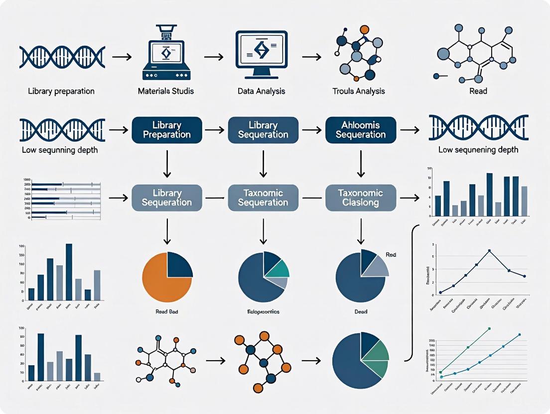
A systematic workflow for diagnosing and addressing low sequencing depth is critical for robust metagenomic analysis.
Diagnostic and Remedial Workflow for Low Sequencing Depth
Step 1: Calculate and Verify Your Actual Sequencing Depth
First, confirm that your observed depth is indeed below the recommended target for your study.
Protocol 1.1: Calculating Average Sequencing Depth
- Determine the total number of usable base pairs generated from your sequencing run (e.g., 90 Gigabases).
- Divide this by the effective genome size or the size of your target region.
- Formula: Average Depth = Total Bases Generated / Target Genome Size [2].
- Example: For a human gut metagenomic sample with an estimated community genome size of 3 Gb, generating 90 Gb of data yields: 90 Gb / 3 Gb = 30x average depth.
Compare your calculated depth against established recommendations for your application:
Table 1: Recommended Sequencing Depths for Various Applications
| Application / Study Goal | Recommended Depth | Key Rationale |
|---|---|---|
| Human Whole-Genome Sequencing (WGS) | 30x - 50x [2] | Balances comprehensive genome coverage with cost for accurate variant calling. |
| Exome / Targeted Gene Mutation Detection | 50x - 100x [2] | Increases confidence for calling variants in specific regions of interest. |
| Cancer Genomics (Somatic Variants) | 500x - 1000x [2] | Essential for detecting low-frequency mutations within a heterogeneous sample. |
| Metagenomic AMR Gene Profiling | 80M+ reads/sample [4] | Required to recover the full richness of antimicrobial resistance gene families. |
| Metagenomic SNP Analysis | Ultra-deep sequencing (e.g., 200M+ reads) [5] | Shallow sequencing misses significant allelic diversity and functionally important SNPs. |
Step 2: Assess and Improve Coverage Uniformity
If depth is adequate but specific genomic regions are consistently poorly covered, investigate coverage uniformity.
Protocol 2.1: Measuring Coverage Uniformity with Interquartile Range (IQR)
- Calculate the read depth for every base position in the genome using tools like
samtools depth. - Compile these depths and calculate the distribution's IQR. A smaller IQR indicates more uniform coverage, while a larger IQR signifies high variability [2].
- Visually inspect the depth distribution across the genome using a plotting tool to identify large gaps or consistently low-coverage regions.
Solution: If uniformity is poor, consider:
- Optimizing Library Preparation: Low-quality or fragmented DNA input can cause biased coverage. Use high-quality, intact DNA and optimize fragmentation and amplification steps [2].
- Utilizing Different Sequencing Technologies: Platforms that produce longer reads can improve coverage in challenging regions like those with high GC content or repeats [2] [3].
Step 3: Verify Sample and Library Quality
Sample issues are a common root cause of low effective depth.
Protocol 3.1: Using Exogenous Spike-Ins for Normalization
- Spike a known amount of exogenous DNA (e.g., from Thermus thermophilus) into your sample before DNA extraction and library preparation [4].
- After sequencing, calculate the ratio of spike-in reads to sample reads.
- This ratio allows for normalization, enabling more accurate cross-sample comparisons and can help diagnose whether low depth is due to sequencing output or sample issues [4].
Step 4: Align Sequencing Strategy with Study Objectives
Ensure your planned depth is sufficient for your specific biological question.
Solution: For applications requiring high sensitivity, such as identifying rare strains or alleles in a metagenomic sample, shallow sequencing is insufficient. One study found that even 200 million reads per sample was not enough to capture the full allelic diversity in an effluent sample, whereas 1 million reads per sample was sufficient for stable taxonomic profiling [4] [5]. If your target is rare variants, you must budget for and plan a significantly higher sequencing depth.
The Scientist's Toolkit: Essential Research Reagents & Materials
Table 2: Key Reagents and Tools for Metagenomic Sequencing Quality Control
| Item / Reagent | Function / Application |
|---|---|
| Tiangen Fecal Genomic DNA Extraction Kit | Standardized protocol for extracting microbial DNA from complex stool samples, critical for reproducible metagenomic studies [5]. |
| Thermus thermophilus DNA (Spike-in Control) | Exogenous control added to samples to normalize AMR gene abundance estimates and correct for technical variation during sequencing [4]. |
| BBMap Suite | A software package containing tools for read subsampling (downsampling), which is used to empirically evaluate the impact of sequencing depth on results [5]. |
| Trimmomatic | A flexible tool for read trimming, used to remove low-quality bases and adapters, which improves overall data quality and mapping accuracy [5]. |
| ResPipe Software Pipeline | An open-source pipeline for automated processing of metagenomic data, specifically for profiling antimicrobial resistance (AMR) gene content [4]. |
| Comprehensive Antimicrobial Resistance Database (CARD) | A curated resource of known AMR genes and alleles, used as a reference for identifying and characterizing resistance elements in metagenomic data [4]. |
In metagenomic sequencing, "sequencing depth" refers to the number of times a given nucleotide in the sample is sequenced, typically measured as the total number of reads generated. This parameter is crucial because it directly determines the resolution and sensitivity of your analysis. Low sequencing depth creates a fundamental challenge: it masks true microbial diversity by failing to detect rare taxa—low-abundance microorganisms that collectively form the "rare biosphere."
The rare biosphere, despite its name, plays disproportionately important ecological roles. These rare taxa can function as a "seed bank" that maintains community stability and robustness, and some contribute over-proportionally to biogeochemical cycles [6]. When sequencing depth is insufficient, these rare taxa are either completely missed or misclassified as sequencing artifacts, leading to a skewed understanding of the microbial community. This problem is particularly acute in clinical and environmental studies where rare pathogens or key functional microorganisms may be present at very low abundances but have significant impacts on health or ecosystem function.
The following sections provide a comprehensive troubleshooting guide to help researchers diagnose, address, and prevent the issues arising from insufficient sequencing depth in their metagenomic studies.
Troubleshooting Guide: Diagnosing and Solving Low Depth Issues
Problem Identification: Symptoms of Insufficient Sequencing Depth
Table: Common Symptoms and Consequences of Low Sequencing Depth
| Symptom | What You Observe | Underlying Issue |
|---|---|---|
| Inflated Alpha Diversity | Higher than expected diversity in simple mock communities [6] | False positive rare taxa from index misassignment inflate diversity metrics |
| Deflated Alpha Diversity | Lower than expected diversity in complex samples [6] | Genuine rare taxa remain undetected below the sequencing depth threshold |
| Unreplicateable Rare Taxa | Rare taxa appear inconsistently across technical replicates [6] | Stochastic detection of low-abundance sequences makes results unreproducible |
| Biased Community Assembly | Skewed interpretation of community assembly mechanisms [6] | Missing rare taxa leads to incorrect inference of ecological processes |
| Reduced Classification Precision | Fewer reads assigned to microbial taxa at lower taxonomic levels [7] | Insufficient data for reliable classification beyond phylum or family level |
Root Cause Analysis: Why Low Depth Masks True Diversity
Index Misassignment (Index Hopping): This phenomenon occurs when indexes are incorrectly assigned during multiplexed sequencing, causing reads to be attributed to the wrong sample. While these misassigned reads represent a small fraction (0.2-6% on Illumina platforms), they can generate false rare taxa that significantly distort diversity assessments in low-depth sequencing [6].
Stochastic Sampling Effects: In complex microbial communities with a "long tail" of rare species, low sequencing depth means that rare taxa may be detected only by chance in some replicates but not others. This leads to significant batch effects and inconsistent results across technical replicates [6].
Insufficient Sampling of True Diversity: Each sequencing read represents a random sample from the total DNA in your specimen. With limited reads, the probability of sampling DNA from genuinely rare organisms decreases dramatically, causing them to fall below the detection threshold [7].
Solution Framework: Strategies for Optimal Depth Selection
Table: Recommended Sequencing Depths for Different Sample Types
| Sample Type | Recommended Depth | Rationale | Supporting Evidence |
|---|---|---|---|
| Bovine Fecal Samples | ~59 million reads (D0.5) | Suitable for describing core microbiome and resistome [7] | Relative abundance of phyla remained constant; fewer taxa discovered at lower depths [7] |
| Human Gut Microbiome | 3 million reads (shallow shotgun) | Cost-effective for species-level resolution in large cohort studies [8] | Balances cost with species/strain-level resolution for high microbial content samples [8] |
| Complex Environmental Samples | >60 million reads | Captures greater diversity of low-abundance organisms | Number of taxa identified increases significantly with depth [7] |
| Skin Microbiome (High Host DNA) | Consider targeted enrichment | Host DNA dominates; standard depth insufficient for rare microbes | Shallow shotgun less sensitive for samples with high non-microbial content [8] |
Frequently Asked Questions (FAQs)
Q1: My sequencing depth seems sufficient based on initial quality metrics, but I'm still missing known rare taxa in mock communities. What could be wrong?
A1: The issue may be index misassignment rather than raw sequencing depth. This phenomenon, where indexes are incorrectly assigned during multiplexed sequencing, creates false rare taxa while obscuring real ones. Studies comparing sequencing platforms have found significant differences in false positive rates (0.08% vs. 5.68%) between platforms [6]. To address this:
- Include negative controls and technical replicates in your sequencing run
- Consider platforms with lower demonstrated index misassignment rates
- Use bioinformatic tools specifically designed to identify and remove potential cross-contaminants
Q2: How does sequencing depth specifically affect the detection of antibiotic resistance genes (ARGs) in metagenomic studies?
A2: Deeper sequencing significantly improves ARG detection sensitivity. Research on bovine fecal samples showed that the number of reads assigned to antimicrobial resistance genes increased substantially with sequencing depth [7]. While relative proportions of major ARG classes may remain fairly constant across depths, the absolute detection of less abundant resistance genes requires sufficient depth to overcome the background of more abundant genetic material.
Q3: What is the relationship between sequencing depth and the ability to identify keystone species in microbial networks?
A3: Inadequate depth can completely alter your interpretation of keystone species. False positive or false negative rare taxa detection leads to biased community assembly mechanisms and the identification of fake keystone species in correlation networks [6]. Since rare taxa can play disproportionate ecological roles, missing them due to low depth fundamentally changes your understanding of community dynamics and the identification of which species are truly crucial for network integrity.
Q4: For large cohort studies where deep sequencing of all samples is cost-prohibitive, what are the best alternatives?
A4: Shallow shotgun sequencing (approximately 3 million reads) provides an excellent balance between cost and data quality for large studies, particularly for high-microbial-content samples like gut microbiomes [8]. This approach offers better species-level resolution than 16S rRNA sequencing while maintaining cost-effectiveness. For samples with high host DNA (e.g., skin, blood), consider hybridization capture using targeted probes to enrich microbial sequences before sequencing [9].
Q5: How can I determine the optimal sequencing depth for my specific study system?
A5: Conduct a pilot study with a subset of samples sequenced at multiple depths. Research on bovine fecal samples demonstrated that while relative abundance of reads aligning to different phyla remained fairly constant regardless of depth, the number of reads assigned to antimicrobial classes and the detection of lower-abundance taxa increased significantly with depth [7]. Your optimal depth depends on your study goals—if seeking core community structure, lower depth may suffice; if characterizing rare taxa or ARGs, deeper sequencing is essential.
Essential Experimental Protocols
Protocol: Determining Optimal Sequencing Depth for Microbial Community Analysis
Principle: Systematically evaluate how increasing sequencing depth affects the detection of microbial taxa, particularly rare species, in your specific sample type.
Materials and Reagents:
- High-quality metagenomic DNA (extracted using standardized protocols)
- Library preparation kit (e.g., Novizan Universal Plus DNA Library Prep Kit for Illumina)
- Quality control instruments (Qubit for DNA concentration, Fragment Analyzer for insert size)
- Illumina sequencing platform
Procedure:
- Sample Preparation: Extract DNA using methods optimized for your sample type (e.g., bead-beating for Gram-positive bacteria in fecal samples) [7].
- Library Preparation and Quality Control: Prepare sequencing libraries and quantify using q-PCR to ensure library effective concentration >3 nM [10].
- Sequencing Design: Sequence the same library at multiple depths (e.g., 26, 59, and 117 million reads) by adjusting the number of sequencing cycles [7].
- Bioinformatic Analysis:
- Process raw data through quality filtering and host read removal
- Classify reads using taxonomic profilers (Kraken, etc.)
- Calculate alpha and beta diversity metrics at each depth
- Compare the number of taxa identified at each taxonomic level
- Threshold Determination: Identify the point where additional sequencing yields diminishing returns in new taxa discovery.
Expected Results: Initially, new taxa discovery will increase rapidly with depth, then plateau. The optimal depth is just before this plateau for your specific research questions.
Protocol: Minimizing Index Misassignment in Multiplexed Sequencing
Principle: Reduce cross-sample contamination that creates false rare taxa through optimized library preparation and sequencing practices.
Materials and Reagents:
- Unique dual indexes (UDIs) with maximum sequence diversity
- High-fidelity DNA polymerase for library amplification
- Platform-specific sequencing reagents
Procedure:
- Experimental Design: Include negative controls (extraction blanks) and technical replicates across sequencing lanes.
- Library Preparation: Use UDIs instead of single indexes to minimize misassignment.
- Pooling Strategy: Avoid overloading sequencing lanes; follow manufacturer recommendations for optimal cluster density.
- Platform Selection: If studying rare biosphere is primary goal, consider platforms with demonstrated lower index misassignment rates (0.0001-0.0004% vs. 0.2-6%) [6].
- Bioinformatic Filtering: Implement strict filtering based on negative controls to remove contaminants.
Expected Results: Significant reduction in false positive rare taxa and improved reproducibility across technical replicates.
Workflow Visualization: From Sample to Analysis
Diagram: Microbial Analysis Workflow and Critical Decision Points. This workflow highlights how decisions at key points (blue diamonds) can lead to consequences (red boxes) that ultimately affect result interpretation.
The Scientist's Toolkit: Essential Research Reagents & Solutions
Table: Key Research Reagents and Solutions for Optimized Metagenomic Sequencing
| Reagent/Solution | Function | Application Notes | References |
|---|---|---|---|
| Bead-beating Matrix | Enhanced cell lysis for Gram-positive bacteria | Critical for representative DNA extraction from diverse communities; improves yield from tough cells | [7] |
| Unique Dual Indexes (UDIs) | Sample multiplexing with minimal misassignment | Reduces index hopping compared to single indexes; essential for rare biosphere studies | [6] |
| Hybridization Capture Probes | Targeted enrichment of microbial sequences | myBaits system enables ~100-fold enrichment; ideal for host-dominated samples | [9] |
| DNA Quality Control Kits | Assess DNA purity and quantity | Fluorometric methods (Qubit) preferred over UV spectrophotometry for accurate quantification | [11] |
| Host DNA Removal Kits | Deplete host genetic material | Critical for samples with high host:microbe ratio (skin, blood); improves microbial signal | [7] |
| Mock Community Controls | Method validation and calibration | ZymoBIOMICS or customized communities essential for assessing sensitivity and specificity | [6] |
Successfully characterizing microbial diversity, particularly the rare biosphere, requires careful consideration of sequencing depth throughout your experimental design. The most critical recommendations include: (1) conducting pilot studies to determine optimal depth for your specific sample type and research questions; (2) implementing controls and replicates to identify technical artifacts; (3) selecting appropriate sequencing platforms and methods based on your focus on rare taxa; and (4) applying bioinformatic filters judiciously to remove false positives without eliminating genuine rare organisms. By addressing the challenge of sequencing depth directly, researchers can unmask the true diversity of microbial communities and gain more accurate insights into the ecological and functional roles of the rare biosphere.
Core Concepts: Sequencing Depth and Resistome Analysis
Why is sequencing depth particularly critical for resistome analysis compared to taxonomic profiling?
Taxonomic profiling stabilizes at much lower sequencing depths, while comprehensive resistome analysis requires significantly deeper sequencing. The richness of Antimicrobial Resistance (AMR) gene families and their allelic variants are particularly depth-dependent.
- Taxonomic Profiling Stability: Achieves less than 1% dissimilarity to the full profile with only 1 million reads per sample [4].
- AMR Gene Family Richness: Requires at least 80 million reads per sample for the number of different AMR gene families to stabilize [4] [12].
- AMR Allelic Diversity: Full allelic diversity may not be captured even at 200 million reads per sample, indicating that discovering rare variants demands extreme depth [4].
Troubleshooting Guide: FAQs & Solutions
FAQ 1: My study involves diverse environmental samples. How do I determine the minimum sequencing depth needed?
The required depth depends on your sample type due to inherent differences in microbial and resistance gene diversity. The table below summarizes findings from a key study that sequenced different sample types to a high depth (~200 million reads) [4].
Table 1: Minimum Sequencing Depth Requirements by Sample Type for AMR Analysis
| Sample Type | Sequencing Depth for AMR Gene Family Richness (d0.95)* | Sequencing Depth for AMR Allelic Variants (d0.95)* | Notes |
|---|---|---|---|
| Effluent | 72 - 127 million reads | ~193 million reads | Very high allelic diversity; richness may not plateau even at 200M reads. |
| Pig Caeca | 72 - 127 million reads | Information Not Specified | High gene family richness. |
| River Sediment | Very low AMR reads | Very low AMR reads | AMR gene content was too low for depth analysis in this study. |
| Human Gut (Typical) | ~3 million reads (shallow shotgun) | Not recommended for allelic diversity | Sufficient for species-level taxonomy and core functional profiling [8]. |
*d0.95 = Depth required to achieve 95% of the estimated total richness.
Recommendation: For complex environments like effluent or soil, pilot studies with deep sequencing are recommended to establish depth requirements for your specific samples [4] [12].
FAQ 2: I have already sequenced my samples at a shallow depth (e.g., 3-5 million reads). Can I salvage the data for resistome analysis?
Yes, but with major caveats regarding the scope of your conclusions. Shallow shotgun sequencing (~3 million reads) is a valid cost-effective method for specific applications [8].
What Shallow Depth is Good For:
Limitations of Shallow Depth Data:
- Poor recovery of AMR gene richness: You will have missed rare AMR gene families [4].
- Incomplete allelic diversity: The full scope of allelic variants, which can have profound functional implications, will be underrepresented [4] [13].
- Inability to perform deep genetic analysis: Strain-level variation, single nucleotide variant (SNV) calling, and metagenome-assembled genomes (MAGs) require deep sequencing [12].
Solution: Clearly frame your research findings to reflect the limitations of your sequencing depth. Use phrases like "detection of abundant AMR genes" rather than "comprehensive resistome characterization."
FAQ 3: Are there wet-lab and bioinformatic methods to improve resistome analysis without the cost of ultra-deep sequencing for all samples?
Yes, alternative strategies can enhance sensitivity and specificity.
1. Wet-Lab Solution: Targeted Sequence Capture This method uses biotin-labeled RNA probes to hybridize and enrich DNA libraries for sequences of interest before sequencing.
- Principle: Design probes complementary to a curated database of AMR genes, which allows you to "pull out" these targets from a complex metagenomic background [14] [15].
- Benefit: Dramatically increases the on-target reads for AMR genes, enabling their detection even when they represent <0.1% of the metagenome [14]. One study reported a 300-fold increase in unequivocally mapped reads compared to standard shotgun sequencing [15].
- Workflow: The following diagram illustrates the typical workflow for a targeted capture experiment like the ResCap method [15].
2. Bioinformatic Solution: Optimized Pipelines and Databases Using specialized, well-curated tools can improve the accuracy and depth of analysis from your existing data.
- Use Updated and Comprehensive Databases: Tools like
sraXcan integrate multiple databases (CARD, ARGminer, BacMet) for a more extensive homology search [16]. - Leverage Assembly-Based Methods: While computationally expensive, assembly-based tools can identify novel ARGs and provide genomic context, which is lost in read-based mapping [16].
- Normalization is Key: When quantifying AMR abundance, normalize read counts by gene length. For cross-sample comparison, consider using an exogenous spike-in (e.g., Thermus thermophilus DNA) to estimate absolute abundance [4].
The Scientist's Toolkit: Essential Research Reagents & Materials
Table 2: Key Research Reagent Solutions for Resistome Analysis
| Reagent / Material | Function / Application | Example / Source |
|---|---|---|
| Comprehensive AMR Databases | Curated collections of reference sequences for identifying AMR genes and variants. | CARD (Comprehensive Antibiotic Resistance Database) [4] [16], ResFinder [15], ARG-ANNOT [15]. |
| Targeted Capture Probe Panels | Pre-designed sets of probes for enriching AMR genes from metagenomic libraries prior to sequencing. | ResCap [15], Custom panels (e.g., via Arbor Biosciences myBaits) [14]. |
| Exogenous Control DNA | Spike-in control for normalizing gene abundances to allow absolute quantification and cross-sample comparison. | Thermus thermophilus DNA [4]. |
| Standardized DNA Extraction Kits | Ensure minimal bias and high yield during DNA extraction, which is critical for downstream representativeness. | Kits optimized by sample type (e.g., Metahit protocol for stool) [15]. |
| Bioinformatics Pipelines | Software for processing sequencing data, mapping reads to AMR databases, and performing normalization and statistical analysis. | ResPipe [4], sraX [16], ARIBA [16]. |
| High-Quality Reference Genomes | Used for alignment, variant calling, and understanding the genomic context of detected AMR genes. | Isolates sequenced with hybrid methods (e.g., Illumina + Oxford Nanopore) [4]. |
Frequently Asked Questions (FAQs)
1. Why can't I resolve strains or call SNPs even when my species-level analysis looks good? Species-level analysis often masks significant genetic diversity. While two strains may share over 95% average nucleotide identity (ANI), this only applies to the portions of the genome they have in common. A single species can have a pangenome containing tens of thousands of genes, but an individual strain may possess only a fraction of these, leading to vast differences in key functional characteristics like virulence or drug resistance. When sequencing depth is low, the reads are insufficient to cover these variable regions or detect single-nucleotide polymorphisms (SNPs) that distinguish one strain from another [17].
2. My sequencing run had good yield; why is my strain-level resolution still poor? Total sequencing yield can be misleading. Strain-level resolution requires sufficient coverage (depth) across the entire genome of each strain present. In a metagenomic sample with multiple co-existing strains, the coverage for any single strain can be drastically lower than the total sequencing depth. Tools for de novo strain reconstruction, for instance, often require 50-100x coverage per strain for accurate results. If your sample contains multiple highly similar strains (with Mash distances as low as 0.0004), the effective coverage for distinguishing them is even lower, making SNP calling unreliable [18] [19].
3. How does sample contamination affect strain-level analysis? In low-biomass samples, contamination is a critical concern. Contaminant DNA can constitute a large proportion of your sequence data, effectively diluting the signal from your target organisms. This leads to reduced coverage for genuine strains and can cause false positives by introducing sequences that look like novel strains. Contamination can originate from reagents, sampling equipment, or the lab environment, and its impact is disproportionate in studies aiming for high-resolution strain detection [20].
4. Are some sequencing technologies better for strain-level SNP calling than others? While short-read technologies (like Illumina) are widely used, their read length can be a limitation. Longer reads are better for spanning repetitive or variable regions, which is often crucial for separating strains. Sanger sequencing, with its longer read length and high accuracy, can improve assembly outcomes but is cost-prohibitive for large metagenomic studies. The error profiles of different platforms also matter; for example, homopolymer errors in 454/Roche pyrosequencing can cause frameshifts that obscure true SNPs [21].
Troubleshooting Guide: Low Sequencing Depth for Strain Resolution
Problem: Inability to call SNPs or distinguish between highly similar strains in a metagenomic sample.
Step 1: Diagnose the Root Cause
- Check Coverage and Complexity: Use tools like
BBMaporCheckMto assess the coverage depth and completeness of your metagenome-assembled genomes (MAGs). A CheckM completeness score below 90% often indicates an inadequate dataset for confident strain-level analysis [22]. - Identify Contamination: Use tools like
KrakenorDecontaMinerto screen for and quantify contaminant sequences. A high percentage of reads classified as common contaminants (e.g., human, skin flora) signals a problem [20]. - Assess Strain Diversity: If possible, use a low-resolution strain tool (e.g.,
StrainGE,StrainEst) to estimate the number and similarity of strains present. Co-existing strains with a Mash distance < 0.005 are exceptionally challenging to resolve [18].
Step 2: Apply Corrective Measures in Wet-Lab Procedures
- Optimize DNA Extraction: For low-biomass samples, use extraction protocols designed to maximize yield while minimizing contamination. Include multiple negative controls (e.g., empty collection vessels, swabs of the air) to track contamination sources [20].
- Increase Sequencing Depth: Based on your initial coverage assessment, you may need to sequence more deeply. As a rule of thumb, strain-resolving analyses require significantly higher depth than species-level profiling.
- Consider Long-Read Sequencing: If the budget allows, supplement your short-read data with long-read sequencing (e.g., PacBio, Oxford Nanopore). Long reads can bridge strain-specific structural variations and improve assembly, providing a better scaffold for SNP calling [19].
Step 3: Optimize Computational Analysis
- Select the Right Tool: Standard species-level classifiers (
Kraken,MetaPhlAn2) are not suitable. Use tools specifically designed for high-resolution strain-level analysis, such asStrainScan, which employs a hierarchical k-mer indexing structure to distinguish highly similar strains [18]. - Use Customized Reference Databases: When targeting specific bacteria, provide a customized, high-quality set of reference genomes for that species to the analysis tool. This increases the chance of matching the strains present in your sample [18].
- Leverage Metagenomic Assembly: For high-coverage samples, use assembly-based methods (
EVORhA,DESMAN) to reconstruct strain genomes de novo. These methods can resolve full strain genomes but require high coverage (50-100x per strain) [19].
Performance Comparison of Strain-Level Analysis Tools
The table below summarizes the key characteristics of various computational approaches for strain-level analysis, highlighting their different strengths and data requirements.
TABLE 1: Strain-Level Microbial Detection Tools
| Tool / Method | Category | Key Principle | Key Strength | Key Limitation / Requirement |
|---|---|---|---|---|
| StrainScan [18] | K-mer based | Hierarchical k-mer indexing (Cluster Search Tree) | High resolution for distinguishing highly similar strains; improved F1 score. | Requires a predefined set of reference strain genomes. |
| EVORhA [19] | Assembly-based | Local haplotype assembly and frequency-based merging. | Can reconstruct complete strain genomes; high accuracy. | Requires extremely high coverage (50-100x per strain). |
| DESMAN [19] | Assembly-based | Uses differential coverage of core and accessory genes. | Resolves strains and estimates relative abundance without a reference. | Requires a group of high-quality Metagenome-Assembled Genomes (MAGs). |
| Pathoscope2 [18] | Alignment-based | Bayesian reassignment of ambiguously mapped reads. | Effectively identifies dominant strains in a mixture. | Computationally expensive with large reference databases. |
| Krakenuniq [18] | K-mer based | Uses k-mer counts for classification and abundance estimation. | Good for species-level and some strain-level identification. | Low resolution when reference strains share high similarity. |
Experimental Protocol: Workflow for Diagnosing Strain-Resolution Failure
This protocol provides a step-by-step method to systematically diagnose the reasons behind failed strain-level SNP calling, integrating both bioinformatic and experimental checks.
Title: Diagnostic Workflow for Strain-Resolution Failure
1. Initial Quality Control and Coverage Assessment
- Input: Filtered metagenomic sequencing reads (FASTQ format).
- Procedure:
- Assemble reads into contigs using a metagenomic assembler (e.g., MEGAHIT, metaSPAdes).
- Bin contigs into Metagenome-Assembled Genomes (MAGs) using a tool like MetaBAT2.
- Run
CheckMorCheckM2on the MAGs to assess completeness and contamination.
- Interpretation: A MAG with a CheckM completeness score below 90% is unlikely to provide sufficient data for strain-level resolution [22]. Low coverage across the target genome (<20x) is a primary indicator of insufficient sequencing depth.
2. Contamination Screening and Quantification
- Input: Raw or filtered sequencing reads (FASTQ format).
- Procedure:
- Classify all reads taxonomically using
Kraken2with a standard database. - Analyze the output to determine the percentage of reads classified as your target organism versus common contaminants (e.g., human, E. coli lab strain).
- Compare the taxonomic profile of your sample with your negative control samples.
- Classify all reads taxonomically using
- Interpretation: A high proportion of contaminant reads (>5%) significantly dilutes your signal and is a likely cause of failure, especially in low-biomass samples [20].
3. Low-Resolution Strain Profiling
- Input: Sequencing reads and a database of reference genomes for your target species.
- Procedure:
- Run a clustering-based strain tool like
StrainGEorStrainEston your data. - These tools will report strains at a lower resolution (e.g., clustering strains with >99.4% ANI).
- If a cluster is identified, calculate the Mash distance between the reference strains within that cluster.
- Run a clustering-based strain tool like
- Interpretation: The presence of a cluster containing multiple reference strains with a very low Mash distance (e.g., <0.005) indicates a challenging scenario of highly similar co-existing strains, explaining the failure of SNP callers [18].
The Scientist's Toolkit: Key Reagents & Materials for Strain-Resolving Studies
TABLE 2: Essential Research Reagents and Materials
| Item | Function / Purpose | Considerations for Strain-Level Resolution |
|---|---|---|
| DNA-Free Collection Swabs/Tubes | To collect samples without introducing contaminant DNA. | Critical for low-biomass samples (e.g., tissue, water). Pre-sterilized and certified DNA-free. [20] |
| DNA Degrading Solution | To remove trace DNA from equipment and surfaces. | Used for decontaminating reusable tools. More effective than ethanol or autoclaving alone. [20] |
| High-Yield DNA Extraction Kit | To maximize recovery of microbial DNA from the sample. | Select kits benchmarked for your sample type (e.g., soil, stool) to minimize bias. [21] |
| Multiple Displacement Amplification (MDA) Kit | To amplify femtograms of DNA to micrograms for sequencing. | Use with caution as it can introduce bias and chimeras; essential for single-cell genomics. [21] |
| Negative Control Kits | To identify contaminating DNA from reagents and the lab environment. | Should include "blank" extraction controls and sampling controls processed alongside all samples. [20] |
| Strain-Specific Reference Genomes | Curated genomic sequences used as a database for strain identification. | Quality and diversity of the reference database directly impact the resolution of tools like StrainScan. [18] |
Frequently Asked Questions (FAQs)
Q1: What are the primary consequences of low sequencing depth on MAG quality? Low sequencing depth directly leads to fragmented assemblies and poor genome recovery [23]. Insufficient reads result in short contigs during assembly, which binning algorithms struggle to group correctly into MAGs. This fragmentation causes lower genome completeness and an increased rate of missing genes, even for high-abundance microbial populations [24]. Furthermore, it reduces the ability to distinguish between closely related microbial strains, as the coverage information used for binning becomes less reliable.
Q2: My MAGs have high completeness scores but are missing known genes from the population. Why? This is a documented discrepancy. A study comparing pathogenic E. coli isolates to their corresponding MAGs found that MAGs with completeness estimates near 95% captured only 77% of the population's core genes and 50% of its variable genes, on average [24]. Standard quality metrics (like CheckM) rely on a small set of universal single-copy genes, which may not represent the entire genome. This indicates that gene content, especially variable genes, is often worse than estimated completeness suggests [24].
Q3: How does high host DNA contamination in a sample affect MAG recovery? Samples with high host DNA (e.g., >90%) drastically reduce the proportion of microbial sequencing reads. This leads to a significant loss of sensitivity in detecting low-abundance microbial species and results in fewer recovered MAGs [25] [26]. To acquire meaningful microbial data from such samples, a much higher total sequencing depth is required to achieve sufficient coverage of the microbial genomes, making studies more costly and computationally intensive.
Q4: Can the choice of binning pipeline influence the recovery of genomes from complex communities? Yes, significantly. Different binning pipelines exhibit variable performance. A 2024 simulation study evaluating three common pipelines found that the DAS Tool (DT) pipeline showed the most accurate results (~92% true positives), outperforming others in the same test [23]. The study also highlighted that some pipelines (like the 8K pipeline) recover a higher number of total MAGs but with a lower accuracy rate, meaning more bins do not necessarily reflect the actual community composition [23].
Q5: What is a major limitation of using mock communities to validate MAG recovery? Traditional mock communities are often constructed from a single genome per organism. They do not capture the full scope of intrapopulation gene diversity and strain heterogeneity found in natural populations [24]. Consequently, a pipeline's performance on a mock dataset may not accurately predict its performance on a real, more complex environmental sample where multiple closely related strains with variable gene content are present.
Troubleshooting Guides
Guide: Diagnosing and Mitigating Effects of Low Sequencing Depth
Problem: Assembled MAGs are highly fragmented (low N50, high contig count), have low estimated completeness, and fail to recover key genes of interest.
Diagnosis: This is a classic symptom of insufficient sequencing depth. Check the following:
- Raw Read Coverage: Calculate the average coverage of your target population(s) from the metagenomic reads. For reliable MAG recovery, a minimum of 10x coverage is often required, with higher depths (e.g., 20-30x) needed for more complete genomes [24] [27].
- Community Profiling: Use a tool like MetaPhlAn2 to profile community composition [25]. If low-abundance taxa are missing from the profile, it indicates they are under-sequenced.
Solutions:
- Increase Sequencing Depth: Sequence the library to a greater depth. Simulation studies show that increasing depth from 10 million to 60 million reads can significantly boost the number of recovered MAGs [23].
- Employ Co-assembly: If you have multiple related samples (e.g., technical or biological replicates), a co-assembly strategy can increase the effective read depth, leading to more robust assemblies and better MAGs [28].
- Use Longer Reads: If possible, leverage long-read sequencing technologies (PacBio, Nanopore) or hybrid assembly approaches. These can span repetitive regions and produce much longer contigs, directly combating fragmentation [29] [30].
Guide: Improving MAG Recovery from Host-Dominated Samples
Problem: Samples like bronchoalveolar lavage fluid (BALF) or oropharyngeal swabs yield a very low percentage of microbial reads, hindering MAG reconstruction.
Diagnosis: The sample has a high host-to-microbe DNA ratio. Confirm this by aligning a subset of your reads to the host genome (e.g., using KneadData/Bowtie2) [25]. A microbial read ratio below 1% is a clear indicator [26].
Solutions:
- Apply Host Depletion Methods: Implement a pre-sequencing host DNA depletion protocol. A 2025 benchmarking study compared seven methods on respiratory samples [26]. The performance of these methods in increasing microbial reads is summarized in the table below.
Table 1: Performance of Host DNA Depletion Methods for Respiratory Samples (Adapted from [26])
| Method Name | Category | Key Principle | Performance in BALF (Fold Increase in Microbial Reads) |
|---|---|---|---|
| K_zym (HostZERO Kit) | Pre-extraction | Chemical & enzymatic host cell lysis & DNA degradation | 100.3x |
| S_ase | Pre-extraction | Saponin lysis & nuclease digestion | 55.8x |
| F_ase (New Method) | Pre-extraction | 10μm filtering & nuclease digestion | 65.6x |
| K_qia (QIAamp Kit) | Pre-extraction | Not specified in detail | 55.3x |
| O_ase | Pre-extraction | Osmotic lysis & nuclease digestion | 25.4x |
| R_ase | Pre-extraction | Nuclease digestion | 16.2x |
| O_pma | Pre-extraction | Osmotic lysis & PMA degradation | 2.5x |
- Increase Sequencing Depth Proactively: When working with host-dominated samples, plan for a much higher total sequencing output to ensure sufficient coverage of the microbial fraction after depletion [25].
Table 2: Impact of Sequencing Depth on MAG Recovery from Simulated Communities [23]
| Sequencing Depth (Millions of Reads) | Trend in MAG Recovery (across 8K, DT, and MM pipelines) |
|---|---|
| 10 million | Low number of MAGs recovered. |
| 30 million | Increasing trend in MAG recovery. |
| 60 million | Increasing trend in MAG recovery; MM pipeline peaks around this depth. |
| 120 million | 8K pipeline recovers more true positives at depths above 60M reads. |
| 180 million | Trend continues for the 8K pipeline. |
Table 3: Quantitative Impact of High Host DNA on Microbiome Profiling Sensitivity [25]
| Host DNA Percentage | Impact on Sensitivity of Detecting Microbial Species |
|---|---|
| 10% | Minimal impact on sensitivity. |
| 90% | Significant decrease in sensitivity for very low and low-abundance species. |
| 99% | Profiling becomes highly inaccurate and insensitive. |
Experimental Protocols
Protocol: Host DNA Depletion using the F_ase Method for Respiratory Samples
This protocol is based on a method benchmarked in a 2025 study, which showed a balanced performance in increasing microbial reads while maintaining good bacterial DNA retention [26].
Principle: Microbial cells are separated from host cells and debris by filtration through a 10μm filter. The filtrate, enriched in microbial cells, is then treated with a nuclease to degrade free-floating host DNA.
Materials:
- Respiratory sample (e.g., BALF, OP swab in suspension)
- Sterile saline solution
- 10μm pore-size filters (e.g., cell strainers)
- Nuclease enzyme (e.g., Benzonase) and corresponding buffer
- Microcentrifuge tubes
- Centrifuge
- DNA extraction kit (for microbial DNA)
Procedure:
- Sample Preparation: Homogenize the respiratory sample in a sterile saline solution.
- Filtration: Pass the homogenized sample through a 10μm filter. The filtrate contains the enriched microbial cells.
- Nuclease Treatment: a. Collect the filtrate in a fresh microcentrifuge tube. b. Add nuclease enzyme and its reaction buffer to the filtrate. c. Incubate at the recommended temperature and duration (e.g., 37°C for 1 hour) to degrade free DNA. d. Heat-inactivate the nuclease as per the manufacturer's instructions.
- DNA Extraction: Pellet the microbial cells by centrifugation. Proceed with DNA extraction from the pellet using a standard microbial DNA extraction kit.
- Quality Control: Quantify the total DNA and assess the host DNA depletion efficiency by qPCR targeting a host-specific gene (e.g., GAPDH) and a universal bacterial gene (e.g., 16S rRNA).
Protocol: Evaluating MAG Quality Against an Isolate Genome
This protocol provides a method for an independent assessment of MAG quality that goes beyond standard completeness/contamination metrics, as described in [24].
Principle: A MAG recovered from a metagenome is directly compared to a high-quality isolate genome obtained from the same sample. This allows for a true assessment of core and variable gene recovery.
Materials:
- Metagenomic sequencing data from a sample.
- Whole-genome sequencing data of an isolate from the same sample.
- Assembly software (e.g., MEGAHIT, SPAdes).
- Binning software (e.g., MaxBin, MetaBAT).
- Genome quality assessment tool (e.g., CheckM).
- Genome comparison tool (e.g., OrthoFinder, Roary, BLAST+).
Procedure:
- Generate the MAG: a. Assemble the metagenomic reads into contigs. b. Bin the contigs to recover the MAG representing the target organism. c. Assess the MAG's quality using CheckM to estimate completeness and contamination.
- Assemble the Isolate Genome: a. Assemble the isolate's sequencing reads into a reference genome.
- Compare Gene Content: a. Annotate both the MAG and the isolate genome to identify all protein-coding genes. b. Perform an all-vs-all BLAST of the genes from both genomes. c. Identify core genes (present in >90% of the population, i.e., in the isolate) and variable genes (shared by >10% but <90%). d. Calculate the percentage of the isolate's core and variable genes that were successfully captured by the MAG. The study in [24] found averages of 77% for core and 50% for variable genes, serving as a benchmark.
Visual Workflows
The Scientist's Toolkit: Research Reagent Solutions
Table 4: Essential Reagents and Kits for MAG Studies from Complex Samples
| Reagent / Kit | Function | Example Use Case |
|---|---|---|
| HostZERO Microbial DNA Kit (K_zym) | Pre-extraction host DNA depletion. | Effectively removing host DNA from samples with very high host content (e.g., BALF), increasing microbial read yield over 100-fold [26]. |
| QIAamp DNA Microbiome Kit (K_qia) | Pre-extraction host DNA depletion. | An alternative commercial kit for host DNA depletion, showing good performance in increasing microbial reads from oropharyngeal swabs [26]. |
| Nextera XT DNA Library Prep Kit | Metagenomic library preparation. | Used for preparing sequencing libraries from normalized DNA, including metagenomic samples, for Illumina platforms [25]. |
| Microbial Mock Community B (BEI Resources) | Positive control for sequencing and analysis. | A defined mix of 20 bacterial genomic DNAs used to benchmark sequencing sensitivity, bioinformatics pipelines, and host depletion methods [25] [26]. |
| RNAlater / OMNIgene.GUT | Nucleic acid preservation. | Stabilizes microbial community DNA/RNA at the point of collection, preventing degradation and shifts in community structure before DNA extraction [29]. |
Strategic Approaches: From Wet-Lab Bench to Bioinformatic Pipelines
Frequently Asked Questions (FAQs) and Troubleshooting Guides
FAQ 1: Why is host DNA depletion critical for shotgun metagenomic sequencing of respiratory samples?
Host DNA depletion is crucial because samples like bronchoalveolar lavage fluid (BALF) can contain over 99.7% host DNA, drastically limiting the sequencing depth available for microbial reads [31]. Without depletion, the overwhelming amount of host DNA overshadows microbial signals, reducing the sensitivity for detecting pathogens [32] [26]. Effective host DNA depletion can increase microbial reads by more than 100-fold in BALF samples, transforming a dataset with minimal microbial information into one suitable for robust analysis [26].
FAQ 2: My microbial sequencing depth remains low after host DNA depletion. What are the primary factors to investigate?
Low microbial sequencing depth post-depletion can stem from several factors. systematically investigate the following, which are common troubleshooting points:
- Sample Type and Initial Biomass: The method's efficiency is highly dependent on the sample type. For instance, oropharyngeal (OP) swabs typically respond better to depletion than BALF due to their inherently higher bacterial load [26]. Check if the starting bacterial DNA is sufficient.
- Depletion Method Selection: Different methods have vastly different efficiencies. As shown in Table 1, some kits are significantly more effective than others. Ensure the selected method is optimal for your specific sample type.
- Cell-Free DNA Contamination: Pre-extraction methods that target intact cells will not remove cell-free microbial DNA. One study noted that ~69% of microbial DNA in BALF and ~80% in OP swabs was cell-free, which can be lost during these protocols, reducing the final yield [26].
- Technical Failures: Some methods, particularly lyPMA (osmotic lysis with PMA) and MolYsis, have been reported to have higher rates of library preparation failure, which would directly result in low output. Always check library quality post-preparation [31].
FAQ 3: How does host DNA depletion impact the representation of the microbial community?
Most studies indicate that while host depletion efficiently removes human DNA, it can introduce biases:
- General Composition: Overall bacterial community composition is often reported to be similar before and after depletion [32].
- Specific Taxa Loss: Some methods can cause a significant reduction in the biomass of certain commensals and pathogens, such as Prevotella spp. and Mycoplasma pneumoniae [26].
- Varying Bacterial Retention: Different methods result in different bacterial DNA retention rates. For example, nuclease digestion (Rase) and the QIAamp kit (Kqia) showed the highest bacterial retention rates in OP swabs (median ~20%), while other methods were more aggressive and led to greater loss [26]. It is critical to validate the method for your microbes of interest, ideally using a mock microbial community.
FAQ 4: What is a sufficient sequencing depth for metagenomic studies after host depletion?
The required depth depends on your research goal. The following table summarizes recommendations from recent studies:
Table 1: Recommended Sequencing Depths for Metagenomic Studies
| Research Goal | Recommended Minimum Depth | Key Rationale |
|---|---|---|
| Metagenome-Wide Association Studies (MWAS) | 15 million reads | Provides stable species richness (changing rate ≤5%) and reliable species composition (ICC > 0.75) [33]. |
| Strain-Level SNP Analysis | Ultra-deep sequencing (>> standard depth) | Shallow sequencing is "incapable of supporting systematic metagenomic SNP discovery." Ultra-deep sequencing is required to detect functionally important SNPs reliably [5]. |
| Rapid Clinical Diagnosis | Low-depth sequencing (<1 million reads) | When coupled with efficient host depletion and a streamlined workflow, this can be sufficient for detecting pathogens at physiological levels [34]. |
Technical Performance Data of Common Host Depletion Methods
The following table consolidates quantitative performance data from recent benchmarking studies to aid in method selection. Note that performance is sample-dependent.
Table 2: Performance Comparison of Host DNA Depletion Methods [32] [26] [31]
| Method (Category) | Key Principle | Host Depletion Efficiency (Fold Reduction) | Microbial Read Increase (Fold vs. Control) | Key Advantages / Disadvantages |
|---|---|---|---|---|
| Saponin + Nuclease (S_ase) | Pre-extraction: Lyses human cells with saponin, digests DNA with nuclease. | BALF: ~10,000-fold [26] | BALF: 55.8x [26] | High efficiency. Requires optimization of saponin concentration. |
| HostZERO (K_zym) | Pre-extraction: Selective lysis and digestion. | BALF: ~10,000-fold [26]; Tissue: 57x (18S/16S ratio) [32] | BALF: 100.3x [26] | Very high host depletion. Can have high bacterial DNA loss and library prep failure risk [26] [31]. |
| QIAamp Microbiome Kit (K_qia) | Pre-extraction: Selective lysis and enzymatic digestion. | Tissue: 32x (18S/16S ratio) [32] | BALF: 55.3x [26] | Good host depletion and high bacterial retention. |
| Benzonase Treatment | Pre-extraction: Enzyme-based digestion. | Effective on frozen BALF, reduces host DNA to low pg/µL levels [31]. | Significantly increases final non-host reads [31]. | Robust performance on previously frozen non-cryopreserved samples [31]. |
| Filtration + Nuclease (F_ase) | Pre-extraction: Filters microbial cells, digests free DNA. | Moderate to High [26] | BALF: 65.6x [26] | Balanced performance with less taxonomic bias [26]. |
| NEB Microbiome Enrichment | Post-extraction: Binds methylated host DNA. | Low in respiratory samples [26]. | Low [26] | Easy workflow. Inefficient for respiratory samples and other types [26]. |
Workflow and Decision Pathway
Use the following diagram to guide your selection and troubleshooting of a host DNA depletion method. The process begins with sample characterization and leads to a method choice optimized for your specific goals.
The Scientist's Toolkit: Key Research Reagent Solutions
This table lists essential reagents and kits commonly used in host DNA depletion protocols, as featured in the cited research.
Table 3: Key Reagents for Host DNA Depletion Workflows
| Reagent / Kit Name | Function / Principle | Example Use Case |
|---|---|---|
| Molzym Ultra-Deep Microbiome Prep | Pre-extraction: Selective lysis of human cells and enzymatic degradation of released DNA. | Evaluated on diabetic foot infection tissue samples [32]. |
| Zymo HostZERO Microbial DNA Kit | Pre-extraction: Selective lysis of human cells and digestion of host DNA. | Efficient host depletion in BALF and tissue samples [32] [26]. |
| QIAamp DNA Microbiome Kit | Pre-extraction: Selective lysis followed by enzymatic digestion of host DNA. | Effective enrichment of bacterial DNA from tissue and respiratory samples [32] [26]. |
| NEBNext Microbiome DNA Enrichment Kit | Post-extraction: Captures methylated host DNA, leaving microbial DNA in solution. | Less effective for respiratory samples [32] [26]. |
| Propidium Monoazide (PMA) | Viability dye: Penetrates compromised membranes of dead cells, cross-linking DNA upon light exposure. | Used in osmotic lysis (lyPMA) protocols to remove free host DNA and indicate viable microbes [26] [31] [35]. |
| Benzonase Endonuclease | Enzyme-based: Digests both host and free DNA in samples. | Effective host depletion for frozen respiratory samples (BAL, sputum) [31]. |
| ArcticZymes Nucleases (e.g., M-SAN HQ) | Enzyme-based: Magnetic bead-immobilized or free nucleases to deplete host DNA under various salt conditions. | Used in rapid clinical mNGS workflows for plasma and respiratory samples [36]. |
Frequently Asked Questions
1. What is the fundamental difference between short-read and long-read sequencing? Short-read sequencing (e.g., Illumina, Element Biosciences AVITI) generates massive volumes of data from DNA fragments that are typically a few hundred base pairs long. These technologies are known for high per-base accuracy (often exceeding Q40) and low cost per base, making them the workhorse for many applications [37] [38] [39]. Long-read sequencing (e.g., PacBio, Oxford Nanopore Technologies), in contrast, sequences DNA fragments that are thousands to hundreds of thousands of base pairs long in a single read. This allows them to span repetitive regions and resolve complex genomic structures without the need for assembly from fragmented pieces [37] [39].
2. How does sequencing depth interact with the choice of technology for metagenomic studies? Sequencing depth requirements are critically influenced by your sample type and research question. In metagenomic samples with high levels of host DNA (e.g., >90%), a much greater sequencing depth is required to obtain sufficient microbial reads for a meaningful analysis [40]. Furthermore, the required depth depends on what you are looking for: profiling taxonomic composition may be stable at around 1 million reads, but recovering the full richness of antimicrobial resistance (AMR) gene families can require at least 80 million reads, with even deeper sequencing needed to discover all allelic variants [4].
3. My metagenomic samples have high host DNA contamination. What can I do? Samples like saliva or tissue biopsies often contain over 90% host DNA, which can waste sequencing resources and obscure microbial signals [40]. Wet-lab and bioinformatics solutions are available:
- Wet-lab depletion kits: Use commercial kits to remove host (e.g., human) DNA before library preparation.
- Bioinformatics decontamination: Employ tools like CLEAN [41] or KneadData [40] to computationally identify and remove reads that align to the host genome after sequencing. Tools like QC-Blind can even perform this without a reference genome for the contaminant, using marker genes of the target species instead [42].
4. Can I combine long-read and short-read sequencing in a single study? Yes, this is a powerful strategy to leverage the strengths of both. You can use the high accuracy and low cost of short-read data for confident SNP and mutation calling, while layering long-read data to resolve complex structural variations and phase haplotypes. This hybrid approach is particularly beneficial for de novo genome assembly and studying rare diseases [38].
5. Have the historical drawbacks of long-read sequencing been overcome? Significant progress has been made. The high error rates historically associated with long-read technologies have been drastically reduced. PacBio's HiFi sequencing method now delivers accuracy exceeding 99.9% (Q30), on par with short-read technologies [37] [38]. While the cost of long-read sequencing was once prohibitive for large studies, platforms like the PacBio Revio have reduced the cost of a human genome to under $1,000, making it more accessible for larger-scale projects [37].
Troubleshooting Guides
Problem: Incomplete or Biased Metagenomic Profiling
Issue: Your sequencing data fails to capture the full taxonomic or functional diversity of your sample, especially low-abundance species or complex gene variants.
Solution:
- Re-assess Required Sequencing Depth:
- Cause: Inadequate sequencing depth, particularly in samples with high host DNA or high microbial diversity, leads to incomplete profiling [4] [40].
- Fix: Conduct a pilot study or use rarefaction analysis on existing data to determine the depth at which diversity metrics plateau. For AMR gene discovery in complex environments, plan for depths of 80-200 million reads or more [4]. The table below summarizes findings from key studies.
| Application / Sample Type | Recommended Sequencing Depth | Key Findings from Research |
|---|---|---|
| Taxonomic Profiling (Mock community) | ~1 million reads | Achieved <1% dissimilarity to the full taxonomic composition [4]. |
| AMR Gene Family Richness (Effluent, Pig Caeca) | ≥80 million reads | Required to recover 95% of estimated AMR gene family richness (d0.95) [4]. |
| AMR Allelic Variant Discovery (Effluent) | ≥200 million reads | Full allelic diversity was still being discovered at this depth [4]. |
| Samples with High (90%) Host DNA | High depth required; >10 million reads fixed | At a fixed depth of 10M reads, profiling becomes inaccurate as host DNA increases. Deeper sequencing is crucial for sensitivity [40]. |
- Switch to or Incorporate Long-Read Sequencing:
- Cause: Short-read technologies struggle with repetitive genomic regions and cannot resolve long-range contiguity, leading to fragmented assemblies and missed structural variants [37] [38].
- Fix: For applications like de novo genome assembly, resolving complex structural variations, or phasing haplotypes, use PacBio HiFi or Oxford Nanopore sequencing. The long reads can span repetitive elements, resulting in more complete genomes and metagenome-assembled genomes (MAGs) [37] [39].
Problem: Contamination in Sequencing Data
Issue: Your datasets are contaminated with host DNA, laboratory contaminants, or control sequences (e.g., PhiX, Lambda phage DCS), leading to misinterpretation of results.
Solution:
- Implement a Rigorous Decontamination Pipeline:
- Cause: Contamination can occur during sample collection, library preparation, or from spike-in controls that are not removed before data analysis [41] [42].
- Fix: Use a reproducible decontamination tool like CLEAN [41]. This pipeline can remove host sequences, control spike-ins (e.g., Illumina's PhiX, Nanopore's DCS), and ribosomal RNA reads from both short- and long-read data in a single step.
Experimental Protocol: Decontaminating Sequencing Data with CLEAN
- Software: CLEAN (https://github.com/rki-mf1/clean) [41]
- Input: Single- or paired-end FASTQ files (from Illumina, ONT, or PacBio).
- Procedure:
- Installation: Install CLEAN via Docker, Singularity, or Conda. Ensure Nextflow is installed.
- Run Basic Decontamination: Execute a command to remove common contaminants and spike-ins.
nextflow run rki-mf1/clean --input './my_sequencing_data/*.fastq' - Custom Reference (Optional): To remove a specific contaminant (e.g., host genome), provide a custom FASTA file.
nextflow run rki-mf1/clean --input './my_data.fastq' --contamination_reference './host_genome.fna' - "Keep" List (Optional): To protect certain reads from being falsely removed (e.g., viral reads in a host background), use the
keepparameter. - Output: CLEAN produces clean FASTQ files, a set of identified contaminants, and a comprehensive MultiQC report summarizing the decontamination statistics [41].
The following workflow diagram outlines the key decision points for choosing a sequencing technology and addressing common issues, integrating the solutions discussed above.
The Scientist's Toolkit: Key Research Reagent Solutions
The following table lists essential materials and tools referenced in this guide for troubleshooting metagenomic sequencing studies.
| Item / Tool Name | Function / Application | Key Features / Notes |
|---|---|---|
| CLEAN Pipeline [41] | Decontamination of sequencing data. | Removes host DNA, spike-ins (PhiX, DCS), and rRNA from short- and long-read data. Ensures reproducible analysis. |
| KneadData [40] | Quality control and host decontamination for metagenomic data. | Integrates Trimmomatic for quality filtering and Bowtie2 for host read removal. Used in microbiome analysis pipelines. |
| QC-Blind [42] | Quality control and contamination screening without a reference genome. | Uses marker genes and read clustering to separate target species from contaminants when reference genomes are unavailable. |
| PacBio HiFi Reads [37] | Long-read sequencing with high accuracy. | Provides reads >10,000 bp with >99.9% accuracy (Q30). Ideal for resolving complex regions and accurate assembly. |
| ResPipe [4] | Processing and analysis of AMR genes in metagenomic data. | Open-source pipeline for inferring taxonomic and AMR gene content from shotgun metagenomic data. |
| Mock Microbial Community (BEI Resources) [40] | Benchmarking and validation of metagenomic workflows. | Composed of genomic DNA from 20 known bacterial species. Used to assess sensitivity, accuracy, and optimal sequencing depth. |
Implementing Rigorous Quality Control (QC) Pipelines to Salvage Data from Low-Quality Reads
Core Concepts: Sequencing Depth and Data Quality
What is the relationship between sequencing depth and the ability to detect rare microbial species or genes? Sequencing depth directly determines your ability to detect low-abundance members of a microbial community. Deeper sequencing (more reads per sample) increases the probability of capturing sequences from rare species or rare genes [12]. For instance, characterizing the full richness of antimicrobial resistance (AMR) gene families in complex environments like effluent required a depth of at least 80 million reads per sample, and additional allelic diversity was still being discovered at 200 million reads [4]. Another study found that a depth of approximately 59 million reads (D0.5) was suitable for robustly describing the microbiome and resistome in cattle fecal samples [7].
How does sequencing depth requirement vary with sample type and study goal? The required depth is not one-size-fits-all and depends heavily on your sample's complexity and your research question [12]. The table below summarizes key considerations:
| Factor | Consideration | Recommended Sequencing Depth (Reads/Sample) |
|---|---|---|
| Study Goal | Broad taxonomic & functional profiling [12] | ~0.5 - 5 million (Shallow shotgun) |
| Detection of rare taxa (<0.1% abundance) or strain-level variation [12] | >20 million (Deep shotgun) | |
| Comprehensive AMR gene richness [4] | >80 million (Deep shotgun) | |
| Sample Type | Low-diversity communities (e.g., skin) | Lower Depth |
| High-diversity communities (e.g., soil, sediment) | Higher Depth [4] [12] | |
| Host DNA Contamination | Samples with high host DNA (e.g., skin swabs with >90% human reads) | Higher Depth [12] |
What are the fundamental steps in a QC and data salvage pipeline? A robust pipeline involves pre-processing, cleaning, and validation. The following workflow outlines the key stages and decision points for processing raw sequencing data into high-quality, salvaged reads ready for analysis.
Troubleshooting Guides & FAQs
FAQ 1: My initial quality control report shows low overall read quality. What steps should I take?
- Problem Identification: Use a tool like FastQC to check for per-base sequence quality, adapter contamination, and overrepresented sequences [43].
- Root Causes & Corrective Actions:
- Degraded Nucleic Acids: Re-assess your DNA extraction method. Ensure proper sample preservation (immediate freezing in liquid nitrogen and storage at -80°C) and use bead-beating to enhance lysis of Gram-positive bacteria [7].
- Enzyme Inhibition from Contaminants: Re-purify input DNA to remove inhibitors like phenol, EDTA, or salts. Check absorbance ratios (260/280 ~1.8, 260/230 >1.8) and use fluorometric quantification (e.g., Qubit) for accuracy [11].
- Adapter Contamination: Use trimming tools like Trimmomatic or Cutadapt to remove adapter sequences. This is crucial for libraries with small fragments [43].
FAQ 2: After cleaning my data, the mapping rate to reference genomes is still low. How can I troubleshoot this?
- Problem Identification: Check the alignment logs from your aligner (e.g., BWA, Bowtie2) for the percentage of unmapped reads.
- Root Causes & Corrective Actions:
- Incorrect Reference Genome: Ensure you are using the correct version of the reference genome (e.g., hg38) and that it has been properly indexed for your specific aligner [43].
- High Host DNA Contamination: For samples like skin swabs or tissue, a high percentage of reads may map to the host genome. Remove these host-derived reads prior to metagenomic analysis using a tool like BWA against the host reference genome [7].
- Presence of PhiX Control Contamination: Illumina sequencing runs often spike in PhiX174 bacteriophage DNA as a control. Map your demultiplexed reads to the PhiX genome and filter them out, as they can be a major contaminant in final datasets [7].
- High Proportion of Novel or Uncharacterized Microbes: In complex environmental samples, a large fraction of reads may not map to any known reference. Consider de novo assembly approaches to characterize these communities [44].
FAQ 3: My library yield is low after preparation. What are the common causes and fixes?
- Problem Identification: Final library concentration is below expectations when measured by fluorometry (e.g., Qubit) or qPCR [11].
- Root Causes & Corrective Actions:
- Inefficient Adapter Ligation: Titrate your adapter-to-insert molar ratio. Excess adapters promote adapter-dimer formation, while too few reduce yield. Ensure fresh ligase and optimal reaction conditions [11].
- Overly Aggressive Size Selection or Purification: Using an incorrect bead-to-sample ratio during clean-up can exclude desired fragments. Re-optimize bead-based cleanup parameters to minimize loss of target fragments [11].
- PCR Amplification Issues: Too many PCR cycles can introduce duplicates and bias, while enzyme inhibitors can cause failure. Use the minimum number of PCR cycles necessary and ensure your template is free of inhibitors [11].
Experimental Protocols for Key QC Experiments
Protocol 1: Read Trimming and Adapter Removal for Data Salvage
This protocol is designed to remove low-quality bases and adapter sequences from raw sequencing reads.
- Quality Assessment: Run FastQC on raw FASTQ files to visualize per-base quality and identify adapter types [43].
- Tool Selection: Choose a trimming tool such as Trimmomatic (for a balance of speed and configurability) or Cutadapt.
- Execute Trimming (Example with Trimmomatic for paired-end reads):
- Command Structure:
- Parameter Explanation:
ILLUMINACLIP: Removes adapter sequences. Specify the adapter FASTA file, and set parameters for palindrome clip threshold, simple clip threshold, and minimum adapter length.LEADING/TRAILING: Remove low-quality bases from the start and end of reads.SLIDINGWINDOW: Scans the read with a window (e.g., 4 bases), cutting when the average quality in the window falls below a threshold (e.g., 15).MINLEN: Discards reads shorter than the specified length after trimming.
- Post-Processing QC: Run FastQC again on the trimmed output files (e.g.,
output_forward_paired.fq.gz) to confirm improved quality and removal of adapters [43].
Protocol 2: Host DNA Removal from Metagenomic Samples
This protocol reduces host-derived reads, enriching for microbial sequences and improving effective sequencing depth.
- Reference Preparation: Download the appropriate host reference genome (e.g., Bos taurus UMD_3.1.1 for cattle) and index it using your chosen aligner (e.g.,
bwa index) [7]. - Alignment to Host Genome: Map all quality-filtered reads to the host reference.
- Example BWA Command:
- Read Extraction: Filter the alignment file to keep only reads that did not map to the host genome.
- Using SAMtools:
- Validation: The resulting FASTQ file (
salvaged_non_host_reads.fq) is now enriched for microbial and other non-host sequences and is ready for metagenomic analysis [7].
The Scientist's Toolkit: Research Reagent Solutions
| Item | Function/Benefit | Application in Metagenomic QC |
|---|---|---|
| Bead-beating Tubes | Ensures mechanical lysis of tough cell walls (e.g., Gram-positive bacteria), improving DNA yield and community representation [7]. | Sample Preparation & DNA Extraction |
| Guanidine Isothiocyanate | A denaturant that inactivates nucleases, preserving DNA integrity after cell lysis during extraction [7]. | Sample Preparation & DNA Extraction |
| Fluorometric Kits (e.g., Qubit) | Provides accurate quantification of double-stranded DNA, superior to UV absorbance for judging usable input material [11]. | Library Preparation QC |
| Size Selection Beads | Clean up fragmentation reactions and selectively isolate library fragments in the desired size range, removing adapter dimers [11]. | Library Purification |
| Thermus thermophilus DNA | An exogenous spike-in control that allows for normalisation of AMR gene counts, enabling more accurate cross-sample comparisons of gene abundance [4]. | Data Normalisation & Analysis |
| PhiX174 Control DNA | Serves as a run quality control for Illumina sequencers. Its known sequence helps with error rate estimation and base calling calibration [7]. | Sequencing Run QC |
A critical decision in metagenomic analysis is whether to use bioinformatic mapping to a reference genome or to perform de novo assembly. This guide provides clear criteria for selecting the appropriate method, especially when dealing with the common challenge of low sequencing depth.
Core Concepts and Definitions
What is Bioinformatic Mapping?
Bioinformatic mapping, or reference-based alignment, involves aligning sequencing reads to a pre-existing reference genome sequence. It is a quicker method that works well for identifying single nucleotide variants (SNVs), small indels, and other variations compared to a known genomic structure [45].
What is De Novo Assembly?
De novo assembly is the process of reconstructing the original DNA sequence from short sequencing reads without the aid of a reference genome. It is essential for discovering novel genes, transcripts, and structural variations, but requires high-quality raw data and is computationally intensive [45].
Decision Framework: Mapping vs. Assembly
The table below summarizes the key factors to consider when choosing your analysis path.
Table 1: A Comparative Overview of Mapping and De Novo Assembly
| Factor | Bioinformatic Mapping | De Novo Assembly |
|---|---|---|
| Primary Use Case | Ideal when a high-quality reference genome is available for the target organism(s). | Necessary for novel genomes, highly diverse communities, or studying structural variations [45]. |
| Sequencing Depth Requirements | Can be effective with lower or shallow sequencing depths (e.g., 2-5 million reads) [46]. | Requires very high sequencing depth and data quality to ensure sufficient coverage across the entire genome [45]. |
| Computational Demand | Relatively fast and less computationally intensive. | A slow process that demands significant computational infrastructure [45]. |
| Key Advantages |
|
|
| Key Limitations |
|
|
The following workflow provides a visual guide for selecting the appropriate analytical path based on your research goals and resources.
Troubleshooting Low Sequencing Depth
Low sequencing depth is a major constraint in metagenomic studies. The following questions address common problems and their solutions.
FAQ 1: My mapping results show a low properly paired rate. Is this due to low sequencing depth?
A low properly paired rate (e.g., ~23%) can indeed be linked to insufficient sequencing depth, but it is often more directly a problem of assembly quality. In a diverse metagenomic community, low sequencing data can result in contigs that are shorter than the insert size of your sequencing library. When these short contigs are used as a reference for mapping, read pairs cannot align within the expected distance, leading to a low properly paired rate [47].
Solution:
- Use Scaffolds for Mapping: If you have performed a de novo assembly, use the resulting scaffold file (
.scaffold.fa) rather than the contig file (.contig.fa) for mapping with tools like Bowtie2. Scaffolds have better contiguity, which can significantly improve proper pairing statistics [47]. - Increase Sequencing Depth: For a very diverse community, 13 million reads is considered very few. Aim for hundreds of millions of reads for robust assembly and mapping [47].
- Review Trimming Stringency: Overly aggressive quality trimming can discard a huge amount of data, exacerbating depth issues. For metagenomics, use a balanced trimming tool and parameters (e.g.,
BBMap'sbbduk.shwithtrimq=8andminlen=70) to preserve more data [47].
FAQ 2: What is the minimum sequencing depth required for a meaningful metagenomic analysis?
The required depth depends heavily on your analysis goal and the method used. The table below provides general guidance.
Table 2: Sequencing Depth Recommendations for Metagenomic Analysis
| Analysis Type | Recommended Depth | Rationale & Evidence |
|---|---|---|
| 16S rRNA Amplicon (Taxonomy) | ~50,000 - 100,000 reads/sample | Covers majority of diversity; deeper sequencing yields diminishing returns. |
| Shallow Shotgun (Taxonomy) | 2 - 5 million reads/sample | Provides lower technical variation and higher taxonomic resolution than 16S sequencing at a comparable cost [46]. |
| Deep Shotgun (AMR Gene Discovery) | 80+ million reads/sample | Required to recover the full richness of different antimicrobial resistance (AMR) gene families in complex samples [4]. For full allelic diversity, even 200 million reads may be insufficient [4]. |
| De Novo Assembly | Varies by genome size and complexity | Requires high coverage (e.g., 20x to 50x) across the entire genome to avoid gaps and fragmentation [48] [45]. |
FAQ 3: Can I use shallow shotgun sequencing for a large-scale study instead of 16S?
Yes. Shallow shotgun (SS) sequencing, defined here as 2-5 million reads per sample, is a powerful alternative to 16S amplicon sequencing for large-scale studies. It provides two key advantages:
- Higher Taxonomic Resolution: SS can classify the majority of abundant taxa to the species level, while 16S sequencing typically cannot resolve beyond genus level [46].
- Lower Technical Variation: SS sequencing demonstrates significantly lower technical variation arising from both library preparation and DNA extraction compared to 16S sequencing [46].
Essential Research Reagent Solutions
The table below lists key reagents and materials used in modern, integrated metagenomic workflows designed for efficiency and host depletion.
Table 3: Key Reagents and Kits for Optimized Metagenomic Workflows
| Reagent / Kit | Function | Application in Troubleshooting |
|---|---|---|
| HostEL Kit | A host depletion strategy that uses magnetic bead-immobilized nucleases to degrade human background DNA after selective lysis. | Enriches for pathogen DNA and RNA, increasing the fraction of informative non-host reads. This effectively increases sequencing depth for microbial content without additional sequencing [34]. |
| AmpRE Kit | A single-tube, combined DNA/RNA library preparation method based on amplification and restriction endonuclease fragmentation. | Streamlines workflow, reduces processing time and costs, and allows for simultaneous detection of both DNA and RNA pathogens from a single sample [34]. |
| ZymoBIOMICS Microbial Community Standard | A defined mock microbial community used as a spike-in control. | Serves as an absolute standard for analytical validation of the entire wet-lab and bioinformatic workflow, helping to identify technical biases and sensitivity limits [34]. |
| Quick DNA/RNA Viral Kit | An integrated nucleic acid extraction kit. | Efficiently co-extracts both DNA and RNA, which is compatible with subsequent combined library preparation protocols [34]. |
Practical Solutions for Diagnosing and Mitigating Insufficient Depth
Frequently Asked Questions (FAQs) on Rarefaction Curve Troubleshooting
1. My rarefaction curve does not plateau, even at high sequencing depths. What does this mean? A non-saturating rarefaction curve indicates that the full species diversity within your sample has not been captured and that further sequencing would likely continue to discover new taxa or features [49]. This is common in highly diverse environmental samples, such as soils or complex fungal communities [50]. Before assuming biological causes, it is critical to rule out technical artifacts. Common culprits include:
- Adapter Contamination: Inefficient removal of adapter sequences during quality control can inflate the number of unique features. Tools like Trim Galore can be used to identify and remove these contaminants [50].
- Index/Barcode Hopping: During demultiplexing, index sequences can be misassigned, making the same feature appear unique to different samples and artificially inflating diversity counts [50].
- Chimeric Sequences: PCR artifacts can create chimeric sequences that are incorrectly identified as novel features. Using denoising methods like DADA2 or stringent chimera removal tools (e.g., UCHIME-denovo) is essential [50].
2. How do I choose an appropriate sampling depth for my diversity analysis? The rarefaction curve is the primary tool for this decision. The goal is to select a depth where the curves for most samples begin to flatten, indicating that sufficient sequencing has been performed to capture the majority of diversity [49] [51]. You should:
- Identify the Plateau: The chosen sampling depth should be where the increase in observed features (e.g., species, OTUs, ASVs) sharply declines [49].
- Prioritize Sample Retention: Set the depth to the maximum value that retains all, or the vast majority, of your samples. If a few samples have significantly lower depth and their curves have already plateaued, you may choose to subsample at a lower depth to keep them in the analysis [51].
- Check All Metrics: Generate rarefaction curves for multiple alpha diversity metrics (e.g., Observed Features, Shannon Diversity). A curve might plateau for one metric but not another, and your choice of depth may depend on your primary diversity measure of interest [51].
3. I have an outlier sample with a much higher read count. Should I remove it? Not necessarily. A high-frequency sample from a genuinely more diverse environment is a valid biological result. However, you should:
- Investigate the Sample: Verify the sample's authenticity by checking for technical issues like contamination (e.g., PhiX control DNA from the Illumina run) [50] [7].
- Examine its Rarefaction Curve: If the outlier sample's curve is still steep at its maximum depth, it may be hyper-diverse. If its curve has plateaued, its higher diversity is likely real [50].
- Consider Independent Filtering: Samples with extremely low read counts (e.g., below 3,000) that have not reached a plateau may need to be removed to avoid skewing the analysis [51].
4. How does sequencing depth requirement differ for taxonomic profiling versus functional gene analysis (e.g., resistome)? The required depth is highly dependent on your research goal. Taxonomic profiling generally stabilizes at a lower depth, while capturing the full richness of functional genes like Antimicrobial Resistance (AMR) genes requires significantly deeper sequencing [4] [7].
Table 1: Impact of Sequencing Depth on Microbiome and Resistome Characterization
| Analysis Type | Minimum Depth for Stabilization | Key Findings from Research |
|---|---|---|
| Taxonomic Profiling (Phylum level) | ~1 million reads/sample | Achieves less than 1% dissimilarity to the full-depth taxonomic composition [4]. Relative abundances remain fairly constant across depths [7]. |
| AMR Gene Family Richness | ~80 million reads/sample | Required to recover the full richness of different AMR gene families in diverse environments like effluent and pig caeca [4]. |
| AMR Allelic Variant Richness | >200 million reads/sample | In effluent samples, allelic diversity was still being discovered at 200 million reads, indicating a very high depth is needed for full allelic resolution [4]. |
Experimental Protocol: Generating a Rarefaction Curve for 16S rRNA Data
This protocol outlines the steps for generating a rarefaction curve using QIIME 2, a standard platform for microbiome analysis [50].
1. Data Preparation and Pre-processing:
- Import Data: Import demultiplexed paired-end sequences into QIIME 2 to create a
Demuxobject. - Denoise and Cluster: Use a denoising algorithm like DADA2 (recommended) or a clustering method like VSEARCH to correct sequencing errors and group sequences into amplicon sequence variants (ASVs) or operational taxonomic units (OTUs). This generates a feature table (counts per ASV/OTU per sample) and representative sequences.
- Filter Features: Filter out low-abundance and spurious features from the table. A common command is: This removes any feature that appears in fewer than 2 samples with a total frequency of less than 5 [50].
- Remove Chimeras: Perform de novo chimera removal using a tool like VSEARCH to eliminate PCR artifacts:
2. Generate the Rarefaction Curve:
- Use the
alpha-rarefactionaction in QIIME 2, which automatically performs repeated subsampling at a series of depths and calculates diversity metrics.--i-table: Your filtered, non-chimeric feature table.--p-metrics: The diversity metric(s) to compute (e.g.,observed_features,shannon_entropy).--p-max-depth: The maximum sequencing depth to subsample. This should be set just above the depth of your smallest sample that you wish to retain [51].
3. Interpretation and Analysis:
- Visualize the resulting
.qzvfile in QIIME 2's visualization platform. - Observe the point at which the increase in the number of observed features slows down and the curve begins to flatten for the majority of samples. This inflection point guides the choice of sampling depth for downstream alpha and beta diversity analyses [49] [51].
Diagnostic Workflow for a Non-Plateauing Curve
The following diagram outlines a logical, step-by-step process for diagnosing the cause of a non-plateauing rarefaction curve and determining the appropriate action.
The Scientist's Toolkit: Essential Research Reagents & Materials
Table 2: Key Reagents and Tools for Metagenomic Sequencing and Analysis
| Item | Function / Purpose |
|---|---|
| Bead-Beating Tubes | Ensures mechanical lysis of robust cell walls (e.g., from Gram-positive bacteria), critical for unbiased DNA extraction from diverse communities [7]. |
| Guanidine Isothyocynate & β-mercaptoethanol | Powerful denaturants used in DNA extraction buffers to inactivate nucleases and protect nucleic acids from degradation after cell lysis [7]. |
| PhiX Control DNA | A bacteriophage genome spiked into Illumina sequencing runs for quality control and calibration. It is a known contaminant and should be bioinformatically filtered from final datasets [7]. |
| Thermus thermophilus DNA | An exogenous spike-in control used for normalization. It allows for estimation of absolute microbial abundances and enables more accurate cross-sample comparisons [4]. |
| Reference Databases (e.g., CARD, UNITE, RefSeq) | Curated collections of known genes (CARD for AMR) or taxonomic sequences (UNITE for fungi, RefSeq for genomes). Essential for assigning taxonomy and function to metagenomic reads [4] [50]. |
| Bioinformatic Pipelines (e.g., ResPipe, QIIME 2, DADA2) | Open-source software suites that automate data processing, from quality filtering and denoising to taxonomic assignment and diversity analysis [4] [50]. |
Frequently Asked Questions (FAQs)
Q1: What is the difference between sequencing depth and sampling depth, and why does it matter for my metagenomic study? A1: Sequencing depth refers to the amount of sequencing data generated for a single sample (e.g., number of reads). In contrast, sampling depth is the ratio between the number of microbial cells sequenced and the total microbial load present in the sample [52]. This distinction is critical because two samples with the same sequencing depth can have vastly different sampling depths if their microbial loads differ. A low sampling depth increases the risk of missing low-abundance taxa and can distort downstream ecological analyses [52].
Q2: My metagenomic study involves low-biomass samples (e.g., blood, tissue). What is the minimal sequencing depth I should target? A2: For low-biomass samples, a precise depth depends on your specific sample type and detection goals. However, one validated workflow demonstrated reliable detection of spiked-in bacterial and fungal standards in plasma with shallow sequencing of less than 1 million reads per sample when combined with robust host depletion and a dual DNA/RNA library preparation method. Clinical validation of this approach showed 93% agreement with diagnostic qPCR results [34].
Q3: How can I determine if my sequencing depth is sufficient to detect true microbial signals and not just contamination? A3: Sufficient depth is just one part of the solution. To distinguish signal from contamination, you must incorporate and sequence multiple types of negative controls (e.g., extraction blanks, no-template PCR controls) from the start of your experiment [20]. The contaminants found in these controls should be used to filter your experimental datasets. Furthermore, for low-biomass samples, using experimental quantitative approaches to account for microbial load, rather than relying solely on relative abundance, significantly improves the detection of true positives and reduces false positives [52].
Troubleshooting Guide: Low Sequencing Depth
Problem: Inconsistent or Low-Depth Sequencing Results This issue can arise from multiple factors, from sample preparation to data processing. The following workflow outlines a systematic approach to diagnose and resolve these problems.
Step 1: Check Sample Quality & Quantity
- Potential Cause: Low microbial biomass in the starting sample.
- Action:
- Quantify total DNA using fluorescence-based methods (e.g., Qubit), which are more accurate for dilute samples than absorbance.
- For extremely low-biomass samples, adopt a quantitative approach by spiking in a known amount of internal standard (e.g., synthetic DNA circles, microbial cells) to determine the absolute microbial load and adjust sequencing efforts accordingly [52].
- Re-evaluate sample collection and storage methods to prevent biomass degradation [20].
Step 2: Verify Library Preparation
- Potential Cause: Inefficient library construction or amplification.
- Action:
- Use a library preparation kit validated for metagenomic samples and compatible with your input DNA range. Some kits, like the AmpRE kit, allow for combined DNA/RNA library prep from low-input samples, improving efficiency [34].
- Ensure accurate quantification of the final library using methods like the Agilent TapeStation or qPCR before pooling and sequencing [34].
Step 3: Inspect the Sequencing Run
- Potential Cause: Technical failures during the sequencing run itself.
- Action:
- Check the sequencing instrument's performance metrics and quality control reports (e.g., Illumina's Sequence Analysis Viewer). Look for low cluster density or high error rates that could indicate a problem with the flow cell or reagents.
Step 4: Analyze Data Output
- Potential Cause: High proportion of host or non-microbial reads consuming sequencing depth.
- Action:
- Implement a computational host depletion step. In one workflow, an average of ~88.4% of reads were removed by aligning to the human genome (GRCh38) using HiSAT2, drastically increasing the effective depth on microbial targets [34].
- Analyze the percentage of reads that remain after quality trimming and host depletion to calculate the effective microbial depth.
Step 5: Implement Solutions
Based on the diagnosis, implement the appropriate solution:
- For low biomass, increase the starting material volume if possible and always include negative controls.
- For high host DNA, integrate a wet-lab host depletion method (e.g., the HostEL kit which uses nucleases to degrade human DNA) prior to library prep [34].
- For general sensitivity, consider using a rapid sequencing platform like the Illumina iSeq 100 or MiniSeq, which have been shown to be compatible with unbiased low-depth metagenomic identification when used with an optimized workflow [34].
Evidence-Based Depth Benchmarks by Sample Type
The following table summarizes recommended sequencing depths and key methodological considerations based on published evidence.
Table 1: Data-Driven Sequencing Depth Benchmarks
| Sample Type | Recommended Sequencing Depth | Key Methodological Considerations | Supporting Evidence |
|---|---|---|---|
| Plasma / Blood | < 1 million reads (with host depletion) | Combine with a host background depletion method (e.g., HostEL) and a DNA/RNA library prep kit. | Clinical validation showed 93% agreement with qPCR (Ct < 33) at this depth [34]. |
| Low-Biomass (General) | Target-inferred; use quantitative methods | Employ quantitative approaches (e.g., spike-ins, cell counting) to transform relative abundances into absolute counts and correct for varying microbial loads. | Correcting for sampling depth significantly improves precision in identifying true associations in low-load scenarios [52]. |
| All Sample Types | N/A (Control-focused) | Incorporate extensive negative controls (extraction blanks, no-template controls) and process them alongside samples. | Essential for identifying and filtering contaminant DNA introduced during sampling and processing, which is critical for low-biomass studies [20]. |
The Scientist's Toolkit: Essential Reagents & Materials
Table 2: Key Research Reagent Solutions for Metagenomic Depth Benchmarking
| Item | Function / Explanation |
|---|---|
| Host Depletion Reagents (e.g., HostEL kit) | Selectively lyses human cells and uses magnetic bead-immobilized nucleases to degrade host DNA, enriching for pathogen nucleic acids and increasing the effective microbial sequencing depth [34]. |
| Combined DNA/RNA Library Prep Kits (e.g., AmpRE kit) | Allows for the preparation of sequencing libraries from both DNA and RNA pathogens in a single, rapid workflow, reducing processing time and costs [34]. |
| Internal Standard Spike-ins (e.g., ZymoBIOMICS Standard) | A defined community of microbial cells or DNA used as a spike-in control to assess sequencing sensitivity, accuracy, and to enable absolute quantification [34] [52]. |
| DNA Degradation Solutions (e.g., Bleach, UV-C) | Used to decontaminate work surfaces and equipment to remove exogenous DNA, which is a major source of contamination in low-biomass microbiome studies [20]. |
| Quantitative DNA Assay Kits (Fluorescence-based) | Essential for accurately measuring the low concentrations of DNA typical in metagenomic samples from low-biomass environments prior to library preparation [20]. |
Frequently Asked Questions (FAQs)
1. What are the most common causes of low library yield, and how can they be fixed? Low library yield is often caused by poor input DNA/RNA quality, inaccurate quantification, or suboptimal fragmentation and ligation. To address this, re-purify input samples to remove contaminants, use fluorometric quantification methods instead of UV absorbance, and optimize fragmentation parameters for your specific sample type [11].
2. How does automation specifically improve reproducibility in NGS workflows? Automation enhances reproducibility by standardizing liquid handling, reducing human variation in pipetting, and ensuring consistent incubation and washing times. This is particularly crucial in library preparation steps where small volumetric errors can lead to significant bias and failed runs [11] [53].
3. My sequencing data shows high duplicate reads. What step in library prep is likely responsible? High duplicate rates are frequently a result of over-amplification during the PCR step. Using too many PCR cycles can introduce these artifacts. The solution is to optimize the number of PCR cycles and use high-fidelity polymerases to maintain library complexity [11].
4. Why is my on-target percentage low in hybridization capture experiments? Low on-target rates can result from several factors, including miscalibrated lab instruments leading to suboptimal hybridization or wash temperatures, insufficient hybridization time, or carryover of SPRI beads into the hybridization reaction. Ensuring instruments are calibrated and strictly adhering to wash and incubation times can mitigate this [54].
5. How can I prevent the formation of adapter dimers in my libraries? Adapter dimers are caused by inefficient ligation and an improper adapter-to-insert molar ratio. To prevent them, titrate your adapter concentrations, ensure efficient ligase activity with fresh reagents, and include robust purification and size selection steps to remove these small artifacts [11].
Troubleshooting Guide: Common Library Preparation Errors
The following table outlines frequent issues, their root causes, and corrective actions [11].
| Problem Category | Typical Failure Signals | Common Root Causes | Corrective Actions |
|---|---|---|---|
| Sample Input / Quality | Low starting yield; smear in electropherogram; low complexity [11] | Degraded DNA/RNA; sample contaminants; inaccurate quantification [11] | Re-purify input; use fluorometric quantification (e.g., Qubit); check 260/230 and 260/280 ratios [11] |
| Fragmentation & Ligation | Unexpected fragment size; inefficient ligation; adapter-dimer peaks [11] | Over-/under-shearing; improper buffer conditions; suboptimal adapter-to-insert ratio [11] | Optimize fragmentation time/energy; titrate adapter ratios; ensure fresh ligase and buffers [11] |
| Amplification / PCR | Overamplification artifacts; high duplicate rate; bias [11] | Too many PCR cycles; inefficient polymerase; primer exhaustion [11] | Reduce the number of PCR cycles; use high-fidelity polymerases; avoid inhibitor carryover [11] |
| Purification & Cleanup | Incomplete removal of adapter dimers; high sample loss; salt carryover [11] | Incorrect bead-to-sample ratio; over-drying beads; inadequate washing [11] | Precisely follow bead cleanup ratios; avoid over-drying beads; ensure proper washing [11] |
Experimental Workflow: From Sample to Sequencer
The diagram below illustrates a generalized NGS library preparation workflow, highlighting key stages where automation and careful troubleshooting are critical for success.
Key Research Reagent Solutions
This table details essential reagents and their critical functions in ensuring a successful and high-quality NGS library preparation [53] [54].
| Reagent / Material | Function | Key Considerations for Optimization |
|---|---|---|
| High-Fidelity Polymerase | Amplifies the adapter-ligated library for sequencing. | Essential for minimizing PCR errors and bias. Using a master mix reduces pipetting error [11] [53]. |
| Hybridization Capture Probes | Enriches for specific genomic targets from the total library. | Panel size and design impact performance. Extending hybridization time to 16 hours can improve performance for small panels [54]. |
| Human Cot DNA | Blocks repetitive sequences in human DNA to reduce non-specific binding during capture. | Amount must be optimized for the chosen DNA concentration protocol (e.g., SpeedVac vs. bead-based) to avoid low on-target percentage [54]. |
| SPRI Beads | Purifies and size-selects DNA fragments at various stages of library prep. | The bead-to-sample ratio is critical. Incorrect ratios or bead carryover can lead to significant data quality issues [11] [54]. |
| NGS Adapters | Provides the sequences necessary for library binding to the flow cell. | The adapter-to-insert molar ratio must be carefully titrated to prevent adapter-dimer formation and ensure high ligation efficiency [11]. |
Detailed Methodologies for Key Protocols
1. Protocol for Automated Library Purification Using SPRI Beads
- Principle: SPRI (Solid Phase Reversible Immobilization) beads bind DNA in a size-dependent manner in the presence of polyethylene glycol (PEG) and salt, allowing for the purification and size selection of library fragments.
- Procedure:
- Combine the library sample with SPRI beads at a precisely calibrated ratio (e.g., 0.8X to 1.8X, depending on the desired size cutoff) on an automated liquid handler.
- Incubate the mixture to allow DNA binding to the beads.
- Place the plate on a magnetic stand to separate beads from the supernatant. The automated system then removes and discards the supernatant.
- Wash the beads with freshly prepared 80% ethanol while the plate is on the magnetic stand. Perform two washes to ensure complete removal of salts and contaminants.
- Air-dry the beads briefly, ensuring they do not become overly dry and cracked, which hinders resuspension.
- Elute the purified DNA in nuclease-free water or a low-salt buffer.
- Troubleshooting Tip: Inconsistent yields can result from inaccurate bead-to-sample ratios or variations in incubation times. Automation ensures these parameters are kept constant across all samples [11] [53].
2. Protocol for Hybridization Capture Target Enrichment
- Principle: Biotinylated DNA probes hybridize to the library fragments of interest, which are then captured using streptavidin-coated magnetic beads, enriching the library for specific genomic regions.
- Procedure:
- Mix the prepared library with hybridization buffers, Human Cot DNA (or species-specific alternative), and the biotinylated probe panel.
- Denature the mixture and incubate at a precise, calibrated temperature for hybridization (e.g., 16 hours for panels <1,000 probes).
- Pre-heat the stringent wash buffers for a minimum of 15 minutes before use to ensure they are at the correct temperature (65°C).
- Add streptavidin beads to the hybridization reaction to capture the probe-bound targets. Vigorously vortex every 10-12 minutes during the 45-minute capture to keep beads suspended and improve kinetics.
- Wash the beads with the pre-heated stringent buffers to remove off-target, non-specifically bound fragments. Do not let the beads dry out during this process.
- Elute the captured library from the beads and perform a final PCR amplification.
- Troubleshooting Tip: Small temperature fluctuations (+/- 2°C) during hybridization or washing can drastically affect on-target percentage and GC bias. Regular calibration of heating blocks and water baths is essential [54].
Troubleshooting Guide: Common Reference Database Issues at Low Sequencing Depth
When analyzing samples with low sequencing depth, the quality and composition of your reference database are not just important—they are critical. Limited sequencing data amplifies the impact of any database imperfections. The guide below outlines common issues, their effects on your analysis, and recommended mitigation strategies.
| Issue | Impact on Low-Depth Analysis | Mitigation Strategies |
|---|---|---|
| Incorrect Taxonomic Labelling [55] | High risk of false positives; rare pathogen reads misassigned to incorrect species. | Validate sequences against type material; use extensively tested, curated databases. |
| Unspecific Taxonomic Labelling [55] | Inability to achieve species-level resolution with limited data. | Review label distribution; filter out unspecific names (e.g., those containing "sp."). |
| Taxonomic Underrepresentation [55] | Increased unclassified reads; failure to detect novel or rare organisms. | Use broad inclusion criteria; source sequences from multiple repositories. |
| Taxonomic Overrepresentation [55] | Biased results; overestimation of certain taxa due to duplicate sequences. | Apply selective inclusion criteria; perform sequence deduplication or clustering. |
| Sequence Contamination [55] | False detection of contaminants as sample content. | Use tools like GUNC, CheckV, or Kraken2 to identify and remove contaminated sequences. [55] |
| Poor Quality Reference Sequences [55] | Poor read mapping; reduced confidence in all taxonomic assignments. | Implement strict quality control for sequence completeness, fragmentation, and circularity. |
Frequently Asked Questions (FAQs)
Q1: Why does my low-depth metagenomic analysis have so many unclassified reads?
This is frequently a problem of database comprehensiveness, not just your data. At low sequencing depths, you have fewer reads to assign to organisms. If a database is taxonomically underrepresented or lacks high-quality genome assemblies for specific groups, the limited reads have nothing to map to, resulting in high rates of unclassified sequences. [55] This can be mitigated by using a broader, more inclusive database or by sourcing sequences from multiple repositories to fill gaps for underrepresented taxa. [55]
Q2: How can I be sure my positive hit isn't a database error?
Database errors, such as contamination or taxonomic mislabeling, are pervasive and can easily lead to false positives. This risk is heightened with low-depth data because a handful of reads might align to an erroneous sequence. [55] To verify a positive hit:
- Check for Contamination: Use tools like GUNC or CheckV to assess if the reference genome itself is contaminated. [55]
- Cross-validate: Correlate your mNGS findings with other methods, such as PCR or culture, if possible. [56] One study noted that metagenomic sequencing identified four times more pathogens than standard blood cultures, but clinical correlation is key. [57]
- Review Controls: Scrutinize your negative controls to ensure the signal is not from contamination introduced during wet-lab processing. [57]
Q3: What is the minimum quality standard for my sequencing data to be reliable?
While there is no universal minimum, you can define one for your own pipeline using a framework like the Quality Sequencing Minimum (QSM). [58] The QSM sets minimum thresholds for three key metrics:
- Depth of Coverage (C): The number of reads covering a base.
- Base Quality (B): The confidence in a base call.
- Mapping Quality (M): The confidence a read is mapped to the correct location.
A QSM format looks like CX_BY(PY)_MZ(PZ), for example, C50B10(85)M20(95). This means a base is only considered if it has ≥50x coverage, with ≥10 base quality in 85% of its reads, and ≥20 mapping quality in 95% of its reads. This automatically flags regions that fall below your quality standards for review. [58]
Q4: Can I use shallow sequencing for clinical diagnostics?
Yes, but with caveats. Metagenomic next-generation sequencing (mNGS) is transforming infectious disease diagnostics by enabling hypothesis-free detection of pathogens. [56] However, its clinical adoption faces hurdles like high host DNA content, a lack of IVDR-certified kits, and unstandardized bioinformatic pipelines. [57] [56] For low-depth data, these challenges are more pronounced. Successful implementation requires:
- Robust Host DNA Depletion: Using kits like MolYsis to remove human DNA, which can constitute over 99% of the sample. [57]
- Rigorous Controls: Implementing a full suite of positive and negative controls at every stage from sample extraction to bioinformatics. [57]
- Validated Bioinformatics: Using curated databases and established pipelines to ensure results are clinically actionable. [57] [56]
Experimental Protocol: Validating a Reference Database for Low-Depth Studies
Objective
To evaluate and curate a custom reference database for its performance in classifying metagenomic data derived from low-depth sequencing.
Methodology
- Database Assembly: Compile an initial database by downloading genomes from primary repositories like NCBI GenBank and RefSeq.
- Quality Control and Curation:
- Run tools like GUNC (to detect chimeric sequences) and CheckM (to assess genome completeness) on all database entries. [55]
- Identify and remove or correct taxonomically mislabeled sequences by comparing them to type material or trusted sources. [55]
- Apply low-complexity masking if compatible with your classification algorithm. [55]
- Performance Benchmarking:
- Create an in-silico mock community by extracting reads from a set of known genomes not in your final database.
- Sequence your actual samples at both low depth (e.g., 0.5-5 million reads) and high depth (e.g., >20 million reads) for comparison. [12]
- Analyze the low-depth and high-depth datasets against your curated database.
- Metrics for Validation:
- Recall: The proportion of expected taxa in the mock community that were correctly identified.
- Precision: The proportion of reported taxa that were actually in the mock community.
- Species-Level Resolution: The percentage of classified reads that could be assigned to a species rather than a higher genus or family.
The following workflow diagram illustrates the key steps in this validation protocol:
The Scientist's Toolkit: Essential Research Reagents & Materials
| Item | Function in Low-Depth mNGS Workflow |
|---|---|
| Host DNA Depletion Kits (e.g., MolYsis) [57] | Selectively degrades host (e.g., human) DNA in clinical samples, dramatically increasing the relative abundance of microbial reads available for sequencing. Critical when host DNA can be >99% of the sample. [57] |
| External Quality Assurance (EQA) Samples [57] | Provides a known positive control with a defined microbial composition. Essential for validating that the entire wet-lab and bioinformatic pipeline is functioning as expected. [57] |
| Standardized Nucleic Acid Extraction Kits | Ensures consistent and efficient lysis of diverse microbial taxa (bacterial, viral, fungal) and high-yield DNA/RNA recovery, minimizing bias before sequencing. |
| Bioinformatic Tools for Curation (e.g., GUNC, CheckM, BUSCO) [55] | Identifies and removes contaminated or poor-quality sequences from custom reference databases, improving classification accuracy and reducing false positives. [55] |
| Curated Reference Databases (e.g., portions of RefSeq, GTDB) | Provides a high-quality, taxonomically accurate ground truth for read classification. Using a curated database is one of the most effective ways to improve results from low-depth data. [55] |
Frequently Asked Questions
Q1: My metagenomic samples have different sequencing depths. How does this impact the detection of antimicrobial resistance (AMR) genes, and what depth is sufficient? Sequencing depth critically affects your ability to fully characterize a sample's resistome. While taxonomic profiling tends to stabilize at lower depths, recovering the full richness of AMR genes requires significantly deeper sequencing [4].
- AMR Gene Families: For complex samples like effluent or pig caeca, the number of unique AMR gene families observed typically stabilizes at a sequencing depth of approximately 80 million reads per sample [4].
- AMR Allelic Variants: Deeper diversity, such as different allelic variants of a specific AMR gene, may require even greater depth. In some cases, new allelic diversity is still being discovered at 200 million reads per sample, indicating the full diversity is not captured [4].
- Recommendation: A depth of 80 million reads is often suitable for characterizing the core microbiome and resistome in samples like cattle feces, providing a balance between cost and meaningful results [7].
Q2: I am analyzing single-cell RNA-seq data with many zero counts. Can Compositional Data Analysis (CoDA) be applied to this sparse data, and what are the advantages? Yes, CoDA is applicable to high-dimensional, sparse data like scRNA-seq. The key challenge is handling zero counts, which are incompatible with log-ratio transformations. Innovative count addition schemes (e.g., SGM) enable the application of CoDA to such datasets [59]. Advantages of using CoDA transformations like the centered-log-ratio (CLR) for scRNA-seq include [59]:
- Improved Visualization: Provides more distinct and well-separated clusters in dimensionality reduction plots like UMAP.
- Robust Trajectory Inference: Improves the accuracy of trajectory inference tools like Slingshot and can eliminate suspicious trajectories likely caused by technical dropouts.
- Theoretical Robustness: Log-ratio representations reduce data skewness and make the data more balanced for downstream analyses.
Q3: What is the fundamental difference between "normalization-based" and "compositional data analysis" methods for differential abundance analysis? Your choice here defines how you handle the compositional nature of your data.
- Normalization-Based Methods: These methods rely on calculating external normalization factors to scale the counts from different samples onto a common scale before applying standard statistical models (e.g., Poisson or Negative Binomial). Examples include RLE (used by DESeq2) and the novel group-wise methods G-RLE and FTSS [60].
- Compositional Data Analysis (CoDA) Methods: These methods use advanced statistical de-biasing procedures to correct model estimates without the need for external normalization. They explicitly treat the data as relative and work in the realm of log-ratios. Examples include LinDA, ANCOM-BC, and ALDEx2 [60].
Q4: What are group-wise normalization methods, and when should I use them? Group-wise normalization is a novel framework that reduces bias by calculating normalization factors using group-level summary statistics, rather than on a per-sample basis across the entire dataset. You should consider these methods, such as Group-wise Relative Log Expression (G-RLE) and Fold Truncated Sum Scaling (FTSS), in challenging scenarios where differences in absolute abundance across study groups are large. These methods have been shown to achieve higher statistical power and better control of the false discovery rate (FDR) in such settings compared to traditional sample-level normalization [60].
Experimental Protocols
Protocol 1: Applying CoDA CLR Transformation to scRNA-seq Data
This protocol outlines the steps to transform raw single-cell RNA-seq count data using the Centered Log-Ratio (CLR) transformation within the CoDA framework [59].
- Input Data: Begin with a raw count matrix of dimensions (genes × cells).
- Handle Zero Counts: Apply a count addition scheme (e.g., the SGM method) or a simple pseudo-count to all features to replace zeros, making the data compatible with log-ratio transformations.
- Transform to Compositions: For each cell, transform the count vector into a composition by dividing each gene count by the total counts for that cell. This creates a vector of proportions that sums to 1.
- Apply CLR Transformation: For each cell, calculate the logarithm of each gene's proportion, divided by the geometric mean of all proportions in that cell.
The formula for a single cell is:
CLR(gene_i) = log( proportion_i / G(proportions) )whereG(proportions)is the geometric mean of all gene proportions in the cell. - Output: The resulting CLR-transformed matrix can be used for downstream analyses like PCA, UMAP, clustering, and trajectory inference.
Protocol 2: Conducting Differential Abundance Analysis with Group-Wise Normalization
This protocol describes how to perform a DAA on microbiome count data using the novel group-wise normalization framework [60].
- Input Data: Start with a taxon count table (taxa × samples) and sample metadata that defines the study groups for comparison.
- Choose a Method: Select a group-wise normalization method. The two primary options are:
- G-RLE (Group-wise Relative Log Expression): Applies the standard RLE method separately within each study group.
- FTSS (Fold Truncated Sum Scaling): Uses group-level summary statistics to identify a stable set of reference taxa for scaling.
- Calculate Normalization Factors: Using the chosen method, compute normalization factors for each sample based on its group's data.
- Integrate with DAA Tool: Use the calculated normalization factors in a normalization-based DAA tool. The study suggests that using FTSS normalization with MetagenomeSeq yields particularly strong results.
- Interpret Results: Analyze the output list of differentially abundant taxa, noting that the group-wise approach should provide more robust inference, especially when compositional bias is large.
Data Presentation
Table 1: Impact of Sequencing Depth on Metagenomic Profiling
This table summarizes how key profiling metrics are affected by the number of sequencing reads per sample, based on studies of complex microbial environments [4] [7].
| Profiling Metric | Impact of Low Sequencing Depth (~1-25 million reads) | Recommended Depth for Stabilization (~80 million reads) |
|---|---|---|
| Taxonomic Composition (Phylum Level) | Profile is stable; achieves <1% dissimilarity to full depth profile [4]. | Not required for phylum-level stability. |
| Taxon Richness (Species Level) | Lower discovery of rare species and taxa [7]. | Higher discovery of low-abundance taxa; richness increases with depth. |
| AMR Gene Family Richness | Significant under-detection of unique gene families [4]. | Number of observed AMR gene families stabilizes. |
| AMR Allelic Variant Richness | Severe under-sampling of allelic diversity [4]. | Additional allelic diversity may still be discovered; depth of 200M may not capture full diversity [4]. |
Table 2: Comparison of Normalization and Compositional Data Analysis Methods
This table compares different classes of methods used for differential abundance analysis in compositional data like microbiome or transcriptome profiles [59] [60].
| Method Class | Core Principle | Example Tools / Methods | Key Considerations |
|---|---|---|---|
| Normalization-Based | Uses an external normalization factor to scale counts onto a common scale prior to analysis. | RLE [60], G-RLE [60], FTSS [60], MetagenomeSeq [60] | Widely used; performance can depend on the choice of normalization method. Group-wise methods (G-RLE, FTSS) offer improved FDR control. |
| Compositional Data Analysis (CoDA) | Applies log-ratio transformations to move data from simplex to Euclidean space; no external normalization. | CLR [59], ALDEx2 [60], LinDA [60], ANCOM-BC [60] | Directly models data as compositions. CLR transformation has shown benefits for scRNA-seq clustering and trajectory inference [59]. |
| Group-Wise Normalization | A subtype of normalization-based methods that calculates factors using group-level summaries. | G-RLE, FTSS [60] | Specifically designed to reduce bias in group comparisons; recommended for scenarios with large compositional bias. |
The Scientist's Toolkit
Research Reagent Solutions
| Item | Function in the Context of Normalization |
|---|---|
| Comprehensive AMR Database (CARD) | A curated resource of antimicrobial resistance genes, used as a reference for mapping reads to identify and quantify AMR genes and their variants in metagenomic samples [4]. |
| Exogenous Spike-in DNA (e.g., T. thermophilus) | Added to samples in known quantities before sequencing. Used to normalize gene counts to absolute abundance by accounting for technical variation, allowing for more accurate cross-sample comparison [4]. |
| CoDAhd R Package | An R package specifically developed for conducting CoDA log-ratio transformations on high-dimensional single-cell RNA-seq data [59]. |
| ResPipe Software Pipeline | An open-source software pipeline for automated processing of metagenomic data, including profiling of taxonomic and AMR gene content [4]. |
Workflow Visualization
Diagram 1: CoDA CLR Transformation for scRNA-seq
The diagram below illustrates the workflow for applying the Centered Log-Ratio (CLR) transformation to single-cell RNA-seq data, from raw counts to a normalized matrix ready for analysis.
Diagram 2: Group-wise vs Sample-wise DAA Workflow
This diagram contrasts the traditional sample-wise normalization approach with the novel group-wise framework for differential abundance analysis, highlighting the key difference in how normalization factors are calculated.
Ensuring Robustness: Validation Techniques and Cross-Method Comparisons
Metagenomic next-generation sequencing (mNGS) provides a powerful, culture-independent method for detecting and characterizing microbial communities directly from complex samples [61]. However, the transition from metagenomic detection to biological understanding or clinical action often requires validation through culture-based techniques. Ground truthing with cultured isolates provides the essential link between computational predictions and biological reality, confirming the presence of viable pathogens, enabling antibiotic susceptibility testing, and supporting the completion of Koch's postulates for novel pathogens [62] [63]. This technical guide addresses the key challenges and solutions for effectively validating metagenomic findings using culture-based methods, with particular emphasis on troubleshooting issues related to low sequencing depth.
Frequently Asked Questions (FAQs)
FAQ 1: Why is culture-based validation necessary if metagenomics can detect unculturable organisms? While metagenomics can identify genetic material from any organism present in a sample, culture confirmation provides critical evidence of viability, pathogenicity, and clinical relevance. Culture isolates allow for functional studies, antimicrobial susceptibility testing, and genome completion—all of which are essential for clinical diagnostics and public health interventions [62] [63]. Furthermore, discrepancies between metagenomic and culture results can reveal limitations in either approach, such as the detection of non-viable organisms or the inability to culture certain pathogens.
FAQ 2: How does low sequencing depth affect my ability to detect pathogens for culture validation? Low sequencing depth significantly reduces detection sensitivity for low-abundance microorganisms. One study found that the number of reads assigned to antimicrobial resistance genes (ARGs) and microbial taxa increased significantly with increasing depth [7]. Shallow sequencing (e.g., 0.5 million reads) may be sufficient for broad taxonomic profiling, but deeper sequencing (>20 million reads) is often required to detect rare taxa (<0.1% abundance) and assemble metagenome-assembled genomes (MAGs) for accurate identification [12]. Without sufficient depth, target organisms may remain undetected or poorly characterized, complicating subsequent culture efforts.
FAQ 3: What are the most common reasons for discrepancies between metagenomic and culture results? Discrepancies can arise from several sources:
- Non-viable organisms: Metagenomics detects DNA from both live and dead cells, while culture only detects viable microorganisms [64].
- Culture bias: Some organisms have specific growth requirements not met by standard culture media [63].
- Sample processing differences: Variations in DNA extraction efficiency between Gram-positive and Gram-negative bacteria can affect metagenomic results [7].
- Bioinformatic limitations: Database errors or incomplete reference genomes can lead to misidentification [65].
- Low abundance: True pathogens may be present below the detection limit of either method [12].
FAQ 4: How can I optimize my sampling strategy to facilitate both metagenomic and culture-based analyses? Employ careful sampling strategies that consider the type, size, scale, number, and timing of samples to ensure they are representative of the habitat or infection [64]. For clinical samples, collect before antibiotic administration when possible. For environmental samples, conduct pilot studies to assess diversity and variability. Always divide samples appropriately for molecular and culture analyses, using sterile techniques to avoid contamination that can severely impact mNGS interpretation [65].
Troubleshooting Common Validation Challenges
Problem 1: Metagenomics Detects a Pathogen That Cannot Be Cultured
Potential Causes and Solutions:
- Cause: The organism may be non-viable, require specialized growth conditions, or be present in low abundance.
- Solution:
- Review metagenomic data for indicators of viability, such as the presence of RNA or high genome completeness [66].
- Optimize culture conditions based on genomic clues (e.g., atmospheric requirements, nutritional needs).
- Use multiple enrichment strategies and culture media tailored to the suspected pathogen.
- Consider using viability dyes or molecular viability assays before investing extensive culture effort.
Problem 2: Culture Grows an Organism Not Detected by Metagenomics
Potential Causes and Solutions:
- Cause: Low sequencing depth or high host DNA concentration may have limited detection sensitivity.
- Solution:
- Increase sequencing depth, particularly if working with samples containing high host DNA (e.g., skin swabs with >90% human reads) [12].
- Implement host DNA depletion methods (e.g., HostEL kit described in [34]) to improve microbial signal.
- Re-examine bioinformatic parameters and databases; ensure they include the cultured organism.
- Check for PCR inhibitors in DNA extraction that may have reduced sequencing efficiency [7].
Problem 3: Inconsistent Identification Between Methods
Potential Causes and Solutions:
- Cause: Taxonomic resolution limitations, especially with 16S sequencing which cannot differentiate closely related species [63].
- Solution:
- Use whole metagenomic shotgun sequencing rather than 16S sequencing for better resolution [63].
- For metagenomic data, aim to assemble Metagenome-Assembled Genomes (MAGs) rather than relying on read-based classification alone [67] [66].
- Employ complementary techniques like MALDI-TOF or specific PCR for confirmation [62].
Experimental Protocols for Integrated Validation
Protocol 1: Combined Metagenomic and Culture Workflow for Pathogen Detection
This integrated protocol, adapted from food safety research [62], provides a systematic approach for validating metagenomic findings through culture.
Sample Processing:
- Collect samples using sterile techniques and divide for molecular and culture analyses.
- For metagenomics: Process 0.25-0.5g of sample using bead-beating enhanced DNA extraction to ensure lysis of both Gram-positive and Gram-negative bacteria [7].
- For culture: Homogenize 10g sample in 90mL appropriate enrichment broth (e.g., Buffered Peptone Water for Salmonella, Bolton broth for Campylobacter) [62].
Metagenomic Sequencing and Analysis:
- Perform shotgun sequencing with sufficient depth (aim for >20 million reads for complex samples) [12].
- Use both direct-read mapping and metagenomic assembly approaches [12] [66].
- Classify reads using curated databases and confirm potential pathogens by checking for multiple marker genes and adequate genome coverage.
Culture and Isolation:
- Incubate enrichment broths under appropriate conditions (temperature, atmosphere) [62].
- Plate serial dilutions on selective agar media (e.g., XLD for Salmonella, PALCAM for Listeria).
- Identify presumptive positive colonies based on morphology and confirm via molecular methods (PCR, sequencing).
Validation and Reconciliation:
- Compare species identification between metagenomic and culture results.
- Investigate discrepancies by checking for database limitations, culture conditions, or sequencing depth issues.
- Use complementary tests (e.g., PCR, antimicrobial susceptibility testing) to resolve conflicting results.
Protocol 2: Mock Community Validation for Method Verification
Create a defined microbial community to validate your integrated metagenomic-culture approach [62]:
Mock Community Preparation:
- Select reference strains of target pathogens (e.g., E. coli ATCC BAA-197, L. monocytogenes ATCC 19115).
- Adjust bacterial suspensions to approximately 2.04 × 10⁸ CFU/mL using optical density measurement.
- Spike known quantities into sterile sample matrix (e.g., lettuce, chicken).
- Process spiked samples through both metagenomic and culture workflows.
Analysis and Quality Control:
- Calculate recovery rates for each method.
- Assess detection limits and quantitative correlation.
- Use results to optimize protocols and establish performance benchmarks.
Impact of Sequencing Depth on Validation Outcomes
Sequencing depth significantly influences the ability to detect microorganisms for subsequent culture validation. The table below summarizes key findings from depth investigation studies:
Table 1: Impact of Sequencing Depth on Microbiome and Resistome Characterization
| Sequencing Depth | Taxonomic Assignments | Detection Capabilities | Suitability for Validation |
|---|---|---|---|
| Low Depth (∼5-10 million reads) | Identifies majority of phyla but misses rare species [7] | Limited detection of low-abundance taxa (<1%) and antimicrobial resistance genes [7] [12] | Poor for comprehensive validation; may miss relevant pathogens |
| Medium Depth (∼20-60 million reads) | Recovers most genera and common species [7] | Good detection of moderate-abundance taxa; some AMR genes detected [7] | Moderate; suitable when target organisms are relatively abundant |
| High Depth (>80 million reads) | Identifies substantially more species and strain variants [7] [12] | Comprehensive detection of rare taxa (<0.1%) and full AMR gene diversity [7] [12] | Excellent; enables detection of most viable organisms for culture |
Research Reagent Solutions
Table 2: Essential Reagents for Integrated Metagenomic-Culture Workflows
| Reagent/Kit | Function | Application Notes |
|---|---|---|
| Bead-beating enhanced DNA extraction kits | Cell lysis and DNA purification, particularly effective for Gram-positive bacteria [7] | Reduces bias in community representation; essential for difficult-to-lyse organisms |
| Selective enrichment broths (e.g., Bolton, BLEB, BPW) | Promotes growth of target pathogens while inhibiting background flora [62] | Critical for detecting low-abundance pathogens; choose based on target organism |
| Selective agar media (e.g., XLD, PALCAM, MAC) | Isolation and presumptive identification based on colony morphology [62] | Allows visual screening for target pathogens; use multiple media for polymicrobial samples |
| Host DNA depletion kits (e.g., HostEL) | Reduces host nucleic acids in samples with high human background [34] | Improves microbial sequencing depth; essential for samples like plasma or tissue |
| DNA/RNA library prep kits (e.g., AmpRE) | Simultaneous preparation of DNA and RNA libraries [34] | Enables comprehensive pathogen detection including RNA viruses; reduces processing time |
Troubleshooting Low Sequencing Depth for Better Validation
Diagnostic Flowchart for Depth-Related Issues
Solutions for Depth-Related Validation Failure:
High Host DNA Contamination: Implement human background depletion methods before sequencing. The HostEL method uses magnetic bead-immobilized nucleases to deplete human DNA after selective lysis, significantly improving microbial signal [34].
Low Abundance Targets: Increase sequencing depth to at least 80 million reads for comprehensive detection of rare taxa and antimicrobial resistance genes [7]. For clinical samples with very low pathogen load, consider increasing sample volume or using targeted enrichment approaches.
Strain-Level Resolution Needs: For detecting single nucleotide variants or assembling high-quality Metagenome-Assembled Genomes (MAGs), ultra-deep sequencing (>20 million reads) is typically required [12]. Shallow sequencing is insufficient for comprehensive strain characterization.
Effective ground truthing of metagenomic findings with culture-based methods remains an essential component of rigorous microbiome research and clinical diagnostics. By understanding the limitations and strengths of both approaches, researchers can design integrated workflows that leverage the comprehensive detection power of metagenomics with the confirmatory viability evidence provided by culture. Particular attention to sequencing depth requirements, appropriate controls, and systematic troubleshooting will significantly enhance the reliability and interpretability of integrated microbial studies. As one study demonstrated, metagenomic analysis was able to produce the same diagnosis as culture methods at the species-level for five of six samples, while 16S analysis achieved this for only two of six samples [63], highlighting the importance of both methodological choices and validation approaches.
FAQs on Controls and Spikes in Metagenomics
What is the fundamental difference between absolute and relative abundance, and why does it matter? Relative abundance is the proportion of a specific microorganism within the entire microbial community, typically summing to 100%. In contrast, absolute abundance is the actual number of that microorganism present in a sample (e.g., cells per gram) [68]. Relative abundance measurements can be misleading; an increase in one taxon's relative abundance could mean it actually grew, or that other taxa decreased. Absolute abundance reveals the true, quantitative changes, providing a more accurate picture of microbial dynamics [69].
When should I use spike-in controls in my metagenomic study? Spike-in controls are synthetic DNA sequences of known concentration added to your sample. You should use them when your goal is to perform absolute quantification, monitor technical variation across different sample processing batches, or account for biases introduced during DNA extraction, library preparation, and sequencing [70]. They are particularly crucial for samples with highly variable microbial loads, low biomass, or when comparing data across multiple laboratories [69] [70].
What is a major limitation of using synthetic spike-in controls? A key limitation is that synthetic spike-ins may not perfectly mimic the behavior of endogenous biological material. They often lack natural modifications (e.g., 2'-O-methylation on RNAs) and may have different sequence composition, which can lead to residual biases in how they are processed during ligation or amplification compared to your native nucleic acids [70].
How can I quantify absolute abundances without commercial spike-in kits? An alternative method is "dPCR anchoring," which uses digital PCR (dPCR) to precisely quantify the total number of 16S rRNA gene copies (or other marker genes) in a DNA sample. This total abundance figure is then used as an "anchor" to convert relative abundances from 16S rRNA gene amplicon or metagenomic sequencing into absolute abundances [69].
What are the recommended sequencing depths for shotgun metagenomics? The optimal depth depends on your study's goal. Shallow shotgun sequencing (e.g., 0.5-5 million reads per sample) is often sufficient for community-level taxonomic and functional profiling and is cost-effective for large-scale studies. Deep shotgun sequencing (e.g., 20-80+ million reads per sample) is necessary for detecting very low-abundance taxa (<0.1%), assembling Metagenome-Assembled Genomes (MAGs), or identifying genetic variations like single nucleotide variants (SNVs) [12].
Troubleshooting Guides
Guide 1: Troubleshooting Low Sequencing Depth
Low sequencing depth can compromise your ability to detect rare species and perform robust statistical analyses. Below are common causes and solutions.
Table: Troubleshooting Low Sequencing Depth
| Problem | Possible Cause | Recommended Solution |
|---|---|---|
| Insufficient reads for analysis | Inadequate sequencing depth per sample; over-multiplexing. | Re-sequence library more deeply. For future studies, determine optimal depth based on goal: >20M reads for MAGs/SNVs; 0.5-5M for shallow profiling [12]. |
| High percentage of host DNA | Sample is from a host-associated environment (e.g., mucosa, biopsy) with high host cell content. | Use physical or enzymatic methods to enrich for microbial cells prior to DNA extraction [21]. Increase sequencing depth to compensate for the dilution of microbial reads [12]. |
| Low microbial biomass in sample | Sample type (e.g., saliva, skin swab, small intestine content) has inherently low numbers of microbial cells [69]. | Use an extraction protocol optimized for low biomass. Employ whole-genome amplification (e.g., MDA) with caution, as it can introduce bias [21]. Use spike-in controls to monitor potential contaminants [70]. |
| Failed or inefficient library preparation | Poor DNA quality, inadequate quantification, or suboptimal adapter ligation. | Check DNA integrity. Use spike-in controls to monitor library prep efficiency and identify the step where failure occurs [70]. |
Guide 2: Addressing Technical Variation with Spike-In Controls
Technical variation can be introduced at every step from sample collection to sequencing. The following workflow integrates spike-in controls to monitor and correct for this variation.
Protocol: Implementing Spike-In Controls for Absolute Quantification
- Selection of Spike-Ins: Choose a diverse panel of synthetic spike-in oligonucleotides that cover a range of GC content, lengths, and sequences to capture various technical biases. For absolute quantification, select a mix that brackets the expected abundance range of your endogenous microbes [70].
- Addition to Sample: Add a precise, known amount of the spike-in mix to your intact sample immediately after collection or, at the latest, at the beginning of the DNA extraction protocol. This allows the spikes to experience the entire experimental process [70].
- Standard Metagenomic Workflow: Proceed with DNA extraction, library preparation, and sequencing as you normally would [21].
- Bioinformatic Processing:
- Process your raw sequencing data.
- Separate the sequencing reads belonging to your spike-in controls from the reads belonging to your endogenous microbial community.
- For each spike-in control, calculate its "observed recovery" (sequencing reads) versus its "expected recovery" (known input amount).
- Data Normalization: Use the relationship between observed and expected spike-in reads to create a normalization factor. This factor can then be applied to your endogenous microbial reads to convert relative abundances into absolute abundances [70].
The Scientist's Toolkit: Research Reagent Solutions
Table: Essential Reagents for Quantification and Control
| Reagent / Tool | Primary Function | Key Considerations |
|---|---|---|
| Synthetic Spike-In Controls | Monitor technical variation and enable absolute quantification. | Select a mix with a variety of sequences and concentrations. Commercial mixes (e.g., miND) are pre-optimized [70]. |
| Digital PCR (dPCR) | Provides absolute quantification of total microbial load (e.g., 16S rRNA gene copies) without a standard curve. | Used as an "anchor" to convert relative sequencing data to absolute abundance. Highly precise for counting DNA molecules [69]. |
| Multiple Displacement Amplification (MDA) | Whole-genome amplification for low-biomass samples. | Can introduce significant amplification bias and chimera formation; use with caution [21]. |
| Restriction Enzymes (e.g., Sau3AI, MluCI) | Used in Hi-C and other library prep methods to digest and fragment genomic DNA. | Enzyme choice can be optimized for different sample types (microbiome, plant, animal) [71]. |
| Proximity Ligation Kit | For preparing Hi-C libraries from intact cells, enabling metagenome deconvolution and genome scaffolding. | Must start with unextracted sample material (cells) [71]. |
Comparative Analysis of Illumina and Oxford Nanopore in Low-Biomass Scenarios
Platform Selection Guide: Illumina vs. Oxford Nanopore
The choice between Illumina and Oxford Nanopore Technologies (ONT) for low-biomass metagenomic studies depends heavily on your specific research objectives, as each platform exhibits distinct strengths and limitations in sensitivity, resolution, and practical application.
Table: Platform Comparison for Low-Biomass Metagenomic Studies
| Feature | Illumina | Oxford Nanopore Technologies (ONT) |
|---|---|---|
| Key Strength | High sensitivity for species richness; ideal for broad microbial surveys [72] | Species-level resolution; real-time sequencing [72] |
| Typical Read Length | Short (~150-300 bp) [72] [73] | Long (full-length 16S rRNA ~1,500 bp) [72] |
| Error Rate | Low (< 0.1%) [72] | Historically higher (5-15%), but improving [72] [73] |
| Taxonomic Resolution | Reliable for genus-level classification [72] [73] | Enables species- and strain-level resolution [72] |
| Best Suited For | Detecting a broader range of taxa, characterizing overall diversity [72] | Identifying dominant species, rapid, in-field applications [72] [74] |
| Low-Biomass Challenge | Requires sufficient DNA input; may miss low-abundance species due to short reads [75] | Requires protocol modification for ultra-low input; susceptible to kitome contamination [76] |
Frequently Asked Questions (FAQs) and Troubleshooting
Q1: Our Nanopore sequencing of low-biomass nasal swabs failed to detect Corynebacterium, which was abundant in Illumina data. What could be the cause?
This is a known issue likely caused by primer mismatches during the amplification step [73]. The primers used in the ONT 16S barcoding kit may not efficiently bind to the 16S rRNA gene of some Corynebacterium species, leading to their underrepresentation.
- Solution: If a specific taxon is crucial for your study, validate your findings with an alternative method, such as qPCR or Illumina sequencing with different primer sets. Keep track of ONT kit updates, as primer compositions may improve.
Q2: We are getting a high percentage of host DNA in our sequences from low-biomass clinical samples. How can we mitigate this?
Host DNA contamination is a major challenge that can overwhelm microbial signals.
- Solution: Consider using selective lysis protocols or physical fractionation during DNA extraction to enrich for microbial cells and deplete host cells [21]. For ONT, a modified rapid PCR barcoding kit protocol with additional PCR cycles has been used successfully for ultra-low biomass samples with high host contamination [76].
Q3: Our negative controls for cleanroom surface sampling show bacterial contamination. How should we handle this?
Contamination from reagents or the kit itself ("kitome") is a critical concern in low-biomass studies [76].
- Solution: It is essential to run multiple negative controls (e.g., reagent blanks, process controls) alongside your samples [76]. During analysis, you can then subtract taxa that appear predominantly in these controls from your sample results. Use DNA-free reagents and collection techniques wherever possible.
Q4: What sequencing depth is sufficient to characterize the resistome in a complex, low-biomass sample?
Characterizing the antimicrobial resistance (AMR) gene repertoire requires significantly greater depth than general taxonomic profiling.
- Research indicates that while 1 million reads per sample may suffice for stable taxonomic composition, recovering the full richness of AMR gene families may require at least 80 million reads per sample. Furthermore, full allelic diversity may not be captured even at 200 million reads [4].
Experimental Protocols for Low-Biomass Scenarios
Modified ONT Protocol for Ultra-Low Biomass Surfaces
This protocol, adapted from NASA cleanroom studies, enables shotgun metagenomic sequencing from ultra-low biomass environments within ~24 hours [76].
- Step 1: High-Efficiency Sampling. Use the Squeegee-Aspirator for Large Sampling Area (SALSA) device or similar to sample large surface areas (up to 1 m²). This device aspirates sampling liquid directly into a collection tube, bypassing the low recovery efficiency of swabs [76].
- Step 2: Sample Concentration. Concentrate the collected liquid sample using a device like the InnovaPrep CP-150 with a hollow fiber concentrating pipette tip. Elute in a small volume (e.g., 150 µL) to maximize DNA concentration [76].
- Step 3: DNA Extraction and Library Prep. Extract DNA using a kit validated for low biomass. For library preparation, use a modified Oxford Nanopore Rapid PCR Barcoding Kit. The modification involves optimizing PCR cycles to amplify the very low input DNA, potentially with additional cycles, though this must be balanced against increased amplification bias [76].
- Step 4: Sequencing and Analysis. Sequence on a MinION flow cell. During bioinformatic analysis, critically compare results against your negative process controls to identify and subtract contamination [76].
2bRAD-M Method for Highly Degraded or Low-Input DNA
The 2bRAD-M method is a highly reduced representation sequencing technique ideal for samples with severe DNA degradation or extremely low biomass (as low as 1 pg total DNA) [75].
Workflow Description:
- Digestion: Total genomic DNA is digested with a Type IIB restriction enzyme (e.g., BcgI). This enzyme cuts at specific sequences, producing uniform, short fragments (e.g., 32 bp) from all genomes present [75].
- Library Preparation: These iso-length fragments (2bRAD tags) are ligated to adaptors, amplified, and sequenced. Their uniform length minimizes PCR amplification bias [75].
- Bioinformatic Analysis: Sequenced tags are mapped to a custom database (2b-Tag-DB) of taxa-specific 2bRAD tags derived from microbial genomes. This allows for species-level identification and relative abundance estimation, even from minute amounts of DNA [75].
The Scientist's Toolkit: Essential Reagents & Materials
Table: Key Reagents and Materials for Low-Biomass Metagenomics
| Item | Function | Example Use Case |
|---|---|---|
| SALSA Sampler | High-efficiency surface liquid collection; improves recovery over swabs [76] | Sampling cleanroom or hospital surfaces for metagenomics. |
| Hollow Fiber Concentrator (e.g., InnovaPrep CP) | Concentrates microbial cells/DNA from large liquid volumes into small eluates [76] | Processing samples from the SALSA device or large volume water samples. |
| ONT Rapid PCR Barcoding Kit | Library prep for low DNA input; requires modification for ultra-low biomass [76] | Enabling shotgun metagenomics from samples with <1 ng DNA. |
| Type IIB Restriction Enzyme (e.g., BcgI) | Produces uniform, short DNA fragments for reduced representation sequencing [75] | 2bRAD-M library preparation for degraded or ultra-low biomass samples. |
| Multiple Displacement Amplification (MDA) Reagents | Whole-genome amplification from femtogram DNA inputs to micrograms [21] | Amplifying DNA from biopsies or groundwater with extremely low yield. |
| DNA-Free Water & Reagents | Minimizes introduction of external DNA contamination during processing [76] | Critical for all steps in low-biomass workflow, especially sample collection and PCR. |
Frequently Asked Questions
How does sequencing depth directly affect my ability to detect rare taxa? Sequencing depth has a profound impact on the detection of low-abundance organisms. While the relative abundance of major phyla remains fairly constant across different depths, the number of taxa identified, especially at finer taxonomic levels (genus and species), increases significantly with greater depth [7]. At lower depths, many of the undetected taxa are very low abundance (sometimes represented by only 1-6 reads in a sample), including certain bacteria, archaea, and bacteriophages [7]. Sufficient depth is required to capture this "rare biosphere."
My taxonomic profile seems stable at low depth, but my resistome analysis is not. Why? This is a common observation. Research shows that taxonomic profiling stabilizes at a much lower sequencing depth than resistome characterization [4]. One study found that while 1 million reads per sample was sufficient to achieve a taxonomic composition with less than 1% dissimilarity to the full-depth profile, at least 80 million reads per sample were required to recover the full richness of different antimicrobial resistance (AMR) gene families [4]. This is because AMR genes are often present at low abundance and have high allelic diversity.
What is a reasonable sequencing depth to start with for a typical fecal metagenomics study? While the optimal depth depends on your specific research question, a study on bovine fecal samples found that a depth of approximately 59 million reads (labeled as D0.5) was suitable for describing both the microbiome and the resistome [7]. Another study suggested that for highly diverse environments like effluent, even 200 million reads per sample may not capture the full allelic diversity of AMR genes, indicating that deeper sequencing is required for comprehensive resistome analysis [4].
How can I normalize my AMR gene counts to make comparisons valid? The method of normalization critically affects the estimated abundance of AMR genes. Two key strategies are:
- Normalizing by Gene Length: This accounts for the fact that longer genes have a higher probability of being sequenced and can dramatically change the distribution and ranking order of AMR allelic variants [4].
- Using an Exogenous Spike-in: Adding a known quantity of DNA from an organism not found in your sample (like Thermus thermophilus) allows for the estimation of the absolute abundance of any given gene variant in the sample, enabling more accurate comparisons between different samples or studies [4].
Troubleshooting Guide
Problem: Incomplete or Unreproducible Community Structure
Issue: Your results show inconsistent or unstable taxonomic profiles and diversity metrics between technical replicates or when re-sampling your data.
Diagnosis and Solutions:
Symptom: Low Taxonomic Richness
- Potential Cause: Insufficient sequencing depth to capture rare species in the community.
- Solution:
- Re-assess Depth Requirements: Pilot studies suggest that for fecal samples, depths of around 60 million reads may be sufficient for taxonomy, but much deeper sequencing is needed for genes [7]. Use rarefaction curves to see if your depth captures the diversity.
- Increase Sequencing Depth: If the rarefaction curve has not plateaued, sequence deeper to capture more of the rare biosphere.
Symptom: Volatile Resistome Profile
- Potential Cause: AMR genes are often low-abundance and highly diverse, making them more sensitive to sampling depth than taxonomy [4].
- Solution:
- Prioritize Depth for Gene-Centric Studies: If your primary goal is to characterize the resistome, plan for significantly higher sequencing depth. One study recommended 80-200 million reads per sample for this purpose [4].
- Use Specific Mapping and Normalization: Adopt a stringent read mapping approach (e.g., requiring 100% identity to a reference database) and normalize your counts using gene length and spike-in controls to improve comparability [4].
Symptom: Poor Replicability Across Spatial Studies
- Potential Cause: Spatial heterogeneity and autocorrelation can cause a model or finding from one location to fail in another, a key challenge in GeoAI and environmental metagenomics [77].
- Solution:
- Account for Spatial Variance: Acknowledge that perfect replicability may not be possible due to inherent spatial heterogeneity [77].
- Generate "Replicability Maps": Incorporate spatial autocorrelation and heterogeneity into your analysis to quantify and visualize how research findings change across geographic space [77].
Table 1: Impact of Sequencing Depth on Microbiome and Resistome Characterization
| Feature | Impact of Low Sequencing Depth | Recommended Depth for Stabilization | Key References |
|---|---|---|---|
| Taxonomic Profiling (Major Phyla) | Minimal impact on relative abundance of major groups | ~1 million reads | [4] |
| Taxonomic Richness (Rare Taxa) | Significant under-sampling of low-abundance species | Increases with depth; >60 million reads for finer taxonomy | [7] |
| AMR Gene Family Richness | Severe under-detection of gene families | ~80 million reads (for 95% of estimated richness) | [4] |
| AMR Allelic Diversity | Incomplete profile of gene variants | May not plateau even at 200 million reads | [4] |
Table 2: Essential Research Reagent Solutions for Metagenomic Sequencing
| Reagent / Material | Function in Experiment |
|---|---|
| Bead-beating Lysis Kit | Ensures mechanical breakdown of tough cell walls (e.g., from Gram-positive bacteria) for unbiased DNA extraction. |
| Guanidine Isothyocynate & β-mercaptoethanol | Denaturants used in DNA extraction to shield nucleic acids from nucleases after cell lysis, improving yield and quality. |
| Exogenous Spike-in DNA (e.g., T. thermophilus) | A known quantity of foreign DNA added to the sample to allow for normalization and estimation of absolute gene abundance. |
| PhiX174 Control DNA | Spiked during Illumina sequencing for quality calibration; must be bioinformatically filtered post-sequencing to prevent contamination. |
Experimental Protocols & Workflows
Protocol: Assessing Sequencing Depth Sufficiency
Objective: To determine if your sequencing depth is adequate to capture the community's diversity.
Methodology:
- Bioinformatic Processing: Process your raw metagenomic reads through a standard quality control (QC) pipeline (e.g., FastQC, Trimmomatic).
- Subsampling: Create randomly subsampled datasets from your full-depth data at various fractions (e.g., 10%, 25%, 50%, 75%).
- Analysis: For each subsampled dataset, perform taxonomic assignment (using tools like Kraken or Centrifuge) and AMR gene profiling (using a database like CARD).
- Generate Rarefaction Curves: Plot the number of unique taxa or AMR gene families discovered against the sequencing effort (number of reads).
- Interpretation: A curve that has reached a plateau indicates sufficient sequencing depth. A curve that is still rising suggests that deeper sequencing would reveal more diversity [4].
Workflow: Diagnostic Pathway for Irreproducible Community Structures
The following workflow provides a logical, step-by-step guide for diagnosing and resolving issues related to poor reproducibility in community structure analysis.
Workflow: From Sample to Reproducible Metagenomic Data
The following workflow outlines a robust methodology for processing samples, with a focus on steps that maximize the reproducibility of community structure data.
Frequently Asked Questions
1. How does primer choice for the 16S rRNA variable region affect my functional interpretation? Primer choice significantly influences the observed taxonomic profile, which can directly impact functional predictions. Specific primer pairs can underrepresent or completely miss certain bacterial taxa. For example, the primer pair 515F-944R was found to miss Bacteroidetes, and the representation of Verrucomicrobia was highly dependent on the primer pair used [78]. Since functional potential is often inferred from taxonomy, such biases can lead to an incomplete or skewed understanding of the community's metabolic capabilities.
2. My sequencing depth seems low. How do I know if it's sufficient for reliable integration with functional data? Shallow sequencing depth is a major limitation for robust analysis, especially for strain-level resolution. While relative abundances of major phyla may appear stable at different depths, the ability to detect less abundant taxa and genetic variants like Single-Nucleotide Polymorphisms (SNPs) increases significantly with greater depth [7]. One study found that conventional shallow sequencing was "incapable to support a systematic metagenomic SNP discovery," which is crucial for linking genetic variation to functional differences [5]. Sufficient depth is required to ensure that the taxonomic profile you generate is a true reflection of the community for correlation with functional assays.
3. What are the key advantages of full-length 16S rRNA gene sequencing over shorter amplicons for integration studies? Sequencing the full-length (~1500 bp) 16S rRNA gene provides superior taxonomic resolution compared to shorter segments targeting individual variable regions (e.g., V4). In silico experiments demonstrate that the V4 region alone may fail to confidently classify over 50% of sequences to the species level, whereas the full-length gene can correctly classify nearly all sequences [79]. This improved resolution is critical when trying to correlate specific bacterial species or strains with functional measurements from assays like metabolomics.
4. Why might my 16S rRNA data and functional assay results show conflicting patterns? Conflicts can arise from several technical and biological sources:
- Primer/Region Bias: Your 16S data may not accurately capture the key taxa driving the functional signal [78].
- Intragenomic Variation: Many bacterial genomes contain multiple copies of the 16S rRNA gene that can have sequence variants (polymorphisms). If not properly accounted for, this can complicate strain-level identification and the correlation with strain-specific functions [79].
- Database Selection: Taxonomic nomenclature differs across databases (e.g., a genus may be called Enterorhabdus in one database and Adlercreutzia in another). Using different databases for taxonomy and functional annotation can create apparent mismatches [78].
- Bioinformatic Parameters: Clustering methods (OTUs vs. ASVs) and quality filtering thresholds (like read truncation length) can alter the resulting microbial composition [78].
Troubleshooting Guides
Issue: Low Sequencing Depth Compromising Data Integration
Problem: The sequencing depth of your 16S rRNA amplicon library is too low to provide a reliable taxonomic profile for correlation with functional data, leading to missed associations with rare taxa and poor strain-level resolution.
Solution:
- Assess Your Current Data: Determine if your depth is truly inadequate. For shotgun metagenomics, one study on bovine feces found that a depth of approximately 59 million reads (D0.5) was suitable for describing the microbiome and resistome, while 26 million reads (D0.25) identified fewer taxa [7]. The table below summarizes the impact of depth.
| Sequencing Depth (Reads) | Impact on Microbiome & Resistome Characterization [7] |
|---|---|
| ~26 million (D0.25) | Identifies fewer taxa; may miss low-abundance members. |
| ~59 million (D0.5) | Suitable for describing the microbiome and resistome. |
| ~117 million (D1) | Captures more taxa, including low-abundance organisms. |
- Plan for Sufficient Depth: For future studies, especially those aiming for strain-level SNP analysis, plan for ultra-deep sequencing. A machine learning model (SNPsnp) has been developed to help determine the optimal sequencing depth for specific projects [5].
- Validate with Mocks: Always include mock communities of known composition and adequate complexity. This allows you to empirically determine the required sequencing depth to detect all members in your specific experimental setup and to validate your bioinformatic pipeline [78].
Issue: Primer Selection Bias Skews Functional Correlation
Problem: The primer pair used to amplify the 16S rRNA gene fails to detect or underrepresents specific bacterial taxa that are functionally relevant to your study, creating a disconnect between the taxonomic and functional data.
Solution:
- Select Primers Informed by Literature: Choose primer pairs demonstrated to effectively amplify the taxa of interest for your sample type. The table below shows the performance of different targeted regions.
| Targeted Variable Region | Example Primer Pairs | Key Performance Characteristics [78] [79] |
|---|---|---|
| V1-V2 | 27F-338R | Poor for classifying Proteobacteria. |
| V3-V4 | 341F-785R | Poor for classifying Actinobacteria. |
| V4 | 515F-806R | Lowest species-level classification rate (56%). |
| V4-V5 | 515F-944R | Can miss Bacteroidetes. |
| V6-V8 | 939F-1378R | Good for Clostridium and Staphylococcus. |
| V1-V9 (Full-length) | Varies by platform | Consistently produces the best species-level results. |
- Test Multiple Primers: If feasible, sequence the same sample with different primer pairs to identify which one provides the most comprehensive and unbiased profile for your system [78].
- Use Complementary Methods: For critical findings, validate the presence or absence of key taxa using a complementary method, such as qPCR.
Issue: Bioinformatics Pipeline Introduces Discrepancies
Problem: The choice of clustering methods, reference databases, and pipeline parameters leads to a taxonomic profile that does not accurately reflect the biological reality, creating artifactual correlations or obscuring real ones with functional data.
Solution:
- Choose Clustering Methods Appropriately:
- ASVs/zOTUs: Use Amplicon Sequence Variants (ASVs) or zero-radius OTUs (zOTUs) for higher resolution and better cross-study comparison. These methods correct for sequencing errors and can resolve subtle differences [78].
- Full-Length 16S: When using full-length 16S sequencing, ensure the analysis pipeline can account for and correctly handle intragenomic sequence variants between 16S gene copies within a single genome [79].
- Select and Standardize Reference Databases: Be aware that different databases (GreenGenes, RDP, Silva) have varying coverages and nomenclatures. Stick to one database for an entire project and confirm it contains the taxa relevant to your study, as some like Acetatifactor may be missing from certain databases [78].
- Optimize Truncation Parameters: Test different truncated-length combinations for quality filtering on a per-study basis. Inappropriate truncation can remove valuable data or introduce errors [78].
Experimental Protocols
Protocol: Systematic Evaluation of Primer Pairs and Sequencing Depth
This protocol is designed to empirically determine the optimal primer and sequencing depth for your specific study, ensuring robust integration with functional assays [78].
1. Sample Selection:
- Select a subset of samples that represent the diversity of your study (e.g., from different treatment groups or time points).
- Include a commercially available mock microbial community with known composition and sufficient complexity.
2. DNA Extraction and Library Preparation:
- Extract DNA from all samples and mock communities using a standardized, optimized protocol that includes bead-beating for lysis of Gram-positive bacteria.
- Aliquot the DNA from each sample and mock community.
- From each aliquot, prepare separate 16S rRNA amplicon libraries using different primer pairs targeting various variable regions (e.g., V1-V2, V3-V4, V4, V6-V8).
3. Sequencing and Bioinformatic Processing:
- Pool all libraries and sequence on a platform that allows for high-depth sequencing (e.g., Illumina MiSeq/NovaSeq or PacBio for full-length).
- Process the raw sequencing data through your standard bioinformatic pipeline (e.g., QIIME2, DADA2, mothur) for each primer set separately.
- For depth investigation, use bioinformatic tools to randomly downsample your sequence reads from each library to various depths (e.g., 10k, 50k, 100k reads per sample).
4. Data Analysis:
- For Mock Communities: Compare the observed composition from each primer pair and depth against the known composition. Calculate metrics like sensitivity (recall) and specificity.
- For Biological Samples: Assess how the observed community structure, alpha-diversity, and beta-diversity change with different primers and sequencing depths.
Protocol: Integrating Full-Length 16S rRNA Sequencing with Metatranscriptomics
This protocol provides a methodology for correlating high-resolution taxonomic data from full-length 16S sequencing with community gene expression profiles [80] [79].
1. Parallel Sample Processing:
- From the same homogenized sample, split into two aliquots.
- Aliquot 1 (for DNA): Use for full-length 16S rRNA gene sequencing (e.g., using PacBio or Oxford Nanopore platforms). This provides high-fidelity taxonomic and strain-level data.
- Aliquot 2 (for RNA): Use for metatranscriptomic sequencing. This involves: a. Total RNA extraction. b. Depletion of ribosomal RNA (rRNA). c. Construction of an mRNA sequencing library.
2. Sequencing:
- Sequence both libraries to an appropriate depth. For metatranscriptomics, this may require deeper sequencing to capture lowly expressed genes.
3. Bioinformatics Analysis:
- 16S Data: a. Process full-length reads using a denoising pipeline (e.g., DADA2) that models and corrects errors. b. Perform taxonomic assignment using a reference database (e.g., Silva) capable of species-level identification. c. Account for intragenomic 16S copy number variation in abundance estimates [79].
- Metatranscriptomics Data: a. Quality filter and trim adapter sequences. b. Align reads to a curated non-redundant gene catalog or a genomic database. c. Quantify gene and/or pathway abundances.
4. Data Integration:
- Perform correlation analysis (e.g., Spearman correlation) between the abundance of specific bacterial taxa (at species or strain level) from the 16S data and the expression levels of key functional pathways from the metatranscriptomics data.
- Use multivariate statistical models or network analysis to identify robust taxon-function relationships.
The Scientist's Toolkit
| Research Reagent / Tool | Function in Experiment |
|---|---|
| Mock Microbial Communities | Serves as a positive control with a known composition to validate the accuracy and sensitivity of the entire workflow, from DNA extraction to bioinformatic analysis [78]. |
| Bead Beating Tubes | Used during DNA extraction to ensure mechanical lysis of tough cell walls (e.g., from Gram-positive bacteria), preventing a bias towards Gram-negative taxa [7]. |
| Full-Length 16S rRNA Primers | PCR primers designed to amplify the entire ~1500 bp 16S rRNA gene, enabling the highest possible taxonomic resolution for distinguishing between closely related species and strains [79]. |
| Host Depletion Kit (e.g., HostEL) | A kit that uses nucleases to selectively degrade host (e.g., human, bovine) DNA in a sample, thereby increasing the proportion of microbial sequences and the efficiency of sequencing [34]. |
| Reference Databases (Silva, RDP, GreenGenes) | Curated collections of 16S rRNA gene sequences used to assign taxonomy to unknown sequencing reads. The choice of database impacts which taxa can be identified and their nomenclature [78]. |
| Standardized DNA/RNA Extraction Kit | A commercial kit that ensures reproducible and unbiased co-extraction of nucleic acids, which is critical for parallel DNA (16S) and RNA (metatranscriptomics) studies [34]. |
Workflow and Relationship Visualizations
The following diagram illustrates the integrated experimental and computational workflow for combining full-length 16S rRNA sequencing with functional metatranscriptomics, highlighting key decision points.
Integrated 16S and Metatranscriptomics Workflow
This decision tree outlines the systematic troubleshooting process for resolving discrepancies between 16S rRNA and functional assay data.
Troubleshooting Discrepancies with Functional Data
Conclusion
Navigating the challenges of low sequencing depth requires a holistic strategy that integrates careful experimental design, informed methodological choices, and rigorous bioinformatic validation. As the field advances towards routine clinical application, establishing standardized depth requirements for specific objectives—such as AMR surveillance or strain-tracking in clinical trials—becomes paramount. Future success in microbiome-based drug development and personalized medicine will hinge on our ability to generate and interpret metagenomic data that is not only deep in sequence but also deep in biological insight, ensuring that critical findings are never lost in the shallow end.
References
- 1. Sequencing Depth vs. Coverage: Key Metrics in NGS ... [https://3billion.io/blog/sequencing-depth-vs-coverage]
- 2. Decoding Sequencing Depth and Coverage [https://www.cd-genomics.com/resource-sequencing-depth-and-coverage.html]
- 3. Sequencing 101: Sequencing coverage [https://www.pacb.com/blog/sequencing-101-sequencing-coverage/]
- 4. The impact of sequencing depth on the inferred taxonomic ... [https://pmc.ncbi.nlm.nih.gov/articles/PMC8204541/]
- 5. Towards Strain-Level Complexity: Sequencing Depth ... [https://www.frontiersin.org/journals/microbiology/articles/10.3389/fmicb.2022.828254/full]
- 6. Sequencing introduced false positive rare taxa lead to biased ... [https://environmentalmicrobiome.biomedcentral.com/articles/10.1186/s40793-022-00436-y]
- 7. Impact of sequencing depth on the characterization ... [https://www.nature.com/articles/s41598-018-24280-8]
- 8. Shallow Shotgun Sequencing Services [https://www.cosmosid.com/shallow-shotgun-sequencing/]
- 9. myBaits® targeted sequencing: revealing hidden microbial ... [https://arborbiosci.com/mybaits-targeted-sequencing-revealing-hidden-microbial-diversity/]
- 10. Effects of wearing masks during COVID-19 pandemic on ... [https://www.frontiersin.org/journals/microbiology/articles/10.3389/fmicb.2023.1274050/full]
- 11. How to Troubleshoot Sequencing Preparation Errors (NGS ... [https://www.cd-genomics.com/resource-sequencing-preparation-troubleshooting.html]
- 12. Shotgun Metagenomic Sequencing: Determining Depth [https://blog.microbiomeinsights.com/shotgun-metagenomic-sequencing-determining-depth]
- 13. Allelic diversity uncovers protein domains contributing to the ... [https://pmc.ncbi.nlm.nih.gov/articles/PMC10079234/]
- 14. Capturing the Resistome: a Targeted Capture Method To ... [https://pmc.ncbi.nlm.nih.gov/articles/PMC7187591/]
- 15. In-depth resistome analysis by targeted metagenomics [https://microbiomejournal.biomedcentral.com/articles/10.1186/s40168-017-0387-y]
- 16. sraX: A Novel Comprehensive Resistome Analysis Tool [https://www.frontiersin.org/journals/microbiology/articles/10.3389/fmicb.2020.00052/full]
- 17. Challenges and opportunities of strain diversity in gut ... [https://pmc.ncbi.nlm.nih.gov/articles/PMC9981649/]
- 18. High-resolution strain-level microbiome composition analysis ... [https://microbiomejournal.biomedcentral.com/articles/10.1186/s40168-023-01615-w]
- 19. Computational Methods for Strain-Level Microbial ... [https://www.frontiersin.org/journals/microbiology/articles/10.3389/fmicb.2020.01925/full]
- 20. Guidelines for preventing and reporting contamination in ... [https://www.nature.com/articles/s41564-025-02035-2]
- 21. Metagenomics - a guide from sampling to data analysis - PMC [https://pmc.ncbi.nlm.nih.gov/articles/PMC3351745/]
- 22. Frequently Asked Questions for Genomes - NCBI - NIH [https://www.ncbi.nlm.nih.gov/genbank/wgsfaq/]
- 23. Simulation of 69 microbial communities indicates ... [https://pmc.ncbi.nlm.nih.gov/articles/PMC11530072/]
- 24. The Reliability of Metagenome-Assembled Genomes ... [https://pmc.ncbi.nlm.nih.gov/articles/PMC8105024/]
- 25. Impact of Host DNA and Sequencing Depth on the ... [https://pmc.ncbi.nlm.nih.gov/articles/PMC6581681/]
- 26. Benefits and challenges of host depletion methods in ... [https://www.nature.com/articles/s41522-025-00762-2]
- 27. Recovery of nearly 8000 metagenome-assembled ... [https://www.nature.com/articles/s41564-017-0012-7]
- 28. Recovery of 679 metagenome-assembled genomes from ... [https://www.nature.com/articles/s41597-025-04884-2]
- 29. Metagenome-Assembled Genomes (MAGs): Advances ... [https://pmc.ncbi.nlm.nih.gov/articles/PMC12114606/]
- 30. A Comprehensive Guide of Metagenome-Assembled ... [https://www.cd-genomics.com/resource-comprehensive-guide-metagenome-assembled-genomes.html]
- 31. Host DNA depletion on frozen human respiratory samples ... [https://pmc.ncbi.nlm.nih.gov/articles/PMC10854296/]
- 32. Host DNA depletion efficiency of microbiome ... [https://pubmed.ncbi.nlm.nih.gov/32007505/]
- 33. Analysis and evaluation of different sequencing depths ... [https://www.sciencedirect.com/org/science/article/pii/S0831279622000199]
- 34. Towards a Rapid-Turnaround Low-Depth Unbiased ... [https://pmc.ncbi.nlm.nih.gov/articles/PMC10215873/]
- 35. An improved workflow for accurate and robust healthcare ... [https://microbiomejournal.biomedcentral.com/articles/10.1186/s40168-022-01412-x]
- 36. Host-DNA Depletion Solutions for Metagenomic Diagnostics [https://www.arcticzymes.com/application/host-cell-dna-depletion-prior-to-diagnostic-metagenomic-analysis]
- 37. Long-read sequencing vs short-read sequencing [https://frontlinegenomics.com/long-read-sequencing-vs-short-read-sequencing/]
- 38. Short-Read Sequencing vs. Long ... [https://seqwell.com/short-read-sequencing-vs-long-read-sequencing-which-technology-is-right-for-your-research/]
- 39. Next-Generation Sequencing Technology: Current Trends ... [https://pmc.ncbi.nlm.nih.gov/articles/PMC10376292/]
- 40. Impact of Host DNA and Sequencing Depth on the ... [https://www.frontiersin.org/journals/microbiology/articles/10.3389/fmicb.2019.01277/full]
- 41. Targeted decontamination of sequencing data with CLEAN [https://pmc.ncbi.nlm.nih.gov/articles/PMC12288876/]
- 42. Using QC-Blind for Quality Control and Contamination ... [https://www.frontiersin.org/journals/microbiology/articles/10.3389/fmicb.2019.01560/full]
- 43. Troubleshooting Your First NGS Dataset [https://genomebeans.com/blog/troubleshooting-your-first-ngs-dataset-how-genomebeans-simplifies-it]
- 44. Quality assessment and control of high-throughput ... [https://pmc.ncbi.nlm.nih.gov/articles/PMC4110477/]
- 45. An Overview of Genome Assembly [https://www.cd-genomics.com/an-overview-of-genome-assembly.html]
- 46. Shallow shotgun sequencing reduces technical variation in ... [https://www.nature.com/articles/s41598-023-33489-1]
- 47. Troubleshooting low quality mapping [https://www.biostars.org/p/233174/]
- 48. Making sense of deep sequencing - PMC [https://pmc.ncbi.nlm.nih.gov/articles/PMC5895101/]
- 49. Rarefaction Curve: A Measure of Species Richness and ... [https://www.cd-genomics.com/microbioseq/rarefaction-curve-a-measure-of-species-richness-and-diversity.html]
- 50. Issues finding sampling depth for Rarefaction - User Support [https://forum.qiime2.org/t/issues-finding-sampling-depth-for-rarefaction/30164]
- 51. Sanity check on the rarefaction curve - User Support [https://forum.qiime2.org/t/sanity-check-on-the-rarefaction-curve/31950]
- 52. Benchmarking microbiome transformations favors ... [https://www.nature.com/articles/s41467-021-23821-6]
- 53. Key Strategies for Optimizing NGS Workflows [https://www.technologynetworks.com/genomics/articles/key-strategies-for-optimizing-ngs-workflows-395232]
- 54. Protocol tips to boost your NGS target capture success [https://eu.idtdna.com/page/support-and-education/decoded-plus/protocol-tips-to-boost-your-ngs-target-capture-success/]
- 55. Ten common issues with reference sequence databases ... [https://pmc.ncbi.nlm.nih.gov/articles/PMC10978663/]
- 56. Metagenomic Next-Generation Sequencing in Infectious ... [https://pmc.ncbi.nlm.nih.gov/articles/PMC12384723/]
- 57. Key Insights from ESCMID 2025: Advancing Metagenomic ... [https://www.vhbio.com/escmid-2025-advancing-metagenomic-sequencing-clinical-practice/]
- 58. The Quality Sequencing Minimum (QSM) [https://pmc.ncbi.nlm.nih.gov/articles/PMC6020721/]
- 59. Compositional data modeling of high-dimensional single ... [https://pmc.ncbi.nlm.nih.gov/articles/PMC12539173/]
- 60. Group-wise normalization in differential abundance analysis ... [https://bmcbioinformatics.biomedcentral.com/articles/10.1186/s12859-025-06235-9]
- 61. Clinical metagenomics—challenges and future prospects - PMC [https://pmc.ncbi.nlm.nih.gov/articles/PMC10337830/]
- 62. Integrating Metagenomic and Culture-Based Techniques to ... [https://pmc.ncbi.nlm.nih.gov/articles/PMC11817458/]
- 63. Metataxonomic and Metagenomic Approaches vs. Culture ... [https://www.frontiersin.org/journals/microbiology/articles/10.3389/fmicb.2016.00484/full]
- 64. Designing a Successful Metagenomics Project - NCBI - NIH [https://www.ncbi.nlm.nih.gov/books/NBK54007/]
- 65. Metagenomic Next Generation Sequencing: How Does It ... [https://asm.org/articles/2019/november/metagenomic-next-generation-sequencing-how-does-it]
- 66. assembly through the lens of Metagenomic : recent... validation [https://pubmed.ncbi.nlm.nih.gov/28968737/]
- 67. bin3C: exploiting Hi-C sequencing data to accurately resolve... [https://genomebiology.biomedcentral.com/articles/10.1186/s13059-019-1643-1]
- 68. Absolute Abundance vs Relative Abundance in the ... [https://www.cd-genomics.com/resource-absolute-abundance-relative-abundance.html]
- 69. A quantitative sequencing framework for absolute ... [https://www.nature.com/articles/s41467-020-16224-6]
- 70. Optimizing Small RNA-Seq with Spike-In Controls. [https://www.revvity.com/blog/optimizing-small-rna-seq-spike-controls]
- 71. FAQs [https://phasegenomics.com/resources-and-support/faqs-2/]
- 72. Comparative analysis of illumina and oxford nanopore ... [https://www.nature.com/articles/s41598-025-18768-3]
- 73. Comparison of Illumina versus Nanopore 16S rRNA Gene ... [https://pmc.ncbi.nlm.nih.gov/articles/PMC7565314/]
- 74. Advancing molecular macrobenthos biodiversity monitoring [https://peerj.com/articles/19158/]
- 75. Species-resolved sequencing of low-biomass or degraded ... [https://pmc.ncbi.nlm.nih.gov/articles/PMC8789378/]
- 76. Analysis of Microbiomes from Ultra-Low Biomass Surfaces ... [https://pmc.ncbi.nlm.nih.gov/articles/PMC10644977/]
- 77. GeoAI Reproducibility and Replicability: a computational ... [https://arxiv.org/html/2404.10108v2]
- 78. Primer, Pipelines, Parameters: Issues in 16S rRNA Gene ... [https://pmc.ncbi.nlm.nih.gov/articles/PMC8544895/]
- 79. Evaluation of 16S rRNA gene sequencing for species and ... [https://www.nature.com/articles/s41467-019-13036-1]
- 80. 16S and ITS rRNA Sequencing | Identify bacteria & fungi ... [https://www.illumina.com/areas-of-interest/microbiology/microbial-sequencing-methods/16s-rrna-sequencing.html]