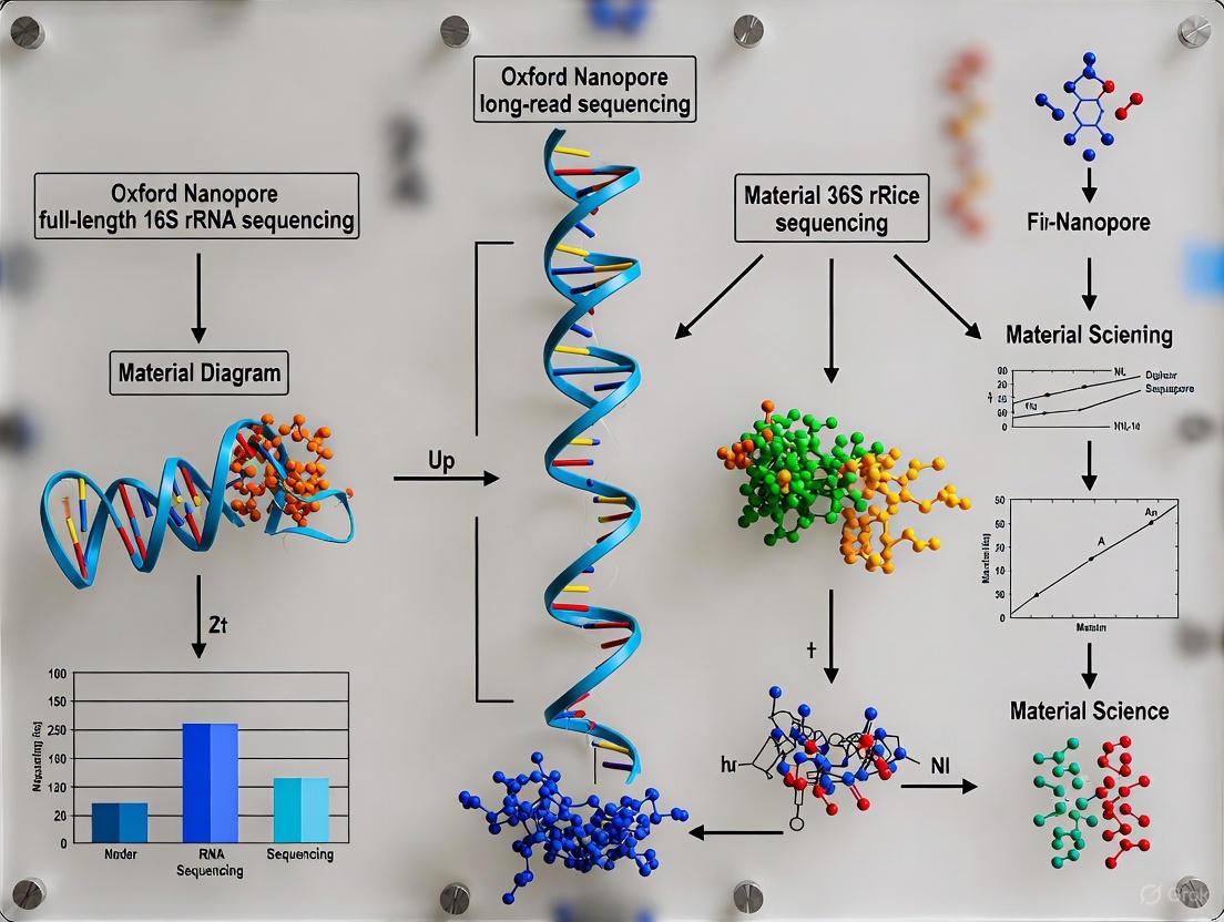
Oxford Nanopore Full-Length 16S rRNA Sequencing: A Comprehensive Guide for Species-Level Microbial Analysis
Full-length 16S rRNA sequencing using Oxford Nanopore Technologies (ONT) is revolutionizing microbial identification by providing species-level resolution critical for biomedical research and drug development.
Oxford Nanopore Full-Length 16S rRNA Sequencing: A Comprehensive Guide for Species-Level Microbial Analysis
Abstract
Full-length 16S rRNA sequencing using Oxford Nanopore Technologies (ONT) is revolutionizing microbial identification by providing species-level resolution critical for biomedical research and drug development. This article explores the transformative potential of long-read sequencing, which overcomes the limitations of short-read methods that target only partial gene regions. We detail the complete workflow from DNA extraction to bioinformatic analysis, leveraging the latest ONT chemistries and kits. The content provides a rigorous comparison with Illumina sequencing, validates performance using clinical and mock community samples, and offers a framework for troubleshooting and optimizing protocols. This guide equips researchers with the methodological knowledge to implement this powerful technology for discovering precise microbial biomarkers and advancing clinical diagnostics.
Unlocking Microbial Diversity: The Power of Full-Length 16S rRNA Sequencing
The 16S ribosomal RNA (rRNA) gene is a ~1.5 kilobase component of the prokaryotic 30S ribosomal subunit, universally present in all self-replicating organisms and comprising nine hypervariable regions (V1-V9) interspersed with highly conserved sequences [1] [2]. Its extensive use in bacterial phylogenetics was pioneered by Carl Woese in 1977 to delineate the previously undescribed taxonomic lineage of Archaea [3]. Woese justified the use of this gene based on its universality in bacteria and its molecular clock-like nature [3]. An important characteristic favoring its use is the presence of these multiple conserved and hypervariable regions, which provide multiple options for PCR primer design [3]. The 16S rRNA gene has served as the cornerstone of microbial identification and phylogenetics for decades, forming the basis of modern microbiology and becoming the gold-standard method for microbiome studies [4] [2].
Strengths and Limitations of the 16S rRNA Gene in Phylogenetics
Advantages as a Phylogenetic Marker
The 16S rRNA gene possesses several key properties that have solidified its role as a primary phylogenetic marker. Its universality ensures it is present in all prokaryotes, allowing for broad comparative analyses across the bacterial and archaeal domains. The functional constancy of the gene, due to its essential role in protein synthesis, means that sequence changes represent evolutionary time rather than functional shifts. The presence of conserved regions enables the design of universal primers for amplification, while the hypervariable regions provide the sequence diversity necessary for taxonomic differentiation at various levels [3] [1]. This combination of features has made 16S rRNA sequencing a powerful tool for classifying uncultivable microorganisms, revolutionizing our understanding of microbial diversity.
Critical Limitations and Evolutionary Dynamics
Recent comparative phylogenomic studies have revealed significant limitations of the 16S rRNA gene that challenge its status as an unequivocal "gold standard" for species identification.
Intragenomic Heterogeneity and Recombination: The 16S rRNA gene often exists in multiple copies within a single genome (from 1 to 27 copies), and these copies can exhibit sequence heterogeneity [3]. Furthermore, the gene is subject to recombination and horizontal gene transfer (HGT) within genera, which can confound phylogenetic inference [3] [2]. One study found evidence of recombination in the 16S rRNA gene in three out of four genera analyzed (Campylobacter, Legionella, and Clostridium) [3].
Poor Phylogenetic Concordance: At the intra-genus level, the 16S rRNA gene shows one of the lowest levels of concordance with core genome phylogeny, averaging only 50.7% [3]. This discordance has direct ramifications for species delineation, phylogenetic inference, and can confound popular community diversity metrics such as Faith's phylogenetic diversity and UniFrac [3].
Evolutionary Rigidity and Species Identification Failure: Contrary to being highly variable, 16S rRNA is actually an evolutionarily rigid sequence, showing extremely low divergence between closely related species compared to the rest of the genome [2]. Analysis of over 1,200 species across 15 bacterial genera identified more than 175 cases where two well-differentiated species (with ~82.5% Average Nucleotide Identity) possessed essentially identical copies of 16S rRNA (>99.9% identity) [2]. This phenomenon questions its applicability as a species-specific marker.
Impact of Analyzed Region: The phylogenetic performance varies significantly across the gene. Concordance for individual hypervariable regions is lower than for the full-length gene, with entropy masking providing little to no benefit [3]. The number of single nucleotide polymorphisms (SNPs) in a region shows a positive logarithmic association with concordance, with approximately 690 ± 110 SNPs required for 80% concordance—a threshold the average 16S rRNA gene (with 254 SNPs) fails to meet [3].
The table below summarizes the concordance of the full-length 16S rRNA gene and its hypervariable regions with core genome phylogenies at different taxonomic levels:
Table 1: Phylogenetic Concordance of the 16S rRNA Gene and Its Regions
| Genetic Region | Intra-genus Concordance with Core Genome | Inter-genus Concordance with Core Genome | Key Findings |
|---|---|---|---|
| Full-length 16S rRNA gene | 50.7% (average) | 73.8% (10th out of 49 loci) | Subject to recombination/HGT; low reliability for species-level phylogenies. |
| Hypervariable Regions (e.g., V3-V4) | Lower than full-length | 60.0% - 62.5% (3rd quartile) | Reduced discriminatory power compared to full-length sequence. |
| Required SNP count for 80% concordance | 690 ± 110 | Not Reported | The average 16S gene has only 254 SNPs, explaining its poor performance. |
Full-Length 16S rRNA Sequencing with Oxford Nanopore Technology
Overcoming the Limitations of Short-Read Sequencing
Legacy short-read sequencing technologies are limited to sequencing partial fragments of the 16S rRNA gene (e.g., V3–V4 or V4–V5), which restricts taxonomic resolution primarily to the genus level [4] [1]. Oxford Nanopore Technology (ONT) overcomes this limitation by generating long reads that span the entire V1–V9 region of the ~1.5 kb 16S rRNA gene in a single read [1]. This full-length sequencing enables high taxonomic resolution for accurate species-level microbial identification from complex, polymicrobial samples [4] [5].
Recent advancements, including the R10.4.1 flow cell and improved basecalling models (e.g., Dorado's super-accurate model), have significantly improved accuracy, facilitating reliable species-level identification [4]. Studies have demonstrated that full-length 16S sequencing with ONT identifies more specific bacterial biomarkers for conditions like colorectal cancer compared to Illumina's V3V4 approach [4]. Furthermore, optimized ONT protocols have been shown to yield higher accuracy for synthetic communities than MiSeq pipelines [5].
Detailed Workflow for Full-Length 16S rRNA Sequencing
The following diagram illustrates the complete workflow for full-length 16S rRNA sequencing using Oxford Nanopore technology:
Key Research Reagent Solutions and Materials
Successful implementation of the full-length 16S rRNA sequencing workflow requires specific reagents and kits. The following table details the essential components.
Table 2: Essential Reagents and Kits for Nanopore 16S rRNA Sequencing
| Item Name | Manufacturer/Kit | Function and Key Features |
|---|---|---|
| 16S Barcoding Kit 24 V14 (SQK-16S114.24) | Oxford Nanopore Technologies | Contains barcoded primers for amplifying and multiplexing up to 24 samples. Includes rapid adapter and buffers for library prep. Compatible with R10.4.1 flow cells. |
| R10.4.1 Flow Cell (FLO-MIN114) | Oxford Nanopore Technologies | The flow cell chemistry required for this protocol, providing high accuracy for full-length 16S rRNA gene sequencing. |
| LongAmp Hot Start Taq 2X Master Mix | New England Biolabs (NEB) | Enzyme master mix recommended for the PCR amplification of the full-length 16S rRNA gene. |
| DNA LoBind Tubes | Eppendorf | Specialized tubes to minimize DNA loss during library preparation steps. |
| AMPure XP Beads | Beckman Coulter | Magnetic beads used for post-PCR clean-up and size selection to purify the library. |
| Qubit dsDNA HS Assay Kit | Thermo Fisher Scientific | For accurate quantification of DNA concentration at critical steps (gDNA and final library). |
The wet-lab protocol can be summarized in four main stages, with specific attention to key details:
DNA Extraction and QC: Extract high-quality genomic DNA using a sample-appropriate method (e.g., QIAamp PowerFecal DNA Kit for stool). Assess DNA quantity and purity. The protocol requires 10 ng of high molecular weight gDNA per barcode [6].
16S Barcoded PCR Amplification: Amplify the full-length 16S rRNA gene using the barcoded primers from the kit and the LongAmp Hot Start Taq Master Mix. A critical requirement is that a minimum of 4 barcodes must be used per flow cell for optimal output. For projects with fewer than 4 samples, the sample must be split across multiple barcodes (e.g., one sample split across barcodes 01-04) [6].
Library Preparation: Pool the barcoded amplicons in equimolar ratios. Perform a bead-based clean-up using AMPure XP Beads to purify the library and remove short fragments and contaminants. Subsequently, attach the rapid sequencing adapters to the DNA ends. The adapted library should be sequenced immediately for best results [6].
Sequencing and Analysis: Prime the flow cell and load the prepared library. Sequence on a MinION or GridION device using the MinKNOW software with the high-accuracy (HAC) basecaller enabled. For analysis, the EPI2ME wf-16s workflow or tools like Emu can be used for real-time or post-run species-level identification and abundance profiling [4] [1].
The 16S rRNA gene remains an indispensable, universal marker in microbial ecology and phylogenetics. However, modern phylogenomic studies have critically revised its role, demonstrating significant limitations due to recombination, horizontal gene transfer, and evolutionary rigidity that can mislead species-level identification and phylogenetic inference. The advent of Oxford Nanopore long-read sequencing directly addresses one of the most significant practical constraints by enabling full-length 16S rRNA gene analysis. This provides a substantial improvement in taxonomic resolution over short-read approaches, moving from genus-level to robust species-level identification. For researchers, this means that while the 16S rRNA gene must be used with a clear understanding of its phylogenetic shortcomings, full-length sequencing on nanopore platforms offers a rapid, accessible, and cost-effective method for accurate microbial profiling in diverse applications from clinical diagnostics to environmental monitoring.
Limitations of Short-Read Sequencing for Species-Level Identification
The accurate identification of microbial species is a cornerstone of microbiology, with profound implications for understanding human health, disease pathogenesis, and ecosystem function. For decades, short-read sequencing technologies, exemplified by Illumina platforms, have been the workhorse of microbial ecology and diagnostics. These methods typically generate reads of 50-600 bases by fragmenting DNA into small segments, amplifying them, and reading these segments as they are synthesized [7] [8]. However, when applied to species-level identification—particularly through 16S rRNA gene sequencing—inherent limitations of these short-read approaches emerge with significant consequences for taxonomic resolution.
This application note details the fundamental constraints of short-read sequencing for species-level microbial identification. It further outlines how the adoption of full-length 16S rRNA sequencing using Oxford Nanopore Technologies (ONT) provides a transformative solution, enabling researchers and drug development professionals to achieve unprecedented taxonomic resolution within complex microbiomes.
Core Technical Limitations of Short-Read Sequencing
The inability of short-read sequencing to reliably resolve microbial identities to the species level stems from several interconnected technical constraints.
Incomplete Gene Capture and Region Selection Bias
The full 16S rRNA gene is approximately 1,500 base pairs (bp) long and contains nine hypervariable regions (V1-V9) interspersed with conserved regions [1]. Short-read platforms cannot sequence this entire gene in a single read, forcing researchers to select one or two hypervariable regions (such as V3-V4 or V4) for amplification and sequencing [9] [10]. The mean read length for the V3-V4 region is typically around 447 bp [11], representing only a fraction of the full gene.
This regional approach introduces substantial bias, as no single variable region provides sufficient phylogenetic signal to distinguish all bacterial species. Different regions exhibit varying degrees of conservation across taxa, meaning that the choice of region directly influences the observed microbial community composition and can miss key discriminatory nucleotides present in unsequenced portions of the gene [12] [10].
Limited Phylogenetic Resolution
The limited length of short reads directly constrains phylogenetic resolution. While often sufficient for genus-level assignments, the sequences lack the informational breadth required to differentiate between closely related species that diverge only in regions not captured by the sequencing strategy [7].
Comparative studies demonstrate this limitation clearly. In mouse gut microbiome studies, short-read (V3-V4) and long-read (full-length) approaches yield highly concordant results at higher taxonomic levels (phylum, family, genus), but the short-read method fails to identify specific species like Bifidobacterium animalis and Bifidobacterium pseudolongum that are readily detected with full-length sequencing [11]. Similarly, in human respiratory microbiome studies, Illumina short-read sequencing struggles with species-level resolution, whereas ONT's full-length 16S rRNA sequencing enables it [10].
Challenges with Repetitive and Conserved Regions
Microbial genomes contain repetitive regions and highly conserved sequences that complicate short-read assembly and analysis. When short reads are derived from these regions, it becomes impossible to uniquely assign them to a specific location in a gene or genome, leading to fragmented assemblies and ambiguous taxonomic assignments [7]. This is particularly problematic in metagenomics, where identical or highly similar sequences may originate from multiple related organisms, further confounding analysis [7].
Table 1: Comparative Analysis of Sequencing Approaches for 16S rRNA Gene Profiling
| Feature | Short-Read Sequencing (e.g., Illumina) | Long-Read Sequencing (e.g., Oxford Nanopore) |
|---|---|---|
| Target Region | Partial gene (e.g., V3-V4, ~447 bp) [11] | Full-length gene (V1-V9, ~1,500 bp) [1] |
| Species-Level Resolution | Limited and unreliable [11] [10] | High and reliable [12] [10] |
| Ability to Resolve Repetitive Regions | Poor, leads to fragmented assemblies [7] | Excellent, spans repetitive regions [7] |
| Primary Limitation | Regional bias; insufficient phylogenetic information per read | Historically higher error rates, though now >99% [7] |
| Data Output for Community Analysis | Coarser resolution, struggles with closely related groups [7] | Finer resolution, can discriminate sub-species clades [7] |
Impact on Microbiome Research and Clinical Applications
The technical limitations of short-read sequencing translate directly into concrete challenges for research and clinical interpretation.
The most significant impact is the incomplete and biased microbial community profiling. Without species- and strain-level data, researchers cannot build accurate hypotheses about the role of specific microbes in health and disease. This is a critical barrier in drug development, particularly for Live Biotherapeutic Products (LBPs), where understanding strain-level pharmacokinetics and pharmacodynamics is essential [12]. While short-read metagenomics can detect an introduced therapeutic strain, detection confidence is notably higher with long-read methods [12].
Furthermore, the lack of resolution obscures microbial diversity. A 2022 comparative study found that long-read 16S-ITS-23S amplicon sequencing provided strain-level community resolution and insights into novel taxa that were inaccessible via ubiquitous short-read V3-V4 profiling [12].
Oxford Nanopore Full-Length 16S rRNA Sequencing as a Solution
Oxford Nanopore Technology directly addresses the gaps left by short-read sequencing by enabling real-time, single-molecule sequencing of the entire ~1.5 kb 16S rRNA gene in a single read [1].
Principle of the Solution
This approach eliminates the need for regional selection bias by capturing all nine hypervariable regions simultaneously. The long reads provide a comprehensive nucleotide signature for each organism in a sample, which dramatically increases the number of informative characters available for taxonomic classification. This allows for discrimination not just at the species level, but often at the strain level, within complex microbiomes [7] [12].
The platform works by threading DNA strands through protein nanopores and detecting changes in an ionic current as each nucleotide passes through the pore. This mechanism does not require DNA amplification for sequencing, thus avoiding associated biases [7] [8].
Experimental Protocol for Full-Length 16S rRNA Sequencing
The following protocol provides a robust framework for species-level microbial identification using Oxford Nanopore technology.
Sample Collection and DNA Extraction
- Sample Collection: Collect samples (e.g., stool, soil, respiratory secretions) using sterile tools and place them in sterile, DNA-free containers. For fecal or gut content samples, standardize collection time relative to feeding to minimize biological variability [13]. Store samples at -80°C immediately or use nucleic acid preservation buffers if freezing is not feasible [13].
- DNA Extraction: Select a method that yields high-molecular-weight DNA. For stool samples, the QIAamp PowerFecal DNA Kit is recommended. For soil, use the QIAGEN DNeasy PowerMax Soil Kit. For environmental water samples, the ZymoBIOMICS DNA Miniprep Kit is suitable [1]. Validate extraction efficiency using well-characterized reference materials like the WHO WC-Gut RR [14].
Library Preparation and Sequencing
- PCR Amplification: Amplify the full-length 16S rRNA gene from 5-50 ng of genomic DNA using universal primers (e.g., 27F: AGAGTTTGATYMTGGCTCAG and 1492R: GGTTACCTTGTTAYGACTT) [9] [1]. Use a PCR protocol: initial denaturation at 95°C for 5 min; 25-30 cycles of 95°C for 30 s, 55-57°C for 30 s, and 72°C for 60 s; final extension at 72°C for 5 min [9] [14].
- Library Preparation: Prepare the sequencing library using the ONT 16S Barcoding Kit (e.g., SQK-16S114). This kit allows for multiplexing up to 24 samples by using barcoded primers during PCR, followed by adapter ligation [10] [1].
- Sequencing: Load the pooled library onto a MinION Flow Cell (R10.4.1 or newer). Sequence on a MinION or GridION device using the MinKNOW software for approximately 24-72 hours, utilizing the high-accuracy (HAC) basecaller to achieve optimal coverage and accuracy [10] [1].
Data Analysis
- Basecalling and Demultiplexing: Perform basecalling and demultiplexing using the Dorado basecaller integrated within MinKNOW or EPI2ME [10].
- Taxonomic Classification: Analyze the resulting FASTQ files using the EPI2ME Labs wf-16s workflow or other specialized pipelines like Emu [9]. These tools classify reads against reference databases (e.g., SILVA) to generate abundance tables and phylogenetic visualizations [10] [1].
Research Reagent Solutions
Table 2: Essential Research Reagents and Kits for Full-Length 16S rRNA Sequencing
| Item | Function | Example Product |
|---|---|---|
| DNA Extraction Kit | Isolates high-quality genomic DNA from specific sample matrices. | QIAamp PowerFecal DNA Kit (stool), QIAGEN DNeasy PowerMax Soil Kit (soil) [1] |
| Full-Length 16S PCR Primers | Amplifies the entire ~1.5 kb 16S rRNA gene from genomic DNA. | 27F (AGAGTTTGATYMTGGCTCAG) / 1492R (GGTTACCTTGTTAYGACTT) [9] |
| Long-Range DNA Polymerase | Performs PCR amplification of long DNA fragments with high fidelity. | Included in ONT 16S Barcoding Kit [1] |
| Barcoding & Library Prep Kit | Multiplexes samples and prepares DNA for nanopore sequencing. | Oxford Nanopore 16S Barcoding Kit 24 (SQK-16S114) [1] |
| Sequencing Flow Cell | The consumable containing nanopores for generating sequence data. | Oxford Nanopore MinION Flow Cell (R10.4.1) [10] |
| Control Material | Validates extraction, amplification, and sequencing accuracy. | WHO International Reference Reagents for Microbiome [14] |
Short-read sequencing technologies have provided invaluable insights into microbial communities but possess inherent limitations that prevent reliable species-level identification. These constraints, including regional bias and insufficient phylogenetic resolution, hinder a complete understanding of microbiome composition and function.
The adoption of Oxford Nanopore's full-length 16S rRNA sequencing effectively overcomes these limitations. By providing comprehensive genetic information in single reads, this method delivers the high taxonomic resolution required for advanced research and the development of targeted therapeutic interventions. For researchers and drug development professionals seeking to move beyond genus-level observations, leveraging this technology is a critical step toward unlocking a more precise and actionable understanding of the microbial world.
The 16S ribosomal RNA (rRNA) gene, approximately 1.5 kilobases in length, serves as a cornerstone for microbial identification and classification [1]. This gene comprises nine hypervariable regions (V1-V9), which are interspersed with highly conserved sequences, providing a genetic barcode for distinguishing bacterial taxa [1] [15]. For decades, short-read sequencing technologies have been constrained to analyzing partial fragments of the gene, such as the V3-V4 or V4-V5 regions, due to their inherent read length limitations [1] [4]. This fragmented approach often limits taxonomic resolution to the genus level, obscuring the precise microbial species present in a sample and hindering the discovery of fine-scale, disease-relevant biomarkers [4].
Oxford Nanopore Technologies (ONT) overcomes this fundamental limitation by generating long-read sequences that can effortlessly span the entire V1-V9 region of the 16S rRNA gene in a single, continuous read [1] [4] [16]. This capability enables high taxonomic resolution for accurate species-level microbial identification, even from complex, polymicrobial samples [1]. The following application note details how this "Nanopore Advantage" is achieved through specific protocols and reagents, and demonstrates its impact on research and diagnostic outcomes.
Quantitative Comparisons: Full-Length vs. Partial Region Sequencing
Sequencing the complete 16S rRNA gene provides a tangible increase in taxonomic classification power. The table below summarizes key performance metrics from recent comparative studies.
Table 1: Performance comparison of 16S rRNA sequencing approaches
| Metric | Illumina (V3-V4) | Nanopore (V1-V9) | PacBio HiFi (V1-V9) | Citation |
|---|---|---|---|---|
| Species-Level Classification Rate | 47% - 48% | 76% | 63% | [16] |
| Genus-Level Classification Rate | 80% | 91% | 85% | [16] |
| Read Length | ~442 bp | ~1,412 - 1,567 bp | ~1,453 bp | [17] [16] |
| Key Finding | Limited species-level resolution; genus-level results | Identified more specific bacterial biomarkers for colorectal cancer | High-fidelity reads; lower species resolution than ONT | [4] [16] |
The correlation between bacterial abundances measured by Illumina (V3-V4) and Nanopore (V1-V9) at the genus level is strong (R² ≥ 0.8) [4]. However, the superior resolution of full-length sequencing enables the discovery of disease-specific bacterial biomarkers that are missed by partial gene analysis. For instance, in a colorectal cancer study, Nanopore sequencing identified pathogens such as Parvimonas micra, Fusobacterium nucleatum, and Peptostreptococcus anaerobius with high specificity [4].
Experimental Protocols for Full-Length 16S rRNA Sequencing
A robust and standardized workflow is critical for generating reliable, reproducible full-length 16S data. The following section outlines a validated, end-to-end protocol.
Sample Collection and DNA Extraction
The selection of a DNA extraction method should be tailored to the sample type to ensure high yield and quality while minimizing bias.
- Sample Types: The protocol is applicable to diverse samples, including stool, soil, water, and clinical specimens like tissue, pus, and body fluids [1] [14].
- Recommended Kits: For environmental water samples, the ZymoBIOMICS DNA Miniprep Kit is recommended. For soil, use the QIAGEN DNeasy PowerMax Soil Kit. For stool, the QIAamp PowerFecal DNA Kit is effective for microbiome DNA extraction [1]. The PureLink Microbiome DNA Purification Kit has also been successfully used with microbial standards [18].
- Critical Step: For clinical samples, especially tissue, a bead-beating step using instruments like a TissueLyser is often necessary to ensure efficient lysis of tough bacterial cell walls, particularly for Gram-positive bacteria [14] [17].
PCR Amplification and Library Preparation
This stage amplifies the target gene and prepares the DNA for sequencing.
- Primer Sets: The full-length ~1.5 kb 16S rRNA gene is amplified using universal primers 27F and 1492R [15] [19]. Primer degeneracy significantly impacts results; a more degenerate 27F primer (e.g., 5'-AGAGTTTGATCMTGGC-3') can reduce amplification bias and yield a more accurate representation of microbial diversity compared to standard primers [15] [19].
- PCR Protocol: Using the 16S Barcoding Kit (SQK-16S114.24), amplify the gene with 25-40 PCR cycles. An increased cycle number (e.g., 40 cycles) is recommended for low-biomass clinical samples [17]. The annealing temperature can be optimized; lowering it from 55°C to 52°C improves sensitivity [17].
- PCR Components: The choice of polymerase (e.g., LongAmp Hot Start Taq) and careful control of cycle numbers are crucial, as elevated cycles can introduce PCR bias [15].
- Library Construction: The amplified products are barcoded to enable multiplexing of up to 24 samples. Sequencing adapters are then ligated to the pooled library, which is loaded onto a flow cell [1].
Sequencing and Basecalling
- Sequencing Device: Sequencing can be performed on MinION, GridION, or PromethION devices. MinION Flow Cells are suitable for portable, at-source sequencing [1].
- Run Time: A typical sequencing run lasts 24-72 hours, depending on sample complexity and desired coverage [1]. For high-accuracy bacterial identification, a minimum Q-score of 10 is recommended during basecalling [17].
- Basecalling Models: The Dorado basecaller offers different models (fast, hac, sup). While the "super-accurate" (sup) model is available, the high-accuracy (hac) model is sufficient for reliable species-level taxonomic identification [4].
The Scientist's Toolkit: Essential Research Reagent Solutions
Table 2: Key reagents and tools for Nanopore 16S rRNA sequencing
| Item | Function | Example Products & Part Numbers |
|---|---|---|
| DNA Extraction Kits | Isolate high-quality DNA from various sample types. | ZymoBIOMICS DNA Miniprep Kit; QIAGEN DNeasy PowerMax Soil Kit; QIAamp PowerFecal DNA Kit [1]. |
| 16S Amplification & Barcoding Kit | Amplify full-length 16S gene and attach unique barcodes for multiplexing. | 16S Barcoding Kit 24 (SQK-16S114.24) [1] [17]. |
| Sequencing Hardware | Platform for generating long-read sequences. | MinION, GridION, PromethION [1]. |
| Flow Cell | Consumable containing nanopores for sequencing. | MinION Flow Cell (R9.4.1 or R10.4.1) [20] [17]. |
| Bioinformatics Pipelines | Analyze sequencing data for taxonomic classification and abundance. | EPI2ME wf-16s, EMU (e.g., GMS-16S pipeline), BugSeq [1] [4] [17]. |
Workflow Visualization: From Sample to Species
The following diagram summarizes the complete end-to-end workflow for full-length 16S rRNA sequencing using Nanopore technology.
The ability of Oxford Nanopore long-read sequencing to span the entire V1-V9 region of the 16S rRNA gene represents a significant leap forward in microbial genomics. This technical advantage directly translates into higher species-level resolution, enabling researchers and drug development professionals to discover more precise biomarkers, characterize complex polymicrobial infections, and achieve a deeper, more accurate understanding of microbial communities in health and disease. As chemistries and protocols continue to standardize, Nanopore sequencing is poised to become an indispensable tool for precision microbiology.
Key Applications in Biomedical Research and Drug Development
The identification of microbial communities at the species level is paramount in biomedical research, influencing everything from understanding disease mechanisms to identifying novel therapeutic targets. The 16S ribosomal RNA (rRNA) gene, approximately 1.5 kb in length, contains nine variable regions (V1-V9) flanked by conserved sequences, providing a genetic barcode for bacterial identification [1]. While short-read sequencing technologies have been the workhorse for 16S studies, they are limited to analyzing partial fragments of the gene (e.g., V3–V4), which restricts taxonomic resolution primarily to the genus level [1] [4] [21]. Oxford Nanopore Technologies (ONT) long-read sequencing overcomes this limitation by generating reads that span the entire V1–V9 region of the 16S rRNA gene in a single read, enabling accurate species-level identification and unlocking new applications in drug development and clinical diagnostics [1] [4]. This application note details the protocols and key applications of this powerful technology.
Key Applications and Comparative Performance
Full-length 16S rRNA sequencing with Oxford Nanopore technology is revolutionizing multiple domains within biomedicine by providing a rapid, cost-effective, and highly resolutive method for microbial identification.
Disease Biomarker Discovery
The ability to resolve bacterial species significantly enhances the discovery of disease-specific microbial biomarkers.
- Colorectal Cancer (CRC): A 2025 study comparing Illumina (V3V4) and ONT (V1V9) sequencing for CRC biomarker discovery demonstrated that Nanopore sequencing identified more specific bacterial biomarkers. Species such as Parvimonas micra, Fusobacterium nucleatum, and Bacteroides fragilis were identified as biomarkers, with a predictive model for CRC achieving an AUC of 0.87 using 14 species, and 0.82 using just 4 species [4].
- Inflammatory Bowel Disease (IBD): Deep learning models like Read2Pheno, applied to full-length 16S reads, can predict host phenotypes such as IBD by identifying informative nucleotide regions within the 16S gene, bypassing the need for traditional abundance tables and enabling direct genotype-to-phenotype linkage [22].
Clinical Diagnostics and Infectious Disease
The speed and portability of Nanopore sequencing make it suitable for near-patient clinical diagnostics.
- Infective Endocarditis (IE): Traditional culture-based identification of IE pathogens has limited sensitivity after antibiotic administration. ONT's full-length 16S sequencing provides a rapid, flexible, and accurate method for species-level identification directly from clinical samples, guiding targeted antimicrobial therapy [23].
- Culture-Negative and Polymicrobial Infections: Implementation of 16S nanopore sequencing in a clinical diagnostic lab demonstrated its value for identifying pathogens without prior enrichment. The method successfully identified bacteria in all culture-positive samples and detected pathogenic bacteria in 15 out of 30 culture-negative samples, even unravelling complex polymicrobial infections [24].
Characterizing Complex Microbial Communities
The high accuracy of the latest ONT chemistry enables reliable profiling of synthetic and environmental microbial communities.
- Synthetic Communities: A 2025 study presented a high-throughput protocol for synthetic communities, showing that the accuracy of the ONT sequencing pipeline was significantly higher than that of a standard MiSeq pipeline, ensuring reproducible and easy characterization of community composition [5].
- Environmental and Gut Microbiota: Research has shown that ONT R10.4.1 analysis produces a community composition similar to PacBio data, a established long-read technology. In human gut microbiota studies, full-length sequencing provides better resolution for discriminating between members of particular taxa like Bifidobacterium, allowing an accurate representation of the sample's bacterial composition [25] [21].
Table 1: Comparative Performance of 16S rRNA Sequencing Approaches
| Parameter | Illumina (Short-Read) | Oxford Nanopore (Long-Read) |
|---|---|---|
| Target Region | Partial (e.g., V3-V4) | Full-length (V1-V9) |
| Primary Resolution | Genus-level | Species-level [4] |
| Read Length | ~400-500 bp | ~1,500 bp (unrestricted) |
| Accuracy | >99.9% (Q30+) | ~99% with R10.4.1/HAC basecalling (Q20) [4] [25] |
| Key Advantage | High raw accuracy, high throughput | Species-level resolution, rapid turnaround, portability |
| Demonstrated Application | General community profiling | Biomarker discovery (CRC), rapid diagnostics (IE), complex community analysis [4] [23] |
Detailed Experimental Protocol
This protocol is adapted from the Oxford Nanopore "Microbial Amplicon Barcoding Sequencing for 16S and ITS" (SQK-MAB114.24) and is designed for multiplexing up to 24 samples [26].
The diagram below illustrates the key steps in the workflow.
Step-by-Step Methodology
Step 1: DNA Extraction and Quality Control (QC)
- Input Material: Begin with your sample (e.g., stool, soil, water, clinical specimen).
- Extraction Kits: Use sample-specific kits for high-quality DNA. Recommendations include:
- Stool samples: QIAmp PowerFecal DNA Kit or QIAGEN Genomic-tip 20/G.
- Soil samples: QIAGEN DNeasy PowerMax Soil Kit.
- Environmental water: ZymoBIOMICS DNA Miniprep Kit [1].
- QC Check: Quantify DNA using a fluorometric method (e.g., Qubit dsDNA HS Assay Kit). The protocol requires 10 ng of high molecular weight genomic DNA per sample for amplification [26].
Step 2: PCR Amplification of Full-Length 16S Gene
- Primers: Use the inclusive 16S primers supplied in the Microbial Amplicon Barcoding Kit 24 V14. These are designed to amplify the full-length ~1.5 kb 16S rRNA gene.
- PCR Reaction: Set up the PCR reaction using LongAmp Hot Start Taq 2X Master Mix.
- Process Time: 10 minutes setup + PCR run time [26].
- Primer Design Note: To avoid amplification bias against certain taxa (e.g., Bifidobacterium), some protocols use primers with degenerate bases to account for sequence mismatches, ensuring a more representative profile [21].
Step 3: Amplicon Barcoding and Pooling
- Barcoding Reaction: Attach unique barcodes from the kit (up to 24) to the amplified DNA from each sample.
- Process Time: 15 minutes [26].
- Pooling and Clean-up: Inactivate the barcoding reaction, pool all barcoded samples into a single tube, and perform a bead-based clean-up (e.g., using AMPure XP Beads) to purify the library.
Step 4: Adapter Ligation and Loading
- Rapid Adapter Attachment: Add the Rapid Sequencing Adapter to the prepared DNA ends to facilitate sequencing.
- Process Time: 5 minutes. It is strongly recommended to sequence the library immediately after this step [26].
- Priming and Loading: Prime the flow cell (e.g., MinION R10.4.1) using the Flow Cell Priming Kit and load the adapted library.
- Process Time: 10 minutes [26].
Step 5: Sequencing and Analysis
- Sequencing: Start the sequencing run on a MinION or GridION device using the MinKNOW software. For high accuracy, use the High Accuracy (HAC) or Super Accuracy (SUP) basecaller within MinKNOW. A typical run can take 24-72 hours depending on the desired coverage and sample complexity [1] [4].
- Analysis: The EPI2ME software platform offers user-friendly bioinformatics workflows. The wf-16s pipeline is designed for real-time or post-run analysis of 16S data, generating an abundance table, bar plots, and interactive visualizations (Sankey, sunburst plots) for taxonomic lineages [1].
Table 2: Essential Research Reagent Solutions
| Item | Function / Purpose | Example Product / Kit |
|---|---|---|
| Sample-Specific DNA Extraction Kit | Obtains high-quality, inhibitor-free gDNA from complex samples. | ZymoBIOMICS DNA Miniprep Kit, QIAGEN DNeasy PowerMax Soil Kit [1] |
| Microbial Amplicon Barcoding Kit | Provides primers for full-length 16S amplification and barcodes for multiplexing. | Oxford Nanopore SQK-MAB114.24 [26] |
| High-Fidelity PCR Master Mix | Ensures accurate and efficient amplification of the target 16S gene. | LongAmp Hot Start Taq 2X Master Mix [26] |
| Magnetic Beads | Purifies and size-selects the DNA library post-amplification and barcoding. | AMPure XP Beads [26] |
| R10.4.1 Flow Cell | The consumable containing nanopores for sequencing; R10.4.1 provides high accuracy. | MinION/GridION Flow Cell (FLO-MIN114) [26] [25] |
| Bioinformatics Tool | Classifies sequencing reads taxonomically and generates abundance profiles. | EPI2ME wf-16s, Emu [1] [4] |
Technical and Biological Validation
The transition to full-length 16S sequencing is supported by rigorous technical validation.
- Accuracy of R10.4.1 Chemistry: The latest Nanopore flow cells (R10.4.1) with Q20+ reagents have substantially improved accuracy, achieving ~99% model read accuracy, which is sufficient for species-level classification (requiring ≥99% identity) [4] [25]. One study noted that error rates, particularly deletions, were greatly reduced in R10.4.1 compared to previous versions [25].
- Impact of Basecalling and Databases: Performance is influenced by bioinformatic choices. A 2025 study reported that lower-quality basecalling models (e.g., "fast") resulted in higher observed species counts, while database choice (e.g., SILVA vs. Emu's Default database) greatly influenced species identification, with the latter yielding higher diversity but potential overclassification [4]. The use of curated, in-house databases is recommended to mitigate errors in public references [27].
- Resolution Power: Research has consistently demonstrated that while the relative abundance of dominant genera is similar between full-length and short-read sequencing, only the full-length method provides the resolution necessary for reliable species-level discrimination, as seen in taxa like Bacillus, Clostridium, and Staphylococcus [21].
Oxford Nanopore's full-length 16S rRNA sequencing represents a significant advancement over traditional short-read methods, providing the species-level resolution required for cutting-edge biomedical research and drug development. Its applications in precise biomarker discovery for conditions like colorectal cancer, rapid diagnosis of challenging infections, and accurate characterization of complex microbial communities make it an indispensable tool for researchers and clinicians alike. The continuously improving chemistry, coupled with streamlined wet-lab and bioinformatic protocols, positions this technology as a cornerstone for future microbiome studies aimed at understanding disease etiology and developing novel therapeutics.
From Sample to Sequence: A Practical Workflow for Nanopore 16S Sequencing
Sample-Specific DNA Extraction Protocols for Optimal Yield
The pursuit of optimal DNA extraction is a foundational prerequisite for successful full-length 16S ribosomal RNA (rRNA) gene sequencing using Oxford Nanopore Technologies (ONT). This targeted approach requires high-molecular-weight (HMW), intact DNA to leverage the primary advantage of long-read sequencing: the generation of reads that span the entire ~1.5 kb V1-V9 region of the 16S rRNA gene. Such comprehensive coverage is essential for achieving species-level taxonomic resolution in complex polymicrobial samples, a level of detail that is often lost with short-read sequencing of partial gene regions [1] [15]. The integrity and purity of the extracted DNA directly influence every subsequent step, from library preparation efficiency to the accuracy of bioinformatic classification. Consequently, the selection of a DNA extraction protocol is not a one-size-fits-all endeavor but must be tailored to the specific biological matrix of the sample to effectively overcome unique biochemical challenges and minimize bias.
This application note provides a detailed framework for selecting and optimizing DNA extraction methods for full-length 16S rRNA sequencing. It outlines sample-specific protocols, presents comparative performance data, and identifies key reagents to ensure the isolation of high-quality DNA suitable for ONT's MinION platform.
Sample-Specific DNA Extraction Methodologies
Stool and Fecal Samples
Primary Challenge: Stool samples contain a complex mixture of microbial organisms with varying cell wall structures (Gram-positive vs. Gram-negative) and high levels of PCR inhibitors and contaminating host DNA [28] [29].
Recommended Protocol:
- Kit: QIAamp PowerFecal Pro DNA Kit (Qiagen) or ZymoBIOMICS DNA Miniprep Kit (Zymo Research) [1] [29].
- Lysis Method: Implement a robust mechanical lysis step, such as bead beating with a homogenizer or vortexing with Pathogen Lysis Tubes containing glass beads, to ensure the disruption of tough Gram-positive bacterial cell walls [28] [30].
- Inhibitor Removal: Utilize kits that incorporate specific reagents to remove humic acids, bile salts, and other complex inhibitors commonly found in stool.
- Input Material: Use 180-220 mg of raw stool or the equivalent from a swab. For samples preserved in stabilization media, note that DNA yield may be lower, and input volume may need adjustment [28].
- Automation: For high-throughput studies, the MagMAX Microbiome Ultra Kit (Thermo Fisher) is compatible with KingFisher instrument systems for automated purification [28].
Tissue Samples (e.g., Liver, Muscle, Biopsies)
Primary Challenge: Tissues are often fibrous and require effective homogenization. Furthermore, endogenous nucleases in tissues like liver can lead to rapid DNA degradation post-collection [28] [31].
Recommended Protocol:
- Homogenization: Use a mechanical homogenizer (e.g., Fisherbrand 850 Homogenizer) or careful bead beating to disrupt the tissue matrix. Inadequate homogenization can cause foaming and make the homogenate difficult to transfer, leading to sample loss [28].
- DNA Stabilization: Flash-freeze tissue samples in liquid nitrogen immediately after collection and store at -80°C to inhibit nuclease activity [31].
- RNase Treatment: Incorporate an RNase A treatment step during extraction to reduce RNA contamination, which can skew quantification and interfere with downstream library preparation [28].
- Validated Kits: The Nanobind PanDNA kit (PacBio) and MagMAX DNA Multi-Sample Ultra 2.0 kit (Thermo Fisher) have been validated for HMW DNA extraction from various tissue types [28] [32].
Buccal and Dry Swabs
Primary Challenge: These samples often contain high concentrations of host cells, bacterial contaminants from the skin or oral microbiome, and potential inhibitors like mucins [28].
Recommended Protocol:
- Increased Yield Strategy: For buccal swabs, using two swabs in a single isolation and extending the lysis incubation time can significantly improve DNA recovery [28].
- Inhibitor Removal: Magnetic bead-based purification methods, such as those used in the MagMAX DNA Multi-Sample Ultra 2.0 chemistry, are effective at removing sample-specific inhibitors while targeting microbial DNA [28].
- Storage: Ensure swabs are thoroughly dried before storage to prevent overgrowth of contaminants.
Formalin-Fixed Paraffin-Embedded (FFPE) Samples
Primary Challenge: The formalin fixation process causes cross-linking and nucleic acid fragmentation, while the paraffin embedding requires additional dewaxing steps [28].
Recommended Protocol:
- Deparaffinization: Replace traditional, hazardous xylene washes with automated, heating-based methods. The Applied Biosystems AutoLys M Tubes and Caps provide an effective and safer alternative [28].
- Lysis and Digestion: Use a combination of heating steps and prolonged proteinase K digestion to reverse cross-links and release DNA from the fixed tissue. The Applied Biosystems MagMAX FFPE DNA/RNA Isolation chemistry is designed for this purpose [28].
Water and Soil Samples
Primary Challenge: Environmental samples can contain particulate matter and environmental inhibitors while often having low microbial biomass [1].
Recommended Protocol:
- Water Filtration: Filter a large volume of water through a 0.22 µm membrane to concentrate microbial biomass.
- Soil Lysis: For soil, use a kit designed for tough environmental matrices, such as the QIAGEN DNeasy PowerMax Soil Kit, which is effective at removing inhibitory humic acids and fulvic acids [1].
- Gentle Lysis Consideration: For studies prioritizing DNA length over total yield, an enzymatic lysis approach (e.g., using lysozyme and MetaPolyzyme) can be superior to harsh bead-beating, as it reduces DNA shearing. Research has shown that enzymatic lysis can increase the average length of microbial reads by a median of 2.1-fold compared to methods without pre-lysis [30].
Comparative Performance of DNA Extraction Methods
The following table summarizes the quantitative performance of several DNA extraction methods evaluated specifically for long-read sequencing applications using defined bacterial mock communities.
Table 1: Performance Comparison of DNA Extraction Methods for Long-Read Sequencing
| Extraction Method | Lysis Technique | Purification Technique | Key Finding | Recommended Application |
|---|---|---|---|---|
| Quick-DNA HMW MagBead Kit [29] | Bead Beating | Magnetic Beads (SPRI) | Produced the best yield of pure HMW DNA; enabled accurate detection of almost all species in a mock community. | Bacterial metagenomics (Gram+ and Gram-). |
| Enzymatic Lysis Method [30] | Enzymatic (MetaPolyzyme) | Spin Column | Increased average microbial read length by 2.1-fold (IQR: 1.7-2.5) vs. control; provided 100% consistent diagnosis vs. clinical culture. | Urine samples; pathogen identification. |
| Mechanical Lysis Method [30] | Bead Beating | Spin Column | Resulted in excessive DNA fragmentation, reducing the advantage of long-read sequencing. | Not recommended for HMW DNA. |
| Phenol-Chloroform (Organic) [29] [31] | Chemical / Bead Beating | Solvent Precipitation | Can yield HMW DNA but uses hazardous chemicals; prone to phase inversion and contamination. | General purpose (with caution). |
| Nanobind PanDNA Kit [32] | Lysis Buffer | Nanobind Disk | Delivers ultra-clean, HMW DNA with little to no shearing; avoids hazardous chemicals. | Broad range: blood, tissue, cells, bacteria. |
The Scientist's Toolkit: Essential Reagents and Kits
Table 2: Key Research Reagent Solutions for DNA Extraction and 16S rRNA Sequencing
| Item | Function/Application | Example Products |
|---|---|---|
| HMW DNA Extraction Kits | Isolation of pure, high-molecular-weight DNA crucial for long-read sequencing. | Quick-DNA HMW MagBead Kit (Zymo) [29]; Nanobind PanDNA Kit (PacBio) [32]. |
| Sample-Specific Kits | Optimized lysis and purification for challenging matrices. | QIAamp PowerFecal Pro DNA Kit (stool) [1]; DNeasy PowerMax Soil Kit (soil) [1]; MagMAX FFPE DNA/RNA Kit (FFPE) [28]. |
| Lytic Enzymes | Gentle, enzymatic cell wall lysis for preserving DNA length. | Lysozyme; MetaPolyzyme [30]. |
| Magnetic Beads | High-throughput, automated DNA purification and size selection. | SPRIselect Beads [15]; MagMAX beads [28]. |
| 16S Barcoding Kit | Targeted amplification and barcoding of the full-length 16S gene for multiplexing. | 16S Barcoding Kit (ONT, SQK-16S024) [1]. |
| Taq Polymerase | Robust amplification of the full-length ~1.5 kb 16S amplicon. | LongAmp Hot Start Taq (NEB) [15]. |
| PCR Barcoding Expansion Kit | Allows multiplexing of up to 96 samples in a single sequencing run. | PCR Barcoding Expansion Kit (ONT, EXP-PBC096) [15]. |
Optimized Experimental Workflow for Full-Length 16S rRNA Sequencing
The following diagram illustrates the integrated workflow from sample collection to data analysis, highlighting critical decision points for DNA extraction.
Figure 1: Optimized end-to-end workflow for full-length 16S rRNA gene sequencing, highlighting sample-specific extraction and critical PCR parameters.
Critical PCR and Sequencing Parameters
Following DNA extraction, the amplification and library preparation steps require careful optimization to minimize bias and ensure high-quality data.
- Primer Selection: Use universal primers targeting the full-length 16S gene. Primer set 27F (5'-AGAGTTTGATCCTGGCTCAG-3') and 1492R (5'-CGGTTACCTTGTTACGACTT-3') is commonly used [15]. In-silico validation with tools like TestPrime is recommended to check for target coverage.
- PCR Cycle Optimization: The number of PCR cycles significantly impacts bias. While 35 cycles are common, studies show that 20-25 cycles provide a better balance between sufficient yield and reduced amplification bias, better preserving the true microbial community structure [15].
- Polymerase Choice: Use a high-fidelity polymerase with long-fragment amplification capability, such as LongAmp Hot Start Taq, which is recommended by ONT protocols [15].
- Sequencing and Analysis: Sequence amplified libraries on MinION flow cells using the high-accuracy (HAC) basecaller in MinKNOW software. For analysis, the EPI2ME-16S workflow (ONT) provides a user-friendly interface, while the BugSeq workflow has demonstrated superior correlation (Pearson r=0.92) with expected abundances at the species level [1] [15].
Successful full-length 16S rRNA sequencing with Oxford Nanopore technology is contingent upon a sample-tailored DNA extraction strategy. As demonstrated, the optimal method balances efficient cell lysis with the gentle recovery of high-molecular-weight DNA, and must be selected based on the sample matrix's specific challenges—whether they are inhibitors in stool, toughness in tissue, or cross-linking in FFPE samples. Adherence to the protocols and recommendations outlined herein, coupled with careful optimization of downstream PCR, will provide researchers with high-quality sequencing data capable of achieving species-level taxonomic resolution for a wide array of biomedical and environmental applications.
Library Preparation with the 16S Barcoding Kit for Multiplexing
The 16S ribosomal RNA (rRNA) gene is approximately 1.5 kilobases in length and contains nine hypervariable regions (V1-V9) that provide phylogenetic signatures for bacterial identification [1]. Oxford Nanopore Technologies (ONT) long-read sequencing enables the amplification and sequencing of the entire ~1.5 kb 16S rRNA gene, overcoming the limitations of short-read technologies that target only partial fragments (e.g., V3–V4) [1] [33]. This full-length sequencing approach provides superior taxonomic resolution, enabling accurate species-level microbial identification from complex, polymicrobial samples [34] [4]. The 16S Barcoding Kit facilitates this targeted sequencing, allowing researchers to multiplex up to 24 samples in a single sequencing run for efficient and cost-effective microbial community analysis [6].
Table 1: Key Advantages of Full-Length 16S rRNA Sequencing with Oxford Nanopore
| Feature | Short-Read Sequencing (e.g., V3-V4) | ONT Full-Length 16S Sequencing |
|---|---|---|
| Sequenced Region | Partial gene (e.g., ~400 bp V3-V4) [4] | Entire ~1,500 bp V1-V9 region [1] [33] |
| Typical Taxonomic Resolution | Genus-level [34] [4] | Species-level [34] [4] |
| Strain-Level Discrimination | Limited | Potential with appropriate analysis [33] |
| Identification of Biomarkers | Less specific genera | Specific species-level biomarkers [4] |
Library Preparation Protocol
This protocol describes the steps for creating sequencing libraries using the 16S Barcoding Kit 24 V14 (SQK-16S114.24), which is compatible exclusively with R10.4.1 flow cells [6].
Equipment and Reagents
Table 2: Research Reagent Solutions and Essential Materials
| Item | Function/Application | Example Products/Components |
|---|---|---|
| 16S Barcoding Kit 24 V14 | Contains all specialized reagents for library prep | 16S Barcode Primers 01-24, Rapid Adapter, Adapter Buffer, AMPure XP Beads, Elution Buffer [6] |
| PCR Master Mix | Amplifies the 16S rRNA gene from gDNA | LongAmp Hot Start Taq 2X Master Mix (NEB, M0533) [6] |
| DNA Quantification Kit | Measures DNA concentration and quality | Qubit dsDNA HS Assay Kit [6] |
| Magnetic Beads | Purifies and size-selects PCR amplicons | AMPure XP Beads [6] |
| Flow Cell | Platform for sequencing | MinION/GridION R10.4.1 Flow Cell (FLO-MIN114) [6] |
| Auxiliary Kits | Support sequencing and flow cell maintenance | Flow Cell Wash Kit (EXP-WSH004), Rapid Adapter Auxiliary V14 (EXP-RAA114) [6] |
Step-by-Step Workflow
Figure 1: Library Preparation Workflow for 16S Barcoding
16S Barcoded PCR Amplification
Begin with extracted high molecular weight genomic DNA. The quality of the input DNA is critical for experimental success [6].
- Input Material: 10 ng of high molecular weight genomic DNA per barcode reaction [6]
- PCR Components:
- 16S Barcode Primers (1 μM each)
- LongAmp Hot Start Taq 2X Master Mix
- Nuclease-free water
- Thermal Cycling Conditions: 10-minute setup followed by PCR amplification [6]
- Stopping Point: PCR products can be held at 4°C overnight [6]
Critical Considerations:
- For optimal results, use a minimum of 4 barcodes, even when processing fewer than 4 samples [6]
- For a single sample: distribute across 4 barcodes (e.g., Barcodes 01-04) [6]
- For 2 samples: use two barcodes each (e.g., Barcodes 01-02 for Sample A, 03-04 for Sample B) [6]
Barcoded Sample Pooling and Bead Clean-up
Following PCR amplification, quantify and pool the barcoded samples, then perform a library clean-up using beads [6].
- Process Time: Approximately 15 minutes [6]
- Clean-up Reagents: AMPure XP Beads and freshly prepared 80% ethanol [6]
- Elution: Use Elution Buffer (EB) provided in the kit [6]
- Stopping Point: Cleaned-up library can be stored at 4°C for short-term storage or repeated use [6]
Rapid Adapter Attachment
The final library preparation step involves attaching rapid sequencing adapters to the prepared DNA ends.
- Process Time: 5 minutes [6]
- Key Reagents: Rapid Adapter (RA) and Adapter Buffer (ADB) [6]
- Critical Note: It is strongly recommended to sequence the library immediately after adapter attachment for optimal results [6]
Priming and Loading the Flow Cell
Prime the flow cell and load the prepared DNA library for sequencing.
- Process Time: 10 minutes [6]
- Required Kits: Flow Cell Priming Kit V14 (EXP-FLP004) [6]
- Flow Cell Compatibility: This protocol requires R10.4.1 flow cells only [6]
Sequencing, Analysis, and Performance
Sequencing and Data Analysis
- Sequencing Device: MinION or GridION device [6] [1]
- Software: MinKNOW software for run control and basecalling [6]
- Basecalling: Use High Accuracy (HAC) basecaller for improved taxonomic resolution [1] [4]
- Recommended Sequencing Time: 24-72 hours, depending on microbial sample complexity [1]
- Analysis Workflow: EPI2ME wf-16S workflow for real-time or post-run analysis [6] [1]
Technical Performance and Applications
Table 3: Performance Comparison of 16S rRNA Sequencing Methods
| Parameter | Illumina V3-V4 Short Reads | ONT Full-Length 16S |
|---|---|---|
| Species-Level Identification | Limited (18.8% of isolates) [34] | High (75% of isolates) [34] |
| Biomarker Discovery Potential | Genus-level biomarkers | Species-specific biomarkers [4] |
| Correlation with Other Methods | Good genus-level correlation (R² ≥ 0.8) [4] | Good genus-level correlation with additional species data [4] |
| Primer Selection Impact | Fixed region sequenced | Critical; affects diversity results [35] |
Full-length 16S rRNA sequencing has demonstrated significant advantages in clinical and research applications. In a study comparing sequencing methods for head and neck cancer tissues, full-length ONT sequencing identified 75% of bacterial isolates at the species level compared to only 18.8% with Illumina V3-V4 sequencing [34]. Similarly, in colorectal cancer biomarker discovery, nanopore sequencing identified specific bacterial pathogens including Parvimonas micra, Fusobacterium nucleatum, and Bacteroides fragilis that could serve as potential diagnostic biomarkers [4].
The selection of primers is a critical factor in full-length 16S sequencing, as different primer sets can significantly impact the observed taxonomic diversity and relative abundance of various taxa [35]. For human fecal microbiome studies, more degenerate primer sets may provide a more accurate representation of community composition compared to conventional primers [35].
Oxford Nanopore Technologies (ONT) sequencing platforms, such as the MinION and GridION, have revolutionized full-length 16S ribosomal RNA (rRNA) gene sequencing. This capability is critical for microbial identification at the species level, enabling advanced insights into complex microbiomes in clinical, environmental, and pharmaceutical research [4] [21]. Unlike short-read sequencing technologies that target partial hypervariable regions (e.g., V3-V4), ONT long-read sequencing spans the entire ~1.5 kb V1-V9 region of the 16S rRNA gene, providing the high taxonomic resolution necessary for discovering precise disease-related bacterial biomarkers [4]. This Application Note provides detailed protocols and experimental parameters for conducting full-length 16S rRNA sequencing on MinION and GridION platforms, framed within the context of microbial biomarker discovery.
The MinION and GridION are versatile sequencing platforms that support a wide range of applications, with full-length 16S rRNA sequencing being a prominent use case. The MinION is a compact, portable device that utilizes a single flow cell, making it ideal for in-field or small-scale laboratory sequencing [36]. The GridION is a benchtop instrument capable of running up to five independent MinION Flow Cells simultaneously, offering greater throughput and integrated computing for real-time analysis without complex IT infrastructure [37]. Both platforms produce reads of unrestricted length, which is fundamental to obtaining full-length 16S rRNA amplicons.
Table 1: Platform Comparison for 16S rRNA Sequencing
| Feature | MinION | GridION |
|---|---|---|
| Flow Cell Capacity | 1 flow cell | Up to 5 flow cells |
| Portability | High (USB-powered) | Low (Benchtop) |
| Typical 16S Output per Flow Cell | Varies with sample complexity and run time [1] | Varies with sample complexity and run time [1] |
| Integrated Compute | No (requires connected computer) | Yes |
| Ideal Use Case | Rapid, on-site pathogen detection; lower-throughput studies [38] | Multi-user, multi-project environments; higher-throughput studies [37] |
Experimental Workflow for Full-Length 16S rRNA Sequencing
The standard workflow for full-length 16S rRNA sequencing on ONT platforms involves DNA extraction, PCR amplification of the target gene using barcoded primers, library preparation, sequencing, and real-time data analysis. The following diagram illustrates the key steps in this workflow.
Figure 1. Full-Length 16S rRNA Sequencing Workflow. The process from sample collection to taxonomic identification, highlighting key wet-lab (green), sequencing (blue), and analysis (red) stages.
Sample Preparation and Library Construction
The initial steps are critical for obtaining high-quality, species-level resolution data.
- DNA Extraction: The selection of an extraction method depends on the sample type. For stool samples, the QIAamp PowerFecal DNA Kit is recommended to efficiently lyse microbial cells. For bronchoalveolar lavage fluid (BALF) or cerebrospinal fluid (CSF), a kit such as the QIAamp DNA Mini Kit is suitable, often following a centrifugation step to pellet microbial material [39] [1]. The goal is to obtain high-molecular-weight DNA free of inhibitors.
- Full-Length 16S Amplification and Barcoding: The 16S Barcoding Kit (e.g., SQK-RAB204) is commonly used. This kit uses PCR to amplify the ~1.5 kb V1-V9 region of the 16S rRNA gene with primers that also incorporate sample-specific barcodes, enabling multiplexing of up to 24 samples in a single sequencing run [1]. To overcome amplification bias against certain taxa like Bifidobacterium, which have primer mismatches, optimized primers with degenerate bases (e.g., 5'-barcode-27F-AGAGTTTGATCMTGGCTCAG-3' and 5'-barcode-1492R-CGGTTACCTTGTTACGACTT-3') have been successfully employed [21] [39]. PCR is typically performed with a high-fidelity master mix.
- Library Preparation: After PCR amplification and purification of the barcoded amplicons, the library is prepared using a ligation sequencing kit (e.g., SQK-LSK110). The steps include end-repair/dA-tailing of the pooled, barcoded amplicons, followed by adapter ligation. This optimized "nanopore barcoding 16S sequencing" (NB16S-seq) method, which incorporates barcodes during the initial PCR, can reduce reagent costs and streamline the workflow to a single reaction step [39].
Sequencing Parameters and Run-Time Configurations
Configuring the sequencing run correctly is essential for balancing data yield, cost, and turnaround time. The table below summarizes key parameters and typical run times for different experimental goals.
Table 2: Sequencing Parameters and Run Times for 16S rRNA Studies
| Experimental Goal | Recommended Flow Cell | Basecalling Model | Approximate Run Time | Key Findings & Performance |
|---|---|---|---|---|
| Rapid Pathogen ID | MinION R9.4.1 [39] | Fast or HAC | 1-8 hours [38] [39] | Pathogen detection from BALF in ~6-8 hours [39]; CSF pathogen ID in 100 minutes [38]. |
| High-Accuracy Microbiome Profiling | MinION/GridION R10.4.1 [4] | Super-accurate (SUP) | 24-72 hours [1] | Higher accuracy (Q20+) enables confident species-level assignment; ideal for biomarker discovery [4]. |
| Multiplexed Sample Screening | GridION (Multiple Flow Cells) [37] | High Accuracy (HAC) | 24-48 hours | Enables parallel processing of multiple projects or large sample sets; run time depends on target coverage. |
Detailed Run-Time Considerations
- Rapid Diagnostic Settings: For time-sensitive applications like identifying the causative agent of meningitis or pneumonia, shorter runs are feasible. One study detected pathogens in CSF samples with only 100 minutes of sequencing on a MinION [38]. Another prospective study on BALF samples from children with severe pneumonia established a complete workflow from DNA extraction to result in 6-8 hours using the NB16S-seq method on a GridION platform [39]. For these rapid runs, the "fast" basecalling model is often used, though "high accuracy" (HAC) is recommended for improved taxonomic resolution.
- High-Throughput, High-Accuracy Profiling: For comprehensive microbiome studies, such as discovering biomarkers for colorectal cancer, longer runs are standard. The official ONT workflow recommends run times of 24-72 hours on a MinION Flow Cell to achieve optimal coverage, particularly for complex microbial communities [1]. The use of the latest R10.4.1 flow cell chemistry with improved basecallers (e.g., Dorado in "super-accurate" mode) is critical for achieving the lowest error rates, which directly translates to more reliable species-level identification [4].
- Basecalling and Data Analysis: Basecalling can be performed in real-time using the MinKNOW software on the connected computer or GridION's integrated compute. The choice of basecalling model (fast, HAC, or SUP) involves a trade-off between speed and accuracy. One study found that while different Dorado basecalling models resulted in similar taxonomic outputs, the SUP model provided the most accurate species identification [4]. For analysis, the EPI2ME platform offers the user-friendly "wf-16s" workflow for real-time or post-run taxonomic assignment, generating abundance tables and interactive plots [1].
The Scientist's Toolkit: Essential Research Reagents and Materials
Successful execution of a full-length 16S rRNA sequencing experiment requires specific reagents and kits. The following table details the essential components.
Table 3: Key Research Reagent Solutions for ONT 16S Sequencing
| Item | Function | Example Product/Specification |
|---|---|---|
| DNA Extraction Kit | Isolates high-quality microbial DNA from complex samples. | QIAamp PowerFecal DNA Kit (stool), QIAamp DNA Mini Kit (BALF/CSF) [1] [39]. |
| 16S Amplification & Barcoding Kit | Amplifies the full-length V1-V9 region and adds sample barcodes for multiplexing. | 16S Barcoding Kit 24 (SQK-RAB204) [1]. |
| Sequencing Adapter Kit | Prepares the amplicon library for loading onto the flow cell. | Ligation Sequencing Kit (e.g., SQK-LSK110) [39]. |
| Flow Cell | The consumable containing nanopores for sequencing. | MinION Flow Cell (R9.4.1 or R10.4.1) [4] [39]. |
| Positive Control DNA | Validates the entire workflow, from extraction to sequencing. | Lambda DNA (supplied in control kits) or mock microbial communities [40] [21]. |
The MinION and GridION platforms provide robust and flexible solutions for full-length 16S rRNA sequencing, a powerful method for achieving species-level resolution in microbial community analysis. The protocols and parameters detailed in this application note provide a framework for researchers to design and execute their experiments, whether the goal is rapid clinical pathogen detection or in-depth microbiome biomarker discovery. As chemistry and basecalling models continue to improve, the accuracy and scope of ONT-based 16S rRNA sequencing will further solidify its role in scientific research and drug development.
Bioinformatic Analysis with EPI2ME and Specialist Tools like Emu
Oxford Nanopore Technologies (ONT) enables a paradigm shift in 16S ribosomal RNA (rRNA) gene sequencing. The ~1.5 kb 16S rRNA gene contains nine variable regions (V1-V9) interspersed with conserved sequences. Short-read sequencing platforms are limited to analyzing partial fragments (e.g., V3–V4 or V4–V5), which often restricts taxonomic resolution to the genus level [1]. In contrast, ONT long-read sequencing can generate reads that span the entire V1–V9 region in a single read [1] [41]. This capability provides the potential for species-level microbial identification directly from complex, polymicrobial samples, revolutionizing applications in clinical microbiology, environmental monitoring, and food safety [1].
However, the unique characteristics of ONT data—long read lengths and a distinct error profile—demand specialized bioinformatics tools. This application note details two primary analytical pathways: the integrated EPI2ME wf-16s workflow and the command-line Emu software. By providing detailed protocols and comparisons, we empower researchers to implement robust, species-level microbial community profiling in their work.
Analysis Tool Comparison: EPI2ME wf-16s vs. Emu
Choosing the appropriate tool depends on the user's technical resources, desired level of control, and specific analytical goals. The table below provides a structured comparison to guide this decision.
Table 1: Comparative overview of EPI2ME wf-16s and Emu
| Feature | EPI2ME wf-16s [42] [43] [44] | Emu [41] [45] |
|---|---|---|
| Primary Interface | Graphical user interface (EPI2ME Desktop) and command-line. | Command-line. |
| Ease of Use | Designed for simplicity; minimal bioinformatics expertise required for the GUI. | Requires comfort with the command line and environment management (e.g., Conda). |
| Core Methodology | Offers a choice between Kraken2 (k-mer based) and Minimap2 (alignment-based) classification. | Uses an expectation-maximization (EM) algorithm that leverages community composition for error-aware abundance estimation. |
| Reference Databases | Pre-configured defaults: ncbi_16s_18s, ncbi_16s_18s_28s_ITS, SILVA_138_1. Supports custom databases. |
A dedicated default database is downloaded separately. Supports the creation and use of custom databases. |
| Key Outputs | Abundance tables, interactive Sankey and sunburst plots, comparative bar plots. | Species-level relative abundance tables. |
| Ideal User | Researchers seeking a rapid, user-friendly, and well-supported solution for routine analysis. | Researchers requiring maximum species-level accuracy for complex communities and those with specific customization needs. |
Integrated Workflow: From Sample to Insight with EPI2ME wf-16s
The EPI2ME wf-16s workflow provides a seamless, end-to-end solution for taxonomic classification of 16S and 18S rRNA amplicon data.
Experimental Protocol: Library Preparation and Sequencing
The wet-lab process is critical for generating high-quality data.
- DNA Extraction: The choice of extraction kit depends on the sample type.
- Library Preparation: Using the 16S Barcoding Kit 24, the full-length ~1.5 kb 16S rRNA gene is amplified via PCR from extracted gDNA using barcoded primers. Sequencing adapters are then ligated, enabling the multiplexing of up to 24 samples in a single sequencing run [1].
- Sequencing: The amplified library is loaded onto a MinION Flow Cell. It is recommended to sequence for ~24–72 hours using the high-accuracy (HAC) basecaller within the MinKNOW software to achieve approximately 20x coverage per microbe in a 24-plex library [1].
Computational Protocol: Running the wf-16s Workflow
The following protocol executes the wf-16s workflow via the command line.
- Install Nextflow: Ensure Nextflow is installed on your system to manage the workflow.
- Obtain the Workflow: Pull the latest version of the workflow and view its parameters.
- Run with Demo Data (Optional): Test the installation using the provided demo dataset.
- Analyze Your Data: To process your own FASTQ or BAM files, a typical command is:
Diagram: The integrated EPI2ME wf-16s analysis pathway
Specialist Tool: Achieving Species-Level Accuracy with Emu
Emu is a specialized software that employs a probabilistic expectation-maximization algorithm to correct for sequencing errors and database incompleteness, enabling highly accurate species-level microbial community profiling [41] [45].
Computational Protocol: Microbial Community Profiling with Emu
This protocol begins with a basecalled FASTQ file from a full-length 16S rRNA sequencing run.
- Download the Emu Database: The pre-built database is required for taxonomic classification.
- Set the Database Environment Variable: Tell Emu where to find the database.
- Install Emu via Bioconda: The simplest installation method is using the Bioconda package manager.
- Test the Installation: Verify everything works using the example data in the Emu repository.
- Run Emu on Your Data: Execute Emu on a single sample. The primary output is a relative abundance table.
- The resulting
*_rel-abundance.tsvfile contains the estimated species-level relative abundances for the sample [41].
- The resulting
Diagram: The Emu analysis workflow emphasizing its core algorithm
The Scientist's Toolkit: Essential Research Reagents and Materials
Successful execution of the full-length 16S rRNA workflow depends on key laboratory and computational resources.
Table 2: Essential materials and software for full-length 16S rRNA analysis
| Category | Item | Function / Description | Source / Example |
|---|---|---|---|
| Wet-Lab Reagents | DNA Extraction Kits | Isolate high-quality, inhibitor-free genomic DNA from specific sample types. | QIAamp PowerFecal Pro DNA Kit (stool) [41], ZymoBIOMICS DNA Miniprep (water) [1]. |
| 16S Barcoding Kit | Contains primers for full-length 16S amplification and reagents for barcoding/adaptor ligation. | Oxford Nanopore 16S Barcoding Kit 24 (SQK-16S114.24) [41]. | |
| Mock Community | Validates the entire workflow, from extraction to bioinformatic analysis. | ZymoBIOMICS Microbial Community Standard II [41]. | |
| Sequencing Hardware | Flow Cell | The consumable device containing the nanopores for sequencing. | MinION Flow Cell (R10.4.1 recommended) [41]. |
| Sequencer | The instrument that controls the flow cell and records raw signal data. | MinION or GridION sequencer [1]. | |
| Software & Databases | MinKNOW | The device control software that manages sequencing runs and performs live basecalling. | Oxford Nanopore Technologies [46]. |
| EPI2ME wf-16s | The integrated workflow for taxonomic classification and visualization. | Oxford Nanopore Technologies [43] [44]. | |
| Emu | The command-line tool for species-level community profiling using an EM algorithm. | Available via Bioconda (conda install -c bioconda emu) [41]. |
|
| Reference Databases | Curated collections of 16S sequences and taxonomy for read classification. | NCBI RefSeq targeted loci, SILVA [43]. |
The combination of Oxford Nanopore's full-length 16S rRNA sequencing and robust bioinformatic tools like EPI2ME wf-16s and Emu provides researchers with a powerful capability for species-level microbial community analysis. The choice between the user-friendly, integrated EPI2ME platform and the highly specialized, accuracy-focused Emu software depends on the project's specific goals and the researcher's technical background. By following the detailed application notes and protocols outlined herein, researchers can confidently implement these methodologies to advance our understanding of complex microbial ecosystems in health, disease, and the environment.
Achieving High Taxonomic Resolution with Full-Length Amplicons
The use of full-length 16S ribosomal RNA (rRNA) gene sequencing has revolutionized microbial ecology and clinical diagnostics by enabling species-level identification in complex microbial communities. While short-read sequencing technologies have been the traditional approach for 16S rRNA gene analysis, their limitation to specific hypervariable regions (e.g., V3-V4) restricts taxonomic resolution predominantly to the genus level [1] [4]. Oxford Nanopore Technologies (ONT) long-read sequencing overcomes this constraint by generating reads that span the entire ~1.5 kb 16S rRNA gene, encompassing the V1-V9 variable regions, thus providing the comprehensive genetic information necessary for high taxonomic resolution [1] [4]. This application note details standardized protocols and experimental frameworks for achieving reliable, species-level bacterial identification using ONT's full-length 16S rRNA gene sequencing, contextualized within the broader thesis of implementing robust long-read sequencing strategies for microbial research.
Technical Advantages of Full-Length 16S rRNA Gene Sequencing
Full-length 16S rRNA gene sequencing with Oxford Nanopore technology provides several distinct technical advantages over short-read approaches. By capturing the complete genetic information from V1-V9 regions, researchers can achieve species-level and often strain-level discrimination of microorganisms [4] [47]. This enhanced resolution is particularly valuable for studying polymicrobial infections where precise pathogen identification is critical for appropriate therapeutic intervention [14] [20].
The capability for real-time sequencing and analysis further distinguishes this technology, enabling rapid diagnostic applications. In clinical settings, ONT sequencing has demonstrated the ability to provide results within 24 hours, significantly reducing the time-to-answer compared to conventional culture methods that require 24-72 hours or longer [14] [20]. This accelerated timeline is crucial for managing life-threatening conditions such as intra-abdominal infections and sepsis, where timely administration of targeted antimicrobial therapy significantly impacts patient outcomes [20].
Recent advancements in ONT chemistry, particularly the R10.4.1 flow cells and improved basecalling algorithms, have substantially enhanced sequencing accuracy, with some reads now achieving Q20 (1% error rate) or higher [4] [48]. This improved accuracy, combined with the portable form factor of MinION devices, enables both laboratory and field-based sequencing applications, expanding the technology's utility across diverse research and clinical environments [1] [49].
Table 1: Comparison of 16S rRNA Gene Sequencing Approaches
| Parameter | Short-Read Sequencing (e.g., Illumina) | Long-Read Sequencing (ONT) |
|---|---|---|
| Target Region | Partial gene (e.g., V3-V4, ~400-500 bp) | Full-length gene (V1-V9, ~1500 bp) [4] |
| Taxonomic Resolution | Primarily genus-level [4] | Species-level and strain-level [4] [47] |
| Polymicrobial Infection Analysis | Limited resolution in mixed samples [14] | High resolution in mixed samples [14] |
| Sequencing Time | Batch processing, longer turnaround | Real-time capability, rapid results (within 24h) [20] |
| Error Rate | <0.1% (Q30+) [48] | 1-5% with latest chemistry (Q20-Q25+) [4] |
| Platform Flexibility | Benchtop instruments | Portable (MinION) to high-throughput (GridION, PromethION) [1] |
Standardized Experimental Workflow
Sample Preparation and DNA Extraction
The initial step in achieving high taxonomic resolution begins with optimized sample preparation and DNA extraction. Selection of an appropriate extraction method depends on sample type, as different matrices require specific processing to maximize DNA yield and quality while minimizing bias [1]. For environmental water samples, the ZymoBIOMICS DNA Miniprep Kit is recommended, while for soil samples, the QIAGEN DNeasy PowerMax Soil Kit provides optimal recovery. For stool samples, either the QIAamp PowerFecal DNA Kit for microbiome-specific extraction or the QIAGEN Genomic-tip 20/G for a balanced host-microbiome DNA ratio is advised [1].
The implementation of bead-beating mechanical lysis is crucial for comprehensive cell wall disruption across diverse bacterial taxa, particularly for Gram-positive species [14]. For clinical samples from sterile sites (e.g., tissue, cerebrospinal fluid, joint fluid), pre-processing with tissue lysis buffer and proteinase K digestion for 2 hours at 56°C prior to bead-beating enhances DNA recovery [14]. Extraction should be performed on 200μL of sample material, with elution in 50-60μL of elution buffer to concentrate the nucleic acids adequately for downstream applications. DNA quality and quantity should be verified using fluorometric methods (e.g., Qubit dsDNA HS Assay Kit) prior to library preparation [26].
Library Preparation and Sequencing
The library preparation process utilizes ONT's Microbial Amplicon Barcoding Kit 24 (SQK-MAB114.24), which enables multiplexing of up to 24 samples in a single sequencing run [26]. The workflow begins with PCR amplification of the full-length 16S rRNA gene using inclusive primers designed for enhanced taxa representation. The reaction utilizes LongAmp Hot Start Taq 2X Master Mix with the following cycling conditions: initial denaturation at 95°C for 5 minutes; 25 cycles of denaturation at 95°C for 30 seconds, annealing at 60°C for 30 seconds, and extension at 72°C for 30 seconds; followed by a final extension at 72°C for 5 minutes [26].
Following amplification, barcode attachment is performed in a 15-minute reaction, after which barcoding reactions are inactivated, and samples are pooled for a combined clean-up using AMPure XP beads [26]. Rapid sequencing adapters are then ligated to the DNA ends in a 5-minute incubation period. The prepared library is immediately loaded onto a primed R10.4.1 flow cell, as this chemistry provides improved basecalling accuracy for the full-length 16S rRNA gene [26] [4]. Sequencing is performed using the MinKNOW software with the high-accuracy (HAC) basecalling model active during the run, typically for 24-72 hours depending on sample complexity and desired coverage [1] [26].
Figure 1: End-to-end workflow for full-length 16S rRNA gene sequencing using Oxford Nanopore Technology, highlighting key steps and processing times.
Bioinformatic Analysis and Taxonomic Classification
The EPI2ME wf-16s workflow serves as the primary bioinformatic pipeline for taxonomic classification of full-length 16S rRNA amplicon data [44]. This workflow supports two classification approaches: Minimap2 (alignment-based) for finer taxonomic resolution, and Kraken2 (k-mer based) for rapid classification [44]. The default database option utilizes the NCBI targeted loci (16S rDNA, 18S rDNA, ITS), though custom databases can be implemented for specific research applications.
For optimal performance with full-length 16S rRNA gene sequences, the Minimap2 classifier with the SILVA 138.1 database is recommended, as this combination has demonstrated superior species-level resolution in validation studies [4] [48]. The bioinformatic process includes quality control, read filtering, and taxonomic assignment, generating comprehensive output including abundance tables, comparative bar plots, and interactive Sankey and sunburst diagrams for visualizing taxonomic lineages [44]. The workflow requires approximately 40 minutes to process 1 million reads across 24 barcodes using standard computing resources (12 CPUs, 32GB RAM) [44].
Experimental Validation and Performance Metrics
Taxonomic Resolution and Biomarker Discovery
Comparative studies have demonstrated the enhanced taxonomic resolution achieved through full-length 16S rRNA gene sequencing. In a comprehensive analysis of colorectal cancer biomarkers, ONT full-length (V1-V9) sequencing identified specific bacterial species, including Parvimonas micra, Fusobacterium nucleatum, Peptostreptococcus stomatis, Peptostreptococcus anaerobius, Gemella morbillorum, Clostridium perfringens, Bacteroides fragilis, and Sutterella wadsworthensis, which were not consistently resolved with Illumina V3-V4 sequencing [4]. The ability to discriminate these species-level biomarkers enabled more accurate prediction models for colorectal cancer, achieving an AUC of 0.87 with 14 species or 0.82 with just 4 key species [4].
In respiratory microbiome studies, while Illumina captured greater species richness due to higher sequencing depth, ONT exhibited improved resolution for dominant bacterial species and more accurate characterization of community evenness [48]. Differential abundance analysis revealed platform-specific biases, with ONT overrepresenting certain taxa (e.g., Enterococcus, Klebsiella) while underrepresenting others (e.g., Prevotella, Bacteroides) [48]. These findings emphasize that platform selection should align with specific research objectives, with ONT excelling in applications requiring species-level resolution and real-time analysis.
Table 2: Performance Metrics for Full-Length 16S rRNA Gene Sequencing
| Performance Metric | Result | Experimental Context |
|---|---|---|
| Species-Level Identification | Achieved for 20 species in mock community [47] | Mock community analysis |
| Biomarker Discovery | 8 specific CRC biomarkers identified [4] | Colorectal cancer study (n=123) |
| Clinical Concordance | Pathogens detected in culture-negative cases [20] | Intra-abdominal infections (n=16) |
| Basecalling Accuracy | Q20 (1% error rate) with SUP model [4] | Comparison of basecalling models |
| Database Impact | Significantly higher diversity with Emu's Default database vs. SILVA (p<0.05) [4] | Database comparison study |
| Time to Result | Up to 24 hours [20] | Clinical diagnostic setting |
Quality Control and Standardization Framework
Implementation of a robust quality control framework is essential for reliable taxonomic assignment. The use of well-characterized reference materials, such as the National Measurement Laboratory (NML) metagenomic control materials (MCM2α and MCM2β) and World Health Organization international reference reagents for microbiome, provides standardized metrics for validating and revalidating long-read sequencing methods [14]. These materials enable laboratories to establish performance benchmarks for PCR amplification efficiency, sequencing accuracy, and bioinformatic classification reliability.
For clinical applications aiming for ISO:15189 accreditation, establishing validation frameworks that incorporate both standardized reference materials and clinical samples is recommended [14]. This approach facilitates continuous monitoring of assay performance and ensures consistency across sequencing runs. Critical quality control checkpoints include DNA quantity and purity assessment pre-library preparation, flow cell pore count verification (>800 active pores for MinION/GridION flow cells), and post-sequencing read quality evaluation (minimum Q-score of 7, read length 400-2000 bp) [26] [20].
The Scientist's Toolkit: Essential Research Reagents
Table 3: Essential Research Reagents for Full-Length 16S rRNA Gene Sequencing
| Reagent/Kits | Function | Specific Application Notes |
|---|---|---|
| Microbial Amplicon Barcoding Kit 24 (SQK-MAB114.24) [26] | Amplification and barcoding of full-length 16S rRNA genes | Enables multiplexing of up to 24 samples; includes inclusive primers for enhanced taxa representation |
| R10.4.1 Flow Cells [26] | Sequencing matrix with improved accuracy | Essential for high-accuracy full-length 16S sequencing; compatible with MinION and GridION |
| QIAmp DNA/Blood Kit [14] | DNA extraction from clinical samples | Optimal for body fluids, tissue samples; elution volume 50-60μL |
| ZymoBIOMICS DNA Miniprep Kit [1] | DNA extraction from environmental water samples | Effective for low-biomass environmental samples |
| LongAmp Hot Start Taq 2X Master Mix [26] | PCR amplification of full-length 16S gene | Provides high-fidelity amplification of ~1.5kb 16S rRNA fragment |
| AMPure XP Beads [26] | Library clean-up and size selection | Included in Microbial Amplicon Barcoding Kit; removes primer dimers and contaminants |
| Flow Cell Wash Kit (EXP-WSH004) [1] | Flow cell wash and recovery | Enables flow cell reuse, reducing cost per sample |
| NCBI 16S/18S/ITS Database [44] | Taxonomic classification | Default database for EPI2ME wf-16s workflow; comprehensive coverage |
Full-length 16S rRNA gene sequencing with Oxford Nanopore Technology represents a significant advancement in microbial taxonomy, enabling researchers to achieve species-level resolution in complex microbial communities. The standardized protocols outlined in this application note provide a framework for implementing this technology across diverse research and clinical applications, from biomarker discovery to infectious disease diagnostics. As sequencing chemistry and bioinformatic tools continue to evolve, the accessibility and accuracy of full-length 16S rRNA gene sequencing will further expand, driving new discoveries in microbial ecology and enhancing clinical diagnostic capabilities.
Maximizing Data Quality: Troubleshooting and Protocol Optimization
Basecalling is a fundamental computational process in Oxford Nanopore Technologies (ONT) sequencing that translates raw electrical signals from DNA or RNA strands passing through nanopores into nucleotide sequences [46] [50]. This conversion relies on sophisticated machine learning algorithms, primarily deep neural networks, which have been trained to recognize the distinctive current patterns associated with different DNA sequences [46]. The accuracy and efficiency of this process are critical for all downstream biological analyses, making the selection of an appropriate basecalling model an essential consideration in experimental design.
Oxford Nanopore's production basecaller, Dorado (integrated within MinKNOW and available as a standalone tool), offers three primary basecalling models that represent different balance points between accuracy and computational demand [46] [51]. The Fast model prioritizes speed to keep pace with data generation during active sequencing runs. The High Accuracy (HAC) model provides improved accuracy with moderate computational requirements. The Super Accuracy (SUP) model delivers the highest possible raw read accuracy at the cost of significantly greater computational intensity [46] [51]. For full-length 16S rRNA gene sequencing, which spans approximately 1.5 kb across the V1-V9 variable regions, this choice directly influences taxonomic resolution and the reliability of species-level identification in microbial community studies [1] [4].
Technical Specifications and Performance Comparison
The three basecalling models leverage similar neural network architectures but differ in their complexity and the computational resources they require. All production models utilize bi-directional Recurrent Neural Networks (RNNs) or transformer models that process raw signal data in the context of both preceding and subsequent measurements [46] [50]. This architectural approach allows the algorithms to interpret each segment of the electrical signal within the broader context of the entire DNA molecule passing through the pore.
Table 1: Comparison of Oxford Nanopore Basecalling Models
| Parameter | Fast Model | HAC Model | SUP Model |
|---|---|---|---|
| Primary Use Case | Real-time basecalling on all devices; rapid insights | High-throughput projects; variant analysis | De novo assembly; low-frequency variant detection; clinical applications |
| Computational Demand | Low | Moderate | High |
| Keep-up Capability | Keeps up with all devices [46] | Keeps up with GridION and PromethION A-Series (18 flow cells) [46] | Catch-up mode (post-run processing) [46] |
| Typical Relative Speed | Fastest | ~50% slower than Fast [46] | ~85% slower than Fast [46] |
| DNA Modification Calling | Not available | Available with SUP models for DNA modifications [46] | Available with specialized models for various DNA/RNA modifications [46] |
| Recommended 16S rRNA Sequencing Duration | ~24-72 hours (for complex microbial samples) [1] | ~24-72 hours (for complex microbial samples) [1] | Flexible; depends on computational resources |
Table 2: Basecalling Accuracy Performance Metrics
| Metric | Fast Model | HAC Model | SUP Model |
|---|---|---|---|
| Raw Read Accuracy (typical) | Not explicitly stated | Not explicitly stated | >99% (Q20) with R10.4.1 chemistry [51] |
| Relative Species Identification | Higher observed species, potentially overclassified [4] | Intermediate performance [4] | Most accurate taxonomic classification [4] |
| 16S rRNA Species-Level Resolution | Lower confidence for closely related species | Moderate confidence for closely related species | Highest confidence for closely related species [4] |
| Best Application in 16S Studies | Rapid community profiling; initial assessment | Routine microbiome analysis; biomarker discovery | Clinical diagnostics; definitive biomarker validation [4] [14] |
The selection of basecalling model directly influences downstream taxonomic classification in 16S rRNA sequencing. A 2025 study evaluating colorectal cancer biomarkers found that while basecalling models broadly resulted in similar taxonomic output, they observed "significantly higher observed species and different taxonomic identification the lower the basecalling quality" [4]. This suggests that the Fast model may over-classify reads to the species level, while the SUP model provides more conservative and reliable species assignments crucial for clinical applications [4] [14].
Experimental Protocols for 16S rRNA Sequencing and Basecalling
Full-Length 16S rRNA Library Preparation Protocol
The following protocol outlines the standard workflow for full-length 16S rRNA gene sequencing using Oxford Nanopore technology, adapted from the ONT 16S Sequencing Workflow [1]:
DNA Extraction: Obtain high-quality genomic DNA from microbial samples using appropriate extraction methods. For polymicrobial samples, recommended kits include:
Library Preparation: Use the 16S Barcoding Kit 24 (SQK-16S024) or similar to multiplex up to 24 samples:
- Amplify the ~1.5 kb full-length 16S rRNA gene using barcoded primers targeting conserved regions flanking V1-V9
- Attach sequencing adapters to amplified products
- Quantify the final library using fluorometric methods (e.g., Qubit dsDNA BR Assay)
- Pool barcoded libraries in equimolar ratios [1]
Sequencing: Load the pooled library onto MinION or PromethION flow cells:
- For MinION Flow Cells, run for approximately 24-72 hours depending on microbial complexity
- Utilize the high accuracy (HAC) basecaller in MinKNOW for real-time basecalling during sequencing
- Aim for 20x coverage per expected microbe for optimal taxonomic resolution [1]
Basecalling Implementation Protocol
The basecalling process can be implemented through different approaches depending on computational resources and experimental needs:
Live Basecalling During Sequencing:
- Configure MinKNOW to select the desired basecalling model (Fast, HAC, or SUP) before starting the sequencing run
- For MinION Mk1B/Mk1D, Flongle, and PromethION 2 Solo, basecalling occurs on the local computer in real-time
- Basecalled reads are displayed in real-time in the MinKNOW interface and written as BAM or FASTQ files [46]
Post-Sequencing Basecalling with Dorado:
- Install Dorado standalone basecaller from Oxford Nanopore's GitHub repository
- For GPU acceleration, ensure NVIDIA GPU with at least 8 GB memory (Volta architecture or newer)
- Execute basecalling with command structure:
- For modified basecalling, specify appropriate modification models (e.g., 5mC, 5hmC for DNA) [46]
Custom Basecaller Training (Advanced):
- Use Bonito software for training custom basecaller models with species-specific data
- Prepare training data using representative subsets of reads basecalled with Dorado and aligned to a ground truth reference
- Validate model quality using reads from genomic regions excluded from training [46]
The Scientist's Toolkit: Essential Research Reagent Solutions
Table 3: Essential Research Reagents and Materials for 16S rRNA Sequencing
| Item | Function | Example Products |
|---|---|---|
| DNA Extraction Kits | Obtain high-quality gDNA from various sample types | ZymoBIOMICS DNA Miniprep Kit, QIAGEN DNeasy PowerMax Soil Kit, QIAmp PowerFecal DNA Kit [1] |
| 16S Amplification & Barcoding Kit | Amplify full-length 16S gene and attach barcodes for multiplexing | 16S Barcoding Kit 24 (SQK-16S024) [1] |
| Sequencing Kit | Prepare library for loading onto flow cells | Ligation Sequencing Kit V14 (SQK-LSK114) [52] |
| Flow Cells | Platform for sequencing reactions | MinION Flow Cells, PromethION Flow Cells [1] |
| Flow Cell Wash Kit | Reuse flow cells for cost-efficient sequencing | Flow Cell Wash Kit (EXP-WSH004) [1] |
| Quality Control Tools | Assess DNA quantity, size, and purity | Qubit Fluorometer, Agilent 2100 Bioanalyzer, Nanodrop 2000 Spectrophotometer [52] |
| Reference Materials | Validate and standardize sequencing performance | NML Metagenomic Control Materials (MCM2α/MCM2β), WHO WC-Gut RR [14] |
The selection of an appropriate basecalling model for full-length 16S rRNA sequencing depends on the specific research objectives, computational resources, and required level of taxonomic precision. For rapid community profiling and initial assessments, the Fast model provides sufficient data quickly. For most research applications involving microbiome analysis and biomarker discovery, the HAC model offers an optimal balance between accuracy and computational efficiency. For clinical diagnostics and definitive biomarker validation where species-level resolution is critical, the SUP model delivers the highest taxonomic fidelity despite its greater computational demands [4].
Recent advancements in nanopore chemistry (R10.4.1) and basecalling algorithms have significantly improved accuracy, making species-level identification from full-length 16S rRNA sequences increasingly reliable [51] [4]. The implementation of standardized protocols using well-characterized reference materials further enhances the reproducibility and comparability of results across different laboratories and studies [14]. As nanopore technology continues to evolve, with ongoing improvements in basecalling accuracy and modification detection, full-length 16S rRNA sequencing is poised to become the gold standard for high-resolution microbial community analysis in both research and clinical settings.
The Impact of Reference Database Choice on Taxonomic Classification
Within the rapidly advancing field of microbial genomics, the selection of an appropriate reference database is a critical determinant of success in taxonomic classification, especially when utilizing the full-length 16S rRNA sequencing capabilities of Oxford Nanopore Technologies (ONT). Long-read sequencing provides the necessary genetic context to resolve classifications to the species level, a task that often eludes short-read technologies limited to partial gene regions [1] [53]. However, this potential is only fully realized when paired with a comprehensive and well-curated database. The choice of database directly influences the accuracy, resolution, and reliability of the resulting microbial community profile, impacting downstream interpretations in research and diagnostic settings [54] [55]. This application note examines the effect of database selection, provides validated experimental protocols for benchmarking, and offers guidance for integrating these components into a robust ONT-based 16S rRNA sequencing workflow.
The Critical Role of Reference Databases in Full-Length 16S Analysis
Full-length 16S rRNA sequencing with ONT captures the entire ~1,500 bp gene, encompassing all nine hypervariable regions (V1-V9). This provides a substantially greater amount of taxonomic information compared to short-read sequencing of partial regions like V3-V4 [1] [53]. The enhanced sequence information improves the ability to distinguish between closely related species and strains. However, this powerful analytical capability is contingent upon the reference database used for classification. A database must be not only extensive but also accurately curated, taxonomically consistent, and updated regularly to include newly discovered species and revised taxonomies [54] [56].
Commonly used public databases, including SILVA, Greengenes, and the RDP database, each possess unique strengths and weaknesses. For instance, a comparative evaluation using defined mock communities revealed that the EzBioCloud database, which is curated for species-level identification, identified over 40 true positive genera, whereas the Greengenes database, which has not been updated since 2013, identified only 30. The Silva database, while comprehensive, resulted in the highest number of false-positive identifications [54]. This demonstrates that the database itself can introduce significant bias, potentially leading to over- or under-estimation of microbial diversity.
Specialized databases have been developed to address specific niches. The expanded Human Oral Microbiome Database (eHOMD) is a prime example, significantly improving classification accuracy for oral microbiota compared to general-purpose databases like NCBI. When processing a mock community of 33 oral species, using eHOMD increased read accuracy from approximately 50% to over 90% for classifiers like Kraken2 and Minimap2 [55]. For clinical or environmental studies focusing on a particular biome, leveraging such specialized databases can yield substantially more accurate results.
Quantitative Database Performance Comparison
The performance of different databases can be quantitatively assessed using metrics such as true positives, false positives, and false negatives from known mock community samples. The following tables summarize key performance indicators and characteristics of widely used databases.
Table 1: Performance Comparison of 16S rRNA Databases Using a Mock Community (59 Strains)
| Database | True Positive Genera (of 44 total) | False Positive Genera | False Negative Genera | Key Characteristics |
|---|---|---|---|---|
| EzBioCloud | >40 | Low | Low | Designed for species-level ID; contains high-quality 16S sequences from genomes [54]. |
| SILVA | ~35 | High (~20% of predicted) | Medium | Comprehensive (Bacteria, Archaea, Eukarya); some species info missing or at strain level only [54]. |
| Greengenes | ~30 | High | High | Not updated since 2013; default for QIIME; many sequences lack species-level resolution [54] [56]. |
Table 2: Characteristics and Taxonomic Resolution of Major 16S rRNA Databases
| Database | Update Status | Number of Sequences | Sequences with Exact Species Name | Primary Application Scope |
|---|---|---|---|---|
| RDP | Regular | 21,295 | 94.86% | Well-curated with high proportion of named species [56]. |
| SILVA | Regular | >430,000 | 16.10% | Large volume but low species-resolution proportion; broad scope [56]. |
| Greengenes | Static (2013) | >200,000 | 10.19% | Legacy database; low species-resolution proportion [56]. |
| eHOMD | Periodic | N/A | High | Specialized for oral and upper respiratory tract species [55]. |
Experimental Protocol for Database Validation and Taxonomic Classification
Implementing a standardized wet-lab and bioinformatic protocol is essential for achieving reliable and reproducible results. The following protocol is adapted from recent studies that established robust workflows for ONT-based 16S rRNA sequencing [14] [55].
Wet-Lab Workflow: Library Preparation and Sequencing
- DNA Extraction: Select a extraction method appropriate for the sample type to obtain high-quality, high-molecular-weight DNA. For complex matrices like soil or stool, recommended kits include the QIAGEN DNeasy PowerMax Soil Kit or the QIAmp PowerFecal DNA Kit [1]. Incorporate bead-beating for thorough cell lysis, particularly for Gram-positive bacteria [14].
- Full-Length 16S Amplification & Barcoding: Use the ONT 16S Barcoding Kit for PCR amplification of the full-length ~1.5 kb 16S rRNA gene. This kit incorporates barcoded primers, enabling multiplexing of up to 24 samples in a single sequencing run, which optimizes cost-efficiency [1].
- Library Preparation & Sequencing: Prepare the library according to the kit's instructions, ligating the sequencing adapter to the amplified product. Load the library onto a MinION Flow Cell (reuse of washed flow cells is feasible to reduce costs). Sequence for 24-72 hours using the MinKNOW software with the high-accuracy (HAC) basecalling model active to achieve sufficient coverage (recommended 20x per microbe) [1].
Bioinformatic Workflow: Data Processing and Classification
- Basecalling and Demultiplexing: Perform basecalling directly from the raw FAST5 files using Guppy or Dorado with the HAC model. Demultiplex the reads based on their unique barcodes.
- Quality Filtering and Preprocessing: Remove low-quality and short reads. A recommended threshold is a minimum read length of 500 bp and a minimum mean quality score (Q-score) of 9 [57].
- Taxonomic Classification: Choose a classifier and a reference database based on the experimental goals.
- Classifier Options: Common choices include Kraken2, Minimap2, and EMU. Notably, benchmarking studies have shown that the EMU pipeline consistently achieves high accuracy (e.g., >95% on mock communities) [55].
- Database Selection: For general microbial profiling, consider curated, integrated databases like 16S-ITGDB, which combine records from RDP, SILVA, and Greengenes to maximize species-level coverage [56]. For specific niches like the human oral microbiome, eHOMD is strongly recommended [55].
- Data Analysis and Visualization: Analyze the classifier's output to generate abundance tables and visualizations such as bar plots, Sankey diagrams, and sunburst plots. The EPI2ME wf-16s pipeline or tools like MARTi, which offers real-time analysis and visualization, can be used for this purpose [1] [57].
The following workflow diagram outlines the key decision points in the bioinformatic process, particularly regarding database selection.
The Scientist's Toolkit: Essential Research Reagents and Materials
Successful implementation of a full-length 16S rRNA sequencing project requires a suite of wet-lab and bioinformatic tools. The following table details key components.
Table 3: Essential Reagents and Tools for ONT Full-Length 16S rRNA Sequencing
| Category | Item | Function/Description |
|---|---|---|
| Wet-Lab Reagents | ONT 16S Barcoding Kit (SQK-16S024) | Amplifies the full-length 16S gene and adds sample barcodes for multiplexing [1]. |
| MinION Flow Cell (FLO-MIN106D) | The disposable device containing nanopores for sequencing [1]. | |
| QIAmp PowerFecal DNA Kit | Optimized for DNA extraction from complex samples like stool [1]. | |
| QIAGEN DNeasy PowerMax Soil Kit | Recommended for efficient DNA extraction from soil and other environmental samples [1]. | |
| Bioinformatic Tools | EPI2ME wf-16s | User-friendly, real-time analysis workflow for taxonomic identification from ONT data [1]. |
| MARTi | Open-source software for real-time analysis and visualization of metagenomic data; supports custom databases [57]. | |
| EMU Classifier | A taxonomic classifier designed for long-read data, showing high accuracy in benchmarks [55]. | |
| Kraken2 | A fast k-mer based taxonomic classifier widely used for metagenomic data [58] [55]. | |
| Reference Databases | 16S-ITGDB | An integrated database that combines RDP, SILVA, and Greengenes to improve species-level classification [56]. |
| eHOMD | Curated database for the human oral and upper respiratory tract microbiome [55]. | |
| EzBioCloud | A curated database emphasizing high-quality, genome-derived 16S sequences for precise species identification [54]. | |
| SILVA | A comprehensive ribosomal RNA database that is regularly updated [54] [56]. |
The power of Oxford Nanopore's full-length 16S rRNA sequencing is inextricably linked to the choice of reference database. As demonstrated, database selection has a profound and quantifiable impact on taxonomic resolution and accuracy, influencing the final biological interpretation. There is no universally "best" database; the optimal choice depends on the research question, the sample type, and the required taxonomic depth. For species-level discrimination in complex microbiomes, leveraging specialized or integrated databases like eHOMD or 16S-ITGDB, in combination with robust classifiers like EMU, provides a significant advantage over generic pipelines. By adopting the standardized experimental and bioinformatic protocols outlined herein, researchers can confidently harness the full potential of long-read sequencing to uncover precise and meaningful insights into microbial community structures.
Strategies for Managing Error Rates and Improving Accuracy
Oxford Nanopore Technologies (ONT) long-read sequencing has revolutionized full-length 16S rRNA research by enabling sequencing of the complete ~1.5 kb gene region (V1-V9) in a single read, providing superior species-level resolution compared to short-read technologies that target only partial segments (e.g., V3-V4). However, the relatively higher error rates historically associated with nanopore sequencing present significant challenges for accurate microbial identification and biomarker discovery. Effective management of these error rates through integrated wet-lab and bioinformatic strategies is therefore paramount for generating reliable, high-fidelity data in microbial ecology and clinical diagnostics [4] [59] [14]. This application note details comprehensive, practical strategies for mitigating errors and enhancing analytical accuracy in full-length 16S rRNA sequencing studies.
The accuracy of ONT sequencing data is influenced by a combination of biochemical, instrumentation, and computational factors. Understanding these sources is the first step in developing effective mitigation strategies.
- Sequencing Chemistry: The fundamental sequencing mechanism involves measuring changes in electrical current as DNA strands pass through nanopores. Imperfections in this process contribute initial errors, though recent chemistry upgrades (R10.4.1) have significantly improved raw accuracy [4].
- Basecalling: The computational process of translating raw electrical signals (squiggles) into nucleotide sequences is a major source of variation. Different basecalling models (e.g., fast, hac, sup) offer trade-offs between speed and accuracy, directly impacting the quality of taxonomic classification [4].
- Wet-Lab Procedures: Sample collection, DNA extraction, and library preparation protocols introduce biases and potential artifacts. Inefficient lysis of certain bacterial species, PCR amplification artifacts, and DNA degradation can all distort the true microbial community structure [14].
Wet-Lab Protocols for Maximizing Data Quality
Robust and standardized laboratory protocols are critical for minimizing errors at the source before sequencing begins.
DNA Extraction and Quality Control
The goal of DNA extraction in microbiome studies is to obtain high-quality, high-molecular-weight DNA that accurately represents the original microbial community composition.
Recommended Protocol:
- Sample Lysis: For complex samples like stool, soil, or tissue, employ mechanical lysis using bead-beating (e.g., with Lysing Matrix E tubes) on a tissue homogenizer set at 50 oscillations/second for 2 minutes. This ensures uniform lysis of both Gram-positive and Gram-negative bacteria [14].
- Extraction Kits: Use standardized, commercially available kits validated for microbiome studies.
- Quality Assessment: Quantify DNA using fluorometric methods (e.g., Qubit) and assess purity via spectrophotometry (A260/A280 ratio ~1.8-2.0). Verify DNA integrity by running a small aliquot on an agarose gel to confirm high molecular weight.
Library Preparation and Sequencing
Targeted amplification and careful library construction are essential for specific and efficient sequencing of the 16S rRNA gene.
Recommended Protocol (Using ONT 16S Barcoding Kit):
- Full-Length 16S Amplification: Amplify the ~1.5 kb V1-V9 region of the 16S rRNA gene using barcoded primers provided in the kit. Use a high-fidelity PCR enzyme to minimize amplification errors [1].
- Library Construction: Follow the manufacturer's instructions for purifying the PCR amplicons and attaching sequencing adapters. This ensures that only the region of interest is sequenced.
- Multiplexing: Use up to 24 barcodes to pool multiple libraries into a single sequencing run, reducing cost per sample [1].
- Sequencing Run: Load the library onto a MinION Flow Cell (capable of being run on MinION or GridION devices). Sequence for 24-72 hours using the MinKNOW software with the High Accuracy (HAC) basecalling model selected in real-time to achieve sufficient coverage (recommended 20x per microbe) [1].
Table 1: Research Reagent Solutions for 16S rRNA Sequencing
| Item | Function | Example Products/Models |
|---|---|---|
| DNA Extraction Kits | To obtain high-quality, inhibitor-free microbial DNA from various sample types. | QIAamp PowerFecal DNA Kit, ZymoBIOMICS DNA Miniprep Kit, QIAGEN DNeasy PowerMax Soil Kit [1] [14] |
| 16S Amplification & Barcoding Kit | To amplify the full-length 16S gene and attach unique barcodes for sample multiplexing. | Oxford Nanopore 16S Barcoding Kit 24 [1] |
| Sequencing Device | To generate long-read sequencing data from the prepared library. | MinION, GridION [1] |
| Flow Cell | The consumable containing nanopores for sequencing. | MinION Flow Cell (washed and reused with Flow Cell Wash Kit) [1] |
| Reference Materials | To validate and QC the entire workflow, from extraction to sequencing. | NML Metagenomic Control Materials (MCM2α/β), WHO WC-Gut RR [14] |
Bioinformatic Strategies for Error Correction and Analysis
Computational methods are powerful tools for correcting errors and refining taxonomic assignments post-sequencing.
Basecalling and Quality Control
The choice of basecalling model directly influences the observed error rate and downstream analysis.
- Basecalling Models: ONT's Dorado basecaller offers models with varying accuracy. The super-accurate (sup) model provides the highest accuracy but requires more computational resources, while the HAC model offers a good balance for standard workflows [4] [59].
- Quality Control: Use tools like LongQC or NanoPack to assess read quality, length distribution, and potential contaminants. Filter out reads with a quality score below Q7 (or higher for more stringent applications) [59].
Table 2: Impact of Basecalling and Database on Taxonomic Assignment
| Factor | Option | Impact on Observed Diversity & Accuracy |
|---|---|---|
| Basecalling Model [4] | Super-accurate (sup) | Highest per-read accuracy; most faithful representation of community. |
| High Accuracy (hac) | Balanced option for routine analysis. | |
| Fast (fast) | Lowest accuracy; can inflate observed species diversity due to errors. | |
| Reference Database [4] | Emu Default Database | Higher number of species IDs; may overclassify unknown species as the closest match. |
| SILVA Database | More conservative classification; may report more unclassified species. |
The following workflow diagram outlines the core bioinformatic steps for processing ONT 16S reads, from raw data to taxonomic abundance.
Denoising and Taxonomic Assignment
Specialized algorithms are required to account for the error profile of ONT reads, which differ from Illumina data.
- Denoising Tools: Avoid tools designed for short-read Amplicon Sequence Variants (ASVs) like DADA2, as they are not optimized for ONT's error profile. Instead, use tools specifically developed for long-read 16S data, such as Emu, which employs a statistical model to project reads into a reference database and account for errors, or NanoClust [4].
- Database Selection: The choice of reference database significantly impacts results. As highlighted in Table 2, while Emu's default database can yield higher species-level identifications, it may sometimes overclassify sequences. SILVA offers a more conservative alternative. The decision should be guided by the study's goals—exploratory biomarker discovery versus conservative clinical reporting [4].
Advanced Hybrid and Self-Correction Methods
For applications requiring the highest possible accuracy, advanced correction methods can be applied.
- Hybrid Error Correction: This method uses highly accurate short-read data (e.g., from Illumina) to correct errors in the long nanopore reads. Tools like Nanocorr were among the pioneers of this approach, which can produce highly contiguous and accurate assemblies [60].
- Long-Read Self-Correction: When short-read data is unavailable, methods like HERRO can be used. HERRO performs haplotype-aware error correction on ultra-long reads by integrating read overlapping with AI-based models, achieving a significant reduction in error rates [61]. For transcriptomic data, tools like isONcorrect leverage shared regions between isoforms for effective error correction [62].
The diagram below illustrates the decision process for implementing these advanced correction strategies.
Validation and Quality Assurance Framework
Implementing a rigorous quality framework is essential, particularly for clinical diagnostics and regulated research.
- Use of Reference Materials: Integrate well-characterized control materials throughout the workflow. The UK National Measurement Laboratory's Metagenomic Control Materials (MCM2α/β) and the WHO International Reference Reagents for microbiome contain defined mixtures of microbial DNA or whole cells, allowing for assessment of PCR bias, sequencing accuracy, and limit of detection [14].
- Standardization for Accreditation: For laboratories seeking accreditation (e.g., ISO:15189), using these reference materials is critical for initial validation and ongoing revalidation of the entire 16S rRNA sequencing method, especially when ONT chemistry or basecalling software is updated [14].
Application in Biomarker Discovery
Implementing these error management strategies directly enhances the reliability of downstream applications, such as disease biomarker discovery.
Research comparing Illumina-V3V4 with ONT-V1V9 sequencing in a colorectal cancer (CRC) cohort demonstrated that the full-length nanopore approach, facilitated by improved accuracy, identified more specific bacterial biomarkers. Species such as Parvimonas micra, Fusobacterium nucleatum, and Bacteroides fragilis were more readily identified. Furthermore, using these species as features in a machine learning model achieved an AUC of 0.87 for predicting CRC, showcasing the translational potential of accurate, species-level data [4].
Managing error rates in Oxford Nanopore full-length 16S rRNA sequencing requires an integrated, end-to-end approach. This involves selecting appropriate wet-lab protocols, leveraging improved sequencing chemistries like R10.4.1, applying specialized bioinformatic tools like Emu, and implementing rigorous validation with standardized reference materials. By systematically applying these strategies, researchers can harness the full potential of long-read sequencing for high-fidelity, species-resolution analysis of microbial communities, thereby advancing research in human health, environmental microbiology, and diagnostic development.
Optimal Coverage and Multiplexing Recommendations for Complex Samples
Within the framework of Oxford Nanopore long-read sequencing for full-length 16S rRNA research, achieving optimal data output and cost-efficiency is paramount. This application note provides detailed, evidence-based protocols for determining sequencing coverage and designing effective multiplexing strategies for complex microbial samples. The ability of nanopore technology to generate long reads spanning the entire ~1.5 kb 16S rRNA gene in a single read overcomes the limitations of short-read platforms, which cannot span the full gene, thereby enabling high taxonomic resolution for accurate species-level identification from polymicrobial samples [1]. This guide synthesizes current best practices for experimental design, wet-lab procedures, and data analysis to maximize the yield and quality of full-length 16S rRNA sequencing studies.
Core Principles of 16S rRNA Sequencing with Oxford Nanopore
Full-length 16S rRNA sequencing on the Oxford Nanopore platform offers significant advantages over short-read approaches that target only partial gene fragments. The technology sequences the entire V1-V9 regions of the 16S rRNA gene, providing superior taxonomic resolution for accurate species identification, even from complex polymicrobial samples [1]. The real-time nature of nanopore sequencing enables immediate data quality assessment and adaptive sampling approaches, while direct DNA sequencing without PCR amplification eliminates PCR biases and allows for simultaneous detection of base modifications [63].
The foundational workflow involves DNA extraction, PCR amplification of the full-length 16S rRNA gene using barcoded primers, library preparation, sequencing, and downstream bioinformatic analysis. Successful outcomes depend critically on appropriate coverage calculations and efficient sample multiplexing, which are explored in detail in the following sections.
Determining Optimal Sequencing Coverage
Coverage Recommendations for 16S rRNA Studies
Achieving sufficient sequencing depth is critical for comprehensive characterization of microbial communities. The recommended coverage varies based on experimental goals and sample complexity.
Table 1: Recommended Sequencing Coverage Guidelines for 16S rRNA Studies
| Application Context | Recommended Coverage | Technical Justification |
|---|---|---|
| Standard Species-Level Identification (24-plex library) | 20x coverage per microbe [1] | Ensures sufficient read depth for reliable taxonomic classification at the species level |
| High-Complexity Microbial Communities | 50,000-75,000 reads per sample [64] | Based on empirical data from 1,711 clinical samples; accommodates diverse community structure |
| Low-Complexity Samples | 10,000-30,000 reads per sample | Enables robust statistical analysis while avoiding unnecessary sequencing costs |
For a standard 24-plex library using the 16S Barcoding Kit, Oxford Nanopore recommends sequencing on a MinION Flow Cell with the high-accuracy (HAC) basecaller in MinKNOW software for approximately 24-72 hours, depending on microbial sample complexity [1]. This timeframe typically generates sufficient data to achieve the recommended 20x coverage per microorganism.
Calculating Sample-Specific Requirements
The relationship between sequencing output, multiplexing level, and per-sample coverage follows this formula:
Total Required Reads = (Number of Samples) × (Desired Reads per Sample)
For example, a 24-plex experiment aiming for 50,000 reads per sample would require approximately 1.2 million total reads. On a MinION Flow Cell capable of generating 2-3 million reads, this provides a comfortable margin to achieve the target depth.
Sequencing run time should be adjusted based on real-time monitoring of data yield. For low-plex libraries, Oxford Nanopore recommends sequencing until enough data is generated to reach optimal coverage rather than for a fixed duration [1].
Multiplexing Strategies for Complex Sample Sets
Barcoding and Sample Multiplexing
Multiplexing multiple samples in a single sequencing run significantly reduces per-sample costs and minimizes batch effects. The 16S Barcoding Kit 24 enables multiplexing of up to 24 DNA samples in a single preparation [1]. The kit uses PCR to amplify the entire ~1.5 kb 16S rRNA gene from extracted gDNA using barcoded 16S primers before adding sequencing adapters.
Recent advancements in indexing strategies have demonstrated the feasibility of highly multiplexed experiments. A 2024 study successfully analyzed 1,711 samples using custom 10-base pair indices, achieving an average of 52,459 reads per sample after quality filtering [64]. The use of 10-nucleotide indices provides a significantly larger pool of unique index combinations compared to shorter index systems, reducing the risk of index collisions in large-scale studies [64].
Table 2: Barcoding and Multiplexing Solutions for Oxford Nanopore 16S Sequencing
| Product/Strategy | Multiplexing Capacity | Key Features and Applications |
|---|---|---|
| 16S Barcoding Kit 24 | Up to 24 samples [1] | Amplifies full-length ~1.5 kb 16S rRNA gene; ideal for standard microbial ecology studies |
| Custom 10-bp Indices | >1,700 samples demonstrated [64] | Enables population-scale studies; minimizes batch effects in large sample sets |
| Native Barcoding Kits | 24 or 96 samples [52] | Flexible barcoding options for various experimental scales |
Cost and Efficiency Optimization
Maximizing efficiency in 16S rRNA sequencing studies involves strategic planning of multiplexing levels and flow cell usage:
- Flow Cell Reuse: Flow cells not run at full capacity can be washed and reused multiple times using the Flow Cell Wash Kit, facilitating efficient sample batching while maintaining low cost per sample [1]
- Library Loading Calculations: For adaptive sampling approaches, calculate DNA input based on molarity rather than mass to optimize pore occupancy. The ideal molarity with V14 chemistry is 50-65 fmol per load [65]
- Index Hopping Mitigation: Empirical data shows that with properly designed indices, index hopping rates can be maintained at negligible levels (0.2% demonstrated in large-scale studies) [64]
Experimental Protocols
Complete Workflow for Full-Length 16S rRNA Sequencing
DNA Extraction and Quality Control
Selecting an appropriate extraction method is critical for obtaining high-quality DNA suitable for full-length 16S rRNA amplification. The optimal method varies by sample type:
- Environmental Water Samples: ZymoBIOMICS DNA Miniprep Kit [1]
- Soil Samples: QIAGEN DNeasy PowerMax Soil Kit [1]
- Stool Samples: QIAmp PowerFecal DNA Kit for microbiome DNA or QIAGEN Genomic-tip 20/G for mixed host and microbiome DNA [1]
After extraction, DNA quality should be assessed using multiple methods:
- Quantification: Use Qubit Fluorometer with dsDNA BR Assay for mass measurement [52]
- Size Distribution: Agilent 2100 Bioanalyzer for fragments <10 kb or Agilent FemtoPulse for fragments >10 kb [52]
- Purity: Nanodrop 2000 Spectrophotometer to assess contamination [52]
Library Preparation and Barcoding Protocol
The 16S Barcoding Kit 24 provides a streamlined workflow for amplifying and barcoding full-length 16S rRNA genes:
- PCR Amplification: Amplify the ~1.5 kb 16S rRNA gene from extracted gDNA using barcoded 16S primers
- Library Construction: Add sequencing adapters to the amplified products
- Quality Assessment: Verify library quality and quantity using appropriate methods
- Pooling: Combine equimolar amounts of each barcoded library into a single multiplexed pool
For large-scale studies (>24 samples), custom barcoding strategies with 10-base pair indices can be implemented following published protocols [64]. These longer indices provide enhanced error correction capabilities and minimize index collisions in highly multiplexed experiments.
Sequencing and Basecalling Parameters
Optimal sequencing results are achieved with the following parameters:
- Basecalling Model: Use High Accuracy (HAC) or Super Accurate (SUP) models in MinKNOW for improved raw read accuracy [46]
- Run Duration: 24-72 hours for 24-plex libraries on MinION Flow Cells, depending on sample complexity [1]
- Active Pore Monitoring: Regularly monitor pore occupancy and active pores throughout the run
- Adaptive Sampling: For targeted studies, implement adaptive sampling by providing MinKNOW with a BED file containing regions of interest and a FASTA reference file to enrich for specific targets [65]
The Scientist's Toolkit: Essential Research Reagents and Materials
Table 3: Key Research Reagent Solutions for Oxford Nanopore 16S rRNA Sequencing
| Product Name | Application Context | Key Function |
|---|---|---|
| 16S Barcoding Kit 24 (SQK-16S024.24) | Standard 16S rRNA studies [1] | Amplifies full-length 16S gene with barcodes for multiplexing up to 24 samples |
| ZymoBIOMICS DNA Miniprep Kit | Environmental water samples [1] | Optimized DNA extraction for microbial community analysis |
| QIAmp PowerFecal DNA Kit | Stool samples [1] | Efficient extraction of microbiome DNA from complex stool matrix |
| Ligation Sequencing Kit V14 (SQK-LSK114) | General library preparation [52] | Core chemistry for sequencing library construction; optimized for R10.4.1 flow cells |
| Flow Cell Wash Kit (EXP-WSH004) | Flow cell maintenance [1] | Enables reuse of flow cells, reducing cost per sample |
| Native Barcoding Kit 96 V14 (SQK-NBD114.96) | Large-scale studies [52] | Extends multiplexing capacity to 96 samples for population-scale studies |
Data Analysis and Interpretation
Bioinformatic Processing Workflow
Analysis Pipelines and Outputs
The EPI2ME platform offers user-friendly analysis solutions for 16S rRNA sequencing data. The wf-16s pipeline is specifically designed for species-level identification from 16S data and offers two analysis modes [1]:
- Real-Time Analysis: Provides rapid results during sequencing
- Post-Run Analysis: Delivers higher accuracy results for publication-quality data
Key outputs from the standard analysis pipeline include:
- Abundance Tables: Counts per taxa across all samples
- Comparative Bar Plots: Visual comparison of taxonomic abundances
- Interactive Plots: Sankey and sunburst diagrams for exploring taxonomic lineages [1]
For large-scale studies, the DADA2 pipeline effectively processes sequencing data through quality filtering, read merging, and chimera removal. In a recent study of 1,711 samples, this approach resulted in retention of 72% of raw reads as high-quality data after processing [64].
Troubleshooting and Optimization Strategies
Common challenges in full-length 16S rRNA sequencing and their solutions include:
- Low Library Complexity: Increase DNA input mass or implement PCR amplification during library preparation
- Reduced Pore Occupancy: Optimize DNA loading amounts based on molarity calculations (50-65 fmol for V14 chemistry) [65]
- Insufficient Coverage: Extend sequencing run duration or reduce multiplexing level
- High Sample Drop-Out: Verify barcode balance and use equimolar pooling
For challenging samples with high host DNA contamination, consider implementing adaptive sampling in depletion mode to selectively remove host DNA, thereby enriching for microbial sequences of interest [65].
Flow Cell Wash Kit Usage for Cost-Effective Reuse
For researchers conducting full-length 16S rRNA sequencing using Oxford Nanopore technology, maximizing data output while minimizing costs is a critical consideration. The Flow Cell Wash Kit (EXP-WSH004 or EXP-WSH004-XL) provides a powerful solution by enabling sequential runs of multiple sequencing libraries on the same flow cell [66]. This approach is particularly valuable for 16S rRNA studies where researchers may process numerous samples from different experiments or time points without the need for batch processing. By effectively removing previous sequencing libraries and refreshing the flow cell, this technology significantly enhances the flexibility and cost-efficiency of long-read sequencing projects, making comprehensive microbiome research more accessible to individual laboratories [67].
The underlying mechanism of the wash kit involves the use of DNase I to digest and remove nucleic acids from the flow cell, effectively clearing the nanopores of previous sequencing libraries and restoring their functionality [66] [68]. This process not only allows for flow cell reuse but can also revitalize pores that have become unavailable during sequencing, thereby extending the operational lifespan of these valuable consumables [67] [69].
Flow Cell Wash Kits: Options and Specifications
Oxford Nanopore Technologies offers two primary wash kit formats designed to accommodate different laboratory scales and usage patterns. The standard kit provides sufficient reagents for 6 flow cell washes, while the XL version supports 48 washes, offering better value for high-throughput laboratories [67] [69].
Table 1: Flow Cell Wash Kit Comparison
| Feature | EXP-WSH004 (Standard) | EXP-WSH004-XL |
|---|---|---|
| Reactions | 6 [67] | 48 [69] |
| Price | $115.00 [67] | $480.00 [69] |
| Price per Wash | ~$19.17 | ~$10.00 |
| Contents | 1x WMX (15 µl), 2x DIL (1,300 µl each), 2x S (1,600 µl each) [67] | 1x WMX (150 µl), 1x DIL (20,000 µl), 1x S (25,000 µl) [69] |
| Best For | Low to moderate usage labs | High-throughput sequencing cores |
Both kits are compatible with all Oxford Nanopore DNA sequencing kits and can be used with MinION and PromethION flow cells, providing flexibility across different sequencing platforms [67] [69]. The kits have a stated shelf life of 3 months from receipt by the customer and are shipped at 2–8°C with recommended long-term storage at -20°C [67].
Detailed Protocol for Flow Cell Washing
Equipment and Reagent Preparation
- Materials Needed: Flow Cell Wash Kit, P1000 and P20 pipettes with tips, 1.5 ml Eppendorf DNA LoBind tubes, ice bucket with ice [66]
- Reagent Preparation:
- Place the Wash Mix (WMX) on ice immediately. Do not vortex [66].
- Thaw one tube of Wash Diluent (DIL) at room temperature, then vortex thoroughly, spin down briefly, and place on ice [66].
- Prepare the Flow Cell Wash Mix in a fresh 1.5 ml Eppendorf DNA LoBind tube by combining 2 µl of WMX with 398 µl of DIL for a total volume of 400 µl per flow cell [66].
- Mix well by pipetting and store on ice until use. Do not vortex the prepared mix [66].
Flow Cell Wash Procedure
Sequencing Run Management: Ensure the sequencing run has been stopped or paused in MinKNOW before beginning the wash procedure [66].
Initial Setup: Keep the flow cell inserted in the sequencing device throughout the entire wash process to maintain proper temperature control and prevent damage [66]. Ensure both the flow cell priming port and SpotON sample port covers are closed [66].
Waste Buffer Removal: Using a P1000 pipette set to 1000 µl, insert the tip into waste port 1 and slowly aspirate to remove all waste buffer from the waste channel [66].
Priming Port Preparation: Slide the flow cell priming port cover clockwise to open. Check for air bubbles between the priming port and sensor array [66]. If bubbles are present, use a P1000 pipette set to 200 µl to draw back 20-30 µl of buffer until continuous liquid is visible across the sensor array [66].
First Wash Mix Loading:
- Allow the prepared Flow Cell Wash Mix to reach room temperature immediately before loading [68].
- Using a P1000 pipette, slowly load 200 µl of the wash mix into the priming port over at least 5-10 seconds, leaving a small volume in the tip to prevent air introduction [66] [68].
- Incubate for 5 minutes at room temperature [66].
Second Wash Mix Loading: Carefully load the remaining 200 µl of wash mix using the same slow, controlled technique [66]. Close the priming port and incubate for 60 minutes [66].
Post-Incubation Cleanup: After the 60-minute incubation, remove all waste buffer from waste port 1 using a P1000 pipette [66].
Post-Wash Options
Option 1: Immediate Reuse
After washing, the flow cell can be immediately reused for a new sequencing run. Run a flow cell check in MinKNOW before priming and loading the next library [68].
Option 2: Storage for Future Use
- Thaw one tube of Storage Buffer (S) at room temperature, mix by pipetting, and spin down briefly [68].
- Open the priming port and check for air bubbles as previously described [68].
- Slowly load 500 µl of Storage Buffer (S) through the priming port over approximately 20 seconds [68].
- Close the priming port and remove all fluid from the waste channel through waste port 1 [68].
- Store the flow cell at 4-8°C in the provided blister pack [68]. When ready to reuse, allow the flow cell to warm to room temperature for approximately 5 minutes before running a flow cell check [68].
Performance and Quality Control
Effectiveness and Contamination Control
The Flow Cell Wash Kit demonstrates high effectiveness in removing previous sequencing libraries, with data showing as little as 0.1% carryover between sequential runs [67] [69]. This minimal contamination level makes the technology suitable for most research applications, though additional precautions are recommended for critical studies.
Table 2: Performance Metrics of Washed Flow Cells
| Parameter | Performance | Notes |
|---|---|---|
| Carryover | ≤0.1% [67] [69] | Barcoding recommended for sample deconvolution |
| Pore Recovery | Significant improvement [67] | "Unavailable" pores revert to "single pore" state |
| Read Length | No deterioration observed [67] | Comparable distributions across multiple uses |
| Flow Cell Reuse | 3-6 times typically [66] | Dependent on individual flow cell characteristics |
To mitigate potential carryover contamination in sensitive 16S rRNA studies, sample barcoding is strongly recommended when using washed flow cells. This allows bioinformatic separation of sequences from different runs, ensuring the integrity of results [66] [68]. Internal validation data from Oxford Nanopore demonstrates successful deconvolution of samples using this approach [68].
Impact on Sequencing Output
Flow cell washing can significantly extend the useful life of flow cells and increase total data output. Research demonstrates that performing wash steps when sequencing performance begins to decline due to pore recovery issues can double the total output from a single flow cell [67] [69]. This is particularly valuable for 16S rRNA studies where consistent read length and quality are essential for accurate taxonomic classification.
A key benefit of the washing procedure is its ability to restore "unavailable" pores to the "single pore" state. One study showed that a MinION flow cell with fewer than 200 available pores (from an initial ~1600) could be restored to approximately 1000 available pores after washing [67]. This pore recovery directly translates to increased sequencing throughput and more cost-effective operation.
Application in 16S rRNA Sequencing Workflow
For full-length 16S rRNA sequencing studies, incorporating flow cell washing requires strategic planning at multiple stages of the experimental design. The following workflow illustrates how wash procedures integrate with the complete research pipeline:
Critical Considerations for 16S rRNA Studies
Experimental Design: Plan sample batches strategically to group similar sample types in sequential runs on washed flow cells, with appropriate negative controls to monitor potential contamination [66] [68].
Barcoding Strategy: Implement comprehensive barcoding for all samples, regardless of whether they will be sequenced on new or washed flow cells. This enables bioinformatic identification and filtering of any residual carryover between runs [68].
Quality Control: Always run a flow cell check before reusing a washed flow cell to assess available pore count and ensure sufficient quality for 16S rRNA sequencing requirements [68].
Data Analysis: In bioinformatic processing, maintain sample run information to facilitate tracking of potential batch effects related to flow cell use history.
The Scientist's Toolkit: Essential Materials
Table 3: Key Research Reagent Solutions for Flow Cell Washing
| Item | Function | Specifications |
|---|---|---|
| Flow Cell Wash Kit | Removes previous sequencing libraries from flow cells | Contains Wash Mix (DNase I), Wash Diluent, Storage Buffer [66] |
| Sequencing Auxiliary Vials | Provides reagents for reloading washed flow cells | Contains Sequencing Buffer, Elution Buffer, Library Solution, Library Beads [66] |
| DNA LoBind Tubes | Prevents reagent loss during preparation | 1.5 ml Eppendorf DNA LoBind tubes recommended [66] |
| High-Quality DNA Extraction Kits | Ensures optimal input material for sequencing | ZymoBIOMICS, Nanobind, or Fire Monkey kits provide high molecular weight DNA [70] |
| Barcoding Expansion Kits | Enables sample multiplexing and tracking | Critical for contamination monitoring in washed flow cells [66] [68] |
The Flow Cell Wash Kit represents an essential tool for maximizing the cost-effectiveness of Oxford Nanopore sequencing in full-length 16S rRNA research. By enabling flow cell reuse with minimal carryover contamination, the technology significantly reduces per-sample sequencing costs while maintaining data quality. The straightforward protocol can be easily integrated into existing laboratory workflows, providing researchers with greater flexibility in experimental planning and execution.
For 16S rRNA studies specifically, the combination of rigorous washing procedures and comprehensive sample barcoding creates a robust framework for generating high-quality taxonomic data across multiple sequencing runs on the same flow cell. This approach aligns with the growing need for cost-effective, scalable microbiome research solutions that maintain scientific rigor while expanding experimental possibilities.
Benchmarking Performance: Nanopore vs. Illumina for Microbial Profiling
For researchers investigating complex microbial communities, the choice of sequencing platform is pivotal for achieving precise taxonomic classification. This application note provides a detailed comparison between Oxford Nanopore Technology (ONT) full-length 16S rRNA sequencing and Illumina V3-V4 short-read sequencing for species-level identification. Evidence from multiple studies consistently demonstrates that while both platforms perform similarly at higher taxonomic levels (phylum to family), ONT's long-read capability provides significantly superior resolution at the species level. This enhanced resolution is critical for drug development and clinical research applications where understanding specific microbial species and their functional roles in disease pathophysiology is essential.
Performance Comparison and Quantitative Data
The following tables summarize key performance metrics from recent comparative studies, highlighting the distinct advantages of each platform.
Table 1: Comparative Performance Metrics for Species-Level Identification
| Metric | ONT Full-Length 16S | Illumina V3-V4 | Research Context |
|---|---|---|---|
| Species-Level Identification Rate | 75% of isolates [34] [71] | 18.8% of isolates [34] [71] | Head and neck cancer tissues (validation via MALDI-TOF MS) |
| Taxonomic Resolution (Species Level) | 76% of sequences classified [16] | 47% of sequences classified [16] | Rabbit gut microbiota |
| Read Length | ~1,500 bp (full-length V1-V9) [10] [1] | ~300-465 bp (V3-V4 region) [10] [71] | Various (HNC, respiratory, gut) |
| Primary Advantage | High species/strain-level resolution [34] [25] | High accuracy & richness for broad surveys [10] | N/A |
Table 2: Diversity Analysis and Platform Characteristics
| Parameter | ONT Full-Length 16S | Illumina V3-V4 |
|---|---|---|
| Alpha Diversity (Richness) | Comparable to Illumina [34] [71] | Captures greater species richness in some complex microbiomes [10] |
| Beta Diversity | Often shows significant differences from Illumina (e.g., PERMANOVA R²=0.131) [34] [71] | Reference standard, but distinct from ONT profiles [34] [10] |
| Typical Error Rate | Historically higher (~5-15%), but R10.4.1 achieves >99% accuracy [10] [25] | Very low (<0.1%) [10] |
| Best-Suited For | Applications requiring species-level resolution, rapid time-to-result, and portability [10] [1] | Large-scale population studies requiring high-throughput and reproducibility [10] |
Experimental Protocols for Platform Comparison
To ensure robust and reproducible comparisons between sequencing platforms, the following detailed methodologies, derived from cited studies, are recommended.
Sample Collection and DNA Extraction (Common Workflow)
The initial steps are critical for both platforms and must be standardized to ensure a valid comparison.
- Sample Collection: For tissue samples (e.g., head and neck cancer), collect specimens using sterile technique immediately after excision and store at -80°C in a sterile cryotube [71].
- Homogenization: Mechanically homogenize tissue samples using stainless steel beads and a tissue lyser (e.g., 23 Hz for 3 minutes) to lyse cells effectively [71].
- DNA Extraction:
- Enzymatic Lysis: Incubate homogenate with lysozyme (1 mg/mL) and lysostaphin (0.2 mg/mL) at 37°C for 1 hour to break down bacterial cell walls [71].
- Protein Digestion: Further incubate with proteinase K (0.5 mg/mL) at 56°C for 2 hours [71].
- Purification: Use a commercial silica-column-based kit (e.g., DNeasy Blood & Tissue Kit, Qiagen) following the manufacturer's protocol [71].
- Quality Control: Quantify DNA concentration using a fluorescence-based method (e.g., Qubit dsDNA HS Assay) and assess purity via spectrophotometry (e.g., Nanodrop) [10] [71].
Library Preparation and Sequencing
After DNA extraction, the workflows diverge based on the platform-specific requirements.
Oxford Nanopore Technology (Full-Length 16S):
- PCR Amplification: Amplify the full-length ~1.5 kb 16S rRNA gene (V1-V9 regions) using barcoded primers from the 16S Barcoding Kit (SQK-16S114.24). This allows for multiplexing up to 24 samples [10] [1].
- Library Preparation: The kit protocol attaches sequencing adapters directly to the PCR amplicons. No further fragmentation is required [1].
- Sequencing: Load the library onto a MinION flow cell (R10.4.1 recommended). Sequence for 24-72 hours using the MinKNOW software with the High Accuracy (HAC) basecalling model enabled [10] [1].
Illumina (V3-V4 16S rRNA Sequencing):
- PCR Amplification: Amplify the V3-V4 hypervariable region (~465 bp) using region-specific primers (e.g., 341F and 806R) [10] [71].
- Library Construction: Attach dual indices and sequencing adapters using a kit such as the Nextera XT Index Kit [10] [16].
- Sequencing: Pool libraries and sequence on an Illumina NextSeq or MiSeq system to generate paired-end reads (e.g., 2 x 300 bp) [10] [71].
The Scientist's Toolkit: Essential Research Reagents
Table 3: Key Reagents and Kits for 16S rRNA Sequencing Workflows
| Item | Function | Example Product |
|---|---|---|
| DNA Extraction Kit | Isolation of high-quality microbial DNA from complex samples. | DNeasy Blood & Tissue Kit (Qiagen) [71], DNeasy PowerSoil Kit (Qiagen) [16] |
| ONT 16S Library Prep Kit | PCR amplification and barcoding of the full-length 16S rRNA gene for multiplexed sequencing. | 16S Barcoding Kit 24 (SQK-16S114.24, Oxford Nanopore) [10] [1] |
| Illumina Library Prep Kit | Amplification of the V3-V4 region and addition of Illumina-compatible adapters/indexes. | QIAseq 16S/ITS Region Panel (Qiagen) [10], Nextera XT Index Kit (Illumina) [16] |
| Quantification Assay | Accurate quantification of DNA concentration for library preparation. | Qubit dsDNA HS Assay Kit (Thermo Fisher Scientific) [71] |
| Positive Control | Synthetic DNA control to monitor library construction efficiency and sequencing performance. | QIAseq 16S/ITS Smart Control (Qiagen) [10] |
Experimental and Data Analysis Workflow
The following diagram illustrates the comparative workflows from sample to taxonomic identification, highlighting the key differences that influence results.
The choice between Oxford Nanopore and Illumina sequencing for 16S rRNA-based studies must align with the primary research objective. Illumina's V3-V4 sequencing remains a powerful and robust tool for large-scale microbial surveys where high throughput, exceptional accuracy, and genus-level profiling are paramount. In contrast, ONT full-length 16S sequencing is the unequivocally superior technology when the research goal demands species-level and strain-level microbial identification. This capability, enabled by long reads spanning the entire 16S gene, is indispensable for advancing our understanding of the specific roles microbes play in health, disease, and drug response. As ONT chemistry and analysis tools continue to improve, its value in both clinical and research settings for precise microbial characterization is set to increase dramatically.
Analysis of Alpha and Beta Diversity Metrics Across Platforms
The selection of a sequencing platform is a critical decision in the design of 16S rRNA microbiome studies, as it directly influences the observed microbial diversity and community composition. While Illumina short-read sequencing has been the benchmark for high-throughput microbial ecology, Oxford Nanopore Technologies (ONT) full-length 16S rRNA sequencing is emerging as a powerful alternative that provides enhanced taxonomic resolution. This Application Note provides a systematic comparison of alpha and beta diversity metrics derived from these platforms, contextualized within a broader thesis on the application of ONT long-read sequencing for full-length 16S rRNA research. Understanding the methodological biases inherent to each platform is essential for researchers, scientists, and drug development professionals to accurately interpret microbial data and select the optimal technology for their specific research objectives.
Comparative Analysis of Diversity Metrics
Key Comparisons from Recent Studies
Table 1: Summary of alpha diversity comparisons between ONT full-length and Illumina V3-V4 sequencing
| Study Context | Sample Type | Alpha Diversity Findings | Species-Level Resolution |
|---|---|---|---|
| Head and Neck Cancer Tissues [34] [71] | 26 tumor tissues | Similar alpha diversity indexes between FL-ONT and V3V4-Illumina. | FL-ONT identified 75% of culture-based isolates vs. 18.8% for V3V4-Illumina. |
| Respiratory Microbiomes [10] | Human & swine respiratory samples | Illumina captured greater species richness, while community evenness was comparable between platforms. | ONT exhibited improved resolution for dominant bacterial species. |
| Rabbit Gut Microbiota [16] | Rabbit soft feces | Significant differences in taxonomic composition were observed across platforms. | ONT classified 76% of sequences to species level, vs. 47% for Illumina. |
Table 2: Summary of beta diversity findings across sequencing platforms
| Study Context | Sample Type | Beta Diversity Findings | Statistical Significance |
|---|---|---|---|
| Head and Neck Cancer Tissues [34] [71] | 26 tumor tissues | Beta-diversity was significantly different between techniques. | PERMANOVA: R2 = 0.131, p < 0.0001 |
| Respiratory Microbiomes [10] | Human & swine respiratory samples | Differences were significant in pig samples but not in human samples. | Platform effects are more pronounced in complex microbiomes. |
| Colorectal Cancer Biomarkers [4] | Human fecal samples | Bacterial abundance at the genus level correlated well between platforms. | R2 ≥ 0.8 at genus level |
Interpretation of Diversity Discrepancies
The observed differences in alpha and beta diversity metrics between ONT and Illumina platforms stem from fundamental technological distinctions. Illumina sequencing, targeting shorter hypervariable regions (e.g., V3-V4), often generates a higher number of reads, which can contribute to increased estimates of species richness in some contexts [10]. Conversely, ONT's long-read capability, spanning the full-length V1-V9 regions of the 16S rRNA gene (~1,500 bp), provides more phylogenetic information per read, which enhances species-level classification and can improve the accuracy of diversity assessments for dominant community members [1] [4] [16].
The significant beta-diversity differences (PERMANOVA R2=0.131, p<0.0001) reported in head and neck cancer tissues indicate that the choice of sequencing platform explains a substantial portion of the variation in microbial community composition [34] [71]. This effect appears to be sample-type dependent, with more pronounced platform-specific biases in complex microbiomes, as demonstrated by significant beta diversity differences in pig respiratory samples but not in human samples [10].
Experimental Protocols for Cross-Platform Comparison
Sample Preparation and DNA Extraction
Protocol 1: DNA Extraction from Tissue Samples (for HNC Study [71])
- Homogenization: Process tissue samples using 3 mm stainless steel beads in a TissueLyser II at 23 Hz for 3 minutes.
- Enzymatic Lysis: Incubate homogenized tissues with:
- 1 mg/mL lysozyme (Sigma L3790)
- 0.2 mg/mL lysostaphin (Sigma L7386)
- Incubate at 37°C for 1 hour
- Protein Digestion: Add 0.5 mg/mL proteinase K (Qiagen) and incubate at 56°C for 2 hours.
- Purification: Complete DNA extraction using the DNeasy Blood & Tissue Kit (Qiagen) following manufacturer's protocol.
- Quantification: Assess DNA concentration using Qubit dsDNA Quantification Assay Kit (Invitrogen).
Protocol 2: DNA Extraction for Clinical Isolates [72]
- Kit-Based Extraction: Use Quick-DNA Fungal/Bacterial Miniprep kit (Zymo).
- Quality Assessment:
- Measure DNA concentration with Qubit 4 fluorometer (Invitrogen) using Qubit 1X dsDNA HS assay kit.
- Assess purity via NanoDrop spectrophotometer; target 260:280 ratio of ~1.8.
Library Preparation and Sequencing
Protocol 3: Illumina V3-V4 Library Preparation [10]
- PCR Amplification:
- Use QIAseq 16S/ITS Region Panel (Qiagen) with V3-V4 primers.
- Program: Denaturation at 95°C for 5 min; 20 cycles of 95°C for 30s, 60°C for 30s, 72°C for 30s; final elongation at 72°C for 5 min.
- Indexing: Attach QIAseq 16S/ITS Index barcodes (Qiagen) in a second amplification step.
- Sequencing: Pool libraries and sequence on Illumina NextSeq platform for 2x300 bp paired-end reads.
Protocol 4: ONT Full-Length 16S Sequencing [1] [26]
- PCR Amplification:
- Use ONT 16S Barcoding Kit 24 (SQK-16S024 or SQK-16S114.24) or Microbial Amplicon Barcoding Kit 24 V14 (SQK-MAB114.24).
- Amplify full-length 16S rRNA gene (~1,500 bp) with barcoded primers.
- Library Preparation:
- Pool barcoded samples (up to 24-plex).
- Attach rapid sequencing adapters.
- Sequencing:
- Prime R10.4.1 flow cell (FLO-MIN114) using Flow Cell Priming Kit.
- Load library onto MinION or GridION sequencer.
- Run with high-accuracy (HAC) basecalling in MinKNOW software for ~24-72 hours.
Bioinformatic Analysis
Protocol 5: Data Processing for Illumina Sequences [10]
- Quality Control: Use FastQC and MultiQC for sequence quality evaluation.
- Primer Trimming: Apply Cutadapt to remove primer sequences.
- Sequence Processing: Use DADA2 within nf-core/ampliseq pipeline for:
- Error correction
- Paired-read merging
- Chimera removal
- Amplicon Sequence Variant (ASV) generation
- Taxonomic Classification: Assign taxonomy using SILVA 138.1 database.
Protocol 6: Data Processing for ONT Sequences [10] [4]
- Basecalling and Demultiplexing: Use Dorado basecaller with High Accuracy (HAC) model integrated in MinKNOW.
- Quality Filtering: Process reads with EPI2ME Labs 16S Workflow or Emu.
- Taxonomic Classification: Classify against SILVA 138.1 database or specialized databases like Emu's Default database.
Protocol 7: Diversity Analysis [10] [16]
- Data Import: Use R package phyloseq for data organization.
- Alpha Diversity: Calculate indices (Shannon, Observed Richness, Pielou Evenness) from rarefied tables.
- Beta Diversity:
- Calculate Bray-Curtis and Jaccard dissimilarities.
- Perform PCoA and PERMANOVA with 10,000 permutations.
- For compositionally aware analysis, use Aitchison distance on CLR-transformed data.
Workflow and Data Relationships
The Scientist's Toolkit: Research Reagent Solutions
Table 3: Essential research reagents and kits for cross-platform 16S rRNA sequencing studies
| Product Name | Manufacturer | Function in Workflow | Key Features/Benefits |
|---|---|---|---|
| DNeasy Blood & Tissue Kit | Qiagen | DNA extraction from tissue samples | Effective lysis with proteinase K; ideal for human tissues [71] |
| Quick-DNA Fungal/Bacterial Miniprep Kit | Zymo Research | DNA extraction from bacterial isolates | High-purity DNA suitable for ONT sequencing [72] |
| QIAseq 16S/ITS Region Panel | Qiagen | Illumina library preparation (V3-V4) | Integrated ISO-certified quality controls [10] |
| 16S Barcoding Kit 24 (SQK-16S114.24) | Oxford Nanopore Tech | ONT full-length 16S library prep | Multiplexes 24 samples; includes barcoded primers [10] [26] |
| Microbial Amplicon Barcoding Kit 24 V14 (SQK-MAB114.24) | Oxford Nanopore Tech | ONT full-length 16S & ITS library prep | Inclusive primers with boosted taxa representation [26] |
| AMPure XP Beads | Beckman Coulter | PCR cleanup and size selection | Magnetic bead-based purification included in ONT kits [26] |
| LongAmp Hot Start Taq 2X Master Mix | New England Biolabs | PCR amplification of 16S gene | High-fidelity amplification of full-length 16S [26] |
| SILVA 138.1 Database | SILVA | Taxonomic classification | Curated 16S rRNA database for consistent taxonomy [10] |
The comparative analysis of alpha and beta diversity metrics across sequencing platforms reveals a complex landscape where platform selection significantly influences research outcomes. While Illumina and ONT platforms show comparable results for higher taxonomic levels (phylum to family) and similar alpha diversity indices, substantial differences emerge at finer taxonomic resolutions and in beta diversity metrics. ONT's full-length 16S rRNA sequencing demonstrates clear advantages for species-level identification, resolving 75% of culture-based isolates compared to 18.8% with Illumina V3-V4 sequencing in head and neck cancer tissues [34] [71]. The significant beta diversity differences between platforms (PERMANOVA R2=0.131, p<0.0001) underscore the non-interchangeable nature of these technologies and highlight the importance of consistent platform use within a study [34] [71]. Platform selection should be guided by research objectives: Illumina remains ideal for broad microbial surveys requiring high sequence volume, while ONT excels in applications demanding species-level resolution, rapid turnaround, and the ability to resolve complex taxonomic relationships through full-length 16S rRNA gene sequencing.
Validation Using Mock Communities and Clinical Isolates
Within the field of clinical microbiology and microbial ecology, the accurate identification of bacterial species is foundational to understanding infectious diseases and dysbiosis. The 16S ribosomal RNA (rRNA) gene has served as a cornerstone for bacterial phylogenetic studies and identification for decades [73]. The full-length 16S rRNA gene (~1,500 bp) encompasses nine variable regions (V1-V9) interspersed with conserved sequences, providing a robust genetic marker for taxonomic classification [33]. While traditional Sanger sequencing and short-read next-generation sequencing (NGS) of hypervariable regions have been widely adopted, they often fail to provide the resolution necessary for definitive species-level identification, particularly in polymicrobial samples [74] [33].
Oxford Nanopore Technologies (ONT) long-read sequencing has emerged as a powerful solution to this limitation, enabling real-time, full-length 16S rRNA gene analysis. This Application Note details the experimental and bioinformatic protocols for validating ONT sequencing using mock communities and clinical isolates, framing the work within the broader thesis that full-length 16S rRNA sequencing provides superior species and strain-level resolution for clinical and research applications. Recent advancements, particularly the introduction of R10.4.1 flow cells and Q20+ chemistry, have elevated the accuracy of ONT sequencing to ~99%, making it a highly viable platform for precise microbial taxonomy [4] [25].
Comparative Performance of Sequencing Platforms
The validation of any new sequencing methodology requires rigorous benchmarking against established standards and reference materials. Mock microbial communities, comprising known quantities of defined bacterial species, provide an essential ground truth for evaluating accuracy, sensitivity, and precision.
Performance Metrics from Mock Community Studies
Table 1: Comparative performance of sequencing platforms and bioinformatics tools for 16S rRNA analysis using a mock community (ZymoBIOMICS) as a reference.
| Sequencing Platform | Bioinformatic Tool | Recall | Precision | F1 Score | Primary Application |
|---|---|---|---|---|---|
| ONT R10.4.1 | Emu | 0.89 | 0.94 | 0.91 | Species-level identification |
| ONT R10.4.1 | LAST | 0.85 | 0.91 | 0.88 | Species-level identification |
| PacBio Sequel II | DADA2 | 0.92 | 0.96 | 0.94 | Species-level identification |
| Illumina NovaSeq (V3-V4) | DADA2 | 0.78 | 0.95 | 0.86 | Genus-level identification |
Data adapted from Zhang et al. (2023) [25]. Recall represents the proportion of true positive taxa correctly identified; Precision represents the proportion of identified taxa that are true positives.
A study comparing ONT's performance with other platforms demonstrated that the R10.4.1 flow cell, combined with updated chemistry, substantially reduced error rates, particularly in resolving homopolymer regions [25]. Analysis of a defined mock community showed that ONT R10.4.1 data, when processed with the Emu taxonomic profiler, achieved a recall of 0.89 and a precision of 0.94, resulting in an F1 score of 0.91. This performance was notably superior to older R9.4.1 chemistry and closely approached the performance of the PacBio Sequel II platform, which is renowned for its high accuracy in full-length 16S sequencing [25]. The Illumina platform, while excellent for genus-level community profiling, struggles with species-level resolution due to the limited phylogenetic information contained in short ~300 bp reads of the V3-V4 regions [33].
Wet-Lab Protocol: Mock Community Sequencing
Objective: To assess the error rate, recall, precision, and quantitative bias (L1 distance) of the ONT full-length 16S rRNA sequencing workflow.
Materials:
- Reference Material: Commercial mock community (e.g., ZymoBIOMICS Microbial Community Standard, Cat. No. D6300).
- DNA Extraction: Use a kit suitable for Gram-positive and Gram-negative bacteria (e.g., ZymoBIOMICS DNA Miniprep Kit).
- 16S PCR Amplification: Use the 16S Barcoding Kit 24 (SQK-16S024) from Oxford Nanopore. This kit contains primers that amplify the full-length ~1.5 kb 16S rRNA gene and attaches sample barcodes for multiplexing.
- Sequencing Device: MinION, GridION, or PromethION.
- Flow Cell: MinION Mk1C (R10.4.1) or PromethION (R10.4.1).
Procedure:
- DNA Extraction: Extract genomic DNA from the mock community according to the manufacturer's protocol. Quantify DNA using a fluorometer (e.g., Qubit).
- PCR Amplification and Library Preparation:
- Perform PCR using the barcoded primers from the 16S Barcoding Kit.
- Use the following thermocycling conditions: 95°C for 1 min; 25 cycles of 95°C for 20 s, 55°C for 30 s, 65°C for 2 min; final extension at 65°C for 5 min.
- Pool the barcoded PCR products in equimolar amounts.
- Prepare the sequencing library according to the SQK-16S024 protocol, which involves an adapter ligation step.
- Sequencing:
- Load the library onto the prepared R10.4.1 flow cell.
- Sequence for 24-72 hours using the MinKNOW software. Select the "Super-accurate" (SUP) basecalling model within MinKNOW for the highest raw read accuracy.
- Data Analysis:
- Basecalled FASTQ files are generated in real-time by MinKNOW.
- Demultiplex the reads by barcode using Guppy or Dorado.
- Analyze the demultiplexed FASTQ files using the EPI2ME wf-16s workflow or the Emu command-line tool for taxonomic assignment.
- Compare the identified taxa and their relative abundances to the known composition of the mock community to calculate error rate, recall, precision, and L1 distance (a measure of compositional bias) [25].
Validation with Clinical Isolates
The ultimate test of a diagnostic method is its performance on complex, real-world clinical samples. These samples often contain multiple bacterial species, are of low biomass, and may have been exposed to antibiotics, making traditional culture challenging.
Clinical Performance against Sanger Sequencing
Table 2: Comparison of ONT and Sanger sequencing for pathogen detection in 101 culture-negative clinical samples [74].
| Metric | Sanger Sequencing | ONT Sequencing |
|---|---|---|
| Positivity Rate (Clinically Relevant Pathogen) | 59% (60/101) | 72% (73/101) |
| Samples with Polymicrobial Presence Detected | 5 | 13 |
| Concordance Between Methods | 80% (81/101) | 80% (81/101) |
| Notable Finding | Missed low-abundance pathogens | Identified Borrelia bissettiiae in a joint fluid sample |
A prospective study of 101 culture-negative clinical samples from sterile sites (e.g., tissue, joint fluid, pleural fluid) demonstrated the superior capability of ONT sequencing. All samples were subjected to both Sanger and ONT sequencing after an initial positive 16S rRNA gene PCR. The positivity rate for clinically relevant pathogens was significantly higher for ONT (72%) than for Sanger sequencing (59%) [74]. Crucially, ONT sequencing detected more than twice the number of polymicrobial samples compared to Sanger sequencing (13 vs. 5), as Sanger sequencing produces uninterpretable chromatograms when multiple templates are present [74]. In one illustrative case, ONT sequencing identified Borrelia bissettiiae in a synovial fluid sample that was missed by Sanger sequencing, highlighting its sensitivity for detecting fastidious or low-abundance pathogens [74].
Wet-Lab Protocol: Clinical Sample Processing
Objective: To identify bacterial pathogens in culture-negative clinical samples from sterile sites using ONT full-length 16S rRNA sequencing.
Materials:
- Clinical Samples: Tissue, joint fluid, pleural fluid, pus, CSF.
- DNA Extraction Kit: Use a kit designed to remove host DNA and enrich for microbial DNA, such as the Molzym Micro-Dx kit in combination with SelectNA plus, as used in the clinical study [74].
- 16S PCR Amplification: Use the 16S Barcoding Kit 24 (SQK-16S024).
- Sequencing Device & Flow Cell: GridION or PromethION with FLO-MIN104 (R9.4.1) or R10.4.1 flow cell.
Procedure:
- Sample Processing and DNA Extraction:
- Process samples according to standard clinical laboratory procedures for molecular diagnostics.
- Extract DNA using the Molzym Micro-Dx kit or equivalent, following the manufacturer's protocol for selective lysis. This step is critical for reducing human background DNA.
- 16S rRNA Gene PCR and Library Preparation:
- Amplify the 16S rRNA gene using the SQK-16S024 kit as described in the mock community protocol.
- The PCR products are then used for library preparation per the kit instructions.
- Sequencing and Basecalling:
- Load the library onto the flow cell.
- Run sequencing on a GridION or PromethION device for up to 72 hours.
- Use the MinKNOW software with the "Super-accurate" (SUP) basecalling model. For older R9.4.1 flow cells, the "High Accuracy" (HAC) model is also suitable.
- Data Analysis and Clinical Reporting:
- Demultiplex and analyze data using the EPI2ME Fastq 16S workflow or an in-house pipeline like the k-mer alignment (KMA) tool [74].
- A senior clinical microbiologist must interpret the results, correlating the identified taxa with clinical data, recent microbiological history, and the patient's response to antibiotics to distinguish contaminants from true pathogens.
The Scientist's Toolkit: Essential Research Reagents
Table 3: Key research reagent solutions for ONT full-length 16S rRNA sequencing.
| Item | Function | Example Product |
|---|---|---|
| R10.4.1 Flow Cell | The latest nanopore sensor array; provides >99% raw read accuracy, improving species-level identification. | MinION Mk1C, PromethION R10.4.1 |
| 16S Barcoding Kit | Contains primers for full-length 16S amplification and barcodes for multiplexing up to 24 samples. | Oxford Nanopore SQK-16S024 |
| Selective DNA Extraction Kit | Selectively lyses human cells and enriches for microbial DNA, increasing pathogen detection sensitivity. | Molzym Micro-Dx with SelectNA plus |
| Basecalling Model | Converts raw electrical signal to nucleotide sequence. SUP model offers highest accuracy. | Dorado "sup" / MinKNOW SUP |
| Taxonomic Profiling Software | Classifies reads to species level, accounting for sequencing error and intragenomic variation. | EPI2ME wf-16s, Emu |
Workflow and Data Analysis Diagrams
The following diagram illustrates the complete experimental and computational workflow for validating and applying ONT full-length 16S rRNA sequencing, from sample preparation to final analysis.
Full-Length 16S rRNA Sequencing and Analysis Workflow. This diagram outlines the key steps for validating and implementing ONT full-length 16S rRNA sequencing, from initial DNA extraction to final analytical output and protocol refinement.
The validation data obtained from both mock communities and clinical isolates robustly supports the use of Oxford Nanopore's long-read sequencing for full-length 16S rRNA research. The high accuracy of the R10.4.1 flow cell, combined with tailored bioinformatic tools like Emu, enables species-level identification that surpasses the capabilities of Sanger sequencing and short-read Illumina platforms, particularly in polymicrobial samples [74] [4] [25]. The detailed protocols provided herein offer a reliable framework for researchers and clinical scientists to implement this powerful technology, thereby enhancing the resolution of microbial diagnostics and biomarker discovery. The ability to perform real-time, on-demand sequencing with minimal upfront investment makes ONT a versatile and powerful tool for the modern microbiology laboratory.
The discovery of precise, non-invasive biomarkers is a critical objective in the fight against colorectal cancer (CRC), the third most commonly diagnosed malignancy worldwide. For years, high-throughput sequencing of the 16S ribosomal RNA (rRNA) gene has been a cornerstone technique for exploring the microbiome's role in CRC development. However, the predominance of short-read sequencing (e.g., Illumina) has limited taxonomic resolution largely to the genus level, obscuring the specific bacterial species involved in tumorigenesis. The advent of Oxford Nanopore Technologies (ONT) long-read sequencing now enables full-length 16S rRNA gene analysis (covering hypervariable regions V1-V9), facilitating accurate species-level bacterial identification. This case study demonstrates how ONT long-read sequencing unveils a more precise microbial signature of colorectal cancer, increasing the fidelity of biomarker discovery and paving the way for novel diagnostic tools [4].
Comparative Analysis: Long-Read vs. Short-Read 16S Sequencing
The fundamental advantage of ONT sequencing in microbiome research lies in its ability to generate reads that span the entire ~1,500 base pair length of the 16S rRNA gene. This contrasts with short-read approaches, which typically sequence only one or two hypervariable regions (e.g., V3-V4, ~400 base pairs). The longer read length provides a greater density of taxonomic information, allowing bioinformatics tools to distinguish between closely related bacterial species with higher confidence [75] [76].
A direct comparison of Illumina (V3V4) and ONT (V1V9) sequencing performed on fecal samples from 123 subjects revealed that while bacterial abundance at the genus level correlated well between the two techniques (R² ≥ 0.8), ONT sequencing identified a broader and more specific set of bacterial biomarkers for colorectal cancer [4]. The table below summarizes the key differences in output and performance.
Table 1: Comparison of Short-Read (Illumina) and Long-Read (Nanopore) 16S rRNA Sequencing for CRC Biomarker Discovery
| Feature | Short-Read Sequencing (e.g., Illumina V3V4) | Long-Read Sequencing (e.g., ONT V1V9) |
|---|---|---|
| Target Region | Partial gene (e.g., V3V4, ~400 bp) | Full-length gene (V1-V9, ~1500 bp) |
| Typical Taxonomic Resolution | Genus-level | Species-level |
| Primary Bioinformatics Method | DADA2 (Amplicon Sequence Variants - ASVs) | Emu (a tool designed for ONT error profile) |
| Identified CRC Biomarkers | General genera (e.g., Bacteroides, Fusobacterium) | Specific species (e.g., Parvimonas micra, Fusobacterium nucleatum) |
| Machine Learning AUC | Not specified in search results | 0.87 (with 14 species); 0.82 (with just 4 species) |
Experimental Protocol: Full-Length 16S rRNA Sequencing with Oxford Nanopore
The following section details a standardized protocol for full-length 16S rRNA sequencing, as applied in recent CRC studies [75] [76].
Sample Collection and DNA Extraction
- Sample Type: The protocol can be applied to fresh fecal samples or fresh-frozen colonoscopic biopsy tissues. Samples should be stored at -80°C until processing.
- DNA Extraction: DNA is isolated using commercial kits, such as the QIAamp DNA Mini Kit (Qiagen) or the DNeasy PowerLyzer PowerSoil kit (Qiagen), following the manufacturer's instructions.
- DNA Quantification: The concentration of purified DNA is accurately measured using a fluorometer (e.g., Qubit with dsDNA BR Assay Kit).
Library Preparation and Sequencing
- PCR Amplification: The full-length 16S rRNA gene is amplified using the 27F/1492R primer set from the 16S Barcoding Kit (SQK-RAB204). The reaction uses a high-fidelity PCR mix with the following thermal profile:
- Initial denaturation: 95°C for 2 minutes
- 30 cycles of: 95°C for 20s, 55°C for 30s, 72°C for 45s
- Final extension: 72°C for 5 minutes
- Library Preparation: The PCR amplicons are pooled, purified (e.g., with AMPure XP beads), and quantified. A total of 200 ng of DNA is used as input for library preparation with the SQK-RAB204 kit.
- Sequencing: The prepared library is loaded onto a MinION sequencer using R9.4.1 or R10.4.1 flow cells and sequenced for approximately 24 hours. Real-time basecalling is performed using MinKNOW software.
Bioinformatics and Data Analysis
- Basecalling and Demultiplexing: Raw signal data (FAST5 files) are basecalled and barcodes are trimmed using Guppy or Dorado. For Dorado, the
sup(super-accurate) model is recommended for the highest quality output, thoughhac(high accuracy) also performs well [4]. - Taxonomic Classification: Quality-filtered reads are assigned taxonomy using a tool designed for ONT's error profile, such as Emu. Database choice is critical; while SILVA is a common reference, Emu's default database may identify more species but requires careful interpretation to avoid overclassification of unknown species as the closest match [4].
- Statistical Analysis: Downstream analysis, including alpha/beta diversity calculations and differential abundance testing, can be performed using R packages like
phyloseq,DESeq2, andvegan.
Key Findings: Microbial Signatures in Colorectal Cancer
Long-read 16S rRNA sequencing has consistently identified specific bacterial species that are enriched in the CRC microenvironment, providing a refined view of microbial dysbiosis.
Table 2: Bacterial Species Identified as CRC Biomarkers via ONT Full-Length 16S Sequencing
| Bacterial Species | Association with CRC | Potential Mechanistic Role in Tumorigenesis |
|---|---|---|
| Fusobacterium nucleatum | Significantly higher in CRC patients [75] [76] | Promotes chronic inflammation; affects anti-tumoral immune activity via adhesins like Fap2 [4]. |
| Parvimonas micra | Identified as a specific biomarker [4] | Induces hypermethylation of genes related to tumor suppression [4]. |
| Bacteroides fragilis (enterotoxigenic) | Identified as a specific biomarker [4] | Secretes BFT toxin, triggering DNA mutagenesis via reactive oxygen species and activating oncogenic signaling pathways (Wnt/NF-κB) [4] [76]. |
| Peptostreptococcus stomatis | Identified as a specific biomarker [4] | Associated with the tumor microenvironment; specific role under investigation. |
| Gemella morbillorum | Identified as a specific biomarker [4] | Associated with the tumor microenvironment; specific role under investigation. |
| Enterococcus spp. | Overabundant in rectal and early-onset CRC [76] | Associated with the tumor microenvironment; specific role under investigation. |
These species-level insights enable the construction of powerful predictive models. One study achieved an Area Under the Curve (AUC) of 0.87 for predicting CRC using a panel of 14 species identified by ONT sequencing. The model performance remained high (AUC 0.82) even when using only four key species: Parvimonas micra, Fusobacterium nucleatum, Bacteroides fragilis, and Agathobaculum butyriciproducens [4].
Visualizing the Workflow and Pathogenic Mechanisms
The following diagrams illustrate the experimental workflow and the complex ways these bacteria contribute to CRC development.
Figure 1: Experimental Workflow for ONT Full-Length 16S rRNA Sequencing
Figure 2: Mechanisms of Bacterial-Driven Colorectal Carcinogenesis
The Scientist's Toolkit: Essential Research Reagents and Materials
Successful implementation of this protocol relies on specific reagents and computational tools.
Table 3: Essential Research Reagents and Solutions for ONT 16S rRNA Sequencing
| Item | Function / Purpose | Example Product / Software |
|---|---|---|
| DNA Extraction Kit | Isolates high-quality microbial genomic DNA from complex samples. | QIAamp DNA Mini Kit; DNeasy PowerLyzer PowerSoil Kit |
| 16S Barcoding Kit | Contains primers and enzymes for PCR amplification and barcoding of the full-length 16S gene. | Oxford Nanopore SQK-RAB204 |
| Flow Cell | The consumable device containing nanopores through which DNA is sequenced. | MinION Flow Cell (R9.4.1 or R10.4.1) |
| Basecaller Software | Translates raw electrical signal data from the sequencer into nucleotide sequences (FASTQ). | Dorado (recommended: sup model); Guppy |
| Taxonomic Classification Tool | Assigns taxonomy to sequencing reads, accounting for ONT's specific error profile. | Emu |
| Reference Database | A curated collection of 16S sequences used for taxonomic classification. | SILVA; Emu's Default Database |
Oxford Nanopore's long-read sequencing technology represents a significant advancement in the field of microbiome and cancer research. By enabling full-length 16S rRNA gene sequencing, it moves beyond the limitations of short-read methods, providing species-level resolution that is critical for discovering specific and actionable bacterial biomarkers for colorectal cancer. The robust experimental and bioinformatics protocols outlined here allow researchers to consistently identify a microbial signature of CRC, enhancing our understanding of the disease's pathogenesis and bringing us closer to the development of novel, non-invasive diagnostic tests.
Standardization Frameworks for Clinical Diagnostic Applications
The integration of long-read sequencing technology, particularly Oxford Nanopore Technologies (ONT), into routine diagnostic laboratories represents a paradigm shift in bacterial infection diagnostics with significant potential to improve patient management [14]. Full-length 16S ribosomal RNA (rRNA) gene sequencing (~1,500 bp spanning regions V1-V9) provides enhanced taxonomic resolution compared to short-read approaches that target only partial fragments (e.g., V3-V4 or V4-V5 regions) [1] [4]. This comprehensive view of microbial communities within clinical samples significantly enhances sensitivity and capacity to analyze mixed bacterial populations, which is particularly valuable for diagnosing culture-negative infections where traditional methods fail [14] [77].
The transition from research to clinical implementation requires robust standardization frameworks to ensure reproducible, reliable results across laboratories. Variations in sample processing, extraction methods, primer design, and instrumentation can result in significant inter-laboratory discrepancies in assay performance and accuracy [14]. This application note presents a comprehensive standardization framework for implementing ONT long-read 16S rRNA sequencing in clinical diagnostics, incorporating validated protocols, quality control measures, and bioinformatic pipelines to support accreditation under international standards such as ISO:15189 [14].
Experimental Design and Methodological Standardization
Sample Processing and DNA Extraction Standards
Proper sample processing and DNA extraction are critical foundational steps that significantly impact downstream sequencing results. The choice of extraction method should be tailored to sample type to ensure optimal yield and representation of all microbial taxa present [1].
Table 1: Recommended DNA Extraction Methods by Sample Type
| Sample Type | Recommended Extraction Kit | Key Considerations |
|---|---|---|
| Environmental Water | ZymoBIOMICS DNA Miniprep Kit | Effective for low biomass samples |
| Soil | QIAGEN DNeasy PowerMax Soil Kit | Handles inhibitory compounds |
| Stool (microbiome focus) | QIAmp PowerFecal DNA Kit | Optimized for microbial DNA |
| Stool (host & microbiome) | QIAGEN Genomic-tip 20/G | Balances host and microbial DNA |
| Clinical Samples (tissue, pus, CSF) | QIAmp DNA/Blood kit | Validated for clinical specimens |
| Hard-to-lyse Bacteria | Bead beating with Lysing Matrix E tubes | Mechanical disruption for Gram-positive species |
For clinical samples from normally sterile sites (tissue biopsies, cerebrospinal fluid, joint fluid, pleural fluid), bead beating using Lysing Matrix E tubes with a TissueLyser set at 50 oscillations per second for 2 minutes is recommended to ensure adequate lysis of hard-to-disrupt organisms [14]. Tissue samples require additional pre-processing with Tissue Lysis Buffer ATL and proteinase K for 2 hours at 56°C before bead-beating [14]. The use of well-characterized reference materials, such as the WHO international whole cell reference reagent for DNA extraction of the gut microbiome (WC-Gut RR, NIBSC 22/210) and metagenomic control materials (MCM2α and MCM2β) developed by the UK National Measurement Laboratory, is essential for validating and monitoring extraction efficiency and bias [14].
Library Preparation Protocol
The 16S Barcoding Kit 24 V14 (SQK-16S114.24) enables multiplexing of up to 24 samples in a single sequencing run, making it cost-effective for clinical laboratories [6]. This protocol uses PCR to amplify the entire ~1.5 kb 16S rRNA gene from extracted genomic DNA using barcoded primers before adding sequencing adapters.
Key modifications for clinical implementation:
- Increased PCR cycles: Raising from 25 to 40 cycles to account for low-concentration samples typical in clinical settings [78]
- Reduced annealing temperature: Lowering from 55°C to 52°C to improve sensitivity and accommodate possible mismatches in primer binding sites [78]
- Input DNA standardization: Using 10 ng high molecular weight genomic DNA per barcode, or 10 μL directly when quantification isn't feasible [6] [78]
- Barcode usage requirement: A minimum of 4 barcodes must be used, even for fewer samples, by splitting samples across multiple barcodes [6]
Library preparation employs LongAmp Hot Start Taq 2X Master Mix with bovine serum albumin (BSA) to improve amplification efficiency. After PCR amplification, products are purified using AMPure XP beads, quantified, and pooled in equimolar ratios. Rapid adapters are then attached, and the prepared library is loaded onto primed flow cells [6].
Sequencing Parameters and Quality Control
Sequencing should be performed using R10.4.1 flow cells, which are specifically recommended for this application due to their improved accuracy [6]. The MinKNOW software controls the sequencing run, with basecalling performed using the high accuracy (HAC) or super-accurate (SUP) models to maximize read quality [1] [4].
Quality Control Checkpoints:
- Flow cell quality assessment: Verify ≥800 active pores within 12 weeks of purchase [6]
- DNA quality: Assess quantity and purity using fluorometric methods (e.g., Qubit dsDNA HS Assay) [6]
- Buffer compatibility: Avoid sodium acetate-containing elution buffers, which cause compatibility issues during library construction [78]
- Read quality filtering: Minimum Q score of 10 during basecalling, with ideal targets of Q15 or higher [4] [78]
For optimal species-level resolution, sequencing should continue until achieving approximately 20x coverage per microbe, typically requiring 24-72 hours depending on microbial sample complexity [1]. For a 24-plex library, this generally produces sufficient data for reliable taxonomic assignment.
Bioinformatic Analysis and Taxonomic Classification
Analysis Pipeline Options
Bioinformatic analysis converts raw sequencing data into actionable taxonomic classifications. Multiple pipelines are available, each with distinct strengths and considerations for clinical implementation.
Table 2: Bioinformatic Analysis Pipelines for ONT 16S rRNA Data
| Pipeline | Methodology | Strengths | Clinical Considerations |
|---|---|---|---|
| EPI2ME wf-16s (minimap2) | Alignment-based classification | Fine taxonomic resolution; user-friendly interface | Default NCBI database; customizable |
| EPI2ME wf-16s (kraken2) | K-mer based classification | Faster processing suitable for real-time analysis | Potentially lower resolution for closely related species |
| GMS-16S (EMU-based) | Expectation-Maximization algorithm | Improved species-level identification, especially for Streptococcus and Staphylococcus | Open-source; requires command-line expertise |
| 1928-16S | Commercial pipeline | Integrated platform with support services | Commercial license required; potentially lower sensitivity for some taxa |
The GMS-16S pipeline, based on the EMU classification tool, has demonstrated superior performance for species-level identification, particularly for closely related taxa within the Streptococcus and Staphylococcus genera [78]. The pipeline includes quality control (FastQC, NanoPlot), length filtering (1,200-1,800 bp using Filtlong), taxonomic profiling with EMU, and visualization using Krona [78].
For real-time analysis during sequencing, the EPI2ME wf-16s workflow offers continuous monitoring of the input directory, enabling preliminary results to guide clinical decision-making while sequencing is ongoing [44]. This can significantly reduce time to diagnosis for critical infections.
Database Selection Considerations
Database choice significantly influences taxonomic classification accuracy. A recent study comparing SILVA and Emu's Default database found that Emu's Default database obtained significantly higher diversity and identified species, though it occasionally overconfidently classified unknown species as the closest match due to its database structure [4]. The NCBI targeted loci databases (ncbi16s18s, ncbi16s18s28sITS) provide balanced classification with minimal false positives [44].
Performance Validation and Quality Assurance
Analytical Sensitivity and Specificity
Establishing performance characteristics through rigorous validation is essential for clinical implementation. The nationwide Swedish multicentre study demonstrated that laboratories using the standardized protocol consistently identified species in samples with high bacterial load, while detection was poorer for low bacterial load samples and hard-to-lyse species [78]. Gram-positive bacteria, in particular, were detected at lower abundance likely due to lysis efficiency challenges [78].
Validation should utilize well-characterized reference materials with known composition, such as:
- NML metagenomic control materials (MCM2α and MCM2β): Contain genomic DNA from 14 clinically relevant bacterial organisms in variable concentrations [14]
- WHO international reference reagents: Whole cell (WC-Gut RR) and DNA (DNA-Gut) reagents with 20 bacterial species in equal abundance [14]
- Mock communities: Custom panels including Gram-positive, Gram-negative, and hard-to-lyse bacteria at varying concentrations [78]
Multicentre Reproducibility
The Swedish nationwide study involving 20 laboratories demonstrated that 17 successfully sequenced and analyzed samples following the standardized protocol, with total reads per run ranging from 606,661 to 7,068,074 after quality filtering [78]. Mean read length was approximately 1,500 bp with average read quality scores of Q16.5-Q17.7, and 77-80% of reads exceeded Q15 quality score [78]. Laboratories that encountered issues typically used sodium acetate-containing elution buffers, highlighting the importance of buffer compatibility [78].
Clinical Applications and Validation Data
Diagnostic Performance in Clinical Samples
ONT long-read 16S rRNA sequencing has demonstrated particular utility for diagnosing infections from normally sterile sites where traditional culture has failed, often due to prior antibiotic administration [14] [77]. Clinical applications include:
- Culture-negative infections: Detection in cerebrospinal fluid, tissue biopsies, joint fluid, and pleural fluid [14]
- Polymicrobial infections: Identification of multiple pathogens in complex samples [78]
- Fastidious or slow-growing organisms: Detection of organisms challenging to cultivate with standard methods [14]
- Infective endocarditis: Rapid identification of causative agents for appropriate antimicrobial therapy [23]
In a comparison with Illumina short-read sequencing for colorectal cancer biomarker discovery, Nanopore full-length 16S rRNA sequencing identified more specific bacterial biomarkers (e.g., Parvimonas micra, Fusobacterium nucleatum, Peptostreptococcus stomatis) and achieved accurate species-level identification that facilitated discovery of more precise disease-related biomarkers [4]. Bacterial abundance between Illumina-V3V4 and ONT-V1V9 at the genus level correlated well (R² ≥ 0.8), supporting the validity of the long-read approach [4].
Turnaround Time and Clinical Utility
A key advantage of ONT sequencing for clinical applications is reduced turnaround time. With library preparation requiring approximately 2 hours and sequencing times of 12-24 hours typically yielding sufficient data for identification, results can be available within 24-48 hours of sample receipt [78] [79]. This contrasts with conventional referral laboratory testing, which often incurs turnaround times exceeding one week due to transport and processing delays [14]. Rapid identification enables earlier transition to targeted antimicrobial therapy, supporting antimicrobial stewardship efforts [14].
The Scientist's Toolkit: Essential Research Reagents and Materials
Table 3: Essential Research Reagents for ONT 16S rRNA Sequencing
| Category | Item | Function | Specific Recommendations |
|---|---|---|---|
| Core Kits | 16S Barcoding Kit 24 V14 (SQK-16S114.24) | Amplification and barcoding of 16S rRNA gene | Compatible with R10.4.1 flow cells only |
| Flow Cell Wash Kit (EXP-WSH004) | Flow cell washing and reuse | Enables cost-effective batching | |
| Extraction Kits | QIAmp DNA/Blood kit | DNA extraction from clinical samples | For tissue, pus, CSF |
| ZymoBIOMICS DNA Miniprep Kit | Environmental water samples | Low biomass optimization | |
| DNeasy PowerLyzer PowerSoil Kit | Soil and sediment samples | Handles inhibitory compounds | |
| PCR Components | LongAmp Hot Start Taq 2X Master Mix | 16S rRNA gene amplification | Manufacturer-validated |
| Bovine Serum Albumin (BSA) | PCR enhancement | Improves amplification efficiency | |
| Clean-up & QC | AMPure XP Beads | PCR product purification | Size selection and clean-up |
| Qubit dsDNA HS Assay Kit | DNA quantification | Fluorometric accuracy | |
| Consumables | R10.4.1 Flow Cells (FLO-MIN114) | Sequencing platform | Required for V14 chemistry |
| 1.5 ml Eppendorf DNA LoBind tubes | Sample storage | Prevents DNA adsorption | |
| Reference Materials | WHO International Reference Reagents | Extraction and process control | WC-Gut RR (NIBSC 22/210) |
| NML Metagenomic Control Materials | Sequencing accuracy control | MCM2α and MCM2β |
Experimental Workflow Visualization
Figure 1: Standardized clinical workflow for Nanopore 16S rRNA sequencing, showing key steps from sample collection to clinical interpretation with quality control checkpoints and validation requirements.
Implementation Considerations for Clinical Laboratories
Successful implementation of ONT 16S rRNA sequencing in clinical diagnostics requires careful consideration of several practical aspects:
Infrastructure Requirements:
- Computing resources: Minimum 6 CPUs and 16GB RAM, with recommended 12 CPUs and 32GB RAM for bioinformatic analysis [44]
- Storage capacity: Approximately 40 minutes processing time per 1 million reads across 24 barcodes [44]
- Personnel expertise: Training in molecular biology techniques, sequencing technology, and basic bioinformatics
Quality Management:
- Internal quality controls: Include extraction controls, negative amplification controls, and positive control materials in each run
- External quality assessment (EQA): Participation in programs such as the QCMD Bacterial 16S EQA Scheme [78]
- Documentation: Comprehensive standard operating procedures covering pre-analytical, analytical, and post-analytical phases
Bioinformatic Validation:
- Pipeline verification: Compare performance across multiple classification tools (e.g., GMS-16S vs. 1928-16S) [78]
- Database validation: Assess classification accuracy with characterized reference materials
- Threshold establishment: Define minimum read counts and abundance thresholds for species reporting
This standardization framework provides clinical laboratories with a comprehensive roadmap for implementing ONT long-read 16S rRNA sequencing, enabling improved diagnostic capabilities for culture-negative infections and complex polymicrobial samples. The standardized protocols, validation approaches, and quality control measures support the generation of reliable, reproducible results that can inform clinical decision-making and ultimately improve patient outcomes through more accurate pathogen identification.
Conclusion
Oxford Nanopore's full-length 16S rRNA sequencing represents a paradigm shift in microbial analysis, delivering the species-level resolution required for advanced biomedical research. By providing complete coverage of the ~1.5 kb 16S gene, this technology overcomes the taxonomic limitations of short-read methods and enables the discovery of precise disease-related biomarkers, as demonstrated in colorectal cancer studies. The establishment of standardized workflows and validation frameworks, as highlighted in recent clinical research, paves the way for its integration into routine diagnostic laboratories. Future directions will focus on refining basecalling accuracy, expanding curated databases, and leveraging machine learning to fully realize the potential of long-read metagenomics in personalized medicine and therapeutic development.
References
- 1. Workflow overview: 16S sequencing [https://nanoporetech.com/resource-centre/workflow-16s-sequencing]
- 2. The overlooked evolutionary dynamics of 16S rRNA ... [https://www.nature.com/articles/s41598-024-59667-3]
- 3. Phylogenies of the 16S rRNA gene and its hypervariable ... [https://pmc.ncbi.nlm.nih.gov/articles/PMC9264627/]
- 4. Nanopore full length 16S rRNA gene sequencing ... [https://www.nature.com/articles/s41598-025-10999-8]
- 5. A high-throughput and time-efficient Nanopore full-length ... [https://pubmed.ncbi.nlm.nih.gov/40204203/]
- 6. Rapid sequencing DNA - 16S Barcoding Kit 24 V14 (SQK- ... [https://nanoporetech.com/document/rapid-sequencing-DNA-16s-barcoding-kit-v14-sqk-16114-24]
- 7. Long-read vs short-read sequencing in microbiome research [https://blog.microbiomeinsights.com/long-read-vs-short-read-sequencing-in-microbiome-research]
- 8. Long-read sequencing vs short-read sequencing [https://frontlinegenomics.com/long-read-sequencing-vs-short-read-sequencing/]
- 9. Comparative evaluation of sequencing platforms [https://pmc.ncbi.nlm.nih.gov/articles/PMC12365774/]
- 10. Comparative analysis of illumina and oxford nanopore ... [https://www.nature.com/articles/s41598-025-18768-3]
- 11. The advantage of long reads in 16S rDNA-based ... [https://nanoporetech.com/resource-centre/advantage-long-reads-16s-rdna-based-microbiome-studies]
- 12. Finding the right fit: evaluation of short-read and long ... [https://pmc.ncbi.nlm.nih.gov/articles/PMC9176275/]
- 13. Metagenome-Assembled Genomes (MAGs): Advances, ... [https://www.mdpi.com/2076-2607/13/5/985]
- 14. Standardization of 16S rRNA gene sequencing using ... [https://pmc.ncbi.nlm.nih.gov/articles/PMC11922894/]
- 15. Optimized bacterial community characterization through full ... [https://bmcmicrobiol.biomedcentral.com/articles/10.1186/s12866-024-03208-5]
- 16. Comparative analysis of Illumina, PacBio, and nanopore ... [https://www.frontiersin.org/journals/microbiomes/articles/10.3389/frmbi.2025.1587712/full]
- 17. Nationwide multicentre study of Nanopore long-read ... [https://pmc.ncbi.nlm.nih.gov/articles/PMC12321653/]
- 18. 16S rRNA long-read nanopore sequencing is feasible and ... [https://www.sciencedirect.com/science/article/abs/pii/S1472648321001449]
- 19. Comparative analysis of full-length 16s ribosomal RNA ... [https://www.frontiersin.org/journals/cellular-and-infection-microbiology/articles/10.3389/fcimb.2025.1658615/full]
- 20. Nanopore 16S-Full Length and ITS Sequencing for ... [https://pmc.ncbi.nlm.nih.gov/articles/PMC12428456/]
- 21. Full-length 16S rRNA gene amplicon analysis of human gut ... [https://bmcmicrobiol.biomedcentral.com/articles/10.1186/s12866-021-02094-5]
- 22. Learning, visualizing and exploring 16S rRNA structure ... [https://pmc.ncbi.nlm.nih.gov/articles/PMC8496832/]
- 23. Long-read 16S rRNA sequencing for rapid bacterial ... [https://nanoporetech.com/resource-centre/long-read-16s-rrna-sequencing-rapid-bacterial-identification-infective-endocarditis]
- 24. Implementation of full-length 16S nanopore sequencing for ... [https://www.sciencedirect.com/science/article/pii/S0732889323002651]
- 25. The newest Oxford Nanopore R10.4.1 full-length 16S rRNA ... [https://pmc.ncbi.nlm.nih.gov/articles/PMC10617388/]
- 26. Microbial Amplicon Barcoding Sequencing for 16S and ITS ... [https://nanoporetech.com/document/microbial-amplicon-barcoding-sequencing-for-16s-and-its-sqk-mab114-24]
- 27. 16S/ITS Amplicon Sequencing Service [https://www.zymoresearch.com/pages/16s-its-amplicon-sequencing?srsltid=AfmBOoq4Pi7hxvRrHxDmfiFXl1y8r6Q3kkI14Gnmep3B-93XwAt1oSy0]
- 28. DNA Extraction Guide: Mastering 7 Essential Sample Types [https://www.thermofisher.com/blog/life-in-the-lab/dna-extraction-guide-mastering-7-essential-sample-types/]
- 29. Comparison of 6 DNA extraction methods for isolation of high ... [https://bmcgenomics.biomedcentral.com/articles/10.1186/s12864-023-09537-5]
- 30. Comparison Analysis of Different DNA Extraction Methods ... [https://pmc.ncbi.nlm.nih.gov/articles/PMC9273838/]
- 31. Sequencing 101: DNA extraction — tips, kits, and protocols [https://www.pacb.com/blog/sequencing-101-dna-extraction/]
- 32. DNA extraction tips and best practices for HiFi sequencing [https://www.pacb.com/blog/dna-extraction-tips-and-best-practices-for-hifi-sequencing/]
- 33. Evaluation of 16S rRNA gene sequencing for species and ... [https://www.nature.com/articles/s41467-019-13036-1]
- 34. A comparison between full-length 16S rRNA Oxford ... [https://pubmed.ncbi.nlm.nih.gov/38713383/]
- 35. Comparative analysis of full-length 16s ribosomal RNA ... [https://www.frontiersin.org/journals/genetics/articles/10.3389/fgene.2023.1213829/full]
- 36. Nanopore sequencing: flourishing in its teenage years [https://www.sciencedirect.com/science/article/abs/pii/S1673852724002443]
- 37. GridION [https://nanoporetech.com/products/sequence/gridion]
- 38. Cerebrospinal fluid MinION sequencing of 16S rRNA gene for ... [https://pmc.ncbi.nlm.nih.gov/articles/PMC7113840/]
- 39. Improved targeting of the 16S rDNA nanopore sequencing ... [https://pmc.ncbi.nlm.nih.gov/articles/PMC9868273/]
- 40. Getting started guide (GSR_9175_v114_revA_16Nov2022) [https://nanoporetech.com/document/guides/getting-started-guide-rapid-114]
- 41. Microbial community profiling protocol with full-length 16S ... [https://pmc.ncbi.nlm.nih.gov/articles/PMC10963033/]
- 42. EPI2ME Workflows [https://epi2me.nanoporetech.com/wfindex/]
- 43. wf-16s - Documentation - EPI2ME Labs [https://epi2me.nanoporetech.com/epi2me-docs/workflows/wf-16s/]
- 44. wf-16s [https://nanoporetech.com/document/epi2me-workflows/wf-16s]
- 45. Emu: species-level microbial community profiling of full ... [https://www.nature.com/articles/s41592-022-01520-4]
- 46. | Data Technologies analysis Oxford Nanopore [https://nanoporetech.com/document/data-analysis]
- 47. Species level resolution of 16S rRNA gene amplicons ... [https://nanoporetech.com/resource-centre/species-level-resolution-16s-rrna-gene-amplicons-sequenced-through-miniontm]
- 48. Comparative analysis of illumina and oxford nanopore ... [https://pmc.ncbi.nlm.nih.gov/articles/PMC12480654/]
- 49. A high-throughput and time-efficient Nanopore full-length ... [https://www.sciencedirect.com/science/article/abs/pii/S1046202325000908]
- 50. How basecalling works [https://nanoporetech.com/platform/technology/basecalling]
- 51. Nanopore sequencing accuracy [https://nanoporetech.com/platform/accuracy]
- 52. Chemistry Technical Document [https://nanoporetech.com/document/chemistry-technical-document]
- 53. The effect of taxonomic classification by full-length 16S ... [https://www.nature.com/articles/s41598-020-80826-9]
- 54. Evaluation of 16S rRNA Databases for Taxonomic ... [https://pmc.ncbi.nlm.nih.gov/articles/PMC6440677/]
- 55. Saliva microbiome profiling by full-gene 16S rRNA Oxford ... [https://www.nature.com/articles/s41522-024-00634-1]
- 56. 16S-ITGDB: An Integrated Database for Improving Species ... [https://www.frontiersin.org/journals/bioinformatics/articles/10.3389/fbinf.2022.905489/full]
- 57. Real-time analysis and visualization of nanopore ... [https://pmc.ncbi.nlm.nih.gov/articles/PMC12581910/]
- 58. Benchmarking bacterial taxonomic classification using ... [https://www.nature.com/articles/s41597-024-03672-8]
- 59. A Hitchhiker's Guide to long-read genomic analysis - PMC [https://pmc.ncbi.nlm.nih.gov/articles/PMC12047252/]
- 60. Oxford Nanopore sequencing, hybrid error correction, and de ... [https://pubmed.ncbi.nlm.nih.gov/26447147/]
- 61. HERRO: haplotype-aware error correction of ultra-long ... [https://nanoporetech.com/resource-centre/herro-haplotype-aware-error-correction-of-ultra-long-nanopore-reads]
- 62. Error correction enables use of Oxford Nanopore ... [https://www.nature.com/articles/s41467-020-20340-8]
- 63. How nanopore sequencing works [https://nanoporetech.com/platform/technology]
- 64. An improved and extended dual-index multiplexed 16S ... [https://pmc.ncbi.nlm.nih.gov/articles/PMC10804484/]
- 65. Adaptive sampling [https://nanoporetech.com/document/adaptive-sampling]
- 66. Flow Cell Wash Kit (EXP-WSH004 or EXP-WSH004-XL) [https://nanoporetech.com/document/flow-cell-wash-kit-exp-wsh004]
- 67. Flow Cell Wash Kit - Nanopore store [https://store.nanoporetech.com/flow-cell-wash.html]
- 68. Flow Cell Wash Kit (Q-EXP-WSH004) [https://nanoporetech.com/document/flow-cell-wash-kit-q-exp-wsh004]
- 69. Flow Cell Wash Kit XL - Nanopore store [https://store.nanoporetech.com/flow-cell-wash-kit-xl.html]
- 70. Comparative evaluation of commercial DNA isolation ... [https://www.nature.com/articles/s41598-024-78066-2]
- 71. A comparison between full-length 16S rRNA Oxford nanopore ... [https://pmc.ncbi.nlm.nih.gov/articles/PMC11076400/]
- 72. Evaluation of long-read 16S rRNA next-generation ... [https://pmc.ncbi.nlm.nih.gov/articles/PMC12077174/]
- 73. Impact of 16S rRNA Gene Sequence Analysis for ... [https://pmc.ncbi.nlm.nih.gov/articles/PMC523561/]
- 74. Next Generation Sequencing Improves Diagnostic 16S ... [https://pmc.ncbi.nlm.nih.gov/articles/PMC12418051/]
- 75. Long-read 16S rRNA amplicon sequencing reveals ... [https://pmc.ncbi.nlm.nih.gov/articles/PMC11789410/]
- 76. Long-read 16S rRNA amplicon sequencing reveals microbial ... [https://gutpathogens.biomedcentral.com/articles/10.1186/s13099-025-00681-9]
- 77. Standardization of 16S rRNA gene sequencing using ... [https://pubmed.ncbi.nlm.nih.gov/40115075/?utm_source=FeedFetcher&utm_medium=rss&utm_campaign=None&utm_content=10MSL6XygFtCE0XpaQpog-pEt8342fXkY9lzrR7q6H0wVRyEa2&fc=None&ff=20250324052224&v=2.18.0.post9+e462414]
- 78. Nationwide multicentre study of Nanopore long-read ... [https://link.springer.com/article/10.1007/s10096-025-05158-w]
- 79. SituSeq: an offline protocol for rapid and remote Nanopore ... [https://www.nature.com/articles/s43705-023-00239-3]