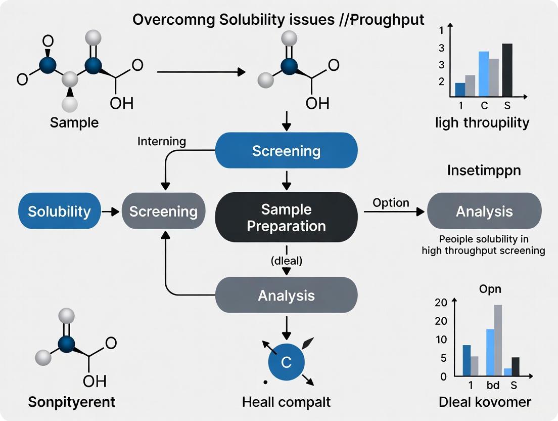
Overcoming Solubility Issues in High-Throughput Screening: Modern Strategies for Drug Development
This article provides a comprehensive guide for researchers and drug development professionals tackling the pervasive challenge of poor solubility in high-throughput screening (HTS).
Overcoming Solubility Issues in High-Throughput Screening: Modern Strategies for Drug Development
Abstract
This article provides a comprehensive guide for researchers and drug development professionals tackling the pervasive challenge of poor solubility in high-throughput screening (HTS). It explores the foundational causes of solubility limitations, details automated and miniaturized methodological approaches for rapid data generation, offers troubleshooting strategies for common pitfalls, and discusses validation frameworks to ensure data reliability. By integrating recent advancements in HTS platforms, computational prediction, and formulation technologies, this resource aims to equip scientists with the knowledge to efficiently identify and advance promising drug candidates with favorable solubility properties.
The Critical Challenge of Solubility in Modern Drug Discovery
Why Solubility is a Primary Bottleneck for Bioavailability
Frequently Asked Questions (FAQs)
1. What does "solubility" mean in a drug discovery context? In drug discovery, solubility is not a single parameter but is measured differently throughout the process. In early discovery, kinetic solubility (the maximum concentration a compound reaches in a short time, often from a DMSO stock) is commonly used for rapid flagging of problematic compounds. In later development stages, thermodynamic (equilibrium) solubility (the concentration at which the solute is in equilibrium with the solid crystalline phase) is measured, as it provides a more reliable basis for formulation development [1].
2. Why is poor solubility a major problem for oral bioavailability? For a drug to be absorbed into the bloodstream after oral administration, it must first dissolve in the fluids of the gastrointestinal (GI) tract. Poorly soluble drugs have limited and variable absorption, leading to low bioavailability. This means that a higher dose might be required to achieve the desired therapeutic effect, which can increase the risk of adverse effects [2]. Solubility and intestinal permeability are the two key factors defining drug absorption in the Biopharmaceutics Classification System (BCS) [3].
3. Which drugs are most at risk from solubility issues? According to the BCS, Class II drugs (low solubility, high permeability) and Class IV drugs (low solubility, low permeability) are most problematic. For BCS Class II drugs, solubility is the rate-limiting step for absorption, meaning that enhancing solubility directly improves bioavailability [4] [3]. It is estimated that over 40% of new chemical entities (NCEs) in the pharmaceutical industry are practically insoluble in water, creating a significant development bottleneck [3].
4. What biological barriers exacerbate solubility limitations? Even if a drug has high intrinsic permeability, several biological barriers can limit the bioavailability of poorly soluble compounds:
- Fluctuating GI pH: The change from stomach acid to the more neutral intestine can affect the dissolution and precipitation of ionizable drugs [4].
- The Mucosal Barrier: The mucus layer lining the GI tract can act as a filter, trapping drug molecules and preventing them from reaching the intestinal wall for absorption [4].
- Metabolic Enzymes and Efflux Pumps: Enzymes like CYP3A4 and efflux transporters like P-glycoprotein (P-gp) can metabolize or actively pump out drugs that have managed to dissolve and enter the intestinal cells, further reducing the amount that reaches circulation [4].
5. What are common experimental artifacts in HTS solubility assays? In high-throughput screening, compounds with low solubility can cause false positives or negatives. A common issue is nonspecific inhibition by aggregates, where compounds form colloidal aggregates that non-specifically inhibit the target, leading to a false readout of activity. Furthermore, precipitation in bioassay buffers can lead to an underestimation of a compound's true potency [1] [5].
Troubleshooting Guides
Guide 1: Addressing Inconsistent Solubility Measurements
Problem: Significant variability in solubility data for the same compound across different tests or laboratories.
| Potential Cause | Explanation & Impact | Solution |
|---|---|---|
| Solid-State Form Variability | The same compound can exist in different solid forms (e.g., amorphous, crystalline, hydrates), each with different solubility. Using an inconsistent form leads to inconsistent data. | Standardize the solid form used for testing. For development, use the most stable crystalline form (thermodynamic solubility) [1]. |
| Experimental Method Differences | Variations in buffer composition, shaking time, temperature control, and filtration methods between labs can yield different results [6]. | Adopt a standardized, well-documented experimental protocol across all tests. Control temperature meticulously [1]. |
| DMSO Stock Solution Effects | In discovery, kinetic solubility is measured from DMSO stocks. Residual DMSO can artificially enhance apparent solubility. Water content in DMSO stocks can also cause precipitation [1]. | Control and minimize DMSO concentration and water content in assay buffers. Be aware that kinetic solubility will often be higher than thermodynamic solubility [1]. |
Experimental Protocol: Measuring Thermodynamic Solubility
- Preparation: Use a well-characterized, pure, and crystalline solid form of the compound.
- Equilibration: Add an excess of the compound to a relevant aqueous buffer (e.g., simulated gastric or intestinal fluid). Agitate the suspension at a constant temperature (e.g., 37°C) for a sufficiently long period (e.g., 24-72 hours) to reach equilibrium.
- Separation: Separate the saturated solution from the undissolved solid using a method like centrifugation or filtration.
- Quantification: Dilute the supernatant appropriately and quantify the dissolved compound concentration using a validated analytical method, such as UV spectroscopy or HPLC [1].
Guide 2: Mitigating Solubility-Related Bioassay Artifacts
Problem: A compound shows high potency in a primary screen but fails in follow-up assays or shows nonspecific activity.
| Potential Cause | Explanation & Impact | Solution |
|---|---|---|
| Promiscuous Aggregation | Compound molecules form colloidal aggregates that non-specifically inhibit multiple protein targets, leading to false positives that are not reproducible [5]. | Confirm activity in a detergent-containing assay (e.g., with 0.01% Triton X-100), as detergents often disrupt aggregates. Use orthogonal, non-binding-based assays to validate hits [5]. |
| Precipitation in Bioassay Buffer | The compound precipitates out of solution at the concentration used in the assay. The observed effect may be due to a lower, unknown concentration of dissolved compound, or the precipitate may interfere with the assay readout [1]. | Measure the kinetic solubility of the compound under the exact conditions of the bioassay (buffer, pH, temperature). Compare the dose-response values with the apparent solubility to flag potential liabilities [1]. |
HTS Solubility Artifact Troubleshooting
Advanced Techniques & Reagent Solutions
Computational Solubility Prediction
Machine learning models are now capable of predicting solubility to help guide experimental work. For instance, the FASTSOLV model, trained on the large BigSolDB dataset, can predict a molecule's solubility in various organic solvents at different temperatures. These models are approaching the practical limit of prediction accuracy (aleatoric uncertainty of 0.5–1 log S) due to inherent variability in experimental training data, but they are invaluable for pre-screening solvents and conditions [7] [6].
Research Reagent Solutions for Solubility Enhancement
The following table details key materials and technologies used to overcome solubility challenges.
| Reagent/Technology | Function & Mechanism | Example Platforms / Components |
|---|---|---|
| Amorphous Solid Dispersions (ASD) | Disperses the API in a polymer matrix, stabilizing it in a high-energy amorphous state with greater solubility than the crystalline form. | Spray Drying, Hot Melt Extrusion, KinetiSol [8] |
| Lipid-Based Delivery Systems | Enhances solubility and permeability by solubilizing the drug in lipid vehicles (oils, surfactants) that form emulsions in the GI tract. | Self-Emulsifying Drug Delivery Systems (SEDDS/SMEDDS) [4] [8] |
| Cyclodextrins | Forms water-soluble inclusion complexes where the lipophilic drug molecule is housed inside the hydrophobic cavity of the cyclodextrin ring. | CycloLab, various cyclodextrin derivatives (e.g., SBE-β-CD) [8] [9] |
| Nanocrystal/Nanosuspension | Increases the surface area-to-volume ratio by reducing particle size to the nanoscale (100-1000 nm), dramatically increasing dissolution rate. | Nanomilling, NanoSol [4] [9] |
| Salts and Co-crystals | Alters the solid-state form of the API through chemical (salt formation) or physical (co-crystal with a coformer) modification to improve solubility. | Various organic acids/bases for salts; sugars, acids for co-crystals [3] [8] |
| Permeation Enhancers | Improves transport across biological barriers by transiently altering membrane integrity or fluidity. Useful for BCS Class IV drugs. | Surfactants, Dimethyl Isosorbide (DMI) [8] |
Solubility Enhancement Techniques
The Impact of High-Throughput Screening on Lead Compound Profiles
Frequently Asked Questions (FAQs)
FAQ 1: Why is solubility testing so critical in early-stage drug discovery? Poor solubility is the most undesirable property in early chemical screening, as it carries a high risk of compound failure. Insufficient solubility can compromise other property assays, mask additional undesirable properties, and influence both pharmacokinetic and pharmacodynamic properties of a compound. Identifying solubility liabilities prior to functional evaluations prevents wasted resources on compounds that are unlikely to succeed [1] [10].
FAQ 2: What is the difference between kinetic and thermodynamic solubility?
- Thermodynamic (Equilibrium) Solubility: This is the concentration of a compound in a saturated solution when solid is present and an equilibrium has been established between the solid and solution phases. It is typically measured using the shake-flask method over at least 24 hours and is considered the "gold standard" for development scientists [1].
- Kinetic Solubility: This is the concentration at which a compound precipitates from a solution, often starting from a stock solution in DMSO. It is a non-equilibrium measurement that offers higher throughput and is typically used for ranking compounds early in the discovery process [1] [10].
FAQ 3: How can automation improve the reliability of HTS solubility data? Automation addresses key challenges in HTS, such as human error and inter-user variability, which are major sources of irreproducible results. Automated liquid handlers enhance precision, especially at low volumes. Technologies like DropDetection can verify that the correct volume has been dispensed into each well, allowing errors to be identified and corrected. This standardization improves assay performance and data reproducibility across users and sites [11].
FAQ 4: What are Pan-Assay Interference Compounds (PAINS) and why are they problematic? PAINS are compounds with functional groups that promiscuously interfere with assay outputs, leading to false positives that can be mistaken for authentic activity. Screening libraries should be filtered to eliminate these and other problematic compounds (e.g., redox cycling compounds, alkyl halides, Michael acceptors) during the library design phase to avoid confounding HTS results [12].
Troubleshooting Guides
Issue 1: High Incidence of False Positives or Negatives
Potential Causes and Solutions:
| Cause | Solution |
|---|---|
| Promiscuous/Interfering Compounds | Apply stringent cheminformatics filters (e.g., PAINS, REOS) during library selection to remove compounds with problematic functionalities [12]. |
| Low Aqueous Solubility of Test Compounds | Integrate a kinetic solubility screen (e.g., via nephelometry) into the primary workflow. Flag or exclude compounds with solubility below the assay concentration [1] [10]. |
| Human Error in Manual Liquid Handling | Implement automated liquid handling systems to improve precision and reduce intra- and inter-user variability [11]. |
| Compound Precipitation in Assay Buffer | Characterize solubility under specific bioassay conditions (pH, temperature, buffer composition) to ensure compounds remain in solution [1]. |
Issue 2: Poor Reproducibility of Solubility Data
Potential Causes and Solutions:
| Cause | Solution |
|---|---|
| Variability in Sample Preparation | Standardize and automate the entire sample preparation workflow, from compound dissolution and dilution to liquid transfers [11]. |
| Inconsistent Detection Methods | Use a uniform, sensitive detection method. Nephelometry is highly suitable for automated, high-throughput kinetic solubility screens as it directly measures light scattered by precipitated particles [10]. |
| Uncontrolled Crystallization/Polymorphism | For thermodynamic solubility, ensure equilibrium is reached by controlling temperature and agitation time. Be aware that different solid forms (polymorphs) can result in different solubility measurements [1]. |
Key Experimental Protocols
Protocol 1: High-Throughput Kinetic Solubility Assay Using Nephelometry
This protocol determines the concentration at which a compound begins to precipitate out of solution, enabling rapid ranking of compound libraries [10].
Workflow Diagram:
Detailed Methodology:
- Sample Preparation: Prepare a series of serial dilutions for each test compound in DMSO.
- Aqueous Dilution: Using an automated liquid handler, transfer a fixed volume of each DMSO stock into a 384-well plate containing an aqueous buffer (e.g., phosphate buffer at pH 7.4). The final DMSO concentration should typically be kept low (e.g., 1%).
- Incubation: Allow the plate to incubate at room temperature for a standardized period (e.g., 15-60 minutes).
- Measurement: Read the plate using a microplate nephelometer (e.g., NEPHELOstar Plus). The instrument passes a laser (635 nm) through each well. Insoluble particles scatter the light, and the intensity of the scattered light is measured by a detector.
- Data Analysis:
- Plot the nephelometry count (scattered light intensity) against the compound concentration for each compound.
- The plot typically shows a low, stable baseline followed by a sharp increase in scattering at the precipitation point.
- Fit two linear regressions to the baseline and the precipitation slope. The point where these two lines intersect is defined as the kinetic solubility of the compound.
Protocol 2: Automated Workflow for Large-Scale Solubility Data Generation
This robotic workflow is designed for collecting large-scale, high-quality solubility data to feed data-driven models and databases [13].
Workflow Diagram:
Detailed Methodology:
- Robotic Setup: A robotically controlled platform handles all liquid transfers, compound weighing, and plate movements.
- Saturation & Equilibrium: Compounds are dispensed into vials or microplates with a relevant solvent (aqueous or non-aqueous). The platform agitates the samples (shaking) at a controlled temperature for a sufficient period (e.g., 24-48 hours) to reach solid-solution equilibrium.
- Phase Separation: After equilibration, the automated system separates the solid from the solution using centrifugation or filtration.
- Concentration Quantification: The concentration of the compound in the supernatant is determined using an integrated analytical method. This could be a direct UV measurement or, for higher accuracy, an automated HPLC system coupled in-line.
- Data Management: The resulting solubility data is automatically uploaded to a centralized database, facilitating data mining and the development of machine learning models for solubility prediction.
Table 1: Comparison of Solubility Measurement Methods in HTS
| Method | Throughput | Key Measurement | Primary Use | Key Advantage | Key Limitation |
|---|---|---|---|---|---|
| Kinetic Solubility (Nephelometry) | High (e.g., 24 compounds in 75 mins) [10] | Precipitation point | Early discovery, compound ranking | Speed and suitability for automation | Does not reflect the stable equilibrium state |
| Thermodynamic Solubility (Shake-Flask) | Low (e.g., ~24 hours) [1] [10] | Equilibrium concentration | Late discovery/early development | Gold standard, reflects true equilibrium | Low throughput, time-consuming, resource-intensive |
| Automated Robotic Platform | High (Large-scale data generation) [13] | Equilibrium concentration | Data-driven discovery & database building | Generates large-scale, high-quality data for ML | Requires significant initial investment in equipment |
Table 2: Common Functional Groups and Compounds to Filter from Screening Libraries
| Compound/Functional Group Type | Examples | Reason for Interference |
|---|---|---|
| Pan-Assay Interference Compounds (PAINS) | Certain aminothiazoles, acyl hydrazides | Promiscuous inhibition via non-specific mechanisms |
| Reactive Functional Groups | Aldehydes, alkyl halides, Michael acceptors, epoxides | Covalent modification of protein targets |
| Redox-Active Compounds | Dihydroxyarenes, trihydroxyarenes | Generate reactive oxygen species that damage targets |
| Aggregators | Certain anthracene derivatives | Form colloidal aggregates that non-specifically inhibit enzymes |
The Scientist's Toolkit: Key Research Reagent Solutions
Table 3: Essential Materials for HTS Solubility Experiments
| Item | Function in Solubility Assays |
|---|---|
| DMSO (Dimethyl Sulfoxide) | Standard solvent for creating and storing compound stock libraries. It is hygroscopic, so water content must be controlled to avoid precipitation and concentration errors [1]. |
| Aqueous Buffers (e.g., PBS) | Simulate physiological conditions (e.g., pH 7.4) for solubility measurements, providing relevant data for predicting in vivo performance [10]. |
| 384-well or 1536-well Microplates | Enable assay miniaturization, drastically reducing reagent consumption and compound requirements while facilitating high-throughput automation [11] [10]. |
| Cyclodextrins | Used as solubility-enhancing additives in solubility studies. They can increase water solubility, bioavailability, and stability of poorly soluble compounds [10]. |
Frequently Asked Questions: Troubleshooting Solubility
Q1: What is the fundamental relationship between LogP, melting point, and intrinsic solubility? The properties are interconnected in their influence on the energy required for dissolution. LogP (partition coefficient) is a direct measure of lipophilicity; a high value indicates a molecule prefers an organic environment over an aqueous one, directly correlating with poor aqueous solubility [14]. The melting point (MP) reflects the energy of the crystal lattice; a high melting point indicates strong, stable intermolecular forces within the solid, which require more energy to break for dissolution to occur [15] [16]. Intrinsic solubility is the result of a balance: it is disfavored by high lipophilicity (high LogP) and a strong crystal lattice (high MP), but favored by the energy released when the molecule solvates in water [17] [18].
Q2: During high-throughput screening, my drug candidate shows promising binding affinity but has a LogP > 5. What are the risks? A LogP value greater than 5 is a major red flag for development. It strongly predicts:
- Extremely poor aqueous solubility, making formulation for oral or intravenous delivery very challenging [14] [19].
- High risk of toxicity due to potential accumulation in lipid-rich tissues [14].
- Violation of the "Rule of Five," which is a strong indicator of poor absorption and permeation for orally administered drugs [16]. You should prioritize structural modification to reduce lipophilicity at this stage.
Q3: My compound has a high melting point (> 200 °C). How will this impact its solubility and which formulation strategies should I consider? A high melting point is a key indicator of high crystal lattice energy, which directly limits solubility in all solvents [18] [16]. For such molecules, conventional methods like particle size reduction may be insufficient. You should focus on formulation technologies that disrupt the crystal lattice itself:
- Amorphous Solid Dispersions (ASDs): These create a high-energy, non-crystalline form of the drug that dissolves more rapidly and completely, leading to supersaturation [20] [19].
- Salt Formation: If the compound is ionizable, forming a salt can significantly alter the crystal packing and melting point, leading to higher solubility and dissolution rate [21] [19].
- Co-crystals: For non-ionizable compounds, co-crystallization with a safe co-former can create a new crystal structure with lower lattice energy and higher apparent solubility [19].
Q4: I have measured the water solubility of my ionizable drug at pH 7.4. Why is this different from its intrinsic solubility, and which value is more important? The solubility you measured at pH 7.4 is the aqueous solubility at that specific pH, which is influenced by the drug's ionization state. The intrinsic solubility (S₀) is the solubility of the neutral, uncharged form of the molecule [17]. For ionizable compounds (approximately 85% of drug substances), these values can be vastly different [17]. The intrinsic solubility is a more fundamental property because it is pH-independent and, together with the pKa, can be used to accurately calculate solubility at any pH. This is critical for predicting behavior throughout the different pH environments of the gastrointestinal tract [17].
Q5: What does a "wide melting range" indicate in my capillary tube experiment, and what should I do? A wide melting range (e.g., > 2-3 °C) typically indicates that your sample is impure or not a single crystalline form [15]. For a pure substance, the phase change from solid to liquid occurs at a sharp, well-defined temperature.
- Troubleshooting Steps:
- Re-purify your sample to remove impurities.
- Ensure proper sample preparation: The sample must be dry and in a fine powder to ensure consistent heat transfer [15].
- Control the heating rate: Heating too quickly is a common experimental error. A slow heating rate of 1-2 °C per minute is recommended to establish thermal equilibrium [15].
- Investigate polymorphism: The sample may exist in multiple crystalline forms, which can melt at different temperatures.
Key Physicochemical Properties at a Glance
Table 1: Interpretation Guide for Key Properties Governing Solubility
| Property | Definition | Ideal Range for Drug-like Compounds | Impact on Solubility & Bioavailability |
|---|---|---|---|
| LogP | The logarithm of the partition coefficient of a compound between octanol and water, measuring lipophilicity [14]. | 0 to 5 (per Lipinski's Rule of Five) [16]. | High LogP (>5) indicates high lipophilicity and very poor aqueous solubility, challenging formulation and increasing risk of toxicity [14] [16]. |
| Melting Point (MP) | The temperature at which a solid substance changes to a liquid state, indicating crystal lattice energy [15]. | Most approved drugs have an MP below 250°C [16]. | High MP indicates strong crystal lattice forces, which require more energy to break during dissolution, resulting in lower solubility [18] [16]. |
| Ionization (pKa) | The acid dissociation constant, defining the pH at which a molecule is 50% ionized. | N/A | Allows for pH modification and salt formation to enhance solubility. Enables calculation of solubility at any pH when used with intrinsic solubility [17] [21]. |
| Intrinsic Solubility (S₀) | The solubility of the neutral, uncharged form of a compound [17]. | N/A | The foundational property for understanding pH-dependent solubility behavior. Poor intrinsic solubility is a primary driver of low bioavailability [17] [20]. |
Table 2: Formulation Strategy Selection Based on Drug Properties
| Primary Challenge | Recommended Formulation Technology | Mechanism of Action | Key Considerations |
|---|---|---|---|
| High Lipophilicity (High LogP) | Lipid-Based Drug Delivery Systems (LBDDS) [19] | Pre-solubilizes the drug in a lipid vehicle, facilitating absorption via the lymphatic system [19]. | Compatibility with capsule shells; requires stabilization to prevent precipitation. |
| High Crystal Energy (High MP) | Amorphous Solid Dispersions (ASDs) [20] [19] | Creates a high-energy, non-crystalline form that generates a supersaturated solution for enhanced absorption [19]. | Physical stability must be managed to prevent re-crystallization over time [19]. |
| Ionizable Compound | Salt Formation [21] [19] | Alters crystal structure and pH microenvironment, leading to higher dissolution rate and solubility [21]. | Risk of precipitation in the GI tract due to pH changes (salt conversion); common ion effect can limit dissolution [19]. |
| Non-Ionizable Compound with High MP | Co-crystals [19] | Uses a co-former to create a new crystalline material with lower lattice energy and higher apparent solubility [19]. | Requires selection of GRAS (Generally Recognized As Safe) co-formers; can also suffer from precipitation in vivo [19]. |
| Particle Size Limited Dissolution | Nanocrystals [19] | Increases the surface area-to-volume ratio dramatically, leading to a faster dissolution rate [19]. | The particles are 100% API, but require stabilizers/surfactants to prevent aggregation [19]. |
Essential Experimental Protocols
Protocol 1: Determining Melting Point via Capillary Tube Method
The capillary method is a standard technique for compound identification and purity assessment [15].
- Sample Preparation:
- Ensure the sample is completely dry and ground into a fine powder [15].
- Press the open end of a capillary tube into the powder several times.
- Tap the closed end of the tube gently on a hard surface or drop it through a long glass tube (approx. 1m) to compact the sample to the bottom. The final packed sample height should be 2-3 mm [15].
- Experimental Setup & Execution:
- Place the capillary tube in a melting point apparatus alongside a thermometer if using a manual setup [15].
- Heat the sample rapidly to about 10-15°C below its expected melting point.
- Crucially, slow the heating rate to 1-2°C per minute as you approach the melting point to establish thermal equilibrium and ensure an accurate reading [15].
- Observe carefully and record the temperature at which the sample begins to melt (initial liquid phase observed) and the temperature at which it becomes completely liquid. This is the melting range [15].
- Troubleshooting Tip: Never re-melt a sample that has already been heated. Always use a fresh sample and a new capillary tube for each measurement [15].
Protocol 2: High-Throughput Solubility Screening Assay (PEG-Induced Precipitation)
This turbidity-based assay is useful for rank-ordering monoclonal antibodies (mAbs) or other biologics based on their relative solubility properties in early discovery [22].
- Principle: A soluble agent like Polyethylene Glycol (PEG) is used to induce precipitation in a manner that correlates with the protein's solubility and self-interaction propensity [22].
- Methodology:
- Prepare a series of solutions or buffers at the desired pH values for screening.
- Dispense these solutions into a multi-well plate.
- Add a constant, small volume of your protein solution to each well.
- Introduce a gradient of PEG solutions to the wells to create a series of known PEG concentrations.
- Incubate the plate to allow for precipitation.
- Quantify the turbidity (a measure of precipitation) in each well using a plate reader.
- Data Interpretation: The relative solubility of different mAbs or formulations can be rank-ordered by the PEG concentration required to induce precipitation. A higher required PEG concentration indicates higher relative solubility [22].
The Scientist's Toolkit: Research Reagent Solutions
Table 3: Essential Materials for Solubility and Formulation Experiments
| Reagent / Material | Function in Experiment |
|---|---|
| Capillary Tubes | Used for sample containment in traditional melting point determination [15]. |
| Melting Point Apparatus | Provides controlled heating and visual observation for determining a compound's melting range [15]. |
| Polyethylene Glycol (PEG) | A polymer used in high-throughput screening to induce precipitation and rank-order the relative solubility of biologics [22]. |
| Lipidic Excipients (e.g., oils, surfactants) | Core components of Lipid-Based Drug Delivery Systems (LBDDS) used to pre-solubilize lipophilic drugs [19]. |
| Polymer Carriers (e.g., HPMC, PVP-VA) | Used to create Amorphous Solid Dispersions (ASDs); they inhibit crystallization and maintain drug supersaturation [20] [19]. |
| Co-formers (e.g., GRAS acids/bases) | Molecules used in co-crystallization to form a new, more soluble crystalline structure with a poorly soluble API [19]. |
Property Interplay and Formulation Strategy
The following diagram illustrates the logical decision-making process for selecting a formulation strategy based on a compound's key physicochemical properties.
Biopharmaceutical Classification System (BCS) and its Relevance to HTS
A technical support center for resolving experimental challenges in high-throughput screening.
BCS and HTS: Fundamental Concepts FAQ
1. What is the Biopharmaceutics Classification System (BCS) and why is it relevant to High-Throughput Screening (HTS)?
The Biopharmaceutics Classification System (BCS) is a scientific framework that classifies drug substances based on their aqueous solubility and intestinal permeability into four classes [23] [24]. It was originally developed to aid in regulatory decisions for bioequivalence studies but is now extensively used as a critical decision-making tool in drug discovery and early development [23]. Its relevance to HTS lies in its ability to guide the prioritization of lead compounds based on their biopharmaceutical properties early in the discovery process, aligning with the "fail early, fail cheap" paradigm of modern drug development [23] [25]. By applying BCS principles during HTS and lead optimization, pharmaceutical scientists can identify compounds with poor solubility or permeability early, allowing for timely intervention through formulation strategies or compound redesign [23].
2. How has the distribution of BCS classes changed in modern drug pipelines, and what are the implications for HTS?
There has been a significant shift in the biopharmaceutical characteristics of new drug candidates compared to marketed drugs. While marketed drugs comprise approximately 40% BCS Class I and 30% BCS Class II compounds, the new drug pipeline shows a marked increase in less-ideal candidates, with BCS Class I compounds decreasing to 10-20% and BCS Class II compounds increasing to 50-60% [23]. This trend underscores the critical importance of integrating solubility and permeability screening into HTS operations, as a majority of new chemical entities now present solubility challenges that can complicate downstream development [23] [26].
3. What are the key differences between kinetic and thermodynamic solubility measurements, and when should each be used in HTS?
Understanding the distinction between kinetic and thermodynamic solubility is crucial for appropriate experimental design in HTS:
- Kinetic Solubility: Measured by incrementally adding a DMSO stock solution to an aqueous buffer until precipitation is detected optically. This method is faster and compatible with HTS workflows but tends to overestimate thermodynamic solubility due to the DMSO cosolvent effect and the compound being in an amorphous state [23] [25].
- Thermodynamic Solubility: Determined by incubating a purified crystalline solid in a liquid for an extended period (typically 24-48 hours) to achieve equilibrium. This method provides more accurate data but is slower, requires more compound material, and is less compatible with true HTS throughput [23] [25].
For HTS environments, kinetic solubility is typically employed initially due to throughput requirements, with thermodynamic solubility studies reserved for later stages of lead optimization [23].
Troubleshooting Guides for Common Experimental Challenges
Issue 1: Inconsistent Solubility Measurements in HTS
Problem: Solubility data shows high variability between assays or between different compound batches, leading to unreliable BCS classification.
Solution:
- Confirm solubility measurement type: Ensure consistency between kinetic and thermodynamic solubility methods. For BCS classification, the FDA requires equilibrium solubility data, though kinetic solubility is often used in early HTS [23] [25].
- Standardize DMSO handling: Since HTS solubility measurements typically start from DMSO stock solutions,严格控制DMSO quality, storage conditions, and freeze-thaw cycles is essential. Implement rigorous DMSO compatibility testing during assay validation [27].
- Control precipitation detection: Use consistent detection methods (nephelometry, UV scattering, or direct UV) across experiments. The light scattering effect from precipitated material should be measured using standardized instrumentation [23].
- Validate with reference compounds: Include compounds with known solubility profiles in each assay plate to monitor inter-assay variability and normalize data accordingly [27].
Table 1: Troubleshooting Solubility Measurement Inconsistencies
| Observation | Potential Cause | Recommended Action |
|---|---|---|
| Higher than expected solubility values | DMSO cosolvent effect in kinetic solubility measurements | Use consistent DMSO concentrations (typically <1% for cell-based assays); confirm findings with thermodynamic measurements for critical compounds [27] [25] |
| Variable results between replicates | Compound precipitation kinetics affected by nucleation | Standardize equilibration times and temperature control; implement automated mixing protocols [23] |
| Discrepancy between HTS and manual solubility data | Different detection methods or sample preparation | Cross-validate HTS methods with shake-flask (equilibrium) methods for a compound subset [23] |
Issue 2: Permeability Assay Artifacts in Caco-2 and MDCK Systems
Problem: Permeability data from cell-based systems (Caco-2/MDCK) does not correlate with in vivo absorption, potentially misclassifying BCS Class 3 and 4 compounds.
Solution:
- Identify false negatives: Recognize that Caco-2 systems may underestimate human permeability for three key reasons: (1) overexpression of P-glycoprotein efflux pumps, (2) reduced paracellular transport due to tighter junctions, and (3) non-specific binding of insoluble compounds to filter supports [23].
- Implement counter-screens: Use the in-situ rat gut perfusion method for compounds that show unexpectedly low permeability in cell-based assays, particularly for compounds suspected of paracellular or transporter-mediated uptake [23].
- Consider alternative cell lines: Evaluate MDCK cells for faster turnaround (3-7 days vs. 21 days for Caco-2), but recognize their limitations with efflux transporter expression [23].
- Account for non-specific binding: For highly lipophilic compounds, include controls to measure non-specific binding to apparatus components, which can significantly reduce apparent permeability [23].
Issue 3: BCS Classification Discrepancies Between Discovery and Development
Problem: Compounds classified as BCS Class 3 or 4 (low permeability) in discovery are reclassified as BCS Class 1 or 2 (high permeability) upon entering development.
Solution:
- Harmonize permeability methods: Recognize that HTS permeability methods (Caco-2/MDCK) have different characteristics than methods used in development (rat gut perfusion). Implement more labor-intensive but accurate methods earlier for lead compounds [23].
- Standardize solubility criteria: Apply consistent criteria for "high solubility" - defined as the highest clinical dose strength being soluble in ≤250 mL of aqueous media over pH 1-7.5 at 37°C [24].
- Incorporate potency considerations: Remember that BCS accounts for potency, as solubility is relative to clinical dose. A compound with poor absolute solubility may still be "highly soluble" if it is highly potent [24].
Experimental Protocols for BCS-Relevant HTS Assays
Protocol 1: HTS Solubility Measurement Using Nephelometry
Purpose: To determine kinetic solubility of compounds from DMSO stock solutions in a high-throughput format [23] [25].
Materials:
- Test compounds in DMSO stock solutions (typically 10 mM)
- Assay buffer (e.g., phosphate-buffered saline, pH 7.4)
- 96-well or 384-well microplates with clear bottoms
- Microplate nephelometer or UV/Vis plate reader with scattering detection
- Liquid handling robotics for precise DMSO transfer
Procedure:
- Prepare assay plates with 200 μL of buffer per well using automated liquid handling.
- Program robotic systems to add DMSO stock solutions incrementally (typically 1 μL at a time) to buffer while monitoring light scattering.
- Detect precipitation point where light scattering intensity increases significantly.
- Calculate kinetic solubility based on the amount of compound added before precipitation occurs.
- Include reference compounds with known solubility profiles in each plate for quality control.
Data Interpretation: Kinetic solubility values obtained are typically higher than thermodynamic solubility. Use this method for ranking compounds during early screening, with follow-up equilibrium studies for lead compounds [23] [25].
Protocol 2: HTS Permeability Assessment Using Caco-2 Cell Monolayers
Purpose: To assess compound permeability across intestinal epithelium in a high-throughput format [23] [24].
Materials:
- Caco-2 cells (human colon adenocarcinoma cell line)
- 96-well or 24-well Transwell plates with filter supports
- Transport buffer (e.g., HBSS with appropriate pH adjustment)
- Test compounds in DMSO
- LC-MS or UV plate reader for compound quantification
- Marker compounds for monolayer integrity (e.g., Lucifer Yellow)
Procedure:
- Culture Caco-2 cells on Transwell filters for 21 days to form confluent, differentiated monolayers. Monitor transepithelial electrical resistance (TEER) to confirm monolayer integrity.
- Add test compounds to donor compartment (apical side for A→B transport, basolateral for B→A transport).
- Incubate for predetermined time (typically 2 hours) at 37°C with gentle agitation.
- Sample from acceptor compartment at multiple time points and analyze compound concentration using LC-MS or direct UV assay.
- Calculate apparent permeability (Papp) using standard equations.
- Include control compounds with known high and low permeability in each assay.
Data Interpretation: Papp values >10×10⁻⁶ cm/s typically indicate high permeability. Be aware of potential false negatives due to efflux transporters or paracellular transport limitations in this system [23].
The Scientist's Toolkit: Essential Research Reagents and Materials
Table 2: Key Research Reagent Solutions for BCS-Related HTS
| Reagent/ Material | Function | Application Notes |
|---|---|---|
| Caco-2 Cell Line | Model human intestinal permeability | Requires 21-day differentiation; may overexpress efflux transporters compared to human intestine [23] [24] |
| MDCK Cell Line | Alternative permeability model | Faster differentiation (3-7 days); lower expression of various efflux pumps [23] |
| DMSO (Cell Culture Grade) | Universal solvent for compound libraries | Maintain strict quality control; limit freeze-thaw cycles; keep final concentration <1% for cell-based assays [27] |
| Kolliphor Surfactants (e.g., RH40, ELP, HS15) | Solubility enhancers for poorly soluble compounds | Used in self-emulsifying drug delivery systems (SEDDS/SMEDDS); enhance solubility and maintain supersaturation [26] |
| Polymeric Excipients (Kollidon VA64, Soluplus) | Amorphous dispersion matrices for solubility enhancement | Used in hot-melt extrusion and spray-dried dispersion technologies to improve dissolution of BCS Class II compounds [26] |
| HTS Solubility Assay Kits | Automated kinetic solubility measurement | Commercial systems available for high-throughput nephelometry or direct UV detection [23] |
BCS-Based Decision Framework for Formulation Strategy
The following workflow illustrates how BCS classification can guide formulation strategy selection in early drug development, particularly for overcoming solubility challenges identified during HTS:
BCS-Based Formulation Decision Tree
Advanced Techniques: Integrating QSPR Modeling with HTS
Problem: How to further accelerate solubility assessment when physical screening capacity is limited.
Solution: Implement Quantitative Structure-Property Relationship (QSPR) modeling as a complementary approach to experimental HTS.
Methodology:
- Use computational models to predict aqueous solubility based on molecular structure alone, requiring no physical compound [25].
- Apply linear regression equations incorporating molecular descriptors to estimate solubility for virtual compounds before synthesis [25].
- Combine with Lipinski's "Rule of Five" and other computational filters to prioritize compounds with higher probability of acceptable biopharmaceutical properties [25].
Advantages:
- Compatible with HTS throughput and limited compound availability in early stages [25].
- Can model structurally diverse drug-like compounds, particularly valuable for predicting solubility of low-solubility compounds that present the greatest development challenges [25].
Implementation Tips:
- Use QSPR for initial compound library triaging before synthesis and experimental testing.
- Validate computational predictions with experimental data for your specific chemical series to improve model accuracy.
- Recognize limitations in predicting solubility for compounds with unusual structural features or complex ionization behavior [25].
High-Throughput and Automated Solubility Screening Platforms
Robotic Platforms and Liquid Handlers for Miniaturized Assays
Troubleshooting Guides
FAQ: Addressing Common Liquid Handler Challenges
1. My liquid handler is dripping or has a drop hanging from the tip. What could be the cause? This is often caused by a difference in vapor pressure between your sample and the water used for instrument adjustment. To resolve this, you can sufficiently prewet the tips or add an air gap after aspiration [28].
2. How can I prevent errors when loading containers onto the liquid handler deck? Implement a "pre-flight check" using your laboratory information management system (LIMS) integration. This check verifies that containers are in the correct deck positions and are the correct containers before any transfers begin, preventing errors related to misplaced or wrong labware [29].
3. I've observed inconsistent data. How can I determine if it's a real problem? First, check if the pattern of "bad data" is repeatable. Run the test again to confirm the error is not a random event. Increasing the frequency of testing for a period can help catch any recurrence and determine the level of mitigation required [28].
4. What are the best practices for dispensing viscous liquids? If you see droplets or trailing liquid during delivery, the liquid's viscosity may be the issue. Adjust the aspirate and dispense speeds, and consider adding air gaps or blow-outs to the protocol to ensure complete liquid delivery [28].
5. When should I use wet dispense versus dry dispense methods? While process requirements may dictate the choice, wet dispensing (where the tip contacts the solution in the well) can often improve accuracy and repeatability. It minimizes carryover or residual solution in the tip by pulling the solution away from the tip upon contact with the well's liquid [28].
Troubleshooting Specific Liquid Handling Errors
The table below summarizes common errors, their possible sources, and recommended solutions [28].
| Observed Error | Possible Source of Error | Possible Solutions |
|---|---|---|
| Dripping tip or drop hanging from tip | Difference in vapor pressure of sample vs water used for adjustment | - Sufficiently prewet tips- Add air gap after aspirate |
| Droplets or trailing liquid during delivery | Viscosity and other liquid characteristics different than water | - Adjust aspirate/dispense speed- Add air gaps/blow outs |
| Dripping tip, incorrect aspirated volume | Leaky piston/cylinder | Regularly maintain system pumps and fluid lines |
| Diluted liquid with each successive transfer | System liquid is in contact with sample | Adjust leading air gap |
| First/last dispense volume difference | Due to sequential dispense | Dispense first/last quantity into reservoir/waste |
| Serial dilution volumes varying from expected concentration | Insufficient mixing | Measure liquid mixing efficiency |
Integration Patterns to Mitigate Common Robotic Problems
The following workflow illustrates the recommended integration of a Liquid Handling Robot (LHR) with a Laboratory Information Management System (LIMS) to prevent common operational errors [29].
LIMS and LHR Integration Workflow
Troubleshooting by Liquid Handler Type
Different liquid handling technologies require specific troubleshooting approaches [28].
Air Displacement Liquid Handlers
- Errors may be caused by insufficient pressure or leaks in the lines.
Positive Displacement Liquid Handlers Troubleshooting should include checking the following:
- Ensure tubing is clean, clear, and free of kinks.
- Check for leaks and ensure connections are tight.
- Verify there are no bubbles in the line and flush lines sufficiently.
- Confirm tubes are not too long or too short.
- Check liquid temperature, as it can affect flow rate.
- Ensure system (working) liquid is not mixing with the sample liquid.
Acoustic Liquid Handlers Best practices for these systems include:
- Ensuring the contents of the plate have reached thermal equilibrium with the environment.
- Centrifuging the source plate prior to use to precipitate insoluble material.
- Optimizing calibration curves based on actual deviation from the expected volume.
Experimental Protocols
Protocol 1: High-Throughput Nephelometry for Qualitative Solubility Assessment
This protocol is designed for the initial solubility profiling of large chemical libraries to guide hit prioritization after High-Throughput Screening (HTS) campaigns [30].
1. Principle Nephelometry measures the turbidity (cloudiness) of a solution caused by suspended particles. This allows for the qualitative classification of compounds as highly, moderately, or poorly water-soluble. It is not intended to yield precise quantitative solubility values but serves as an efficient primary assessment.
2. Workflow Diagram
Qualitative Solubility Screening Workflow
3. Step-by-Step Procedure
- Step 1: Sample Preparation. Prepare the compound library, typically as stock solutions in DMSO.
- Step 2: Dilution. Using an automated liquid handler, transfer a small aliquot of each compound into a microplate and dilute with an aqueous buffer. This shift to aqueous conditions may cause precipitation of poorly soluble compounds.
- Step 3: Incubation. Seal the plate and allow it to incubate at a constant temperature to reach equilibrium.
- Step 4: Measurement. Read the plate using a nephelometer. This instrument passes a beam of light through the sample and measures the amount of light scattered by suspended particles.
- Step 5: Analysis. Classify compounds based on the measured turbidity signal. High turbidity indicates low solubility (many suspended particles), while low turbidity indicates high solubility.
Protocol 2: Automated Solubility Determination Using a Turbidity Probe
This protocol describes a more automated workflow for determining solubility endpoints in various solvents, suitable for chemical development [31].
1. Principle A customized robotic system fully automates the process of solid dispensing, weighing, solvent addition, and turbidity measurement to determine solubility.
2. Step-by-Step Procedure
- Step 1: Solid Dispensing. The robotic system automatically dispenses and weighs a solid material directly into the assay vial or well.
- Step 2: Solvent Addition. The system adds a precise volume of solvent (aqueous or organic) to the solid.
- Step 3: Mixing. The platform mixes the contents to facilitate dissolution.
- Step 4: Turbidity Measurement. An integrated three-wavelength turbidity probe directly measures the turbidity of the mixture in the vial.
- Step 5: Reporting. The system generates a report containing all solubility information in a clearly arranged format.
The Scientist's Toolkit: Research Reagent Solutions
Key Materials for Solubility and Formulation Research
The following table details key reagents and materials used to overcome solubility challenges in pharmaceutical development [19].
| Item | Function in Research |
|---|---|
| Polymers for Amorphous Solid Dispersions (ASDs) | Act as a matrix to maintain the drug in a high-energy amorphous state, enhancing solubility and providing a "parachute" effect to inhibit precipitation in the GI tract. |
| Lipid-Based Excipients | Form the core of Lipid-Based Drug Delivery Systems (LBDDS), enhancing solubility and permeability of lipophilic drugs for oral delivery. |
| Surfactants | Act as stabilizers in nanocrystal formulations to prevent aggregation and can be used in self-emulsifying drug delivery systems. |
| Co-formers for Co-crystals | Co-precipitate with non-ionizable APIs to form a single-phase crystalline material with lower lattice energy and higher apparent solubility. |
| Salt Formers | React with ionizable APIs to form salts, which can significantly increase solubility and dissolution rate compared to the free acid or base form. |
Advanced Formulation Technologies for Poorly Soluble Compounds
When standard solubilization methods fail, several advanced technologies can be employed [19].
| Technology | Typical Application | Key Consideration |
|---|---|---|
| Micronization | Particle size reduction to increase surface area and dissolution rate. | A practical approach, but may be a high-energy process unsuitable for heat-sensitive APIs. |
| Nanocrystals | Particle size reduction to the nanoscale for a further increase in surface area and apparent solubility. | Particles are 100% API but require surfactants as stabilizers, adding formulation complexity. |
| Amorphous Solid Dispersions (ASDs) | Ideal for APIs with high crystalline lattice energy; eliminates crystal structure. | Requires polymers to maintain stability and prevent re-crystallization; poor flowability can be a challenge. |
| Lipid-Based Formulations | Effective for lipophilic compounds; range from simple oils to self-emulsifying systems. | Versatile, but the large number of excipients can make development complex. |
| Salt Formation | Increases solubility and dissolution rate of ionizable APIs. | Performance can be limited in vivo by the common ion effect or precipitation in the GI tract. |
| Co-crystals | Improves solubility of non-ionizable APIs through co-precipitation with a soluble co-former. | More stable than ASDs but still subject to the "spring and parachute" effect without crystallization inhibition. |
Shake-Flask and Turbidity-Based Methods in Multi-Well Plates
Frequently Asked Questions (FAQs) and Troubleshooting
Q1: What are the common causes of poor reproducibility in shake-flask solubility assays, and how can they be mitigated? Poor reproducibility often stems from inconsistent incubation times, incomplete solid precipitation, or variable filtration efficiency [32] [33]. To mitigate this:
- Ensure Proper Equilibration: For thermodynamic solubility, ensure long incubation times (24 hours or more) with continuous shaking to reach equilibrium [32]. For kinetic solubility, standardize the incubation time (e.g., 2 hours) [33].
- Prevent Supersaturation: A modified shake-flask method using heating to accelerate dissolution followed by seeding with the solid compound after cooling can promote precipitation and generate reliable data [34].
- Standardize Filtration: Use specialized solubility filter plates and carefully control vacuum pressure during filtration (e.g., 0.2 atm) to ensure consistent removal of precipitates without clogging [33].
Q2: When measuring turbidity in multi-well plates, how do I ensure my measurements are in the linear range of the detector? The linearity of turbidity measurement is highly dependent on the instrument settings and sample preparation.
- Check Absorbance Range: For nephelometry-based instruments, ensure the turbidity (absorbance) reading falls within the instrument's linear response region. Pre-dilute samples to maintain turbidity between 0.5 and 0.8 to minimize multiple scattering effects [35].
- Validate Linearity: The linear relationship between Relative Nephelometry Units (RNUs) and Nephelometric Turbidity Units (NTUs) can be maintained up to at least ~200 NTUs. Establish a standard curve using formazin standards to confirm your instrument's linear range [36].
Q3: My oil-in-water emulsions for turbidity assays are unstable. What factors can improve their stability? Emulsion stability is critical for a reproducible Turbidity-based Emulsion Agglutination (TEA) assay [35].
- Controlled Sonication: Prepare emulsions using a tip sonicator in an ice bath (e.g., 60% amplitude for 1 hour with 10-second on/off cycles) to control droplet size and prevent overheating [35].
- Stabilizing Lipids: Use a mixture of phospholipids (e.g., POPC) and other lipids (e.g., glycolipids) to form a stable monolayer around the oil droplets, mimicking the fluidity of cell membranes [35].
- Confirm Droplet Size: Use Dynamic Light Scattering (DLS) to determine the size distribution of the oil droplets after preparation, ensuring consistent starting conditions [35].
Q4: How can I adapt shake-flask methods for insoluble substrates like waste animal fats (WAF) in microbial cultivations? Working with hydrophobic materials like WAF is challenging due to film formation and poor mass transfer [37].
- Use Baffled Vessels: Bottom-baffled shake flasks improve mixing and oxygen transfer, preventing fat from forming an inaccessible film on the vessel walls [37].
- Pre-emulsification: Improve pre-emulsification conditions before transferring to round microwell plates. This enhances the fat's accessibility to microbes, significantly improving growth and product yield [37].
- Monitor Oxygen Transfer: Measure the dissolved oxygen concentration in-line to identify design variants and cultivation conditions that provide sufficient oxygen mass transfer [37].
Experimental Protocols and Data
Shake-Flask Aqueous Solubility Assay (Kinetic Solubility)
This protocol is used for early-stage drug discovery to determine the kinetic solubility of compounds [33].
Table 1: Key Reagents and Equipment
| Item | Function in the Assay |
|---|---|
| Phosphate Buffered Saline (PBS), pH 7.4 | Aqueous buffer to simulate physiological conditions for solubility measurement [33]. |
| DMSO | Solvent for preparing high-concentration stock solutions of the test compound [33]. |
| MultiScreen HTS 96-Well Filter Plates | Used to separate precipitated solid from the saturated solution after incubation [33]. |
| UV-Star 96-Well Microplate | Optically clear plate for measuring the UV absorbance of the filtrate [33]. |
| Microplate Reader | Instrument to measure UV-Vis absorbance for concentration determination [33]. |
Detailed Methodology [33]:
- Stock Solution Preparation: Prepare 20 mM stock solutions of test compounds in DMSO.
- Incubation:
- In a 1.4 mL tube, add 490 µL of PBS buffer.
- Add 10 µL of the 20 mM stock solution to the buffer, creating a 400 µM incubation mixture.
- Incubate the mixture in a thermomixer at 850 rpm for 2 hours.
- Filtration:
- Transfer 290 µL of the incubation mixture to a 96-well filter plate placed on a vacuum manifold.
- Apply a vacuum of 0.2 atm to filter the solution, separating the saturated solution from any precipitate.
- Sample Preparation for UV Measurement:
- Mix 250 µL of the filtrate with 250 µL of a quenching solution (Acetonitrile:DMSO, 98:2 v/v) to dissolve any potential micro-precipitates and ensure compatibility with UV measurement.
- Measurement and Analysis:
- Transfer 200 µL of the final solution to a UV-transparent microplate.
- Measure the UV absorbance using a microplate reader.
- Calculate the compound concentration in the filtrate using a separately built calibration curve. The effective range of this assay is typically 2-400 µM.
Turbidity-Based Emulsion Agglutination (TEA) Assay
This protocol is used as a high-throughput tool to screen ligands involved in hetero-multivalent binding, such as lectin-glycan interactions [35].
Table 2: Key Reagents for TEA Assay
| Item | Function in the Assay |
|---|---|
| Silicone Oil | Forms the core of the oil-in-water emulsion droplets [35]. |
| Phospholipids (e.g., POPC) | Main structural lipid to form the emulsion droplet monolayer [35]. |
| Glycolipids (e.g., Gb3, LacCer) | Functional ligands presented on the droplet surface for binding studies [35]. |
| Tris-Buffered Saline (TBS) with CaCl₂ | Aqueous buffer providing the necessary ionic environment for lectin binding [35]. |
| 96-Well Plate & UV/Vis Spectrophotometer | Platform for high-throughput measurement and instrument for detecting turbidity changes [35]. |
Detailed Methodology [35]:
- Emulsion Preparation:
- Mix desired compositions of lipids (e.g., POPC and glycolipids) in chloroform in a round-bottom flask and dry using a rotary evaporator to form a thin film.
- Reconstitute the dried lipids with TBS buffer containing CaCl₂ to form multilamellar vesicles (MVs).
- Create an emulsion by mixing silicone oil, buffer, and the MV solution, then sonicating the mixture on ice using a tip sonicator (e.g., 60% amplitude for 1 hour with 10-second on/off cycles).
- Characterize the size distribution of the resulting oil droplets using Dynamic Light Scattering (DLS).
- Agglutination Measurement:
- Dilute the emulsion in a 96-well plate with buffer to an optimal turbidity range (e.g., absorbance between 0.5 and 0.8).
- Initiate the aggregation by adding the lectin (e.g., LecA) to the wells.
- Immediately monitor the change in turbidity (absorbance) over time using a UV/Vis spectrophotometer.
- Data Analysis:
- The initial rate of turbidity change (dτ/dt) is proportional to the emulsion aggregation rate constant, which reflects the binding strength between the lectin and the surface ligands [35].
High-Throughput Lipophilicity Measurement (Log P)
This 96-well method measures the polymer-water partition coefficient (log Ppw) as a high-throughput alternative to the traditional shake-flask method for determining lipophilicity [38].
Detailed Methodology [38]:
- Polymer Film Preparation:
- Dispense a solution of plasticized poly(vinyl chloride) (PVC) in tetrahydrofuran (THF) into the wells of a polypropylene 96-well microplate.
- Allow the THF to evaporate, forming a thin polymer film at the bottom of each well.
- Partitioning:
- Dispense an aqueous solution of the solute (e.g., 200 µL of 0.5 mM) into the wells containing the polymer film.
- Seal the plate with an adhesive film and equilibrate in a shaker (500 rpm, 25°C) for 4 hours.
- Concentration Measurement:
- After equilibration, transfer a portion of the supernatant from each well to a UV-transparent microplate.
- Measure the UV absorbance of the supernatant and compare it to the absorbance of the initial standard solution.
- Calculation:
- Calculate the partition coefficient, Ppw, using the formula:
Ppw = (C₀ - C₁) / C₁ * Φwhere C₀ is the initial concentration, C₁ is the equilibrium concentration, and Φ is the phase ratio (volume of polymer film / volume of aqueous solution) [38].
- Calculate the partition coefficient, Ppw, using the formula:
The Scientist's Toolkit: Research Reagent Solutions
Table 3: Essential Materials for Featured Experiments
| Category | Item | Critical Function |
|---|---|---|
| Assay Platforms | 96-Well Filter Plates | High-throughput separation of solids from saturated solutions in solubility assays [33]. |
| UV-Transparent Microplates | Optimal clarity for accurate UV-Vis absorbance measurements in solubility and turbidity assays [33] [38]. | |
| Polypropylene Microplates | Chemical resistance for preparing polymer films in lipophilicity measurements [38]. | |
| Key Reagents | Plasticized PVC Film | Acts as a lipophilic phase for high-throughput partitioning in log P measurements [38]. |
| Dioctyl Sebacate (DOS) | Plasticizer used to create the flexible PVC film for partitioning [38]. | |
| Formazin Standards | Calibrate nephelometers and turbidimeters for accurate turbidity quantification in NTUs [36]. |
Workflow Diagrams
Shake-Flask Solubility Assay
Turbidity Emulsion Agglutination
High-Throughput Lipophilicity Measurement
## Frequently Asked Questions (FAQs)
1. What are the primary mechanisms by which PEG and Ammonium Sulfate induce precipitation?
Ammonium sulfate functions as a salting-out agent. At high concentrations, kosmotropic ions (like sulfate) bind water molecules more tightly than water binds to itself, increasing the surface tension of the solution. This competes with the protein for hydration, effectively dehydrating the protein surface and driving protein molecules to self-associate and precipitate [39] [40]. In contrast, Polyethylene Glycol (PEG) is a long-chain polymer that acts primarily through an excluded volume mechanism. The polymer occupies space in the solution, crowding the protein out of the solvent and making protein-protein interactions more favorable than protein-solvent interactions, leading to precipitation [40].
2. How do I choose between PEG and Ammonium Sulfate for my screening project?
The choice depends on your protein's stability and your experimental goals. Ammonium sulfate stabilizes protein structure and increases the melting temperature (Tm) of proteins, making it a good choice for proteins where maintaining the native fold is a concern [40]. PEG-8000 generally has a minimal effect on protein stability, though it can slightly destabilize some proteins. Furthermore, the constant derived from PEG precipitation curves can be used to estimate a protein's solubility in buffer alone, which is not directly possible with ammonium sulfate [40]. A comparative approach using both precipitants is often recommended as they probe similar protein properties relevant to solubility [40].
3. My protein is precipitating too quickly or forming amorphous aggregates. What can I do?
Rapid, amorphous precipitation often indicates that the system is entering a state of labile supersaturation too abruptly. To encourage crystal formation, you should aim to slow down the process and approach the metastable zone more gently. Consider the following:
- Fine-tune precipitant concentration: Use finer concentration gradients around the condition where precipitation occurs.
- Optimize protein concentration: High protein concentrations can lead to overcrowding and amorphous aggregation. Try a lower concentration.
- Introduce additives: Small molecules or salts that mildly interact with the protein can sometimes promote order.
- Change methods: Switch from vapor diffusion to batch methods under oil, which can offer more control over the initial supersaturation state [41].
4. What are the best practices for validating a high-throughput precipitation screening assay?
A robust validation process is critical for a successful HTS campaign. This involves running the assay on multiple days with appropriate positive and negative controls to assess reproducibility. Key statistical metrics should be calculated [42]:
- Z'-factor: A dimensionless parameter that assesses the quality and robustness of an assay. A Z'-factor > 0.4 is generally considered acceptable for HTS.
- Coefficient of Variation (CV): The CV of assay controls should typically be less than 20%.
- Signal Window: This should be greater than 2 to ensure sufficient distinction between positive and negative controls. Systematic errors can be identified by distributing controls in an interleaved pattern across plates and visualizing data in scatter plots to detect trends or edge effects [42].
## Troubleshooting Guide
| Symptom | Potential Causes | Recommended Solutions |
|---|---|---|
| No precipitation in any condition | Protein concentration is too low; Precipitant concentration is insufficient; Protein is highly stable/soluble | Increase protein concentration; Expand precipitant screen to higher concentrations; Alter pH towards protein's isoelectric point (pI); Introduce a crowding agent |
| Only amorphous precipitate (no crystals) | Excessively rapid precipitation; Protein denaturation; Heterogeneous protein sample | Slow down equilibration (e.g., different vapor diffusion method); Screen additives/stabilizing ligands; Improve protein purity and homogeneity; Fine-tune precipitant and protein concentration |
| High plate-to-plate or day-to-day variability | Inconsistent liquid handling; reagent degradation; Edge evaporation in plates; Assay protocol not robust | Calibrate automated liquid handlers; Prepare fresh reagent batches; Use plates with seals or low-evaporation lids; Perform full assay validation to establish a robust protocol [42] |
| High rate of false positives/negatives in HTS | Assay signal window is too small; Compound interference (e.g., aggregation, fluorescence); Systematic positional effects on plate | Re-optimize assay conditions to improve Z'-factor [42]; Include counter-screens for compound interference [43]; Use interleaved control placement to identify/diagnostic patterns [42] |
## The Scientist's Toolkit: Key Research Reagent Solutions
| Reagent / Material | Function in Screening | Key Considerations |
|---|---|---|
| Ammonium Sulfate | Salting-out precipitant. Induces precipitation by dehydrating protein surface and increasing surface tension [39] [40]. | Follows the Hofmeister series. Highly soluble, inexpensive, and stabilizes native protein fold. Can be corrosive. |
| Polyethylene Glycol (PEG) | Excluded volume precipitant. Crowds protein out of solution, favoring protein-protein interactions [40]. | Available in various molecular weights (e.g., PEG 8000). Mechanism is largely non-specific and minimally perturbing. |
| Microtiter Plates | Standardized platform for high-throughput experimentation. | Available in 96-, 384-, and 1536-well formats. Material (e.g., polystyrene) and well geometry can influence meniscus and evaporation. |
| Automated Liquid Handlers | Precisely dispense nanoliter-to-microliter volumes of protein and precipitant solutions [43]. | Essential for reproducibility and miniaturization. Requires regular calibration and maintenance. |
| Controls ("High", "Medium", "Low" Signal) | Benchmark for assay quality and performance during validation and screening [42]. | "High" and "Low" define assay dynamic range. "Medium" (e.g., EC50 concentration) tests assay sensitivity. |
## Experimental Protocols & Data Analysis
### Detailed Methodology: Precipitant Screening via Microscale Vapor Diffusion
This protocol is adapted for high-throughput screening in 96- or 384-well format plates.
1. Reagent Preparation:
- Precipitant Stock Solutions: Prepare a wide-ranging set of conditions. For ammonium sulfate, prepare stocks from 1.0 M to 4.0 M. For PEG 8000, prepare stocks from 5% to 30% (w/v). Include buffers at various pH levels (e.g., pH 4.0, 7.0, 9.0) and additives as needed.
- Protein Solution: Centrifuge the purified protein at high speed to remove any aggregates. Dilute the protein to the target concentration in its storage buffer. A common starting point is 10 mg/mL.
2. Plate Setup:
- Reservoir Solution: Dispense 50-100 µL of each precipitant condition into the wells of the reservoir.
- Protein-Precipitant Mix: In a sitting drop plate, mix equal volumes (e.g., 100 nL - 1 µL) of the protein solution and the reservoir precipitant solution directly on the plate's sitting drop bridge. This is typically done with an automated liquid handler.
- Sealing and Incubation: Seal the plate with a clear, adhesive seal to prevent evaporation. Incubate the plate at a constant temperature (e.g., 20°C) without disturbance.
3. Imaging and Analysis:
- Schedule Imaging: Use an automated imaging system to take pictures of each drop at regular intervals (e.g., daily for the first week, then weekly).
- Scoring Results: Score each well based on its outcome: clear, precipitate, phase separation, microcrystals, or crystals.
### Quantitative Precipitant Data Analysis
The relationship between precipitant concentration and protein solubility is described by the general expression: Log(S) = constant - β[Precipitant], where S is the measured solubility at a given precipitant concentration, and β is the dependence of solubility on precipitant concentration for a given protein [40].
The following table summarizes the distinct solubility behavior and influence on protein stability of the two key precipitants:
| Property | Ammonium Sulfate | Polyethylene Glycol (PEG) |
|---|---|---|
| Mechanism | Preferential solvation / Dehydration [39] [40] | Excluded volume / Molecular crowding [40] |
| Solubility Trend | Salting-in at low concentration, salting-out at high concentration [39] | Linear decrease in log(solubility) with increasing concentration [40] |
| Effect on Protein Stability | Stabilizes native fold; Increases melting temperature (Tm) [40] | Minimal effect on stability; may slightly destabilize some proteins [40] |
| Estimation of S₀ (Solubility in Buffer) | Not directly possible from salting-out constant [40] | Constant from linear regression is Log(S₀), the estimated solubility in buffer [40] |
### Workflow and Troubleshooting Logic
The diagram below outlines the core experimental workflow and key decision points for troubleshooting precipitation screens.
Solubility of redox-active materials is a paramount physicochemical property in redox flow battery (RFB) research and development because it directly governs the system's energy density [13]. Overcoming solubility limitations is a significant challenge in the design and discovery of novel electrolytes. High-throughput screening (HTS) methodologies provide an efficient pathway to generate large-scale, high-quality solubility data, accelerating the development of next-generation energy storage materials [13]. This case study examines the implementation of high-throughput solubility determination within the broader thesis of overcoming solubility issues in screening research, providing a technical support framework for researchers encountering experimental hurdles.
High-Throughput Solubility Determination: Core Methodologies
Automated Robotic Screening Platforms
The primary advanced methodology involves a robotically controlled platform integrated with high-throughput workflows to systematically collect solubility data for redox-active materials. This automated process significantly accelerates data acquisition while maintaining high quality and reproducibility [13] [44]. The platform enables researchers to study both aqueous and non-aqueous systems simultaneously, along with the effects of various additives on solubility behavior, providing comprehensive datasets for redox flow battery optimization [13].
The core workflow combines traditional shake-flask method principles with automation, enabling parallel processing of multiple samples under controlled conditions. This approach has demonstrated practical utility in developing optimized electrolyte formulations that boost energy density by 24% while maintaining stable performance over extended cycling (>100 cycles) [13] [44].
Comparative Analysis of Solubility Determination Techniques
Table 1: Comparison of Solubility Determination Methods
| Method | Throughput | Principle | Detection Limit | Key Advantages | Key Limitations |
|---|---|---|---|---|---|
| Automated Robotic Platform | High | Robotic shaking & automated analysis | Varies by detection method | Standardized workflow, large-scale data generation | Significant initial setup investment [13] |
| Shake-Flask (Gold Standard) | Low | Equilibrium saturation & quantification | Limited for near-insoluble compounds | Reliability, established protocol | Time-consuming, large solvent volumes [45] |
| Second Harmonic Scattering (SHS) | High | Non-resonant light scattering | High sensitivity | Minimal compound consumption, kinetic profiling | New method, requires validation [45] |
| Nephelometry | Medium | Light scattering by suspended particles | ~20 µM | Rapid measurement, versatility | Higher detection limit [45] |
| Potentiometric Titration | Low | Acid-base titration for ionizable compounds | N/A | Full solubility-pH profiles | Limited to ionizable compounds [45] |
Troubleshooting Guides for High-Throughput Solubility Experiments
Common Experimental Challenges and Solutions
Problem: Inconsistent Solubility Measurements Across Plates Root Cause: Evaporation effects in outer wells of multi-well plates due to uneven heating or airflow. Solution: Implement humidity-controlled chambers during equilibrium steps and use blank correction wells to normalize evaporation effects. Include internal standards in each plate to validate measurement consistency [13] [45].
Problem: Precipitation Issues During Electrochemical Cycling Root Cause: Solution supersaturation creating metastable systems that eventually precipitate. Solution: Incorporate supersaturation propensity assessment by comparing 1-hour vs. 24-hour solubility measurements. Identify compounds with glass-forming ability that maintain supersaturation longer, such as ketoconazole and tamoxifen analogs [45].
Problem: Low Correlation Between Predicted and Experimental Solubility Root Cause: Inadequate accounting for molecular asymmetry and solute-solvent interactions in prediction models. Solution: Apply COSMO-RS for qualitative assessment of structural modifications and their impact on solubility trends. Introduce molecular asymmetry through alkyl side chains or functional group repositioning to systematically lower melting points [46].
Problem: Aggregation and Micellization Interfering with Measurements Root Cause: Amphiphilic molecules forming self-assembled structures above solubility limit. Solution: Utilize second harmonic scattering to detect micellization patterns. For problematic compounds, reduce concentration below critical micelle concentration or modify molecular structure to reduce amphiphilic character [45].
Data Quality and Validation Issues
Problem: Poor Reproducibility Between Technical Replicates Root Cause: Inadequate equilibration time or temperature fluctuations. Solution: Extend protocol to several hours or days to approach true thermodynamic solubility. Implement temperature monitoring with ±0.5°C control and validate equilibrium through consecutive measurements [45].
Problem: High Background Signal in Light-Based Detection Root Cause: Particulate contamination or solvent impurities. Solution: Implement rigorous solvent filtration (0.22 µm) and include background subtraction wells. For SHS, optimize forward-scattering detection geometry to reduce noise [45].
Frequently Asked Questions (FAQs)
Q1: What minimum sample quantity is required for high-throughput solubility screening? Modern high-throughput methods like second harmonic scattering require minimal compound consumption compared to traditional shake-flask methods, making them particularly valuable for early-stage material development when compound availability is limited [45].
Q2: How can we distinguish between thermodynamic and kinetic solubility in automated platforms? Kinetic measurements typically overestimate true equilibrium solubility due to drug supersaturation. By extending the measurement protocol to several hours or days, allowing sufficient time for the system to reach equilibrium, the measured values more closely approach thermodynamic solubility [45].
Q3: What strategies effectively increase solubility for redox-active organic molecules? Six key thermodynamic strategies include: (1) Introducing molecular asymmetry to lower melting properties; (2) Tuning alkyl side chains; (3) Repositioning functional groups; (4) Modifying ligands; (5) Using task-specific co-solvents; (6) Employing salting-in agents to optimize solute-solvent interactions [46].
Q4: How do carbonate solvent mixtures impact solubility in non-aqueous systems? Mixtures of linear and cyclic carbonates have been shown to improve both solubility and conductivity. Specific mixtures demonstrate improved solubility (>0.5 M) while maintaining electrolyte conductivity (>5 mS/cm). The addition of LiTFSI as a salt or co-salt further enhances both solubility and ionic conductivity [47].
Q5: What validation methods ensure high-throughput data quality? Strong correlation (r = 0.9273) has been demonstrated between high-throughput methods like SHS and gold-standard HPLC measurements. Cross-validation with established techniques, internal standards, and replicate measurements ensures data reliability [45].
Experimental Protocols for Key Workflows
High-Throughput Solubility Screening Protocol
Materials and Equipment:
- Robotically controlled liquid handling platform [13]
- Multi-well plates (96 or 384-well format)
- Temperature-controlled shaking incubator
- Centrifuge with plate adapters
- Appropriate detection system (HPLC/UV, SHS, or nephelometry) [45]
Procedure:
- Sample Preparation: Prepare stock solutions of redox-active materials in appropriate solvents (e.g., DMSO), keeping final organic solvent concentration ≤1% [45].
- Dispensing: Using robotic platform, dispense varying concentrations of target compounds into wells containing electrolyte solutions.
- Equilibration: Incubate plates with continuous shaking (200-300 rpm) at constant temperature (25°C recommended) for 24-48 hours to reach equilibrium.
- Phase Separation: Centrifuge plates (3000 rpm, 10 minutes) to separate undissolved material.
- Quantification: Analyze supernatant using appropriate detection method. For SHS, measure non-resonant second harmonic scattering signals; for HPLC, inject supernatant directly after filtration [45].
- Data Analysis: Plot concentration vs. response curves to determine saturation point where solubility limit is reached.
Critical Parameters:
- Maintain consistent temperature throughout equilibration
- Minimize organic solvent content to avoid cosolvency effects
- Include blank controls and internal standards in each plate
- Perform replicate measurements (n≥3) for statistical significance
Protocol for Assessing Additive Effects on Solubility
Materials:
- Target redox-active material (e.g., DBBB for non-aqueous systems) [47]
- Candidate additives (salts, co-solvents, salting-in agents)
- Carbonate solvent mixtures (cyclic and linear combinations) [47]
Procedure:
- Prepare base electrolyte solution with primary solvent system.
- Using automated platform, create additive gradient across plate wells.
- Dispense fixed concentration of target compound into all wells.
- Follow standard equilibration and detection protocol as above.
- Measure both solubility and ionic conductivity to identify optimal formulations that balance both parameters [47].
Research Reagent Solutions and Essential Materials
Table 2: Essential Research Reagents for High-Throughput Solubility Studies
| Reagent Category | Specific Examples | Function/Purpose | Application Notes |
|---|---|---|---|
| Redox-Active Materials | DBBB, Ferrocyanide, Vanadium complexes | Target compounds for solubility assessment | Select based on aqueous/non-aqueous system requirements [13] [47] |
| Carbonate Solvents | Ethylene carbonate, Dimethyl carbonate, Mixtures | Non-aqueous electrolyte base solvents | Cyclic:linear mixtures optimize solubility/conductivity [47] |
| Conducting Salts | LiTFSI, LiBF₄, Alkali ion salts | Enhance ionic conductivity | LiTFSI particularly improves both solubility and conductivity [47] |
| Solubility Enhancers | Molecular asymmetry inducers, Co-solvents, Salting-in agents | Increase solubility through thermodynamic manipulation | Asymmetric structures lower melting points [46] |
| Aqueous System Additives | Supporting electrolytes, pH buffers | Modify solute-solvent interactions in water | Phosphate buffer common for aqueous RFBs [45] |
Workflow and Relationship Visualizations
High-Throughput Solubility Screening Workflow
Solubility Enhancement Strategies Diagram
High-throughput solubility determination represents a paradigm shift in redox flow battery development, enabling data-driven materials design through rapid generation of standardized, high-quality datasets. The robotic platforms and methodologies detailed in this technical support center provide researchers with robust tools to overcome traditional solubility bottlenecks. By implementing these automated workflows, troubleshooting guides, and strategic approaches, research teams can accelerate the development of higher-performance energy storage systems with optimized energy density and cycling stability. The integration of these high-throughput methodologies with machine learning approaches promises continued advancement in overcoming solubility challenges for next-generation redox flow batteries.
FAQs: Core Concepts and Workflow
What is the primary advantage of using a high-throughput approach for protein solubility screening?
The key advantage is a dramatic increase in efficiency. An automated, high-throughput method allows researchers to prepare and analyze 96 samples simultaneously, compared to the lengthy process of testing variables one by one. This miniaturized and automated workflow enables the rapid screening of numerous conditions, such as different buffer compositions, pH levels, or excipients, facilitating data-driven decisions in formulation development [48].
Which proteins are the focus of this case study and why?
This case study focuses on two key categories:
- Plant Proteins: Ingredients like those from soy, pea, faba bean, and canola are increasingly important in the food industry, but they often face native solubility challenges, with typical rates ranging from 40-70% for legume proteins [49].
- Monoclonal Antibodies (mAbs): As a major class of biotherapeutic proteins, mAbs are frequently expressed recombinantly and can be prone to aggregation and low solubility, impacting yield, stability, and efficacy [50].
How does the refined Developability Classification System (rDCS) aid in formulation?
The rDCS is a framework that helps visualize how a formulation strategy improves a drug's absorption potential. For poorly soluble compounds, a successful formulation (like an Amorphous Solid Dispersion) can cause a "left-shift" in its classification. For example, a pure API might be in the challenging Class IIb, but its optimized ASD formulation could shift to the more favorable Class I, indicating a major improvement in dissolution performance and a higher likelihood of sufficient bioavailability [51].
Troubleshooting Guides
Issue 1: Low Solubility Recovery in Plant Protein Assays
| Observed Problem | Potential Cause | Recommended Solution |
|---|---|---|
| Low measured solubility across many samples [49] | Protein aggregation or complex native structure | Employ enzymatic modification (e.g., phospholipase) during extraction [49]. |
| Suboptimal extraction pH | Systematically screen pH during extraction and precipitation; alkaline extraction (pH ≥ 8) is often beneficial [39]. | |
| High variability in solubility measurements (High CV) [48] | Pipetting errors due to sample foaming or viscosity | Optimize liquid handler settings to account for these physical properties [48]. |
| Inhomogeneous protein powder | Ensure consistent sample preparation and powder dispersion. |
Experimental Protocol: To systematically address low solubility, follow this high-throughput screening workflow [48]:
- Sample Preparation: Use an automated liquid handler to dispense protein powder into a 96-well plate. The buffer conditions (pH, ionic strength) can be varied across the plate.
- Solubilization: The plate is mixed uniformly by orbital shaking.
- Clarification: Centrifuge the plate to separate soluble protein from insoluble aggregates.
- Concentration Measurement: Transfer supernatant to a new plate and use an automated Bicinchoninic Acid (BCA) Assay to determine soluble protein concentration.
- Data Validation: For key hits, validate the solubility values against a standard reference method like Kjeldahl digestion to ensure accuracy [48].
Issue 2: Poor Recovery or Aggregation of mAbs
| Observed Problem | Potential Cause | Recommended Solution |
|---|---|---|
| mAb precipitates during purification or storage [50] | Buffer conditions are not optimized for the specific mAb | Screen buffers at different pH levels, targeting a point away from the mAb's isoelectric point (pI). Explore various ionic strengths using salts like NaCl [50]. |
| Lack of stabilizing agents | Introduce additives such as glycerol or sucrose (as stabilizers) or specific surfactants (to prevent surface-induced aggregation) [50]. | |
| mAb is expressed insolubly in E. coli as inclusion bodies [52] | Overexpression overwhelms folding machinery | Use a fusion partner strategy. Fusing the mAb (or its fragment) to a highly soluble tag like Maltose-Binding Protein (MBP) can dramatically enhance soluble expression [52]. |
| Incorrect expression temperature | Lower the induction temperature (e.g., to 18-25°C) to slow down expression and allow for proper folding [50]. |
Experimental Protocol: For recombinant mAb expression and solubility screening:
- Construct Design: Clone the mAb gene sequence into an expression vector downstream of an N-terminal MBP tag, separated by a protease cleavage site (e.g., TEV protease site) [52].
- Small-Scale Expression: Test expression in small cultures (e.g., 96-deep well blocks), varying conditions like temperature, inducer concentration, and media.
- High-Throughput Solubility Analysis: Lyse cells in a high-throughput format and separate soluble and insoluble fractions by centrifugation. Use an SDS-PAGE automated analysis system or a solubility-specific immunoassay to quantify the amount of mAb in the soluble fraction.
- Purification: Purify soluble MBP-mAb fusions using amylose resin affinity chromatography and cleave the MBP tag to isolate the pure mAb [52].
Issue 3: Inconsistent Dissolution Performance of Solid Formulations
| Observed Problem | Potential Cause | Recommended Solution |
|---|---|---|
| Poor and variable dissolution rate [51] | Crystallization of the API from the amorphous solid dispersion (ASD) | Screen different polymeric carriers (e.g., Eudragit, HPMC, Soluplus) that inhibit crystallization [51]. |
| Non-optimal drug load in the ASD | Use high-throughput screening (e.g., SPADS method) to test a range of drug loads (e.g., 10-50%) to identify the most stable and soluble formulation [51]. |
The Scientist's Toolkit: Key Research Reagent Solutions
| Item | Function in Solubility Screening |
|---|---|
| Automated Liquid Handler | Precisely dispenses reagents and protein samples in 96-well or 384-well plates, enabling high-throughput and miniaturized assays [48]. |
| Bicinchoninic Acid (BCA) Assay Kit | A colorimetric method for determining protein concentration in solution; adaptable to automated plate readers [48]. |
| Maltose-Binding Protein (MBP) Tag | A fusion partner that enhances the solubility of recombinant proteins (like mAb fragments) during expression in E. coli [52]. |
| Polymeric Carriers (HPMC, Eudragit, Soluplus) | Used in Amorphous Solid Dispersions (ASDs) to maintain poorly soluble drugs in a supersaturated state, thereby improving dissolution rate and apparent solubility [51]. |
| Phospholipase Enzyme | Used in processing plant proteins to modify and reduce lipid content, which can improve the final protein isolate's flavor and solubility [49]. |
Experimental Workflow and Decision Framework
The following diagram illustrates the high-level workflow for automated solubility screening, integrating key decision points.
The troubleshooting logic for resolving a confirmed solubility issue is detailed in the following decision tree.
Leveraging Fusion Tags and Expression Strains for Recombinant Protein Solubility
In high-throughput screening (HTS) research, the production of soluble, functional recombinant proteins is a critical bottleneck. A significant portion of recombinant proteins, particularly when expressed in prokaryotic systems like E. coli, fail to fold correctly, resulting in insoluble aggregates known as inclusion bodies [53]. This challenge is compounded in HTS pipelines, where the goal is to rapidly test hundreds or thousands of protein variants, conditions, or constructs. This technical support guide outlines proven strategies, centered on the strategic use of fusion tags and expression strains, to overcome solubility issues and ensure the success of your high-throughput research and drug development projects.
FAQs: Core Concepts for Solubility Challenges
1. Why are my recombinant proteins insoluble in E. coli, and how can fusion tags help?
Protein misfolding in E. coli often arises from an "evolutionary mismatch"; the heterologous protein may require specialized chaperones or specific redox conditions for folding that the bacterial cytoplasm cannot provide [53]. Furthermore, high-level expression can overwhelm the host's natural "proteostasis" network—the system that maintains protein health and folding [53].
Fusion tags act as folding scaffolds or solubility enhancers. They can provide a well-folded structural nucleus that guides the correct folding of the attached target protein, increase the overall solubility of the fusion complex, and even recruit the host's chaperone systems to aid folding [54]. This approach converts insoluble aggregates into functionally active conformations [53].
2. Which fusion tag should I choose first for a high-throughput solubility screen?
For an initial HTS screen, it is effective to test a panel of tags empirically, as the optimal tag is often protein-dependent [55]. Research shows that larger tags like NusA (55 kDa) and Maltose-Binding Protein (MBP, 42.5 kDa) often demonstrate higher success rates in producing soluble fusion proteins [55]. The table below summarizes key tags for solubility.
Table 1: Common Fusion Tags for Enhancing Solubility
| Tag Name | Size | Mechanism of Action | Pros | Cons |
|---|---|---|---|---|
| NusA [54] | ~55 kDa | Very strong solubility enhancer; acts as a large folding scaffold. | Highest reported success rates for insoluble proteins [55]. | Very large size may alter target protein activity; usually requires removal [54]. |
| MBP (Maltose-Binding Protein) [54] | ~42.5 kDa | Strong solubility enhancer; may function as an intramolecular chaperone [53]. | Also allows for affinity purification on amylose resin [54]. | Large size; may interfere with the activity or structure of the target protein [54]. |
| Trx (Thioredoxin) [54] | ~12 kDa | Enhances folding in E. coli; may create a more reduced environment conducive to folding. | Small size; successful with disulfide-rich proteins in specific strains [54]. | Limited use for purification unless combined with another tag. |
| SUMO (Small Ubiquitin-like Modifier) [54] | ~11 kDa | Enhances folding and solubility; precise cleavage with SUMO protease. | High-yield and specific tag removal. | Requires the specific SUMO protease, adding a step and cost [54]. |
| GST (Glutathione S-transferase) [54] | ~26 kDa (monomer) | Moderate solubility enhancer; affinity purification via glutathione resin. | Simple and robust purification. | Dimerization may artificially oligomerize fusion partners or lead to false positives in interaction screens [54]. |
3. Besides fusion tags, what other strategies can improve soluble yield?
A comprehensive approach involves both intrinsic and extrinsic strategies:
- Intrinsic Molecular Redesign: Modify the protein itself through truncation of disordered regions, rational design, or ancestral sequence reconstruction to improve inherent stability and solubility [53].
- Extrinsic Folding Modulation: Co-express molecular chaperones (like DnaK/DnaJ/GrpE and GroEL/GroES) to help the host cell fold the recombinant protein [53]. You can also add chemical chaperones (e.g., betaine, arginine) to the culture medium to stabilize folding intermediates [53].
- Culture Condition Optimization: Lowering the induction temperature (e.g., to 20-25°C) and extending induction time is a simple and effective way to slow down protein production and facilitate correct folding [55].
4. How do I select the right E. coli expression strain?
The choice of strain is crucial for addressing specific folding issues. The table below provides a strategic overview.
Table 2: Selecting *E. coli Expression Strains for Solubility*
| Strain | Key Features | Best For |
|---|---|---|
| BL21(DE3) and derivatives | Deficient in the Lon and OmpT proteases, reducing protein degradation. | Standard protein expression; the default starting point. |
| BL21(DE3)pLysS | Contains a plasmid expressing T7 lysozyme, which suppresses basal expression of the T7 polymerase. | Expressing proteins that are toxic to the host cell. |
| Origami(DE3) | Mutations in the thioredoxin reductase (trxB) and glutathione reductase (gor) pathways create a more oxidizing cytoplasm. |
Producing proteins that require disulfide bonds for proper folding. |
| Rosetta(DE3) | Supplies tRNAs for codons rarely used in E. coli (e.g., AGA, AGG, AUA, CUA, GGA). | Expressing proteins from organisms with high GC-content or different codon usage bias. |
| SHuffle | Engineered to have an oxidizing cytoplasm and constitutively express disulfide bond isomerase (DsbC). | The premier strain for robust, cytoplasmic expression of disulfide-bonded proteins. |
Troubleshooting Guides
Problem: Consistently Low Soluble Protein Yield
Possible Causes & Solutions:
Inefficient Folding Machinery:
- Solution: Co-express molecular chaperones. Transform your plasmid into strains engineered to overexpress chaperone systems like GroEL/GroES or DnaK/DnaJ/GrpE. Alternatively, use plasmids that co-express chaperones alongside your target protein [53].
Suboptimal Expression Conditions:
- Solution: Implement a high-throughput condition screen. In a 96-well plate, test a matrix of different induction temperatures (e.g., 16°C, 25°C, 30°C), inducer concentrations (e.g., 0.1 mM, 0.5 mM, 1.0 mM IPTG), and media types (e.g., Rich LB, Terrific Broth, auto-induction media) to identify the sweet spot for soluble expression [56].
Protein-Specific Instability:
- Solution: Add chemical chaperones to the culture medium. Compounds like betaine, sorbitol, or arginine can stabilize proteins during the folding process and prevent aggregation. Test these in small-scale cultures [53].
Problem: Protein is Soluble but Inactive
Possible Causes & Solutions:
Improper Folding:
- Solution: The protein may be soluble but misfolded. Verify folding using analytical techniques like circular dichroism (CD) spectroscopy. Consider switching to a more sophisticated fusion tag like SUMO, which is known to enhance correct folding, not just solubility [54].
Missing Post-Translational Modifications:
- Solution: If your protein requires modifications (e.g., glycosylation, complex disulfide bonds) that E. coli cannot provide, you may need to switch to a eukaryotic expression system like yeast, insect, or mammalian cells [57].
Interference from the Fusion Tag:
- Solution: Cleave the fusion tag off. Design your construct with a protease cleavage site (e.g., TEV, HRV 3C) between the tag and your protein. After purification of the fusion protein, perform a cleavage reaction and re-purify to isolate the untagged, native protein [54].
The Scientist's Toolkit: Essential Research Reagents
Table 3: Key Reagents for High-Throughput Solubility Screening
| Reagent / Material | Function | Example Use Case |
|---|---|---|
| pMCSG53 Vector [56] | An expression vector with an N-terminal, cleavable hexa-histidine tag. | Standardized cloning and initial solubility screening with a small affinity tag. |
| VNp (Vesicle Nucleating Peptide) Tag [58] | A short peptide tag that promotes export of recombinant proteins from E. coli into extracellular vesicles. | Producing functional, pre-packaged protein of sufficient purity for direct use in plate-based enzymatic or binding assays without purification. |
| 96-Well Deep Well Plates | Allow for high-throughput parallel culture growth with sufficient aeration. | Growing many different clones or conditions simultaneously for expression screening. |
| Liquid Handling Robot [56] | Automates repetitive pipetting tasks (transformation, media dispensing, cell lysis). | Enables reproducible, large-scale (100s-1000s of samples) HTP pipelines. |
| TEV Protease | A highly specific protease used to remove fusion tags after purification. | Cleaving tags like His-SUMO or His-MBP to obtain the native target protein for functional assays. |
Experimental Protocols for High-Throughput Screening
Basic Protocol: High-Throughput Screening of Fusion Tag Panels for Solubility
This protocol allows you to rapidly test multiple fusion tags for your protein of interest in a 96-well format [55].
- Cloning: Clone your target gene into a set of fusion protein expression vectors (e.g., NusA, MBP, GST, Trx) using ligation-independent or restriction-based methods. The goal is to create an array of constructs differing only in their N-terminal fusion tag.
- Transformation: Transform each plasmid into an appropriate E. coli expression strain (e.g., BL21(DE3)) using a high-throughput transformation protocol in 96-well plates [56].
- Expression:
- Inoculate 1-2 mL of culture in a deep-well 96-block for each construct.
- Grow at 37°C to mid-log phase (OD600 ~0.6).
- Induce protein expression with IPTG (e.g., 0.5 mM final concentration).
- Incubate at 25°C with shaking for 18-24 hours. This lower temperature is critical for promoting proper folding.
- Lysis & Solubility Analysis:
- Harvest cells by centrifugation.
- Lyse cells using chemical lysis (e.g., lysozyme) or physical methods in a format compatible with 96-well plates.
- Centrifuge the lysates at high speed (e.g., >15,000g) to separate soluble protein from insoluble pellets.
- Analyze the total lysate (T), soluble supernatant (S), and insoluble pellet (P) fractions by SDS-PAGE to determine which fusion tag produces the highest yield of soluble protein.
Advanced Protocol: Single-Plate Expression and Assay Using VNp Technology
This innovative protocol uses the Vesicle Nucleating Peptide (VNp) tag to express, export, and assay recombinant proteins directly in a microplate well, dramatically simplifying HTS [58].
- Construct Design: Fuse your protein of interest to an N-terminal VNp tag. Optimize the construct by testing it with solubilization tags like MBP or mNeongreen if necessary [58].
- Transformation & Culture: Perform a 96-well plate cold-shock transformation of the VNp-POI construct into E. coli. Grow the cultures in the microplate [58].
- Expression & Export: Induce protein expression. The VNp tag will cause the functional protein to be exported from the cells and packaged into membrane-bound vesicles in the culture medium. Typical yields can range from 40 to 600 µg of exported protein per 100-µL culture in a 96-well plate [58].
- Vesicle Isolation: Centrifuge the plate to separate the cells from the vesicle-containing culture medium. Transfer the cleared medium (containing the vesicles) to a fresh plate.
- Assay: The vesicles can be stored or used directly. For enzymatic or binding assays, lyse the vesicles with a mild detergent to release the functional protein. The protein is pure enough to be assayed directly in the plate without further purification [58].
The following workflow diagram illustrates this streamlined process.
Strategic Selection of Solubility Enhancement Methods
Choosing the right approach depends on your protein's characteristics and project goals. The following decision diagram outlines a logical pathway for method selection.
Overcoming solubility challenges in high-throughput research requires a systematic and multi-faceted strategy. By strategically employing a panel of fusion tags, selecting specialized expression strains, and optimizing culture conditions, researchers can significantly increase the yield of soluble, functional protein. The integration of innovative technologies like the VNp export system and AI-driven prediction tools is transforming HTS pipelines from empirical guesswork to rational design. By applying the troubleshooting guides and protocols outlined in this document, scientists and drug development professionals can accelerate their research and bring critical discoveries to market faster.
Optimizing Assay Conditions and Overcoming Common Pitfalls
In high-throughput screening (HTS) research, the challenge of working with low-quantity candidate molecules is increasingly common. This technical support center provides targeted strategies to overcome solubility and bioavailability issues when material is limited. The following guides and FAQs synthesize current methodologies to help researchers maximize data quality while conserving precious compounds.
Frequently Asked Questions (FAQs)
1. How can I accurately determine solubility with minimal compound usage? A high-throughput approach miniaturizes sample preparation and analysis in multi-well plates automated with a liquid handler. This method allows 96 samples to be prepared and analyzed simultaneously, significantly reducing the quantity of compound required per test while maintaining accuracy comparable to reference methods like Kjeldahl digestion [48].
2. What computational tools can predict solubility before I use my material? Machine learning models like FASTSOLV and CHEMPROP can predict organic solubility at arbitrary temperatures for a wide range of small molecules. These open-source tools are accessible via Python packages or web interfaces and provide rapid inference times suitable for high-throughput workflows, helping prioritize experiments before using physical materials [6].
3. How can I address assay interference with low-quantity compounds? Implement counter screens and orthogonal assays to identify and eliminate false positives. For low-quantity scenarios, focus on assays that bypass the actual reaction or interaction to measure compound action on detection technology specifically. Adding bovine serum albumin (BSA) or detergents to buffer conditions can counteract unspecific binding or aggregation [59].
4. What formulation strategies work best for poorly soluble compounds when material is limited? High-throughput screening tools like SoluHTS can identify suitable excipients early in formulation development. Lipid-based excipients and polymeric nanocarriers effectively improve solubility and bioavailability, with film casting helping expedite compound screening in various polymers/solubilizers to establish maximum miscibility [26].
Troubleshooting Guides
Problem: Inconsistent Solubility Measurements with Limited Compound
Potential Causes and Solutions:
Cause: Pipetting errors due to protein foaming and viscosity.
Cause: Compound aggregation or nonspecific binding.
Cause: Experimental variability exceeding 0.5-1 log units.
- Solution: Implement standardized protocols across laboratories. Understand that this variability represents the aleatoric limit, and ensure your sample size accounts for this inherent uncertainty [6].
Problem: Poor Bioavailability with Low-Quantity Candidates
Formulation Strategies for Limited Material:
Table 1: Advanced Formulation Technologies for Poorly Soluble Compounds
| Technology | Mechanism of Action | Suitability for Low-Quantity |
|---|---|---|
| Lipid-based drug delivery systems (SEDDS/SMEDDS) | Enhances solubility via emulsification; influences biliary secretion and absorption barriers | Moderate; requires solubility screening in excipient mixtures [26] |
| Amorphous solid dispersions (Spray-dried dispersions) | Transforms crystalline compounds to amorphous dispersions in polymers | High; compatible with high-throughput screening for polymer selection [26] |
| Polymeric nanocarriers | Provides suspending vehicle for drug transport across intestinal wall | Low to moderate; may require larger quantities for nanoparticle formation |
| Nanocrystals | Increases dissolution rate via high surface area-to-volume ratio | Moderate; nanomilling can be optimized with small samples [26] |
| Phospholipid Gel Depot | Sustained release platform for parenteral delivery | High; effective for peptides and proteins with limited availability [26] |
Research Reagent Solutions
Table 2: Essential Materials for Solubility Enhancement with Low-Quantity Candidates
| Reagent Category | Specific Examples | Function | Throughput Compatibility |
|---|---|---|---|
| Polymeric excipients for solid dispersions | Kollidon VA64, Soluplus | Maintains drug in supersaturated state without crystallization | High; compatible with hot-melt extrusion, spray drying [26] |
| Surfactants/solubilizers for liquid systems | Kolliphor series (RH40, EL, HS15), TPGS | Enables self-emulsifying drug delivery systems (SEDDS/SMEDDS) | High; suitable for high-throughput screening [26] |
| Lipid excipients | Various lipid-based systems (Gattefossé) | Influences in vivo processes, drug supersaturation, and absorption pathways | Moderate; requires guidance documents for formulation decisions [26] |
| Biomimetic chromatography stationary phases | CHIRALPAK HSA and AGP columns | Mimics molecular interactions with biological targets for high-throughput ADMET profiling | High; requires minimal compound [60] |
| Bicinchoninic acid (BCA) assay reagents | Standard BCA assay components | Quantifies protein concentration in high-throughput solubility determination | High; 96-well plate format [48] |
Experimental Protocols & Workflows
High-Throughput Solubility Determination Protocol
Methodology: Adapted from automated BCA approach for plant protein solubility [48]
Sample Preparation:
- Prepare compound solutions in desired buffers at varying concentrations
- Utilize 96-well plates with automated liquid handling
- Optimize pipetting settings to account for protein foaming and viscosity
Incubation and Separation:
- Incubate plates under controlled temperature conditions
- Separate soluble and insoluble fractions via centrifugation
- Transfer soluble fractions to new multi-well plates using automated systems
Analysis:
- Perform BCA assay to determine protein concentration in soluble fractions
- Compare against standard curves generated in parallel
- Validate method against reference Kjeldahl digestion for accuracy assessment
Data Analysis:
- Calculate solubility percentages across conditions
- Determine coefficient of variation (<15% indicates satisfactory precision)
Orthogonal Assay Validation Protocol
Purpose: To confirm bioactivity and eliminate false positives with minimal compound usage [59]
Primary Screening:
- Conduct initial HTS at single compound concentration
- Use 384- or 1536-well formats to conserve material
Dose-Response Confirmation:
- Test primary hit compounds in broad concentration range
- Generate dose-response curves to calculate IC50 values
- Remove compounds with steep, shallow, or bell-shaped curves indicating toxicity, poor solubility, or aggregation
Orthogonal Validation:
- Implement assays with different readout technologies
- Replace fluorescence-based readouts with luminescence- or absorbance-based methods
- Utilize biophysical assays (SPR, ITC, MST) for target-based approaches
Cellular Fitness Assessment:
- Evaluate general toxicity using cell viability assays (CellTiter-Glo, MTT)
- Assess cytotoxicity (LDH assay, CytoTox-Glo)
- Perform high-content analysis for single-cell effects
Advanced Technical Notes
Understanding Aleatoric Limits in Solubility Prediction
When working with low-quantity candidates, recognize that experimental solubility measurements typically have inherent variability of 0.5-1 log unit between laboratories, even with standardized materials and methods. This represents the aleatoric limit - the irreducible error below which model performance improvements cannot be discerned. Factor this variability into your experimental design and data interpretation [6].
Mass Spectrometry in HTS with Limited Compound
High-throughput mass spectrometry (HT-MS) enables label-free in vitro assays that reduce false positives and mitigate assay interference. Techniques include:
- RapidFire system with BLAZE mode for cycling times of 2.5 seconds per sample
- Acoustic droplet ejection open port interface (ADE-OPI) MS
- Desorption electrospray ionization (DESI) MS approaching 10,000 reactions per hour [61]
These methods allow direct, label-free quantitative measurement of substrates and products in enzymatic assays with minimal compound usage, expanding the breadth of targets for HTS campaigns with limited material.
Optimizing Buffer Composition, pH, and Ionic Strength
A technical guide for researchers overcoming solubility challenges in high-throughput screening.
Q: How can suboptimal buffer conditions negatively impact my High-Throughput Screening (HTS) results?
Suboptimal buffer conditions are a major source of assay interference and poor data quality in HTS. The effects can be direct, such as causing compound precipitation or protein instability, or more subtle, by altering enzymatic kinetics and detection sensitivity. Specifically, incorrect pH or ionic strength can modify the charge state of proteins and compounds, leading to:
- Reduced Solubility: Precipitated compounds or proteins can cause inconsistent results, increased scattering in optical assays, and even clog automated liquid handlers.
- Poor Reproducibility: Inconsistent buffer preparation leads to day-to-day and operator-to-operator variability, making it difficult to compare results across screens.
- Increased False Positives/Negatives: Unstable reaction conditions can enhance chemical compound interference or quench the detection signal, leading to a high rate of false results that waste follow-up resources [62].
- Loss of Sensitivity: A low signal-to-background ratio obscures the detection of weak but biologically relevant hits [27] [63].
Q: What is a systematic approach to optimizing buffer conditions for a new assay?
A one-factor-at-a-time (OFAT) approach can take more than 12 weeks. A more efficient method is to use Design of Experiments (DoE), which can identify critical factors and optimal conditions in a matter of days [64]. The workflow below outlines this systematic process.
Step-by-Step Protocol:
- Define the System: Identify your enzyme or target and the primary reaction to be measured (e.g., product formation) [63].
- Select a Detection Method: Choose a method (e.g., Fluorescence Intensity, TR-FRET) compatible with your assay format and HTS infrastructure [63] [43].
- Initial Factor Screening: Use a fractional factorial DoE design to test a wide range of buffer components (e.g., pH 6-8, ionic strength 0-150 mM NaCl, various cofactors) with a minimal number of experiments. The goal is to identify which factors significantly impact your key response, which is often the Z'-factor (a measure of assay robustness) or signal-to-background ratio [64] [27].
- Response Surface Optimization: For the significant factors (e.g., pH and ionic strength), use a response surface methodology (e.g., Central Composite Design) to model their interaction and pinpoint the optimal levels that maximize assay performance [64].
- Final Validation: Conduct a plate uniformity study over 2-3 days using the optimized conditions. Test "Max," "Min," and "Mid" signals across multiple plates to confirm robustness and reproducibility before initiating the full HTS campaign [27].
Q: What are the specific effects of pH and ionic strength, and how do I troubleshoot them?
The following table summarizes common symptoms, their root causes in buffer composition, and recommended solutions.
| Symptom | Root Cause | Solution |
|---|---|---|
| Poor Resolution/Peak Tailing (in HPLC-based detection) | - pH mismatch: Basic compounds interacting with ionized silanol groups on the column.- Insufficient buffer capacity: Inability to maintain stable pH. | - Use high-purity silica or polar-embedded phase columns.- Add a competing base (e.g., triethylamine) to mobile phase.- Increase buffer concentration [65] [66]. |
| High Background Noise/Instability | - Contaminated or impure reagents: Leading to high background signal.- Inadequate buffering: Causing pH drift during the assay. | - Use high-purity solvents and water. Filter and degas mobile phases.- Ensure buffer has sufficient capacity for the assay duration [65] [66]. |
| Precipitation of Compounds or Proteins | - Solution conditions beyond solubility limit: pH too close to compound's pI or pKa.- Too high ionic strength: Salting out effect. | - Adjust pH to increase charge and solubility.- Reduce ionic strength or add mild detergents to improve solubility [62]. |
| Irreproducible Results (Day-to-Day) | - Inconsistent mobile phase/buffer preparation: Slight variations in pH or ionic strength.- Reagent instability: Buffers or cofactors degrading over time. | - Standardize preparation with high-quality solvents and SOPs.- Determine storage stability of all reagents; use fresh aliquots [27] [66]. |
| Low Signal-to-Background Ratio | - Suboptimal pH: Affecting enzyme kinetics or fluorescence detection.- Non-ideal ionic strength: Impacting binding affinity. | - Use DoE to find pH that maximizes enzyme velocity and detection signal.- Titrate ionic strength to optimize biomolecular interactions without causing interference [63]. |
The Scientist's Toolkit: Essential Reagents for Buffer Optimization
This table lists key reagents and materials you will need for developing and validating your buffer system.
| Item | Function in Optimization |
|---|---|
| High-Purity Buffers (e.g., HEPES, Tris, Phosphate) | Provides a stable chemical environment and resists pH changes during the assay. Choice depends on desired pH range and compatibility [27] [63]. |
| Cofactors & Cations (e.g., Mg²⁺, Ca²⁺, ATP, NADH) | Essential for the activity of many enzymes; their concentration must be optimized as part of the buffer system [63]. |
| Detergents & Stabilizers (e.g., Tween-20, BSA, DTT) | Improves solubility of proteins and compounds, reduces non-specific binding, and prevents oxidation [62]. |
| DMSO | Universal solvent for compound libraries. The final concentration (typically kept below 1%) and its compatibility with assay reagents must be validated [27]. |
| Reference Agonists/Antagonists | Pharmacologically relevant controls used to define "Max," "Min," and "Mid" signals during assay validation and optimization [27]. |
| Statistical Software | Required for designing efficient DoE experiments and analyzing the complex multivariate data they generate to find optimal conditions [64]. |
Dealing with Viscosity, Foaming, and Non-Specific Binding
Frequently Asked Questions (FAQs)
Q1: Why are high-concentration monoclonal antibody (mAb) formulations particularly prone to high viscosity, and what can be done about it?
At high concentrations, mAb molecules are densely packed, which increases molecular crowding and the likelihood of unintended attractive molecular interactions. These interactions significantly increase viscosity, which can impair syringeability, complicate manufacturability, and affect drug stability [67] [68]. Mitigation strategies include:
- Excipient Screening: The judicious selection of appropriate excipients, such as viscosity-reducing agents (e.g., amino acids like L-arginine) and stabilizers, can effectively mitigate viscosity by disrupting attractive protein-protein interactions [67] [68].
- Computational Prediction: Integrating in silico modeling early in development can predict protein-protein interactions and identify molecules with a high risk of causing viscosity issues, allowing for proactive selection of better candidates [67].
- pH and Buffer Optimization: A robust formulation strategy relies on identifying the optimal pH and buffer system to maintain colloidal stability, thereby reducing viscosity. High-throughput screening of various buffer conditions is essential for this [69] [68].
Q2: How does foaming occur during bioprocessing, and how can it be minimized?
Foaming is often caused by the introduction of shear forces during mixing, filtration, filling operations, or by agitation during shipping. Proteins, which are surfactants, accumulate at the air-liquid interface, stabilizing foam bubbles. To minimize foaming:
- Use of Surfactants: Incorporating surfactants (e.g., polysorbates) is a common and effective strategy. They compete with the protein for the air-liquid interface, preventing protein denaturation and aggregation at the surface [68].
- Process Optimization: Adjusting processing parameters to minimize air entrapment, such as using low-foaming disposable tubing or optimizing tank mixing speeds, can reduce foam formation.
- Formulation Characterization: Conducting stability studies under stress conditions, including oscillation, helps identify formulations prone to foaming so they can be optimized early [67].
Q3: What are the primary causes of non-specific binding in assays, and how can it be reduced?
Non-specific binding (NSB) typically arises from weak, low-affinity interactions between antibodies or other assay components and non-target surfaces or molecules. These interactions are characterized by a dissociation constant (KD) with a high value and a Gibbs free energy change (ΔG) near zero, representing a shallow energy well compared to the deep energy well of specific, high-affinity binding [70]. Reduction strategies include:
- Optimized Blocking: Using effective blocking reagents (e.g., proteins like BSA or proprietary commercial blockers) to occupy potential non-specific binding sites on surfaces such as microplates or blotting membranes.
- Buffer Optimization: Adjusting the pH, ionic strength, and adding mild detergents to the assay buffer can shield low-affinity electrostatic and hydrophobic interactions that cause NSB [70].
- Affinity Reagent Quality: Using high-affinity antibodies with a low KD (highly negative ΔG) ensures stable, specific binding. The application of thermodynamic principles during assay development helps diagnose NSB as a suboptimal kinetic or thermodynamic condition [70].
Troubleshooting Guides
Problem: High Viscosity in High-Concentration Formulations
Issue: Solution viscosity is too high, leading to challenges with filtration, syringeability, and injection.
| Troubleshooting Step | Key Actions | Experimental Protocol / Technique |
|---|---|---|
| 1. Feasibility Assessment | Conduct a "Concentration Gate Check" using Tangential Flow Filtration (TFF) to determine if the target concentration is achievable with the current molecule and buffer [68]. | Concentrate the protein solution to the target concentration using TFF. Assess filter clogging and the viscosity of the resulting solution visually or with a micro-viscometer. |
| 2. Excipient Screening | Screen a broad matrix of excipients for their viscosity-reducing potential. Key candidates include amino acids (e.g., L-arginine-HCl), salts, and surfactants [67] [68]. | Use high-throughput screening systems. Prepare formulations with different excipients, concentrate them, and measure viscosity. Dynamic light scattering (DLS) can also be used to predict interaction parameters (kD) related to viscosity [71]. |
| 3. pH & Buffer Optimization | Systematically evaluate the impact of pH and buffer species on colloidal stability and viscosity [69] [68]. | Use a high-throughput platform to screen colloidal stability across a pH range (e.g., pH 5-8). Techniques like CEX-HPLC and SE-HPLC can evaluate chemical and physical stability under different pH conditions [68]. |
| 4. In-silico Modeling | Implement computational models early to predict mAb developability based on structural characteristics, identifying viscosity risks before costly experiments [67]. | Use software to predict protein-protein interactions and protein-excipient interactions based on the antibody's sequence and structural model. |
High-Throughput Viscosity Mitigation Workflow
Problem: Foaming During Processing or Shipping
Issue: Excessive foam formation during mixing, filtration, or after agitation, risking protein denaturation and aggregation.
| Troubleshooting Step | Key Actions | Experimental Protocol / Technique |
|---|---|---|
| 1. Surfactant Screening | Identify the optimal type and concentration of surfactant (e.g., Polysorbate 20/80) to displace protein from the air-liquid interface [68]. | Perform a surfactant screen by adding different surfactants to the formulation. Subject samples to shaking stress (e.g., on an orbital shaker) and quantify foam formation (e.g., by foam height or stability over time). |
| 2. Process Parameter Adjustment | Modify unit operations to minimize shear and air entrainment. | In a development setting, test different mixing speeds, types of impellers, or filling needle designs to find parameters that reduce foaming. |
| 3. Formulation Stress Testing | Validate formulation robustness against foaming under stress conditions. | Include oscillation and shaking stress tests in stability studies. Monitor for protein aggregation post-stress using SE-HPLC to ensure surfactants adequately protect the protein [67]. |
Problem: Non-Specific Binding in High-Throughput Screening Assays
Issue: High background signal, false positives, or reduced assay sensitivity due to non-specific interactions.
| Troubleshooting Step | Key Actions | Experimental Protocol / Technique |
|---|---|---|
| 1. Buffer and Blocking Optimization | Systematically vary blocking agents, buffer pH, ionic strength, and detergents to find conditions that minimize NSB. | Coat a microplate with your target. Test different blocking buffers (e.g., with BSA, casein, commercial blockers) and assay buffers (varying pH/salt). Measure background signal in wells without the primary target. |
| 2. Thermodynamic & Kinetic Characterization | Apply biophysical principles to understand antigen-antibody interactions. The goal is to maximize specific binding (low KD, negative ΔG) over non-specific binding (high KD, ΔG near zero) [70]. | Use techniques like Bio-Layer Interferometry (BLI) or Surface Plasmon Resonance (SPR) to determine the association (kon) and dissociation (koff) rates of your antibody, calculating its KD. This helps select high-affinity reagents. |
| 3. Reagent Quality and Concentration | Use high-purity reagents and titrate antibodies to find the concentration that maximizes specific signal while minimizing background. | Perform a cross-titration of capture and detection antibodies to find the optimal concentration. Avoid using antibodies at concentrations far above their KD. |
A Biophysical Framework for Understanding Non-Specific Binding
The Scientist's Toolkit: Key Research Reagent Solutions
The following table details essential materials and their functions for addressing the challenges discussed.
| Research Reagent / Tool | Primary Function | Key Application Notes |
|---|---|---|
| Amino Acids (e.g., L-arginine-HCl) | Viscosity reducer & stabilizer [67] | Disrupts protein-protein interactions in high-concentration mAb formulations. Concentration must be optimized. |
| Surfactants (e.g., Polysorbates) | Stabilizer & anti-foaming agent [68] | Prevents surface-induced aggregation and minimizes foaming by competing for the air-liquid interface. |
| Polyethylene Glycol (PEG) | Precipitant for solubility screening [69] | Used in high-throughput assays to rank-order mAbs based on relative solubility and colloidal stability. |
| High-Throughput Nephelometer | Instrument for kinetic solubility measurement [10] | Rapidly detects insoluble particles in solution via light scattering, enabling fast compound solubility screens in microplate format. |
| Blocking Reagents (BSA, Casein) | Reduces non-specific binding [70] | Used to coat unused surface sites on assay plates or membranes to prevent unwanted adherence of detection reagents. |
Temperature and Induction Optimization for Protein Expression
FAQs: Core Concepts for Practitioners
Q1: Why does lowering the induction temperature often improve protein solubility?
Lowering the induction temperature is a key strategy to enhance solubility because it slows down all cellular processes. This provides newly synthesized polypeptide chains more time to fold correctly before interacting with other chains, thereby reducing improper hydrophobic interactions that lead to aggregation and inclusion body formation [72]. The slower rate of translation at lower temperatures also better coordinates transcription and translation, which is particularly beneficial for expressing complex heterologous proteins in E. coli [73] [72]. Furthermore, reduced temperatures decrease the activity of endogenous bacterial proteases, minimizing protein degradation and potentially increasing yield [72].
Q2: My protein is toxic to the expression host. What temperature and induction strategies can help?
Protein toxicity often stems from basal expression of the target gene before induction. To combat this, you can:
- Use tighter regulation systems: Employ specialized cell strains like BL21(DE3) pLysS or BL21(DE3) pLysE, which contain T7 lysozyme to inhibit basal T7 RNA polymerase activity. The BL21-AI strain, which requires arabinose for T7 RNA polymerase expression, provides even tighter control [74].
- Add glucose to repressive media: Supplementing your growth medium with 0.1%-1% glucose can help repress basal expression from lac-based promoters [74].
- Induce at lower temperatures: Conducting induction at 18°C or 25°C reduces the metabolic activity of the cells and the rate of protein production, which can mitigate the toxic effects of the recombinant protein [74].
Q3: How do I balance the trade-offs between high yield and high solubility?
Achieving both high yield and high solubility often requires a compromise, as the conditions that maximize one can be detrimental to the other. The table below summarizes the trade-offs associated with different expression temperatures [72]:
Table: Trade-offs of Expression Temperature on Yield and Solubility
| Temperature Range | Impact on Yield | Impact on Solubility | Recommended Use Case |
|---|---|---|---|
| High (37°C) | High yield | Lower solubility; increased aggregation | Proteins known to be robust and soluble |
| Low (10°C - 15°C) | Lower yield | Higher solubility; reduced aggregation | Aggregation-prone, large, or complex proteins |
| Room Temp (~25°C) | Moderate yield | Moderate solubility | A good starting point for troubleshooting |
Q4: In a high-throughput pipeline, what is a practical first approach to temperature screening?
For high-throughput (HTP) workflows, testing a small range of temperatures in parallel using a 96-well plate format is efficient. A common strategy is to test three broad conditions after induction: a standard condition (37°C for 3-4 hours), a moderate condition (25°C overnight), and a low-temperature condition (18°C overnight) [56] [55]. This approach allows you to quickly identify the most promising condition for a large number of proteins before moving to larger-scale expression.
Troubleshooting Guides
Problem: Insoluble Protein Expression
Potential Causes and Solutions:
- Cause 1: Overly rapid expression at 37°C. The fast transcription/translation rates in E. coli do not allow sufficient time for proper protein folding [73].
- Cause 2: Insufficient time for folding.
- Solution: Implement a "cool before induce" protocol. After the culture reaches the desired OD, cool the flask on ice or in an incubator at the target lower temperature for 20-30 minutes before adding IPTG. This ensures the cellular machinery is already acclimated to the slower pace [73].
- Cause 3: Suboptimal bacterial strain.
- Cause 4: Protein sequence itself.
- Solution: For persistent issues, consider protein engineering. Using bioinformatic tools to design constructs that exclude disordered regions can improve solubility [56]. Alternatively, test different solubility-enhancing fusion tags (e.g., MBP, NusA, Trx) in parallel to find the most effective one [55].
Problem: Low Protein Yield
Potential Causes and Solutions:
- Cause 1: Protein degradation.
- Cause 2: Poor codon usage.
- Cause 3: Plasmid loss during culture.
- Solution: This is common with ampicillin resistance. Use carbenicillin instead, as it is more stable. Always start expressions from a freshly transformed colony and ensure antibiotic is present in all culture media [74].
- Cause 4: Low induction efficiency.
- Solution: Optimize inducer concentration. For IPTG, test a range from 0.1 mM to 1.0 mM, as lower concentrations can sometimes improve solubility without sacrificing too much yield [74].
Experimental Protocol: A High-Throughput Workflow for Screening Expression Conditions
This protocol is adapted for a 96-well plate format to efficiently screen multiple variables [56].
Objective: To rapidly identify the optimal temperature and induction conditions for soluble expression of multiple recombinant proteins in E. coli.
Materials:
- Hardware: Multichannel pipettes, 96-well deep-well plates, plate shaker/incubator capable of temperature control, microplate centrifuge, spectrophotometer for measuring OD600 in plates.
- Software: (Optional) Liquid handling robot for automation.
- Strains: Chemically competent E. coli BL21(DE3) or other appropriate strains.
- Media: LB broth supplemented with the appropriate antibiotic.
Procedure:
- Transformation and Inoculation: Transform expression plasmids into your expression strain. In a 96-well plate, inoculate each well with 500 µL of LB medium containing antibiotic from a single transformed colony.
- Growth: Seal the plate with a breathable seal and incubate at 37°C with shaking (~250 rpm) until the cultures reach mid-log phase (OD600 ~0.6-0.8).
- Induction and Temperature Shift:
- This is the key screening step. Divide your plate logically to test different conditions. For example, you might test:
- Condition A (Standard): Add IPTG to 1 mM, continue incubation at 37°C for 4 hours.
- Condition B (Moderate): Add IPTG to 0.5 mM, shift temperature to 25°C, induce overnight (~16 hours).
- Condition C (Low): Add IPTG to 0.2 mM, shift temperature to 18°C, induce overnight (~16 hours).
- This is the key screening step. Divide your plate logically to test different conditions. For example, you might test:
- Harvesting: Centrifuge the plate at 4,000 x g for 20 minutes to pellet the cells. Discard the supernatant.
- Lysis and Solubility Analysis: Resuspend cell pellets in a standard lysis buffer. Lyse cells by lysozyme treatment, sonication, or freeze-thaw. Following lysis, centrifuge the plate at high speed (e.g., 15,000 x g) for 30 minutes to separate the soluble fraction (supernatant) from the insoluble fraction (pellet).
- Analysis: Analyze the total lysate, soluble fraction, and insoluble fraction for each well by multi-channel SDS-PAGE or a colorimetric protein assay in a 96-well plate (e.g., BCA assay) to determine expression levels and solubility ratio [55] [48].
The workflow for this screening process is summarized in the following diagram:
Key Reagents and Materials
Table: Essential Research Reagents for Optimization
| Reagent / Material | Function / Explanation | Example Use Case |
|---|---|---|
| Specialized E. coli Strains | Host cells engineered for specific challenges. | C41(DE3) for membrane proteins [75]; BL21-CodonPlus for rare codons [55]. |
| pET Vector Systems | A family of expression vectors with a strong T7/lac promoter. | pMCSG53 for structural genomics with cleavable His-tag [56]. |
| IPTG (Inducer) | A molecular mimic of lactose that induces protein expression. | Test concentrations from 0.1 mM to 1.0 mM to optimize yield/solubility [74]. |
| Solubility-Enhancing Fusion Tags | Proteins fused to the target to improve folding and solubility. | NusA, MBP, and Trx are larger tags that often yield high solubility [55]. |
| Protease Inhibitors (e.g., PMSF) | Chemicals that inhibit cellular proteases. | Add to lysis buffer to prevent degradation; must be fresh [74]. |
| Alternative Inducers (Arabinose) | For use with tightly regulated systems like the pBAD promoter. | Induction with 0.2% arabinose in BL21-AI strain minimizes basal expression [74]. |
Decision Pathway for Optimization
When faced with a protein expression problem, follow this logical decision pathway to identify and implement a solution.
Polymer and Excipient Screening for Amorphous Solid Dispersions
Frequently Asked Questions (FAQs)
FAQ 1: What is the primary goal of polymer screening for Amorphous Solid Dispersions (ASDs)? The primary goal is to identify a polymer that is miscible with the drug and can effectively stabilize it in the amorphous form. A successful polymer forms a true solid solution, dispersing the API molecules to prevent interactions that lead to crystallization. This stabilization is both thermodynamic (through strong drug-polymer interactions) and kinetic (by reducing molecular mobility). The ideal polymer also maintains supersaturation of the API in aqueous media during dissolution, thereby enhancing bioavailability [76] [77].
FAQ 2: Which polymer families are most commonly used in ASDs? The pharmaceutical industry most commonly relies on three families of polymers for ASDs, favoring those with existing regulatory approval [76]:
- Cellulose-based polymers: Examples include hypromellose acetate succinate (HPMCAS) and hydroxypropylmethyl cellulose (HPMC).
- Polyvinylpyrrolidone-based polymers: Examples include polyvinylpyrrolidone (PVP or povidone) and copovidone (PVP-VA64).
- Acrylate-based polymers: These are copolymers of methacrylates with acrylic acid in different ratios.
FAQ 3: What are the key challenges in selecting excipients for spray-dried dispersions? Key challenges include [76]:
- Chemical Compatibility: The polymer must be chemically compatible with the API. Some hygroscopic or acidic polymers can be inappropriate for compounds prone to hydrolysis or acid degradation.
- Solvent Selection: A solvent must be found that provides sufficient solubility and chemical stability for both the API and the polymer.
- Physical Instability: Any instability (e.g., hygroscopicity, reactivity) present in the crystalline API is often more pronounced in the high-energy amorphous state.
- Process-Induced Instability: The spray-drying process itself must not induce chemical degradation of the API, and residual solvent levels must be controlled.
FAQ 4: How can in-silico models assist in the experimental screening of polymers? Computational tools can pre-select promising polymers, reducing reliance on traditional trial-and-error methods and minimizing the use of valuable API. For instance:
- COSMO-RS (Conductor like Screening Model for Real Solvents) can calculate activity coefficients to investigate the interaction strength between a drug and polymer. A stronger interaction, indicated by a lower activity coefficient, generally leads to better supersaturation maintenance and physical stability. This method can effectively differentiate between polymers with strong precipitation inhibition functionality and those with weaker efficacy [78] [79].
- Machine Learning (ML) and Artificial Intelligence (AI) are reshaping formulation strategies by enabling accurate predictions of drug-polymer interactions and physical stability, allowing for high-throughput in-silico screening [80].
Troubleshooting Guides
Poor Physical Stability: Phase Separation and Recrystallization
Issue: The ASD is physically unstable, showing signs of phase separation or recrystallization during storage or stability testing, which can reduce dissolution rate and bioavailability [80].
| Possible Cause | Diagnostic Tests | Potential Solutions |
|---|---|---|
| Weak drug-polymer interaction | - Calculate activity coefficients via COSMO-RS [78] [79].- Use thermal analysis (DSC) to detect a single glass transition (Tg) [80].- Utilize solid-state NMR or IR spectroscopy to probe molecular interactions [80]. | - Switch to a polymer with stronger affinity for the API (e.g., one with proton donor/acceptor groups for your drug) [78].- Increase the polymer-to-drug ratio to improve kinetic stabilization [79]. |
| High molecular mobility | - Measure the wet Tg of the ASD using DSC. A Tg close to or below storage temperature is a risk [79].- Perform stability testing under accelerated conditions (high temperature/humidity) [80]. | - Select a polymer with a higher Tg to raise the overall Tg of the ASD [80].- Incorporate additives (e.g., surfactants) to improve physical stability [76].- Use protective packaging to control moisture uptake [76]. |
| Suboptimal API/Polymer ratio | - Conduct high-throughput stability screening at different ratios using polarized light microscopy (PLM) to monitor instability onset time [79]. | - Find the "sweet spot" ratio where the mixture is thermodynamically and/or kinetically stable [76]. |
Inadequate Supersaturation and Dissolution Performance
Issue: The ASD dissolves but fails to generate or maintain a sufficient supersaturated concentration of the drug in solution, leading to poor absorption.
| Possible Cause | Diagnostic Tests | Potential Solutions |
|---|---|---|
| Poor precipitation inhibition | - Perform a solvent shift assay to evaluate the polymer's ability to inhibit drug crystallization from a supersaturated solution [81].- Use a high-throughput dissolution method to monitor supersaturation over time [78]. | - Select a polymer with strong precipitation inhibition (PI) functionality, such as HPMCAS [78] [76].- Add a small amount (< 5-10%) of a surfactant like Vitamin E TPGS or SLS to prevent API precipitation during dissolution [76]. |
| Drug crystallization upon dissolution | - Use microscopy or solution-based analytics (e.g., Raman) to observe crystallization in real-time during dissolution. | - Re-evaluate polymer selection; some polymers are more effective at maintaining supersaturation in the GI pH environment [78].- Consider a third-generation ASD formulation that includes a surfactant [76]. |
Challenges in Thermal Processing (Hot-Melt Extrusion)
Issue: Processing an ASD via Hot-Melt Extrusion (HME) is challenging due to high processing temperatures or unsuitable material viscoelastic properties.
| Possible Cause | Diagnostic Tests | Potential Solutions |
|---|---|---|
| High melting point (M.P.) or degradation temperature (Tdeg) of API | - Perform Thermogravimetric Analysis (TGA) and Differential Scanning Calorimetry (DSC) on the API and polymer [82]. | - Use a polymer with a lower processing window. Plasticizers can also lower the required processing temperature [82].- Consider alternative technologies like KinetiSol Dispersing, which is less dependent on thermal properties [82]. |
| Polymer viscoelasticity is unsuitable | - Measure the complex viscosity (η) of the neat polymer by melt rheology. A range between 1,000 - 10,000 Pa s is typically required for extrusion [82]. | - For high viscosity polymers, use a temporary plasticizer like supercritical CO2 [82].- Select a polymer with a wider extrusion temperature range (e.g., PVP-VA64, Soluplus) [82]. |
Experimental Protocols
High-Throughput Solvent Shift Assay for Polymer Screening
This protocol is used as a preliminary screening tool to evaluate a polymer's ability to maintain supersaturation and inhibit crystallization [81].
Methodology:
- Preparation: Prepare stock solutions of the drug and each polymer in a suitable organic solvent (e.g., DMSO).
- Supersaturation Generation: Create a supersaturated solution of the drug in an aqueous buffer (e.g., pH 6.8) by adding a small volume of the drug stock solution.
- Polyber Addition: Simultaneously, add a small volume of the polymer stock solution to the aqueous medium. The final concentration of the drug should be above its thermodynamic solubility.
- Monitoring: Use an automated system like the Crystal16 to monitor the solution turbidity (via nephelometry) and/or concentration over time.
- Analysis: Polymers that maintain low turbidity (no precipitation) and high drug concentration for the longest duration are identified as the most effective precipitation inhibitors.
Miniaturized Stability Screening via Polarized Light Microscopy (PLM)
This high-throughput method assesses the physical stability of ASDs under accelerated conditions with minimal material [79].
Methodology:
- Sample Preparation: Prepare ASDs at various drug-polymer ratios (e.g., via solvent casting in 96-well plates) [78] [79].
- Stress Testing: Expose the samples to accelerated stress conditions, typically elevated temperature and humidity (e.g., 40°C/75% RH).
- Detection: Use an automated polarized light microscope to periodically scan each well.
- Endpoint Measurement: The onset of instability (crystallization) is detected by the appearance of birefringent crystals under polarized light. The time to onset is recorded for each formulation.
- Data Alignment: The experimental instability data is compared with in-silico predictions (e.g., activity coefficients from COSMO-RS and wet Tg estimations) to validate the computational model [79].
The Scientist's Toolkit: Research Reagent Solutions
| Category | Item | Function & Rationale |
|---|---|---|
| Polymers | HPMCAS (Hypromellose Acetate Succinate) | A cellulose-based polymer, often the first choice for spray-dried dispersions. It provides excellent supersaturation maintenance, especially in intestinal pH conditions [76]. |
| PVP-VA64 (Copovidone) | A widely used synthetic copolymer with a good balance of processability, stabilizing properties, and low hygroscopicity. Common in HME products [82] [76]. | |
| HPMC (Hypromellose) | A common cellulose ether used in ASD formulations for its stabilizing and matrix-forming properties [76]. | |
| Surfactants | Vitamin E TPGS | A non-ionic surfactant used at low levels (<5-10%) to prevent drug precipitation during dissolution by forming micelles, thereby maintaining supersaturation [76]. |
| Sodium Lauryl Sulfate (SLS) | An ionic surfactant used to enhance wettability and prevent precipitation in dissolution media [76]. | |
| Solvents | DMSO (Dimethyl Sulfoxide) | A common solvent for creating stock solutions in early discovery bioassays and for solvent casting during high-throughput ASD screening [83] [79]. |
| Analytical Tools | COSMO-RS Software | An in-silico tool for predicting drug-polymer miscibility and interaction strength by calculating activity coefficients, guiding polymer pre-selection [78] [79]. |
| Crystal16 | An automated parallel crystallizer used for high-throughput screening of polymers via the solvent shift method and for measuring solubility [81]. |
In high-throughput screening research for drug development, overcoming solubility issues is a fundamental challenge. More than 80% of new chemical entities (NCEs) belong to Biopharmaceutical Classification System (BCS) Class II and IV, characterized by poor solubility, which can impede development and lead to failure in early-stage research [20]. Computational pre-screening has emerged as a powerful strategy to address these challenges by using artificial intelligence (AI) and solubility parameters to guide experimental workflows, reduce resource consumption, and prioritize the most promising candidates.
The core principle behind solubility prediction is "like dissolves like," formalized through Hansen Solubility Parameters (HSP) which quantify a molecule's capacity for dispersion (D), polar (P), and hydrogen-bonding (H) interactions [84]. By calculating the HSP distance between an API and potential carriers, researchers can predict solubility behavior before conducting wet-lab experiments. AI and machine learning (ML) models dramatically enhance this capability by learning complex relationships between molecular structures, physicochemical properties, and solubility outcomes, enabling data-driven decision-making in experimental design [85] [86].
Key Computational Methods and Tools
Thermodynamic and Equation-Based Models
Thermodynamic models based on equations of state provide a physics-based approach to solubility prediction. The Perturbed Chain Statistical Associating Fluid Theory (PC-SAFT) equation of state has shown particular promise for pharmaceutical applications.
- PC-SAFT EoS: This method calculates solubility parameters from binary experimental solubility data and explicitly considers association interactions between drug-drug and drug-solvent molecules. Research highlights that hydrogen-bonding interaction plays a critical role in accurate predictions [87].
- Association Considerations: Unlike simpler models, PC-SAFT can account for specific molecular interactions such as steric hindrance and intramolecular hydrogen bonding, which are often missed by group contribution methods [87].
Machine Learning and AI Approaches
Machine learning algorithms can learn from existing solubility data to predict parameters for new, untested compounds. These data-driven approaches are particularly valuable when experimental data is scarce.
- Algorithms and Performance: Studies evaluating multiple ML algorithms for predicting polymer solubility parameters found that CatBoost, Artificial Neural Networks (ANNs), and Convolutional Neural Networks (CNNs) outperformed other techniques, achieving superior accuracy with the highest R-squared values and lowest error rates [86].
- Descriptor Significance: Sensitivity analysis has identified that the dielectric constant is the most significant feature influencing solubility parameters in polymers, followed by other inputs like molecular weight, melting point, and dipole moment [86].
- Deep Neural Networks for Toxicity: Beyond solubility, conditional generative adversarial networks (cGANs) and Deep Neural Networks (DNNs) have been successfully applied to predict toxic outcomes of untested chemicals, demonstrating AI's broader utility in pre-screening [85].
Hansen Solubility Parameters (HSP)
HSP provides a practical, three-parameter framework that is relatively simple to use while maintaining sufficient accuracy for practical formulation guidance [84].
- The Three Parameters: The total solubility parameter (δ) is divided into:
- δD: Dispersion (van der Waals) component
- δP: Polar component
- δH: Hydrogen-bonding component
- Calculating Affinity: The "likeness" between two materials (e.g., an API and a polymer) is calculated as the three-dimensional distance between their HSP values:
Distance = [4(δD₁-δD₂)² + (δP₁-δP₂)² + (δH₁-δH₂)²]^0.5. A smaller distance indicates higher predicted solubility [84]. - Determining HSP: A compound's HSP can be determined experimentally by measuring its solubility in a variety of solvents and finding the center of the sphere in HSP space where it dissolves best, or predicted using group contribution methods [84].
Table 1: Comparison of Computational Pre-Screening Methods
| Method | Key Features | Data Requirements | Best Use Cases |
|---|---|---|---|
| PC-SAFT EoS | Thermodynamic model; accounts for hydrogen bonding & specific interactions [87] | Binary experimental solubility data | Optimizing solvent selection; detailed thermodynamic understanding |
| Machine Learning | Data-driven; can handle diverse chemical structures; high throughput [86] | Large datasets of chemical structures and properties | Early screening of large compound libraries; when experimental data is limited |
| Hansen Solubility Parameters | Practical & intuitive; based on "like dissolves like" [84] | Experimental solubility in multiple solvents or group contributions | Polymer & excipient selection; solvent mixture optimization |
The Scientist's Toolkit: Essential Research Reagents and Materials
Successful implementation of computational pre-screening requires both digital tools and physical materials for experimental validation.
Table 2: Key Research Reagent Solutions for Solubility Screening
| Item | Function/Description | Example Technologies/Brands |
|---|---|---|
| Polymers for Solid Dispersions | Create amorphous solid dispersions to enhance solubility and bioavailability [88]. | Methylacrylate polymers (Eudragit), Polyvinylpyrrolidone (PVP), Copovidone, Cellulose derivatives (HPMC, HPC) [88] |
| Lipid-Based Excipients | Formulate self-emulsifying drug delivery systems (SEDDS/SNEDDS) for permeability enhancement [20]. | Various lipid mixtures for creating microemulsions and lipid nanoparticles (SLN, NLC) [20] |
| Specialized Solubilizers | Polymeric solubilizers designed for hot-melt extrusion and spray-drying applications [88]. | Soluplus [88] |
| Robotic Liquid Handling Systems | Enable automated, high-throughput solubility screening with minimal human intervention [89] [13]. | Custom or commercial robotic platforms for solvent titration and turbidity monitoring [89] |
Experimental Protocols and Workflows
Protocol: High-Throughput Automated Solubility Screening
This protocol leverages automation for efficient data generation to feed AI/ML models [89] [13].
- Platform Setup: Configure a modular, closed-loop robotic platform with integrated solid and liquid dosing capabilities.
- Computer Vision Integration: Implement a system that uses computer vision and turbidity monitoring to detect solubility endpoints without requiring HPLC analysis.
- Iterative Feedback: Program the system to perform automated solvent titration informed by the real-time turbidity readings.
- Data Collection: Run the system autonomously to screen multiple solvent systems. A typical screening run for a single solvent system can take between 20-80 minutes [89].
- Validation: Compare solubility values obtained by the automated system with those from traditional manual techniques to ensure accuracy.
The workflow for this automated process is structured as follows:
Protocol: Determining Hansen Solubility Parameters Experimentally
This protocol is used to determine the HSP of a new API or material empirically [84].
- Solvent Selection: Select a diverse set of 20-30 solvents covering a broad range of HSP space.
- Solubility Testing: Test the solubility of the compound in each solvent. This can be done qualitatively (e.g., soluble/insoluble) or quantitatively.
- Data Fitting: Input the results (solvents and whether the compound dissolved) into a software program like HSPiP (Hansen Solubility Parameters in Practice).
- Sphere Generation: The software will generate a Hansen sphere, where solvents that dissolve the compound lie mostly inside a sphere in the 3D HSP space, and solvents that do not dissolve it lie outside.
- Parameter Extraction: The center of this sphere represents the estimated HSP (δD, δP, δH) of your compound.
Troubleshooting Guides and FAQs
FAQ 1: What should I do when my AI/ML solubility predictions disagree with my initial experimental results?
- Check the Domain of Applicability: AI/ML models are only reliable for compounds within the chemical space they were trained on. Ensure your compound's features (e.g., functional groups, molecular weight) fall within the model's training set domain [85] [86].
- Verify Data Quality and Preprocessing: Discrepancies often stem from errors in input data. Double-check that molecular descriptors were calculated correctly and that the input format matches the model's requirements.
- Consider Model Limitations: Remember that ML models may not fully capture specific solid-state properties like crystallinity or polymorphism, which can drastically impact solubility. Use AI predictions as a guide, not an absolute truth [90].
- Actionable Step: Use the disagreement as an opportunity. Add the new experimental data to your training set to iteratively improve your model's accuracy for future predictions—a core principle of data-driven materials design [13].
FAQ 2: How can I obtain reliable solubility parameters for a novel drug compound when experimental data is limited?
- Leverage PC-SAFT with Minimal Data: The PC-SAFT approach can estimate solubility parameters using binary experimental solubility data, which requires less data than full HSP determination [87].
- Use a Tiered Prediction Approach: Start with a quick group contribution method for an initial estimate. Then, employ a more sophisticated ML model (like CatBoost or ANN) that has been trained on a diverse dataset of polymers and small molecules [86].
- Focus on Relative Ranking: If absolute values are uncertain, use computational methods to rank-order potential excipients or solvents by predicted solubility (smallest HSP distance), and prioritize the top candidates for experimental testing [84].
- Actionable Step: Employ the "Optimum" or "Solution Engine" platforms, which use solubility parameters and miniaturized screening to identify appropriate polymer/API combinations with only 100-200mg of API, conserving scarce material [20].
FAQ 3: My computational pre-screen suggests a polymer is a good fit, but the resulting solid dispersion has poor stability. What went wrong?
- Kinetic vs. Thermodynamic Stability: A good HSP match predicts thermodynamic miscibility but does not guarantee long-term kinetic stability. The API may still crystallize over time if the polymer has insufficient mobility or lack of specific interactions to inhibit nucleation [88].
- Investigate Hydrogen Bonding: Even with a good overall HSP distance, the specific hydrogen-bonding capacity between the API and polymer is critical. Re-evaluate your PC-SAFT or model parameters to ensure hydrogen-bonding interactions were properly accounted for [87].
- Processing Conditions: The method used to create the solid dispersion (e.g., hot-melt extrusion, spray drying) can induce degradation or create non-equilibrium states that are unstable.
- Actionable Step: Use a tool like MEMFIS, which performs a quantitative evaluation that includes specific hydrogen-bonding capabilities at a molecular level, going beyond simple solubility parameter matching to assess miscibility more deeply [88].
FAQ 4: How can I effectively bridge the solubility assessment methods between drug discovery and development stages?
- Implement an Integrated Solubility Approach: Use different methods aligned with each stage. In early discovery, use in silico and high-throughput kinetic solubility assays. As compounds advance, shift towards more rigorous equilibrium methods like shake-flask to obtain development-reliable data [90].
- Maintain a Centralized Database: Build a unified solubility database that contains data from all stages (computational predictions, HTS results, and development-grade measurements). This repository is invaluable for refining AI models and making cross-stage comparisons [13].
- Standardize Workflows: Develop standardized protocols for how solubility is measured and reported across different teams to ensure data consistency and comparability [90] [13].
- Actionable Step: Adopt a high-throughput experimentation (HTE) platform that uses a robotic shake-flask method to generate large-scale, high-quality solubility data suitable for both informing late-stage discovery and de-risking early development [13].
The following diagram illustrates a robust, iterative workflow that integrates computational and experimental approaches to effectively bridge discovery and development:
Validating HTS Data and Comparing Technological Approaches
Benchmarking HTS Results Against Gold-Standard Methods (e.g., Kjeldahl)
In high-throughput screening (HTS) for drug discovery, accurately determining properties like protein content and solubility is paramount. However, the miniaturized and automated nature of HTS assays can make them prone to interference and false positives, creating a critical need to validate their results against established, reliable reference methods. The classic Kjeldahl method has long served as such a benchmark for total nitrogen and protein analysis. This guide provides troubleshooting and procedural support for researchers validating their HTS outcomes against these gold-standard methods, framed within the essential context of overcoming solubility challenges in modern screening research.
FAQs: Core Concepts and Troubleshooting
1. Why is it necessary to benchmark my high-throughput protein solubility results against the Kjeldahl method?
Benchmarking against the Kjeldahl method is crucial for validating the accuracy of your HTS workflow. While HTS methods are fast and efficient, they can be susceptible to interference from compounds that affect assay detection technology, such as autofluorescence or chemical reactivity [91]. The Kjeldahl method is a wet-chemical digestion process that is widely accepted as a fundamental reference for total nitrogen (and thus protein) content [92] [93]. By comparing your HTS results to this gold standard, you confirm that your automated, miniaturized assay is not being skewed by such interferences, ensuring data reliability before proceeding with costly development stages [48].
2. My HTS protein solubility data shows a consistent positive bias compared to Kjeldahl results. What could be causing this?
A consistent positive bias often indicates assay interference. Common culprits include:
- Chemical Reactivity: Thiol-reactive or redox-active compounds in your sample can interact with assay reagents, generating a false signal that is misinterpreted as higher protein concentration [91].
- Autofluorescence: Some plant-based protein compounds may be intrinsically fluorescent, artificially inflating readings in fluorescence-based HTS assays [91] [43].
- Compound Aggregation: At high concentrations used in screening, compounds can form colloidal aggregates that nonspecifically perturb assay components, leading to inaccurate solubility readings [91]. To troubleshoot, run the HTS assay with known interferents or use in silico tools like Liability Predictor to flag potential nuisance compounds in your library [91].
3. What are the practical advantages and disadvantages of using the Kjeldahl method as a benchmark today?
The Kjeldahl method is a robust and widely accepted technique, but it has modern limitations [93].
- Advantages: It is compatible with nearly all food and feed materials, and its principles are well-understood, making it a trusted reference.
- Disadvantages:
- It is time-consuming, taking several hours per sample.
- It requires highly trained technicians, concentrated acids, proper PPE, fume hoods, and specialized waste disposal.
- The most efficient traditional catalysts use mercury or selenium, which are now avoided for environmental and regulatory reasons. Less efficient catalysts can lead to incomplete nitrogen recovery and inaccurate results [93].
4. Are there combustion-based methods that can serve as a faster alternative to Kjeldahl for validation?
Yes, the Dumas method is a combustion-based technique that is increasingly recognized as a modern alternative for total nitrogen analysis [93]. It involves combusting the sample, converting nitrogen to nitrogen gas, and quantifying it with a thermal conductivity detector. Modern automated instruments, like the LECO 828 series, make this method fast (approximately 3 minutes per sample), safer for technicians and the environment, and cheaper per analysis than Kjeldahl [93]. For many applications, it is an excellent high-throughput complementary or alternative reference method.
Experimental Protocols for Benchmarking
Protocol 1: Reference Method - Kjeldahl Determination of Total Nitrogen
This classic method serves as the foundational benchmark [93].
- Principle: Organic nitrogen in a sample is converted to ammonium sulfate through acid digestion, which is then distilled into ammonia gas and quantified by titration.
- Materials:
- Digestion unit with fume control
- Distillation unit
- Kjeldahl flasks
- Concentrated sulfuric acid (H₂SO₄)
- Catalyst tablets/powder (e.g., based on copper or titanium, avoiding Hg/Se)
- Sodium hydroxide (NaOH) solution (~40%)
- Boric acid solution for trapping ammonia
- Standardized acid for titration (e.g., HCl or H₂SO₄)
- Procedure:
- Digestion: Weigh 100 mg to 1 g of sample into a Kjeldahl flask. Add a catalyst and ~20 mL of concentrated H₂SO₄. Heat gently until frothing stops, then boil until the solution clears and becomes a pale green/blue. This may take 1-2 hours.
- Distillation: After cooling, carefully dilute the digestate with water and transfer to the distillation unit. Add a sufficient volume of concentrated NaOH to make the solution strongly alkaline. Distill the liberated ammonia into a known excess of boric acid solution.
- Titration: Titrate the trapped ammonia in the boric acid solution with standardized acid, using an appropriate indicator. Calculate the nitrogen content based on the volume of acid used.
Protocol 2: High-Throughput Protein Solubility Determination via Miniaturized BCA Assay
This protocol is adapted from a recent study that successfully benchmarked an HTS approach against the Kjeldahl method [48].
- Principle: Protein solubility is determined by measuring protein concentration in a supernatant after dissolution and centrifugation, using a bicinchoninic acid (BCA) assay in a multi-well plate format.
- Materials:
- Automated liquid handler
- Centrifuge with microplate rotor
- 96-well or 384-well microplates
- Plate reader (for absorbance measurement at ~562 nm)
- BCA assay kit
- Protein samples and appropriate buffer (e.g., phosphate-buffered saline)
- Procedure:
- Sample Preparation: Using a liquid handler, prepare a series of protein concentrations in a buffer. The liquid handler's settings should be optimized to minimize pipetting errors caused by protein foaming and viscosity [48].
- Solubilization & Clarification: Seal the plate, mix thoroughly, and incubate for a set time (e.g., 60 minutes) to allow for full solubilization. Centrifuge the plate to pellet insoluble material.
- Protein Assay: Carefully transfer a aliquot of the supernatant to a new assay plate using the liquid handler. Add the working BCA reagent and incubate according to the kit's protocol.
- Measurement & Analysis: Measure the absorbance in the plate reader. Generate a standard curve from known standards and calculate the protein concentration in the supernatants. The percentage solubility is calculated as (protein in supernatant / total protein added) × 100.
Data Presentation: Method Comparison
Table 1: Key Characteristics of Protein Analysis Methods
| Attribute | Classic Kjeldahl | Modern Dumas | HTS BCA Assay |
|---|---|---|---|
| Principle | Wet digestion & titration | Combustion & gas detection | Colorimetric chemical reaction |
| Throughput | Low (hours/sample) | High (~3 min/sample) [93] | Very High (96+ samples simultaneously) [48] |
| Sample Prep | Complex | Simple | Automated, miniaturized |
| Hazardous Waste | Yes (strong acids) [93] | No | Minimal |
| Primary Use | Gold-standard reference | Modern reference/quality control | High-throughput screening |
Table 2: Benchmarking Performance of an HTS BCA Method vs. Kjeldahl
| Metric | Value | Interpretation |
|---|---|---|
| Correlation (R²) | 0.90 | Strong agreement with the reference method [48] |
| Precision (Coefficient of Variation) | < 15% | Satisfactory repeatability for an HTS workflow [48] |
| Key Advantage | Allows 96 samples to be prepared and analyzed simultaneously [48] | Drastically increases efficiency for solubility screening |
The Scientist's Toolkit: Essential Research Reagents & Materials
Table 3: Key Reagents for HTS Solubility Benchmarking
| Item | Function in the Experiment |
|---|---|
| HSA & AGP Chromatography Columns | Biomimetic chromatography tools for high-throughput prediction of plasma protein binding, a key parameter influencing solubility and distribution [60]. |
| Liability Predictor (Webtool) | A free resource to predict HTS artifacts like thiol reactivity and luciferase interference, helping triage false positives during hit validation [91]. |
| Automated Liquid Handler | Enables precise, miniaturized liquid dispensing in 96- or 384-well formats, critical for HTS assay reproducibility and managing viscous protein samples [48] [43]. |
| C18 Stationary Phases | Used in Reversed-Phase HPLC to determine ChromlogD, a high-throughput measure of lipophilicity, which is a fundamental driver of solubility [60]. |
| Dumas/Nitrogen Analyzer | A modern combustion instrument providing a fast, safe, and automated alternative to Kjeldahl for total nitrogen/protein analysis [93]. |
Workflow Visualization
HTS Benchmarking Workflow
Solubility Determination Strategy
Correlating Predictive Assays with High-Concentration Behavior
In the development of biopharmaceuticals, particularly for subcutaneous delivery, achieving high protein concentration formulations is often essential. However, these concentrations introduce challenges like aggregation, high viscosity, and opalescence. Direct characterization of these behaviors at an early development stage is frequently impractical due to limited sample availability. This guide details troubleshooting strategies and predictive methodologies to correlate small-scale, high-throughput assay data with actual high-concentration behavior, enabling rapid and de-risked candidate selection.
FAQs: Predictive Assays and High-Concentration Behavior
1. Why can't I directly measure the solubility and stability of my protein at high concentrations during early development?
Direct measurement of high-concentration behavior during early-stage development is often constrained by the very limited quantities of protein available. Producing sufficient material for traditional formulation screening can be slow, costly, and can prevent promising therapeutics from moving rapidly into the clinic [94]. Predictive, miniaturized assays are therefore essential for early screening.
2. What is the most common physical instability encountered at high protein concentrations?
Aggregation is a substantial concern at high concentrations [94]. It occurs when proteins form high molecular weight species through weak nonspecific interactions (self-association) or covalent bonds [94]. Aggregates are generally viewed as immunogenic and may limit product shelf-life [94].
3. My protein is stable at 2-8°C, but aggregates during frozen storage of the drug substance. Why?
Frozen storage (e.g., -20°C) can cause instability through several mechanisms:
- Cryoconcentration: Protein and small excipients concentrate differently during freezing, leading to local variations in protein-to-stabilizer ratios and instability [94].
- Cold Denaturation: Some mAbs can undergo cold denaturation at temperatures near -20°C [94].
- Excipient Crystallization: If a stabilizer like trehalose crystallizes (e.g., when stored above its glass transition temperature, Tg'), it can destabilize the protein [94]. Stability is often better at -40°C or, in some cases, -10°C [94].
4. What high-throughput assay can I use to rank the solubility of my biologic candidates?
A miniaturized, high-throughput polyethylene glycol (PEG) precipitation (mini-PEG) assay is an effective tool for ranking the apparent solubility of biologics, including mAbs and bispecific antibodies [95]. This method uses only microgram quantities of protein and provides results consistent with solubility in formulation without PEG [95].
5. What does a sub-optimal Z'-factor in my high-content screening (HCS) assay mean for my screen?
While a Z'-factor > 0.5 is ideal for most high-throughput screening (HTS) assays, a value in the 0 – 0.5 range is often acceptable for complex HCS phenotypic assays [96]. The hits from these assays may be more subtle but still biologically valuable [96]. The decision to proceed should factor in the complexity and value of potential hits, and the cost of missing a false negative [96].
Troubleshooting Guide: Predictive Assays
Problem 1: High Aggregation Propensity in Predictive Assays
| Observation | Potential Root Cause | Recommended Action |
|---|---|---|
| High aggregation in PEG precipitation or thermal stress assays [95] | Unfavorable buffer conditions (pH, ionic strength) [50] | Optimize buffer pH to be near the protein's isoelectric point and adjust ionic strength by adding salts like NaCl to shield electrostatic interactions [50]. |
| Lack of stabilizing additives [50] | Introduce excipients such as glycerol, sucrose, or amino acids (e.g., arginine) to provide a more favorable stabilizing environment [50]. | |
| Inherently hydrophobic protein surface [50] | If possible, use protein engineering (e.g., site-directed mutagenesis) to replace hydrophobic surface residues with hydrophilic ones [50]. |
Problem 2: High Viscosity Prediction at High Concentration
| Observation | Potential Root Cause | Recommended Action |
|---|---|---|
| High viscosity in concentrated samples, leading to challenges in manufacturing or administration [94] | Strong self-association driven by protein-protein interactions [94] | Modify formulation conditions, such as pH and excipient type, to disrupt attractive interactions [94]. |
| During candidate selection, use developability assessments to select molecules with lower inherent self-association propensity [94]. | ||
| For administration, consider instructions for warming the drug product to room temperature to reduce viscosity, if compatible with stability [94]. |
Problem 3: Poor Correlation Between Predictive Assay and Formulation Behavior
| Observation | Potential Root Cause | Recommended Action |
|---|---|---|
| A candidate performs well in a mini-PEG assay but poorly in the actual formulation [95] | Differences in buffer composition and ionic strength between the assay and the formulation [95] | Ensure the buffer system used in the predictive assay closely mirrors the final formulation buffer. Be aware that buffer agents can differentially impact ionic strength and have preferential interactions with the protein [95]. |
| The assay may not capture all relevant stress conditions | Use the predictive assay for ranking rather than absolute prediction. Supplement with other small-scale stress studies (e.g., mechanical agitation, freeze-thaw) to build a more comprehensive stability profile. |
Experimental Protocols
Protocol 1: Miniaturized PEG Precipitation Assay for Solubility Ranking
This protocol is used for high-throughput ranking of the apparent solubility of biologic candidates using minimal sample [95].
Key Research Reagent Solutions
| Reagent | Function |
|---|---|
| Polyethylene Glycol (PEG) | Induces a controlled precipitation (phase separation) of the protein, mimicking crowded solution conditions. |
| Phosphate Buffered Saline (PBS) | A standard buffer to maintain a consistent pH and ionic environment. |
| 96- or 384-well Microplates | Enable high-throughput screening of multiple candidates or conditions simultaneously. |
| Microplate Reader | Measures turbidity or static light scattering to quantify the amount of precipitate formed. |
Methodology:
- Sample Preparation: Dialyze all protein samples into a consistent buffer, such as PBS, to standardize initial conditions [95].
- PEG Solution: Prepare a stock solution of PEG (e.g., PEG 10,000) in the same buffer.
- Assay Setup: In a 96- or 384-well plate, prepare a series of PEG concentrations. A typical range is 0% to 20% PEG.
- Incubation: Add a fixed, small volume of protein solution (e.g., 100 μg) to each well containing the PEG solution. Mix thoroughly and allow the plate to incubate at a constant temperature (e.g., 20-25°C) for a set period (e.g., 1-2 hours) to reach equilibrium [95].
- Measurement: Measure the turbidity of each well using a microplate reader at a wavelength of 350-400 nm. Alternatively, static light scattering can be used.
- Data Analysis: The apparent solubility is determined as the PEG concentration (% w/v) at which the turbidity reaches 50% of its maximum value. Candidates can be ranked based on this value, with a higher PEG concentration indicating greater apparent solubility [95].
Protocol 2: Assay Quality Control for High-Content Screening
This protocol ensures that a high-content phenotypic assay is robust enough to detect biologically relevant hits.
Methodology:
- Control Selection: Include both positive and negative controls on every assay plate.
- Negative Control: Cells or samples with no treatment that induces the phenotype.
- Positive Control: A reagent known to induce the phenotypic change of interest. It is best to use a control of the same type as the screen (e.g., a known small molecule for a small molecule screen). If not available, an "artificial" control like a constitutively active protein or an siRNA knockdown can be used [96].
- Plate Layout: To minimize spatial bias (edge effects), alternate positive and negative controls across the available control wells on rows and columns [96].
- Replicates: Perform the assay in duplicate or triplicate to decrease false positive and false negative rates [96].
- Calculate Z'-factor: After the assay run, calculate the Z'-factor as a measure of assay quality and dynamic range [96].
- Formula: Z' = 1 - [3*(σp + σn) / |μp - μn| ]
- Where μp and σp are the mean and standard deviation of the positive control, and μn and σn are those of the negative control.
- Interpretation: An assay with Z' > 0.5 is considered excellent. For complex HCS assays, a Z' between 0 and 0.5 may still be acceptable for identifying valuable, subtle hits [96].
Workflow and Data Analysis Diagrams
Predictive Assay Workflow
High-Concentration Liability Relationships
This section provides a detailed comparison of three leading solubilization technologies, highlighting their core principles, advantages, and limitations to guide selection for specific research needs.
Table 1: Key Characteristics of Solubilization Technologies
| Feature | Spray-Dried Dispersions (SDDs) | Hot Melt Extrusion (HME) | Lipid-Based Systems (e.g., SEDDS) |
|---|---|---|---|
| Core Principle | Dissolve or suspend drug-polymer mix in solvent, then spray dry to create amorphous solid particles [97]. | Heat and mix drug-polymer blend without solvents to form a molten mass, then extrude and shape into solid dispersion [98] [99]. | Dissolve drug in isotropic mixture of oils, surfactants, and co-solvents that self-emulsify in aqueous media to form fine emulsion [100]. |
| Key Advantage | Broad polymer compatibility; avoids thermal stress on API [97]. | Continuous, solvent-free process; suitable for high-potency, low-dose drugs [98] [101]. | Enhances solubilization in vivo; can inhibit efflux transporters and promote lymphatic transport [100] [102]. |
| Key Limitation | Challenges with powder flow and low bulk density for capsule filling; potential for gelation in capsules [97]. | High processing temperatures; potential for drug degradation or polymer thermal instability [98]. | Risk of drug precipitation upon dilution; compatibility issues with capsule shells; liquid handling challenges [100]. |
| Ideal Drug Properties | Poorly water-soluble drugs (PWSDs) with thermal instability [97]. | PWSDs stable at elevated temperatures; suitable for low-dose, high-potency drugs [101]. | Lipophilic drugs (Log P >5), low-dose compounds [100]. |
Table 2: Formulation and Performance Comparison
| Aspect | Spray-Dried Dispersions (SDDs) | Hot Melt Extrusion (HME) | Lipid-Based Systems (e.g., SEDDS) |
|---|---|---|---|
| Typical Carrier/Matrix | Polymers (e.g., HPMC, HPMCAS) [97]. | Polymers (e.g., Soluplus, Kollidon VA64) and mesoporous carriers (e.g., Neusilin) [99] [101]. | Oils (MCTs/LCTs), Surfactants (e.g., Kolliphor), Co-solvents (e.g., Transcutol) [100]. |
| Final Dosage Form | Powder in capsules, tablets [97]. | Extrudates (pellets, granules), which can be milled and compressed into tablets or filled into capsules [99]. | Liquid-filled capsules, or solid-SEDDS adsorbed onto carriers for powder compression/filling [100]. |
| Dissolution Performance | Rapid dissolution, but can be impeded by gelation in capsules [97]. | Improved dissolution rate; performance depends on polymer and drug load [51] [99]. | Rapid self-emulsification; droplet size (150-200 nm) critical for performance [101]. |
| Process Scalability | Well-established but requires solvent handling and removal [97]. | Highly scalable, continuous process [98]. | Scalable, but solidification can be complex [100]. |
Technology Selection Workflow
Troubleshooting Guides and FAQs
Spray-Dried Dispersions (SDDs)
Q: Our encapsulated SDD formulation shows slow and incomplete drug release. What could be the cause?
- A: This is often caused by gelation of the hydrated SDD within the capsule shell, which physically traps the drug and prevents dispersal.
- Solution 1: Incorporate an osmogen (e.g., a salt or sugar) into the powder blend. This reduces the water activity gradient, limiting the rate and extent of water absorption and subsequent gelation [97].
- Solution 2: Consider dry granulating the SDD to densify the particles. This reduces the specific surface area available for rapid hydration, thereby mitigating gelation [97].
- Solution 3: Evaluate different capsule shell materials. Hypromellose (HPMC) capsules have different dissolution and water uptake rates compared to gelatin and may help reduce gelation tendencies. HPMC shells can also act as a precipitation inhibitor [97].
Q: Our SDD powder has poor flowability, causing issues with capsule filling. How can we improve it?
- A: Poor flow is common with SDDs due to low bulk density (often 0.15-0.30 g/cm³) and fine particle size.
- Solution: Coat the SDD particles with a glidant. Adding <1% w/w of a micron or submicron glidant like colloidal silica (CAB-O-SIL, AEROSIL) or magnesium aluminometasilicate (Neusilin) can significantly improve flowability and increase bulk density by 10-80% [97].
Hot Melt Extrusion (HME)
Q: We are observing degradation of our active ingredient during the HME process. What are the primary levers to control this?
- A: Degradation is typically thermally driven. Mitigation strategies focus on reducing the effective temperature experienced by the drug.
- Solution 1: Utilize plasticizers. Incorporating lipid-based excipients like SEDDS can have a strong plasticizing effect, lowering the glass transition temperature (Tg) of the polymer blend and allowing for a lower processing temperature and torque [101].
- Solution 2: Optimize screw configuration and speed. A higher screw speed can reduce residence time in the extruder barrel, minimizing heat exposure [98].
- Solution 3: Select a polymer with a lower melting point or Tg. This allows the extrusion to be run at a lower temperature while still achieving adequate mixing and conveying [98].
Q: How can we ensure content uniformity for a low-dose, high-potency drug in HME?
- A: Achieving homogeneity with drug loads below 1% w/w is challenging with conventional powder feeding.
- Solution: Pre-dissolve the drug in a liquid vehicle. A study with carvedilol showed that introducing the drug pre-dissolved in a liquid SEDDS via a secondary feeding port resulted in a homogeneous distribution, whereas feeding the drug as a powder led to non-homogeneity [101].
Lipid-Based Systems (SEDDS)
Q: Our liquid SEDDS precipitates the drug upon dilution in aqueous media. How can we prevent this?
- A: Precipitation occurs when the dilution volume exceeds the solubilizing capacity of the formulation.
- Solution 1: Formulate a supersaturable SEDDS (s-SEDDS) by adding a polymeric precipitation inhibitor (PPI) such as HPMC or HPMCAS. The polymer inhibits drug nucleation and crystal growth, maintaining a metastable supersaturated state long enough for absorption to occur [98] [100].
- Solution 2: Solidify the liquid SEDDS. Adsorbing the SEDDS onto a solid carrier like mesoporous silica (Syloid) or Neusilin via HME or spray drying can create a solid-SEDDS. Upon contact with water, the drug is released in a controlled manner, which can reduce the risk of precipitation [99] [100].
Q: What is the best way to transform a liquid SEDDS into a solid dosage form?
- A: Solidification is key to improving stability and manufacturability.
- Solution 1: Adsorption onto solid carriers. This is a common method where the liquid is absorbed into the pores of a material like Neusilin US2 or Syloid 244FP to create a free-flowing powder [99] [100].
- Solution 2: Hot Melt Extrusion. The liquid SEDDS can be fed into an extruder alongside a polymeric matrix (e.g., Soluplus, Kollidon VA64) and processed into a solid form. This is a continuous, solvent-free method for creating solid-SEDDS [98] [101].
- Solution 3: Spray Drying. The liquid SEDDS can be spray-dried with a solid carrier to produce a dry powder [100].
Experimental Protocols for High-Throughput Screening
This section provides standardized protocols for the rapid evaluation and development of solubilization formulations.
High-Throughput ASD Screening with rDCS Classification
Purpose: To rapidly screen multiple polymer-carrier combinations for amorphous solid dispersions and classify their performance improvement using the refined Developability Classification System (rDCS) [51].
Workflow:
- Preparation: Select APIs (e.g., befetupitant, celecoxib, itraconazole) and a panel of polymeric carriers (e.g., Eudragit E, HPMC 100LV, Soluplus).
- Film Fabrication: Use the Screening of Polymers for Amorphous Drug Stabilization (SPADS) approach. Prepare solutions of drug and polymer at various drug loads in a volatile solvent. Dispense into microplates and evaporate the solvent under controlled conditions to form thin, uniform ASD films.
- Dissolution Testing: Perform small-volume, high-throughput dissolution experiments on the films. Monitor the concentration of dissolved API over time.
- Data Analysis & rDCS Mapping: Calculate key dissolution metrics (e.g., maximum supersaturation achieved, area under the dissolution curve). Translate these results into the rDCS framework by comparing the dissolution performance of the pure API (often Class IIb) to the ASD films. A successful formulation will show a "left-shift" to a more favorable class (e.g., Class I), indicating a major improvement in dissolution and absorption potential [51].
ASD Screening and rDCS Workflow
Protocol for Comparing Melt-Based Technologies
Purpose: To directly compare the properties of ASDs prepared by Hot Melt Extrusion (HME) and High-Shear (HS) Melt Granulation using the same drug and carrier materials [99].
Workflow:
- Formulation: Use a model drug like carvedilol with mesoporous carriers (Syloid 244FP or Neusilin US2) and polymers (PEG 6000 or Soluplus).
- Processing:
- HME: Process pre-blended mixtures of polymer and carrier (e.g., 1:1 ratio) in a twin-screw extruder with appropriate temperature and screw speed profiles.
- HS Melt Granulation: Process the same components (e.g., at a 3:1 polymer:carrier ratio) in a high-shear granulator with applied thermal energy.
- Solid-State Characterization:
- Use Differential Scanning Calorimetry (DSC) and X-Ray Diffraction (XRD) to confirm the conversion to the amorphous state and absence of crystalline drug.
- Product Characterization:
- Particle Properties: Analyze particle size and shape using Sieve Analysis and Scanning Electron Microscopy (SEM). HME typically produces larger particles with a smoother surface, leading to better flowability.
- Surface Area: Measure specific surface area (e.g., via BET). HS granules often have a rougher, more porous surface and higher surface area.
- Dissolution Testing: Perform USP dissolution testing. Correlate the faster dissolution rate often seen with HS granules to their higher specific surface area [99].
Protocol for Solubility Enhancement for Bioassays
Purpose: To improve the aqueous solubility of insoluble compounds to enable reliable bioassay testing (e.g., for TNF-α inhibitors) and develop a predictive solubility model [103].
Workflow:
- Solubilization Screening: Test parameters such as dissolution time, initial state of the solute (solid vs. liquid), and the use of co-solvents (e.g., DMSO, PEG3350) to establish a robust solubilization protocol.
- Solubility Measurement: Measure the aqueous solubility of a compound series (e.g., 55 potential inhibitors) in the optimized medium (e.g., with 5% DMSO).
- Model Development: Use the collected solubility data to build a validated Quantitative Structure-Property Relationship (QSPR) model. This classification model can then predict the solubility of new, analogous compounds with high accuracy (e.g., 81.2%), guiding which compounds are suitable for further bioassay development [103].
The Scientist's Toolkit: Essential Research Reagents & Materials
Table 3: Key Excipients and Their Functions in Solubilization Technologies
| Category | Material/Tool | Primary Function | Key Considerations |
|---|---|---|---|
| Polymeric Carriers | Soluplus | Amphiphilic polymer for HME and ASDs; enhances solubility and acts as a matrix former [99] [101]. | Reconstitution in water can be slow; droplet size may be temperature-sensitive [101]. |
| Kollidon VA64 | Copolymer for HME and ASDs; promotes rapid self-emulsification in solid-SEDDS [101]. | Forms emulsions with small droplet sizes (~150-200 nm) quickly (<15 min) [101]. | |
| HPMC / HPMCAS | Cellulose-based polymers for SDDs and ASDs; inhibit precipitation and sustain supersaturation [97]. | Can cause gelation in capsule formulations; may require osmogens [97]. | |
| Lipidic Excipients | Medium-Chain Triglycerides (MCTs) | Oil phase in SEDDS; high solvent capacity, low viscosity, rapid hydrolysis [100]. | Labrafac Lipophile WL 1349 is a common example [101]. |
| Kolliphor RH40 | Non-ionic surfactant in SEDDS; enables self-emulsification and enhances drug solubility [101]. | Used in concentrations of 30-60%; high concentrations may cause GI irritation [100]. | |
| Capmul MCM | Modified lipid (mono-/diglyceride); acts as both oil and co-surfactant, improving emulsification [101]. | Has high solubilizing capacity for many lipophilic drugs [101]. | |
| Transcutol HP | Hydrophilic co-solvent; improves miscibility of components and drug solubility in SEDDS [100]. | Can migrate to the aqueous phase upon dilution, potentially triggering precipitation [100]. | |
| Solid Carriers | Neusilin / Syloid | Mesoporous materials for adsorbing liquid SEDDS or forming ASDs; create free-flowing powders [99] [97]. | High surface area stabilizes the amorphous state and improves dissolution [99]. |
| Analytical & Process Aids | Colloidal Silica (AEROSIL) | Glidant; improves flowability of SDD powders for capsule filling [97]. | Typically used at <1% w/w to coat particle surfaces [97]. |
| Classification Framework | Refined DCS (rDCS) | A tool for rational formulation selection based on dose, solubility, and permeability [51]. | Used to visualize performance improvement (e.g., shift from Class IIb to I) [51]. |
Evaluating Machine Learning Models for pH-Dependent Solubility Prediction
Technical Support Center
Frequently Asked Questions (FAQs)
FAQ 1: What is the fundamental difference between intrinsic solubility (S₀) and aqueous solubility (Saq), and why is this critical for pH-dependent modeling?
The intrinsic solubility (S₀) of a molecule is defined as the maximum concentration of the neutral compound that can dissolve in aqueous solution. In contrast, the total aqueous solubility (Saq) includes the concentrations of all dissolved microspecies, including those formed by (de)protonation, which are often highly water-soluble. This distinction is critical because the pH dependence of solubility arises from the changing population of these charged microstates. For ionizable molecules, Saq is almost always larger than S₀. Accurate pH-dependent prediction, therefore, relies on first determining the intrinsic solubility of the neutral compound and then calculating how the ensemble of ionized species increases the total dissolved material at a given pH [104] [105].
FAQ 2: My model performs well on the training set but generalizes poorly to new compound series. What could be the cause?
This is a classic sign of data leakage or an ill-defined applicability domain. Many published solubility datasets, even large ones, suffer from significant overlaps and hidden duplicates, where nearly identical compounds appear in both training and test sets after data merging from multiple sources. This gives a false impression of model performance. To mitigate this:
- Use Structure-Based Splitting: Employ Butina splitting or scaffold splitting based on molecular fingerprints to ensure that structurally similar molecules are grouped together in the train/validation/test sets, providing a more realistic assessment of prospective performance [104] [105].
- Define an Applicability Domain: Implement checks based on molecular weight, atom types, calculated logP, and polar surface area of your training data. Molecules falling outside these predefined ranges should be flagged as unreliable predictions [105] [106].
FAQ 3: Which machine learning architecture is best for predicting pH-dependent solubility?
Current research indicates that no single architecture is universally superior. A wide range of modern machine-learning approaches can yield similar outcomes when properly trained and evaluated [104]. The choice of strategy for handling the pH component can be as important as the model architecture itself. The table below summarizes the performance of various modeling strategies and architectures as reported in a benchmark study.
Table 1: Comparison of ML Modeling Strategies and Architectures for Solubility Prediction
| Modeling Strategy | Description | Suitable Model Architectures | Key Considerations |
|---|---|---|---|
| Aqueous | Directly predicts log₁₀(Saq) at a specific pH. | Boosted Trees (XGBoost, LightGBM), ESOL [104] | Simple but may struggle with extrapolation to different pH values. |
| Intrinsic | Predicts log₁₀(S₀) and uses a pKa model to convert to Saq(pH). | Graph Neural Networks (GCN, GIN, GAT), Message Passing Neural Networks (MPNN) [104] [107] | Separates solid-state and ionization effects. Performance can degrade when the neutral fraction is near zero. |
| Multi-task Graph Transformer | Simultaneously predicts S₀ and related properties (e.g., logP, logD). | Graph Transformer [107] | Leverages shared learning across tasks; reported state-of-the-art performance for S₀ prediction. |
FAQ 4: How do I convert between intrinsic and pH-dependent aqueous solubility for a polyprotic drug-like molecule?
For a monoprotic acid, the Henderson-Hasselbalch equation can be used. However, most drug-like molecules are polyprotic. The generalized approach uses the neutral fraction (F_N), which is the fraction of total dissolved material in a neutral microstate (with zero net charge) at a specific pH. The relationship is given by:
Saq(pH) = S₀ / F_N(pH)
You can obtain F_N(pH) numerically using a macroscopic pKa prediction model, such as a physics-informed neural network, which can handle complex systems with several coupled protonation states [104]. The workflow for this process is outlined in the diagram below.
Workflow for pH-Dependent Solubility Prediction
Troubleshooting Guides
Issue 1: Inaccurate pKa predictions leading to large errors in calculated intrinsic solubility.
Problem: The calculation of intrinsic solubility (S₀) from a pH-dependent measurement is highly sensitive to the accuracy of the pKa value. A small error in pKa can lead to a large error in S₀, especially when the compound is highly ionized at the measurement pH [107].
Solution:
- Use the Least Ionized State: Always calculate S₀ from a solubility measurement taken at a pH where the compound is predominantly in its neutral form. For example, use solubility at pH 2 for compounds with only basic pKa values and solubility at pH 7 for compounds with only acidic pKa values [107].
- Employ a High-Quality pKa Predictor: Use a modern, robust pKa prediction tool that is well-validated on drug-like molecules. The accuracy of this tool is paramount for the entire workflow [104] [107].
- Inspect Discrepancies: If S₀ values calculated from solubility measurements at different pH values show significant disagreement, this is a strong indicator of pKa prediction inaccuracy or issues with the experimental data.
Issue 2: Model performance is inconsistent with published benchmarks.
Problem: The model you built or a published model does not perform as well on your proprietary data as the reported metrics suggested.
Solution:
- Audit Your Data Quality: Ensure your experimental solubility data is thermodynamically measured and includes critical metadata like temperature, pH, and the solid-state form of the solute post-measurement (e.g., crystalline vs. amorphous). Inconsistent data sources and measurement protocols are a major source of error [105].
- Check for Data Contamination: Replicate the train/test splitting procedure described in the benchmark paper, typically using a structure-clustering method like Butina splitting. Random splitting of solubility datasets often leads to over-optimistic performance metrics [104] [105].
- Verify the Solubility Type: Confirm whether your experimental data represents intrinsic, apparent, or water solubility. Mixing these different types in a single dataset without correction will degrade model performance [105].
Issue 3: The model provides unreliable predictions for compounds with specific functional groups.
Problem: The model was not trained on a chemically diverse enough dataset and has a limited applicability domain.
Solution:
- Implement a Confidence Metric: Use methods like a convex hull approach or a probabilistic neural network to assess whether a query molecule falls within the model's trained chemical space. Predictions for molecules outside this domain should be treated with low confidence [106].
- Perform Local Interpretation: Use model-agnostic interpretation tools like LIME (Local Interpretable Model-agnostic Explanations) to understand which atoms or fragments in a molecule are driving a particular prediction. This can help identify model biases or unreasonable chemical reasoning [106].
- Expand Training Data: If certain chemotypes are consistently poorly predicted, targeted data generation or transfer learning from a related property (like pKa) may be necessary to improve performance for those series [107].
Table 2: Key Resources for pH-Dependent Solubility Modeling
| Resource Name | Type | Function in Research |
|---|---|---|
| Falcón-Cano "Reliable" Dataset | Curated Dataset | A cleaned, de-duplicated aqueous solubility dataset used for training and benchmarking models to minimize data quality issues [104]. |
| Starling Macroscopic pKa Model | Computational Tool | Predicts microstate populations and the neutral fraction (F_N) as a function of pH for complex, polyprotic molecules [104]. |
| Butina Splitting Algorithm | Computational Method | Clusters molecules based on structural fingerprints to create meaningful train/validation/test splits and prevent data leakage [104]. |
| Graph Neural Network (GNN) Architectures | Model Architecture | Learns molecular representations directly from graph structures (atoms and bonds), suitable for predicting intrinsic solubility [104]. |
| Multi-task Graph Transformer | Model Architecture | Simultaneously learns multiple related properties (S₀, logP, logD), improving data efficiency and prediction accuracy [107]. |
| LIME (Local Interpretable Model-agnostic Explanations) | Interpretation Tool | Provides atom- or fragment-based explanations for a model's solubility prediction, aiding in debugging and trust-building [106]. |
Assessing Correlation Between Solubility, Stability, and In Vivo Performance
Frequently Asked Questions (FAQs)
What is an In Vitro-In Vivo Correlation (IVIVC) and why is it important? An In Vitro-In Vivo Correlation (IVIVC) is a predictive mathematical model that describes the relationship between an in vitro property of a drug (typically its dissolution rate) and a relevant in vivo response (such as plasma drug concentration or amount absorbed). Its establishment is a critical objective in pharmaceutical development because it allows for the prediction of in vivo performance based on in vitro data. A successful IVIVC can serve as a surrogate for bioequivalence studies, improve product quality, and reduce regulatory burden during drug development [108].
Why might a drug with excellent in vitro solubility show poor in vivo absorption? This common issue can arise from the solubility-permeability interplay. While solubilization carriers (e.g., surfactants, cyclodextrins) can enhance the apparent solubility of a poorly soluble drug in a test tube, they can simultaneously lower the thermodynamic activity of the drug molecules. This reduced activity can diminish the driving force for passive diffusion across intestinal membranes, thereby lowering permeability. Consequently, the gain in solubility may not translate to improved transmembrane flux or in vivo absorption [109]. Other factors include degradation in the GI tract, poor permeability, and efflux by transporters [102].
How do physiological factors complicate the prediction of in vivo performance? In vitro systems are simplified models that cannot fully replicate the dynamic physiological environment of the human gastrointestinal (GI) tract. Key complicating factors include [108]:
- pH Gradients: The GI pH ranges from 1-2 in the stomach to 7-8 in the colon, which can dramatically affect a drug's solubility, stability, and permeability.
- Transit Time: The limited residence time in different segments of the GI tract (e.g., ~2-3 hours for solid materials in the stomach) constrains the time available for drug release and absorption.
- Dilution and Interaction: In vivo, the drug is diluted by intestinal fluid and may interact with bile salts, food components, and the gut microbiome, factors often absent in in vitro assays [109].
What are the key physicochemical properties to consider when developing an IVIVC? Developing a robust IVIVC requires careful consideration of several drug-specific properties [108] [102]:
- Solubility and pH-solubility profile: Determines the dissolution potential under different physiological pH conditions.
- Ionization constant (pKa): Governs the fraction of unionized drug, which influences membrane permeability according to the pH-partition hypothesis.
- Lipophilicity (LogP/LogD): A key determinant of membrane permeability; an optimal range (often LogP 1-3) is required to balance solubility and permeability.
- Particle size and salt form: These can significantly alter the dissolution rate and subsequent absorption.
Can artificial intelligence improve the prediction of solubility and bioavailability? Yes, AI and machine learning are increasingly valuable tools for predicting critical properties like aqueous solubility. These models can quickly evaluate large libraries of molecules, aiding in hit identification and lead optimization. The performance of these AI models is heavily dependent on the volume and quality of the training data. Curating large, high-quality datasets has been shown to significantly boost predictive accuracy, even achieving performance comparable to some physics-based approaches but with a substantial computational time advantage [110].
Troubleshooting Guide
Problem 1: Lack of Correlation Between In Vitro Dissolution and In Vivo Absorption
Symptoms: Your drug product shows acceptable, rapid dissolution in vitro, but in vivo pharmacokinetic studies reveal slow and incomplete absorption.
Possible Causes and Solutions:
| Cause | Diagnostic Steps | Solution |
|---|---|---|
| Solubility-Permeability Interplay | Review formulation composition for solubilizing excipients (e.g., cyclodextrins, surfactants). Conduct in vitro permeability assays (e.g., Caco-2, PAMPA) with and without the solubilizer. | If permeability is compromised, consider reducing the level of the solubilizing agent, using a different solubilization technology (e.g., nanocrystals), or adding a competitive "permeation enhancer." [109] |
| Non-Biorelevant In Vitro Dissolution Media | Compare dissolution profiles in compendial media (e.g., pH 1.2, 4.5, 6.8 buffers) versus biorelevant media (containing bile salts and phospholipids). | Switch to a biorelevant dissolution medium that better simulates the composition and surface tension of human intestinal fluids. This can provide a more physiologically realistic assessment. [108] |
| Ignoring Physiological Factors | Evaluate the drug's stability across the physiological pH range. Review GI transit times and the impact of co-administered food. | Reformulate using pH-protective coatings (e.g., enteric coatings) or modulate release profiles to target specific regions of the GI tract where absorption is optimal. [108] [102] |
Problem 2: Poor Assay Performance and High Variability in High-Throughput Screening
Symptoms: In high-throughput screens (e.g., for solubility or permeability), you observe a lack of assay window, high data variability (poor Z'-factor), or inconsistent results between labs.
Possible Causes and Solutions:
| Cause | Diagnostic Steps | Solution |
|---|---|---|
| Incorrect Instrument Setup | Verify that all emission filters, especially in TR-FRET-based assays, are set exactly as recommended for your specific instrument. | Consult instrument setup guides from the reagent manufacturer. Perform a pre-experiment instrument test using control reagents. [111] |
| Inconsistent Stock Solution Preparation | Audit the process for preparing and storing compound stock solutions, especially DMSO stocks. | Standardize stock solution preparation protocols across all labs. Ensure consistent solvent quality, temperature, and handling procedures. Use freshly prepared stocks when possible. [111] |
| Suboptimal Liquid Handling | Check for dispensing errors, droplet landing inaccuracies, or air bubbles in source wells. | Clean and calibrate liquid handling equipment. Use appropriate liquid classes for the specific solvent and plate type being used. Verify droplet formation and positioning. [112] |
| Poor Data Quality Metrics (Z'-factor) | Calculate the Z'-factor, which assesses the robustness of an assay by considering both the assay window and the data variation. | An assay with a Z'-factor > 0.5 is considered suitable for screening. To improve it, focus on reducing standard deviations by optimizing reagent concentrations, incubation times, and minimizing background noise, rather than just maximizing the assay window. [111] |
Problem 3: In Vitro Permeability Data Does Not Predict In Vivo Absorption
Symptoms: Your compound demonstrates good permeability in cellular monolayers (e.g., Caco-2) or artificial membrane assays, but shows low oral bioavailability in vivo.
Possible Causes and Solutions:
| Cause | Diagnostic Steps | Solution |
|---|---|---|
| Over-simplified In Vitro Models | Simple cell-free permeability models may miss key biological processes. | Utilize more complex in vitro systems such as Caco-2 cells or excised intestinal tissues, which contain relevant transporters and metabolizing enzymes. Follow up with in situ studies (e.g., closed-loop intestinal perfusion) for a more predictive assessment. [109] |
| First-Pass Metabolism | Investigate the metabolic stability of the drug in liver microsome or hepatocyte assays. | The drug may be metabolized in the gut wall or liver before reaching systemic circulation. This is not captured by simple permeability models. Consider structural modifications to block metabolic soft spots or develop prodrugs. [102] |
| Efflux by Transporters | Conduct bidirectional permeability assays in Caco-2 or MDCK cells. A ratio of efflux (B-A / A-B) greater than 2 suggests active efflux. | If the drug is a substrate for efflux transporters like P-glycoprotein (P-gp), consider formulating with excipients that inhibit these transporters or explore structural modification to reduce substrate recognition. [102] |
Experimental Protocols & Data
Key Quantitative Parameters for IVIVC Development
The following parameters are fundamental for building a mathematical model that links in vitro and in vivo performance [108] [102].
| Parameter | Formula / Description | Role in IVIVC |
|---|---|---|
| Dissolution Rate (Noyes-Whitney) | dM/dt = (D * S * (C_s - C_b)) / h Where M=mass dissolved, D=diffusion coefficient, S=surface area, Cs=drug solubility, Cb=bulk concentration, h=diffusion layer thickness. |
Describes the rate-limiting step of drug release from the dosage form. |
| Maximum Absorbable Dose (MAD) | MAD = S * K_a * SIWV * SITT Where S=solubility at pH 6.5, K_a=absorption rate constant, SIWV=small intestinal water volume (~250 mL), SITT=small intestinal transit time (~3 h). |
Provides an initial, simplistic estimate of the maximum amount of drug that could be absorbed. |
| Transcellular Permeability (P_m) | P_m = (K_p * D_m) / L_m Where Kp=membrane-water partition coefficient, Dm=membrane diffusivity, L_m=membrane thickness. |
Quantifies the drug's ability to passively cross biological membranes. |
| Absorption Potential (AP) | AP = log ( (P * F_un) / D_0 ) Where P=partition coefficient, Fun=fraction unionized at pH 6.5, D0=dose number (dose concentration/solubility). |
A useful indicator that correlates with the fraction of drug absorbed. |
Detailed Protocol: Investigating the Solubility-Permeability Interplay
This protocol systematically assesses whether a solubilizing excipient enhances or hinders overall absorption by measuring both solubility and permeability.
1. Objective: To evaluate the impact of a solubilizing agent (e.g., cyclodextrin) on the transmembrane flux of a model drug, bridging the gap between simplified in vitro models and in vivo relevance [109].
2. Materials:
- Test Compound: Poorly soluble drug (e.g., Dexamethasone).
- Solubilizer: e.g., β-cyclodextrin (CD).
- Buffers: Hank's Balanced Salt Solution (HBSS), pH 7.4.
- Permeability Models:
- Simple: Polymeric membrane in a Franz diffusion cell apparatus.
- Intermediate: Artificial lipid membrane (e.g., PAMPA).
- Advanced: Caco-2 cell monolayers or excised rodent intestinal tissue.
3. Methodology:
- Solubility Measurement:
- Prepare a series of solutions with increasing concentrations of the solubilizer (e.g., 0, 2, 4, 6, 8 mM CD) in HBSS.
- Add an excess of the drug to each tube.
- Agitate in a water bath at 37°C for 24-72 hours to reach equilibrium.
- Centrifuge and analyze the supernatant using a validated HPLC-UV method to determine the equilibrium solubility.
- Permeation Studies:
- Using a diffusion cell apparatus (e.g., Franz cell), fill the donor chamber with the drug solution in the presence and absence of the solubilizer. The receptor chamber contains a sink buffer.
- For each permeability model (polymeric, artificial lipid, Caco-2), conduct experiments with donor solutions at a fixed drug concentration, both with and without the solubilizer.
- Sample the receptor medium at regular intervals over several hours and analyze for drug content.
- Calculate the apparent permeability coefficient (P_app) for each condition.
4. Data Analysis:
- Plot the solubility versus solubilizer concentration to determine the solubilization capacity.
- Compare the P_app values across different permeability models and between formulations with and without the solubilizer.
- A successful solubilizer should show a significant increase in solubility without a proportional decrease in Papp. A decrease in Papp indicates a potential solubility-permeability trade-off.
Pathway and Workflow Visualizations
Diagram: IVIVC Development Pathway
Diagram: Solubility-Permeability Interplay
The Scientist's Toolkit: Research Reagent Solutions
Essential materials and computational tools for studying solubility, stability, and in vivo correlation.
| Tool / Reagent | Function & Application |
|---|---|
| Caco-2 Cell Line | A human colon adenocarcinoma cell line that, upon differentiation, forms monolayers with morphologic and functional characteristics of small intestinal enterocytes. It is the gold standard in vitro model for predicting drug permeability and active transport/efflux. [109] |
| PAMPA (Parallel Artificial Membrane Permeability Assay) | A high-throughput, non-cell-based assay that uses an artificial phospholipid membrane to model passive transcellular permeability. It is useful for early-stage screening of large compound libraries due to its speed and low cost. [109] |
| Biorelevant Dissolution Media | Dissolution media designed to mimic the composition, surface tension, and pH of human intestinal fluids (e.g., FaSSIF - Fasted State Simulated Intestinal Fluid, FeSSIF - Fed State). They provide a more physiologically relevant in vitro dissolution profile compared to standard buffers. [108] |
| Cyclodextrins (e.g., β-Cyclodextrin) | Cyclic oligosaccharides that form inclusion complexes with hydrophobic drug molecules, significantly enhancing their apparent aqueous solubility. They are commonly used solubilizing agents, though their impact on permeability must be evaluated. [109] |
| Chemprop | An advanced deep learning software that uses a directed-message passing neural network (D-MPNN) to predict molecular properties directly from molecular structures. It has demonstrated state-of-the-art performance in predicting aqueous solubility and other properties critical for bioavailability. [110] |
| μFlux Apparatus | An instrument used for in vitro permeation studies. It allows for real-time monitoring of drug transport across various membranes (polymeric, artificial lipid, cellular) under controlled conditions, facilitating the study of the solubility-permeability interplay. [109] |
Conclusion
Overcoming solubility issues in high-throughput screening requires a multifaceted strategy that integrates robust automated platforms, intelligent computational pre-screening, and strategic formulation interventions. The successful implementation of HTS solubility assays enables the early identification of developability risks, preventing costly late-stage failures. As the field advances, the convergence of high-quality experimental data from HTS with powerful AI-driven predictive models will create a new paradigm for solubility assessment. Future directions point toward even more integrated workflows that seamlessly link solubility screening with other critical developability assays, ultimately accelerating the delivery of more effective therapeutics to patients. The continued evolution of these technologies is paramount for tackling the growing number of complex, poorly soluble new chemical entities emerging from discovery pipelines.
References
- 1. High throughput solubility measurement in drug discovery ... [https://www.sciencedirect.com/science/article/abs/pii/S0169409X07000786]
- 2. How does solubility affect oral bioavailability? [https://synapse.patsnap.com/article/how-does-solubility-affect-oral-bioavailability]
- 3. Drug Solubility: Importance and Enhancement Techniques [https://pmc.ncbi.nlm.nih.gov/articles/PMC3399483/]
- 4. Advanced Drug Delivery Strategies to Overcome Solubility ... [https://pmc.ncbi.nlm.nih.gov/articles/PMC12444610/]
- 5. The essential roles of chemistry in high-throughput screening ... [https://pmc.ncbi.nlm.nih.gov/articles/PMC4465542/]
- 6. Data-driven organic solubility prediction at the limit of ... [https://www.nature.com/articles/s41467-025-62717-7]
- 7. A new model predicts how molecules will dissolve in ... [https://news.mit.edu/2025/new-model-predicts-how-molecules-will-dissolve-in-different-solvents-0819]
- 8. SPECIAL FEATURE - Improving Bioavailability & Solubility [https://drug-dev.com/special-feature-improving-bioavailability-solubility-the-never-ending-quest/]
- 9. Overcoming Solubility Challenges: Techniques To Improve ... [https://agnopharma.com/blog/overcoming-solubility-challenges-techniques-to-improve-dissolution-rate-and-increase-bioavailability/]
- 10. Drug solubility: why testing early matters in drug discovery [https://www.bmglabtech.com/en/blog/drug-solubility-why-testing-early-matters-in-drug-discovery/]
- 11. How Automation Overcomes Variability & Limitations in ... [https://dispendix.com/blog/how-automation-overcomes-variability-limitations-in-hts-troubleshooting]
- 12. Selecting, Acquiring, and Using Small Molecule Libraries for ... [https://pmc.ncbi.nlm.nih.gov/articles/PMC4687755/]
- 13. High-throughput solubility determination for data-driven ... [https://www.sciencedirect.com/science/article/pii/S2666386423004563]
- 14. - MolModa Documentation logP [https://durrantlab.pitt.edu/molmoda/docs/structures/molprop/logp/]
- 15. Measuring the Melting Point - Westlab Canada [https://www.westlab.com/blog/measuring-melting-point]
- 16. Melting Point Distribution Analysis of Globally Approved and ... [https://pmc.ncbi.nlm.nih.gov/articles/PMC4981057/]
- 17. Intrinsic Aqueous Solubility: Mechanistically Transparent ... [https://pmc.ncbi.nlm.nih.gov/articles/PMC9611068/]
- 18. Validation of quantitative structure–activity relationship ... [https://www.sciencedirect.com/science/article/abs/pii/S004896971300733X]
- 19. Solving Tough Solubility Issues With Fewer Compromises ... [https://drug-dev.com/formulation-development-solving-tough-solubility-issues-with-fewer-compromises-better-outcomes/]
- 20. Solubility & Bioavailability: Difficult Beasts to Tame [https://drug-dev.com/special-feature-solubility-bioavailability-difficult-beasts-to-tame/]
- 21. Insoluble drug delivery strategies: review of recent advances ... [https://pmc.ncbi.nlm.nih.gov/articles/PMC4629443/]
- 22. Development of a high-throughput solubility screening ... [https://pubmed.ncbi.nlm.nih.gov/30913963/]
- 23. Use of the Biopharmaceutical Classification System in ... [https://pmc.ncbi.nlm.nih.gov/articles/PMC2751465/]
- 24. Biopharmaceutical Classification System [https://agnopharma.com/technical-briefs/biopharmaceutical-classification-system/]
- 25. New QSPR study for the prediction of aqueous solubility ... [https://www.sciencedirect.com/science/article/abs/pii/S0968089608006810]
- 26. Bioavailability & Solubility, Drug Delivery, Featured ... [https://drug-dev.com/special-feature-advancements-in-drug-delivery-technologies-tackle-solubility-bioavailability-challenges/]
- 27. HTS Assay Validation - NCBI [https://www.ncbi.nlm.nih.gov/books/NBK83783/]
- 28. Your liquid handling robot is broken. Or is it? (Part 2) [https://www.aicompanies.com/education-training/knowledge-center/your-liquid-handling-robot-is-broken-or-is-it-part-2/]
- 29. How to Mitigate Three Common Liquid Handler Robot ... [https://www.semaphoresolutions.com/post/how-to-mitigate-three-common-liquid-handler-robot-problems]
- 30. High-throughput nephelometry methodology for qualitative ... [https://www.sciencedirect.com/science/article/pii/S247255522400011X]
- 31. Automated Solubility Determination Using a Customized ... [https://www.sciencedirect.com/science/article/pii/S1535553505003084]
- 32. Shake-Flask Solubility Assay [https://enamine.net/biology/services/admet-and-pharmacokinetics/shake-flask-solubility-assay]
- 33. Shake-Flask Aqueous Solubility assay (Kinetic solubility) [https://www.protocols.io/view/shake-flask-aqueous-solubility-assay-kinetic-solub-djtz4np6.html]
- 34. Determination of aqueous solubility by heating and ... [https://pmc.ncbi.nlm.nih.gov/articles/PMC2750711/]
- 35. Evaluation of Hetero-multivalent Lectin Binding Using a ... [https://pmc.ncbi.nlm.nih.gov/articles/PMC10079213/]
- 36. Nephelometric turbidity unit determination [https://www.bmglabtech.com/en/application-notes/improving-throughput-for-assessing-nephelometric-turbidity-units-ntus-using-the-nephelostar-plus/]
- 37. Workflow for shake flask and plate cultivations with fats ... [https://pmc.ncbi.nlm.nih.gov/articles/PMC10313549/]
- 38. A High-Throughput Method for Lipophilicity Measurement [https://pmc.ncbi.nlm.nih.gov/articles/PMC2538958/]
- 39. Protein precipitation: A comprehensive guide [https://www.abcam.com/en-us/knowledge-center/proteins-and-protein-analysis/protein-precipitation]
- 40. Toward a Molecular Understanding of Protein Solubility : Increased... [https://pmc.ncbi.nlm.nih.gov/articles/PMC3328702/]
- 41. Lessons from high-throughput protein crystallization ... [https://pmc.ncbi.nlm.nih.gov/articles/PMC3637395/]
- 42. Assay Validation in High Throughput Screening [https://www.intechopen.com/chapters/48130]
- 43. High-Throughput Screening - Drug Discovery [https://www.technologynetworks.com/drug-discovery/articles/high-throughput-screening-principles-applications-and-advancements-405121]
- 44. High-throughput solubility determination for data-driven ... [https://chemrxiv.org/engage/chemrxiv/article-details/64907699853d501c0023e040]
- 45. High-throughput analysis of aqueous drug solubility, ... [https://www.pharmaexcipients.com/news/high-throughput-analysis/]
- 46. Thermodynamic strategies to increase solubility for higher ... [https://www.sciencedirect.com/science/article/pii/S2352152X25017669]
- 47. Electrolyte Development for Non-Aqueous Redox Flow ... [https://www.osti.gov/pages/biblio/2205076]
- 48. An automated high-throughput approach for determining ... [https://www.sciencedirect.com/science/article/pii/S0023643825006607]
- 49. Techniques to Improve Solubility of Plant Proteins - XRAY [https://xray.greyb.com/plant-protein/increase-solubility]
- 50. Troubleshooting Guide for Common Protein Solubility Issues [https://synapse.patsnap.com/article/troubleshooting-guide-for-common-protein-solubility-issues]
- 51. Combining high-throughput ASD screening with the rDCS ... [https://pubmed.ncbi.nlm.nih.gov/40383401/]
- 52. Improving Recombinant Protein Solubility Using the MBP Tag [https://www.genscript.com/bacterial-soluble-protein-expression-MBP-tag.html]
- 53. Optimizing protein folding in prokaryotes: Strategies to ... [https://www.sciencedirect.com/science/article/abs/pii/S0960852425012337]
- 54. Beyond Purification: Evolving Roles of Fusion Tags in ... [https://pmc.ncbi.nlm.nih.gov/articles/PMC12468823/]
- 55. High-throughput screening of soluble recombinant proteins [https://pmc.ncbi.nlm.nih.gov/articles/PMC2373646/]
- 56. High‐Throughput Pipeline for Protein Expression and ... [https://pmc.ncbi.nlm.nih.gov/articles/PMC12306927/]
- 57. Troubleshooting Guide for Common Recombinant Protein ... [https://synapse.patsnap.com/article/troubleshooting-guide-for-common-recombinant-protein-problems]
- 58. A High‐Throughput Multi Well Plate–Based Approach for ... [https://pmc.ncbi.nlm.nih.gov/articles/PMC12590519/]
- 59. Brief Guide: Experimental Strategies for High-Quality Hit ... [https://pmc.ncbi.nlm.nih.gov/articles/PMC8293735/]
- 60. Development of Prediction Capabilities for High ... [https://www.mdpi.com/1420-3049/30/23/4528]
- 61. Advances in high‐throughput mass spectrometry in drug ... [https://pmc.ncbi.nlm.nih.gov/articles/PMC9832828/]
- 62. A Researcher's Guide to Optimizing High-throughput ... [https://www.ranomics.com/navigating-the-hurdles-a-researchers-guide-to-optimizing-high-throughput-screens-library-design-and-protein-engineering]
- 63. Biochemical Assay Development: Strategies to Speed Up ... [https://bellbrooklabs.com/accelerate-biochemical-assay-development/?srsltid=AfmBOopAinb9kWmjGgF3RsFal7035H4YywzGmM-Gptuauju_jTib22lk]
- 64. A General Guide for the Optimization of Enzyme Assay ... [https://www.sciencedirect.com/science/article/pii/S247255522206779X]
- 65. HPLC Troubleshooting Guide [https://www.thermofisher.com/us/en/home/industrial/chromatography/chromatography-learning-center/high-performance-liquid-chromatography-hplc-support/hplc-troubleshooting.html]
- 66. Advanced Guide to HPLC Troubleshooting: Solve common ... [https://pharmacores.com/advanced-guide-to-hplc-troubleshooting/]
- 67. Integrating computational simulation and high-throughput ... [https://pubmed.ncbi.nlm.nih.gov/41193251/]
- 68. Overcoming Challenges to High-Concentration ... [https://www.pharmasalmanac.com/articles/overcoming-challenges-to-high-concentration-formulation-development]
- 69. Development of a high-throughput solubility screening ... [https://pmc.ncbi.nlm.nih.gov/articles/PMC6601704/]
- 70. Integrating thermodynamics and molecular kinetics into ... [https://www.frontiersin.org/journals/molecular-biosciences/articles/10.3389/fmolb.2025.1640740/full]
- 71. High-Throughput Screening of Viscosity, Aggregation and ... [https://www.wyatt.com/library/webinars/advances-in-high-throughput-screening-of-aggregation-stability-and-viscosity-utilizing-light-scattering-in-microwell-plates.html]
- 72. Determining the Optimal Temperature of Protein Expression [https://arvysproteins.com/blog/2021/04/19/determining-the-optimal-temperature-of-protein-expression/]
- 73. Strategies to Optimize Protein Expression in E. coli - PMC [https://pmc.ncbi.nlm.nih.gov/articles/PMC7162232/]
- 74. Bacteria Expression Support—Troubleshooting [https://www.thermofisher.com/us/en/home/technical-resources/technical-reference-library/protein-expression-support-center/bacterial-expression-support/bacterial-expression-support-troubleshooting.html]
- 75. Optimization of CYP27A1 recombinant protein expression [https://www.sciencedirect.com/science/article/pii/S1046592825000907]
- 76. Overcoming Excipient Challenges in Spray-Dried ... [https://www.pharmtech.com/view/overcoming-excipient-challenges-spray-dried-dispersions]
- 77. Amorphous Solid Dispersions: Utilization and Challenges ... [https://pubmed.ncbi.nlm.nih.gov/26175316/]
- 78. Guiding excipient selection for amorphous solid ... [https://www.sciencedirect.com/science/article/pii/S0378517325008130]
- 79. Guiding excipient selection for physically stable ... [https://www.sciencedirect.com/science/article/pii/S0928098725001514]
- 80. Recent Techniques to Improve Amorphous Dispersion ... [https://pmc.ncbi.nlm.nih.gov/articles/PMC12567211/]
- 81. Polymer screening for amorphous solid dispersions (ASDs) ... [https://www.crystallizationsystems.com/webinars/polymer-screening-for-amorphous-solid-dispersions-asds-using-the-crystal16/]
- 82. Challenges and Strategies in Thermal Processing of ... [https://link.springer.com/article/10.1208/s12249-015-0393-y]
- 83. Biological assay challenges from compound solubility [https://www.sciencedirect.com/science/article/abs/pii/S135964460600047X]
- 84. Ideal Solubility & Solubility Models [https://agnopharma.com/technical-briefs/solubility/]
- 85. Leveraging high-throughput screening data, deep neural ... [https://pmc.ncbi.nlm.nih.gov/articles/PMC8301607/]
- 86. Data-driven frameworks to robustly predict solubility ... [https://www.nature.com/articles/s41598-025-12758-1]
- 87. Prediction of Small-molecule Pharmaceuticals Solubility ... [https://www.sciencedirect.com/science/article/pii/S0928098725003367]
- 88. Meeting Solubility Challenges [https://www.pharmtech.com/view/meeting-solubility-challenges]
- 89. Automated solubility screening platform using computer ... [https://www.sciencedirect.com/science/article/pii/S2589004221001449]
- 90. Bridging solubility between drug discovery and development [https://www.sciencedirect.com/science/article/abs/pii/S1359644611004247]
- 91. Lies and Liabilities: Computational Assessment of High- ... [https://pmc.ncbi.nlm.nih.gov/articles/PMC11724736/]
- 92. High-throughput analysis of total nitrogen content that ... [https://pubmed.ncbi.nlm.nih.gov/11599991/]
- 93. Kjeldahl vs Dumas: Modern Protein Analysis [https://www.leco.com/news/kjeldahl-vs-dumas-modern-protein-analysis/]
- 94. High concentration formulation developability approaches and ... [https://pmc.ncbi.nlm.nih.gov/articles/PMC10190182/]
- 95. Correlating high concentration behavior of commercial or ... [https://www.sciencedirect.com/science/article/pii/S0022354925004538]
- 96. Advanced Assay Development Guidelines for Image-Based ... [https://www.ncbi.nlm.nih.gov/books/NBK126174/]
- 97. Formulation Considerations for Spray Dried Dispersions [https://www.americanpharmaceuticalreview.com/Featured-Articles/345541-Formulation-Considerations-for-Spray-Dried-Dispersions/]
- 98. Lipid-based solubilization technology via hot melt extrusion [https://pubmed.ncbi.nlm.nih.gov/35943158/]
- 99. The comparison of melt technologies based on ... [https://www.sciencedirect.com/science/article/pii/S0928098724001921]
- 100. Solidification Materials and Technology for Solid Self ... [https://pmc.ncbi.nlm.nih.gov/articles/PMC12566748/]
- 101. Comparing Low-Dose Carvedilol Continuous ... [https://www.mdpi.com/1424-8247/17/10/1290]
- 102. Overcoming Challenges in Small-Molecule Drug ... [https://www.mdpi.com/1422-0067/25/23/13121]
- 103. Aqueous Solubility Enhancement for Bioassays of ... [https://www.sciencedirect.com/science/article/pii/S2472555222068241]
- 104. Machine-Learning Methods for pH-Dependent Aqueous ... [https://rowansci.com/publications/ph-dependent-aqueous-solubility-prediction]
- 105. Will we ever be able to accurately predict solubility? [https://www.nature.com/articles/s41597-024-03105-6]
- 106. Solubility Prediction for Small Molecules using ML and ... [https://docs.eyesopen.com/floe/modules/ml-modelbuilding/docs/source/floedocs/floe3.html]
- 107. Improved estimation of intrinsic solubility of drug-like ... [https://pmc.ncbi.nlm.nih.gov/articles/PMC12516829/]
- 108. In vitro-In vivo Correlation: Perspectives on Model Development [https://pmc.ncbi.nlm.nih.gov/articles/PMC3099258/]
- 109. Bridging the gap between in vitro and in vivo solubility- ... [https://www.sciencedirect.com/science/article/abs/pii/S0022354924004465]
- 110. Boosting the predictive performance with aqueous ... [https://www.nature.com/articles/s41597-022-01154-3]
- 111. Drug Discovery Assays Support—Troubleshooting [https://www.thermofisher.com/us/en/home/technical-resources/technical-reference-library/drug-discovery-development-support-center/drug-discovery-assays-support/drug-discovery-assays-support-troubleshooting.html]
- 112. FAQ & Troubleshooting [https://dispendix.com/knowledge-center]