Optimizing Gut Microbiome Insights: A Comprehensive Guide to DNA Extraction Methods for Researchers
This article provides a systematic evaluation of DNA extraction methodologies for gut microbiome research, a critical factor influencing metagenomic study outcomes.
Optimizing Gut Microbiome Insights: A Comprehensive Guide to DNA Extraction Methods for Researchers
Abstract
This article provides a systematic evaluation of DNA extraction methodologies for gut microbiome research, a critical factor influencing metagenomic study outcomes. Tailored for researchers and drug development professionals, we explore foundational principles, compare commercial and non-commercial protocols, and address troubleshooting for challenging fecal samples. The content synthesizes recent evidence on how extraction choices impact microbial community profiling, data reproducibility, and the accuracy of downstream biological interpretations in biomedical applications.
Why DNA Extraction is the Keystone of Reliable Gut Microbiome Research
The Critical Impact of DNA Extraction on Metagenomic Data Quality and Reproducibility
In gut microbiome research, the accuracy of metagenomic analysis is fundamentally dependent on the initial steps of sample processing. DNA extraction methods introduce significant bias that can skew microbial community profiles and impact the biological interpretation of data. This variability poses a substantial challenge for reproducibility and comparability across different studies and laboratories. The selection of an appropriate DNA extraction protocol is therefore not merely a technical consideration but a critical methodological decision that can determine the success and validity of metagenomic investigations. This application note synthesizes current evidence on how DNA extraction methodologies impact data quality in gut microbiome studies, providing structured comparisons and detailed protocols to guide researchers in optimizing their experimental workflows for more reliable and reproducible results.
The Technical Challenge: How DNA Extraction Introduces Bias
DNA extraction efficiency varies considerably across different bacterial populations due to fundamental differences in cellular structure. Gram-positive bacteria, with their thick peptidoglycan layer, present a particular challenge for complete lysis compared to Gram-negative bacteria with their thinner cell walls [1]. Without rigorous standardization, this differential lysis efficiency directly leads to underrepresentation of certain bacterial taxa in downstream sequencing data, creating a distorted view of the actual microbial community structure [2].
This technical variability has been demonstrated to significantly impact research outcomes. In comparative studies, the DNA extraction method alone accounted for 21.4% of the overall microbiome variation and significantly affected the abundances of 32% of detected microbial species [2]. Such substantial effects underscore why DNA extraction represents one of the most critical confounding factors in metagenomic studies, particularly for gut microbiome research where accurate representation of both Gram-positive and Gram-negative communities is essential for biological relevance.
Comparative Performance of DNA Extraction Methods
Quantitative Metrics for Protocol Evaluation
Researchers should evaluate DNA extraction protocols using multiple complementary metrics:
- DNA Yield: Quantity of recovered DNA, crucial for subsequent library preparations
- DNA Quality: Assessed via A260/280 and A260/230 ratios, indicating protein or organic contamination
- Fragment Size: Particularly important for long-read sequencing technologies
- Microbial Diversity Representation: Alpha-diversity metrics indicating coverage of microbial communities
- Gram-positive Bacteria Recovery: Efficiency in lysing challenging cell walls
- Technical Reproducibility: Consistency across replicate extractions
Performance Comparison of Commercial Kits
Table 1: Comparative Performance of DNA Extraction Methods for Gut Microbiome Studies
| Extraction Method | DNA Yield | Fragment Size | Gram-positive Efficiency | Alpha-diversity | Best Application |
|---|---|---|---|---|---|
| DNeasy PowerLyzer PowerSoil (DQ) | High | ~18,000 bp | High | High | General gut microbiome studies |
| SPD + DNeasy PowerLyzer (S-DQ) | High | ~18,000 bp | Very High | Very High | High-priority samples |
| QIAamp PowerFecal Pro (PF) | High | Variable | High | High | Large-scale studies |
| NucleoSpin Soil (MN) | Low | ~12,000 bp | Moderate | Moderate | Budget-conscious projects |
| ZymoBIOMICS DNA Miniprep | Moderate | ~18,000 bp | High | High | Standard microbiome profiling |
Table 2: Impact of Protocol Modifications on Performance Metrics
| Modification | Effect on DNA Yield | Effect on Diversity | Effect on Gram-positive Recovery | Recommended Use |
|---|---|---|---|---|
| Bead-beating | Increases significantly | Increases significantly | Increases dramatically | Essential for all gut microbiome studies |
| Stool Preprocessing Device (SPD) | Increases for most protocols | Improves | Enhances Gram-positive recovery | Recommended for standardization |
| Mechanical lysis with small beads | Increases | Improves | Enhances | Optimal for difficult-to-lyse bacteria |
Key Findings from Comparative Studies
Recent systematic evaluations have yielded several critical insights for method selection:
Bead-beating integration consistently emerges as the most important factor for comprehensive microbial representation, with studies showing it has "the greatest effect on gut microbiome composition" [3]. Protocols without robust mechanical lysis significantly underrepresent Gram-positive taxa.
Kit performance varies significantly in side-by-side comparisons. The DNeasy PowerLyzer PowerSoil protocol, particularly when combined with a stool preprocessing device (SPD), demonstrated superior overall performance in terms of DNA extraction yield, sample alpha-diversity, and recovery of Gram-positive bacteria [1].
Automation compatibility is an important consideration for large-scale studies. The QIAamp PowerFecal Pro and DNeasy PowerSoil HTP kits have been specifically noted as "notably simple to execute and automation-friendly," though at a relatively higher cost [2].
Detailed Experimental Protocols
Optimized DNA Extraction Protocol for Gut Microbiome Studies
Protocol: SPD-enhanced DNeasy PowerLyzer PowerSoil Method (S-DQ) [1]
Reagents and Equipment:
- DNeasy PowerLyzer PowerSoil Kit (QIAGEN)
- Stool Preprocessing Device (SPD, bioMérieux)
- Bead-beater with appropriate tube adapters
- Microcentrifuge
- Ethanol (96-100%)
- Sterile spatulas or toothpicks
- Microcentrifuge tubes
Procedure:
- Sample Preprocessing:
- Using the SPD device, homogenize approximately 200 mg of fecal sample according to manufacturer's instructions.
- Transfer the homogenized material to a PowerBead Tube provided in the kit.
Cell Lysis:
- Add 60 μL of Solution C1 to the PowerBead Tube.
- Secure tubes horizontally in a bead beater adapter and process at maximum speed for 10 minutes.
- Centrifuge the tubes at 10,000 × g for 30 seconds at room temperature.
DNA Binding and Purification:
- Transfer supernatant to a clean 2 mL collection tube.
- Add 250 μL of Solution C2 and vortex for 5 seconds.
- Incubate at 4°C for 5 minutes, then centrifuge at 10,000 × g for 1 minute.
- Transfer up to 600 μL of supernatant to a new collection tube.
DNA Precipitation and Washing:
- Add 200 μL of Solution C3 and vortex for 5 seconds.
- Incubate at 4°C for 5 minutes, then centrifuge at 10,000 × g for 1 minute.
- Transfer up to 750 μL of supernatant to a new collection tube.
- Add 1,200 μL of Solution C4 and vortex for 5 seconds.
- Load 675 μL of the mixture onto a MB Spin Column and centrifuge at 10,000 × g for 1 minute. Discard flow-through and repeat with remaining mixture.
DNA Elution:
- Add 500 μL of Solution C5 to the MB Spin Column.
- Centrifuge at 10,000 × g for 1 minute and discard flow-through.
- Centrifuge again at 10,000 × g for 2 minutes to dry the membrane.
- Transfer column to a clean 1.5 mL microcentrifuge tube.
- Add 100 μL of Solution C6 (elution buffer) to the center of the membrane.
- Centrifuge at 10,000 × g for 30 seconds to elute DNA.
Quality Control:
- Quantify DNA using fluorometric methods (e.g., Qubit) rather than spectrophotometry for accuracy.
- Assess DNA fragment size using agarose gel electrophoresis or fragment analyzer.
- Store purified DNA at -20°C or -80°C for long-term preservation.
Special Considerations for Low-Biomass Samples:
- Process samples immediately after collection when possible
- Use bead-beating kits (PowerSoil or ZymoBIOMICS) rather than non-bead-beating alternatives
- For storage, charcoal swabs enabled DNA recovery after 6 weeks at 4°C
- PowerSoil kit produced longer sequencing reads and higher-quality assemblies compared to ZymoBIOMICS for neonatal samples
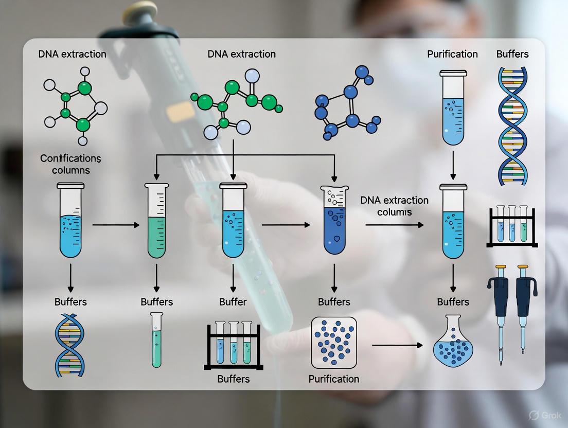
Visualizing the DNA Extraction Workflow and Critical Control Points
The Scientist's Toolkit: Essential Research Reagent Solutions
Table 3: Key Research Reagent Solutions for Optimal DNA Extraction
| Product/Kit | Manufacturer | Key Features | Optimal Use Case |
|---|---|---|---|
| DNeasy PowerLyzer PowerSoil | QIAGEN | Bead-beating integration, inhibitor removal | General gut microbiome studies |
| QIAamp PowerFecal Pro | QIAGEN | Automation-friendly, high yield | Large-scale cohort studies |
| ZymoBIOMICS DNA Miniprep | Zymo Research | Balanced yield/quality, cost-effective | Standardized research protocols |
| NucleoSpin Soil | Macherey-Nagel | Moderate cost, DNA clean-up | Budget-conscious projects |
| Stool Preprocessing Device (SPD) | bioMérieux | Standardized homogenization, improved yield | Multi-center studies requiring standardization |
The evidence consistently demonstrates that DNA extraction methodology profoundly impacts metagenomic data quality and reproducibility in gut microbiome research. Based on current comparative studies, the following recommendations emerge:
Implement mechanical lysis through bead-beating as a non-negotiable step for comprehensive representation of both Gram-positive and Gram-negative bacteria.
Standardize protocols across studies and laboratories to ensure comparability, considering the use of stool preprocessing devices for improved standardization.
Select methods based on research priorities - for highest data quality, the SPD-enhanced DNeasy PowerLyzer PowerSoil protocol currently demonstrates superior performance, while for large-scale studies, the QIAamp PowerFecal Pro offers an optimal balance of performance and automation capability.
Maintain consistency in DNA extraction methods throughout a given study, as protocol switching introduces significant technical variation that can confound biological interpretation.
Implement rigorous quality control at the DNA extraction stage, including assessment of yield, fragment size, and purity, to prevent downstream sequencing failures or biased results.
By adhering to these evidence-based recommendations and selecting appropriate extraction methodologies, researchers can significantly enhance the reliability, reproducibility, and biological relevance of their gut microbiome studies.
The reliability of any gut microbiome study is fundamentally contingent on the quality of the extracted DNA, which serves as the primary template for downstream molecular analyses. Fecal material presents a uniquely challenging matrix for DNA extraction due to its complex biochemical composition. Efficient cell lysis is paramount for achieving a representative microbial profile, as biases at this stage can systematically skew community composition. Furthermore, co-extracted PCR inhibitors and inherent DNA degradation pathways can severely compromise sequencing results and quantitative analyses. This application note delineates these sample-specific challenges within the context of gut microbiome research and provides detailed, evidence-based protocols to mitigate them, ensuring the generation of robust and reproducible data for researchers and drug development professionals.
Core Challenges in Fecal DNA Extraction
PCR Inhibitors in Fecal Material
Fecal samples contain a complex mixture of substances that can co-purify with nucleic acids and inhibit downstream enzymatic reactions, such as PCR and sequencing. The table below summarizes the primary classes of inhibitors and their effects.
Table 1: Common PCR Inhibitors in Fecal Material and Their Effects
| Inhibitor Class | Specific Examples | Primary Effect on Downstream Processes |
|---|---|---|
| Complex Polysaccharides | Glycans, cellulose | Bind to DNA polymerases, impeding enzyme activity [4] [5]. |
| Bile Salts | Various bile acids | Disrupt DNA polymerase function [4] [5]. |
| Lipids | Fatty acids | Interfere with the DNA binding to purification matrices [5]. |
| Metabolic Byproducts | Urate, bilirubin | Inhibit PCR amplification [4]. |
| Humic Substances | Fulvic and humic acids | Mimic DNA and inhibit polymerases; common in soil and sediment contamination [5]. |
Cell Lysis Efficiency and Taxonomic Bias
The physical and chemical disruption of microbial cell walls is a major source of bias. Methods that do not fully address the diversity of cell wall structures lead to the under-representation of certain taxa, creating a distorted picture of the microbial community.
- Gram-Positive vs. Gram-Negative Bacteria: Gram-positive bacteria possess a thick, cross-linked peptidoglycan layer that is notoriously difficult to lyse. Consequently, studies have demonstrated that mechanical lysis methods, particularly bead-beating, yield a significantly higher DNA quantity and Shannon's diversity index compared to methods relying solely on chemical or enzymatic lysis, which favor Gram-negative bacteria [6] [7]. For instance, one study found that the ratio of Gram-positive to Gram-negative bacteria in a mock community was profoundly skewed by the choice of lysis method [4].
- Systematic Bias: This lysis-induced bias is not random noise but a systematic reshaping of the data. It can lead to incorrect conclusions, such as misclassifying predominant taxa as rare or altering fundamental metrics like the Firmicutes-to-Bacteroidetes ratio [5]. The distinction between merely killing a cell and fully lysing it is critical; a dead but intact Gram-positive bacterium will still not release its DNA for extraction [5].
DNA Degradation Pathways
Post-collection, DNA integrity is threatened by several degradation pathways that can fragment nucleic acids and render them unusable. The following diagram illustrates the primary degradation mechanisms and their drivers.
Diagram 1: DNA Degradation Pathways. ROS: Reactive Oxygen Species.
Excessive mechanical shearing during homogenization is another significant contributor to DNA fragmentation, underscoring the need for optimized and controlled lysis protocols [8].
Quantitative Comparison of DNA Extraction Methods
The selection of a DNA extraction method profoundly impacts DNA yield, quality, and the resulting microbial community profile. The following table synthesizes key findings from comparative studies.
Table 2: Comparative Performance of Selected Fecal DNA Extraction Methods
| Extraction Method / Kit | Key Lysis Mechanism | Reported DNA Yield (Mean ± SD) | Key Findings & Performance |
|---|---|---|---|
| QIAamp PowerFecal Pro DNA Kit | Mechanical (bead-beating) | 93.97 ± 27.73 ng/μL [7] | Highest DNA yield & purity (A260/280 ~1.88); Shannon's diversity comparable to SB protocol; recommended replacement for discontinued kits [6] [7]. |
| QIAamp DNA Stool Mini Kit (with bead-beating) | Mechanical & Chemical | 35.84 ± 27.46 ng/μL [7] | Lower yield than PowerFecal Pro; higher and more consistent diversity than non-bead-beating protocol [7]. |
| QIAamp DNA Stool Mini Kit (without bead-beating) | Chemical & Enzymatic | 23.74 ± 18.33 ng/μL [7] | Lowest DNA yield and diversity; significant under-representation of Gram-positive bacteria [7]. |
| NucleoSpin Soil Kit | Mechanical & Chemical | Not specified in results | Associated with highest alpha diversity estimates in terrestrial ecosystem study; effective inhibitor removal [4]. |
Detailed Experimental Protocols
Protocol A: optimized mechanical lysis for comprehensive taxonomic recovery
This protocol is adapted from studies utilizing the QIAamp PowerFecal Pro DNA Kit, which has been shown to provide high DNA yield and integrity while effectively minimizing the bias against tough-to-lyse bacteria [6] [7].
Workflow Overview:
Diagram 2: Optimized Lysis Workflow.
Step-by-Step Procedure:
- Sample Homogenization: Weigh 180-220 mg of fecal material directly into a PowerBead Tube provided in the kit. For consistent results, use an automated weighing system.
- Lysis Buffer Addition: Add the recommended volume of lysis buffer (e.g., Solution CD1). Include Proteinase K if provided for enhanced protein digestion.
- Mechanical Lysis (Bead-Beating)
- Secure tubes securely in a bead-beater instrument.
- Homogenize at a high speed (e.g., 5.5 m/s for 1-2 minutes) [8].
- Critical Step: To prevent excessive heat buildup that promotes DNA degradation, use an instrument with cooling functionality or perform the bead-beating in short, pulsed intervals with cooling periods on ice.
- Chemical Lysis & Incubation: Briefly centrifuge the tubes to remove droplets from the lid. Incubate the lysate at 70°C for 10-15 minutes. This thermal step helps to further lyse cells and inactivate nucleases.
- Inhibitor Removal & DNA Binding: Centrifuge the tubes at high speed (≥13,000 × g) for 1 minute to pellet beads and debris. Transfer the supernatant to a microcentrifuge tube containing an inhibitor removal solution. Vortex and centrifuge. The resulting supernatant is then loaded onto a silica spin column.
- Wash and Elute: Wash the column membrane twice with wash buffers. Perform a final spin with an empty tube to ensure all ethanol is removed. Elute the DNA in 50-100 μL of elution buffer pre-heated to 55-70°C to maximize yield.
Protocol B: sample preservation and handling for metabolite and dna co-analysis
For multi-omics studies that require both DNA and metabolites, preservation from the moment of collection is critical. This protocol is based on findings that specific buffers outperform others in preserving community structure and metabolomic profiles [9].
Workflow Overview:
Diagram 3: Sample Preservation Workflow.
Step-by-Step Procedure:
- Preservation Buffer Selection: For optimal preservation of both microbial DNA and metabolites like short-chain fatty acids (SCFAs) at room temperature (up to 3 days), use PSP (Stratech PSP buffer) or a specialized Lysis Buffer. These have been shown to most closely recapitulate the profile of immediately frozen samples [10] [9]. RNAlater can be used but requires a PBS washing step prior to DNA extraction to achieve adequate DNA yield [9].
- Sample Collection: Using a sterile spoon or spatula, aliquot approximately 1 g of fresh stool into a tube containing 8 mL of the chosen preservation buffer. Homogenize thoroughly by vortexing.
- Storage and Transport: Samples can be stored at room temperature (20°C) or 4°C for up to 72 hours before processing. Lysis buffer has been demonstrated to provide superior DNA integrity and higher subsequent sequencing read counts compared to ethanol [10].
- DNA Extraction: Proceed with DNA extraction directly from the preserved sample using a robust mechanical lysis protocol as described in Protocol A. The preservation buffer is compatible with standard kit protocols.
The Scientist's Toolkit: research reagent solutions
Table 3: Essential Reagents and Kits for Fecal DNA Extraction
| Reagent / Kit | Primary Function | Key Advantage |
|---|---|---|
| QIAamp PowerFecal Pro DNA Kit | DNA extraction | Integrated bead-beating and inhibitor removal technology; high yield and reproducibility [6] [7]. |
| NucleoSpin Soil Kit | DNA extraction | Effective for diverse sample types; excellent inhibitor removal and high alpha diversity recovery [4]. |
| PSP Stool Stabilising Buffer | Sample preservation | Maintains microbial community structure and metabolomic profiles at room temperature for several days [9]. |
| Lysis Buffer (e.g., for transport) | Sample preservation | Superior to ethanol for preserving DNA quantity, quality, and integrity during transport [10]. |
| Lysozyme | Enzymatic Lysis | Targeted digestion of Gram-positive bacterial cell walls; often used as a supplement to mechanical lysis [4]. |
| Proteinase K | Enzymatic Digestion | Degrades proteins and inactivates nucleases, improving DNA yield and purity. |
| Inhibitor Removal Technology | Purification | Specific resins or buffers to remove humic substances, bile salts, and other complex inhibitors [5]. |
Accurate characterization of the gut microbiome hinges on recognizing and mitigating the technical challenges inherent to fecal DNA extraction. The methods outlined herein provide a robust framework for overcoming these obstacles. Mechanical lysis via bead-beating is non-negotiable for unbiased representation of Gram-positive taxa. The choice of sample preservation buffer, particularly for multi-omic studies, must be validated to ensure integrity of both DNA and metabolites. Finally, the use of kits with advanced inhibitor removal capabilities is critical for successful downstream amplification and sequencing. By adhering to these optimized protocols, researchers can minimize technical variability and advance our understanding of the gut microbiome's role in health and disease.
Within gut microbiome studies, the accuracy of microbial community profiling is fundamentally dependent on the initial DNA extraction process. The core challenge for researchers lies in balancing the often-competing parameters of DNA yield, purity, and integrity, as the chosen methodology can introduce significant bias in the subsequent microbial representation [11] [12]. The pursuit of a standardized protocol is essential for generating reproducible and comparable data across studies, particularly as the field moves toward clinical diagnostics and therapeutic development [11] [13]. This application note delineates the critical principles and protocols for optimizing DNA extraction to faithfully represent the complex microbial communities of the gut.
The Impact of DNA Extraction on Microbial Profiling
The DNA extraction procedure is a critical source of bias in microbiome analysis. Different lysis efficiencies, particularly for robust Gram-positive bacterial cell walls, and fungal elements, can drastically alter the apparent composition of the microbial community [11] [12] [14].
- Lysis Efficiency: Methods that combine mechanical, chemical, and enzymatic lysis are superior to those relying on a single mechanism. The inclusion of a mechanical bead-beating step is consistently identified as crucial for the effective disruption of Gram-positive bacteria, leading to a more comprehensive and accurate microbial profile [12] [14].
- Kit-Related Contamination: The analysis of low-biomass communities, such as the gut mycobiome, is particularly vulnerable to contamination. Reagents and kits can introduce exogenous DNA, necessitating the inclusion of appropriate negative controls in every experimental run to identify and account for this background noise [11].
- Standardization for Comparability: The International Human Microbiome Standards (IHMS) project has identified protocol Q (the repeated bead beating column method) as a benchmark for bacterial microbiome research due to its performance in quality, transferability, and reproducibility [11].
Table 1: Comparative Performance of Selected DNA Extraction Kits in Gut Microbiome Studies
| Kit / Method Name | Lysis Principle | Impact on Gram-positive Bacteria | Impact on Fungal DNA | Key Findings |
|---|---|---|---|---|
| IHMS Protocol Q [11] | Mechanical & Chemical | Comprehensive lysis | Effective recovery | Recommended as a standard; performs well for both bacterial and fungal communities. |
| PowerLyzer PowerSoil Kit [12] | Bead-beating | Superior lysis | Information Missing | Outperformed QIAamp kit mainly due to better lysis of Gram-positive bacteria. |
| QIAamp DNA Stool Mini Kit [11] [12] | Primarily Chemical | Lower lysis efficiency | Affected by contamination | Yields higher DNA but with potential bias against Gram-positives; worse DNA integrity in one study. |
| PureLink Microbiome Kit [11] | Information Missing | Information Missing | Information Missing | Evaluated, but IHMS Protocol Q performed best among the methods tested. |
Essential Metrics: Yield, Purity, and Integrity
A holistic assessment of extracted DNA requires the evaluation of three interdependent metrics.
- DNA Yield: Quantified fluorometrically using assays like Qubit, yield must be interpreted with caution. A high yield does not guarantee an accurate microbial representation, as it may be disproportionately derived from easily-lysed cells or, problematically, from human host DNA [11] [13].
- DNA Purity: Assessed via spectrophotometric ratios (A260/A280 and A260/230). Ideal A260/A280 ratios are ~1.8, while lower A260/230 ratios can indicate the presence of salts or organic contaminants which may inhibit downstream enzymatic reactions like PCR [11] [12].
- DNA Integrity: Refers to the fragment size and degradation level. It can be measured by the Genomic Quality Number (GQN) or by analyzing the proportion of short DNA fragments. Methods that preserve high molecular weight DNA are essential for sequencing applications like shotgun metagenomics [12].
Table 2: Quantitative Comparison of DNA Extraction Kit Performance from Human Stool
| Performance Metric | PowerLyzer PowerSoil Kit [12] | QIAamp DNA Stool Mini Kit [12] | Significance |
|---|---|---|---|
| DNA Yield | Lower | Significantly higher (q<0.01) | Higher yield may not correlate with better microbial representation. |
| DNA Purity (A260/A280) | Within expected range (1.8-2.0) | Within expected range (1.8-2.0) | No significant difference found. |
| DNA Integrity (GQN) | Higher | Lower (especially with stool container) | Suggests less degraded DNA with the PowerSoil kit. |
| PCR Inhibition | Less inhibitor presence in stool containers | Less inhibitor presence in stool containers | Sample dilution in container sampling reduces inhibition. |
| Observed OTUs | Significantly increased | Lower | Indicates higher microbial diversity detection. |
Recommended Protocols for Optimal Microbial Representation
Protocol A: IHMS Protocol Q for Comprehensive Microbiome Analysis
This non-commercial, standardized protocol is recommended for studies aiming for high reproducibility and accurate profiling of both bacterial and fungal communities [11].
- Homogenization: Weigh 0.1-0.2 g of stool and resuspend in a suitable buffer (e.g., PBS) to create a homogeneous slurry.
- Mechanical Lysis: Transfer the suspension to a tube containing lysing matrix (e.g., silica beads). Subject to vigorous bead-beating using a homogenizer for a defined period (e.g., 3 minutes at maximum speed).
- Chemical/Enzymatic Lysis: Incubate the lysate with a lysis buffer and proteinase K at elevated temperature (e.g., 70°C for 10-30 minutes).
- Purification: Pellet debris by centrifugation. Transfer the supernatant and purify the DNA using a column-based method, following standard wash and elution steps.
- Quality Control: Determine DNA concentration fluorometrically, assess purity via spectrophotometry, and check integrity by gel electrophoresis or similar methods. Include a negative control.
Protocol B: Bead-Beating Enhanced Commercial Kits
For researchers requiring a commercial solution, kits that incorporate a bead-beating step are strongly advised.
- Sample Preparation: Aliquot a defined mass or volume of stool sample into the provided lysis tube, which contains beads.
- Intensive Lysis: Securely fix the tubes in a bead-beater and process for the manufacturer's recommended time and speed. This step is critical for disrupting tough cell walls.
- Completion of Protocol: Follow the kit's standard protocol for incubation, binding, washing, and elution.
- Inhibition Check: For downstream PCR, assess the presence of inhibitors using a spike-in control or by evaluating amplification efficiency [12].
The Scientist's Toolkit: Research Reagent Solutions
Table 3: Essential Materials for DNA Extraction in Gut Microbiome Research
| Item | Function / Application | Example Products / Notes |
|---|---|---|
| Lysing Matrix | Mechanical disruption of tough cell walls (Gram-positive bacteria, fungi). | Silica/zirconia beads in lysis tubes [14]. |
| DNA Standards | Quantification calibration and sensitivity assessment for qPCR. | Genomic DNA extracts or synthetic gBlocks for standard curves [15] [16]. |
| Inhibition Controls | Detect PCR inhibitors in extracted DNA to prevent false negatives. | Internal positive controls (IPC) or efficiency calculations from standard curves [12]. |
| NGS Standards | Validate entire workflow from extraction to sequencing. | Mock microbial communities with known composition. |
| Automated Platforms | Improve throughput, reproducibility, and reduce hands-on time. | GraBon system; effective for Gram-positive bacteria [17]. |
Achieving an accurate representation of the gut microbiome is predicated on a DNA extraction method that strategically balances yield, purity, and integrity. The evidence consistently demonstrates that protocols incorporating robust mechanical lysis, such as the standardized IHMS Protocol Q or bead-beating enhanced commercial kits, provide the most comprehensive and unbiased microbial community profiles. Adherence to these core principles, coupled with rigorous quality control, is fundamental for generating reliable, reproducible data that can effectively advance research in human health, disease, and drug development.
In gut microbiome research, the accuracy and reproducibility of study outcomes are foundational to scientific progress. The initial and most critical wet-lab step—DNA extraction—is a significant source of technical variation that can distort microbial community profiles and impact subsequent biological interpretations. This application note delineates how choices in DNA extraction protocols introduce variability and provides structured experimental data and protocols to guide researchers in making informed decisions that enhance data reliability in drug development and clinical research.
The Impact of DNA Extraction on Microbiome Profiles
Evidence from comparative metagenomic studies consistently demonstrates that the DNA extraction method accounts for a substantial portion of the observed variation in microbiome data.
- Overall Microbiome Variation: In an analysis of fecal samples, the DNA extraction method itself was responsible for 21.4% of the overall microbiome variation observed and significantly affected the abundances of 32% of detected microbial species [2].
- Species-Level Abundances: A large-scale study of 745 paired fecal samples found that over 75% of bacterial species were differentially abundant when comparing results from two common DNA extraction kits [18].
- Diversity Metrics: The extraction protocol significantly influences both alpha (within-sample) and beta (between-sample) diversity estimates, which are crucial for drawing ecological inferences. This effect is consistent across diverse sample types, including soil, invertebrate, and mammalian feces [4].
The technical variability introduced during DNA extraction stems from several critical procedural differences. The following workflow illustrates the key decision points and their impacts on downstream results:
- Cell Lysis Efficiency: The method of cell wall disruption is a primary factor. Protocols incorporating mechanical lysis with small beads (bead-beating) demonstrate significantly higher efficiency in lysing Gram-positive bacteria, which have more robust cell walls, compared to methods relying solely on enzymatic or chemical lysis [2] [18]. This directly influences the observed microbial diversity.
- Kit Chemistry and Automation: The chemical composition of lysis and binding buffers varies between commercial kits, affecting the efficiency of inhibitor removal (e.g., humic substances, bilirubin, polysaccharides) and subsequent DNA yield and quality [4]. Furthermore, some kits are more amenable to automation, which can reduce hands-on time but may come at a higher cost [2].
- Impact on Fungal Communities (Mycobiota): The extraction method also profoundly affects the assessment of the gut mycobiota. The inclusion of a mechanical bead-beating step favors the recovery of DNA from filamentous fungi, whereas methods without this step can lead to an overrepresentation of yeast fungi [19].
Comparative Performance of DNA Extraction Methods
Systematic evaluations of common DNA extraction methods reveal clear differences in their performance. The following tables summarize key quantitative findings from comparative studies.
Table 1: Performance Comparison of Selected DNA Extraction Kits in Human Gut Microbiome Studies
| Extraction Kit | Key Lysis Method | DNA Yield | Performance in Mock Community | Impact on Diversity | Best Use Case |
|---|---|---|---|---|---|
| QIAamp PowerFecal Pro (PF) [2] | Mechanical (bead-beating) | High | High similarity to theoretical composition; low variability [2] | High microbial diversity; efficient for Gram-positive bacteria [2] | Large-scale human gut metagenomic studies [2] |
| DNeasy PowerSoil HTP (PS) [2] | Mechanical (bead-beating) | High | High similarity to theoretical composition; low variability [2] | High microbial diversity; efficient for Gram-positive bacteria [2] | High-throughput, automation-friendly gut microbiome studies [2] |
| AllPrep DNA/RNA Mini Kit (APK) [18] | Enzymatic + Mechanical (bead-beating) | High | Higher accuracy in recovering microbial relative abundances [18] | Higher microbial diversity compared to FSK [18] | Studies requiring high accuracy and parallel RNA analysis [18] |
| QIAamp Fast DNA Stool Mini (FSK) [18] | Chemical/Enzymatic (No bead-beating) | Lower than APK | Underrepresentation of Gram-positive bacteria [18] | Lower microbial diversity [18] | Rapid processing where comprehensive diversity is not the primary goal [18] |
| NucleoSpin Soil Kit [4] | Not Specified | Variable by sample | High contribution to overall sample diversity [4] | Highest alpha diversity estimates in multi-ecosystem study [4] | Terrestrial ecosystem studies (soil, rhizosphere, invertebrates) [4] |
Table 2: Impact of Lysis Method on Gram-Positive Bacterial Recovery (Mock Community Data) [4]
| Lysis Characteristic | Example Kits/Methods | Observed Ratio (Gram-:Gram+) | Theoretical Ratio | Interpretation |
|---|---|---|---|---|
| With Lysozyme/Mechanical Lysis | QBT Kit [4] | 0.71 ± 0.08 | 0.43 | More efficient lysis of Gram-positive bacteria, bringing observed ratio closer to theoretical. |
| Without Bead-Beating | FSK Kit [18] | Underrepresentation of Gram-positive taxa | - | Fails to lyse tough cells, leading to skewed community profiles. |
Detailed Experimental Protocols
To ensure reproducibility, below are detailed methodologies for key experiments cited in this note.
Objective: To assess the accuracy and bias of different DNA extraction methods by processing a microbial mock community (MMC) of known composition.
Materials:
- Mock Community: Commercially available MMC (e.g., ZymoBIOMICS D6300).
- Kits for Evaluation: e.g., QIAamp PowerFecal Pro Kit, DNeasy PowerSoil HTP Kit, QIAamp Fast DNA Stool Mini Kit.
- Equipment: Vortexer with adapter for bead-beating, microcentrifuge, thermal shaker/heat block, Qubit Fluorometer, NanoDrop spectrophotometer.
Procedure:
- Sample Preparation: Prepare multiple aliquots of the MMC according to the manufacturer's instructions. Include technical replicates for each extraction method.
- DNA Extraction: Extract genomic DNA from each aliquot using the different kits, strictly adhering to the respective manufacturer's protocols. Note: Do not deviate from the prescribed lysis conditions (e.g., inclusion or omission of bead-beating).
- DNA Quantification and Qualification:
- Measure DNA concentration using a fluorescence-based method (e.g., Qubit) for accuracy.
- Assess DNA purity by measuring A260/A280 and A260/A230 ratios with a spectrophotometer (e.g., NanoDrop).
- Downstream Analysis:
- Perform shotgun metagenomic sequencing or 16S rRNA gene amplicon sequencing on all extracted DNA samples.
- Bioinformatically profile the resulting sequences and compare the observed microbial composition to the theoretical composition of the MMC.
Objective: To evaluate the effect of mechanical bead-beating on the recovery of Gram-positive bacteria and overall microbial diversity from human fecal samples.
Materials:
- Samples: Human fecal samples (fresh or frozen at -80°C).
- Lysis Tubes: Tubes containing lysing matrix (e.g., 0.1mm glass beads).
- DNA Extraction Kit: A kit that allows for a customizable lysis step (e.g., AllPrep DNA/RNA Mini Kit or MagMax Microbiome kit).
- Equipment: Bead-beater or vortexer with a high-intensity adapter, microcentrifuge.
Procedure:
- Sample Homogenization: Weigh ~100 mg of fecal material into two separate lysing tubes containing beads.
- Differential Lysis:
- Condition A (With Bead-Beating): Add lysis buffer and perform mechanical disruption on a bead-beater or vortexer at high speed for a defined period (e.g., 10 minutes).
- Condition B (Without Bead-Beating): Add lysis buffer and incubate without mechanical disruption, relying only on chemical/enzymatic lysis.
- DNA Extraction: Continue the DNA extraction protocol as per the kit's instructions for both conditions.
- Analysis:
- Quantify DNA yield and purity.
- Perform metagenomic or metataxonomic profiling.
- Compare the relative abundances of key Gram-positive (e.g., Firmicutes, Actinobacteria) and Gram-negative (e.g., Bacteroidetes) phyla between the two conditions.
The Scientist's Toolkit: Essential Research Reagents & Materials
Table 3: Key Reagents and Materials for DNA Extraction in Microbiome Studies
| Item | Function/Application | Example Products/Catalog Numbers |
|---|---|---|
| Mechanical Lysis Beads | Physical disruption of tough microbial cell walls (e.g., Gram-positive bacteria, fungal spores). Essential for unbiased community representation. | 0.1mm and 0.5mm glass beads, Zirconia/Silica beads [2] [19] |
| Mock Microbial Communities | Internal controls for evaluating extraction bias, sequencing accuracy, and bioinformatic pipeline performance. | ZymoBIOMICS Microbial Community Standards (e.g., #D6300) [18] |
| Automated Nucleic Acid Extractors | For high-throughput, reproducible DNA extraction; reduces hands-on time and inter-operator variability. | QIAcube (QIAGEN), KingFisher (Thermo Fisher) systems [18] |
| Inhibitor Removal Resins/Buffers | Critical for removing PCR inhibitors common in complex samples like stool and soil (e.g., humic acids, bile salts). | Components of PowerSoil, PowerFecal, and NucleoSpin Soil kits [2] [4] |
| Fluorometric DNA Quantification Kits | Highly specific quantification of double-stranded DNA, superior to spectrophotometry for accurate sequencing library preparation. | Qubit dsDNA HS Assay Kit (Thermo Fisher) [18] |
The choice of DNA extraction protocol is not merely a technical preliminary but a fundamental determinant of data quality in gut microbiome research. To minimize technical variability and ensure robust, reproducible results:
- Standardize and Document: Use a single, consistent DNA extraction method throughout a study and report the complete protocol, including any deviations, in publications.
- Validate with Controls: Incorporate mock communities to quantify bias and confirm the accuracy of your extraction and sequencing workflow.
- Prioritize Mechanical Lysis: Select methods that include a robust mechanical lysis step (bead-beating) to ensure equitable recovery of both Gram-negative and Gram-positive bacteria.
- Align Method with Goal: Choose a kit validated for your specific sample type (e.g., human feces, soil) and research objectives (e.g., bacterial vs. fungal community analysis).
By critically evaluating and strategically selecting wet-lab protocols, researchers can significantly reduce technical artifacts, thereby uncovering true biological signals and advancing the discovery of reliable microbiome-based biomarkers and therapeutic targets.
A Practical Guide to Current DNA Extraction Protocols and Kits
In gut microbiome studies, the accuracy of microbial community profiling is fundamentally influenced by the initial step of cell lysis for DNA extraction. The structural differences between Gram-positive and Gram-negative bacteria necessitate careful selection of lysis methods to avoid biasing the representation of taxa. Gram-positive bacteria possess a thick, multi-layered peptidoglycan cell wall, while Gram-negative bacteria have a more complex envelope consisting of a thin peptidoglycan layer sandwiched between an inner cytoplasmic membrane and an outer membrane containing lipopolysaccharides [20]. This application note provides a detailed comparison of mechanical and enzymatic lysis techniques, offering structured protocols and analytical frameworks to guide researchers in selecting appropriate methods for comprehensive microbiome DNA extraction.
Structural Basis for Differential Lysis
Bacterial Cell Envelope Architecture
The effectiveness of lysis methods is dictated by the structural properties of bacterial cell envelopes. Understanding these differences is prerequisite for selecting appropriate lysis techniques.
Bacterial Cell Envelope Structural Differences
The diagram illustrates the fundamental architectural differences that dictate lysis efficiency. Gram-positive bacteria present a thick peptidoglycan layer comprising 50-80% of the cell envelope as the primary barrier, often decorated with teichoic acids that provide additional structural resistance [20]. In contrast, Gram-negative bacteria feature a triple-layer defense with a protective outer membrane of lipopolysaccharides, a thin peptidoglycan layer (only 10-20% of the cell envelope), and an inner cytoplasmic membrane [20] [21]. This structural complexity explains why Gram-negative bacteria generally require more rigorous lysis conditions or combinatorial approaches.
Comparative Analysis of Lysis Methods
Performance Characteristics Across Bacterial Types
Table 1: Comprehensive Comparison of Lysis Methods for Gram-Positive and Gram-Negative Bacteria
| Parameter | Mechanical Lysis | Enzymatic Lysis |
|---|---|---|
| Mechanism of Action | Physical shearing of cell membranes via bead beating or homogenization [20] | Biochemical degradation of peptidoglycan layer using lysozyme, lysostaphin, or mutanolysin [22] |
| Efficiency for Gram-Positive | High efficiency due to direct physical disruption of thick peptidoglycan layer [2] | Variable efficiency; species-specific enzymes may be required for optimal lysis |
| Efficiency for Gram-Negative | Moderate to high; outer membrane provides initial resistance but can be overcome with sufficient shear forces [22] | Low as standalone method; requires outer membrane permeabilizers (EDTA, AMPs) for effectiveness [23] [21] |
| DNA Yield | Higher microbial DNA content and read counts in WGS [22] | Lower DNA yield, particularly from Gram-positive organisms [22] [24] |
| DNA Quality/Integrity | Potential for DNA shearing with excessive processing; optimized protocols yield high molecular weight DNA [24] | Generally high-quality, high molecular weight DNA with minimal fragmentation |
| Throughput & Scalability | High-throughput capabilities with automation; suitable for large sample sizes [2] | Moderate throughput; incubation times can limit processing speed |
| Cost Considerations | Higher initial equipment investment; lower per-sample cost for reagents | Lower equipment needs; higher recurrent costs for enzymes and reagents |
| Technical Complexity | Standardized protocols with minimal hands-on time once optimized | Requires optimization of enzyme cocktails, concentrations, and incubation conditions |
| Community Representation Bias | More comprehensive representation of Gram-positive taxa; reduced bias in community profiling [2] | Potential underrepresentation of Gram-positive bacteria; may skew community analysis [22] [24] |
Quantitative Performance Metrics
Table 2: Experimental Performance Metrics from Comparative Studies
| Performance Metric | Mechanical Lysis | Enzymatic Lysis | Experimental Context |
|---|---|---|---|
| Total Bacterial DNA Yield | 2-fold higher proportion of bacterial mapped reads in WGS [22] | Lower bacterial read counts in downstream sequencing [22] | Gastric, esophageal, and colorectal biopsies [22] |
| Gram-Positive Recovery | Significantly improved efficiency with small beads [2] | Lower recovery without species-specific enzymes | Mock communities and fecal samples [2] |
| Gram-Negative Recovery | Effective with optimized parameters [22] | Requires complementation with EDTA or outer membrane permeabilizers [23] | Pure cultures and complex communities [22] [23] |
| Community Representation | Higher microbial diversity, particularly for Gram-positive bacteria [2] | Potential underrepresentation of resistant taxa | Gut microbiome standard reference materials [24] [2] |
| Process Time | Rapid lysis (minutes) with bead beating [20] | Prolonged incubation (30 minutes to several hours) [22] | Standardized protocols across sample types |
| Reproducibility | Lower variability across technical replicates [2] | Higher variability depending on enzyme activity and conditions | Mock community assessments [2] |
Detailed Experimental Protocols
Mechanical Lysis Protocol for Complex Gut Microbiome Samples
Principle: This protocol utilizes mechanical shearing forces generated by rapid bead agitation to physically disrupt both Gram-positive and Gram-negative bacterial cell walls, providing comprehensive DNA recovery for metagenomic studies [22] [2].
Materials:
- Lysis Buffer: Tris-EDTA-SDS buffer (pH 8.0) with proteinase K
- Bead Tubes: Silica/zirconia beads (0.1 mm and 0.5 mm mixture)
- Equipment: High-speed bead beater or vortex adapter
- Purification: Commercial DNA purification kit (e.g., QIAamp PowerFecal Pro Kit, DNeasy PowerSoil HTP kit)
- Safety: Personal protective equipment including lab coat, gloves, and safety glasses
Procedure:
- Sample Preparation: Transfer 180-220 mg of fecal material to bead tube containing lysis buffer.
- Initial Incubation: Incubate at 65°C for 10 minutes to pre-digest proteins and initiate wall weakening.
- Mechanical Disruption:
- Secure tubes in bead beater and process at maximum speed for 2-3 minutes.
- Alternatively, vortex continuously for 10-15 minutes using vortex adapter.
- Critical Step: Perform in short bursts (30s on/30s off) to prevent excessive heat generation.
- Centrifugation: Spin samples at 13,000 × g for 5 minutes to pellet debris.
- Supernatant Transfer: Carefully transfer supernatant to fresh tube without disturbing pellet.
- DNA Purification: Continue with standard silica-column based purification per manufacturer's instructions.
- Quality Assessment: Evaluate DNA yield via fluorometry and integrity by agarose gel electrophoresis.
Technical Notes:
- Bead composition and size significantly impact efficiency; 0.1 mm beads enhance Gram-positive lysis [2].
- For difficult-to-lyse Actinobacteria, extend bead beating time to 5 minutes with cooling intervals.
- DNA shearing can be minimized by optimizing beating duration and speed.
Enzymatic Lysis Protocol with Outer Membrane Permeabilization
Principle: This protocol employs enzymatic degradation of peptidoglycan layers complemented with outer membrane disrupting agents (EDTA) to facilitate access to Gram-negative bacteria while maintaining DNA integrity [22] [23].
Materials:
- Enzymes: Lysozyme (20-50 mg/mL), mutanolysin (optional for Gram-positive)
- Permeabilization: EDTA (10-20 mM) or antimicrobial peptides
- Buffer: Tris-EDTA buffer (pH 8.0) with or without detergent
- Incubation: Water bath or thermal block (37°C and 56°C)
- Inactivation: Heat inactivation at 95°C or protease treatment
Procedure:
- Sample Preparation: Suspend bacterial pellet or fecal sample (180-220 mg) in 500 μL TE buffer.
- Outer Membrane Disruption: Add EDTA to 10 mM final concentration and incubate at room temperature for 5 minutes with mixing.
- Enzymatic Digestion:
- Add lysozyme to 2 mg/mL final concentration.
- For comprehensive lysis, supplement with mutanolysin (0.1 U/μL) for Gram-positive taxa.
- Mix thoroughly by inversion.
- Primary Incubation: Incubate at 37°C for 30-60 minutes with gentle agitation.
- Proteinase K Treatment: Add proteinase K (0.2 mg/mL) and SDS (1% final concentration).
- Secondary Incubation: Incubate at 56°C for 60 minutes to complete lysis and protein digestion.
- Enzyme Inactivation: Heat at 95°C for 10 minutes or proceed directly to purification.
- DNA Purification: Use standard phenol-chloroform extraction or commercial kit purification.
Technical Notes:
- EDTA concentration can be optimized (5-20 mM) based on sample composition [23].
- For maximal Gram-positive recovery, extend enzymatic incubation to 2 hours with mutanolysin supplementation.
- Enzyme activity varies by source; verify activity with control organisms before use.
Hybrid Mechanical-Enzymatic Protocol for Comprehensive Lysis
Principle: This integrated approach combines enzymatic pre-treatment to weaken cell walls with brief mechanical disruption, maximizing DNA yield across diverse bacterial taxa while maintaining DNA integrity [22] [24].
Procedure:
- Enzymatic Pre-treatment: Follow enzymatic lysis protocol steps 1-4 (30-minute incubation).
- Brief Mechanical Lysis: Transfer samples to bead tubes and process in bead beater for 60 seconds at moderate speed.
- Complete Digestion: Return samples to 56°C for additional 30 minutes with proteinase K.
- Purification: Proceed with standard DNA purification methods.
Advantages: This method demonstrates 15-20% higher recovery of Gram-positive organisms compared to mechanical-only methods while maintaining Gram-negative representation comparable to optimized mechanical protocols [22].
The Scientist's Toolkit: Essential Research Reagents
Table 3: Key Research Reagents for Bacterial Lysis Protocols
| Reagent/Category | Specific Examples | Function & Mechanism | Application Notes |
|---|---|---|---|
| Mechanical Disruption Beads | Zirconia/silica beads (0.1, 0.5 mm) | Physical shearing of cell walls through high-speed impact | 0.1 mm beads enhance Gram-positive lysis; mixture improves comprehensive recovery [2] |
| Enzymatic Lysis Reagents | Lysozyme, mutanolysin, lysostaphin | Hydrolyzes specific bonds in peptidoglycan layer | Species-specific enzymes required for optimal Gram-positive lysis [22] |
| Outer Membrane Permeabilizers | EDTA, citric acid, antimicrobial peptides (AMPs) | Chelates divalent cations destabilizing LPS layer; disrupts membrane integrity | Essential for enzymatic lysis of Gram-negative bacteria [23] [21] |
| Commercial Kits (Mechanical) | QIAamp PowerFecal Pro Kit, DNeasy PowerSoil HTP kit | Integrated bead beating and purification | Demonstrate superior recovery of Gram-positive bacteria in comparative studies [2] |
| Detergents & Surfactants | SDS, Triton X-100, sarkosyl | Solubilizes membrane lipids and facilitates enzyme access | Concentrations must be optimized to balance efficiency with DNA purity |
| Endolysin-Based Reagents | Engineered endolysins (LysBT1, fused constructs) | Phage-derived peptidoglycan hydrolases with enhanced permeability | Emerging technology with activity against both Gram-positive and Gram-negative species [25] [23] [26] |
Method Selection Workflow
Lysis Method Selection Workflow
Selection between mechanical and enzymatic lysis methods represents a critical methodological determinant in gut microbiome studies. Mechanical approaches, particularly bead beating homogenization, provide more comprehensive representation of Gram-positive taxa and higher overall DNA yields, making them preferable for community diversity studies [22] [2]. Enzymatic methods offer advantages in DNA integrity and can be optimized for specific applications, particularly when supplemented with outer membrane permeabilizing agents for Gram-negative bacteria [23]. The emerging integration of these approaches in hybrid protocols, along with developing technologies like engineered endolysins and AMP-based permeabilization, promises enhanced lysis efficiency across the taxonomic spectrum [25] [23] [21]. Method selection should be guided by research priorities, sample type, and the specific balance required between DNA yield, integrity, and community representation.
The accuracy and reliability of gut microbiome studies are fundamentally dependent on the initial step of DNA extraction. Variations in extraction methodologies can introduce significant biases, impacting microbial community profiles and potentially leading to erroneous biological interpretations. This application note provides a comparative evaluation of two leading commercial DNA extraction kits—the QIAamp PowerFecal Pro DNA Kit (Qiagen) and the DNeasy PowerSoil Pro Kit (Qiagen)—within the context of gut microbiome research. We synthesize recent scientific evidence to outline their performance characteristics, experimental applications, and suitability for different research scenarios, providing researchers with a clear framework for kit selection in experimental design.
The QIAamp PowerFecal Pro DNA Kit is specifically designed for the isolation of microbial DNA from stool and gut samples [27]. Its proprietary technology centers on a novel bead tube combined with optimized chemistry to enhance the lysis of bacteria, including tough-to-lyse Gram-positive species, and fungi. A key feature is its streamlined Inhibitor Removal Technology (IRT), which efficiently purifies DNA from common stool-derived inhibitors such as bilirubin and bile salts, yielding inhibitor-free DNA ready for sensitive downstream applications like next-generation sequencing (NGS) [27].
The DNeasy PowerSoil Pro Kit, meanwhile, is renowned for its effectiveness in extracting DNA from a wide range of challenging environmental samples, including soil and, by extension, gut content material characterized by high microbial diversity and low biomass [28] [29]. It employs a similar mechanical bead-beating approach for comprehensive cell disruption.
While both kits utilize mechanical lysis and are respected for their performance, their underlying chemistry and optimization differ, which can lead to variations in the representation of the microbial community. A primary differentiator noted in the literature is that the PowerFecal Pro kit is often highlighted for its superior DNA yield, whereas the PowerSoil Pro kit is frequently recognized for the high purity of its resulting DNA [30] [29].
Comparative Performance Analysis
Quantitative and Qualitative Metrics
Independent studies have systematically compared the performance of these kits across several critical parameters. The findings are summarized in the table below.
Table 1: Comparative Performance of PowerFecal Pro and PowerSoil Pro Kits
| Performance Metric | QIAamp PowerFecal Pro | DNeasy PowerSoil Pro | Supporting Evidence |
|---|---|---|---|
| DNA Yield | Up to 20-fold more DNA compared to alternative methods; demonstrated high yield from stool [27]. | Variable yields; outperformed in purity; highest yield in garden soil among tested kits [28] [30]. | [27] [30] |
| DNA Purity (A260/A280) | Near 1.8, indicating absence of inhibitors [27]. | Provided highest purity DNA in cockle gut study [29]. | [27] [29] |
| Microbial Diversity (Alpha Diversity) | Yielded higher alpha diversity in sequencing; greater number of observed OTUs [27] [2]. | Performance varied; less variation in sequence similarity between replicates in permafrost [28]. | [27] [2] [28] |
| Efficiency in Lysis | Efficient lysis of Gram-positive bacteria and fungi due to mechanical and chemical lysis [27] [6]. | Effective cell lysis with mechanical bead-beating; performance can be sample-dependent [28] [29]. | [27] [6] [28] |
| Automation Compatibility | Can be automated on QIAcube Connect [27]. | Suitable for high-throughput workflows [2]. | [27] [2] |
Impact on Microbial Community Structure
The choice of extraction kit significantly influences the observed microbial community structure. A comparative metagenomic study found that the DNA extraction method alone accounted for 21.4% of the overall microbiome variation and significantly affected the abundances of 32% of detected microbial species [2]. Both the PowerFecal Pro and PowerSoil Pro kits, which employ mechanical lysis with small beads, demonstrated superior efficiency in extracting DNA from Gram-positive bacteria, leading to a more representative and diverse microbial profile compared to kits that rely solely on chemical or enzymatic lysis [2] [6].
In a study using a microbial mock community (MMC), both kits exhibited higher similarity with the theoretical composition and lower variability across technical replicates, underscoring their reliability and precision [2]. Furthermore, research on bovine fecal samples revealed that kit-specific biases impact taxa representation, with distinct clustering in principal-coordinate analysis based on the isolation procedure [30].
Detailed Experimental Protocols
Workflow for QIAamp PowerFecal Pro DNA Kit
The following diagram illustrates the generalized workflow for DNA extraction using the QIAamp PowerFecal Pro DNA Kit, highlighting its key stages from sample input to final elution.
Figure 1: A generalized workflow for DNA extraction using the QIAamp PowerFecal Pro DNA Kit.
Detailed Procedure:
- Sample Preparation: Weigh approximately 200 mg of stool sample and transfer it to the provided bead tube [27].
- Lysis and Homogenization: Add the recommended buffers to the tube. Securely close the tube and vortex thoroughly. This step utilizes mechanical lysis via bead beating and chemical lysis through optimized buffers to efficiently disrupt microbial cells, including tough Gram-positive bacteria and fungi [27] [6].
- Inhibitor Removal: Centrifuge the sample. The patented Inhibitor Removal Technology (IRT) is applied to the supernatant, effectively removing PCR inhibitors commonly found in stool, such as bilirubin and biliverdin [27].
- DNA Binding: Transfer the purified supernatant to a QIAamp spin column. Centrifuge to bind the genomic DNA to the silica membrane [27].
- Washing: Perform two wash steps using the provided wash buffers to remove salts and other residual impurities [27].
- Elution: Elute the pure, inhibitor-free genomic DNA in a small volume of elution buffer or nuclease-free water. The eluted DNA is ready for downstream applications, including qPCR and NGS [27].
Key Protocol Considerations for PowerSoil Pro Kit
While the specific steps for the DNeasy PowerSoil Pro Kit follow a similar bead-beating and spin-column format, critical protocol variations can impact outcomes:
- Lysis Method: The kit relies on a vigorous mechanical lysis step. Consistency in bead-beating time and speed is crucial for reproducible results across samples [2] [28].
- Protocol Modifications: Some studies have evaluated modifications to the standard protocol. For instance, one investigation on permafrost samples found that the addition of Qiagen Buffer ATL to the PowerSoil Pro protocol resulted in significantly higher DNA yields, though this came with challenges in subsequent 16S rRNA amplification due to potentially low starting DNA quantity [28]. Such modifications should be validated for specific sample types.
Essential Research Reagent Solutions
The table below catalogues the core materials and reagents that are fundamental to executing the protocols with these kits and ensuring successful downstream analysis.
Table 2: Key Research Reagents and Materials for DNA Extraction and Analysis
| Item | Function/Application | Relevance to Protocol |
|---|---|---|
| QIAamp PowerFecal Pro DNA Kit | All-in-one solution for microbial DNA isolation from stool. | Contains bead tubes, lysis buffers, IRT reagents, spin columns, and elution buffer [27]. |
| DNeasy PowerSoil Pro Kit | All-in-one solution for DNA isolation from soil and other complex samples. | Contains PowerBead Tubes, lysis solutions, spin columns, and collection plates [28]. |
| Vortexer with Adapter | Ensures thorough and consistent sample homogenization during lysis. | Critical for effective mechanical cell disruption in both kits. |
| Microcentrifuge | Facilitates all spin steps for buffer changes, binding, and washing. | Essential for the spin-column procedure in both kits. |
| Fluorometer (e.g., Qubit) | Accurately quantifies double-stranded DNA concentration. | Preferred over spectrophotometry for assessing DNA yield without contamination from RNA or salts [27] [28]. |
| Thermal Cycler & PCR Reagents | Enables 16S rRNA gene amplification and qPCR. | Used for assessing DNA quality and for library preparation prior to sequencing [2] [28]. |
| NGS Platform | For shotgun metagenomic or 16S rRNA amplicon sequencing. | Downstream application for analyzing microbial community structure and function [2]. |
Application in Downstream Analyses
The quality of DNA extracted directly influences the success of advanced molecular techniques. Both kits produce DNA compatible with shotgun metagenomic sequencing, providing a comprehensive view of the microbiome's taxonomic and functional potential [2]. For 16S rRNA amplicon sequencing, studies indicate that DNA extracted with these kits, particularly the PowerFecal Pro, reveals higher alpha diversity and a greater number of operational taxonomic units (OTUs), which is critical for detecting rare taxa and achieving a more complete community profile [27] [2].
Furthermore, the effective removal of inhibitors is paramount for sensitive downstream applications like quantitative PCR (qPCR). The high purity of the DNA extracted with these kits ensures efficient and accurate amplification, minimizing false negatives and quantitative biases [27] [6].
The choice between the QIAamp PowerFecal Pro and DNeasy PowerSoil Pro kits is not a matter of one being universally superior, but rather of selecting the optimal tool for a specific research context.
- For stool-specific gut microbiome studies prioritizing high yield and diversity: The QIAamp PowerFecal Pro DNA Kit is often the recommended choice. Its design is explicitly tailored to overcome the challenges of stool samples, and it consistently demonstrates superior DNA yields and recovery of higher alpha diversity, making it ideal for studies where capturing the full breadth of the microbial community is the primary goal [27] [2] [6].
- For complex, low-biomass samples or studies requiring high DNA purity: The DNeasy PowerSoil Pro Kit is an excellent option. Its proven efficacy on a wide range of challenging environmental samples, including permafrost, and its performance in delivering high-purity DNA make it suitable for samples beyond standard human stool, or where inhibitor removal is a paramount concern [28] [29].
- For consistency and comparability in large-scale or longitudinal studies: The most critical factor is consistency. Once a kit is selected, it should be used throughout the entire study to avoid introducing methodological bias, as the extraction method can account for a significant proportion of the observed variation [2] [30].
In summary, researchers must align their kit selection with their specific sample type, research questions, and the relative priority of DNA yield versus purity. This strategic decision, coupled with strict adherence to a standardized protocol, forms the foundation for robust, reliable, and reproducible gut microbiome research.
Magnetic bead-based purification has emerged as a foundational technology for nucleic acid extraction, particularly within the demanding field of gut microbiome research. This method utilizes paramagnetic beads coated with a surface chemistry that binds nucleic acids in the presence of specific binding agents like polyethylene glycol (PEG) and salts, a principle known as Solid Phase Reversible Immobilization (SPRI) [31]. The process is simple yet powerful: after binding, a magnetic stand is used to immobilize the bead-DNA complexes, allowing contaminants to be washed away efficiently, resulting in highly pure nucleic acids that are essential for sensitive downstream applications [32].
The adoption of this technology is driven by the critical need for standardized and reproducible methods in metagenomic analyses. Studies consistently demonstrate that the choice of DNA extraction method accounts for a substantial proportion of observed variation in microbiome studies—up to 21.4% of overall microbiome variation and significantly affecting the abundances of nearly a third of detected microbial species [2]. For gut microbiome research specifically, which involves challenging bacterial cell walls (particularly from Gram-positive organisms), magnetic bead-based methods employing mechanical lysis have shown superior efficiency in extracting DNA that accurately represents the true microbial community composition [2] [1].
Advantages for Automated High-Throughput Workflows
The transition from manual processing to automated high-throughput workflows represents a paradigm shift in molecular biology, and magnetic bead-based purification sits at the center of this transformation. Unlike traditional methods such as spin columns, magnetic beads are uniquely suited for automation, offering significant improvements in efficiency, reproducibility, and scalability [32] [31].
Key Benefits Over Traditional Methods
Enhanced Consistency and Reproducibility: Automated systems perform accurate pipetting procedures through standardized protocols, achieving a uniform cycle of sample processing steps (bind, wash, elute). This consistency minimizes technical variability, which is crucial for comparative metagenomic analyses where extraction methods can significantly influence results [32] [2].
Minimized Cross-Contamination: With less sample handling, automated magnetic bead protocols pose a lower risk of contamination and sample carryover. The closed-tube nature of many magnetic bead procedures reduces the opportunity for environmental contamination, whether the procedure is performed in a sterile environment or not [32].
Superior Throughput and Scalability: Magnetic bead systems enable researchers to process larger sample numbers with greater speed, often in 96-well plate formats. This scalability is essential for expansive gut metagenomic studies that may involve hundreds or thousands of samples [32] [31]. Increasing throughput is particularly important when scaling up experiments, as it allows researchers to broaden the scope of their research by testing multiple variables simultaneously [32].
Significant Time Savings: Manual extractions require undivided attention throughout the entire process. In contrast, automation allows researchers to initiate the protocol and attend to other tasks while the robot handles all repetitive liquid handling steps. This walk-away time represents a substantial efficiency gain in laboratory operations [32].
Streamlined Integrated Workflows: Depending on the automation platform, it's possible to integrate nucleic acid extraction with downstream applications like PCR preparation, creating a seamless workflow from sample to analysis. This integration reduces hands-on time and potential points of error [32].
Quantitative Performance Comparison
The advantages of magnetic bead-based purification translate into measurable performance improvements, as shown in the following comparative analysis:
Table 1: Performance Comparison: Magnetic Beads vs. Spin Columns
| Feature | Magnetic Beads | Spin Columns |
|---|---|---|
| Recovery Yield | 94–96% [31] | 70–85% [31] |
| DNA Size Range | 100 bp – 50 kb [31] | 100 bp – 10 kb [31] |
| Throughput | High (96-well & automation compatible) [31] | Low (manual only) [31] |
| Size Selection | Yes (via bead ratio adjustment) [31] | No [31] |
| Automation Compatibility | Yes [32] [31] | No [31] |
| Price per Sample | Low (~$0.90) [31] | High (~$1.75) [31] |
| Protocol Time | <15 minutes [31] | 20–30 minutes [31] |
| Waste Generation | Lower [31] | Higher [31] |
Table 2: Impact of DNA Extraction Methods on Microbiome Analysis
| Parameter | Mechanical Lysis with Beads | Methods without Bead Beating |
|---|---|---|
| Gram-positive Bacteria Recovery | Increased efficiency [2] | Reduced efficiency [2] |
| DNA Yield | Higher from complex samples [1] | Variable, often lower [1] |
| Microbial Diversity Assessment | Higher observed alpha-diversity [2] [1] | Lower observed alpha-diversity [2] |
| Fungal Community Representation | Better recovery of filamentous fungi [19] | Higher relative abundance of yeast fungi [19] |
| Inter-Sample Variability | Lower variability across technical replicates [2] | Higher variability [2] |
Essential Reagents and Equipment
Implementing an effective magnetic bead-based purification workflow requires specific reagents and equipment. The following toolkit outlines the essential components:
Table 3: Research Reagent Solutions for Magnetic Bead-Based Purification
| Item | Function | Example Products/Formats |
|---|---|---|
| Magnetic Beads | SPRI beads for nucleic acid binding; surface chemistry determines binding specificity | HighPrep PCR Beads [31], Norgen Biotek magnetic bead kits [32] |
| Lysis Buffers | Disrupt sample matrix and cell walls; often contain chaotropic salts | Components of DNeasy PowerLyzer PowerSoil kit [1], MagMax Microbiome kit [19] |
| Wash Buffers | Remove contaminants while keeping nucleic acids bound to beads; typically ethanol-based | Commercial buffer solutions included in extraction kits [31] |
| Elution Buffers | Release pure nucleic acids from beads; low-salt buffers like TE or nuclease-free water | Nuclease-free water [31], TE buffer [31] |
| Binding Enhancement Reagents | Promote nucleic acid binding to beads; PEG and salt solutions for SPRI | PEG-based binding solutions [31] |
| Mechanical Lysis Aids | Enhance cell disruption, particularly for tough cell walls; various bead types and sizes | Ceramic, glass, or zirconia beads [1] |
| Automation-Compatible Plates | High-throughput format for automated processing | 96-well plates compatible with liquid handlers [31] |
The selection of appropriate magnetic beads is critical, as different bead types are optimized for specific applications. For gut microbiome studies, beads designed for soil and stool samples often yield the best results due to their effectiveness in disrupting the tough cell walls of Gram-positive bacteria [2] [1].
Protocols for Gut Microbiome DNA Extraction
Manual Magnetic Bead-Based DNA Extraction from Fecal Samples
This protocol is optimized for the recovery of representative microbial community DNA from human fecal samples, incorporating mechanical lysis to ensure efficient disruption of both Gram-positive and Gram-negative bacteria [2] [1].
Reagents and Equipment:
- Magnetic bead-based DNA extraction kit (e.g., DNeasy PowerLyzer PowerSoil kit or equivalent)
- Magnetic stand appropriate for tube format
- Mechanical bead beater with zirconia/silica beads (0.1mm diameter recommended)
- Centrifuge
- Ethanol (80% and absolute)
- Nuclease-free water or TE buffer
- Microcentrifuge tubes (2ml)
Procedure:
- Sample Homogenization: Weigh approximately 180-220mg of fecal material into a 2ml lysing tube containing mechanical beads. Include a stool preprocessing device if available to improve standardization [1].
Lysis: Add the recommended lysis buffer (often containing guanidine hydrochloride and detergents) to the sample. Secure tubes in the bead beater and process at high speed for 3-5 minutes to ensure complete mechanical disruption of microbial cells [1].
Incubation: Incubate the lysate at 70°C for 5-10 minutes to further facilitate lysis and inactivate nucleases.
Binding: Transfer supernatant to a new tube, avoiding bead transfer. Add 1.8x volume of homogenized magnetic bead suspension to the cleared lysate. Mix thoroughly by pipetting or vortexing and incubate at room temperature for 5 minutes to allow DNA binding [31].
Separation: Place the tube on a magnetic stand and allow the beads to pellet completely (approximately 2 minutes) until the solution clears [31] [33].
Washing: Carefully remove and discard the supernatant without disturbing the bead pellet. Add 500μl of 80% ethanol while the tube remains on the magnetic stand. Incubate for 30 seconds, then remove and discard the ethanol wash. Repeat this wash step a second time [31].
Drying: Air-dry the bead pellet for 3-5 minutes at room temperature to ensure complete ethanol evaporation. Avoid over-drying, which can reduce DNA elution efficiency [31] [33].
Elution: Remove the tube from the magnetic stand and resuspend the beads in 50-100μl of nuclease-free water or TE buffer. Mix thoroughly and incubate at room temperature for 2 minutes. Place the tube back on the magnetic stand, allow separation, and transfer the eluted DNA to a clean tube [31].
Quality Control: Assess DNA concentration by fluorometry and purity by A260/A280 ratio (target ~1.8) [34]. Verify fragment size by gel electrophoresis if needed for downstream applications.
Automated High-Throughput Protocol for 96-Well Format
This protocol is designed for automated liquid handling systems such as the Thermo Fisher KingFisher Flex, Hamilton Microlab STAR, or Beckman Coulter Biomek i-Series [31].
Reagents and Equipment:
- Automation-compatible magnetic bead kit (e.g., HighPrep PCR in bulk packaging)
- Deep-well 96-well plates
- Automated magnetic particle processor
- Multichannel pipette or automated liquid handler
Procedure:
- Plate Setup: Aliquot 200μl of fecal sample lysate (prepared as in manual protocol steps 1-3) into each well of a deep-well 96-well plate.
Binding: Program the liquid handler to add 1.8x volume (360μl) of homogenized magnetic beads to each sample. Mix by repeated aspiration and dispensing or plate shaking, then incubate at room temperature for 5 minutes [31].
Separation: Engage the magnetic module to immobilize beads against the well walls. Program a pause for 2 minutes to ensure complete clearance [31].
Washing: Aspirate and discard supernatant. Add 200μl of 80% ethanol to each well, incubate for 30 seconds, then aspirate and discard. Repeat for a total of two washes [31].
Drying: After removing the final wash, program a drying time of 3-5 minutes to allow residual ethanol to evaporate [31].
Elution: Add 50μl of nuclease-free water or TE buffer to each well. Resuspend beads by mixing, incubate for 2 minutes, then immobilize beads and transfer eluted DNA to a clean collection plate [31].
Automation Considerations:
- Homogenize the bead slurry immediately before loading to ensure even suspension [33].
- Calibrate pipetting heights carefully to maximize bead recovery and minimize carryover.
- Include positive and negative controls in each run to monitor extraction efficiency and contamination.
Automation Systems and Integration
Types of Automation Platforms
The successful implementation of high-throughput magnetic bead-based extraction depends on selecting the appropriate automation platform. Two primary system types are available, each with distinct advantages:
Open Systems (e.g., Hamilton Vantage, KingFisher) offer compatibility with diverse reagents, kits, and labware [32]. These systems are designed for adaptability, allowing users to customize their extraction process and configure the platform for additional liquid handling processes such as PCR setup and aliquoting [32]. This flexibility is particularly valuable in research settings where protocols may evolve or require optimization for specific sample types.
Closed Systems (e.g., Qiagen QIAsymphony) typically employ specific protocols and designated reagents optimized for particular applications [32]. These systems generally provide a more user-friendly interface and require less optimization, as they have been pre-validated for specific workflows using proprietary reagents. While less flexible, they often deliver more standardized results with minimal setup time.
Workflow Integration and Process Mapping
The following diagram illustrates the automated magnetic bead purification workflow, highlighting key decision points and process steps:
Automated Magnetic Bead Purification Workflow
System Selection Criteria
Choosing between open and closed automation systems depends on several factors relevant to gut microbiome research:
Protocol Flexibility vs. Standardization: Research environments requiring frequent protocol modifications benefit from open systems, while clinical or production environments may prefer the standardization of closed systems [32].
Throughput Requirements: For large-scale epidemiological studies or longitudinal microbiome analyses processing hundreds of samples daily, open systems often provide superior scalability and walk-away time [32] [31].
Downstream Integration: Platforms that can integrate nucleic acid extraction with subsequent setup of PCR, sequencing library preparation, or other downstream applications offer significant workflow advantages and reduce hands-on time [32].
Cost Considerations: While closed systems may have higher per-sample reagent costs, open systems require greater initial investment in validation and protocol development [31].
Critical Factors for Optimization and Troubleshooting
Successful implementation of magnetic bead-based purification requires attention to several critical parameters that significantly impact DNA yield, quality, and representation of microbial communities.
Bead Handling and Ratio Optimization
Proper bead handling is fundamental to achieving consistent results:
Bead Homogenization: Magnetic beads settle during storage, creating a non-homogeneous slurry. Vortex beads immediately before use until the slurry displays an even color and consistency to ensure accurate pipetting and consistent binding capacity [33].
Temperature Equilibrium: While stored at 2-8°C, magnetic beads should be brought to room temperature approximately 30 minutes before use to optimize binding kinetics [33].
DNA-Bead Ratio Optimization: The bead-to-sample ratio directly determines the size range of DNA fragments recovered. As shown in the table below, adjusting this ratio enables selective recovery of specific fragment sizes for different applications [31].
Table 4: Magnetic Bead Ratio for DNA Size Selection
| Bead-to-Sample Ratio | DNA Fragment Size Retained |
|---|---|
| 0.6x | >500 bp |
| 0.8x | >300 bp |
| 1.0x | >100 bp |
| 1.8x | >50 bp |
Lysis Efficiency and Microbial Representation
The lysis step profoundly influences the representative recovery of diverse microbial taxa:
Mechanical Bead Beating: Incorporation of vigorous mechanical lysis using small beads (0.1-0.5mm diameter) is essential for efficient disruption of Gram-positive bacterial cell walls, which have thick peptidoglycan layers that resist chemical lysis alone [2] [1]. Protocols incorporating bead beating demonstrate increased microbial diversity, particularly from Gram-positive organisms [2].
Inhibition Removal: Fecal samples contain complex mixtures of PCR inhibitors (bile salts, complex polysaccharides). Magnetic bead systems effectively remove these inhibitors through the washing steps, but may require additional purification for difficult samples [31].
Mycobiota Considerations: For comprehensive gut microbiome analysis that includes fungal communities (mycobiota), mechanical lysis similarly improves recovery of filamentous fungi compared to yeast forms, potentially biasing community representation if omitted [19].
Troubleshooting Common Issues
Even with optimized protocols, challenges may arise that require troubleshooting:
Table 5: Troubleshooting Magnetic Bead-Based Purification
| Problem | Possible Causes | Solutions |
|---|---|---|
| Low DNA Yield | Over-dried beads, incorrect bead ratio, insufficient lysis | Elute promptly after drying; optimize bead:sample ratio; increase mechanical lysis intensity/duration [31] [33] |
| Residual Ethanol | Inadequate drying time | Extend drying time by 1-2 minutes; ensure complete evaporation before elution [31] |
| Incomplete Size Selection | Incorrect bead ratio | Reassess and optimize SPRI ratios for target fragment size [31] |
| Bead Carryover | Incomplete magnetic separation, aggressive pipetting | Wait longer on magnetic stand; pipette carefully away from bead pellet [33] |
| Inhibitors in Downstream Applications | Incomplete washing | Increase wash volumes or repetitions; ensure proper mixing during washes [31] |
Magnetic bead-based purification represents a transformative technology for gut microbiome research, offering unparalleled opportunities for automation, standardization, and scalability. The method's compatibility with high-throughput workflows, combined with its superior recovery of representative microbial communities—particularly when incorporating mechanical lysis—makes it an indispensable tool for large-scale metagenomic studies.
As research continues to elucidate the critical role of gut microbiota in human health and disease, the demand for robust, reproducible DNA extraction methods will only intensify. Magnetic bead technology, with its flexibility for both manual and automated processing, cost-effectiveness at scale, and ability to deliver high-quality nucleic acids suitable for sensitive downstream applications like next-generation sequencing, is poised to remain at the forefront of microbiome research methodologies. By adhering to optimized protocols and recognizing the critical factors that influence extraction efficiency, researchers can leverage this powerful technology to generate reliable, comparable data that advances our understanding of the human gut ecosystem.
In gut microbiome studies, the DNA extraction step is not merely a preliminary procedure but a critical determinant of study outcomes and biological interpretations [2]. The choice of extraction protocol directly influences the observed microbial community structure, affecting the apparent abundance of key species and the overall diversity metrics. Rapid advancements in research scale and sequencing technologies necessitate a careful evaluation of extraction methods to ensure both the reliability and precision of downstream microbial community profiling [2]. This application note provides a structured framework for selecting the optimal DNA extraction protocol, grounded in comparative metagenomic evaluations and tailored to specific research objectives in gut microbiota analysis. The goal is to empower researchers to make informed decisions that enhance data quality, reproducibility, and biological relevance in their studies of the complex gut ecosystem.
Comparative Performance of DNA Extraction Methods
A systematic evaluation of eight recent and commonly used DNA extraction methods provides a quantitative basis for selection. The performance was assessed using a microbial mock community (MMC) and human fecal samples, incorporating bacterial, archaeal, and fungal constituents. Key metrics included nucleic acid yield and quality, accuracy in representing theoretical microbial composition, technical reproducibility, and efficiency in extracting DNA from challenging Gram-positive bacteria [2].
Table 1: Performance Comparison of Selected DNA Extraction Kits
| Kit Name | Similarity to Theoretical Composition (MMC) | Technical Reproducibility | Efficiency for Gram-Positive Bacteria | Automation Friendliness | Relative Cost |
|---|---|---|---|---|---|
| QIAamp PowerFecal Pro Kit (PF) | High | High | High | Excellent | Relatively High |
| DNeasy PowerSoil HTP Kit (PS) | High | High | High | Excellent | Relatively High |
| Other Methods Evaluated | Variable | Lower | Lower | Variable | Variable |
Despite variations in DNA quantity and quality, all evaluated methods yielded sufficient DNA for shotgun metagenomic sequencing [2]. However, for fecal samples, the extraction method alone accounted for 21.4% of the overall microbiome variation and significantly affected the measured abundances of nearly one-third (32%) of detected microbial species [2]. Methods utilizing mechanical lysis with small beads, such as the QIAamp PowerFecal Pro (PF) and DNeasy PowerSoil HTP (PS) kits, demonstrated superior performance, particularly in breaking down the tough cell walls of Gram-positive bacteria, leading to a more representative and increased estimation of microbial diversity [2].
Diagram 1: A workflow for selecting a DNA extraction method based on primary research goals. PF: QIAamp PowerFecal Pro Kit; PS: DNeasy PowerSoil HTP kit.
Detailed Experimental Protocols
Protocol A: DNA Extraction from Fecal Samples using Mechanical Lysis Kits
This protocol is optimized for the QIAamp PowerFecal Pro Kit and DNeasy PowerSoil HTP Kit, which have demonstrated high performance in comparative studies [2].
1. Sample Preparation:
- Homogenize the fecal sample thoroughly before sampling.
- Weigh approximately 180-220 mg of wet fecal material into a PowerBead Tube provided in the kit. Precise weighing is critical for reproducibility.
- For longitudinal studies, record stool moisture content, as it is a significant covariate of temporal microbiota variation [35].
2. Cell Lysis:
- Add the recommended volumes of lysis buffer and proteinase K to the PowerBead Tube.
- Secure tubes in a vortex adapter and vortex at maximum speed for 10-15 minutes. This mechanical lysis with small beads is essential for efficient disruption of Gram-positive bacterial cell walls [2].
3. DNA Binding and Washing:
- Centrifuge the lysate to pellet debris.
- Transfer the supernatant to a clean microcentrifuge tube and add the appropriate binding buffer.
- Load the mixture onto a spin column and centrifuge. Discard the flow-through.
- Wash the column twice with the provided wash buffers, centrifuging after each addition to ensure complete removal of contaminants and inhibitors.
4. DNA Elution:
- Transfer the column to a clean elution tube.
- Apply 50-100 µL of elution buffer (10 mM Tris-HCl, pH 8.5) or nuclease-free water to the center of the column membrane.
- Incubate at room temperature for 1-5 minutes, then centrifuge to elute the purified DNA.
Protocol B: Validation and Quantification using qPCR
For the rapid and quantitative detection of specific gut core microbes, a qPCR-based validation is recommended. This method shows high consistency with metagenomic sequencing (Pearson’s r = 0.8688, P < 0.0001) but is faster and more cost-effective for targeted analysis [36].
1. Primer Design and Validation:
- Select novel species-specific genetic markers through comparative genomic analysis. For 45 core gut microbes, specific primers have been developed and validated [36].
- Verify primer specificity using in silico tools like BLAST against genome databases of other bacteria.
2. qPCR Reaction Setup:
- Use a SYBR Green–based master mix.
- Set up reactions in a total volume of 20 µL, containing 10 µL of master mix, forward and reverse primers (final concentration as optimized), and 2-5 µL of template DNA.
- Include a standard curve of known copy numbers (e.g., 10^1 to 10^8 copies/µL) and no-template controls in each run.
3. Amplification and Detection:
- Perform amplification using a standard protocol on a real-time PCR instrument (e.g., Roche LightCycler 480 System):
- Initial denaturation: 95°C for 5 minutes
- 40 cycles of:
- Denaturation: 95°C for 15 seconds
- Annealing/Extension: 60°C for 1 minute (optimize temperature based on primer Tm)
- Perform a melt curve analysis post-amplification to confirm amplification specificity.
4. Data Analysis:
- Determine the quantitative concentration of each target microbe in the sample by interpolating the cycle threshold (Ct) values against the standard curve.
- The limit of detection for well-designed assays typically ranges from 0.1 to 1.0 pg/µL of genomic DNA [36].
The Scientist's Toolkit: Essential Reagents and Materials
Table 2: Key Research Reagent Solutions for Gut Microbiome DNA Studies
| Item | Function/Description | Example Product(s) |
|---|---|---|
| Mechanical Lysis Kit | DNA extraction using bead beating for efficient disruption of tough cell walls (e.g., Gram-positive bacteria). | QIAamp PowerFecal Pro Kit, DNeasy PowerSoil HTP Kit [2] |
| Species-Specific qPCR Primers | For absolute quantification of specific, prevalent gut core microbes (e.g., Bacteroides, Faecalibacterium prausnitzii). | Custom-designed primers [36] |
| SYBR Green Master Mix | Fluorescent dye for detection and quantification of amplified DNA in real-time PCR. | Various commercial suppliers |
| Standard Curve DNA | DNA of known concentration for generating a standard curve in qPCR, enabling absolute quantification. | Genomic DNA from target bacterial strains [36] |
| Metagenomic Sequencing Kit | For library preparation from extracted DNA for shotgun sequencing, allowing for untargeted community profiling. | Various commercial suppliers |
Navigating Research Design: Implications of Technical and Biological Variation
The selection of a DNA extraction protocol must be situated within a broader research design that accounts for both technical and biological sources of variation.
Temporal Variability: The gut microbiome exhibits significant natural fluctuations over time. A dense longitudinal study revealed that for 78% of microbial genera, day-to-day absolute abundance variation is substantially larger within than between individuals, with some genera showing up to 100-fold shifts over a six-week period [35]. This has profound implications for clinical research:
- Single measurements are often poor estimators of a person's temporal average and carry a high risk of misclassification.
- Repeated measurement designs are recommended to increase diagnostic and target discovery power, especially when within-subject variation exceeds between-group differences [35].
Choice of Quantitative Metric:
- Absolute Abundance (QMP): Provides information on the extent and directionality of changes and avoids compositionality effects. Reveals more temporal variation, including fluctuations in total microbial load [35].
- Relative Abundance (RMP): Shows similar but less pronounced temporal variation. Can mask true biological changes due to the "closed sum" constraint [35].
Diagram 2: The interrelationship between research goals, the choice of quantitative metric, and the implications for kit selection and data interpretation.
The selection of a DNA extraction protocol is a foundational decision that should be aligned with the overarching research goals, whether they involve absolute quantification, strain-level discovery, or high-throughput screening. Based on current comparative evidence, kits employing mechanical lysis with small beads, such as the QIAamp PowerFecal Pro Kit and DNeasy PowerSoil HTP Kit, are recommended as optimal for expansive gut metagenomic research due to their high efficiency, reproducibility, and automation compatibility [2]. For large-scale or longitudinal studies, the most critical practice is maintaining strict consistency in the DNA extraction method across all samples to minimize technical confounding and ensure reliable comparative analyses [2]. By integrating a carefully chosen extraction protocol with a research design that accounts for biological variation, researchers can significantly enhance the validity and impact of their gut microbiome studies.
Adapting Extraction Methods for Long-Read Sequencing Technologies
The accurate characterization of complex microbial communities, such as the gut microbiome, is fundamentally reliant on the integrity of the input genetic material. Long-read sequencing technologies from PacBio and Oxford Nanopore Technologies (ONT) have revolutionized metagenomics by providing high-quality reads that span repetitive regions and enable full-length 16S rRNA sequencing, taxonomically resolved metagenome-assembled genomes (MAGs), and precise functional profiling [37] [38]. However, the superior performance of these platforms is contingent upon the extraction of high-quality, high-molecular-weight (HMW) DNA. This application note details the adaptation of nucleic acid extraction protocols specifically for long-read sequencing in gut microbiome studies, providing validated methodologies and performance metrics to guide researchers and drug development professionals.
The Critical Role of Extraction in Long-Read Sequencing Success
The transition from short-read to long-read sequencing necessitates a parallel evolution in DNA extraction methodologies. Short-read technologies (50-300 bp) are more tolerant of fragmented or sheared DNA, whereas long-read sequencing performance is directly correlated with input DNA integrity [38]. The grand challenge in terrestrial and gut metagenomics has been the efficient recovery of high-quality microbial genomes from highly complex habitats [39]. Recent advances demonstrate that long-read sequencing, when coupled with optimized extraction, can overcome this challenge, yielding thousands of previously undescribed microbial genomes from complex samples [39].
The table below summarizes the key differences between sequencing technologies that necessitate adapted extraction protocols.
Table 1: Sequencing Technology Comparison and Implications for DNA Extraction
| Feature | Short-Read Sequencing (Illumina) | Long-Read Sequencing (PacBio HiFi, ONT) | Extraction Implication |
|---|---|---|---|
| Typical Read Length | 50-300 bp [38] | 500 bp -> 20 kb (HiFi); 20 kb -> 4 Mb (ONT) [40] | HMW DNA is critical; minimal shearing required to preserve native fragment length. |
| Input DNA | Amplified, fragmented DNA [38] | Native DNA (preferred) [40] | Avoid enzymatic amplification steps that introduce bias; preserve epigenetic marks. |
| Primary Challenge | Limited in repetitive regions, cannot resolve full-length genes [38] | Lower raw accuracy (ONT), higher system cost (HiFi) [40] | Extraction must maximize yield and purity to offset sequencing costs and improve data quality. |
| Ideal Application | High-throughput variant calling, gene expression | De novo assembly, structural variant detection, haplotype phasing, full-length amplicons [37] [38] | Protocols must support complex analysis goals by providing comprehensive, unbiased genomic representation. |
Protocol: DREX for Hologenomic Data from Fecal Samples
The DREX (DNA and RNA Extraction) protocol is an open-source, magnetic bead-based procedure standardized within the Earth Hologenome Initiative. It is designed for the simultaneous purification of host genomic and microbial metagenomic DNA from fecal samples, making it ideal for non-invasive gut microbiome studies [41]. The protocol is modular, offering two pathways: DREX1 for separate RNA and DNA fractions, and DREX2 for total nucleic acids. The workflow below illustrates the key steps.
Figure 1: DREX workflow for hologenomic data. The flowchart outlines the two pathways of the DREX protocol for purifying nucleic acids from fecal samples, from lysis to final high-quality extract.
Detailed DREX2 Protocol for Total Nucleic Acid Extraction
This protocol is optimized for high-throughput, automation-friendly DNA extraction for shotgun metagenomic sequencing [41].
Reagents and Equipment
- Lysing Matrix E Tubes (MP Biomedicals, cat. no. 116984010)
- DNA/RNA Shield (Zymo Research, cat. no. R1100-250)
- Guanidinium Thiocyanate Lysis/Binding Buffer: Prepare with 4 M guanidinium thiocyanate, 25 mM sodium citrate, and 0.5% Sarkosyl.
- Silica-coated Magnetic Beads
- Freshly prepared 80% Ethanol
- Nuclease-free Water (low TE buffer, 10 mM Tris-HCl, 0.1 mM EDTA, pH 8.0)
- TissueLyser II (Qiagen) or similar bead-beater
- Magnetic Stand for 96-well plates
- Centrifuge for deep-well plates
Step-by-Step Procedure
Sample Homogenization and Lysis:
- Transfer up to 100 mg of fecal material preserved in DNA/RNA Shield to a tube containing Lysing Matrix E.
- Bead-beat on a TissueLyser II for two 6-minute intervals at 30 Hz. Invert the samples between runs to ensure complete homogenization.
- Centrifuge briefly to pellet the debris and transfer up to 200 µL of the homogenized lysate to a deep-well plate.
Binding:
- Add 300 µL of guanidinium thiocyanate lysis/binding buffer and 50 µL of silica-coated magnetic beads to the lysate.
- Mix thoroughly by pipetting and incubate at room temperature for 10 minutes to allow nucleic acids to bind to the beads.
Washing:
- Place the plate on a magnetic stand until the supernatant is clear. Carefully aspirate and discard the supernatant.
- With the plate on the magnetic stand, add 500 µL of freshly prepared 80% ethanol. Incubate for 30 seconds, then aspirate and discard the ethanol.
- Repeat the wash step a second time. Ensure all residual ethanol is removed by air-drying the bead pellet for 5-10 minutes.
Elution:
- Remove the plate from the magnetic stand.
- Resuspend the beads in 50-100 µL of nuclease-free water or low TE buffer.
- Incubate at room temperature for 2 minutes to allow the nucleic acids to dissociate from the beads.
- Return the plate to the magnetic stand. Once the supernatant is clear, transfer the eluate containing the purified total nucleic acids to a new tube.
Performance Benchmarking
The DREX protocol was benchmarked against a commercial reference kit (ZymoBIOMICS MagBead DNA/RNA kit) using fecal samples from 12 vertebrate species. The results demonstrate its suitability for long-read sequencing.
Table 2: Performance Benchmarking of DREX vs. Commercial Kit (REF) [41]
| Metric | DREX1 | DREX2 | REF (Commercial) |
|---|---|---|---|
| DNA Yield | Comparable to REF | Comparable to REF | Benchmark |
| Purity (A260/A280) | Slight variation, but overall comparable | Slight variation, but overall comparable | Benchmark |
| Host Genome Coverage | Comparable | Comparable | Comparable |
| Microbial Community Complexity | Highly similar profiles | Highly similar profiles | Benchmark |
| Cost-Effectiveness | High (open-source reagents) | High (open-source reagents) | Lower (proprietary reagents) |
| Automation Friendliness | High (modular design) | Very High (simplified workflow) | Moderate |
Essential Research Reagent Solutions
The table below lists key reagents and their critical functions in ensuring successful long-read sequencing from complex gut microbiome samples.
Table 3: Essential Reagents for Long-Read Sequencing DNA Extraction
| Research Reagent | Function in Protocol |
|---|---|
| DNA/RNA Shield (Zymo Research) | Preserves nucleic acid integrity immediately upon sample collection by inactiating nucleases and protecting against degradation [41]. |
| Lysing Matrix E (MP Biomedicals) | Provides a mixture of ceramic and silica beads for mechanical disruption of robust microbial cell walls (e.g., Gram-positive bacteria) during homogenization [41]. |
| Guanidinium Thiocyanate | A chaotropic salt that denatures proteins, inactivates nucleases, and promotes the binding of nucleic acids to silica surfaces in the presence of a detergent like Sarkosyl [41]. |
| Silica-coated Magnetic Beads | Selectively bind nucleic acids in the presence of chaotropic salts, enabling efficient purification and concentration through magnetic separation and washing [41]. |
| Sarkosyl (N-Lauroylsarcosine) | An ionic detergent that aids in cell lysis and membrane disruption, working synergistically with guanidinium thiocyanate [41]. |
Application in Gut Microbiome Research
Adapted extraction methods are pivotal for advanced gut microbiome applications. The high-quality DNA obtained enables strain-resolved analysis and functional profiling that is not possible with short-read technologies. For instance, HiFi metagenomic sequencing of mother-child dyads in populations with high chronic malnutrition can provide critical insights into microbiome-growth links by enabling precise taxonomic and functional comparisons [37]. Similarly, in inflammatory bowel disease (IBD) research, optimized protocols for long-read sequencing allow for strain-resolved analysis and precise functional gene profiling of samples from large cohorts, positioning long-read sequencing as the platform of choice for advanced microbiome discovery [37].
The successful implementation of a project using long-read sequencing involves several connected steps, from sample preservation to final data analysis, as shown below.
Figure 2: Long-read sequencing project workflow. The diagram visualizes the key stages of a gut microbiome study utilizing long-read sequencing (LRS), highlighting the foundational role of adapted extraction methods. HMW: High-Molecular-Weight; MAGs: Metagenome-Assembled Genomes.
Solving Common DNA Extraction Problems and Improving Yield from Complex Samples
In gut microbiome studies, the accuracy of PCR-based diagnostics is paramount, yet the reliability of these assays is frequently compromised by the co-extraction of PCR inhibitors from complex sample matrices. Fecal samples, a primary source for gut microbiota analysis, are particularly rich in polysaccharides and polyphenols, which can inhibit polymerase activity and lead to false-negative results or an inaccurate representation of the microbial community [42] [43]. The presence of these inhibitors can severely impact downstream applications, including 16S rRNA metagenomic sequencing and quantitative PCR (qPCR), which are central to understanding microbial ecology and function [42] [44]. The effectiveness of any molecular diagnostic is inherently dependent on the quality of the extracted DNA, making the removal of these inhibitors a critical first step in the analytical pipeline. This document outlines targeted strategies and provides detailed protocols for the effective removal of polysaccharides and polyphenols to ensure the integrity of DNA in gut microbiome research.
Understanding the Challenge and Its Impact
The structural complexity of microbial cell walls, particularly the thick peptidoglycan layer in Gram-positive bacteria, already presents a challenge for efficient DNA extraction [42]. This is compounded in fecal and plant-derived samples by high concentrations of endogenous inhibitors. Polysaccharides and polyphenols are two of the most common classes of compounds that co-purify with nucleic acids. During PCR, these substances can interfere with the DNA polymerase, either by binding directly to the enzyme or by chelating essential divalent cations like Mg²⁺, which are co-factors for polymerase activity [43]. The matrix effect of the sample is a significant factor, as the biological complexity can interfere with both DNA extraction efficiency and quality [42].
The impact on microbiome profiling is substantial. Inhibitors can cause:
- Reduced sensitivity, leading to failure in detecting low-abundance taxa.
- Biased community representation, as inhibitors may not affect all samples equally.
- Inaccurate quantification in qPCR and ddPCR assays, compromising absolute microbial load assessments [44]. Studies have shown that the choice of DNA extraction method can significantly influence the observed taxonomic profile, with some methods preferentially enriching for either Gram-positive or Gram-negative bacteria due to differential lysis efficiency and inhibitor removal [42]. Therefore, selecting an appropriate de-inhibition strategy is not merely a technical consideration but a fundamental step in ensuring data quality and biological relevance.
Comparative Evaluation of Inhibitor Removal Methods
Several methods are available for the removal of PCR inhibitors, each with distinct mechanisms, advantages, and limitations. The following table summarizes the key characteristics of various approaches, drawing on comparative studies.
Table 1: Comparison of PCR Inhibitor Removal Methods and Kits
| Method/Kit Name | Principle/Methodology | Effectiveness Against Polysaccharides/Polyphenols | Key Advantages | Key Limitations |
|---|---|---|---|---|
| Silica-Based Kits (e.g., PowerClean , NucleoSpin ) | Selective binding of DNA to silica membrane in the presence of chaotropic salts, followed by wash steps to remove impurities [45] [46]. | Very effective; specifically designed to remove a wide range of inhibitors, including polyphenols and polysaccharides [46]. | High purity of DNA; effective for challenging samples; commercial convenience and reproducibility [46] [43]. | Can be costly for large-scale studies; may prioritize purity over DNA yield [47]. |
| Chemical Additives (e.g., PVP, SDS) | Addition of compounds like Polyvinylpyrrolidone (PVP) to bind and precipitate polyphenols; SDS aids in cell lysis and protein denaturation [47] [48]. | Highly effective for polyphenol removal; PVP can be added directly to crude sample preparations [47] [48]. | Low cost; can be easily integrated into custom protocols (e.g., CTAB, HotShot); highly customizable [47]. | Requires optimization; additional steps for precipitation and separation are needed [43]. |
| CTAB-Based Methods | Uses Cetyltrimethylammonium bromide (CTAB) to form complexes with polysaccharides and other organic compounds in low-salt conditions, which are then separated [43]. | Effective for polysaccharide removal; a classical approach for complex plant and fecal tissues [43]. | Provides high DNA yield at a low cost; well-established for difficult samples [47]. | Labor-intensive and time-consuming; involves hazardous chemicals like chloroform [47]. |
| Magnetic Bead Kits (e.g., DNA IQ) | DNA binding to silica-coated magnetic beads in the presence of chaotropic salts; impurities are removed with washes using a magnetic stand [46]. | Very effective for removing various inhibitors, similar to column-based silica kits [46]. | Amenable to automation; no centrifugation steps; efficient for high-throughput processing. | Higher cost than traditional methods; requires specialized equipment. |
| Phenol-Chloroform Extraction | Liquid-liquid extraction using organic solvents to separate DNA (aqueous phase) from proteins, polysaccharides, and polyphenols (organic interface) [46] [44]. | Moderately effective; can remove some inhibitors like proteins and certain polyphenols [46]. | Effective for protein removal; can be combined with other methods. | Less effective for some inhibitors like humic acid; involves highly toxic chemicals [46]. |
A focused comparison of four methods for removing eight common PCR inhibitors, including humic acid and collagen, found that the PowerClean DNA Clean-Up Kit and the DNA IQ System were very effective, generating more complete STR profiles than the phenol-chloroform extraction and Chelex-100 methods [46]. This underscores the efficacy of modern silica-based purification for comprehensive inhibitor removal.
Recommended Protocols for Gut Microbiome Studies
Based on the evaluated literature, the following protocols are recommended for obtaining high-quality, PCR-ready DNA from fecal samples for gut microbiome analysis.
Protocol 1: Integrated PVP-Modified HotShot Method for Rapid Extraction
This protocol is adapted from the "HotShot Vitis" method, which was optimized for polyphenol-rich grapevine tissues and is highly suitable for fast, efficient processing of fecal samples [47]. The entire protocol can be completed in approximately 30 minutes.
Workflow Diagram: PVP-Modified HotShot Method
Detailed Reagents and Procedure:
- Alkaline Lysis Buffer Preparation: Prepare a buffer containing 60 mM NaOH, 0.2 mM disodium EDTA, 1% (w/v) polyvinylpyrrolidone (PVP-40), 0.1% (w/v) sodium dodecyl sulfate (SDS), and 0.5% (w/v) sodium metabisulfite. Adjust the pH to 12. The PVP is critical for binding polyphenols, while SDS aids in cell lysis [47] [48].
- Homogenization: Place 500 mg of fecal sample in a bag or tube with 3 mL of the alkaline lysis buffer and homogenize thoroughly at room temperature.
- Lysis and De-inhibition: Transfer 500 µL of the homogenate to a 1.5 mL microcentrifuge tube. Incubate at 95°C for 10 minutes in a thermo-mixer with shaking at 300 rpm. This heat step lyses cells and facilitates inhibitor binding to PVP.
- Neutralization: Cool the samples on ice for 3 minutes. Add an equal volume (500 µL) of neutralization buffer (40 mM Tris-HCl, pH 5). Mix gently.
- Clarification: Centrifuge at 10,000 × g for 5 minutes at 12°C to pellet debris and the PVP-polyphenol complex.
- Recovery: Carefully transfer the supernatant containing the purified DNA to a new tube. The DNA extract is now ready for PCR or can be stored at -20°C.
Protocol 2: Optimized Silica Column-Based Kit Protocol
For higher purity requirements, commercial silica-membrane kits are recommended. The following protocol is based on the PowerClean DNA Clean-Up Kit and QIAamp Fast DNA Stool Mini Kit procedures, which have been validated for inhibitor removal in fecal and other complex samples [46] [44].
Workflow Diagram: Silica Column Purification
Detailed Procedure:
- Initial Lysis: Begin with a standard lysis step. For fecal samples, it is beneficial to include a pre-wash with PBS to remove some soluble inhibitors [44]. Resuspend the pellet in the kit's lysis buffer, add proteinase K, and incubate at 56°C until completely lysed.
- Binding: Load the lysate onto the silica membrane column. The binding step typically requires the presence of a chaotropic salt (e.g., guanidine HCl) which facilitates DNA binding to the silica matrix while allowing inhibitors to flow through.
- Washing: Perform the first wash with the kit's specific inhibitor removal buffer (e.g., a solution containing ethanol and other detergent components). This step is crucial for the removal of residual polysaccharides, polyphenols, and bile salts [45] [46]. A second wash with a standard wash buffer (often containing salt and ethanol) ensures the removal of salts and other contaminants.
- Elution: Elute the pure DNA in a low-salt buffer (e.g., TE buffer or nuclease-free water). Pre-heating the elution buffer to 55-65°C can increase DNA yield.
The Scientist's Toolkit: Essential Reagents for Inhibitor Removal
Table 2: Key Research Reagent Solutions for Overcoming PCR Inhibition
| Reagent/Material | Function in Inhibitor Removal | Example Application/Protocol |
|---|---|---|
| Polyvinylpyrrolidone (PVP) | Binds to and co-precipitates polyphenols via hydrogen bonding, preventing them from inhibiting polymerase [47] [48]. | Added to lysis buffer in "HotShot Vitis" and CTAB protocols at 1-2% (w/v) concentration [47]. |
| Silica Membranes/Columns | Provides a solid phase for selective binding of DNA in the presence of chaotropic salts, allowing impurities to be washed away [45] [46]. | Core component of commercial kits like PowerClean and NucleoSpin [46]. |
| Cetyltrimethylammonium bromide (CTAB) | Precipitates polysaccharides and other complex carbohydrates in low-salt conditions, facilitating their separation from DNA [43]. | Used in classical CTAB extraction protocols for plant and fecal materials [43]. |
| Sodium Metabisulfite | An antioxidant that helps prevent oxidation of polyphenols into darker-colored, more inhibitory compounds during extraction [47]. | Included at 0.5% (w/v) in the alkaline lysis buffer of the HotShot Vitis protocol [47]. |
| Chaotropic Salts | Disrupt hydrogen bonding, denature proteins, and enable DNA to bind to silica surfaces, which is foundational to column-based purification [45]. | Key component of binding buffers in all silica-membrane and magnetic bead kits [45] [46]. |
The reliable profiling of the gut microbiome is fundamentally dependent on the quality of the input DNA. The strategic removal of polysaccharides and polyphenols is therefore not an optional refinement but a necessity. The protocols and methods detailed here—from the rapid, cost-effective PVP-modified HotShot method to the high-purity silica column-based kits—provide researchers with a robust toolkit to overcome PCR inhibition. The choice of method should be guided by the specific requirements of the study, balancing factors such as throughput, cost, required yield, and purity. By implementing these optimized strategies, scientists in gut microbiome and drug development can significantly enhance the accuracy and sensitivity of their molecular analyses, ensuring that the data generated truly reflects the underlying biological reality.
Optimizing Lysis Conditions to Maximize Recovery of Tough-to-Lyse Microbes
Within the framework of gut microbiome research, obtaining a comprehensive and unbiased microbial community profile is a foundational requirement. The DNA extraction process, and the lysis step in particular, is a critical determinant of downstream sequencing results. Tough-to-lyse microbes, primarily Gram-positive bacteria with their thick peptidoglycan cell walls, are frequently underrepresented in sequencing data if lysis conditions are not optimized. This application note details evidence-based strategies to maximize the recovery of these resilient microorganisms, ensuring that metagenomic analyses more accurately reflect the true composition of the gut microbiome.
The Core Challenge: Lysis Efficiency and Microbial Representation
The fundamental challenge in DNA extraction for microbiome studies lies in the diverse cellular structures of microorganisms. Standard enzymatic lysis protocols often fail to disrupt the robust cell walls of bacteria such as Firmicutes (e.g., Blautia, Ruminococcus) and Actinobacteria (e.g., Bifidobacterium). This failure introduces a systematic bias, leading to an overrepresentation of easily-lysed Gram-negative bacteria and a distorted view of the microbial community [49] [50].
Evidence consistently shows that incorporating mechanical lysis methods, such as bead beating, is essential for equitable recovery. One study found that bead beating significantly increased the detected diversity, notably enhancing the signal from Gram-positive genera like Blautia, Bifidobacterium, and Ruminococcus [49]. Conversely, research using the ZymoBIOMICS Gut Microbiome Standard demonstrated that all methods showed limited ability to detect taxa below 0.5% relative abundance, highlighting the sensitivity limits and the need for optimized protocols to capture low-abundance, hard-to-lyse members [24].
Optimization Strategies for Effective Lysis
Optimizing lysis involves combining and fine-tuning various physical, chemical, and enzymatic parameters. The following table summarizes key strategies and their impacts, as demonstrated in recent studies.
Table 1: Lysis Optimization Strategies and Their Effects on DNA Recovery from Tough-to-Lyse Microbes
| Strategy | Key Finding | Experimental Context | Impact on Tough-to-Lyse Microbes |
|---|---|---|---|
| Mechanical Bead Beating | Significantly increased bacterial diversity and detection of Gram-positive genera (Blautia, Bifidobacterium, Ruminococcus) [49]. | Human fecal samples processed in a 96-well plate format using a TissueLyser II. | Greatly improved recovery and representation. |
| Integrated Mechanical-Chemical Lysis | Superior recovery of Gram-positive bacteria compared to standard kits; yielded HMW DNA suitable for long-read sequencing [24]. | Custom protocol for gut microbiome standards, combining mechanical disruption with chemical lysis. | Enhanced yield and DNA integrity. |
| Heating and Enzymatic Pre-Treatment | A protocol for hard-to-lyse organisms (e.g., Mycobacterium) uses an 80°C heat step followed by lysozyme incubation, yielding high-integrity DNA [51]. | Specific protocol for microbial isolates using lysozyme and proteinase K. | Effective for organisms with exceptionally robust cell walls. |
| Lysis Method Comparison | Mechanical lysis provided more consistent microbial community profiles and higher DNA purity than enzymatic lysis, despite lower yield [52] [53]. | Epiphytic phyllosphere samples; findings on consistency are transferable to complex gut samples. | Reduced bias and improved reproducibility. |
Workflow for Lysis Optimization
The following diagram illustrates a systematic workflow for developing an optimized lysis protocol, integrating the strategies discussed above.
Detailed Experimental Protocols
Protocol 1: High-Throughput Bead Beating for Fecal Samples
This protocol is adapted for processing 96-well plates, making it suitable for large-scale gut microbiome studies [49].
- Sample Preparation: Suspend 200 µL of fecal sample preserved in OMNIgeneGUT or DNA/RNA shield in 800 µL of lysis buffer (e.g., Chemagic Lysis Buffer 1).
- Mechanical Lysis:
- Transfer the mixture to a 96-well PowerBead Pro Plate containing 0.1 mm glass beads.
- Seal the plate with a sealing film.
- Shake the plate using a TissueLyser II at a frequency of 15 Hz for 2 cycles of 5 minutes each.
- Chemical Lysis: Incubate the lysate with 15 µL of proteinase K at 70°C for 10 minutes, followed by 95°C for 5 minutes to inactivate the enzyme.
- DNA Purification: Centrifuge the plate at high speed for 5 minutes. Transfer 800 µL of the supernatant to a new sample plate and proceed with a magnetic bead-based purification method, such as the Chemagic DNA Stool 200 H96 kit on a Magnetic Separation Module I (MSM I) instrument.
Protocol 2: Intensive Lysis for Hard-to-Lyse Isolates
This column-based protocol is designed for challenging organisms like Mycobacterium and is optimized to preserve high molecular weight (HMW) DNA [51].
- Sample Input: Harvest 5-10 mg of cells from solid or liquid media.
- Heat Activation: Resuspend the pellet in 500 µL of PBS and incubate at 80°C for 20 minutes. Centrifuge, remove the supernatant, and resuspend in PBS for a wash step.
- Primary Enzymatic Lysis: Resuspend the washed pellet in 100 µL of TE buffer. Add two 4 mm glass beads and vortex for 30 seconds. Add 10 µL of lysozyme (10 mg/mL) and incubate at 37°C for 15 minutes at 800 RPM in a thermomixer.
- Secondary Chemical Lysis: Transfer the lysate to a new tube. Add 10 µL of proteinase K and 3 µL of RNase A. Vortex and add 200 µL of CTAB buffer. Incubate at 56°C for 30 minutes at 1000 RPM.
- Purification: Add 100 µL of 5 M sodium chloride and centrifuge at 17,000 x g for 5 minutes. Transfer 200 µL of the supernatant to a new tube, combine with 400 µL of binding buffer, and complete the purification using a commercial spin column kit (e.g., NEB Monarch Spin gDNA Extraction Kit), eluting in a pre-heated buffer.
The Scientist's Toolkit: Essential Research Reagents
Table 2: Key Reagents for Optimizing Lysis of Tough-to-Lyse Microbes
| Reagent / Kit | Function in Lysis | Specific Application Note |
|---|---|---|
| Glass Beads (0.1 mm & 4 mm) | Mechanical disruption of tough cell walls. | 0.1 mm beads are used for full community lysis in plates [49]; 4 mm beads are for more gentle homogenization of isolates [51]. |
| PowerBead Pro Plates | High-throughput mechanical lysis. | Enables simultaneous, consistent bead beating of 96 samples [49]. |
| Lysozyme | Enzymatic degradation of peptidoglycan in bacterial cell walls. | Essential for pre-treating Gram-positive bacteria; used prior to or in conjunction with mechanical methods [51]. |
| Proteinase K | Broad-spectrum protease that digests proteins and inactivates nucleases. | Used after mechanical disruption to further break down cellular debris [49] [51]. |
| CTAB Buffer | Chemical surfactant that aids in cell lysis and removes polysaccharides. | Critical for sequencing health and output when extracting from hard-to-lyse organisms [51]. |
| Quick-DNA HMW MagBead Kit | Integrated lysis & purification for HMW DNA. | Identified as producing high yield of pure HMW DNA suitable for Nanopore sequencing from complex bacterial mixes [54]. |
| ZymoBIOMICS Gut Microbiome Standard | Positive process control. | Validates lysis efficiency across a defined community of easy- and hard-to-lyse bacteria [24] [49]. |
The accurate profiling of the gut microbiome is contingent upon a DNA extraction protocol that does not discriminate against tough-to-lyse microbes. The integration of robust mechanical disruption via bead beating with targeted chemical and enzymatic treatments is a proven strategy to overcome cellular robustness. By adopting the optimized protocols and reagents detailed herein, researchers can minimize bias, thereby generating more reliable and representative data for downstream applications in drug development and clinical research. Consistent use of standardized positive controls is paramount for validating lysis efficiency and ensuring comparability across studies.
Within the framework of DNA extraction methods for gut microbiome studies, accurate DNA quantification and purity assessment are critical prerequisites for downstream analytical success. The reliability of next-generation sequencing (NGS) and other molecular analyses is fundamentally dependent on the quality of the input nucleic acids [55] [56]. This application note provides a detailed comparison of two principal quantification technologies—spectrophotometry and fluorometry—focusing on their application in evaluating DNA extracted from complex biological samples, particularly fecal specimens. We present standardized protocols, performance data, and practical guidance to enable researchers to implement robust quality control checkpoints in their microbiome research workflows, ensuring the generation of reproducible and meaningful data.
Technology Comparison: Spectrophotometry vs. Fluorometry
The two most prevalent methods for DNA quantification operate on distinct principles: spectrophotometry measures the absorbance of ultraviolet light by nucleic acids, while fluorometry relies on the fluorescence emitted by dyes upon binding specifically to DNA [57]. This fundamental difference dictates their respective strengths, limitations, and optimal applications in microbiome research.
Table 1: Core Principles and Characteristics of DNA Quantification Methods
| Feature | Spectrophotometry (e.g., NanoDrop) | Fluorometry (e.g., Qubit) |
|---|---|---|
| Measurement Principle | Absorbance of UV light at 260 nm [58] [57] | Fluorescence emission from DNA-binding dyes [59] [57] |
| DNA Specificity | Low; detects ssDNA, dsDNA, RNA, and free nucleotides [55] [59] | High; dyes are selective for dsDNA, even with RNA present [55] [59] |
| Purity Assessment | Yes; via A260/A280 and A260/A230 ratios [58] [57] | No; does not provide purity ratios [55] [60] |
| Sensitivity | Lower (2 ng/µL for samples <100 ng/µL) [55] | Higher; Qubit dsDNA HS Assay range: 0.2–100 ng [59] |
| Sample Volume | Typically 1-2 µL | As little as 1 µL [59] |
Table 2: Performance Comparison in Research Contexts
| Performance Aspect | Spectrophotometry | Fluorometry |
|---|---|---|
| Reported Accuracy | Often overestimates concentration due to contaminants [55] [60] | Provides accurate concentration of dsDNA [55] [61] |
| Effect of Contaminants | Highly susceptible; chemicals, proteins, and phenols skew results [60] [57] | Generally unaffected by common contaminants like salts, solvents, and proteins [59] |
| Ideal Application | Initial check for sample integrity and gross contaminants [59] | Accurate quantification of dsDNA for sensitive downstream applications [55] [18] |
Detailed Experimental Protocols
Protocol A: Spectrophotometric DNA Quantification (e.g., NanoDrop)
Principle: This method quantifies DNA by measuring the absorbance of ultraviolet light at 260 nm. The presence of contaminants is assessed by calculating the ratio of absorbance at 260 nm to that at 280 nm (protein contamination) and 230 nm (organic compound and salt contamination) [58] [57].
Materials:
- Spectrophotometer with UV capability (e.g., NanoDrop)
- UV-transparent cuvettes (if required by the instrument)
- Elution buffer or nuclease-free water for blanking
- Microcentrifuge tubes
Procedure:
- Instrument Initialization: Power on the spectrophotometer and allow the lamp to warm up as per the manufacturer's instructions.
- Blank Measurement: Pipette 1-2 µL of the elution buffer used in your DNA extraction (e.g., TE buffer or nuclease-free water) onto the measurement pedestal. Perform a blank measurement to calibrate the instrument.
- Sample Measurement:
- Wipe the pedestal clean with a lint-free tissue.
- Pipette 1-2 µL of the DNA sample onto the measurement surface.
- Lower the arm and initiate the measurement.
- Record the concentration and the A260/A280 and A260/A230 ratios.
- Clean the pedestal thoroughly between samples.
- Data Analysis:
Protocol B: Fluorometric DNA Quantification (e.g., Qubit Assay)
Principle: This method uses a fluorescent dye that emits light only when bound to dsDNA. The fluorescence intensity is measured and compared to a standard curve of known DNA concentrations, providing highly specific quantification of dsDNA [59] [57].
Materials:
- Fluorometer (e.g., Qubit 4 or Qubit Flex)
- Appropriate Qubit dsDNA assay kit (e.g., High Sensitivity or Broad Range)
- Qubit assay tubes
- DNA standards (provided in the kit)
Procedure:
- Working Solution Preparation:
- Use the onboard reagent calculator to determine the required volumes.
- Prepare the working solution by diluting the fluorescent dye in the specified buffer at a 1:200 ratio. Mix thoroughly by vortexing.
- Standard and Sample Preparation:
- For each standard, pipette 190 µL of working solution into a Qubit assay tube and add 10 µL of the respective standard. Mix by vortexing.
- For each unknown sample, pipette 199 µL of working solution into a tube and add 1–20 µL of the DNA sample (volume depends on the expected concentration). Mix by vortexing.
- Incubation: Incubate all tubes at room temperature for 2 minutes, protected from light.
- Measurement:
- On the fluorometer, select the appropriate assay (e.g., "Qubit dsDNA HS Assay").
- Insert the standard tubes first and follow the prompts to read them and create the standard curve.
- Subsequently, read the unknown sample tubes. The instrument will display the concentration directly.
- Data Analysis: The concentration is automatically calculated by interpolating the sample fluorescence against the standard curve. Account for any dilution factor if a sample was diluted prior to the assay.
Diagram 1: DNA QC Workflow. An integrated workflow for DNA quality control, leveraging both spectrophotometry and fluorometry.
Application in Gut Microbiome Research
The choice of quantification method is particularly consequential in gut microbiome studies due to the complex nature of fecal samples. DNA extracts from feces often contain co-purified contaminants from the matrix, dietary components, and extraction reagents, which can interfere with accurate quantification [56] [60]. Studies have demonstrated that spectrophotometry tends to overestimate DNA concentration in such complex matrices due to interference from these contaminants that also absorb at 260 nm [60]. In contrast, fluorometry, being specific to dsDNA, provides a more accurate measure of the actual amplifiable template, which is crucial for sequencing library preparation [55] [18].
Furthermore, the DNA extraction method itself—including the use of mechanical bead-beating—significantly impacts the yield and microbial community profile recovered. Protocols that incorporate bead-beating yield higher DNA concentrations and microbial diversity by more effectively lysing hard-to-break gram-positive bacteria [18]. This makes accurate quantification of the resulting diverse DNA populations even more critical. A recommended best practice is to use an integrated approach: employ spectrophotometry for a rapid assessment of sample purity and the presence of contaminants, followed by fluorometry for precise quantification of dsDNA concentration to normalize loading amounts for sequencing [55] [59].
Table 3: Essential Research Reagent Solutions for DNA QC
| Reagent/Kit | Function | Application Note |
|---|---|---|
| Qubit dsDNA HS Assay Kit | Fluorometric quantification of dsDNA in low-concentration samples (0.2–100 ng) [59]. | Ideal for quantifying precious or low-biomass microbiome samples prior to NGS. |
| AccuGreen/AccuClear Kits | Alternative fluorometric dsDNA quantification kits [55]. | Provide options for researchers; performance comparable to Qubit [55]. |
| PicoGreen dsDNA Dye | Fluorescent dye for dsDNA quantification, compatible with various fluorometers and plate readers [57]. | Enables high-throughput quantification in 96-well plates. |
| Nuclease-free Water | Diluent for DNA samples and blanking solution in spectrophotometry. | Using the same elution buffer as the sample for blanking is critical for accurate spectrophotometric readings [60]. |
| TE Buffer | Common elution buffer for DNA; helps maintain DNA stability. | pH can affect A260/A280 ratios; pure DNA in TE buffer has a ratio of ~1.8 [58]. |
Robust quality control of DNA is a non-negotiable checkpoint in gut microbiome research. Neither spectrophotometry nor fluorometry is universally superior; rather, they offer complementary data. Spectrophotometry provides a quick assessment of sample purity, while fluorometry delivers precise quantification of amplifiable dsDNA. For the most reliable results, a combined workflow is strongly recommended: first, use spectrophotometry to identify samples with significant contaminants (evidenced by abnormal A260/A280 or A260/230 ratios), and then use fluorometry to accurately determine the dsDNA concentration for critical downstream steps like sequencing library normalization [55] [59]. This two-tiered approach ensures the integrity of the starting material, thereby maximizing the reliability and reproducibility of microbiome data.
The successful application of long-read sequencing technologies, such as those from Oxford Nanopore Technologies (ONT) and PacBio, has transformed our ability to resolve complex genomic regions, structural variations, and phased haplotypes in gut microbiome studies [62]. These advancements are inherently dependent on the isolation of high-quality, high molecular weight (HMW) genomic DNA exhibiting exceptional integrity and minimal fragmentation [62]. DNA shearing—the mechanical or chemical fragmentation of DNA molecules into shorter fragments—represents a significant bottleneck, particularly for metagenomic studies aiming to reconstruct complete genomes from complex microbial communities [63]. For long-read sequencing, maintaining DNA fragments ideally >50 kb and preferably >100 kb is critical for achieving optimal read lengths and assembly contiguity [62] [64]. The integrity of the starting DNA material directly influences pivotal metrics such as read length N50 values, which are fundamentally linked to genome assembly quality and the accurate detection of large-scale genomic alterations [62] [65]. This application note details the primary sources of DNA shearing and provides validated protocols for isolating HMW DNA from challenging gut microbiome samples, thereby enabling robust long-read sequencing outcomes.
The process of DNA extraction is fraught with opportunities for shearing, which can be broadly categorized into mechanical, enzymatic, and chemical sources. Understanding these sources is the first step in mitigating their effects.
Table 1: Common Sources of DNA Shearing and Preventive Strategies in HMW DNA Extraction
| Source Category | Specific Examples | Impact on DNA Integrity | Preventive Strategy |
|---|---|---|---|
| Mechanical Forces | Vortexing, vigorous pipetting, harsh centrifugation [62]. | Physical breakage of long DNA strands, reducing average fragment size [62]. | Use wide-bore tips, mix by inversion, employ low-speed centrifugation [62]. |
| Enzymatic Degradation | Nuclease activity from delayed processing or inefficient lysis [62]. | DNA nicking and strand breaks, leading to fragmentation [62]. | Use EDTA tubes, process samples promptly, optimize proteinase K digestion [62]. |
| Chemical & Lysis Factors | Harsh detergents, high salt conditions, excessive incubation temperatures [62]. | DNA denaturation and strand breaks [62]. | Employ mild, non-ionic detergents; carefully balance incubation time and temperature [62]. |
| Sample Handling | Freeze-thaw cycles, prolonged storage at sub-optimal temperatures [62]. | Ice crystal formation and enzymatic degradation increase fragmentation risk [62]. | Aliquot samples, avoid repeated freeze-thaws, use cryoprotectants [62]. |
The biases introduced during DNA extraction are not trivial; they can significantly distort the observed microbial community structure. Studies have demonstrated that the choice of lysis method—mechanical versus enzymatic—can preferentially lyse certain cell types, leading to the under-representation of tough-to-lyse Gram-positive bacteria (e.g., Lactobacillus, Staphylococcus) and an over-representation of easy-to-lyse Gram-negative bacteria [66] [67]. This lysis bias is a major confounder in gut microbiome studies aiming to quantify the absolute and relative abundance of community members [68].
Optimized Workflow for HMW DNA Isolation from Complex Gut Samples
The following workflow integrates best practices from recent benchmarking studies to minimize shearing and maximize DNA integrity for downstream long-read sequencing. The diagram below outlines the core procedural pathway and critical decision points.
Sample Preparation and Lysis
For fecal samples, begin by homogenizing the material in a suitable buffer (e.g., PBS) using slow, end-over-end tube rotation instead of vortexing to prevent initial shearing [63]. Cell lysis is a critical step where bias can be introduced.
- Strategy 1: Modified Mechanical Lysis. Using a DNeasy PowerSoil kit (Qiagen) as a base, transfer the sample to a PowerBead tube and agitate it flat on a IKA Works MS2S8 Minishaker at 2400 rpm for 10 minutes in 1-minute pulses to minimize velocity gradients and DNA shearing [63].
- Strategy 2: Enzymatic Lysis. To entirely avoid mechanical shear, replace bead beating with a heated, enzymatic treatment. Add a lytic cocktail to the sample containing lysozyme (10 mg/ml) and mutanolysin (10 U/µl), then incubate at 37°C for 60 minutes with gentle inversion every 15 minutes [63]. This method is particularly effective for maximizing the recovery of Gram-positive bacteria, which have robust cell walls [24].
DNA Purification and Handling
Following lysis, purification must be performed with techniques that preserve molecular length.
- Magnetic Bead-Based Purification. Kits like the PureLin Microbiome DNA Purification Kit or the MagMAX HMW DNA Kit are preferred for their relative gentleness [24] [64]. When using magnetic beads, ensure mixing is slow and steady to prevent the long DNA molecules from wrapping around and shearing between multiple tiny beads [65]. The Monarch HMW DNA Extraction Kit utilizes large 4mm glass beads, around which DNA winds in a relaxed form, facilitating easier elution with minimal shear [65].
- Phenol-Chloroform Extraction. This traditional method can yield ultra-long DNA fragments (>100 kb) but involves hazardous chemicals and often results in a viscous, difficult-to-handle DNA pellet that is challenging to resuspend [65] [63]. A recent benchmarking study on human oral microbiome samples found that column-based kits with enzyme supplementation, rather than phenol-chloroform methods, may be more appropriate for long-read metagenomic sequencing in terms of overall read-size distribution and assembly performance [63].
Throughout the process, use wide-bore pipette tips for all liquid transfers after lysis, limit pipetting cycles, and avoid vortexing. Centrifugation steps should be performed at low speeds (<3000 x g) with gentle acceleration and deceleration profiles [62] [64].
Validation and Quality Control of HMW DNA
Accurate validation is non-negotiable to ensure the extracted DNA meets the stringent requirements of long-read sequencing platforms. The table below compares the common QC methods.
Table 2: Methods for Validating DNA Integrity and Quantity for Long-Read Sequencing
| Method | Principle | Key Metrics | Optimal Output for HMW DNA |
|---|---|---|---|
| Pulsed-Field Gel Electrophoresis (PFGE) | Periodically changes electric field direction to separate very large DNA molecules [62]. | Fragment size distribution from 50 kb to >1 Mb [62]. | Strong intensity above 50 kb; a distinct band >150 kb is ideal [62]. |
| Capillary Electrophoresis | Automated high-resolution sizing and quantification (e.g., Agilent Femto Pulse, Fragment Analyzer) [62]. | Genomic Quality Number (GQN), percentage of DNA >50 kb and >100 kb [62]. | GQN >8.0; dominant peak >100 kb for ultra-long Nanopore reads [62]. |
| Fluorometric Quantification | Fluorescent dyes binding specifically to DNA (e.g., Qubit, PicoGreen) [62]. | Accurate concentration without overestimation from RNA or contaminants [62]. | Concentration typically 10–100 ng/µL for library preparation [62]. |
It is crucial to use fluorometric quantification (Qubit) over spectrophotometry (NanoDrop), as the latter can overestimate concentration due to RNA or degraded nucleic acid contamination, which is detrimental to library preparation [62].
Essential Reagents and Tools for HMW DNA Isolation
Table 3: Research Reagent Solutions for HMW DNA Extraction
| Reagent / Kit | Primary Function | Key Features & Considerations |
|---|---|---|
| K2/K3 EDTA Tubes | Blood collection & anticoagulation [62]. | Chelates divalent cations to inhibit nuclease activity; essential for sample preservation [62]. |
| PureLin Microbiome DNA Purification Kit | DNA purification from complex microbial communities [24]. | Provides superior recovery of DNA from Gram-positive bacteria; magnetic bead-based [24]. |
| Monarch HMW DNA Extraction Kit | HMW DNA extraction from cells, blood, & tissue [65]. | Uses large 4mm glass beads for gentle DNA winding; quick 30-90 minute workflow [65]. |
| MagMAX HMW DNA Kit | HMW DNA purification for long-read sequencing [64]. | Optimized for use with KingFisher automation systems; suitable for fresh/frozen whole blood, cells, and tissues [64]. |
| Proteinase K | Enzymatic digestion of proteins & nucleases [62]. | Critical for degrading histones and nucleases; must be properly reconstituted and stored at -20°C to maintain activity [62]. |
| Wide-Bore Pipette Tips | Liquid handling of lysed samples [62]. | Reduces fluid shear forces during pipetting, preserving DNA strand length [62]. |
The reliability of long-read sequencing data in gut microbiome research is fundamentally constrained by the quality of the input DNA. Preventing shearing to isolate pristine HMW DNA is not merely a technical detail but a foundational requirement for generating accurate and biologically meaningful genomic information. By adhering to the gentle handling practices, optimized lysis strategies, and rigorous quality control measures outlined in this application note, researchers can effectively minimize technical bias and unlock the full potential of long-read sequencing technologies for de novo genome assembly, structural variant detection, and comprehensive microbial community profiling.
Automation Solutions for Standardization in Large-Scale Cohort Studies
The reliability of large-scale cohort studies, particularly in gut microbiome research, is fundamentally challenged by procedural inconsistencies that compromise data quality and study reproducibility. Manual data handling and cohort selection processes are not only time-consuming but also introduce significant variability, especially during the DNA extraction phase, which critically impacts downstream microbial community analysis [2] [69]. Automation solutions address these challenges by replacing labor-intensive, error-prone manual workflows with standardized, scalable processes. This integration of automation ensures that large-scale studies can maintain methodological consistency across multiple collection sites, processing batches, and timepoints, thereby enhancing the validity of research findings and enabling robust cross-study comparisons [70] [71]. The transition to automated systems represents a paradigm shift in research methodology, transforming the approach to data generation and cohort management in population-scale studies.
The Critical Role of Standardization in Microbiome Research
In gut microbiome studies, lack of standardized protocols introduces substantial technical variation that can obscure biological signals and lead to irreproducible findings. Evidence demonstrates that DNA extraction methodology alone accounts for approximately 21.4% of the overall observed microbiome variation and significantly affects the abundance estimates of nearly one-third of detected microbial species [2]. This technical variability presents a formidable challenge for distinguishing true biological effects from methodological artifacts, particularly in large-scale cohorts where processing occurs across multiple sites or over extended timeframes.
The compositional differences introduced by methodological choices are not merely statistical abstractions but have practical implications for biological interpretation. Studies evaluating multiple DNA extraction methods have revealed that protocols utilizing mechanical lysis with small beads, such as the QIAamp PowerFecal Pro Kit and DNeasy PowerSoil HTP kit, demonstrate superior efficiency in extracting DNA from Gram-positive bacteria, thereby producing more comprehensive microbial diversity profiles [2]. This methodological bias directly impacts study conclusions, as the failure to effectively lyse certain bacterial cell types leads to their underrepresentation in subsequent sequencing data.
International initiatives like the Clinical-Based Human Microbiome Research and Development Project (cHMP) in Korea highlight the growing recognition of standardization needs across the research community [71]. Such projects implement comprehensive standardized procedures spanning clinical metadata collection, specimen handling, DNA extraction, sequencing methods, and quality control measures. This systematic approach ensures consistent data quality and facilitates valid comparisons across studies—a critical foundation for advancing microbiome science toward clinical applications.
Automation Solutions for Cohort Selection and Data Collection
Self-Service Cohort Selection Platforms
The California Teachers Study (CTS) Researcher Platform exemplifies how automation transforms cohort selection in large-scale observational research. This researcher-facing web application guides users through defining complex study parameters via intuitive menus and prompts, capturing analytic designs, inclusion/exclusion criteria, endpoint definitions, censoring rules, and covariate selections [72] [73]. The platform automatically generates custom analysis-ready datasets, corresponding code, data dictionaries, and documentation, effectively replacing inefficient traditional processes that required manual intervention by data analysts [73].
The technical architecture supporting such platforms incorporates high-performance column-oriented database management systems (e.g., ClickHouse) specifically designed for rapid querying of large datasets [72]. This infrastructure enables researchers to iteratively refine cohort definitions while viewing real-time dashboards displaying frequency distributions based on their selection criteria [72]. The system's flexibility allows research teams to review, revise, and update their choices throughout the project lifecycle without restarting the selection process, significantly accelerating the research timeline while maintaining methodological rigor.
Automated Data Collection Tools
Innovative tools such as the CHB-EDC (Chronic Hepatitis B-Electronic Data Capture) system demonstrate how automation streamlines data collection in real-world cohort studies [74]. This intelligent tool incorporates optical character recognition (OCR) and natural language processing (NLP) technologies to automatically process various data formats, including image-based raw data, and populate electronic case report forms (eCRFs) designed in the REDCap system [74].
The efficiency gains from such automated approaches are substantial. Comparative validation has demonstrated that while manual data collection achieved an accuracy of 98.65% requiring 63.64 minutes per patient, the CHB-EDC system maintained comparable accuracy (98.66%) while reducing processing time to just 3.57 minutes per patient [74]. This approximately 18-fold reduction in processing time without compromising accuracy represents a significant advancement for large-scale studies where data collection represents a major resource investment.
Table 1: Comparative Performance of Automated vs. Manual Data Collection
| Method | Average Accuracy (%) | Average Time per Patient (minutes) | Statistical Significance |
|---|---|---|---|
| Manual Data Collection | 98.65 | 63.64 | Reference |
| CHB-EDC Automated System | 98.66 | 3.57 | p < 0.05 |
Standardized Protocols for DNA Extraction in Microbiome Cohort Studies
Evidence-Based DNA Extraction Method Selection
DNA extraction methodology significantly influences microbiome profiling outcomes, necessitating careful protocol selection based on empirical evidence. Comparative metagenomic studies evaluating multiple extraction methods have identified that kits employing mechanical lysis with small beads (e.g., QIAamp PowerFecal Pro Kit and DNeasy PowerSoil HTP kit) yield microbial compositions most similar to theoretical expectations while exhibiting lower variability across technical replicates [2]. These methods demonstrate particular advantage in extracting DNA from challenging Gram-positive bacteria, resulting in more comprehensive diversity assessments.
The impact of extraction choice extends to quantitative assessments, with different protocols recovering varying DNA quantities and qualities despite all yielding sufficient material for shotgun metagenomic sequencing [2]. This methodological influence underscores the necessity of maintaining consistency in DNA extraction methods throughout a cohort study to ensure reliable comparative analyses. For specialized sample types, such as avian feces, protocol efficiency may vary considerably by species, requiring empirical validation before large-scale implementation [69].
Comprehensive Specimen Handling Framework
Standardized specimen collection and handling represents a critical pre-analytical phase that directly impacts DNA extraction quality. The cHMP guidelines provide a robust framework for standardized collection protocols across various sample types [71]:
- Fecal specimens: Minimum 1g solid stool or 5mL liquid stool, with condition recorded via Bristol stool chart
- Rectal swabs: Used selectively due to high human DNA contamination risk
- Biopsy samples: Collected during clinical procedures using standardized stabilization methods
- Other specimens: Urogenital, respiratory, oral, and skin samples with specific collection guidelines
Proper pre-storage processing and stabilization is essential, with recommendations for immediate homogenization followed by flash-freezing in liquid nitrogen or dry ice/ethanol slurry when feasible [75]. For practical considerations in large decentralized cohorts, room temperature stabilization methods including FTA cards, fecal occult blood test cards, or dry swabs of fecal material left on bathroom tissue provide acceptable alternatives, though with recognized systematic shifts in taxon profiles [75].
Implementation Workflow for Standardized Processing
The following workflow diagram outlines a comprehensive standardized processing pipeline for microbiome cohort studies, integrating both automated systems and manual procedures:
Essential Research Reagents and Technologies
Table 2: Key Research Reagent Solutions for Automated Microbiome Studies
| Reagent/Kit | Primary Function | Application Notes |
|---|---|---|
| QIAamp PowerFecal Pro Kit | DNA Extraction | Utilizes mechanical lysis with small beads; optimal for Gram-positive bacteria; automation-friendly [2]. |
| DNeasy PowerSoil HTP Kit | High-throughput DNA Extraction | Efficient for diverse sample types; compatible with automated platforms; reduces technical variability [2]. |
| MagMAX Microbiome Kit | DNA Extraction from challenging samples | Effective for low-biomass specimens; recommended for specific applications like avian feces [69]. |
| RNAlater | Nucleic Acid Stabilization | Preserves RNA and DNA integrity; renders samples unsuitable for metabolomics [75]. |
| FTA Cards | Room Temperature Sample Storage | Practical for decentralized cohorts; induces systematic shifts in taxon profiles [75]. |
| microshades R Package | Data Visualization | Color-blind accessible palettes for microbiome data; enhances accessibility of results [76]. |
Implementation Considerations and Quality Assurance
Strategic Planning for Automated Workflows
Successful implementation of automation solutions requires careful consideration of integration with existing laboratory information management systems (LIMS) and compatibility with current research infrastructure. The selection of automated platforms should prioritize systems with demonstrated contamination control features, such as disposable picking pins and UV decontamination between runs, which are particularly crucial when processing low-biomass samples or working with fastidious organisms [70]. Additionally, platforms offering barcode tracking capabilities ensure sample traceability throughout processing, preventing misidentification that could compromise data integrity in large cohort studies [70].
The flexibility of automated systems to accommodate diverse research needs represents another critical consideration. Next-generation automation platforms offer user-defined parameters for colony morphology assessment, including edge sharpness, circularity, and color contrast, enabling researchers to fine-tune selection criteria for engineered constructs, rare variants, or slow-growing organisms [70]. This adaptability ensures that automated solutions can support evolving research questions without requiring complete workflow overhaul.
Comprehensive Quality Control Framework
Robust quality assurance in automated microbiome studies requires multipoint quality control assessments spanning pre-analytical, analytical, and post-analytical phases. The STORMS (Strengthening The Organization and Reporting of Microbiome Studies) checklist provides comprehensive guidance for standardized reporting, encompassing key elements from epidemiological study design through bioinformatic processing [77]. Adherence to such reporting frameworks enhances study reproducibility and facilitates meta-analyses across multiple cohorts.
Batch effect monitoring and correction represents an essential component of quality assurance, particularly when processing large sample sets across multiple sequencing runs or processing batches [77]. The implementation of technical controls, including mock microbial communities with known composition, enables systematic monitoring of technical performance across processing batches, identifying potential drift in methodology that could introduce artifactual variation [2] [77]. This rigorous approach to quality control ensures the validity of findings and enhances confidence in research outcomes.
Automation solutions represent a transformative approach to addressing standardization challenges in large-scale cohort studies for gut microbiome research. The integration of self-service cohort selection platforms, automated data collection tools, and standardized DNA extraction protocols establishes a robust methodological foundation that enhances reproducibility while significantly improving operational efficiency. The implementation frameworks and technical specifications detailed in this protocol provide researchers with practical guidance for deploying these solutions in diverse research settings. As microbiome research continues to evolve toward clinical applications, the adoption of such automated, standardized approaches will be essential for generating high-quality, comparable data that can reliably inform diagnostic and therapeutic development.
Benchmarking Performance: How Extraction Methods Influence Microbial Community Profiles
Standardized Evaluation Using Microbial Mock Communities (MMCs)
In gut microbiome research, technical variability introduced during DNA extraction represents a critical challenge that can compromise data reliability and cross-study comparisons. Microbial Mock Communities (MMCs) have emerged as essential calibration tools to quantify technical biases and validate experimental protocols. These defined mixtures of microorganisms with known compositions enable researchers to assess the accuracy and efficiency of DNA extraction methods by providing a ground truth reference.
Recent large-scale studies demonstrate that DNA extraction methodologies account for substantial variation in microbiome profiles—up to 21.4% of total observed variation in some analyses [2]. This technical variability significantly impacts downstream analyses, including taxonomic assignment, diversity metrics, and phenotype-microbiome associations [18]. Standardized evaluation using MMCs provides a robust framework for optimizing and validating DNA extraction protocols, thereby enhancing reproducibility across gut microbiome studies.
The Critical Role of MMCs in Method Validation
Understanding Method-Dependent Biases
MMCs enable precise quantification of extraction efficiency across diverse bacterial taxa. Comparative studies reveal that gram-positive bacteria are particularly susceptible to extraction bias due to their complex cell wall structures. Protocols incorporating mechanical lysis with bead-beating demonstrate significantly improved recovery of these challenging-to-lyse organisms compared to methods relying solely on enzymatic or chemical lysis [18].
In one comprehensive evaluation, extraction method choice affected the measured abundances of 32% of detected microbial species in fecal samples [2]. This substantial impact underscores why protocol selection can fundamentally alter biological interpretations. When validated using MMCs, methods employing mechanical lysis with small beads (e.g., QIAamp PowerFecal Pro Kit and DNeasy PowerSoil HTP kit) showed higher similarity to theoretical compositions and lower variability across technical replicates [2].
Impact on Microbiome-Phenotype Associations
The choice of DNA extraction method directly influences the detection of biologically significant relationships. A recent study comparing two commercially available kits across 745 paired fecal samples found that extraction protocol differences led to remarkable variations in microbiome-phenotype associations with anthropometric and lifestyle factors [18]. This demonstrates that uncontrolled technical variation can obscure or distort true biological signals.
Table 1: Comparative Performance of DNA Extraction Methods Using MMCs
| Evaluation Metric | APK Method | FSK Method | PowerFecal Pro | PowerSoil HTP |
|---|---|---|---|---|
| Gram-positive recovery | High | Low (without beads) | High | High |
| DNA concentration | Higher [18] | Lower [18] | Sufficient [2] | Sufficient [2] |
| Similarity to theoretical | Higher [18] | Lower [18] | High [2] | High [2] |
| Technical reproducibility | Moderate | Moderate | High [2] | High [2] |
| Inter-individual variation | Preserved [18] | Attenuated [18] | Preserved [2] | Preserved [2] |
Experimental Protocol: Standardized MMC Validation
MMC Preparation and Composition
Recommended MMC Source: ZymoBIOMICS Microbial Community Standard (Catalog #D6300) provides an optimal reference standard comprising eight bacterial species (five gram-positive and three gram-negative) and two yeast species [18]. This composition represents a diverse range of microorganisms with varying levels of resistance to cell-wall lysis, enabling comprehensive evaluation of extraction efficiency across different cellular structures.
Reconstitution Protocol:
- Storage Condition: Maintain MMC aliquots at -80°C until use
- Rehydration: Add 1 mL of sterile PBS to one vial of lyophilized MMC
- Equilibration: Incubate at room temperature for 15 minutes with gentle vortexing every 5 minutes
- Aliquoting: Prepare 100 μL aliquots for individual extraction tests
- Quality Control: Verify cell viability through culture-based methods for select constituents
DNA Extraction Comparison Methodology
Sample Processing:
- Experimental Design: Process a minimum of three technical replicates per extraction method to assess technical variability [18]
- Extraction Protocols: Apply each DNA extraction method to identical MMC aliquots following manufacturer instructions without modification
- Lysis Variants: For methods lacking mechanical lysis, include a modified protocol with added bead-beating step (e.g., 0.1mm glass beads, 5-minute bead-beating) [18]
- Negative Controls: Include extraction blanks to monitor contamination
DNA Quality Assessment:
- Quantification: Measure DNA concentration using fluorometric methods (e.g., Qubit Fluorometer) for accurate double-stranded DNA quantification [18]
- Purity Assessment: Determine 260/280 and 260/230 ratios via spectrophotometry (NanoDrop)
- Integrity Verification: Assess DNA fragmentation patterns using microfluidic electrophoresis (e.g., Bioanalyzer)
Downstream Analysis and Validation
Library Preparation and Sequencing:
- Uniform Library Prep: Process all extracted DNA samples using the same library preparation kit to eliminate this variable
- Sequencing Depth: Target 5-10 million reads per sample using shotgun metagenomic sequencing
- Platform Consistency: Conduct all sequencing on a single platform to minimize batch effects
Bioinformatic Processing:
- Quality Control: Remove low-quality reads and adapter sequences using KneadData (v.0.7.4) [18]
- Taxonomic Profiling: Generate taxonomic profiles using MetaPhlAn4 with the CHOCOPhlAn database [18]
- Contamination Assessment: Filter out potential contaminants using the extraction blanks as reference
Table 2: Key Performance Metrics for MMC Validation
| Performance Category | Specific Metrics | Acceptance Criteria |
|---|---|---|
| DNA Yield | Total DNA concentration (ng/μL) | Within 20% of expected yield based on MMC composition |
| Taxonomic Accuracy | Relative abundance of gram-positive vs. gram-negative bacteria | <15% deviation from theoretical composition |
| Technical Precision | Coefficient of variation across replicates | <10% for dominant taxa (>5% abundance) |
| Lysis Efficiency | Recovery of difficult-to-lyse species (e.g., Lactobacillus) | >80% recovery compared to best-performing method |
| Method Sensitivity | Limit of detection for low-abundance species | Consistent detection of species present at >0.1% abundance |
Experimental Workflow Visualization
Diagram 1: MMC Validation Workflow. This workflow outlines the standardized procedure for evaluating DNA extraction methods using Microbial Mock Communities, from preparation to performance evaluation.
The Scientist's Toolkit: Essential Research Reagents
Table 3: Key Research Reagent Solutions for MMC Validation
| Reagent/Kit | Manufacturer | Primary Function | Performance Notes |
|---|---|---|---|
| ZymoBIOMICS Microbial Community Standard | Zymo Research | Defined MMC with known composition | Contains 8 bacteria (5 gram+, 3 gram-) and 2 yeasts [18] |
| AllPrep DNA/RNA Mini Kit | QIAGEN | Simultaneous nucleic acid extraction | Higher DNA yield and diversity recovery; includes bead-beating [18] |
| QIAamp Fast DNA Stool Mini Kit | QIAGEN | Fecal DNA isolation | Lower gram-positive recovery without bead-beating; automatable [18] |
| QIAamp PowerFecal Pro Kit | QIAGEN | Fecal DNA extraction with mechanical lysis | High similarity to theoretical composition; low variability [2] |
| DNeasy PowerSoil HTP Kit | QIAGEN | High-throughput soil/microbiome DNA extraction | Efficient gram-positive recovery; automation-friendly [2] |
| MetaPhlAn4 | Huttenhower Lab | Taxonomic profiling from metagenomic data | Uses curated marker genes for accurate classification [18] |
| KneadData | Huttenhower Lab | Quality control of metagenomic sequences | Removes host contamination and adapter sequences [18] |
Decision Framework for Method Selection
Diagram 2: Extraction Method Selection. This decision framework guides researchers in selecting appropriate DNA extraction methods based on their specific research requirements, with final validation using MMCs.
Standardized evaluation using Microbial Mock Communities represents a critical quality control measure in gut microbiome research. As the field advances toward clinical applications, including therapeutic development for conditions ranging from recurrent C. difficile infection to metabolic disorders [78] [79], methodological rigor becomes increasingly important. The consistent implementation of MMC validation across studies will enhance reproducibility, enable meaningful cross-study comparisons, and strengthen the biological conclusions drawn from microbiome research. As new extraction technologies emerge, MMCs provide an unchanging reference point for evaluating their performance and integration into the researcher's toolkit.
Within the context of gut microbiome studies for drug development and precision medicine, the accurate profiling of microbial communities is paramount. The initial step of DNA extraction is a critical source of technical variation that can significantly influence downstream metagenomic analysis and interpretation. This Application Note synthesizes recent research to quantify the effect of DNA extraction protocols on microbial diversity metrics, providing validated experimental protocols and analytical frameworks to ensure reproducible and reliable results in comparative metagenomic studies. Standardization of this initial step is essential for robust biomarker discovery and the development of microbiome-based therapeutics.
Quantitative Impact of DNA Extraction Methods
The choice of a DNA extraction method systematically influences the observed composition and diversity of microbial communities. This effect is quantifiable across multiple sample types, including mammalian feces, and is driven by differences in cell lysis efficiency, particularly for tough-to-lyse microorganisms such as Gram-positive bacteria.
Table 1: Impact of DNA Extraction Kits on Microbial Diversity and Composition
| Sample Type | Kits Compared | Impact on Alpha Diversity | Key Taxonomic Shifts | Recommended Kit(s) |
|---|---|---|---|---|
| Passerine Bird Feces (Black-capped chickadee, Blue tit) [80] | Five commercial kits (PowerSoil, QuickDNA, MagMAX, etc.) | Kit choice significantly influenced measured diversity. | Bacterial community composition was altered. | PowerSoil (Qiagen) or QuickDNA (Zymo) for chickadee; MagMAX (Fisher) for blue tit. |
| Human Gut Microbiome (Fecal samples) [14] | Nine methods from three kits (QS, QP, TS) with/without bead-beating | Bead-beating resulted in higher observed microbial diversity. | Overall community composition clustered by extraction method. | Protocols including a bead-beating step are recommended. |
| Human Gut Mycobiota (Fecal samples) [19] | Qiagen DNeasy (DNgb), MagMax (MM), MagMax with beads (MMgb) | Bead-beating (MMgb) influenced alpha and beta diversity metrics. | MMgb exposed low-abundance taxa; enriched for filamentous fungi vs. yeasts in non-bead methods. | MagMax with additional glass bead lysis (MMgb). |
| Terrestrial Ecosystems (Soil, rhizosphere, invertebrate, feces) [4] | Five kits (QBT, QMC, MNS, QPS, QST) | MNS kit associated with the highest alpha diversity estimates. | Relative abundance of hundreds of ASVs changed with the kit. | NucleoSpin Soil (MNS) for cross-ecosystem studies. |
The data demonstrates that the extraction method effect is not uniform. Its magnitude depends on the sample type, as evidenced by the varying performance of kits across passerine bird species [80]. Furthermore, the inclusion of mechanical lysis, such as bead-beating, is a major factor for comprehensive profiling, improving the recovery of DNA from tough-to-lyse organisms like Gram-positive bacteria and filamentous fungi, thereby reducing a significant bias [14] [19]. Consequently, comparisons of microbial communities across studies that employed different DNA extraction methods should be interpreted with caution [80].
Detailed Experimental Protocols
Protocol 1: DNA Extraction from Fecal Samples with Bead-Beating for Bacterial Community Analysis
This protocol is optimized for the robust lysis of a wide spectrum of gut bacteria, including Gram-positive species, from human fecal samples [14].
- 1. Sample Preparation: Aliquot approximately 180-220 mg of homogenized fecal material into a lysing matrix tube.
- 2. Cell Lysis: Add the recommended lysis buffer from the kit. Subsequently, perform mechanical disruption using a bead-beater for 2-3 minutes at a high speed. Alternatively, vortex adapters can be used.
- 3. Incubation: Incubate the sample at 70°C for 5-10 minutes to enhance lysis efficiency.
- 4. DNA Purification: Continue with the manufacturer's instructions for the selected commercial kit (e.g., QIAamp PowerFecal DNA Kit or TianLong Stool DNA/RNA Extraction Kit). This typically involves steps for inhibitor removal, binding of DNA to a silica membrane, washing, and final elution in a low-EDTA TE buffer or nuclease-free water.
- 5. DNA Quality Control: Quantify the DNA using a fluorescence-based method (e.g., Qubit). Assess purity by measuring absorbance ratios (A260/280 and A260/230) via spectrophotometry. The DNA should be stored at -20°C or -80°C.
Protocol 2: DNA Extraction for Gut Fungal Community (Mycobiota) Assessment
Accurate characterization of the gut mycobiota requires protocols that effectively break open fungal cell walls, which can differ from bacterial lysis requirements [19].
- 1. Sample Preparation: Aliquot 180-220 mg of fecal sample into a tube suitable for bead-beating.
- 2. Enhanced Lysis for Fungi: Add the appropriate lysis buffer. For optimal fungal DNA recovery, combine the use of a commercial automated nucleic acid isolation system (e.g., MagMax Microbiome Ultra kit on a KingFisher instrument) with an additional mechanical lysis step using acid-washed glass beads (≤0.5 mm diameter). Vortex or shake vigorously for 10-15 minutes.
- 3. DNA Isolation: Transfer the lysate to a deep-well plate and proceed with the automated MagMax protocol as per the manufacturer's instructions. This system efficiently purifies nucleic acids through magnetic bead-based technology.
- 4. Elution and Storage: Elute the purified DNA in a final volume of 50-100 µL. Quantify and quality-check the DNA as in Protocol 1 before storage at -80°C.
Protocol 3: Absolute Quantification of Genes Using Spike-In Facilitated Metagenomics
This assembly-independent method allows for the conversion of relative metagenomic abundances into absolute gene copy numbers per mass or volume of sample, enabling direct cross-sample comparisons [81].
- 1. Internal Standard Spike-In: After DNA extraction from the environmental or fecal sample, spike a known concentration of genomic DNA from an foreign organism (e.g., Marinobacter hydrocarbonoclasticus) into the extracted DNA. Spiking after extraction controls for sequencing and mapping biases only.
- 2. Metagenomic Library Prep & Sequencing: Prepare sequencing libraries from the combined DNA (sample + spike-in) using a standard shotgun metagenomic protocol and sequence on an Illumina platform.
- 3. Bioinformatic Processing and Quantification:
- Read Mapping: Map the sequencing reads to a database of target genes (e.g., CARD for antimicrobial resistance genes) and to the spike-in genome using a tool like GROOT. Do not perform metagenomic assembly.
- Calculate Normalization Factor (η): For each gene i in the spike-in genome, calculate the ratio of its known copy concentration (cs,i) to its length-normalized read count (zs,i / Ls,i). Average these ratios across all n spike-in genes to obtain η [81].
- Determine Target Gene Concentration: For a target gene from the sample with length-normalized read count (zt / Lt), its concentration in the DNA extract is calculated as: ĉt = η (zt / Lt) [81].
- Convert to Sample Copies: Multiply ĉ_t by the elution volume and divide by the original sample mass/volume to obtain absolute copies per sample unit [81].
The Scientist's Toolkit: Research Reagent Solutions
Table 2: Essential Materials for DNA Extraction in Metagenomic Studies
| Reagent / Kit | Function / Application | Key Characteristic / Rationale for Use |
|---|---|---|
| QIAamp PowerFecal DNA Kit (QIAGEN) | DNA extraction from stool. | Incorporates a bead-beating step for efficient mechanical lysis of diverse microbes [14]. |
| NucleoSpin Soil Kit (MACHEREY–NAGEL) | DNA extraction from a wide range of environmental samples. | Recommended for cross-ecosystem studies (soil to feces) due to high alpha diversity recovery [4]. |
| MagMAX Microbiome Ultra Kit (Thermo Fisher) | Automated nucleic acid isolation. | Effective for fungal DNA; performance is enhanced when combined with glass bead lysis [19]. |
| Internal Standard DNA (e.g., M. hydrocarbonoclasticus) | Absolute quantification in metagenomics. | Genomic DNA spiked post-extraction to calibrate read counts to gene copy numbers [81]. |
| Acid-Washed Glass Beads (≤0.5 mm) | Mechanical cell disruption. | Critical for lysing tough cell walls (e.g., Gram-positive bacteria, fungal spores) [14] [19]. |
Data Analysis Workflow
The analysis of sequencing data to quantify the extraction method effect involves specific pipelines and normalization strategies.
- Sequencing Data Import: For amplicon data (e.g., 16S rRNA, ITS), raw FASTQ files must be properly imported into analysis frameworks like QIIME 2. This involves creating a manifest file that links sample IDs to filepaths for demultiplexed data [82].
- From Reads to Features: Standard processing includes quality filtering, denoising, and merging of paired-end reads to generate Amplicon Sequence Variants (ASVs) or Operational Taxonomic Units (OTUs).
- Diversity Analysis: Calculate alpha diversity metrics (e.g., Shannon, Chao1) and beta diversity metrics (e.g., UniFrac, Bray-Curtis) using tools integrated in QIIME 2 or packaged in pipelines like those described by [83].
- Accounting for Extraction Bias: For absolute quantification, the spike-in normalization method should be applied as detailed in Protocol 3. For relative abundance data, this technical variation must be considered a primary factor during statistical modeling and interpretation.
The DNA extraction protocol is a fundamental, non-negligible variable in gut metagenomic studies, directly impacting resulting diversity metrics and community composition. This effect is quantifiable and can be mitigated through the careful selection of a validated, bead-beating-inclusive method appropriate for the sample type under investigation. For drug development professionals and researchers, standardizing the DNA extraction step across a study is critical for generating reliable, comparable, and meaningful data. The adoption of absolute quantification methods, such as spike-in internal standards, further enhances the rigor of metagenomic analysis by moving beyond compositional data to true gene abundance, thereby strengthening the foundation for future microbiome-based diagnostics and therapeutics.
Within the context of gut microbiome research, the accuracy of taxonomic profiling is paramount for identifying microbial signatures associated with health and disease. The DNA extraction step is a critical foundation of the analytical pipeline, and methodological choices at this stage can systematically skew the observed microbial abundances, leading to biased biological conclusions. This Application Note details how different DNA extraction protocols can impact taxonomic abundance profiles in gut microbiome studies, providing validated methodologies to mitigate these effects and enhance data reproducibility for researchers and drug development professionals.
Case Study: DNA Extraction Methods for Human Gut Mycobiome Analysis
Experimental Findings on Skewed Profiles
A focused investigation into DNA extraction methods for human gut mycobiome analysis revealed that protocol choice significantly alters observed fungal community structure [84]. The study compared two commercial kits and one manual method for extracting DNA from human fecal samples, using a defined mock fungal community to establish ground truth.
Quantitative Impact on DNA Yield and Purity:
| Extraction Method | Bead-Beating Instrument | Average DNA Concentration (ng/µL) | Average Purity (A260/A280) |
|---|---|---|---|
| DNeasy PowerSoil Pro Kit | Mini-Beadbeater-16 | Highest Yield | Optimal Purity (~1.8) |
| QIAamp Fast DNA Stool Mini Kit | FastPrep-24 5G | Moderate Yield | Lower Purity |
| IHMS Protocol Q | Mini-Beadbeater-16 | Low Yield | Suboptimal Purity |
Impact on Microbial Community Observation: The method utilizing the DNeasy PowerSoil Pro Kit with the Mini-Beadbeater-16 instrument demonstrated the most accurate representation of the fungal community, providing the best results for extracting DNA from human fecal samples [84]. The rigid cell walls of fungi, composed of complex polysaccharides like chitin, make mechanical lysis through bead-beating a crucial step for efficient DNA release [84]. Protocols that omitted or used less effective bead-beating resulted in lower DNA yields and potentially skewed community profiles due to inefficient lysis of certain fungal taxa.
Detailed Experimental Protocol
The following protocol was identified as providing the most reliable and accurate taxonomic profiles for fungal communities in human gut samples [84].
Title: DNA Extraction from Human Fecal Samples for Mycobiome Analysis
Key Research Reagent Solutions:
| Item | Function |
|---|---|
| DNeasy PowerSoil Pro Kit (Qiagen) | Efficient lysis and purification of genomic DNA from tough microbial cells in feces. |
| Mini-Beadbeater-16 (BioSpec) | Mechanical disruption of robust fungal and bacterial cell walls via bead-beating. |
| DNA/RNA Shield (Zymo Research) | Preservative for nucleic acid stabilization in samples post-collection. |
| Human fecal sample | Source material for gut microbiome analysis. |
Step-by-Step Procedure:
- Sample Preparation: Homogenize 0.25 g of human fecal sample. For a spike-in control, add a known quantity of Cryptococcus neoformans H99 cells and vortex thoroughly for 10 minutes.
- Cell Lysis: Transfer the sample to a PowerBead Pro tube provided in the kit. Subject the tube to mechanical bead-beating using the Mini-Beadbeater-16 for a specified duration to ensure complete disruption of microbial cells.
- DNA Extraction and Purification: Follow the manufacturer's instructions for the DNeasy PowerSoil Pro Kit. This generally involves:
- Incubating the lysate at a defined temperature to further facilitate lysis.
- Binding DNA to a silica membrane in a spin column.
- Washing the membrane with provided buffers to remove impurities.
- Eluting purified DNA in a low-salt elution buffer.
- Quality Control: Determine the concentration and purity (A260/A280 ratio) of the extracted DNA using a spectrophotometer (e.g., NanoDrop ND-1000). Store the DNA at -80°C until downstream library preparation and sequencing.
Broader Context: Standardization of Metagenomic Measurements
Interlaboratory Validation of Protocols
A large-scale, industry-based collaborative study systematically compared and validated protocols for DNA extraction and library construction to support the industrialization of human microbiome research [85]. This study underscored that methodological bias is a significant source of variability in observed microbiota profiles.
Key Quantitative Findings on Protocol Performance:
| Performance Metric | Description | Impact on Taxonomic Abundance |
|---|---|---|
| Trueness (gmAFD)* | Closeness to known "ground truth" mock community composition. | gmAFD ranged from 1.06× (high trueness) to 1.24× (lower trueness) across protocols [85]. |
| GC Bias | Over-/under-representation of genomes based on guanine-cytosine content. | Some protocols overrepresented low-GC genomes by 1.14-fold for a 10% GC difference; others overrepresented high-GC genomes by up to 1.25-fold [85]. |
| Precision (qmCV) | Variability of repeated measurements. | PCR-free library construction showed the lowest variability (metric variance) [85]. |
Geometric mean of taxon-wise absolute fold-differences. *Quadratic mean of taxon-wise coefficients of variation.
The use of PCR during library construction, particularly starting from low DNA input amounts, was pinpointed as a major source of bias, leading to higher variability and overrepresentation of genomes with specific GC content [85]. This demonstrates that biases introduced during later steps can compound with those from DNA extraction to further skew taxonomic abundance.
DNA Extraction and Library Construction Workflow
The following diagram illustrates the key decision points in a metagenomics workflow that influence taxonomic abundance profiles, based on the validated studies [84] [85].
The pursuit of accurate and reproducible gut microbiome research requires careful consideration of pre-analytical steps. Evidence from targeted and large-scale collaborative studies demonstrates that the choice of DNA extraction and library construction protocols can significantly skew observed taxonomic abundances. These biases can be mitigated by adopting validated, performant protocols that include robust mechanical lysis and minimize PCR amplification. Standardization of these methodologies, informed by empirical data from mock communities, is essential for advancing reliable biomarker discovery and the development of robust microbiome-based products.
Assessing Technical Reproducibility and Inter-Laboratory Variability
The reproducibility of gut microbiome studies is fundamentally challenged by technical variability introduced during DNA extraction. This variability can obscure true biological signals and complicate cross-study comparisons, presenting a significant hurdle for both research and clinical applications [86]. The choice of DNA extraction method is a critical pre-analytical variable that significantly influences downstream results, including microbial community composition and diversity metrics [69] [6]. This application note systematically assesses the impact of different DNA extraction protocols on technical reproducibility and inter-laboratory variability in gut microbiome studies, providing evidence-based recommendations for method selection and standardization.
The Impact of DNA Extraction on Microbiome Data
DNA extraction methodologies directly influence the apparent structure of microbial communities by varying in their efficiency to lyse different cell wall types, recover DNA of varying fragment sizes, and co-extract PCR inhibitors.
Lysis Efficiency and Taxonomic Bias
The lysis step is a primary source of bias, as Gram-positive bacteria with thick peptidoglycan layers require more rigorous lysis conditions than Gram-negative species [6]. Mechanical lysis methods, such as bead beating, consistently demonstrate superior efficiency for disrupting resilient cell walls. A comparative evaluation of 12 DNA extraction methods found that mechanical lysis provided stable and high DNA yields, particularly for Gram-positive bacteria like Lactobacillaceae and Clostridium leptum, whereas chemical and enzymatic methods showed lower efficiency [6]. This differential lysis efficiency directly alters observed taxonomic proportions, potentially leading to misinterpretation of community structures.
Inter-Laboratory Variability
Even when using identical extraction kits, significant inter-laboratory variability persists. An interlaboratory study evaluating high molecular weight DNA extraction methods observed considerable variation in DNA yield between laboratories, which showed an interaction with the extraction method used [87]. This highlights that protocol implementation—including subtle differences in technical execution—contributes substantially to overall variability.
Comparative Performance of DNA Extraction Methods
Quantitative and Qualitative Assessment
Table 1: Performance Metrics of Selected DNA Extraction Methods
| Extraction Method | DNA Yield | Inhibitor Removal | Gram-Positive Efficiency | Gram-Negative Efficiency | Inter-Lab Reproducibility |
|---|---|---|---|---|---|
| QIAamp PowerFecal Pro | High [88] [6] | Effective [88] | High [6] | High [6] | Moderate-High [87] |
| MagMAX Microbiome | Moderate [69] | Effective [69] | Moderate [69] | High [69] | Moderate [69] |
| PureGene Tissue | Variable [87] | Moderate [87] | Variable [87] | Variable [87] | Moderate [87] |
| Quick-DNA Fecal/Soil | Moderate-High [69] | Effective [69] | High [69] | High [69] | Moderate [69] |
Method Selection for Specific Research Applications
The optimal DNA extraction method depends on specific research objectives and sample types. For comprehensive community representation, the QIAamp PowerFecal Pro DNA Kit demonstrated superior performance in multiple studies, outperforming other methods in DNA yield and effective lysis of both Gram-positive and Gram-negative bacteria [88] [6]. For challenging sample types like bird feces, which contain diverse inhibitors, the MagMAX Microbiome Kit showed particular efficacy for certain species [69]. These findings emphasize that no single method universally outperforms others across all assessment metrics, necessitating careful selection based on specific research requirements [89].
Standardized Experimental Protocol for Method Assessment
DNA Extraction Workflow for Fecal Samples
Sample Preparation and Homogenization
- Collection: Collect fresh fecal samples using sterile containers
- Preservation: Immediately freeze at -80°C or place in DNA/RNA stabilizer buffers to prevent microbial community shifts [8]
- Homogenization: Weigh 0.1-0.3 g of fecal material and homogenize in appropriate lysis buffer using a mechanical homogenizer (e.g., Bead Ruptor Elite) with ceramic or stainless steel beads for 10 minutes at maximum speed [8] [88]
- Aliquoting: Divide homogenized sample into multiple aliquots for parallel extraction using different methods to enable direct comparison
DNA Extraction with Mechanical Lysis
Protocol A: QIAamp PowerFecal Pro DNA Kit (Optimized)
- Add 500 μL of CD1 lysis buffer to 0.3 g homogenized fecal material
- Mechanical lysis: Process using Vortex-Genie 2 at maximum speed for 10 minutes [88]
- Incubate at 65°C for 10 minutes with occasional vortexing
- Centrifuge at 13,000 × g for 1 minute
- Transfer supernatant to a new tube and add 200 μL of solution C2
- Incubate on ice for 5 minutes, then centrifuge at 13,000 × g for 1 minute
- Transfer supernatant to MB Spin Column and centrifuge at 13,000 × g for 1 minute
- Wash with 500 μL of C5 solution (split into two steps of 250 μL each with 5-minute ice incubation) [88]
- Centrifuge at 13,000 × g for 1 minute to dry membrane
- Open column lids for 10 minutes to ensure ethanol evaporation
- Elute DNA in 50 μL of Solution C6
Protocol B: Magnetic Silica Bead-Based Extraction (SHIFT-SP Method)
- Prepare lysis binding buffer (LBB) at pH 4.1 to enhance DNA binding efficiency [90]
- Add 30-50 μL of magnetic silica beads to lysate
- Use "tip-based" binding method: Aspirate and dispense repeatedly for 2 minutes to maximize DNA binding [90]
- Separate beads using magnetic rack and discard supernatant
- Wash twice with wash buffer (e.g., 80% ethanol)
- Elute in low-salt elution buffer (pH 8.0-8.5) at 62°C for 1-2 minutes [90]
Quality Control and Validation
DNA Quality Assessment
- Quantity and Purity: Measure DNA concentration using fluorometric methods (e.g., Qubit) and assess purity via spectrophotometric ratios (A260/280 ≥1.8, A260/230 ≥2.0) [87]
- Fragment Size Analysis: Evaluate DNA integrity using pulsed-field gel electrophoresis (PFGE) or digital PCR linkage assays [87]
- Inhibitor Screening: Test for PCR inhibitors through spiked amplification controls
Microbiome Community Validation
- Mock Communities: Include standardized mock communities of known composition in each extraction batch to assess quantitative bias and limit of detection [88]
- 16S rRNA Gene Sequencing: Amplify and sequence the V3-V4 hypervariable regions using primers 341F/806R with Illumina MiSeq platform
- Shotgun Metagenomics: For comprehensive functional profiling, perform whole-genome sequencing with minimum 10 million reads per sample [86]
The Researcher's Toolkit: Essential Reagents and Equipment
Table 2: Key Research Reagent Solutions for DNA Extraction
| Category | Specific Product/Kit | Key Features | Optimal Application |
|---|---|---|---|
| Commercial Kits | QIAamp PowerFecal Pro DNA Kit | Mechanical and chemical lysis; effective inhibitor removal | General fecal microbiome studies [88] [6] |
| MagMAX Microbiome Kit | Magnetic bead-based; automated platform compatible | Challenging samples (e.g., bird feces) [69] | |
| NucleoSpin Soil Kit | Efficient lysis of Gram-positive bacteria | Samples rich in firmicutes [88] | |
| Lysis Additives | Lysozyme (100 mg/mL) | Enzymatic cell wall degradation | Enhanced Gram-positive bacterial lysis [91] |
| Proteinase K (20 mg/mL) | Protein degradation; inhibitor neutralization | All protocol types [91] [88] | |
| Mutanolysin | Specific peptidoglycan hydrolysis | Difficult-to-lyse Gram-positive species | |
| Binding Technologies | Magnetic Silica Beads | Rapid binding; automation compatible | High-throughput applications [90] |
| Guanidine Thiocyanate | Chaotropic salt; nuclease inactivation | Silica-based binding methods [90] | |
| Equipment | Bead Ruptor Elite | Controlled mechanical homogenization | Standardized cell disruption [8] |
| ThermoMixer Comfort | Precise temperature control during incubation | Enzymatic lysis steps [88] |
Data Analysis and Interpretation Framework
Statistical Approaches for Technical Variability
- Multivariate Analysis: Perform Principal Coordinates Analysis (PCoA) on Bray-Curtis dissimilarity matrices to visualize extraction-induced variation compared to biological variation
- Alpha Diversity Comparisons: Calculate within-sample diversity metrics (Shannon, Chao1, Faith's PD) across extraction methods using non-parametric Kruskal-Wallis tests
- Differential Abundance: Identify taxa with significantly different abundances across extraction methods using specialized tools (e.g., DESeq2, LEfSe)
- Intra-class Correlation: Calculate ICC values to quantify proportion of variance attributable to extraction method versus biological variation
Reporting Standards for Enhanced Reproducibility
To facilitate cross-study comparisons and meta-analyses, comprehensive reporting of DNA extraction parameters is essential. This should include: specific kit details (including lot numbers), sample mass, lysis conditions (time, temperature, mechanical parameters), elution volume, quality control metrics, and any protocol deviations [89] [86].
Technical reproducibility in gut microbiome studies remains challenging due to significant variability introduced by DNA extraction methods. Based on current evidence, the following recommendations can enhance reproducibility:
- Implement Mechanical Lysis: Incorporate rigorous bead-beating steps to ensure efficient lysis of Gram-positive bacteria [6]
- Standardize Protocols Within Studies: Use identical extraction kits and protocols across all samples within a study to minimize technical variability
- Include Controls: Utilize mock communities and extraction controls in each batch to monitor technical performance [88]
- Report Comprehensive Metadata: Document all extraction parameters and quality metrics to enable proper interpretation and future meta-analyses [86]
- Validate Method-Specific Findings: Confirm key biological discoveries using multiple extraction approaches when feasible
The field continues to grapple with the tension between method standardization for comparability and protocol optimization for specific research questions [89]. As microbiome research advances toward clinical applications, developing and validating standardized DNA extraction protocols will be essential for generating reliable, reproducible data that can effectively inform diagnostic and therapeutic development.
The translation of gut microbiome research into clinically actionable insights hinges on the robustness of biomarker discovery. The DNA extraction method used during sample processing is a critical pre-analytical variable that directly influences microbial community profiles and, consequently, the validity of identified clinical correlations. Inconsistent DNA extraction can introduce technical biases that obscure true biological signals, compromise statistical power, and ultimately hinder the development of reliable microbiome-based diagnostics and therapeutics. This Application Note outlines the impact of extraction methodologies on downstream clinical data, provides standardized protocols validated for clinical correlation studies, and presents a framework for selecting and optimizing DNA extraction to ensure that resulting biomarkers are reproducible and clinically meaningful.
The Impact of DNA Extraction on Clinical Biomarker Reliability
The choice of DNA extraction method is not merely a technical consideration but a fundamental determinant of data quality in clinical microbiome studies. Different protocols vary in their efficiency at lysing diverse bacterial cell types, leading to skewed representations of the microbial community.
Table 1: Impact of DNA Extraction Protocol on Microbial Community Representation
| Extraction Protocol | Reported Impact on Microbial Composition | Effect on Clinical Interpretation |
|---|---|---|
| Protocols without bead-beating [92] | Significantly lower abundance of Firmicutes; higher relative abundance of Bacteroidetes and Proteobacteria. | Potential misclassification of patient enterotypes; false association of phyla with disease states. |
| Protocols with vigorous bead-beating [1] [50] | Improved recovery of Gram-positive bacteria (e.g., Firmicutes); higher observed alpha-diversity. | More accurate diversity metrics, which are crucial biomarkers for conditions like CDI and IBD. |
| Use of a Stool Preprocessing Device (SPD) [1] | Increased DNA yield and sample alpha-diversity; improved recovery of Gram-positive bacteria. | Enhanced sensitivity for detecting low-abundance taxa that may be therapeutic targets or prognostic indicators. |
| Bead-beating vs. Manual Homogenization [93] | Pestle homogenization yielded higher bacterial species richness in a model system. | Impacts the detection of rare taxa, which can be crucial for defining specific disease-associated microbial signatures. |
The move towards absolute quantification, integrating sequencing data with techniques like qPCR or synthetic spike-in standards, is gaining traction to overcome the limitations of relative abundance data, which is prone to compositional bias [94]. Ensuring that an extraction protocol does not systematically under-represent a bacterial group is therefore essential for discovering biomarkers that are causally linked to clinical outcomes, rather than being mere artifacts of the laboratory method.
Optimized Experimental Protocols for Robust Biomarker Discovery
The following protocols have been benchmarked in studies designed to correlate microbiome composition with host health status, making them particularly suitable for clinical biomarker research.
Protocol: S-DQ Method for Enhanced Diversity and Gram-Positive Recovery
This protocol combines a stool preprocessing device (SPD) with the QIAGEN DNeasy PowerLyzer PowerSoil kit, which was identified as having the best overall performance in a comparative study, improving standardization and DNA quality [1].
Workflow: S-DQ DNA Extraction Method
Step-by-Step Procedure:
Sample Input and Lysis:
- Input: For neonatal stool, use the entire sample if volume is low (<100 mg), or a defined aliquot. For swabs, use the entire swab head.
- Reagents: DNeasy PowerSoil Pro Kit (QIAGEN).
- Procedure: a. Place the sample in a PowerBead Pro Tube. b. Add Solution CD1. c. Securely vortex and incubate at 70°C for 10 minutes. d. Vortex vigorously for 10-15 minutes using a vortex adapter.
DNA Binding and Purification:
- This follows a similar principle to the S-DQ protocol but uses the reagents specific to the PowerSoil Pro kit: a. Centrifuge and transfer supernatant. b. Add Solution CD2, incubate at 4°C, and centrifuge. c. Transfer supernatant, add Solution CD3, and load onto an MB Spin Column. d. Centrifuge, wash with Solution EA and then Solution C5.
DNA Elution:
- Elute with 50-100 µL of pre-warmed Solution C6 or TE Buffer.
Key Considerations for Low-Biomass Samples:
- Storage: DNA yield drops most significantly within the first 24 hours of storage. Same-day processing is recommended for optimal results [95].
- Controls: The inclusion of negative controls (e.g., empty collection tubes, swabs exposed to air) is non-negotiable to detect contamination from reagents or the environment [96].
- Absolute Quantification: For clinical biomarker work, consider using a spike-in control (a known quantity of non-native DNA) added to the sample at the lysis step. This allows for the correction of technical variation and estimation of absolute microbial abundances, providing more robust data for correlating bacterial load with clinical outcomes [94].
The Scientist's Toolkit: Essential Research Reagents and Materials
Table 2: Key Research Reagent Solutions for DNA Extraction in Biomarker Studies
| Item | Function/Description | Considerations for Biomarker Studies |
|---|---|---|
| DNeasy PowerLyzer PowerSoil (QIAGEN) | Bead-beating-based kit for efficient lysis of diverse bacteria. | High performance in standardized evaluations; combined with an SPD (S-DQ protocol) it shows top-tier performance for DNA yield and diversity [1]. |
| ZymoBIOMICS DNA Miniprep (Zymo Research) | Bead-beating-based kit for microbial DNA isolation. | Consistently recovers high DNA yield and performs well in comparative studies; a robust alternative to PowerSoil [1] [95]. |
| Stool Preprocessing Device (SPD) | Standardizes and homogenizes stool samples prior to DNA extraction. | Improves reproducibility and DNA yield across different sample consistencies, reducing pre-analytical variability [1]. |
| Mock Microbial Communities | Defined mixtures of microbial cells or DNA with known composition. | Serves as a positive control to validate the accuracy and bias of the entire extraction-to-sequencing pipeline [1] [50]. |
| Inhibitor Removal Solutions | Reagents (e.g., Solution CD2) that precipitate non-DNA organic and inorganic material. | Critical for samples like stool that contain PCR inhibitors, ensuring high-quality DNA for reliable amplification and sequencing [1]. |
| DNA Elution Buffers | Low-salt solutions (e.g., TE Buffer, Solution C6) for resuspending purified DNA. | Using a consistent, nuclease-free buffer is essential for long-term DNA stability and downstream enzymatic reactions. |
The path to discovering robust, clinically relevant microbiome biomarkers is inextricably linked to the rigor of the DNA extraction process. Protocols that incorporate vigorous mechanical lysis, such as the S-DQ method or the PowerSoil Pro protocol for low-biomass samples, provide a more accurate representation of the in vivo microbial community, particularly by ensuring the recovery of tough-to-lyse Gram-positive bacteria. By adopting these standardized, validated protocols and adhering to strict quality control measures—including the use of controls and, where possible, moving towards absolute quantification—researchers can minimize technical noise and enhance the signal of true biological associations. This disciplined approach is a prerequisite for generating microbiome data that can confidently inform drug development, patient stratification, and the creation of novel microbiome-based diagnostics.
Conclusion
The choice of DNA extraction method is a non-trivial, foundational decision that significantly shapes all subsequent gut microbiome data, accounting for a substantial portion of observed variation. For comparative studies, methodological consistency is paramount. Future directions should focus on international standardization efforts, the development of methods that further minimize bias, and the creation of DNA extraction protocols specifically optimized for integration with long-read sequencing and multi-omics approaches in clinical and pharmaceutical research.
References
- 1. Comparison of DNA extraction methods for 16S rRNA gene ... [https://www.nature.com/articles/s41598-023-33959-6]
- 2. Impact of DNA Extraction Methods on Gut Microbiome ... [https://pubmed.ncbi.nlm.nih.gov/40313603/]
- 3. Comparison of DNA extraction methods for human gut ... [https://pubmed.ncbi.nlm.nih.gov/29305057/]
- 4. Comparison of DNA extraction methods on different ... [https://www.nature.com/articles/s41598-024-59086-4]
- 5. How to Master the Microbiome: Lysis and Extraction [https://www.zymoresearch.com/blogs/blog/how-to-master-the-microbiome-lysis-and-extraction?srsltid=AfmBOoqZrZElOAxk-ySQRF2FdEZueJiA_LpyyZhfgH9Gai_4RDL6eVJe]
- 6. Comparative Evaluation of DNA Extraction Methods from ... [https://link.springer.com/10.1134/S0026893325700517]
- 7. Evaluation of fecal DNA extraction protocols for human gut ... [https://bmcmicrobiol.biomedcentral.com/articles/10.1186/s12866-020-01894-5]
- 8. How to Work with Challenging or Limited Genomic ... [https://blog.omni-inc.com/blog/how-to-work-with-challenging-or-limited-genomic-samples-updated-for-2025]
- 9. Optimised human stool sample collection for multi-omic ... [https://www.nature.com/articles/s41598-024-67499-4]
- 10. Comparison of DNA quantity and quality from fecal ... [https://www.sciencedirect.com/science/article/pii/S2352771424000570]
- 11. The Impact of DNA Extraction Methods on Stool Bacterial and Fungal... [https://pmc.ncbi.nlm.nih.gov/articles/PMC6479168/]
- 12. Stool sampling and DNA isolation kits affect DNA quality and bacterial... [https://www.nature.com/articles/s41598-019-49520-3?error=cookies_not_supported]
- 13. Comparison of Three DNA Isolation Methods and Two ... [https://pmc.ncbi.nlm.nih.gov/articles/PMC12028492/]
- 14. Comparison of DNA extraction methods for human gut ... [https://www.sciencedirect.com/science/article/pii/S0723202017301790]
- 15. DNA Standards for PCR [https://minervabiolabs.us/124-dna-standards-en]
- 16. A Custom qPCR Assay to Simultaneously Quantify Human ... [https://pmc.ncbi.nlm.nih.gov/articles/PMC11431276/]
- 17. Comparison evaluation of bacterial DNA extraction ... [https://www.nature.com/articles/s41598-025-87225-y]
- 18. Choice of DNA extraction method affects stool microbiome ... [https://www.nature.com/articles/s41598-024-54353-w]
- 19. Effect of DNA extraction method in gut fungal ... [https://www.sciencedirect.com/science/article/pii/S0325754124001615]
- 20. A Review on Macroscale and Microscale Cell Lysis Methods - PMC [https://pmc.ncbi.nlm.nih.gov/articles/PMC6190294/]
- 21. Combating Gram-negative infections: The role of ... [https://pmc.ncbi.nlm.nih.gov/articles/PMC12546895/]
- 22. A Comparison of Homogenization vs. Enzymatic Lysis for Microbiome Profiling in Clinical Endoscopic Biopsy Tissue Samples - PMC [https://pmc.ncbi.nlm.nih.gov/articles/PMC6331478/]
- 23. Enzymatic property and stabilization mechanism of LysBT1 ... [https://pmc.ncbi.nlm.nih.gov/articles/PMC12285257/]
- 24. Impact of DNA Extraction Techniques and Sequencing ... [https://www.frontiersin.org/journals/microbiomes/articles/10.3389/frmbi.2025.1688681/full]
- 25. Endolysins and membrane-active peptides - PubMed Central [https://pmc.ncbi.nlm.nih.gov/articles/PMC12170589/]
- 26. Endolysins and membrane-active peptides: innovative ... [https://www.frontiersin.org/journals/microbiology/articles/10.3389/fmicb.2025.1603380/full]
- 27. QIAamp PowerFecal Pro DNA Kit | Stool DNA Isolation [https://www.qiagen.com/us/products/discovery-and-translational-research/dna-rna-purification/dna-purification/genomic-dna/qiaamp-powerfecal-pro-dna-kit]
- 28. Comparison of three DNA extraction methods for recovery ... [https://pmc.ncbi.nlm.nih.gov/articles/PMC10320573/]
- 29. Efficiency comparison of DNA extraction kits for analysing ... [https://www.sciencedirect.com/science/article/pii/S2405844024148773]
- 30. Comparative analysis of commercially available kits for ... [https://pubmed.ncbi.nlm.nih.gov/38900289/]
- 31. SPRI Beads vs Spin Columns [https://www.magbiogenomics.com/latest/post/magnetic-bead-dna-cleanup-protocols-optimizing-pcr-cleanups-with-highprep-vs-spin-columns?srsltid=AfmBOooTfPgz5lrn0DkhtG7ose7kL3SWDjm4IjJKsEm-SsDenrZ1tSZt]
- 32. The Right Platform for High-Throughput DNA Extraction [https://norgenbiotek.com/blog/automation-right-platform-high-throughput-dna-extraction?srsltid=AfmBOor4QDEoLArLTHWWph1LISTz6xCQrMMZ_owVa_XJ0uBEcmkNxDIk]
- 33. 7 Tips for using Magnetic Beads for DNA Cleanup [https://bitesizebio.com/30856/wash-well-use-magnetic-beads-and-good-technique/]
- 34. Assessment of direct versus indirect magnetic bead-based ... [https://pmc.ncbi.nlm.nih.gov/articles/PMC4964240/]
- 35. Temporal variability in quantitative human gut microbiome ... [https://www.nature.com/articles/s41467-021-27098-7]
- 36. Quantitative and dynamic profiling of human gut core ... [https://pmc.ncbi.nlm.nih.gov/articles/PMC11208268/]
- 37. 2025 Microbiome SMRT Grant Awardees [https://www.pacb.com/blog/2025-microbiome-smrt-grant-awardees/]
- 38. Why Are Long-Read Sequencing Methods Revolutionizing ... [https://pmc.ncbi.nlm.nih.gov/articles/PMC12388134/]
- 39. Genome-resolved long-read sequencing expands known ... [https://www.nature.com/articles/s41564-025-02062-z]
- 40. Comparing long-read sequencing technologies [https://www.pacb.com/blog/sequencing-101-comparing-long-read-sequencing-technologies/]
- 41. Robust, Open‐Source and Automation‐Friendly DNA ... [https://pmc.ncbi.nlm.nih.gov/articles/PMC12550468/]
- 42. Impact of DNA Extraction and 16S rRNA Gene ... [https://pmc.ncbi.nlm.nih.gov/articles/PMC12154973/]
- 43. Comparative evaluation of various DNA extraction methods ... [https://bmcbiotechnol.biomedcentral.com/articles/10.1186/s12896-024-00933-7]
- 44. Highly accurate and sensitive absolute quantification of ... [https://microbiomejournal.biomedcentral.com/articles/10.1186/s40168-024-01881-2]
- 45. PCR inhibitor removal using the NucleoSpin® DNA Clean ... [https://www.sciencedirect.com/science/article/abs/pii/S1872497312001469]
- 46. A comparison of four methods for PCR inhibitor removal [https://www.sciencedirect.com/science/article/abs/pii/S1872497314002762]
- 47. An optimized DNA extraction protocol for reliable PCR-based ... [https://pmc.ncbi.nlm.nih.gov/articles/PMC12532460/]
- 48. US9856515B2 - Removal of PCR inhibitors [https://patents.google.com/patent/US9856515B2/en]
- 49. High-throughput DNA extraction strategy for fecal ... [https://pmc.ncbi.nlm.nih.gov/articles/PMC11237708/]
- 50. Optimising DNA extraction for microbiome studies [https://blog.microbiomeinsights.com/optimising-dna-extraction-for-microbiome-studies]
- 51. Hard to lyse organisms gDNA extraction for Nanopore-only ... [https://nanoporetech.com/document/extraction-method/hard-lyse-no-miss]
- 52. Evaluation and optimization of lysis method for microbial ... [https://pubmed.ncbi.nlm.nih.gov/36716930/]
- 53. Evaluation and optimization of lysis method for microbial ... [https://www.sciencedirect.com/science/article/abs/pii/S0167701223000118]
- 54. Comparison of 6 DNA extraction methods for isolation of high ... [https://bmcgenomics.biomedcentral.com/articles/10.1186/s12864-023-09537-5]
- 55. Performance of Spectrophotometric and Fluorometric DNA ... [https://www.mdpi.com/2673-4532/3/3/25]
- 56. DNA extraction for human microbiome studies - Genome Biology [https://genomebiology.biomedcentral.com/articles/10.1186/s13059-019-1843-8]
- 57. How do I determine the concentration, yield and purity of ... [https://www.promega.com/resources/pubhub/enotes/how-do-i-determine-the-concentration-yield-and-purity-of-a-dna-sample/]
- 58. Spectrophotometric measurement of DNA concentration [https://www.qiagen.com/us/knowledge-and-support/knowledge-hub/bench-guide/dna/analysing-dna/spectrophotometric-measurement-of-dna-concentration]
- 59. Qubit Fluorometric Quantification System Features [https://www.thermofisher.com/us/en/home/industrial/spectroscopy-elemental-isotope-analysis/molecular-spectroscopy/fluorometers/qubit/features.html]
- 60. The suitability of using spectrophotometry to determine ... [https://www.sciencedirect.com/science/article/abs/pii/S0889157522003076]
- 61. Comparison of Fluorometric and Spectrophotometric DNA ... [https://www.nist.gov/publications/comparison-fluorometric-and-spectrophotometric-dna-quantification-real-time]
- 62. How to Minimize DNA Shearing During Blood Extraction ... [https://www.cambrianbioworks.com/blog/how-to-minimize-dna-shearing-during-blood-extraction-for-long-read-sequencing-applications]
- 63. High molecular weight DNA extraction strategies for long‐ ... [https://pmc.ncbi.nlm.nih.gov/articles/PMC9177515/]
- 64. High Molecular Weight (HMW) DNA Extraction [https://www.thermofisher.com/blog/life-in-the-lab/hmw-dna-extraction-101/]
- 65. High Molecular Weight (HMW) DNA Extraction - minimizing ... [https://www.neb.com/en-us/nebinspired-blog/hmw-dna---minimizing-the-bottleneck?srsltid=AfmBOoradZ8NWao1s3ziiACYMbBtHAODlXfPC6-6qrRsV7cXuDR11Ug4]
- 66. Evaluating variation in human gut microbiota profiles due ... [https://pmc.ncbi.nlm.nih.gov/articles/PMC4332372/]
- 67. Evaluation of microbiome enrichment and host DNA ... [https://www.nature.com/articles/s41598-022-08003-8]
- 68. Identifying biases and their potential solutions in human ... [https://microbiomejournal.biomedcentral.com/articles/10.1186/s40168-021-01059-0]
- 69. The choice of DNA extraction protocol affects ... [https://www.sciencedirect.com/science/article/pii/S0167701225000600]
- 70. How Automation Transforms Microbial Research [https://www.moleculardevices.com/lab-notes/clone-screening/how-automation-transforms-microbial-research]
- 71. Standardizing the approach to clinical-based human ... [https://pmc.ncbi.nlm.nih.gov/articles/PMC12245529/]
- 72. Automated self-service cohort selection for large-scale ... [https://pmc.ncbi.nlm.nih.gov/articles/PMC12068635/]
- 73. Automated self-service cohort selection for large-scale ... [https://journals.plos.org/plosone/article?id=10.1371/journal.pone.0296611]
- 74. Automated data collection tool for real-world cohort studies ... [https://www.sciencedirect.com/science/article/pii/S2052297524002531]
- 75. Microbiome 101: Studying, Analyzing, and Interpreting Gut ... [https://pmc.ncbi.nlm.nih.gov/articles/PMC6391518/]
- 76. microshades: An R Package for Improving Color ... [https://pmc.ncbi.nlm.nih.gov/articles/PMC9670991/]
- 77. Reporting guidelines for human microbiome research [https://www.nature.com/articles/s41591-021-01552-x]
- 78. Human Microbiome Market & Therapeutic Pipeline 2025 [https://www.strategicmarketresearch.com/blogs/human-microbiome-market-clinical-trials-pipeline]
- 79. Present and future of microbiome-targeting therapeutics [https://pmc.ncbi.nlm.nih.gov/articles/PMC12126222/]
- 80. The choice of DNA extraction protocol affects ... [https://pubmed.ncbi.nlm.nih.gov/40345500/]
- 81. Metagenomic Quantification of Genes with Internal Standards [https://pmc.ncbi.nlm.nih.gov/articles/PMC7858063/]
- 82. How to import data for use with QIIME 2 - Microbiome marker gene... [https://amplicon-docs.qiime2.org/en/latest/how-to-guides/how-to-import.html]
- 83. Analysis and Interpretation of metagenomics data: an approach [https://biologicalproceduresonline.biomedcentral.com/articles/10.1186/s12575-022-00179-7]
- 84. Assessment of DNA extraction methods for human gut ... [https://pmc.ncbi.nlm.nih.gov/articles/PMC10776226/]
- 85. Validation and standardization of DNA extraction and library ... [https://microbiomejournal.biomedcentral.com/articles/10.1186/s40168-021-01048-3]
- 86. Human gut microbiome study through metagenomics [https://www.sciencedirect.com/science/article/abs/pii/S2529993X25001972]
- 87. Interlaboratory evaluation of high molecular weight DNA ... [https://bmcgenomics.biomedcentral.com/articles/10.1186/s12864-025-11792-7]
- 88. Optimization of DNA extraction methods from pig farm ... [https://pmc.ncbi.nlm.nih.gov/articles/PMC12551764/]
- 89. Wet Lab Protocols Matter: Choice of DNA Extraction and ... [https://pmc.ncbi.nlm.nih.gov/articles/PMC12550470/]
- 90. A rapid and high-yield method for nucleic acid extraction [https://www.nature.com/articles/s41598-025-95226-0]
- 91. Advances in DNA Extraction: Methods, Improvement and ... [https://www.cd-genomics.com/blog/dna-extraction-methods-optimization-troubleshooting/]
- 92. Comparison of four DNA extraction methods for 16s rRNA ... [https://pmc.ncbi.nlm.nih.gov/articles/PMC10422837/]
- 93. Optimizing Tissue Lysis and DNA Extraction Protocols to ... [https://pmc.ncbi.nlm.nih.gov/articles/PMC12642230/]
- 94. The Gut Microbiome as a Biomarker and Therapeutic ... [https://www.mdpi.com/2073-4409/14/22/1779]
- 95. Comparative evaluation of DNA extraction protocols for ... [https://www.sciencedirect.com/science/article/pii/S2950194625001669]
- 96. Guidelines for preventing and reporting contamination in ... [https://www.nature.com/articles/s41564-025-02035-2]