NGS Validation of CRISPR Editing Efficiency: A Comprehensive Guide for Researchers
This article provides a comprehensive guide for researchers and drug development professionals on utilizing Next-Generation Sequencing (NGS) for validating CRISPR editing efficiency.
NGS Validation of CRISPR Editing Efficiency: A Comprehensive Guide for Researchers
Abstract
This article provides a comprehensive guide for researchers and drug development professionals on utilizing Next-Generation Sequencing (NGS) for validating CRISPR editing efficiency. It covers foundational principles, establishing NGS workflows from library preparation to data analysis, troubleshooting common pitfalls, and comparing NGS performance against alternative methods like T7E1, TIDE, and ICE. By synthesizing current methodologies and emerging innovations, this resource aims to empower scientists with the knowledge to implement robust, quantitative validation strategies essential for reliable genetic research and therapeutic development.
Why NGS is the Gold Standard for CRISPR Validation
The Critical Role of Validation in CRISPR Workflows
The advent of CRISPR-Cas9 technology has revolutionized genetic engineering, enabling precise genome editing across diverse biological systems. However, the inherent variability in editing outcomes—including heterogeneous insertion/deletion profiles (indels) and potential off-target effects—poses significant challenges for research reproducibility and therapeutic safety. Consequently, robust validation has become a non-negotiable cornerstone of the CRISPR workflow. This guide objectively compares the performance of key validation methodologies, framing the discussion within the critical context of Next-Generation Sequencing (NGS) as the foundational standard for accuracy in CRISPR research and development.
Comparative Analysis of CRISPR Validation Methods
The choice of validation method directly impacts the reliability and depth of editing efficiency data. The table below summarizes the core characteristics, performance metrics, and ideal use cases for the most common techniques.
Table 1: Comparison of Primary CRISPR Analysis Methods
| Method | Underlying Principle | Key Performance Metrics | Experimental Workflow Complexity | Best-Suited Applications |
|---|---|---|---|---|
| Next-Generation Sequencing (NGS) [1] [2] | High-throughput sequencing of PCR-amplified target loci; provides base-resolution data on indel spectra. | Considered the gold standard; high sensitivity and accuracy; enables detection of large deletions and complex rearrangements [3] [2]. | High; requires DNA extraction, library preparation, sequencing, and bioinformatic analysis [1]. | Definitive validation for publication and therapeutics; quantifying complex editing outcomes; single-cell resolution analysis [3]. |
| Inference of CRISPR Edits (ICE) [1] | Computational deconvolution of Sanger sequencing traces from edited cell pools to infer indel mixtures. | High correlation with NGS (R² = 0.96); provides an ICE score (indel frequency) and a Knockout Score [1]. | Medium; relies on standard Sanger sequencing followed by web-based analysis. | High-throughput screening; labs seeking NGS-level accuracy with Sanger sequencing cost and speed [1]. |
| Tracking of Indels by Decomposition (TIDE) [1] [2] | Decomposition of Sanger sequencing chromatograms to estimate indel frequencies and types. | Accurately predicts overall sgRNA activity in cell pools; can miscall specific alleles in cloned cells [2]. | Medium; standard Sanger sequencing with web-based decomposition. | Rapid assessment of editing efficiency during sgRNA optimization; less ideal for clonal analysis [1]. |
| T7 Endonuclease I (T7E1) Assay [1] [2] | Enzyme-based cleavage of heteroduplex DNA formed by re-annealing wild-type and mutant PCR amplicons. | Low dynamic range; often underestimates high efficiency and misses low efficiency editing; qualitative rather than quantitative [2]. | Low; involves PCR, heteroduplex formation, enzyme digestion, and gel electrophoresis [1]. | Low-cost, fast initial check during protocol development where sequence-level data is not required [1]. |
Experimental Protocols for Key Validation Methods
Targeted Next-Generation Sequencing (NGS)
This protocol is designed to provide a comprehensive, quantitative analysis of CRISPR editing outcomes in a pooled cell population [2].
- Step 1: Genomic DNA Extraction. Harvest cells 3-4 days post-transfection or transduction. Isolate genomic DNA using a standard purification kit [2].
- Step 2: PCR Amplification. Design primers flanking the on-target site to generate an amplicon of suitable length for your sequencing platform (e.g., ~300-500 bp for Illumina MiSeq). Use a high-fidelity polymerase to minimize PCR errors [2].
- Step 3: NGS Library Preparation. Attach dual-indexed sequencing adapters to the purified PCR amplicons via a second, limited-cycle PCR. This step creates the tailed library ready for sequencing [2].
- Step 4: Sequencing and Bioinformatic Analysis. Pool libraries and sequence on an appropriate platform (e.g., 2x250 bp on Illumina MiSeq). Process the raw data: demultiplex samples, align reads to the reference genome, and use specialized tools (e.g., CRISPResso2) to quantify the spectrum and frequency of indels [2].
T7 Endonuclease I (T7E1) Assay
A mismatch cleavage assay used for a rapid, though less quantitative, assessment of nuclease activity [1] [2].
- Step 1: PCR Amplification. Amplify the target region from the purified genomic DNA [2].
- Step 2: DNA Denaturation and Re-Annealing. Purify the PCR product. Denature it at 95°C for 10 minutes, then slowly cool to room temperature (ramp rate of ~0.1°C/sec) to allow formation of heteroduplexes between wild-type and mutant DNA strands [2].
- Step 3: T7E1 Digestion. Incubate the re-annealed DNA with T7 Endonuclease I enzyme at 37°C for 15-60 minutes. The enzyme cleaves DNA at the mismatch sites in heteroduplexes [2].
- Step 4: Analysis by Gel Electrophoresis. Resolve the digestion products via agarose gel electrophoresis. Compare the banding pattern to an undigested control. Editing efficiency can be estimated using densitometry with the formula: % Indel = 100 × (1 - [1 - (b+c)/(a+b+c)]^1/2), where
ais the integrated intensity of the undigested PCR product, andbandcare the intensities of the cleavage products [2].
Visualizing CRISPR Validation Workflows
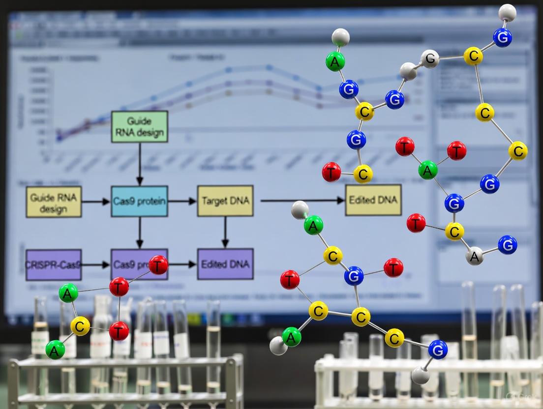
The following diagram illustrates the key decision points and steps involved in the two primary validation pathways: the comprehensive NGS workflow and the rapid T7E1 assay.
The Scientist's Toolkit: Essential Reagents & Solutions
Successful execution of CRISPR validation experiments relies on a foundation of high-quality reagents and tools. The following table details key materials and their functions.
Table 2: Essential Research Reagent Solutions for CRISPR Validation
| Reagent / Tool | Function in Workflow | Key Considerations |
|---|---|---|
| High-Fidelity DNA Polymerase | Amplifies the target genomic locus for all PCR-based methods with minimal errors. | Critical for NGS to prevent false positives from PCR artifacts; ensures accurate representation of indel spectra [2]. |
| NGS Library Prep Kit | Facilitates the attachment of platform-specific adapters and barcodes to PCR amplicons. | Choose kits optimized for amplicon sequencing; efficiency impacts final library complexity and sequencing depth [2]. |
| T7 Endonuclease I | Recognizes and cleaves mismatched DNA in heteroduplexes for the T7E1 assay. | Enzyme activity and buffer conditions can affect cleavage efficiency and background noise [2]. |
| Bioinformatics Software (e.g., CRISPResso2, ICE) | Analyzes sequencing data to quantify editing efficiency, indel distribution, and potential off-target events. | Tool selection dictates the accuracy and depth of analysis; some require command-line expertise while others offer web interfaces [1] [4]. |
| Validated sgRNA | Directs the Cas9 nuclease to the specific genomic target site. | Activity is a primary variable; must be validated itself. Tools like DeepCRISPR use AI to predict high-efficacy guides [4]. |
| Cas9 Nuclease | Executes the double-strand break at the DNA target site. | Options include plasmid DNA, mRNA, or recombinant protein (RNP); delivery method influences on-target efficiency and off-target rates [5] [6]. |
Validation is the critical link between CRISPR experimental design and reliable, interpretable results. While traditional methods like the T7E1 assay offer speed and low cost for initial screens, their limitations in quantitative accuracy and resolution are well-documented [2]. For rigorous research and any clinical application, NGS provides the unparalleled depth and sensitivity required to fully characterize the complex landscape of CRISPR editing outcomes, from precise indel quantification to the detection of large deletions and off-target effects [3] [2]. The evolving toolkit, augmented by AI-powered design and analysis tools like DeepCRISPR and ICE, empowers researchers to implement these robust validation frameworks, thereby ensuring the integrity and safety of their genome engineering efforts [1] [4].
The advent of CRISPR-Cas9 technology has revolutionized genetic engineering, enabling precise modifications across diverse biological systems. However, the full potential of this technology can only be realized with equally advanced validation methodologies. Within the context of CRISPR editing efficiency research, the analytical platform used for validation becomes paramount. Traditional methods, while historically valuable, present significant limitations in comprehensiveness and quantitative precision. Next-Generation Sequencing (NGS) has emerged as a powerful alternative, providing unprecedented depth and accuracy in characterizing editing outcomes. This guide objectively compares the performance of NGS with traditional validation techniques, providing researchers and drug development professionals with the experimental data necessary to select the optimal analytical platform for their CRISPR validation workflows.
Comparative Performance: NGS vs. Traditional Methods
Quantitative Assessment of Editing Efficiency
A critical study directly comparing the T7 Endonuclease 1 (T7E1) assay—a traditional validation method—with targeted NGS for assessing CRISPR-Cas9 editing at 19 genomic loci revealed striking differences in performance [2]. The research demonstrated that the T7E1 assay consistently underestimated editing efficiency, reporting an average indel frequency of just 22% across all tested guide RNAs. In stark contrast, targeted NGS detected a much higher average efficiency of 68%, with nine individual sgRNAs yielding indel frequencies exceeding 70% [2]. This discrepancy is quantified in the table below.
Table 1: Comparison of Editing Efficiency Detection Between T7E1 and Targeted NGS
| sgRNA Group | Average Efficiency by T7E1 | Average Efficiency by NGS | Discrepancy |
|---|---|---|---|
| Human (H1-H9) | 22% | 68% | 46% |
| Mouse (M1-M10) | 22% | 68% | 46% |
| Overall Average | 22% | 68% | 46% |
The limitations of traditional methods extend beyond simple underestimation. The same study found that sgRNAs with apparently similar activity by T7E1 (e.g., M2 and M6, both at ~28%) proved dramatically different by NGS, with actual efficiencies of 92% and 40%, respectively [2]. This low dynamic range and requirement for DNA heteroduplex formation fundamentally limit the reliability of traditional assays for accurately quantifying CRISPR editing efficiency.
Comprehensive Mutation Spectrum Analysis
Beyond simple efficiency quantification, NGS provides a comprehensive view of the entire mutation spectrum, including precise indel characterization, which is largely inaccessible to traditional methods. While techniques like T7E1 and TIDE (Tracking of Indels by Decomposition) can indicate that editing has occurred, they struggle to accurately identify the specific sequences of the resulting alleles, particularly in complex editing scenarios [2].
Targeted NGS enables researchers to simultaneously detect a wide range of editing outcomes, from single-nucleotide changes to large deletions and complex rearrangements. This capability is crucial for thorough characterization of CRISPR experiments, as it reveals the full heterogeneity of editing products within a cell population. Furthermore, NGS can be applied to analyze thousands of samples in parallel through multiplexing, enabling high-throughput screening of CRISPR libraries that is simply not feasible with traditional methods [7] [8].
Table 2: Capability Comparison Between Traditional Methods and NGS
| Analysis Capability | T7E1 Assay | TIDE/IDAA | Targeted NGS |
|---|---|---|---|
| Quantitative Efficiency | Limited (underreports) | Moderate | High accuracy |
| Identifies Specific Indels | No | Partial (size only) | Yes (exact sequence) |
| Detects Complex Rearrangements | No | No | Yes |
| Multiplexing Capacity | Low | Low | High (1000s of samples) |
| Sensitivity for Rare Variants | Low | Low | High (<1%) |
Experimental Applications and Protocols
NGS Workflow for CRISPR Validation
The application of NGS for CRISPR editing validation typically follows a targeted amplicon sequencing approach. This method focuses sequencing power on specific genomic regions of interest, providing deep coverage to detect even rare editing events with high confidence [8]. The workflow can be visualized as follows:
Figure 1: NGS Workflow for CRISPR Validation. This diagram illustrates the key steps in preparing and analyzing CRISPR-edited samples using targeted next-generation sequencing, from initial DNA extraction to final computational analysis.
Detailed Protocol for Targeted Amplicon Sequencing
Step 1: Primer Design - Design gene-specific primers flanking the CRISPR target site, ensuring the amplicon size is appropriate for the sequencing platform (typically <450bp for Illumina). Add partial Illumina adapter sequences to the 5' ends: Forward DS tag: 5'-CTACACGACGCTCTTCCGATCT-3' and Reverse DS tag: 5'-CAGACGTGTGCTCTTCCGATCT-3' [8].
Step 2: PCR Amplification (Step 1) - Perform the first PCR using primers with partial adapters to amplify the target region from genomic DNA. The target modification site should be positioned close to the center of the amplicon with primers binding at least 50bp away from the cut site [8].
Step 3: Indexing PCR (Step 2) - Use the initial PCR product as a template for a second PCR with indexing primers that add unique sample barcodes and complete Illumina adapter sequences. This enables multiplexing of numerous samples in a single sequencing run [8].
Step 4: Sequencing and Analysis - Pool the indexed libraries and sequence on an appropriate NGS platform (e.g., Illumina MiSeq). Analyze the resulting FASTQ files using specialized tools like CRIS.py, which automatically quantifies editing efficiency and characterizes specific indel patterns [8].
Table 3: Essential Research Reagents and Computational Tools for NGS-based CRISPR Validation
| Item | Function | Specification/Example |
|---|---|---|
| High-Fidelity DNA Polymerase | Amplifies target region with minimal errors | Platinum SuperFi II PCR Master Mix |
| NGS Library Prep Kit | Prepares amplicons for sequencing | Illumina-compatible kits |
| Indexing Primers | Adds unique barcodes for sample multiplexing | Illumina i5/i7 index sets |
| NGS Platform | Generates sequence data | Illumina MiSeq, NextSeq |
| CRIS.py Software | Analyzes NGS data for editing outcomes | Python-based, processes FASTQ files |
| Genomic DNA Isolation Reagents | Extracts high-quality DNA from edited cells | Proteinase K-based extraction buffers |
Case Studies in Method Performance
Validation of AI-Designed CRISPR Editors
Recent advances in artificial intelligence have enabled the design of novel CRISPR-Cas proteins with minimal sequence similarity to natural systems. The characterization of these AI-generated editors, such as OpenCRISPR-1, relies heavily on NGS for validation [9]. In one landmark study, researchers used NGS to demonstrate that OpenCRISPR-1, despite being "400 mutations away" from SpCas9 in sequence space, achieved comparable or improved editing efficiency and specificity [9]. This level of precise quantification and specificity assessment would be challenging with traditional methods, highlighting NGS's critical role in validating next-generation genome editing tools.
High-Throughput Functional Genomics
The application of NGS in CRISPR screening has enabled systematic interrogation of gene function at an unprecedented scale. Recent research has focused on optimizing guide RNA design and library size to improve screening efficiency. One study demonstrated that minimal genome-wide CRISPR-Cas9 libraries designed using principled criteria and validated by NGS performed as well or better than larger conventional libraries while reducing costs and increasing feasibility for complex models like organoids and in vivo systems [7]. The quantitative precision of NGS was essential for determining that libraries with fewer guides per gene could maintain sensitivity while dramatically improving scalability.
Discussion and Future Perspectives
The comprehensive comparison presented in this guide clearly demonstrates the superiority of NGS over traditional methods for validating CRISPR editing efficiency. The quantitative precision, comprehensive mutation profiling, and scalability of NGS make it an indispensable tool for researchers requiring accurate characterization of editing outcomes. While traditional methods like T7E1 may still serve as rapid preliminary checks, their technical limitations—particularly in underestimating efficiency and failing to characterize the full spectrum of edits—render them inadequate for rigorous scientific research and therapeutic development.
Looking forward, the integration of NGS with emerging technologies like artificial intelligence and long-read sequencing will further enhance CRISPR validation capabilities. AI-designed editors [9] and advanced screening approaches [7] already rely on NGS for characterization, and this synergy will likely strengthen as the field progresses. For researchers and drug development professionals, investing in NGS-based validation workflows represents not merely a methodological upgrade, but a fundamental requirement for generating robust, reproducible, and clinically relevant data in the genome editing era.
Next-Generation Sequencing (NGS) has become an indispensable technology in the validation of CRISPR-Cas9 genome editing, providing researchers with powerful tools to assess editing efficiency, specificity, and safety. As CRISPR applications advance toward clinical therapies, rigorous evaluation of editing outcomes becomes increasingly critical. NGS offers the precision and depth required to characterize intended edits while identifying unintended consequences that could compromise therapeutic safety. This guide examines the key NGS applications in CRISPR validation—genotyping, off-target detection, and large-scale screening—comparing methodological approaches, their performance characteristics, and appropriate contexts for implementation within drug development and research workflows.
NGS for CRISPR Genotyping: Verifying On-Target Edits
CRISPR genotyping with NGS provides a comprehensive analysis of editing outcomes at the intended target site, offering significant advantages over traditional methods like T7E1 mismatch assays or Sanger sequencing with TIDE/ICE analysis [10] [11]. While these conventional methods provide initial efficiency estimates, NGS delivers precise quantification and full characterization of insertion/deletion (indel) profiles.
Amplicon sequencing represents the most common NGS approach for genotyping, where the genomic region surrounding the target site is PCR-amplified, barcoded, and sequenced at high depth [12]. This method enables researchers to:
- Precisely quantify editing efficiency by calculating the percentage of reads containing indels at the target site
- Characterize the complete spectrum of indel sequences and their relative frequencies
- Detect low-frequency editing events with sensitivity to alleles present at <1% frequency [13]
- Determine zygosity of edits when analyzing clonal populations
- Simultaneously assess homology-directed repair (HDR) efficiency when a donor template is provided
For research requiring the highest resolution of editing outcomes, single-cell DNA sequencing platforms like Tapestri enable genotyping at individual cell resolution [14]. This advanced approach reveals editing co-occurrence, zygosity, and cell clonality patterns that remain obscured in bulk sequencing data.
Table 1: Comparison of CRISPR Genotyping Methods
| Method | Detection Capability | Sensitivity | Throughput | Key Applications |
|---|---|---|---|---|
| T7E1 / Surveyor Assay | Mismatch detection | Low | Medium | Initial efficiency screening |
| Sanger + TIDE/ICE | Indel estimation | Medium | Low | Efficiency and rough indel profile |
| NGS Amplicon Sequencing | Full sequence characterization | High (<1%) | High | Precise efficiency, full indel spectrum, low-frequency edits |
| Single-Cell DNA Sequencing | Per-cell genotypes | High | Medium | Co-editing patterns, zygosity, clonality |
Comprehensive Off-Target Detection Strategies
Off-target effects remain a primary safety concern in CRISPR applications, as the Cas9 nuclease can cleave at genomic sites with sequence similarity to the intended target [15]. Multiple NGS-based approaches have been developed to identify and quantify these unintended editing events, each with distinct strengths and applications.
Computational Prediction Tools
In silico tools provide the most accessible starting point for off-target assessment by nominating potential off-target sites based on sequence homology to the guide RNA [15] [16]. These algorithms scan reference genomes for sites with partial complementarity to the gRNA spacer sequence, typically allowing for a specified number of mismatches or bulges.
Table 2: Major In Silico Off-Target Prediction Tools
| Tool | Algorithm Type | Key Features | Limitations |
|---|---|---|---|
| Cas-OFFinder | Alignment-based | Adjustable sgRNA length, PAM type, mismatch/bulge tolerance | Reference genome-dependent; misses structural variants |
| COSMID | Scoring-based | Stringent mismatch criteria; applies cutoff scores | Conservative; may miss valid off-targets |
| CCTop | Scoring-based | Considers mismatch distances to PAM | Moderate sensitivity and positive predictive value |
| DeepCRISPR | Machine learning | Incorporates sequence and epigenetic features | Requires computational resources |
While computationally efficient, these tools primarily identify sgRNA-dependent off-target sites and may miss edits influenced by cellular context or structural variations [15].
Cell-Free Empirical Methods
Cell-free approaches offer enhanced sensitivity for off-target nomination by detecting Cas9 cleavage events in vitro using purified genomic DNA. These methods typically involve:
- CIRCLE-Seq: Genomic DNA is circularized, incubated with Cas9-gRNA ribonucleoprotein (RNP), then linearized fragments are sequenced [15] [12]
- SITE-Seq: Cas9-cleaved fragments are selectively biotinylated and enriched before sequencing [16] [12]
- Digenome-Seq: Purified genomic DNA is digested with Cas9 RNP then subjected to whole-genome sequencing [15] [12]
These approaches achieve high sensitivity but may overreport off-target sites due to the absence of cellular context like chromatin organization and DNA repair mechanisms [15].
Cell-Based Empirical Methods
Cell-based methods identify off-target sites within their native cellular context, providing more physiologically relevant nomination:
- GUIDE-seq: Double-stranded oligodeoxynucleotides are integrated into double-strand breaks, enabling genome-wide profiling of cleavage sites [16] [12]
- DISCOVER-seq: Utilizes DNA repair protein MRE11 as bait to perform ChIP-seq, identifying active off-target sites [15] [12]
- BLISS: Captures double-strand breaks in situ using dsODNs with T7 promoter sequence [15]
Recent comparative studies in primary human hematopoietic stem and progenitor cells (HSPCs) found that DISCOVER-seq and GUIDE-seq achieved the highest positive predictive value among off-target detection methods [16].
Off-Target Validation and Quantification
After nomination, potential off-target sites require validation through targeted amplicon sequencing. Systems like the rhAmpSeq CRISPR Analysis System enable multiplexed amplification and sequencing of nominated sites across many samples simultaneously [12]. This approach provides precise quantification of editing frequencies at each potential off-target locus.
(Off-Target Assessment Workflow)
Large-Scale Screening Applications
NGS enables unprecedented scale in CRISPR validation, particularly through high-throughput genotyping approaches that streamline the analysis of thousands of edited samples. Automated platforms like genoTYPER-NEXT allow researchers to process up to 10,000 samples per run by combining cell lysis, barcoded PCR, and multiplexed sequencing [13]. This scalability addresses a critical bottleneck in large-scale projects such as:
- Cell line engineering for bioproduction and disease modeling
- Functional genomics screens using CRISPR libraries
- Therapeutic cell product development requiring comprehensive characterization
The integration of single-cell multi-omics approaches further enhances large-scale screening capabilities. The Tapestri platform, for example, simultaneously assesses DNA editing outcomes and surface protein expression through antibody-oligo conjugates, enabling direct correlation of genotype to functional phenotype [14].
Experimental Protocols for Key Applications
Targeted Amplicon Sequencing for On-Target Genotyping
Workflow:
- Design and synthesize target-specific primers flanking the CRISPR target site (amplicon size: 200-300 bp)
- Extract genomic DNA from edited cells (crude lysates may be sufficient for some applications)
- Perform PCR amplification using barcoded primers to enable sample multiplexing
- Purify and pool amplicons at equimolar ratios
- Sequence on an Illumina platform (MiSeq or similar) with sufficient coverage (typically >10,000x)
- Bioinformatic analysis:
- Demultiplex samples by barcode
- Align reads to reference sequence
- Identify and quantify indels using tools like CRISPResso2
Off-Target Assessment Using GUIDE-seq
- Transfert cells with Cas9-gRNA RNP complex along with GUIDE-seq dsODN
- Allow 48-72 hours for dsODN integration into double-strand breaks
- Extract genomic DNA and perform library preparation
- Enrich for dsODN-integrated fragments via PCR
- Sequence using Illumina platforms
- Bioinformatic analysis to identify off-target integration sites
Single-CDNA Sequencing with Tapestri
Workflow [14]:
- Prepare single-cell suspension of edited cells
- Stain with antibody-oligo conjugates (AOCs) if protein co-detection is desired
- Encapsulate cells in droplets with lysis reagents
- Perform multiplex PCR using custom panel targeting on/off-target sites
- Sequence and analyze using automated Tapestri GE pipeline
Comparative Performance Analysis
Recent comparative studies provide valuable insights into the performance of different off-target detection methods. In primary human hematopoietic stem and progenitor cells edited with high-fidelity Cas9, researchers found:
- Off-target activity is exceedingly rare, with an average of less than one off-target site per guide RNA [16]
- Virtually all true off-target sites were identified by available detection methods
- COSMID, DISCOVER-Seq, and GUIDE-seq attained the highest positive predictive value [16]
- Empirical methods did not identify off-target sites that were not also identified by refined bioinformatic methods [16]
Table 3: Method Performance in Primary HSPCs (Cromer et al., 2023)
| Method | Sensitivity | Positive Predictive Value | Key Advantages | Implementation Context |
|---|---|---|---|---|
| In Silico (COSMID) | High | High | Computational efficiency; rapid results | Initial screening; resource-limited settings |
| GUIDE-seq | High | High | Cellular context; genome-wide profiling | Comprehensive assessment; translational research |
| DISCOVER-seq | High | High | In vivo application; native cellular state | Therapeutic development; safety assessment |
| CIRCLE-seq | Very High | Medium | Ultra-sensitive detection | Maximum sensitivity; regulatory submissions |
Essential Research Reagent Solutions
Successful implementation of NGS-based CRISPR validation requires specific reagents and systems designed for these applications:
Table 4: Key Research Reagents and Systems for NGS CRISPR Validation
| Reagent/System | Primary Function | Key Features | Representative Providers |
|---|---|---|---|
| rhAmpSeq CRISPR Analysis System | Targeted amplicon sequencing | Multiplexed on/off-target site amplification; cloud-based analysis | IDT |
| Alt-R CRISPR-Cas9 System | Genome editing | High-specificity Cas9 variants; modified gRNAs with improved specificity | IDT |
| genoTYPER-NEXT | High-throughput genotyping | Automated workflow; thousands of samples per run | GENEWIZ (Azenta) |
| Tapestri Platform | Single-cell DNA sequencing | Single-cell resolution; DNA + protein multi-omics | Mission Bio |
| GeneArt Genomic Cleavage Detection Kit | Initial efficiency screening | Rapid cleavage detection; gel-based analysis | Thermo Fisher Scientific |
NGS technologies provide an essential toolkit for comprehensive CRISPR validation, spanning from basic genotyping to sophisticated off-target detection and large-scale screening applications. The optimal approach depends on the specific research context:
- For basic research validation, targeted amplicon sequencing of on-target sites provides sufficient characterization
- For therapeutic development, a multi-tiered approach combining computational prediction with empirical validation (e.g., GUIDE-seq or DISCOVER-seq followed by targeted sequencing) offers the most rigorous safety assessment
- For complex editing strategies involving multiple targets, single-cell DNA sequencing reveals co-editing patterns and cellular heterogeneity unavailable through bulk methods
As CRISPR applications advance toward clinical implementation, NGS methodologies continue to evolve, with emerging approaches like long-read sequencing and improved computational prediction algorithms further enhancing our ability to characterize editing outcomes with precision and confidence.
Next-Generation Sequencing (NGS) has emerged as the gold standard for validating CRISPR-Cas9 genome editing experiments, offering unparalleled accuracy and sensitivity for characterizing editing outcomes such as insertion and deletion mutations (indels) [1] [2]. However, its adoption in research and drug development is tempered by significant challenges related to cost, bioinformatics, and workflow complexity. For researchers and scientists, a clear understanding of these limitations is crucial for selecting the appropriate validation method and effectively planning projects. This guide objectively compares NGS with alternative CRISPR analysis techniques, providing a detailed examination of their performance, supported by experimental data and a breakdown of essential research reagents.
Comparison of CRISPR Analysis Methods
The selection of a validation method involves balancing cost, time, and the required level of detail. The table below summarizes the key characteristics of the most common techniques.
| Method | Key Principle | Typical Data Output | Relative Cost | Hands-on & Analysis Time | Key Limitations |
|---|---|---|---|---|---|
| Next-Generation Sequencing (NGS) [1] [2] | Deep, targeted sequencing of PCR-amplified edited region | Comprehensive spectrum and precise frequency of all indels | High | High (DNA extraction, library prep, sequencing, bioinformatics) | High cost; requires bioinformatics expertise and infrastructure |
| Inference of CRISPR Edits (ICE) [1] | Computational decomposition of Sanger sequencing traces | Editing efficiency (ICE score), types and distributions of indels | Low | Medium (PCR, Sanger sequencing, web-based analysis) | Inference based on sequence trace decomposition |
| Tracking of Indels by Decomposition (TIDE) [1] [2] | Computational decomposition of Sanger sequencing traces | Estimated editing efficiency and predominant indel types | Low | Medium (PCR, Sanger sequencing, web-based analysis) | Limited ability to detect complex or large indels without manual parameter adjustment |
| T7 Endonuclease I (T7E1) Assay [1] [2] | Enzyme cleavage of heteroduplex DNA formed by mismatched amplicons | Gel-based estimation of total editing efficiency | Very Low | Low (PCR, digestion, gel electrophoresis) | Not quantitative; lacks sequence-level data; unreliable at high (>30%) or low (<10%) efficiency [2] |
| Genomic Cleavage Detection (GCD) [10] | Similar to T7E1; gel-based detection of cleaved PCR products | Gel-based estimation of total editing efficiency | Very Low | Low (PCR, digestion, gel electrophoresis) | Not quantitative; lacks sequence-level data |
Quantitative data from a comparative study highlights the accuracy gap between methods. When compared to NGS, the T7E1 assay consistently underestimated editing efficiency, particularly for highly active sgRNAs. For example, in edited mammalian cell pools, two sgRNAs (M2 and M6) showed similar activity (~28%) by T7E1, but NGS revealed dramatically different true efficiencies of 92% and 40%, respectively [2]. Another study demonstrated that the ICE analysis tool provided results highly comparable to NGS (R² = 0.96), offering a cost-effective alternative for achieving sequence-level detail [1].
Experimental Protocols for Key Methods
Targeted Next-Generation Sequencing (NGS) for CRISPR Validation
Methodology:
- Step 1: DNA Extraction and PCR Amplification. Genomic DNA is extracted from edited cells (e.g., 3-4 days post-transfection). The target locus is amplified using high-fidelity PCR primers designed to flank the CRISPR cut site [2].
- Step 2: Library Preparation. The PCR amplicons are processed into an NGS library. This typically involves fragmentation, size selection, and the ligation of platform-specific adapters and sample barcodes (indices) to enable multiplexed sequencing [2].
- Step 3: Sequencing. The pooled library is sequenced on a platform such as the Illumina MiSeq, using a 2x250 bp paired-end run to ensure sufficient coverage and read length to span the edited region [2].
- Step 4: Bioinformatic Analysis. The resulting sequencing reads are demultiplexed and analyzed using a specialized pipeline. Key steps include:
- Alignment: Reads are aligned to the reference genome sequence using tools like BWA.
- Variant Calling: Specialized algorithms (e.g., CRISPResso2) are used to identify insertions and deletions around the expected cut site.
- Quantification: The frequency of each unique indel and the total editing efficiency are calculated [2].
T7 Endonuclease I (T7E1) Mismatch Cleavage Assay
Methodology:
- Step 1: PCR Amplification. The target locus is PCR-amplified from genomic DNA of edited and control cells [2].
- Step 2: Heteroduplex Formation. The PCR products are denatured at 95°C and then slowly re-annealed by cooling. This allows strands from differently sized indels to hybridize, forming heteroduplexes with bulges at the mismatch sites [2].
- Step 3: T7E1 Digestion. The re-annealed DNA is incubated with the T7 Endonuclease I enzyme, which recognizes and cleaves the heteroduplex structures at the mismatch sites [2].
- Step 4: Visualization and Analysis. The digestion products are separated by agarose gel electrophoresis. The gel is visualized, and the intensity of the cleaved and uncleaved bands is analyzed by densitometry. The editing efficiency is estimated using formulas that compare the band intensities, though this is considered semi-quantitative at best [2].
Workflow and Logical Relationship Diagrams
CRISPR Validation Method Selection
NGS Workflow Complexity
The Scientist's Toolkit: Research Reagent Solutions
Successful execution of CRISPR validation experiments requires specific reagents and tools. The following table details key materials and their functions.
| Reagent / Tool | Function in Experiment | Key Considerations |
|---|---|---|
| High-Fidelity DNA Polymerase [2] | Amplifies the target genomic locus for sequencing or assay with minimal errors. | Critical for reducing PCR-introduced artifacts that can confound NGS or Sanger results. |
| NGS Library Prep Kit [2] | Prepares the PCR amplicons for sequencing by adding platform-specific adapters and indices. | Choice affects library complexity, preparation time, and compatibility with multiplexing. |
| T7 Endonuclease I [2] | Cleaves heteroduplex DNA formed by re-annealing of wild-type and indel-containing amplicons. | Sensitivity is affected by mismatch type and location; not all indels are cleaved efficiently. |
| Sanger Sequencing Service/Kit | Generates sequence traces for input into ICE or TIDE decomposition algorithms. | Purity and concentration of the PCR amplicon are crucial for high-quality sequence data. |
| ICE (Synthego) & TIDE Web Tools [1] | Computational platforms that analyze Sanger sequencing traces to infer indel types and frequencies. | ICE is reported to detect a broader range of outcomes (e.g., large indels) than TIDE [1]. |
| Validated Control gRNA [10] | A gRNA with known high efficiency (e.g., targeting human AAVS1 or HPRT locus) serves as a positive control. | Essential for confirming that the entire workflow from transfection to analysis is functioning correctly. |
NGS remains the most comprehensive and accurate method for validating CRISPR genome editing, providing the depth of information necessary for critical applications in therapeutic development [3] [2]. However, its significant limitations in cost, workflow complexity, and bioinformatics dependency are real barriers. For many research applications, Sanger sequencing-based computational methods like ICE offer a compelling compromise, delivering NGS-comparable accuracy for efficiency and basic indel characterization at a fraction of the cost and time [1]. Conversely, while inexpensive and fast, enzyme-based assays like T7E1 are unsuitable for any application requiring quantitative precision or sequence-level detail [2]. The optimal validation strategy depends on a clear-eyed assessment of the project's requirements, resources, and goals, often leading to a tiered approach where rapid initial screening is followed by confirmatory, deep analysis with NGS for critical samples.
Building Your NGS Validation Workflow: From Sample to Sequence
In the pipeline for Next-Generation Sequencing (NGS) validation of CRISPR-Cas9 genome editing, the initial and critical wet-lab step is the PCR amplification of the target genomic locus and the subsequent preparation of sequencing-ready libraries [8] [17]. This step is fundamental for transforming the minute amounts of genomic DNA (gDNA) extracted from edited cells into a format compatible with high-throughput sequencers. The accuracy and efficiency of this process directly determine the reliability of all downstream analyses, including the quantification of editing efficiency (on-target analysis) and the investigation of unintended, off-target effects [18] [2].
The primary goal is to selectively amplify the genomic region of interest from a background of billions of base pairs, creating a pool of DNA fragments (amplicons) that can be sequenced in parallel. For CRISPR validation, this involves special considerations, such as ensuring the amplicon flankes the Cas9 cut site and is designed to detect a wide variety of insertion/deletion mutations (indels) [8].
Core PCR and Library Preparation Strategies
Two primary methodological frameworks exist for preparing samples for targeted NGS: the single-step, amplicon-based approach and the more flexible two-step PCR strategy. The choice between them depends on the scale of the project, the available resources, and the required throughput.
Amplicon-Based Sequencing (One-Step Strategy)
This strategy is often the go-to method for lower-throughput projects or when using centralized sequencing core facilities that handle library preparation. In this approach, a single PCR reaction is performed using gene-specific primers that already contain full Illumina adapter sequences [2]. This means each sample receives a unique pair of primers, and the resulting PCR product is fully ready for sequencing after a simple clean-up step and normalization. While straightforward, this method can become costly and labor-intensive when scaling to hundreds of samples, as each requires a dedicated, custom primer pair.
Two-Step PCR Strategy
The two-step PCR strategy is widely adopted for high-throughput genotyping of CRISPR-edited cells, especially when screening hundreds of single-cell clones [8] [13]. This method decouples the target amplification from the indexing step, offering significant advantages in flexibility and cost-efficiency.
- Step 1 - Target Amplification: The first PCR uses gene-specific primers that have short, universal overhangs (partial Illumina adapter sequences). These primers amplify the target locus from the gDNA template. All samples for a given target locus can use the same pair of Step 1 primers, making reagent design and inventory simpler [8].
- Step 2 - Indexing and Adapter Addition: The products from Step 1 are used as templates in a second, much shorter PCR. This reaction uses universal primers that bind to the overhangs added in Step 1. These indexing primers contain the full Illumina adapter sequences, unique dual indices (UDIs), and sequencing primers [8]. The UIs allow multiple samples to be pooled together in a single sequencing run and computationally demultiplexed after sequencing.
The workflow and key components of the two-step PCR strategy are detailed in the diagram below.
Strategy Comparison and Selection
The table below summarizes the key characteristics of the two main library preparation strategies to guide researchers in selecting the most appropriate method for their project.
Table 1: Comparison of NGS Library Preparation Strategies for CRISPR Validation
| Feature | Amplicon-Based (One-Step) | Two-Step PCR |
|---|---|---|
| Workflow Complexity | Lower; single PCR reaction [2] | Higher; requires two sequential PCR reactions [8] |
| Primer Design & Cost | Custom primers for each sample; higher cost at scale [2] | Universal indexing primers; lower cost per sample for high-throughput projects [8] |
| Throughput & Scalability | Ideal for low to medium throughput (e.g., dozens of samples) | Ideal for high-throughput screening (e.g., hundreds to thousands of samples) [8] [13] |
| Experimental Flexibility | Lower; primer sets are sample-specific | Higher; same indexing primers can be used for different projects and target loci [8] |
| Primary Application Context | Initial sgRNA validation, small-scale pool analysis [2] | Large-scale single-cell clone screening, multiplexed target analysis [8] [13] |
Practical Implementation and Experimental Protocols
Primer Design Guidelines
The design of the initial gene-specific primers is a critical determinant of success. Adhering to the following guidelines ensures optimal results [8]:
- Amplicon Length: The total amplicon length must be less than the combined length of the paired-end sequencing reads. For 2x250 bp sequencing, amplicons should be under 450 bp to ensure full coverage of both ends.
- Cut Site Placement: The Cas9 cut site should be positioned close to the center of the amplicon. Primers should bind at least 50 bp away from the cut site to allow for the detection of larger indels.
- Specificity: Tools like Primer-Blast should be used to check for and minimize potential off-target amplification [8].
- Adapter Sequences: For the two-step method, the gene-specific primers must have the appropriate partial Illumina adapter sequences added to their 5' ends [8]:
- Forward Primer Partial Adapter:
5′-CTACACGACGCTCTTCCGATCT-3′ - Reverse Primer Partial Adapter:
5′-CAGACGTGTGCTCTTCCGATCT-3′
- Forward Primer Partial Adapter:
Step-by-Step Two-Step PCR Protocol
The following protocol, adapted from high-throughput genotyping workflows, outlines the detailed steps for preparing an NGS library from gDNA of CRISPR-edited cells [8].
Table 2: Key Reagent Solutions for Two-Step PCR Library Preparation
| Reagent / Tool | Function / Description | Example Product / Note |
|---|---|---|
| gDNA Extraction Buffer | Lyses cells and digests protein, releasing gDNA for PCR [8] | Crude extract buffer: Tris, EDTA, Triton X-100, Proteinase K [8] |
| High-Fidelity DNA Polymerase | Amplifies target locus with high accuracy and yield, minimizing PCR errors [8] | Platinum SuperFi II PCR Master Mix [8] |
| Indexing Primers with UDIs | Adds full Illumina adapters and unique barcodes to amplicons in PCR Step 2 for sample multiplexing [8] | Commercially available sets or custom-designed primers [8] |
| NGS Analysis Pipeline | Bioinformatic tool for analyzing sequencing data to quantify editing outcomes [8] | CRIS.py, CRISPResso, others [8] [19] |
PCR Step 1: Target Amplification
- Reaction Setup: Set up PCR reactions using gDNA template (from crude lysates or purified) and gene-specific primers with partial adapter overhangs.
- Cycling Conditions: Use a high-fidelity polymerase and cycling conditions suitable for the polymerase and amplicon length. Typically, this involves an initial denaturation (98°C for 30 sec), followed by 25-35 cycles of denaturation (98°C for 10 sec), annealing (55-65°C for 15 sec), and extension (72°C for 15-30 sec/kb), with a final extension (72°C for 5 min).
- Product Clean-up: Purify the PCR products using magnetic beads or columns to remove primers, enzymes, and salts.
PCR Step 2: Indexing and Adapter Addition
- Reaction Setup: Use the purified Step 1 product as the template. Set up reactions with universal forward and reverse indexing primers. Each sample in the experiment must receive a unique combination of i7 and i5 indices to allow for multiplexing.
- Cycling Conditions: This is typically a shorter PCR (e.g., 8-12 cycles) using the same thermal profile as Step 1, but with a shorter extension time as the amplicon is already the correct size.
- Product Clean-up: Purify the final PCR products.
Library Pooling, Quantification, and Sequencing
- Pooling: Combine equal molar amounts of each uniquely indexed library into a single tube.
- Quantification: Precisely quantify the pooled library using methods like qPCR or the Bioanalyzer system to ensure optimal loading on the sequencer.
- Sequencing: Sequence the pool on an Illumina MiSeq, HiSeq, or similar platform, using a paired-end 250 bp or 300 bp run to ensure sufficient read length to cover the entire amplicon.
The entire workflow, from gDNA to a sequenced library, is visualized below.
Performance Data and Method Comparison
The choice of validation method, which is directly enabled by the PCR and library preparation strategy, has a profound impact on the accuracy of the results. A seminal study compared the popular T7 Endonuclease I (T7E1) assay against targeted NGS for quantifying CRISPR editing efficiency and revealed significant discrepancies [2].
Table 3: Quantitative Comparison of Editing Efficiencies Measured by T7E1 vs. NGS
| sgRNA Example | Editing Efficiency (T7E1) | Editing Efficiency (NGS) | Discrepancy & Implication |
|---|---|---|---|
| M2 | ~28% [2] | 92% [2] | Severe underestimation by T7E1; NGS reveals near-saturating editing. |
| M6 | ~28% [2] | 40% [2] | Same T7E1 value as M2, but true efficiency is 2.3-fold lower, a critical difference masked by T7E1. |
| H3 | Appears inactive [2] | <10% [2] | T7E1 lacks sensitivity for low-activity guides, potentially leading to false negatives. |
| M1 / M5 | Appears modestly active [2] | >90% [2] | T7E1 has a low dynamic range and cannot accurately report high editing efficiencies. |
This experimental data underscores that while enzymatic assays like T7E1 are cost-effective, they are not quantitative. The NGS-based approach, for which proper PCR amplification is the cornerstone, provides a true and quantitative measure of editing outcomes, which is essential for rigorous validation [2].
The initial step of PCR amplification and library preparation is a foundational element in the NGS validation workflow for CRISPR genome editing. The strategic choice between a one-step amplicon approach and a two-step PCR strategy dictates the scale, cost, and efficiency of a project. Adherence to rigorous primer design rules and protocols ensures the generation of high-quality sequencing data. As the experimental data demonstrates, moving away from traditional, less quantitative methods to an NGS-based approach is critical for obtaining an accurate and comprehensive view of CRISPR editing outcomes, enabling confident decision-making in both basic research and therapeutic development.
In the meticulous process of validating CRISPR genome editing efficiency, the choice of next-generation sequencing (NGS) library preparation method is a pivotal decision that directly impacts data quality and experimental conclusions. This step determines how the edited DNA fragments are processed, amplified, and prepared for sequencing, influencing the accuracy with which on-target edits and unwanted off-target effects are captured. The core decision facing researchers lies in selecting between PCR-based and PCR-free library preparation methods. PCR-based methods, which employ polymerase chain reaction to amplify the genetic material before sequencing, are renowned for their high sensitivity and low input requirements. In contrast, PCR-free methods, which omit this amplification step, are celebrated for providing superior sequencing uniformity and reduced bias. This guide provides a detailed, data-driven comparison of these two approaches, equipping researchers and drug development professionals with the information needed to select the optimal protocol for their specific CRISPR validation workflow.
Core Principles and Comparative Workflows
The fundamental difference between the two methods lies in the inclusion or omission of a PCR amplification step after the initial DNA fragmentation and adapter ligation.
- PCR-based Workflow (Red): Incorporates a PCR amplification step after adapter ligation. This enables work with very low input DNA amounts (as little as 1 ng or less) and generates high yields from limited material [20]. However, the amplification process can introduce duplicates and sequence-specific biases.
- PCR-free Workflow (Blue): Omits the PCR amplification step. The adapter-ligated DNA is directly cleaned and prepared for sequencing. This avoids amplification-induced biases but requires significantly more input DNA (often 300-1000 ng) to generate sufficient library yield for sequencing [21] [20].
Performance Comparison: A Data-Driven Analysis
The choice between PCR-based and PCR-free methods has measurable consequences on data quality. The following table summarizes key performance characteristics, with supporting data from independent evaluations of commercial kits.
Table 1: Performance Characteristics at a Glance
| Feature | PCR-based | PCR-free |
|---|---|---|
| GC Bias | Higher, with under-coverage in GC-rich regions [21] [20] | More uniform coverage, including GC-rich promoters [21] [20] |
| Variant Calling (F1-Score) | Generally high (e.g., SNP: ~0.978, Indel: ~0.973) [21] | Excellent (e.g., SNP: ~0.984, Indel: ~0.982) [21] |
| Duplicate Reads | Higher, due to amplification of identical fragments [22] | Significantly reduced, preserving library complexity [22] |
| Input DNA Requirement | Low (1 ng - 500 ng) [20] | High (300 ng - 1 µg) [21] [20] |
| Assay Time | Faster (e.g., ~2-4 hours for some kits) [20] | Can be faster (e.g., ~1.5 hours for some kits) [20] |
Independent studies comparing multiple commercial kits provide quantitative evidence for these performance differences. Research evaluating eight commercially available PCR-free library prep solutions demonstrated that they consistently deliver high-quality, uniform WGS results with minimal GC bias [21]. The same study highlighted that PCR-free libraries achieve robust variant calling, with F1-scores for SNPs and indels often exceeding those of PCR-based methods [21]. Furthermore, a streamlined "Trinity" hybrid capture workflow that eliminates post-hybridization PCR reported a reduction in false positive indels by 89% and false negatives by 67%, underscoring the substantial improvement in accuracy achievable with PCR-free or reduced-PCR workflows [22].
Detailed Experimental Protocols
Protocol 1: Standard PCR-based Library Preparation
This protocol is widely used for its robustness and compatibility with low-input samples, a common scenario when cultivating edited clones is challenging.
- DNA Fragmentation and Size Selection: Fragment genomic DNA (100 pg - 1 µg) via enzymatic or mechanical shearing. Perform solid-phase reversible immobilization (SPRI) bead cleanup to select for a target insert size (e.g., ~350 bp) [22] [20].
- End Repair and A-tailing: Use a master mix to repair fragment ends and add an 'A' base to the 3' ends, preparing them for adapter ligation. This is a standard step in kits like the KAPA HyperPrep [21].
- Adapter Ligation: Ligate indexed adapters containing sequencing primer sites and sample barcodes to the fragments. This allows for multiplexing of samples in a single sequencing run [8].
- PCR Amplification: Amplify the adapter-ligated library for 5-10 cycles using a high-fidelity PCR master mix to generate sufficient material for sequencing [22]. The number of cycles should be minimized to reduce bias.
- Final Library Cleanup and QC: Purify the amplified library with SPRI beads and quantify using fluorometry (e.g., Qubit) and qPCR. Validate library size distribution using an instrument such as the TapeStation [22] [21].
Protocol 2: PCR-free Library Preparation
This protocol is the gold standard for variant calling applications, as it avoids the introduction of amplification artifacts, making it ideal for comprehensive CRISPR off-target assessment [21] [17].
- High-Input DNA Fragmentation: Fragment a larger quantity of high-quality genomic DNA (300 - 1000 ng). Mechanical shearing (e.g., with a Covaris sonicator) is often preferred for its reproducibility [22] [21].
- End Repair and A-tailing: Similar to the PCR-based protocol, this step prepares the blunt-ended, fragmented DNA for ligation.
- Adapter Ligation: Ligate specialized adapters in a reaction that may be scaled up to account for the higher DNA input. This step is performed without subsequent PCR amplification [21].
- Size Selection and Cleanup: Perform a double-sided SPRI bead cleanup to stringently select for the desired insert size range (e.g., 300-350 bp). This is critical for removing adapter dimers and optimizing library efficiency [22] [21].
- Final Library QC: Quantify the final library precisely using qPCR, as the absence of PCR amplification results in lower yields. Verify the library profile with a fragment analyzer [22] [21].
The Scientist's Toolkit: Essential Reagents and Kits
Table 2: Key Research Reagent Solutions
| Item | Function in CRISPR NGS Validation | Example Products |
|---|---|---|
| High-Fidelity DNA Polymerase | Accurately amplifies target loci for PCR-based library prep or amplicon sequencing. Reduces PCR errors. | Platinum SuperFi II Master Mix [8], MyTaq Red Mix [8] |
| SPRI Beads | Purifies and size-selects DNA fragments after enzymatic reactions (e.g., ligation, PCR). Critical for optimizing library insert size. | Various suppliers (e.g., Beckman Coulter) |
| Indexed Adapters | Dual-indexed oligonucleotides ligated to DNA fragments, enabling multiplexing of samples. Unique barcodes differentiate samples post-sequencing. | IDT xGen Stubby Adapter-UDIs [22], Illumina TruSeq DNA UD Indexes |
| PCR-free Library Prep Kit | All-in-one reagent set for constructing unbiased NGS libraries without amplification. Ideal for WGS and off-target discovery. | Illumina DNA PCR-Free Prep [20], Watchmaker DNA Library Prep Kit with Fragmentation [21] |
| Hybrid Capture Panel | Biotinylated oligonucleotide baits that enrich specific genomic regions (e.g., a set of potential off-target sites) from a complex library before sequencing. | IDT xGen Exome Panel [22], Twist Core Exome Panel |
| NGS Analysis Software | Bioinformatics tool specifically designed to analyze CRISPR editing outcomes from NGS data, quantifying indel frequencies and types. | CRIS.py [8], Synthego ICE [1] |
Strategic Guidance for Your CRISPR Workflow
The optimal choice between PCR-based and PCR-free methods depends on the specific goals and constraints of your CRISPR validation experiment. The following decision pathway can help guide your selection:
For PCR-free protocols, prioritize: Comprehensive off-target analysis [17], whole-genome sequencing to discover unpredicted edits [17], and high-confidence characterization of complex edits like indels [22] [21]. This is crucial for preclinical therapeutic development where accuracy is paramount.
For PCR-based protocols, prioritize: High-throughput screening of on-target efficiency across many samples [1], situations with limited DNA input (e.g., single-cell CRISPR assays or precious edited clones) [20], and targeted sequencing where a specific amplicon is being tracked.
Newer streamlined workflows, such as the Trinity hybrid capture approach, demonstrate that innovations in library preparation can successfully eliminate post-hybridization PCR and washing steps, reducing turnaround time by over 50% while simultaneously improving data quality [22]. Staying informed of these technological advancements is key to optimizing your CRISPR validation pipeline.
Selecting the appropriate sequencing platform and determining the required depth are critical steps in designing robust NGS experiments for validating CRISPR-Cas9 editing efficiency. The choice impacts the resolution of editing outcomes, capability to detect rare events, and overall experimental cost. This guide objectively compares current sequencing methodologies, supported by experimental data, to inform researchers and drug development professionals.
Comparison of Sequencing Platforms for CRISPR Validation
The following table summarizes the key characteristics of mainstream and emerging sequencing platforms used for CRISPR validation.
Table 1: Platform Comparison for CRISPR Editing Analysis
| Platform / Method | Key Strength | Throughput & Scalability | Reported Editing Outcome Concordance | Primary Applications in CRISPR Validation |
|---|---|---|---|---|
| Sanger Sequencing + TIDE/ICE | Cost-effective for single-gene/sgRNA analysis [23] | Low throughput; suitable for small-scale validation [23] | High concordance with NGS for common indels [23] | Initial gRNA screening, routine knockout validation [23] |
| Short-Read NGS (Illumina) | High accuracy for small indel quantification [2] | High throughput; highly scalable for multiple targets [2] | Considered the benchmark for indel frequency measurement [2] | High-throughput sgRNA validation, precise indel frequency and spectrum analysis [2] |
| Long-Read Sequencing (Oxford Nanopore) | Resolves complex edits and phasing [23] [24] | Moderate to high throughput; flexible (flow cell choice) [23] | ICE: >99% (vs. Sanger/TIDE) [23] | Characterization of large deletions, structural variations, and haplotype phasing [23] [3] |
| Single-Cell DNA Sequencing (Tapestri) | Reveals clonality and zygosity in edited cell populations [3] | Targeted approach for hundreds of loci across thousands of cells [3] | Provides unique resolution not available with bulk methods [3] | Preclinical safety assessment, off-target profiling, and clonal heterogeneity in therapeutic cell products [3] |
| CRISPR-Cas9 Targeted Enrichment (Context-Seq) | Enables sequencing of low-abundance targets and their genomic context [24] [25] | High multiplexing capability; cost-effective for targeted regions [24] | 7-15x enrichment over untargeted methods [24] | Investigating antimicrobial resistance gene transmission, complex genomic regions, and rare editing events [24] [25] |
Experimental Protocols for Key Methodologies
Oxford Nanopore Sequencing for Indel Analysis
This protocol, as described by McFarlane et al., streamlines routine gRNA validation [23].
- Step 1: DNA Extraction and PCR Amplification. High molecular weight genomic DNA is extracted from edited cells. The target region spanning the gRNA cut site is amplified via PCR, generating amplicons of >600 bp.
- Step 2: Library Preparation. PCR products are prepared using a Native Barcoding Kit, which allows for multiplexing samples. Barcoded libraries are pooled and loaded onto MinION flow cells for sequencing on a GridION device.
- Step 3: Data Analysis with nCRISPResso2. The basecalled sequencing data is analyzed using a nanopore-compatible version of CRISPResso2 (nCRISPResso2). This bioinformatic tool aligns reads to a reference sequence to quantify the percentage of reads with insertions, deletions, or other modifications at the target site, providing indel frequency and spectrum [23].
CRISPR-Cas9 Targeted Enrichment (Context-Seq)
This protocol enriches for specific genomic regions, such as antibiotic resistance genes (ARGs), allowing for deep sequencing of their genomic context from complex samples [24].
- Step 1: Guide RNA Design. Multiple gRNAs are designed to flank the target ARG (e.g., blaCTX-M and blaTEM). Guides are selected using software like CHOPCHOP, with optimization for on-target efficiency and minimal off-target activity in complex communities.
- Step 2: Cas9 Cleavage and Adapter Ligation. Extracted, high molecular weight DNA is dephosphorylated. The Cas9 enzyme, complexed with the designed gRNAs, is used to create double-strand breaks at the target sites. Sequencing adapters are then selectively ligated to the Cas9-cut ends.
- Step 3: Proteinase K Digestion and Sequencing. A digestion step with thermolabile Proteinase K is performed to improve assay performance by removing Cas9 protein. The prepared library is sequenced on a long-read platform (MinION).
- Step 4: Analysis of Genomic Context. Sequencing reads, which are several kilobases long, are clustered and polished to generate consensus sequences. These sequences are annotated to identify the ARG, its flanking mobile genetic elements (e.g., transposases), and the broader genomic context to understand transmission dynamics [24].
Workflow Diagram: NGS Validation for CRISPR Editing
The following diagram illustrates the logical workflow and decision process for selecting the appropriate NGS validation strategy based on experimental goals.
The Scientist's Toolkit: Essential Reagents and Materials
Table 2: Key Research Reagent Solutions for NGS Validation of CRISPR Editing
| Item | Function in Workflow | Specific Example / Note |
|---|---|---|
| Cas9 Nuclease | Creates double-strand breaks at DNA target sites for validation or enrichment assays [24]. | Used in CRISPR-Cas9 targeted enrichment (Context-Seq) to cleave genomic DNA at specific loci [24]. |
| Guide RNA (gRNA) | Directs Cas9 to a specific genomic locus via complementary base pairing [24] [9]. | Designed using tools like CHOPCHOP; multiple gRNAs can be pooled for multiplexed target enrichment [24]. |
| Long-Range PCR Kit | Amplifies the target genomic region for sequencing, especially critical for long-read platforms [23]. | Generates amplicons >600 bp to encompass the gRNA cut site and sufficient flanking sequence for confident analysis [23]. |
| Native Barcoding Kit | Allows for multiplexing of samples by tagging each with a unique nucleotide sequence before pooling [23]. | Enables sequencing of multiple samples or targets on a single Oxford Nanopore flow cell, reducing cost per sample [23]. |
| Analysis Software (nCRISPResso2) | A bioinformatics tool specifically designed to analyze sequencing data and quantify CRISPR-induced indel frequencies [23]. | A nanopore-compatible version (nCRISPResso2) provides results highly concordant with ICE and TIDE analysis [23]. |
Within the framework of Next-Generation Sequencing (NGS) validation of CRISPR editing efficiency, bioinformatic analysis for indel quantification and variant calling is the critical step that transforms raw sequencing data into interpretable, actionable results. Following the confirmation of successful CRISPR component delivery and initial editing checks, this analytical phase precisely measures the spectrum and frequency of insertion and deletion mutations (indels) introduced by the non-homologous end joining (NHEJ) repair pathway [1]. In both basic research and therapeutic drug development, rigorous quantification of these on-target edits, coupled with comprehensive off-target profiling, is indispensable for assessing the efficacy and safety of a CRISPR intervention [26] [27]. This guide objectively compares the leading bioinformatics tools and pipelines available for this task, detailing their methodologies, performance characteristics, and suitability for different experimental scales.
Comparison of Bioinformatics Tools and Pipelines
The selection of a bioinformatics tool depends on the sequencing method, the scale of the experiment, and the required depth of information. The table below summarizes the primary tools and their optimal use cases.
Table 1: Comparison of Bioinformatics Tools for CRISPR Analysis
| Tool Name | Primary Data Input | Key Functionality | Throughput & Scalability | Key Performance Metrics/Output |
|---|---|---|---|---|
| Targeted NGS (Gold Standard) [1] | NGS Reads (FASTQ) | Detects all variant types (indels, SNVs); identifies precise sequences and their relative abundances. | High-throughput; suitable for large sample numbers. | Editing efficiency, full indel spectrum, precise allele frequencies. |
| ICE (Inference of CRISPR Edits) [1] [28] | Sanger Sequencing (.ab1) | Determines indel percentage and profiles from Sanger data. | Scalable for hundreds of samples via batch upload. | ICE Score (indel %), KO Score (frameshift frequency), R² value for model fit. |
| TIDE (Tracking of Indels by Decomposition) [1] [29] | Sanger Sequencing (.ab1) | Decomposes Sanger traces to estimate indel frequencies. | Best for small-scale experiments. | Indel frequency, goodness of fit (R²). Limited to +1 bp insertions. |
| TIDER (Tracking of Ins, Dels, and Recombination events) [29] | Sanger Sequencing (.ab1) | Quantifies HDR and specific nucleotide substitutions in addition to NHEJ indels. | Best for small-scale experiments. | HDR efficiency, specific mutation frequency, background indel frequency. |
| CRISPR-detector [30] | NGS Reads (FASTQ/BAM) | Co-analysis of treated & control samples to filter background; detects on/off-target edits and structural variations. | Optimized for WGS data analysis; highly scalable. | Annotated list of high-confidence editing-induced mutations, including SVs. |
Experimental Protocols for Key Platforms
Protocol: Whole-Genome Sequencing (WGS) Analysis with CRISPR-detector
For the most comprehensive validation, including genome-wide off-target detection, WGS followed by analysis with a pipeline like CRISPR-detector is recommended [26] [30].
- Sample Preparation: Genomic DNA is extracted from both the CRISPR-edited population and a matched control (un-edited) sample. The quality and integrity of the DNA are confirmed.
- Library Preparation & Sequencing: Whole-genome sequencing libraries are prepared from both samples. These are then sequenced on an NGS platform to a sufficient depth (e.g., >30x coverage) to confidently call variants.
- Bioinformatic Analysis with CRISPR-detector:
- Alignment: Raw sequencing reads (FASTQ) are aligned to the reference genome.
- Variant Calling: The core of CRISPR-detector uses the Sentieon TNscope pipeline for haplotype-based variant calling, which improves accuracy by handling sequencing errors effectively [30].
- Background Variant Removal: A co-analysis of the treated and control samples is performed. Variants present in both are identified as pre-existing background polymorphisms and filtered out [30].
- Annotation & Visualization: The final, high-confidence list of editing-induced mutations is annotated for functional impact (e.g., using a Variant Effect Predictor) and clinical relevance, and can be visualized within the tool [30].
Protocol: Targeted NGS for High-Throughput On-Target Analysis
When the focus is on deep sequencing of specific on-target loci, a targeted amplicon sequencing approach is more cost-effective [1].
- PCR Amplification: The genomic region surrounding each CRISPR target site is PCR-amplified from edited and control samples.
- Library Preparation & Sequencing: Amplicons are converted into an NGS library, often with the addition of sample barcodes to multiplex many samples in a single run. This is sequenced on a benchtop sequencer.
- Bioinformatic Analysis (Typical Workflow):
- Demultiplexing: Sequences are separated by sample based on their barcodes.
- Alignment & Variant Calling: Reads are aligned to the reference sequence for the amplicon. Tools like CRISPResso2 or custom scripts are used to quantify the percentage of reads containing indels precisely at the cut site.
- Quantification: Editing efficiency is calculated as the percentage of total reads that contain a non-wild-type sequence at the target locus. The output is the full spectrum of indel sequences and their individual frequencies [1].
Protocol: Rapid Analysis with Sanger Sequencing and ICE/TIDE
For a fast and cost-effective assessment of editing efficiency, Sanger sequencing of PCR amplicons followed by computational decomposition is a widely used method [1] [28].
- PCR and Sanger Sequencing: The target locus is PCR-amplified from genomic DNA of edited and control cells. The purified PCR products are submitted for Sanger sequencing.
- Data Analysis with ICE:
- Upload: The Sanger sequencing chromatogram files (.ab1) for both control and edited samples are uploaded to the ICE web tool.
- Input Parameters: The user provides the gRNA target sequence and selects the nuclease used (e.g., SpCas9).
- Analysis: The ICE algorithm aligns the edited sequence trace to the control trace and uses linear regression to deconvolute the mixed signals, inferring the identities and proportions of different indels [28].
- Output: The tool provides an ICE Score (equivalent to indel percentage), a Knockout Score (proportion of frameshift or large indels), and a detailed list of all detected indels and their relative abundances [1] [28].
Diagram: Bioinformatic Analysis Workflow for CRISPR Validation. This flowchart outlines the key decision points and processes for analyzing CRISPR editing outcomes, from sample preparation to data interpretation.
Critical Considerations for Analytical Validation
Addressing Off-Target Effects
A complete validation must account for unintended, off-target edits. WGS is the most thorough method for unbiased genome-wide off-target discovery [26]. As demonstrated in the validation of NF-κB reporter mice, WGS data can be aligned against predicted off-target sites from tools like Cas-OFFinder to confirm the absence of modifications in critical related genes [26]. For targeted approaches, guides should be designed using tools like CHOPCHOP or the Broad Institute's GPP sgRNA Designer to minimize off-target potential, and predicted off-target sites should be included in the sequencing panel [31].
The Importance of Controls and Background Mutation Filtering
The use of appropriate controls is non-negotiable for accurate variant calling. The best practice is to sequence a paired, unedited control sample (e.g., from the same cell line or organism) simultaneously with the edited sample. Tools like CRISPR-detector are specifically designed to perform a co-analysis, subtracting background variants present in the control from those in the treated sample. This step is crucial for eliminating false positives arising from pre-existing genetic variations or sequencing artifacts, ensuring that the reported variants are a direct consequence of the CRISPR editing process [30].
The Scientist's Toolkit: Essential Research Reagents and Materials
Table 2: Key Reagents and Materials for CRISPR Analysis Workflows
| Item | Function in Analysis |
|---|---|
| High-Quality Genomic DNA Extraction Kit | Provides pure, intact DNA template for accurate PCR and sequencing, minimizing artifacts. |
| PCR Reagents & Target-Specific Primers | Amplifies the genomic region of interest for Sanger sequencing or NGS library preparation. |
| NGS Library Prep Kit | Prepares amplified target sequences (amplicons) for sequencing on high-throughput platforms. |
| CRISPR Control (Un-edited) gRNA | Provides a genetically matched negative control sample essential for distinguishing true editing events from background noise [30]. |
| Reference Genomic Sequence | The standard sequence (e.g., GRCh38 for human) used as a baseline for aligning reads and calling variants. |
| Bioinformatics Pipelines | Software tools (e.g., CRISPR-detector, ICE) that process raw data into quantifiable editing metrics [1] [30]. |
| Validated Cell Line or Tissue Sample | A well-characterized biological source material that ensures reproducibility and reliability of the editing and analysis process. |
Bioinformatic analysis for indel quantification and variant calling is the cornerstone of rigorous CRISPR validation. The choice between gold-standard NGS and rapid, cost-effective Sanger-based methods hinges on the project's requirements for detail, throughput, and budget. While NGS provides an unparalleled, comprehensive view of editing outcomes both on- and off-target, tools like ICE offer a highly accurate and accessible alternative for high-throughput quantification of on-target efficiency that correlates strongly with NGS data [1] [28]. For clinical-grade validation, WGS with pipelines like CRISPR-detector that remove background variants represents the most rigorous standard [26] [30]. By systematically applying these tools and protocols, researchers and drug developers can confidently quantify CRISPR editing efficacy, fully characterize the resulting genetic landscape, and advance therapies with a robust understanding of both their intended and potential unintended effects.
Maximizing NGS Accuracy and Overcoming Common Challenges
In Next-Generation Sequencing (NGS) validation of CRISPR editing experiments, low efficiency presents a major hurdle, potentially leading to inconclusive results and failed validation. Editing efficiency is fundamentally governed by two pillars: the design of the single-guide RNA (sgRNA) that directs the Cas nuclease to its target, and the delivery system that transports CRISPR components into the cell. This guide provides an objective comparison of current strategies and technologies for optimizing both sgRNA design and delivery, presenting critical experimental data to inform the development of robust and reliable NGS validation protocols.
Optimizing sgRNA Design for Enhanced Efficiency
The sgRNA is not merely a targeting mechanism; its sequence and structural properties directly determine the success rate of editing, a key metric measured by NGS.
Key Design Parameters and Pitfalls
Effective sgRNA design requires balancing multiple sequence-based factors. The optimal target sequence length is 17-23 nucleotides; longer sequences increase off-target risk, while shorter ones compromise specificity [32]. The GC content should ideally be maintained between 40% and 60% [32]. Excessively high GC content can cause sgRNA rigidity and Cas9 misfolding, while low GC content results in unstable binding. Furthermore, consecutive nucleotide repeats (e.g., poly-T sequences) can disrupt transcription and should be avoided [32]. A critical prerequisite is the presence of a Protospacer Adjacent Motif (PAM) immediately downstream of the target site. For the commonly used SpCas9, the PAM sequence is 5'-NGG-3' [32].
Comparative Performance of sgRNA Design Libraries
The choice of sgRNA library directly impacts the efficiency of pooled CRISPR screens. Recent benchmark studies comparing genome-wide libraries reveal significant differences in their performance. The table below summarizes the key findings from a 2025 benchmark study that evaluated libraries in essentiality screens across multiple cell lines [7].
Table 1: Benchmark Comparison of Genome-wide CRISPR-Cas9 sgRNA Libraries
| Library Name | Guides per Gene | Key Performance Finding | Relative Depletion of Essential Genes |
|---|---|---|---|
| Vienna (top3-VBC) | 3 | Strongest depletion curve; performance equal to or better than larger libraries. | Strongest |
| Yusa v3 | 6 | One of the best-performing pre-existing libraries. | Strong |
| Croatan | 10 | One of the best-performing pre-existing libraries. | Strong |
| Brunello | 4 | Intermediate performance. | Intermediate |
| Toronto v3 | 4 | Intermediate performance. | Intermediate |
| MinLib (2-guide) | 2 | Incomplete benchmark, but suggested potentially strongest average depletion [7]. | Very Strong (incomplete data) |
The data demonstrates that libraries with fewer, highly functional guides selected using advanced scoring algorithms like the Vienna Bioactivity CRISPR (VBC) score can outperform larger, more traditional libraries. This "less is more" approach reduces screening costs and increases feasibility for complex models like organoids [7].
Advanced Strategies: Dual-Targeting sgRNAs
Dual-targeting, where two sgRNAs are used to knockout a single gene, is a strategy to enhance functional knockout rates. Benchmark studies show dual-targeting guides produce stronger depletion of essential genes and weaker enrichment of non-essential genes compared to single guides [7]. This is attributed to a higher likelihood of generating a deletion between the two cut sites. However, a cautionary note is that dual-targeting can trigger a modest DNA damage response fitness cost, even in non-essential genes, which may be undesirable in some screening contexts [7].
The Role of Machine Learning and AI
Machine learning (ML) and deep learning (DL) are revolutionizing sgRNA design by predicting on-target and off-target activity from sequence data [33]. For instance, the DeepMEns model uses an ensemble approach to predict sgRNA on-target activity based on multiple features [32]. Beyond guide design, AI is now used to generate entirely new CRISPR systems. Researchers have used large language models trained on 1 million CRISPR operons to design novel editors, such as OpenCRISPR-1, which shows comparable or improved activity and specificity relative to SpCas9 despite being highly divergent in sequence [9].
Optimizing Delivery Systems for Maximum Efficiency
Even a perfectly designed sgRNA is useless without efficient delivery into the nucleus of the target cell. The choice of delivery vehicle affects cargo stability, cellular uptake, and off-target rates.
Types of CRISPR Cargo
CRISPR components can be delivered in three primary forms, each with distinct implications for editing efficiency and kinetics [34]:
- DNA Plasmid: Encodes Cas nuclease and sgRNA sequences. Can cause cytotoxicity, variable editing efficiency, and prolonged Cas9 expression leading to higher off-target effects.
- mRNA/sgRNA: Delivers mRNA for Cas translation and a separate sgRNA. Offers more transient expression than plasmids.
- Ribonucleoprotein (RNP): Complex of pre-assembled Cas protein and sgRNA. This format is immediately active upon delivery, leading to rapid editing, high precision, and significantly reduced off-target effects. It is the preferred cargo for many therapeutic applications [34].
Comparative Analysis of Delivery Vehicles
Delivery methods are broadly categorized into viral, non-viral, and physical systems. The table below compares the most common viral and non-viral vectors.
Table 2: Comparison of Key CRISPR-Cas9 Delivery Systems
| Delivery Vehicle | Mechanism | Cargo Capacity | Advantages | Disadvantages |
|---|---|---|---|---|
| Adeno-Associated Virus (AAV) | Viral transduction | ~4.7 kb | Low immunogenicity; non-pathogenic; non-integrating; FDA-approved for some therapies [34]. | Small payload is incompatible with full SpCas9 + sgRNA; requires smaller Cas variants or dual-AAV systems [34]. |
| Lentivirus (LV) | Viral transduction (integrates) | ~8 kb | High efficiency; infects dividing & non-dividing cells; can be pseudotyped [34]. | Integrates into host genome, raising safety concerns for therapeutics; immune responses [34]. |
| Lipid Nanoparticles (LNPs) | Endocytosis/ fusion | Varies (high) | Low immunogenicity; proven clinical success (mRNA vaccines); suitable for in vivo delivery; allows for redosing [34] [35]. | Can trigger infusion reactions; often trapped in endosomes; primarily targets liver without modification [34] [36]. |
| Virus-Like Particles (VLPs) | Viral transduction (non-replicative) | Varies | Non-integrating; reduced safety concerns vs. viral vectors; transient expression [34]. | Manufacturing challenges; stability issues; cargo size limitations [34]. |
Emerging Delivery Technologies
Recent advances in nanomedicine are pushing the boundaries of delivery efficiency. A groundbreaking development from Northwestern University involves Lipid Nanoparticle Spherical Nucleic Acids (LNP-SNAs) [36]. This structure features a standard LNP core packed with CRISPR machinery, coated with a dense shell of DNA. This DNA shell actively promotes cellular uptake by interacting with cell surface receptors. In lab tests, LNP-SNAs demonstrated a dramatic boost in performance [36]:
- Entered cells three times more effectively than standard LNPs.
- Tripled gene-editing efficiency.
- Improved the success rate of precise DNA repairs by over 60%.
- Showed decreased toxicity compared to current methods.
This highlights how the structure of the nanomaterial, not just its ingredients, is a critical determinant of potency [36].
Experimental Protocols for NGS Validation
To accurately assess editing efficiency, the following experimental workflows and controls are recommended.
Workflow for Assessing sgRNA Efficiency
The following diagram outlines a standard workflow for testing and validating sgRNA performance, culminating in NGS analysis.
Key Metrics and Data Analysis for NGS Data
Following NGS, bioinformatic analysis is used to calculate critical efficiency metrics [32]:
- Indel Frequency: The rate of insertions or deletions at the target site, indicating successful gene disruption.
- On-target Cleavage Efficiency: The percentage of sequencing reads with modifications at the intended target site.
- Overall Editing Efficiency: The fraction of cells in a population that harbor the targeted mutation.
Tools like MAGeCK and Chronos are commonly used for robust analysis of CRISPR screen NGS data, generating gene fitness estimates and identifying significant hits [7].
The Scientist's Toolkit: Essential Reagents and Solutions
The following table details key reagents and their functions critical for conducting CRISPR efficiency experiments.
Table 3: Essential Research Reagents for CRISPR Editing Efficiency Validation
| Reagent / Solution | Function & Importance | Examples / Notes |
|---|---|---|
| High-Fidelity Cas Nuclease | Executes DNA cleavage; high-fidelity variants reduce off-target effects. | SpCas9-HF1 [32], hfCas12Max [34], AI-designed OpenCRISPR-1 [9]. |
| Synthetic sgRNA | Guides Cas nuclease to target; synthetic RNA with chemical modifications enhances stability and reduces immune response [32]. | Chemically modified to resist nucleases [32]. |
| Delivery Vehicle | Transports CRISPR cargo into cells. Choice depends on application (in vitro/ in vivo). | LNPs (for in vivo) [35], LNP-SNAs (advanced nanostructure) [36], AAV (for specific tissue targeting) [34]. |
| NGS Library Prep Kit | Prepares amplified target DNA for sequencing on NGS platforms. Critical for accurate efficiency quantification. | Kits from Illumina, Thermo Fisher, etc. |
| Bioinformatics Software | Analyzes NGS data to calculate indel %, variant calls, and detect off-target effects. | MAGeCK [7], Chronos [7], BATCH-GE, CRISPResso2. |
| VBC Score / Rule Set 3 | Computational scores that predict sgRNA on-target activity, guiding optimal sgRNA selection. | Used to design minimal, high-performance libraries (e.g., Vienna library) [7]. |
Optimizing CRISPR editing efficiency for conclusive NGS validation is a multi-faceted challenge. As the data shows, the synergistic combination of computationally designed sgRNAs (e.g., from minimal Vienna libraries) with advanced delivery platforms (e.g., LNP-SNAs) sets the stage for the highest possible editing rates. The emergence of AI-designed editors and highly functional nanostructures promises to further push these boundaries. For researchers, a rigorous approach that integrates optimal sgRNA selection, efficient delivery, and robust NGS analysis is paramount to generating reliable, validated data that can accelerate therapeutic development.
Mitigating PCR Bias and Artifacts in Library Preparation
Next-Generation Sequencing (NGS) validation is indispensable for assessing CRISPR-Cas9 genome editing efficiency, quantifying on-target modifications, and detecting unintended off-target effects. However, the accuracy of these analyses is fundamentally compromised by PCR bias and artifacts introduced during library preparation. Amplification biases can skew the representation of editing outcomes, while chimeric PCR products can generate false-positive structural variants, leading to inaccurate quantification of CRISPR-induced mutations. This guide objectively compares current methodologies designed to mitigate these artifacts, providing experimental data to inform selection of appropriate protocols for precise CRISPR editing analysis.
The Impact of PCR Artifacts on CRISPR Editing Analysis
PCR-based library preparation for NGS introduces two primary categories of artifacts that critically impact CRISPR editing assessment: amplification biases and chimeric amplicons.
Standard short-range PCR amplicon sequencing (S-R NGS) reliably detects small insertions and deletions (INDELs) but systematically fails to detect large deletions and complex rearrangements exceeding a few hundred base pairs [37]. This limitation arises because standard NGS libraries utilize amplicons up to 300 bp, making them incapable of resolving deletions larger than approximately 100 bp or insertions beyond 50 bp [37]. Consequently, studies relying exclusively on S-R NGS significantly underreport the full spectrum of CRISPR-induced modifications.
Furthermore, long-range PCR (L-R PCR) used to overcome size limitations introduces substantial artifacts. Multitemplate L-R PCR generates erroneous chimeras and heteroduplexes due to template switching and incomplete amplification, misrepresenting the true abundance and diversity of edited alleles in bulk cell populations [37]. These artifacts manifest as false-positive structural variations, complicating the accurate quantification of large gene modifications essential for therapeutic safety assessment.
Comparison of Methods for Mitigating PCR Artifacts
The table below summarizes the quantitative performance of four advanced methods for mitigating PCR artifacts in CRISPR editing analysis.
Table 1: Performance Comparison of Methods for Mitigating PCR Artifacts
| Method | Key Principle | Maximum Deletion Size Detectable | Large Deletion Frequency Reported | Artifact Reduction Efficiency | Key Limitations |
|---|---|---|---|---|---|
| SMRT-seq with UMI [37] | Long-read sequencing with unique molecular identifiers for error correction | Several thousand base pairs | 11.7% to 35.4% at HBB gene in HSPCs | High (quantified via predetermined allele standards) | Specialized sample preparation; core facility access often needed |
| LongAmp-seq [37] | Illumina NGS of fragmented long PCR products | Several thousand base pairs | Comparable to SMRT-seq validation | High (provides both small INDEL and LD profiles) | Accessible to labs experienced with S-R NGS |
| Single-Cell DNA Sequencing (Tapestri) [3] | Single-cell partitioning eliminates PCR competition | Simultaneous analysis at >100 loci | Reveals unique editing pattern in nearly every edited cell | Eliminates inter-allelic competition | Not detailed in available sources |
| Amplification-Free CRISPR Enrichment [38] | CRISPR-Cas9 targeted enrichment without PCR | Native large fragments | Enables detection of structural variants and fusion genes | Avoids amplification artifacts entirely | Requires high-input DNA; lower sensitivity for low-frequency events |
Detailed Experimental Protocols
Protocol 1: SMRT-seq with Dual UMI for Accurate Quantification of Large Deletions
SMRT-seq with dual UMI was developed to accurately quantify the full spectrum of CRISPR-Cas9-induced modifications, including large deletions, in bulk edited cell populations [37].
Reagents and Workflow:
- Genomic DNA Extraction: Extract gDNA from CRISPR-edited cells.
- UMI Adapter Ligation: Ligate dual UMIs to gDNA fragments. This step tags each original DNA molecule with a unique barcode to track and eliminate PCR chimeras during bioinformatic analysis.
- Long-Range PCR Amplification: Perform L-R PCR targeting the edited genomic locus with primers designed to span the anticipated large deletions.
- SMRTbell Library Construction: Prepare the PCR products into SMRTbell libraries for sequencing on the PacBio platform, which generates long, high-fidelity (HiFi) reads.
- Bioinformatic Analysis: Use a custom pipeline to cluster reads by UMI, generate consensus sequences, and accurately quantify editing outcomes, including large deletions.
Diagram: SMRT-seq with UMI Workflow
Supporting Data: Application of this protocol in hematopoietic stem and progenitor cells (HSPCs) edited at the HBB gene revealed large deletion frequencies of 11.7% to 35.4%, which were vastly underreported by S-R NGS [37]. The method was benchmarked using DNA libraries with artificial large deletions of predetermined allele frequencies to validate its quantification accuracy.
Protocol 2: LongAmp-seq for Accessible Large Deletion Profiling
LongAmp-seq provides a more accessible, high-throughput alternative for comprehensive deletion profiling using Illumina NGS platforms [37].
Reagents and Workflow:
- Long-Range PCR Amplification: Amplify the target locus from gDNA using L-R PCR primers.
- Library Fragmentation and Preparation: Fragment the L-R PCR products mechanically or enzymatically.
- Standard Illumina Library Prep: Perform end-repair, adapter ligation, and size selection on the fragmented DNA following standard Illumina protocols.
- Illumina Sequencing: Sequence the prepared library on an Illumina sequencer.
- Bioinformatic Analysis: Map the short reads to the reference genome. Unlike S-R NGS, the fragmented nature of the sequenced material allows for the detection of large deletions by identifying read pairs with non-contiguous mapping or internal gaps in coverage.
Diagram: LongAmp-seq vs Standard Amplicon Sequencing
Supporting Data: LongAmp-seq generated both small INDEL and large deletion profiles consistent with SMRT-seq findings, confirming high frequencies of large deletions at multiple genomic loci (HBB, HBG, BCL11A) in HSPCs and PD-1 in T cells [37]. This protocol provides a viable solution for labs to quantify large deletions without requiring long-read sequencers.
Protocol 3: Amplification-Free CRISPR Enrichment for Direct Target Sequencing
Amplification-free CRISPR enrichment strategies utilize CRISPR-Cas9 to directly isolate native genomic regions of interest for sequencing, entirely bypassing PCR [38].
Reagents and Workflow:
- CRISPR-Cas9 Complex Formation: Incubate purified Cas9 protein with sgRNAs targeting the flanking regions of the genomic locus of interest to form ribonucleoprotein (RNP) complexes.
- Genomic DNA Digestion: Digest the gDNA with the prepared RNP complexes. This cleaves the target region from the bulk genome.
- Target Fragment Isolation: Isolate the cleaved target fragments using size selection methods (e.g., gel extraction or magnetic bead-based cleanup) or affinity-based purification (e.g., biotinylated oligos complementary to the Cas9-cleaved ends).
- Direct Library Preparation and Sequencing: Prepare the isolated, amplification-free fragments for NGS using ligation-based library prep kits compatible with low-input DNA, followed by sequencing on either short- or long-read platforms.
Diagram: Amplification-Free Enrichment Workflow
Supporting Data: This approach enables the assessment of genetic and epigenetic composition from native DNA fragments, proving particularly effective for identifying structural variants, short tandem repeats, and fusion genes [38]. It also allows for the enrichment of rare mutant DNA fragments (e.g., from tumors) from a background of wild-type sequences.
The Scientist's Toolkit: Essential Research Reagent Solutions
Table 2: Key Reagents and Tools for Advanced CRISPR Editing Analysis
| Research Reagent/Tool | Function | Example Use Case |
|---|---|---|
| Unique Molecular Identifiers (UMIs) [37] | Short random nucleotide sequences that uniquely tag individual DNA molecules before amplification to correct for PCR duplicates and chimeras in bioinformatic analysis | Accurate quantification of allele frequencies in SMRT-seq and LongAmp-seq protocols |
| High-Fidelity SpCas9 (HiFi Cas9) [37] | An engineered variant of Streptococcus pyogenes Cas9 with reduced off-target activity while maintaining high on-target efficiency | Used in RNP complexes for CRISPR editing to minimize confounding off-target effects in follow-up NGS analysis |
| PacBio SMRTbell Templates [37] | Circular DNA templates used for Single Molecule, Real-Time (SMRT) sequencing on the PacBio platform, enabling long-read sequencing | Essential component of the SMRT-seq with UMI protocol for generating long HiFi reads to span and detect large deletions |
| Cre-inducible sgRNA Vectors (e.g., CRISPR-StAR) [39] | A vector system allowing controlled, stochastic activation of sgRNA expression after a cellular bottleneck via Cre recombination | Enables internally controlled in vivo CRISPR screens by generating isogenic control and edited populations within the same clone, controlling for heterogeneity |
| CRISPR-Cas9 Targeted Enrichment Probes [38] | sgRNA complexes designed to cleave specific genomic loci for amplification-free enrichment of target regions | Used in amplification-free enrichment protocols to isolate native DNA fragments for sequencing, avoiding PCR bias |
The choice of methodology for mitigating PCR bias in CRISPR editing analysis involves a critical trade-off between accessibility and comprehensiveness. While SMRT-seq with UMI provides the most accurate and quantitative data for large modifications, LongAmp-seq offers a practical balance for most laboratories. Amplification-free approaches represent the gold standard for eliminating amplification artifacts but may present sensitivity challenges. For therapeutic applications where accurate quantification of all editing outcomes is paramount, implementing UMI-based or amplification-free protocols is strongly recommended to ensure patient safety and regulatory compliance.
Strategies for Improving Data Quality and Analysis Confidence
Next-generation sequencing (NGS) has become the gold standard for validating CRISPR-Cas9 genome editing experiments, providing unparalleled accuracy for assessing on-target editing efficiency and detecting off-target effects [40] [1] [41]. Unlike traditional methods that offer limited insights, NGS delivers comprehensive data on insertion-deletion (indel) frequencies, specific mutation types, and editing heterogeneity within cell populations. However, achieving high-confidence results requires careful implementation of strategies throughout the experimental workflow—from initial design to final bioinformatic analysis. This guide compares validation methodologies and provides detailed protocols for maximizing data quality in CRISPR editing verification.
Comparative Analysis of CRISPR Validation Methods
Various methodologies exist for validating CRISPR editing efficiency, each with distinct advantages, limitations, and appropriate use cases. The table below provides a systematic comparison of the most common techniques:
| Method | Detection Principle | Optimal Applications | Key Advantages | Significant Limitations |
|---|---|---|---|---|
| Next-Generation Sequencing (NGS) [40] [1] [41] | High-throughput sequencing of amplified target regions | • Gold standard validation• Off-target profiling• Comprehensive indel characterization | • High accuracy & sensitivity• Detects low-frequency edits• Provides complete sequence context | • Higher cost & time• Requires bioinformatics expertise• Complex data analysis |
| T7 Endonuclease 1 (T7E1) Assay [2] [1] | Enzyme cleavage of heteroduplex DNA at mismatch sites | • Initial, rapid screening• Low-cost preliminary assessment | • Fast & inexpensive• Technically simple• No sequencing required | • Not quantitative• Low dynamic range• Underreports high efficiency editing• No indel sequence data |
| TIDE (Tracking of Indels by Decomposition) [2] [1] | Decomposition of Sanger sequencing chromatograms | • Efficiency estimation in edited pools• Labs with Sanger capability | • Cost-effective• Provides indel proportions• Statistical assessment | • Limited detection of complex edits• Can miscall alleles in clones• Challenging parameter optimization |
| ICE (Inference of CRISPR Edits) [1] | Computational analysis of Sanger sequencing data | • Detailed editing analysis without NGS• Multi-guide experiments | • User-friendly interface• Detects unexpected outcomes• High correlation with NGS (R² = 0.96) | • Limited to Sanger sequencing resolution• May miss very complex editing patterns |
Table 1: Comparison of major CRISPR validation methods with their respective characteristics and performance parameters.
Quantitative comparisons reveal substantial accuracy differences between methods. When compared to NGS, the T7E1 assay demonstrates significant limitations, frequently reporting 20-30% editing efficiency for sgRNAs that actually achieve 70-90% efficiency when measured by NGS [2]. Furthermore, sgRNAs with similar apparent activity by T7E1 can show dramatic differences (e.g., 40% vs. 92%) when analyzed by NGS [2]. Computational approaches like ICE show strong correlation with NGS (R² = 0.96) while maintaining the accessibility of Sanger sequencing [1].
Experimental Design for Quality Assurance
Sample Preparation and Library Construction
Robust experimental design begins with appropriate sample handling and library preparation. The following workflow outlines key steps for generating high-quality NGS data for CRISPR validation:
Diagram 1: Experimental workflow for CRISPR validation showing critical quality control checkpoints.
Critical Quality Control Checkpoints:
Nucleic Acid Quality Assessment: Assess DNA quality and quantity using spectrophotometry (e.g., NanoDrop) with A260/A280 ratios of ~1.8 for DNA indicating high purity [42]. For RNA-involved workflows, use electrophoresis methods (e.g., Agilent TapeStation) producing RNA Integrity Numbers (RIN) approaching 10 for optimal quality [42].
Library Preparation Specifics: For CRISPR validation, use a two-step PCR protocol where the target genomic site is first amplified with primers containing partial Illumina sequencing adapters, followed by a second PCR with primers containing complete indices and adapters [41]. Employ robust library preparation workflows known to minimize GC-bias by optimizing PCR enrichment steps and minimizing PCR cycles [43].
Library Quality Control: Determine library size distribution and integrity before sequencing using appropriate methods (e.g., TapeStation, Bioanalyzer). Quality control checks ensure samples meet specific requirements set by the NGS platform [42].
NGS Quality Control Metrics and Interpretation
Targeted NGS experiments generate multiple quality metrics that researchers must understand to properly evaluate data quality. The table below outlines key metrics and their implications for data confidence:
| Quality Metric | Definition | Target Values | Impact on Data Confidence |
|---|---|---|---|
| Depth of Coverage [43] | Number of times a base is sequenced | >100X for variant calling | Higher coverage increases confidence in indel identification, especially for rare variants |
| On-Target Rate [43] | Percentage of reads mapping to target regions | >70% for hybrid capture | Low rates indicate poor probe design, protocol issues, or low-quality reagents |
| Duplicate Rate [43] | Percentage of identical reads mapped to same location | <20% for well-balanced libraries | High rates indicate PCR over-amplification or low complexity libraries, inflating coverage |
| Q Score [42] | Probability of incorrect base call (Q = -10log₁₀P) | >30 (99.9% accuracy) | Scores below 30 increase false positive/negative mutation calls |
| Fold-80 Base Penalty [43] | Measure of coverage uniformity | Closer to 1.0 indicates better uniformity | Values >1 indicate uneven coverage, requiring more sequencing for confident variant calling |
Table 2: Key NGS quality control metrics with their target values and impact on data interpretation.
Addressing Common Quality Issues:
GC-Bias: Disproportionate coverage in AT-rich or GC-rich regions appears as uneven coverage in GC-bias distribution plots [43]. This bias can be introduced during library preparation, hybrid capture, or sequencing. Minimize it by using properly calibrated thermocyclers, reducing PCR cycles, and employing well-designed probes [43].
Adapter Contamination: Occurs when DNA fragments are shorter than read length, incorporating adapter sequences into reads [42]. Remove using tools like CutAdapt or Trimmomatic with known adapter sequences before alignment [42].
Low-Quality Bases: Quality typically decreases toward the 3' end of reads [42]. Trim low-quality bases using tools like FASTQ Quality Trimmer (quality threshold commonly set to 20) before alignment to improve mapping accuracy [42].
Analytical Frameworks and Computational Tools
Bioinformatics Pipelines for CRISPR-Specific Analysis
Specialized computational tools have been developed specifically for analyzing CRISPR editing outcomes from NGS data:
CRISPResso: A widely used tool for quantifying editing efficiency from NGS data that provides precise measurement of indel percentages and types [41].
Digenome-Seq: An in vitro genome-wide method that identifies off-target effects by computationally analyzing Cas9-digested genomic DNA sequenced through NGS [41].
SITE-Seq: Biochemical method that identifies Cas9 cleavage sites through biotin-based tagging and enrichment followed by NGS analysis [41].
Advanced Detection Methods
For comprehensive off-target characterization, several advanced methods have been developed:
BLISS (Breaks Labeling In Situ and Sequencing): Labels DNA double-strand breaks which are then PCR amplified and analyzed by NGS to detect off-target activity [41].
LAM-HTGTS: Identifies chromosomal translocations between on-target and off-target breaks through PCR amplification and NGS analysis [41].
Successful CRISPR validation requires specific reagents and computational resources. The following table details essential components:
| Resource Category | Specific Examples | Function & Application |
|---|---|---|
| Validation Kits | GeneArt Genomic Cleavage Detection Kit [10] | Rapid evaluation of indel formation efficiency via mismatch detection assay |
| Control gRNAs | TrueGuide Synthetic gRNA Controls (AAVS1, HPRT, CDK4) [10] | Positive controls for optimizing editing efficiency in human and mouse models |
| Design Tools | CRISPR Efficiency Predictor [44], GeneArt CRISPR Search and Design [10] | gRNA design and efficiency prediction algorithms for optimal target selection |
| Sequencing Platforms | Illumina MiSeq [41], Ion Torrent [10] | Targeted NGS platforms for high-resolution editing efficiency analysis |
| Analysis Software | CRISPResso [41], ICE (Inference of CRISPR Edits) [1], TIDE [1] | Computational tools for quantifying editing efficiency and characterizing indels |
Table 3: Essential research reagents and resources for CRISPR validation experiments.
Emerging Technologies and Future Directions
The field of CRISPR validation continues to evolve with several promising developments:
AI-Designed CRISPR Systems: Large language models now generate novel CRISPR-Cas proteins with optimal properties. One example, OpenCRISPR-1, shows comparable or improved activity and specificity relative to SpCas9 while being 400 mutations different in sequence [9].
Enhanced Specificity Detection: New methods like CIRCLE-seq and DISCOVER-Seq offer improved sensitivity for identifying off-target effects with higher precision and lower false-positive rates [41] [45].
Point-of-Care Applications: CRISPR-based diagnostics (CRISPRdx) are being refined for single-nucleotide variant detection through strategic gRNA design, effector selection, and reaction condition optimization [45].
High-quality data and analytical confidence in CRISPR validation require integrated strategies spanning experimental design, quality control, and appropriate method selection. While NGS remains the gold standard for comprehensive editing assessment, alternative methods like ICE provide reliable alternatives when NGS is impractical. By implementing rigorous quality control metrics, utilizing CRISPR-specific analytical tools, and maintaining standardized protocols, researchers can significantly enhance the reliability of their genome editing validation data. The continued development of AI-designed editors and improved detection methods promises to further refine these strategies, enabling more precise and confident characterization of CRISPR editing outcomes.
Integrating NGS with Other Methods for Rapid Initial Screening
The validation of CRISPR-Cas9 editing efficiency represents a critical step in genome engineering workflows. While next-generation sequencing (NGS) provides the most comprehensive analysis, its application for every screening step is often impractical due to cost, time, and computational requirements [1]. This creates a compelling need for strategic integration of rapid initial screening methods with definitive NGS validation. This guide objectively compares the performance of available validation techniques, providing researchers with a framework for designing efficient CRISPR validation pipelines that leverage the strengths of multiple methodologies while acknowledging their limitations. The optimal approach often involves using rapid, cost-effective methods for preliminary screening followed by NGS confirmation for selected candidates [46].
CRISPR validation methods can be broadly categorized into sequencing-based and enzyme-based approaches, each with distinct advantages for different stages of the screening pipeline [46]. Sequencing methods, including NGS and Sanger sequencing, provide direct assessment of DNA sequences, enabling precise characterization of editing outcomes. In contrast, enzyme mismatch cleavage techniques like the T7E1 assay exploit the ability of enzymes to target and cleave mismatched DNA as an indicator of successful editing [46]. A third category encompasses computational tools that analyze Sanger sequencing data to infer editing efficiency, serving as an intermediate option.
The logical relationship between these methods in an integrated screening workflow can be visualized as a multi-tiered system where rapid initial screening filters samples for subsequent detailed NGS analysis:
Figure 1: Integrated workflow for CRISPR validation showing the sequential application of methods from rapid screening to comprehensive confirmation.
Comparative Performance Analysis of CRISPR Validation Methods
Quantitative Method Comparison
The selection of an appropriate validation method requires careful consideration of performance characteristics, resource requirements, and application-specific needs. The following table summarizes key metrics for major CRISPR validation techniques:
| Method | Detection Principle | Approximate Cost | Time to Results | Sensitivity | Information Obtained |
|---|---|---|---|---|---|
| T7E1 Assay [46] [1] | Enzyme mismatch cleavage | Low | 1 day | Low (≥10% indels) [2] | Editing efficiency only |
| Computational Tools (ICE/TIDE) [1] [47] | Sanger trace decomposition | Medium | 1-2 days | Medium (≥5% indels) | Efficiency and indel spectra |
| NGS [46] [1] [2] | Direct sequencing | High | 3-7 days | High (≤1% indels) [2] | Comprehensive indel characterization |
Accuracy and Sensitivity Assessment
Critical evaluation of experimental data reveals significant differences in method performance. A comprehensive study comparing T7E1 with targeted NGS across 19 genomic loci demonstrated that T7E1 consistently underestimated editing efficiency, particularly for highly active guides [2]. While NGS detected average editing efficiencies of 68% across all tested guides, T7E1 reported only 22% average efficiency [2]. Furthermore, guides with >90% editing efficiency by NGS appeared only moderately active (20-30%) by T7E1 assessment [2].
The T7E1 assay exhibits limited dynamic range due to its dependence on heteroduplex formation during DNA reannealing [46] [2]. This fundamental limitation constrains its accuracy across varying editing efficiencies. Computational methods like ICE and TIDE provide improved accuracy over T7E1, with ICE demonstrating strong correlation to NGS (R² = 0.96) while utilizing more accessible Sanger sequencing data [1].
Experimental Protocols for Key Methods
T7E1 Assay Protocol
The T7E1 assay provides a rapid, cost-effective method for initial assessment of CRISPR editing efficiency [46]. The following protocol outlines key steps:
- Genomic DNA Isolation and PCR Amplification: Harvest genomic DNA from edited cells 3-4 days post-transfection. Amplify the target region using high-fidelity DNA polymerase (e.g., AccuTaq LA DNA polymerase) to prevent PCR-introduced errors [46].
- DNA Denaturation and Renaturation: Denature PCR products at 95°C for 10 minutes, then slowly cool to reanneal DNA strands. This process generates heteroduplexes between wild-type and mutant strands where indels are present [46].
- T7E1 Digestion: Incubate reannealed DNA with T7 Endonuclease I, which recognizes and cleaves mismatched heteroduplexes [46] [2].
- Analysis by Gel Electrophoresis: Separate digestion products by agarose gel electrophoresis. Calculate editing efficiency based on band intensity ratios using the formula: Editing efficiency = 1 - [1 - (sum of cleaved products / sum of all products)]^{1/2} [46].
The experimental workflow for the T7E1 assay involves sequential steps from sample preparation to final analysis:
Figure 2: T7E1 assay workflow showing the sequential steps from DNA isolation to efficiency calculation.
NGS-Based Validation Protocol
Targeted NGS provides comprehensive characterization of editing outcomes through the following protocol:
- Library Preparation: Amplify target loci from edited and control samples using primers with adapter sequences compatible with your NGS platform [10] [2]. Include sample barcodes to enable multiplexing.
- Sequencing: Perform sequencing on an appropriate platform (e.g., Illumina MiSeq for targeted amplicon sequencing) with sufficient coverage (typically >1000X) to detect low-frequency indels [2].
- Bioinformatic Analysis: Process raw sequencing data through quality filtering, alignment to reference sequence, and indel calling using specialized tools such as CRISPResso or MAGeCK [47] [48].
- Comprehensive Characterization: Calculate precise editing efficiency, determine indel size distribution, identify specific sequences, and detect potential off-target effects [1] [2].
Computational Tool Implementation (ICE)
For researchers seeking intermediate analysis between T7E1 and full NGS, computational tools like ICE provide detailed information from Sanger sequencing data:
- PCR and Sanger Sequencing: Amplify and sequence the target region from both edited and control samples [1] [47].
- Data Upload: Submit Sanger sequencing trace files (.ab1) and the sgRNA target sequence to the ICE web interface [1].
- Automated Analysis: The algorithm decomposes the complex sequencing trace from mixed populations to quantify indel percentages and types [1].
- Results Interpretation: Review the ICE score (indel frequency), knockout score (frameshift frequency), and specific indel distributions provided [1].
The Scientist's Toolkit: Essential Research Reagents
Successful implementation of CRISPR validation workflows requires specific reagents and tools. The following table details essential materials and their functions:
| Reagent/Tool | Function | Application Examples |
|---|---|---|
| High-Fidelity DNA Polymerase [46] | Accurate amplification of target locus without introducing errors | AccuTaq LA DNA Polymerase for T7E1 and NGS library amplification |
| T7 Endonuclease I [46] [2] | Recognizes and cleaves mismatched DNA heteroduplexes | T7E1 assay for initial screening of editing efficiency |
| NGS Platform [49] | High-throughput sequencing of amplified target regions | Illumina MiSeq for comprehensive indel characterization |
| Computational Analysis Tools [1] [47] | Decompose complex sequencing data to quantify editing outcomes | ICE, TIDE, CRISPResso for detailed analysis without full NGS |
| Control sgRNAs [46] [10] | Assess background editing and experimental performance | Non-targeting controls and positive control guides (e.g., targeting HPRT, AAVS1) |
Strategic Implementation Guidelines
Method Selection Framework
Choosing the appropriate validation strategy depends on multiple factors, including project stage, resource availability, and required data resolution:
- Guide RNA Screening: Utilize T7E1 for rapid, low-cost assessment of multiple guide RNAs during initial optimization [46] [1].
- Intermediate Analysis: Employ computational tools (ICE/TIDE) when sequence-level information is needed without NGS resources [1] [47].
- Definitive Characterization: Implement targeted NGS for comprehensive analysis of editing efficiency, spectrum, and potential off-target effects in final validation [2].
Quality Control Considerations
Robust experimental design requires appropriate controls and quality assessment:
- Positive Controls: Include pre-validated, high-efficiency sgRNAs under identical experimental conditions to ensure procedure validity [46].
- Negative Controls: Use non-targeting sgRNAs to distinguish specific editing effects from background [46].
- PCR Fidelity: Utilize high-fidelity polymerases to prevent amplification errors from being misinterpreted as editing events [46].
- Replication: Incorporate biological and technical replicates to assess reproducibility, especially in NGS workflows [48].
Strategic integration of NGS with rapid screening methods enables efficient and comprehensive validation of CRISPR editing experiments. While T7E1 provides an accessible entry point for initial assessment, researchers must recognize its limitations in sensitivity and accuracy [2]. Computational tools like ICE offer an effective intermediate solution, delivering NGS-like analysis from Sanger sequencing data [1]. Ultimately, targeted NGS remains the gold standard for definitive characterization, particularly when precise sequence information or detection of low-frequency events is required [1] [2]. By implementing a tiered validation approach that matches method selection to experimental needs, researchers can optimize resource allocation while maintaining confidence in their genome editing outcomes.
NGS vs. Other Methods: A Data-Driven Comparison for Informed Choice
Accurately quantifying the efficiency and outcomes of CRISPR genome editing is a cornerstone of research and therapeutic development. While numerous techniques exist, they vary dramatically in their accuracy, sensitivity, cost, and complexity. Next-generation sequencing (NGS) is widely considered the "gold standard" for comprehensive editing analysis but can be prohibitively expensive for routine use. This has led to the widespread adoption of alternative methods, including the T7 Endonuclease I (T7E1) assay, Tracking of Indels by Decomposition (TIDE), and Inference of CRISPR Edits (ICE).
Framed within the broader thesis that NGS validation is crucial for benchmarking CRISPR editing efficiency, this guide provides an objective, data-driven comparison of these common techniques. It is designed to help researchers, scientists, and drug development professionals select the most appropriate method for their specific application.
Methodologies at a Glance
The following workflow illustrates how these four core methods fit into a typical CRISPR editing validation pipeline.
Performance Comparison & Experimental Data
Understanding the quantitative performance of each method is critical for accurate interpretation of editing data. The following table summarizes key metrics based on recent benchmarking studies.
| Method | Principle | Reported Dynamic Range | Accuracy vs. NGS (as Benchmark) | Key Advantages | Key Limitations |
|---|---|---|---|---|---|
| NGS (AmpSeq) | High-throughput sequencing of target amplicons [50] | 0.1% - 100% [50] | Gold Standard | High sensitivity & accuracy; Comprehensive indel characterization [50] [3] | High cost, long turnaround, complex data analysis [50] |
| T7E1 Assay | Cleavage of heteroduplex DNA at mismatch sites [2] | Up to ~30% reliably [2] | Inaccurate; Often underestimates efficiency, especially >30% editing [2] [51] | Low cost, technically simple, fast [2] | Semi-quantitative; Requires heteroduplex formation; Low sensitivity & dynamic range [50] [2] |
| TIDE | Decomposition of Sanger sequencing chromatograms [52] | ~5% - 80% (Highly quality-dependent) [51] | High correlation with NGS for pools (R² > 0.9 reported) [50] [51] | Cost-effective; Quantitative indel spectrum; Faster than NGS [52] [51] | Can miscall alleles in clones; Limited detection of large/complex edits [2] [52] |
| ICE | Algorithmic decomposition of Sanger sequencing traces [28] | ~5% - 80% (Highly quality-dependent) [28] | High correlation with NGS; Accurate for knockout and knock-in analysis [50] [28] | User-friendly web tool; Analyzes complex edits (multiple gRNAs, various nucleases) [28] | Accuracy depends on sequencing quality; May struggle with very low-frequency edits [50] |
A 2025 systematic benchmarking study in plants, which used targeted amplicon sequencing (AmpSeq) as a benchmark, found that different methods show significant differences in quantified editing frequency [50]. This study noted that while T7E1 and Sanger-based methods often showed discrepancies, PCR-capillary electrophoresis and droplet digital PCR (ddPCR) methods were more accurate when benchmarked against AmpSeq [50].
Another critical study highlighted the specific inaccuracies of the T7E1 assay, reporting that editing efficiencies of CRISPR-Cas9 complexes with similar activity by T7E1 proved dramatically different by NGS. For example, two sgRNAs that both exhibited ~28% activity in the T7E1 assay showed vastly different true efficiencies of 40% and 92% when measured by NGS [2].
Detailed Experimental Protocols
Next-Generation Sequencing (AmpSeq)
Protocol Summary: The most comprehensive method involves PCR amplification of the target locus from genomic DNA, followed by library preparation and high-throughput sequencing [50].
- Genomic DNA Extraction: Extract high-quality genomic DNA from edited cells or tissues.
- Target Amplification: Design and validate primers to amplify a 300-500 bp region surrounding the CRISPR target site. Use a high-fidelity PCR enzyme to minimize amplification errors.
- Library Preparation: Attach sequencing adapters and sample barcodes to the amplicons via a second PCR or ligation.
- Sequencing: Pool libraries and sequence on an NGS platform (e.g., Illumina MiSeq) to achieve high coverage (>10,000x per sample).
- Data Analysis: Process raw reads through a bioinformatics pipeline: demultiplex samples, align reads to a reference sequence, and quantify insertion/deletion (indel) frequencies and spectra [50] [3].
T7 Endonuclease I (T7E1) Assay
Protocol Summary: This method detects mismatches in heteroduplex DNA formed between wild-type and edited alleles [2] [53].
- PCR Amplification: Amplify the target locus from genomic DNA.
- Heteroduplex Formation: Denature and reanneal the PCR products by heating to 95°C for 5-10 minutes and then slowly cooling to room temperature.
- T7E1 Digestion: Digest the reannealed products with T7 Endonuclease I, which cleaves at heteroduplex sites. A typical reaction includes 8 μL of PCR product, 1 μL of NEBuffer 2, and 1 μL of T7E1 enzyme, incubated at 37°C for 30 minutes [51].
- Analysis: Separate the digestion products by agarose gel electrophoresis. Editing efficiency is estimated semi-quantitatively using the formula: % Indel = (1 - sqrt(1 - (b + c)/(a + b + c))) * 100, where
ais the intensity of the undigested PCR product, andbandcare the intensities of the cleavage products [2].
TIDE & ICE Analysis
Protocol Summary: Both TIDE and ICE analyze Sanger sequencing chromatograms from edited samples but use different algorithms and interfaces [52] [28] [51].
- Sample Preparation:
- Control Sample: Sequence the target locus from a wild-type or untreated control.
- Edited Sample: Sequence the target locus from the edited cell pool.
- Sanger Sequencing: Use standard capillary sequencing. The projected break site should be located preferably ~200 bp downstream from the sequencing start site for robust analysis [52].
- Web Tool Analysis:
- TIDE: Upload the control (.ab1) and test (.ab1) chromatogram files to the TIDE web tool (http://shinyapps.datacurators.nl/tide/). Input the sgRNA target sequence (20nt, excluding PAM). The tool will decompose the mixed sequence trace to quantify indel frequencies and spectra [52].
- ICE: Upload the Sanger sequencing files (.ab1 or .scf) to Synthego's ICE tool (https://ice.synthego.com). Input the gRNA sequence and select the nuclease used. ICE provides an indel percentage, a knockout score (proportion of frameshifting indels), and a detailed profile of edit types [28].
| Item | Function in CRISPR Validation |
|---|---|
| High-Fidelity PCR Master Mix | Accurate amplification of the target genomic locus for all downstream analysis methods, minimizing PCR-introduced errors [51]. |
| T7 Endonuclease I | Enzyme that cleaves heteroduplex DNA at mismatch sites, forming the basis of the T7E1 assay [2] [53]. |
| Sanger Sequencing Services | Generation of capillary sequencing chromatograms for input into decomposition algorithms like TIDE and ICE [52] [28]. |
| NGS Library Prep Kit | Preparation of amplified target DNA for high-throughput sequencing on platforms like Illumina [50]. |
| sgRNA (for control experiments) | A known, highly active sgRNA serves as a positive control for the CRISPR editing system itself [50]. |
| Droplet Digital PCR (ddPCR) | Provides absolute quantification of editing efficiency using fluorescent probes; shown to be highly accurate when benchmarked to NGS [50]. |
The choice of a CRISPR validation method involves a clear trade-off between comprehensiveness and practicality. NGS provides the most complete and accurate data, which is indispensable for therapeutic applications and definitive characterization. For rapid, cost-effective screening, Sanger sequencing-based methods like TIDE and ICE offer a strong balance of quantitative accuracy and throughput. The T7E1 assay, while simple and inexpensive, should be used with caution due to its documented inaccuracies and limited dynamic range. Ultimately, validating key findings with a gold-standard NGS method, especially for clinical or high-stakes research, remains a critical best practice.
The emergence of CRISPR technology has revolutionized biological research and therapeutic development by enabling precise genome editing. However, accurately quantifying the efficiency and outcomes of CRISPR editing remains a critical challenge for researchers and drug development professionals. As the field progresses toward clinical applications, rigorous validation of editing efficiency becomes paramount for ensuring safety and efficacy. Next-generation sequencing (NGS) has emerged as the gold standard for validating CRISPR editing efficiency, providing unprecedented sensitivity and accuracy in detecting diverse editing outcomes. This comprehensive guide objectively compares the performance of current CRISPR quantification methods against NGS benchmarks, providing experimental data and protocols to inform method selection for research and therapeutic applications.
Comparative Performance of CRISPR Quantification Methods
Multiple molecular techniques have been developed or adapted to detect and quantify CRISPR edits, each with distinct advantages and limitations in sensitivity, specificity, accuracy, and practical implementation. Understanding these metrics is essential for selecting appropriate methods for specific applications, from initial guide RNA validation to clinical assessment of edited therapeutic products.
Table 1: Quantitative Benchmarking of CRISPR Editing Efficiency Methods
| Method | Reported Sensitivity Range | Specificity Considerations | Accuracy Relative to NGS | Key Limitations |
|---|---|---|---|---|
| Targeted Amplicon Sequencing (AmpSeq) | <0.1% [50] | High (sequence-based) | Gold standard [50] | Higher cost, longer turnaround, specialized facilities required [50] |
| PCR-CE/IDAA | Not explicitly quantified | High (fragment size analysis) | Highly accurate when benchmarked to AmpSeq [50] | Limited information on indel sequence identity |
| Droplet Digital PCR (ddPCR) | Not explicitly quantified | High (sequence-specific probes) | Accurate when benchmarked to AmpSeq [50] | Requires specific probe design, limited to known variants |
| Sanger Sequencing + ICE | Low-frequency edits detectable [50] [1] | High (sequence-based) | Highly comparable to NGS (R² = 0.96) [1] | Accuracy affected by base caller software [50] |
| Sanger Sequencing + TIDE | Varies with editing efficiency | Moderate | Similar editing efficiencies for pools [2] | Limited capability for +1 insertions, complex parameter modulation [1] |
| T7 Endonuclease 1 (T7E1) Assay | Poor for <10% or >90% editing [2] | Low (cleaves mismatched DNA) | Often inaccurate, underestimates high efficiency edits [2] | Non-quantitative, no indel sequence information [1] [2] |
| PCR-RFLP | Not explicitly quantified | Moderate (requires specific restriction site) | Not systematically benchmarked | Limited to edits affecting restriction sites |
Experimental Protocols for Method Validation
Benchmarking Experimental Design
The fundamental approach for validating CRISPR quantification methods involves comparing their performance against the gold standard of targeted amplicon sequencing (AmpSeq). A robust benchmarking protocol should include:
Sample Preparation: Generate diverse editing efficiencies by targeting multiple genomic loci with guides predicted to have a range of activities. For example, one study designed 20 sgRNA targets across six endogenous N. benthamiana genes with varying predicted efficiency scores [50]. Transient expression systems, such as agroinfiltration in plant leaves or transfection in mammalian cells, provide heterogeneous populations ideal for benchmarking across a wide efficiency spectrum [50] [2].
Control Considerations: Include both negative controls (non-edited samples) and positive controls with known editing efficiencies where possible. For cellular systems, include samples with no sgRNA, non-targeting sgRNAs, and sgRNAs with known high efficiency.
Replication: Implement three to four biological replicates per target to account for biological variability [50]. Technical replicates ensure methodological consistency.
DNA Extraction: Harvest genomic DNA at consistent timepoints post-editing (e.g., 7 days for plant systems, 3-4 days for mammalian cells) using standardized extraction protocols [50] [2].
Targeted Amplicon Sequencing Protocol
Step 1: PCR Amplification Amplify target regions using primers flanking the CRISPR target site. Keep amplicon size appropriate for the sequencing platform (typically 300-500 bp for Illumina MiSeq). Use a limited number of PCR cycles (typically 18-25) to minimize amplification bias.
Step 2: Library Preparation Attach platform-specific adapters and barcodes via a second PCR or ligation approach. Barcoding enables multiplexing of samples. Purify libraries using solid-phase reversible immobilization (SPRI) beads.
Step 3: Sequencing Sequence on an appropriate platform (Illumina MiSeq, NovaSeq, etc.) with sufficient depth. Minimum coverage of 100,000 reads per sample is recommended for detecting low-frequency edits.
Step 4: Bioinformatics Analysis
- Demultiplex samples by barcodes
- Align reads to reference sequence (tools: BWA, Bowtie2)
- Identify indels and their frequencies (tools: CRISPResso2, MAGeCK) [54]
- Filter low-quality reads and potential PCR artifacts
T7E1 Mismatch Cleavage Assay
Step 1: PCR Amplification Amplify the target region using high-fidelity DNA polymerase. Determine optimal cycle number to remain in exponential amplification phase.
Step 2: DNA Denaturation and Renaturation Purify PCR products and subject to heteroduplex formation: denature at 95°C for 10 minutes, then slowly cool to 25°C at a rate of 0.1°C/second.
Step 3: T7 Endonuclease I Digestion Incubate 200-500 ng of reannealed DNA with T7E1 enzyme in provided buffer at 37°C for 15-60 minutes.
Step 4: Fragment Analysis Separate cleavage products by agarose or polyacrylamide gel electrophoresis. Visualize with ethidium bromide or SYBR Safe staining.
Step 5: Densitometry Analysis Quantify band intensities using image analysis software (ImageJ or similar). Calculate editing efficiency using the formula: % gene modification = 100 × [1 - (1 - (a + b)/(a + b + c))^0.5], where c is the intensity of the undigested PCR product, and a and b are the intensities of the cleavage products [2].
Sanger Sequencing with ICE Analysis
Step 1: PCR Amplification and Purification Amplify target region and purify products using column-based or SPRI bead cleanup.
Step 2: Sanger Sequencing Sequence purified amplicons using standard Sanger protocols with the same primer as used for amplification.
Step 3: Data Upload Upload sequencing trace files (.ab1) to the ICE web tool (Synthego) or use standalone version.
Step 4: Analysis Parameters
- Input sgRNA target sequence
- Define analysis window around expected cut site
- Select appropriate baseline (non-edited control)
Step 5: Interpretation ICE provides an editing efficiency score (comparable to indel frequency), knockout score (focusing on frameshift mutations), and detailed breakdown of specific indel types [1].
Advanced Methodologies for Specialized Applications
Single-Cell DNA Sequencing for Editing Assessment
Recent advances in single-cell DNA sequencing enable unprecedented resolution in characterizing editing outcomes. The Tapestri platform allows simultaneous genotyping of multiple edits at single-cell resolution, revealing editing zygosity, structural variations, and cell clonality [3]. This approach identifies unique editing patterns in nearly every edited cell, highlighting the importance of single-cell resolution for comprehensive safety assessment in therapeutic applications [3].
CRISPR-StAR for Complex In Vivo Screening
CRISPR-StAR (Stochastic Activation by Recombination) addresses challenges of genetic screening in complex models like organoids or in vivo tumors. This method uses internal controls generated by activating sgRNAs in only half the progeny of each cell after clonal expansion, overcoming bottleneck effects and biological heterogeneity [39]. Benchmarking demonstrates improved accuracy in hit calling compared to conventional CRISPR screening, particularly valuable for identifying in-vivo-specific genetic dependencies [39].
AI-Designed CRISPR Editors
Large language models trained on biological diversity now enable design of novel CRISPR-Cas proteins with optimal properties. One study generated OpenCRISPR-1, an AI-designed editor with comparable or improved activity and specificity relative to SpCas9 while being 400 mutations away in sequence [9]. These advances demonstrate how computational approaches expand the CRISPR toolkit beyond natural diversity constraints.
The Scientist's Toolkit: Essential Research Reagents
Table 2: Key Research Reagent Solutions for CRISPR Quantification
| Reagent/Resource | Function | Application Notes |
|---|---|---|
| High-fidelity DNA Polymerase | PCR amplification of target loci | Essential for all sequencing-based methods; minimizes amplification errors |
| T7 Endonuclease I | Recognition and cleavage of mismatched DNA | Core enzyme for T7E1 assay; sensitive to buffer conditions [2] |
| ICE Analysis Tool (Synthego) | Deconvolution of Sanger sequencing traces | Web-based tool for indel quantification; requires .ab1 files [1] |
| TIDE Algorithm | Decomposition of Sanger sequencing traces | Alternative to ICE; limited capability for +1 insertions [1] |
| CRISPResso2 | Bioinformatics analysis of NGS data | Widely used tool for quantifying editing from amplicon sequencing [54] |
| MAGeCK | Computational analysis of CRISPR screens | First workflow designed for CRISPR/Cas9 screen analysis; uses robust rank aggregation [54] |
| Droplet Digital PCR System | Absolute quantification of editing events | Requires specific probe design; provides absolute quantification without standards |
| Single-cell DNA Sequencing Platform | High-resolution genotyping of edited cells | Enables assessment of zygosity, structural variations, and clonality [3] |
Technological Advances and Future Directions
CRISPR-Diagnostic Integration
CRISPR-based detection systems like SHERLOCK (Cas13) and DETECTR (Cas12a) leverage collateral cleavage activity for nucleic acid detection with attomolar sensitivity [55]. These platforms outperform traditional methods in speed, sensitivity, and cost, making them ideal for point-of-care applications. Integration with amplification techniques, lyophilized formats, and lateral flow assays further enhances their utility in resource-limited settings [55].
Single-Cell Multiomics Integration
The convergence of CRISPR technology with single-cell platforms enables investigation of gene function and perturbation effects at unprecedented resolution. CRISPR pooled screens integrated with single-cell RNA sequencing (scRNA-seq) facilitate identification of gene regulatory networks and cellular responses [56]. Computational approaches enhance precision and interpretability, with machine learning models optimizing on-target and off-target specificity [56].
Quantitative benchmarking of CRISPR editing efficiency reveals a clear hierarchy of methods based on sensitivity, specificity, and accuracy. While NGS-based approaches represent the gold standard for comprehensive editing assessment, Sanger sequencing coupled with advanced deconvolution algorithms (ICE) provides a cost-effective alternative with comparable accuracy for many applications. Enzymatic methods like T7E1, despite their historical popularity, demonstrate significant limitations in accuracy and dynamic range. The choice of quantification method should be guided by experimental needs, resources, and required resolution—from rapid initial screening to comprehensive characterization of editing outcomes for therapeutic applications. As CRISPR technology advances toward clinical implementation, rigorous validation using appropriate quantification methods remains essential for establishing safety and efficacy.
In CRISPR genome editing research, validating editing efficiency is a critical step that can determine a project's success. While several analysis methods are available, they are not interchangeable. The choice between sophisticated, comprehensive techniques and faster, low-cost alternatives represents a fundamental trade-off between data depth and practical efficiency. Next-Generation Sequencing (NGS) stands as the gold standard for comprehensive validation, offering base-pair resolution and quantitative data on editing outcomes [1]. However, methods like T7E1 assays, TIDE, and ICE provide more accessible and rapid alternatives for specific scenarios [1]. This guide objectively compares these CRISPR analysis methods, providing experimental data and protocols to help researchers, scientists, and drug development professionals make informed decisions aligned with their project goals, resources, and required data integrity.
Understanding CRISPR Analysis Methods
The Gold Standard: Next-Generation Sequencing (NGS)
NGS-based analysis involves targeted deep sequencing of the CRISPR-edited genomic region. This high-throughput approach sequences millions of DNA fragments simultaneously, providing a comprehensive, quantitative profile of all editing outcomes in a heterogeneous cell population [1]. Its primary strength lies in its unparalleled sensitivity and ability to detect a wide spectrum of mutations—from single-nucleotide changes to large insertions or deletions—without prior assumptions about the expected edits [57]. This makes it indispensable for applications requiring absolute precision and full characterization, such as clinical therapeutic development and rigorous functional genomics research.
Faster, Low-Cost Alternatives
Inference of CRISPR Edits (ICE): ICE is a user-friendly software tool that uses Sanger sequencing data to deconvolve a mixed population of edited and unedited sequences. It calculates editing efficiency (ICE score), identifies the types and distributions of indels, and can even detect large, unexpected edits. Its key advantage is providing NGS-like data from more accessible Sanger sequencing, with studies showing a high correlation (R² = 0.96) with NGS results [1].
Tracking of Indels by Decomposition (TIDE): A predecessor to ICE, TIDE also utilizes Sanger sequencing chromatograms from edited and control samples. It decomposes the complex sequencing traces to estimate the prevalence and nature of insertions and deletions. However, it is less capable than ICE in detecting longer insertions and requires manual parameter adjustments for optimal results [1].
T7 Endonuclease I (T7E1) Assay: This is a non-sequencing-based method. After PCR amplification of the target site, the DNA is denatured and re-annealed, creating heteroduplexes where edited and wild-type strands mismatch. The T7E1 enzyme cleaves these mismatches, and the fragments are visualized on a gel. It is the fastest and most cost-effective method but is not quantitative and provides no information on the specific sequences of the indels [1].
The following workflow outlines the fundamental process for NGS-based validation of CRISPR edits, from sample preparation to final analysis:
Figure 1: NGS Workflow for CRISPR Validation. The process involves sample preparation followed by sophisticated data analysis.
Direct Method Comparison: Data, Protocols, and Applications
Quantitative Comparison of Key Performance Metrics
The following table summarizes the core characteristics of each major CRISPR analysis method, providing a direct comparison to guide selection.
Table 1: Direct Comparison of CRISPR Analysis Methods
| Feature | NGS | ICE | TIDE | T7E1 Assay |
|---|---|---|---|---|
| Data Type | Comprehensive sequence data for all edits | Sequence data for indels | Sequence data for indels | Presence/Absence of editing |
| Quantitative Output | Yes (precise efficiency & spectra) | Yes (ICE score & indel profiles) | Yes (R² & p-values for indels) | No (semi-quantitative) |
| Detection of Large Indels | Yes | Yes | Limited | No |
| Detection of SNVs/Precise Edits | Yes | No | No | No |
| Sensitivity | Very High (<1% variant frequency) [58] | High | Moderate | Low |
| Throughput | High (batch processing) | Moderate (batch upload) | Low (single samples) | Low |
| Hands-on Time & Cost | High (weeks, $$$) | Moderate (days, $) | Moderate (days, $) | Low (hours, $) |
| Bioinformatics Expertise | Required [1] | Not Required | Not Required | Not Required |
| Ideal Use Case | Therapeutic validation, novel editor characterization, deep phenotyping | Routine lab validation of knockout efficiency | Basic confirmation of editing activity | Initial sgRNA screening and optimization |
Detailed Experimental Protocols
Protocol 1: NGS-Based Validation (Amplicon Sequencing)
This protocol is adapted from recent studies utilizing tools like CrisprStitch for automated analysis [57].
- DNA Extraction & PCR: Extract genomic DNA from CRISPR-edited and control cell populations. Design primers to amplify a ~200-400 bp region surrounding the CRISPR target site, incorporating Illumina sequencing adapters.
- Library Preparation & Sequencing: Purify the PCR amplicons and prepare the NGS library following standard protocols (e.g., Illumina). Pool libraries and perform high-throughput sequencing on a platform like NovaSeq X to achieve high coverage (>10,000x) [59].
- Data Analysis (using CrisprStitch):
- Input: Upload paired-end FASTQ files and provide the target reference sequence and sgRNA sequence.
- Processing: The tool performs paired-end read merging, aligns reads to the target region, and classifies them as "wild-type," "insertion," "deletion," or "substitution."
- Output: The software generates mutation frequency charts, detailed editing efficiency statistics, and visualizations of the mutation spectrum around the cut site [57]. This entire process can be completed locally in a web browser for data security and speed, handling large datasets (e.g., 348.4 MB) in under 30 seconds [57].
Protocol 2: ICE Analysis
- Sample Preparation & Sanger Sequencing: PCR-amplify the target locus from edited and control cells. Purify the PCR products and submit them for Sanger sequencing.
- Data Upload & Analysis:
- Input: Upload the Sanger sequencing chromatogram (.ab1) files for both the edited and control samples to the ICE web tool (Synthego), along with the sgRNA sequence.
- Processing: The algorithm aligns the sequences and decomposes the complex chromatogram of the edited sample into its constituent sequences.
- Output: The tool provides an "ICE Score" (indel percentage), a "Knockout Score" (frameshift-indel percentage), and a detailed breakdown of the top indel sequences and their relative abundances [1].
Essential Research Reagent Solutions
The following table lists key materials and tools required for implementing these validation methods.
Table 2: Research Reagent Solutions for CRISPR Validation
| Reagent / Tool | Function | Example Use-Case |
|---|---|---|
| NovaSeq X System (Illumina) | High-throughput sequencer for NGS | Generating massive sequencing data for genome-wide CRISPR screens or deep validation of edits [60]. |
| CrisprStitch Web App | Server-less NGS data analysis tool | Rapid, local analysis of amplicon sequencing data from CRISPR experiments without uploading to a server [57]. |
| ICE Software (Synthego) | Web-based Sanger sequence deconvolution | Routine, cost-effective validation of knockout efficiency in a research lab without a bioinformatician [1]. |
| T7 Endonuclease I | Mismatch-cleavage enzyme | Quick, initial check for CRISPR activity during sgRNA screening. |
| DRAGEN Bio-IT Platform | Accelerated secondary analysis | Rapidly processing and analyzing large single-cell or bulk NGS datasets from CRISPR screens [60]. |
| PIPseq Kits (Illumina) | Scalable single-cell RNA prep | Linking CRISPR perturbations to transcriptomic outcomes in Perturb-seq screens at a scale of up to 1 million cells [60]. |
When to Choose NGS: Critical Use-Case Scenarios
The following decision pathway provides a visual guide for selecting the appropriate analysis method based on project requirements, highlighting the specific scenarios where NGS is indispensable.
Figure 2: Decision Workflow for CRISPR Analysis Method Selection.
Based on the decision tree, NGS is non-negotiable in the following high-stakes scenarios:
Therapeutic Development and Clinical Applications: For any CRISPR-based therapy, regulatory approval requires a comprehensive safety profile. NGS is the only method that can provide the precise, sensitive, and exhaustive dataset needed to rule out off-target effects and fully characterize on-target editing outcomes. Single-cell DNA sequencing, an advanced NGS application, is being leveraged for an in-depth analysis of editing outcomes, including zygosity and structural variations, to ensure the highest safety standards [3].
Characterization of Novel CRISPR Systems and AI-Designed Editors: The field is advancing with new editors like AI-designed OpenCRISPR-1 [9] and various Cas variants. Fully understanding the efficiency, specificity, and editing signature of these novel tools requires the unbiased, detailed profile that only NGS can deliver. It is essential for benchmarking their performance against established systems.
Complex Edit Analysis and Single-Cell Resolution Studies: When experiments involve precise knock-ins via HDR, base editing, or multiplexed editing, NGS is critical for verifying the correct sequence integration and identifying heterogeneous outcomes. Furthermore, advanced single-cell NGS methods (e.g., Illumina's PIPseq-based technology) are indispensable for Perturb-seq, where the goal is to link thousands of individual CRISPR perturbations to their resulting transcriptomic changes in a massively parallel screen [60].
The choice between NGS and its faster, lower-cost alternatives is not about finding a universally superior tool, but about matching the method to the research question. For preliminary data, routine knockout validation, and resource-limited settings, ICE and T7E1 offer practical and efficient pathways. However, in scenarios where data comprehensiveness, clinical safety, and absolute precision are paramount—such as therapeutic development, novel editor characterization, and complex functional genomics—NGS remains the indispensable gold standard. As CRISPR technology evolves towards more sophisticated applications, the depth of validation provided by NGS will only grow in importance, solidifying its critical role in the responsible advancement of genome engineering.
Correlating NGS Data with Functional Assays for Phenotypic Confirmation
Next-Generation Sequencing (NGS) has revolutionized the identification of genetic variants in CRISPR-edited cells, yet sequencing data alone cannot reveal the functional consequences of these edits. The integration of NGS data with functional phenotyping represents a critical frontier in genomics, ensuring that observed genetic changes translate to biologically and clinically relevant phenotypes. This is particularly vital in CRISPR genome editing research, where confirming that a genetic modification produces the intended functional outcome is paramount for both basic research and therapeutic development [61]. While NGS technologies—including targeted panels, whole-exome sequencing (WES), and whole-genome sequencing (WGS)—can efficiently detect a spectrum of variants from single-nucleotide changes to large indels, these findings require functional validation to confirm their biological impact [62]. The field is now moving beyond simply identifying variants to understanding their functional significance through advanced multi-optic technologies and AI-assisted design, creating a new paradigm where genotypic data and phenotypic confirmation are inextricably linked [63] [9].
Comparative Analysis of CRISPR Analysis Methods
The selection of an appropriate method to validate CRISPR editing efficiency is fundamental to any functional genomics study. Researchers must choose from several established techniques, each with distinct advantages, limitations, and optimal use cases. The following comparison outlines the key characteristics of major CRISPR analysis methods, highlighting their relative performance in detecting and quantifying editing outcomes.
Table 1: Comparison of Major CRISPR-Cas9 Editing Analysis Methods
| Method | Key Principle | Quantitative Capability | Sensitivity & Specificity | Information on Indel Spectrum | Best Use Cases |
|---|---|---|---|---|---|
| Targeted NGS [2] [1] | Deep sequencing of amplified target region | High (Gold Standard) | High sensitivity and specificity; detects low-frequency variants and mosaicism | Comprehensive: identifies all indel types and frequencies | Definitive validation, clinical applications, characterizing complex edits |
| T7 Endonuclease 1 (T7E1) [2] [1] | Cleavage of heteroduplex DNA by mismatch-sensitive enzyme | Low to Moderate; inaccurate at high/low efficiency | Low dynamic range; fails below 10% and above 30% efficiency [2] | None | Low-cost, rapid initial screening during guide RNA optimization |
| Tracking of Indels by Decomposition (TIDE) [1] | Decomposition of Sanger sequencing chromatograms | Moderate for common indels | Good for simple indel mixtures; struggles with complex patterns | Limited, best for +1 insertions | Projects needing sequence-level data without NGS cost/complexity |
| Inference of CRISPR Edits (ICE) [1] | Computational analysis of Sanger sequencing data | High (R² = 0.96 vs. NGS) | High; comparable to NGS for detecting diverse indels | Detailed identification of multiple indel types | Cost-effective alternative to NGS for detailed editing analysis |
The data clearly establishes targeted NGS as the gold standard for comprehensive editing analysis. However, studies have demonstrated significant discrepancies between NGS and other methods. For instance, the T7E1 assay often dramatically misrepresents true editing efficiency; sgRNAs with >90% efficiency by NGS can appear only modestly active by T7E1, while sgRNAs with seemingly similar T7E1 activity (~28%) showed a two-fold difference in efficiency when measured by NGS (40% vs. 92%) [2]. For researchers requiring detailed information without the resources for NGS, the ICE method provides a robust and cost-effective alternative, delivering NGS-comparable accuracy (R² = 0.96) from Sanger sequencing data [1].
Advanced Workflow: Integrating Single-Cell Multi-Omic Sequencing
To confidently link precise genotypes to functional phenotypes in their native context, a combined single-cell genomic DNA (gDNA) and RNA assay is required. Single-cell DNA–RNA sequencing (SDR-seq) represents a significant advancement, enabling simultaneous profiling of up to 480 genomic DNA loci and the transcriptome in thousands of single cells [63]. This technology allows researchers to directly associate coding and noncoding variants with gene expression changes in the same cell, providing a powerful platform for functional phenotyping.
Detailed SDR-seq Experimental Protocol
The following workflow describes the key steps for implementing SDR-seq, as utilized in a 2025 study published in Nature Methods [63]:
Cell Preparation and Fixation:
- Prepare a single-cell suspension from your cell culture or primary tissue sample.
- Fix cells using either paraformaldehyde (PFA) or glyoxal. The study notes that glyoxal fixation, which does not cross-link nucleic acids, generally provides a more sensitive RNA readout while maintaining high-quality gDNA detection [63].
In Situ Reverse Transcription (RT):
- Permeabilize the fixed cells.
- Perform in situ RT using custom primers containing:
- A poly(dT) sequence to capture mRNA.
- A Unique Molecular Identifier (UMI) to tag individual cDNA molecules for accurate quantification.
- A Sample Barcode (BC) to multiplex samples and later remove doublets.
- A Capture Sequence (CS) for downstream amplification.
Droplet-Based Partitioning and Lysis (Tapestri Platform):
- Load the cells containing cDNA and gDNA onto the Tapestri microfluidics instrument (Mission Bio).
- The platform generates the first droplet emulsion.
- Within the droplets, lyse the cells and treat with proteinase K to release and digest proteins.
Multiplexed PCR Amplification:
- Merge the first droplet with a second droplet containing:
- Target-specific reverse primers for gDNA and RNA.
- Forward primers with a CS overhang.
- PCR reagents.
- Barcoding beads with cell BC oligonucleotides bearing complementary CS overhangs.
- Perform a multiplexed PCR to co-amplify both gDNA and RNA targets. Cell barcoding occurs via the complementary CS overhangs, linking all amplicons from a single cell with the same BC.
- Merge the first droplet with a second droplet containing:
Library Preparation and Sequencing:
- Break the emulsions and pool the amplicons.
- Use distinct overhangs on the gDNA (R2N) and RNA (R2) reverse primers to separate and create two sequencing-ready libraries.
- Sequence the gDNA library for full-length variant information and the RNA library for transcript, cell BC, sample BC, and UMI information.
The power of this integrated approach was demonstrated in primary B-cell lymphoma samples, where SDR-seq successfully correlated a higher mutational burden with elevated B-cell receptor signaling and tumorigenic gene expression, directly linking genotype to a disease-relevant phenotype [63].
Figure 1: SDR-seq Workflow for Integrated Genotype-Phenotype Analysis.
The Scientist's Toolkit: Essential Reagents and Technologies
Successful integration of NGS with functional assays relies on a suite of specialized reagents and platforms. The following table details key solutions used in the advanced methodologies cited in this guide.
Table 2: Key Research Reagent Solutions for Functional Genomics
| Tool / Reagent | Provider / Reference | Primary Function in Workflow |
|---|---|---|
| Tapestri Platform & Kits | Mission Bio [63] [3] | Microfluidics platform and reagents for targeted single-cell DNA and DNA-RNA sequencing. |
| SDR-seq Wet-Lab Protocol | Nature Methods (2025) [63] | Detailed methodology for simultaneous single-cell gDNA and RNA library preparation. |
| CRISPR–Cas Atlas | Nature (2025) [9] | A curated dataset of >1.2 million CRISPR operons for training AI models to design novel editors. |
| OpenCRISPR-1 | Nature (2025) [9] | An AI-designed, highly functional Cas9-like gene editor with high activity and specificity. |
| Pythia Design Tool | Nature Biotechnology (2025) [64] | Computational tool for designing microhomology-based repair templates for precise integrations. |
| ICE Analysis Software | Synthego [1] | Web-based tool for analyzing Sanger sequencing data to infer CRISPR editing efficiency and indel patterns. |
| inDelphi Algorithm | Nature Biotechnology (2019) [64] | Deep learning model predicting microhomology-mediated end joining (MMEJ) repair outcomes. |
Emerging Frontiers: AI-Designed Editors and Predictable Integration
The convergence of NGS data, functional phenotyping, and artificial intelligence is pushing the boundaries of genome engineering. A landmark 2025 study detailed the use of large language models (LMs) trained on a massive dataset of 1.2 million CRISPR operons to generate entirely new, functional CRISPR-Cas proteins [9]. This approach yielded a 4.8-fold expansion of diversity compared to known natural proteins, leading to the creation of OpenCRISPR-1—an AI-designed editor that is 400 mutations away from SpCas9 yet shows comparable or improved activity and specificity [9]. This demonstrates how AI can leverage genomic data to bypass evolutionary constraints and create optimized tools for research and therapy.
Simultaneously, advances are being made in controlling how edits are integrated into the genome. Another 2025 study highlighted a deep-learning-assisted strategy using microhomology (µH)-based templates to achieve highly precise and predictable genome integrations [64]. The researchers used the inDelphi algorithm to predict repair outcomes and devised a method using tandem repeats of µH sequences as repair arms. This approach promotes frame-retentive cassette integration, minimizes deletions at the target site and within the transgene, and facilitates editing in both dividing and non-dividing cells, including adult mouse neurons [64]. The provided design tool, Pythia, enables researchers to apply this strategy for precise genomic integration across diverse cell types and applications.
Conclusion
NGS validation stands as the unparalleled method for achieving a comprehensive and quantitative assessment of CRISPR editing outcomes, providing the depth and accuracy required for rigorous research and clinical applications. While methods like T7E1 and ICE offer valuable, cost-effective preliminary data, NGS delivers the complete genotyping landscape, including precise indel characterization and critical off-target analysis. The future of CRISPR validation will be shaped by emerging trends such as AI-designed editors for enhanced specificity, novel systems like ProPE that expand editing capabilities, and continued workflow optimizations to improve efficiency and accessibility. As CRISPR technologies advance toward therapeutic reality, robust NGS validation will remain the cornerstone for ensuring efficacy and safety, solidifying its indispensable role in the translation of gene editing from the lab to the clinic.
References
- 1. How to Pick the Best CRISPR Data Analysis Method for ... [https://www.synthego.com/guide/how-to-use-crispr/analysis-methods/]
- 2. A Survey of Validation Strategies for CRISPR-Cas9 Editing [https://www.nature.com/articles/s41598-018-19441-8]
- 3. Precise measurement of CRISPR genome editing ... [https://www.sciencedirect.com/science/article/pii/S2329050125000440]
- 4. Integrating Artificial Intelligence in Next-Generation Sequencing [https://pmc.ncbi.nlm.nih.gov/articles/PMC12191491/]
- 5. Comprehensive protocols for CRISPR/Cas9-based gene ... [https://pmc.ncbi.nlm.nih.gov/articles/PMC4988528/]
- 6. Computationally guided high-throughput engineering of an ... [https://pmc.ncbi.nlm.nih.gov/articles/PMC11574282/]
- 7. A benchmark comparison of CRISPRn guide-RNA design ... [https://bmcgenomics.biomedcentral.com/articles/10.1186/s12864-025-11386-3]
- 8. High Throughput Analysis of CRISPR-Cas9 Editing ... [https://pmc.ncbi.nlm.nih.gov/articles/PMC10427880/]
- 9. Design of highly functional genome editors by modelling ... [https://www.nature.com/articles/s41586-025-09298-z]
- 10. Verification of CRISPR Gene Editing Efficiency [https://www.thermofisher.com/us/en/home/life-science/genome-editing/genome-editing-learning-center/genome-editing-resource-library/crispr-validated-protocols/verification-crispr-gene-editing-efficiency.html]
- 11. CRISPR Validation Tools Selection Chart [https://www.neb.com/en-us/tools-and-resources/selection-charts/crispr-validation-tools-selection-chart?srsltid=AfmBOoqNeGt92qjjU8hfxNP004p2GKkcZSUgITzfRTekU4OOBIdAG1Rl]
- 12. CRISPR off-target detection with NGS | IDT [https://www.idtdna.com/pages/technology/crispr/crispr-genome-editing/crispr-detection/next-generation-sequencing]
- 13. High-Throughput Genotyping | CRISPR Validation [https://www.genewiz.com/public/services/next-generation-sequencing/high-throughput-genotyping]
- 14. Precise measurement of CRISPR genome editing ... [https://pmc.ncbi.nlm.nih.gov/articles/PMC11987616/]
- 15. Off-target effects in CRISPR/Cas9 gene editing - PMC [https://pmc.ncbi.nlm.nih.gov/articles/PMC10034092/]
- 16. Comparative analysis of CRISPR off-target discovery tools ... [https://pmc.ncbi.nlm.nih.gov/articles/PMC10124080/]
- 17. CRISPR Genome Editing & NGS | Off-target analysis and ... [https://www.illumina.com/techniques/popular-applications/genome-editing.html]
- 18. Methods for Optimizing CRISPR-Cas9 Genome Editing ... [https://pmc.ncbi.nlm.nih.gov/articles/PMC4976696/]
- 19. A qPCR method for genome editing efficiency ... [https://www.nature.com/articles/s41598-019-55463-6]
- 20. Which NGS DNA Library Prep Kit should you choose? [https://frontlinegenomics.com/which-ngs-dna-library-prep-kit-should-you-choose/]
- 21. High-Quality Genome Sequencing Using PCR-Free Library ... [https://www.technologynetworks.com/genomics/posters/high-quality-genome-sequencing-with-pcr-free-library-preparation-391852]
- 22. A simplified hybrid capture approach retains high ... [https://pmc.ncbi.nlm.nih.gov/articles/PMC12403294/]
- 23. Case study: streamlining routine CRISPR validation with ... [https://nanoporetech.com/resource-centre/case-study-streamlining-routine-crispr-validation-with-oxford-nanopore-sequencing]
- 24. Context-Seq: CRISPR-Cas9 targeted nanopore ... [https://www.nature.com/articles/s41467-025-60491-0]
- 25. An efficient CRISPR-Cas9 enrichment sequencing strategy for ... [https://plantmethods.biomedcentral.com/articles/10.1186/s13007-022-00937-4]
- 26. Whole genome sequencing of CRISPR/Cas9-engineered ... [https://www.nature.com/articles/s41597-024-04064-8]
- 27. Guidelines for Validation of Next-Generation Sequencing– ... [https://pmc.ncbi.nlm.nih.gov/articles/PMC6941185/]
- 28. How To Use ICE: A Detailed Guide for Analyzing CRISPR ... [https://www.synthego.com/guide/how-to-use-crispr/ice-analysis-guide/]
- 29. Easy quantification of template-directed CRISPR/Cas9 ... [https://pmc.ncbi.nlm.nih.gov/articles/PMC6007333/]
- 30. CRISPR-detector: fast and accurate detection, visualization ... [https://www.sciencedirect.com/science/article/pii/S1673852723000747]
- 31. CRISPR-Cas9 and Its Bioinformatics Tools: A Systematic ... [https://pmc.ncbi.nlm.nih.gov/articles/PMC12109910/]
- 32. Design for Optimizing -Cas9 Gene sgRNA CRISPR Editing [https://lifesciences.danaher.com/us/en/library/sgrna-crispr-design.html]
- 33. Review Advancing CRISPR with deep learning [https://www.sciencedirect.com/science/article/pii/S2162253125002458]
- 34. CRISPR Delivery Methods: Cargo, Vehicles, and Challenges [https://www.synthego.com/blog/delivery-crispr-cas9/]
- 35. CRISPR Clinical Trials: A 2025 Update [https://innovativegenomics.org/news/crispr-clinical-trials-2025/]
- 36. Scientists just made CRISPR three times more effective [https://www.sciencedaily.com/releases/2025/09/250907024543.htm]
- 37. Comprehensive analysis and accurate quantification of ... [https://pmc.ncbi.nlm.nih.gov/articles/PMC9586483/]
- 38. CRISPR-Cas9 Targeted Enrichment and Next-Generation ... [https://www.sciencedirect.com/science/article/pii/S1525157823000296]
- 39. CRISPR-StAR enables high-resolution genetic screening ... [https://www.nature.com/articles/s41587-024-02512-9]
- 40. CRISPR Validation Tools Selection Chart [https://www.neb.com/en-us/tools-and-resources/selection-charts/crispr-validation-tools-selection-chart?srsltid=AfmBOopaEhX54F53NEqmyUJE_72jsZTxL4s2irVJxWZn8_m12HfuavHs]
- 41. Next Generation Sequencing Validating Your CRISPR ... [https://www.cd-genomics.com/next-generation-sequencing-validating-your-crispr-cas9-edit.html]
- 42. How-to: NGS Quality Control [https://frontlinegenomics.com/how-to-ngs-quality-control/]
- 43. Making the Most of Your NGS Data: Understanding Metrics ... [https://sequencing.roche.com/us/en/article-listing/metrics-for-target-enriched-ngs.html]
- 44. CRISPR Efficiency Prediction [https://fgr.hms.harvard.edu/crispr-efficiency]
- 45. Recent advances in CRISPR-based single-nucleotide fidelity ... [https://pmc.ncbi.nlm.nih.gov/articles/PMC12219407/]
- 46. /Cas9-mediated Gene Validating CRISPR Editing [https://www.sigmaaldrich.com/US/en/technical-documents/technical-article/genomics/advanced-gene-editing/validating-crispr-cas9-mediated-gene-editing]
- 47. CRISPR 101: Validating Your Genome Edit [https://blog.addgene.org/crispr-101-validating-your-genome-edit]
- 48. How To Analyze CRISPR Screen Data For Complete Beginners [https://ngs101.com/how-to-analyze-crispr-screen-data-for-complete-beginners-from-fastq-files-to-biological-insights/]
- 49. Next-Generation Sequencing Technology: Current Trends ... [https://pmc.ncbi.nlm.nih.gov/articles/PMC10376292/]
- 50. Comprehensive benchmarking of genome editing ... [https://pmc.ncbi.nlm.nih.gov/articles/PMC12144422/]
- 51. Comparative Analysis of Methods for Assessing On-Target ... [https://pmc.ncbi.nlm.nih.gov/articles/PMC11932317/]
- 52. TIDE: Tracking of Indels by DEcomposition [https://apps.datacurators.nl/tide/]
- 53. A simple validation and screening method for CRISPR/Cas9 ... [https://journals.plos.org/plosone/article?id=10.1371/journal.pone.0312722]
- 54. Bioinformatics approaches to analyzing CRISPR screen data [https://pmc.ncbi.nlm.nih.gov/articles/PMC10019185/]
- 55. CRISPR‐driven diagnostics: Molecular mechanisms, clinical ... [https://pmc.ncbi.nlm.nih.gov/articles/PMC12464570/]
- 56. Unraveling the future of genomics: CRISPR, single-cell ... [https://www.frontiersin.org/journals/genome-editing/articles/10.3389/fgeed.2025.1565387/full]
- 57. CrisprStitch: Fast evaluation of the efficiency of CRISPR ... [https://pmc.ncbi.nlm.nih.gov/articles/PMC10943576/]
- 58. An NGS-based approach for precise and footprint-free ... [https://www.sciencedirect.com/science/article/pii/S1046202325001197]
- 59. 2025 and Beyond: The Future of Genomic Data Analysis ... [https://blog.crownbio.com/2025-and-beyond-the-future-of-genomic-data-analysis-and-innovations-in-genomics-services]
- 60. Illumina's high-throughput single-cell CRISPR prep makes ... [https://www.illumina.com/content/illumina-marketing/en/company/news-center/feature-articles/illumina-s-high-throughput-single-cell-crispr-prep-makes-gene-ed.html]
- 61. Next Generation Sequencing Demands ... [https://pmc.ncbi.nlm.nih.gov/articles/PMC3327792/]
- 62. NGS Approaches in Clinical Diagnostics: From Workflow to ... [https://pmc.ncbi.nlm.nih.gov/articles/PMC12524640/]
- 63. Functional phenotyping of genomic variants using joint ... [https://www.nature.com/articles/s41592-025-02805-0]
- 64. Precise, predictable genome integrations by deep-learning ... [https://www.nature.com/articles/s41587-025-02771-0]