Metatranscriptomics: Analyzing Active Microbial Communities for Biomedical Breakthroughs
This article provides a comprehensive overview of metatranscriptomics, a powerful method for profiling gene expression in entire microbial communities.
Metatranscriptomics: Analyzing Active Microbial Communities for Biomedical Breakthroughs
Abstract
This article provides a comprehensive overview of metatranscriptomics, a powerful method for profiling gene expression in entire microbial communities. Tailored for researchers, scientists, and drug development professionals, it explores the foundational principles that distinguish this technique from metagenomics, detailing its workflows from sampling to bioinformatics. The content covers diverse methodological applications in human health and drug discovery, addresses key technical challenges and optimization strategies, and validates the approach through comparative benchmarking and multi-omics integration. The article concludes by synthesizing how metatranscriptomics is revolutionizing our understanding of active microbial functions in disease and health, offering critical insights for developing novel therapeutic and diagnostic strategies.
Beyond DNA: How Metatranscriptomics Reveals the Active Microbiome
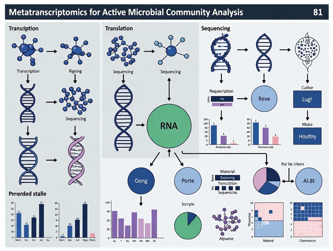
Metatranscriptomics is the set of techniques used to study the gene expression of microbes within natural environments, collectively known as the metatranscriptome [1]. While metagenomics provides a taxonomic profile of a microbial community by revealing "who is there," metatranscriptomics advances this understanding by characterizing the active functional profile, showing what functions the community is performing at a specific point in time [1] [2]. This approach provides a dynamic picture of the state and activity of a microbiome by focusing on changes in gene expression, capturing the collective mRNA transcripts of an entire microbial community to reveal actively expressed genes and metabolic activities [3] [4].
The fundamental advantage of metatranscriptomics lies in its ability to provide information about differences in the active functions of microbial communities that would otherwise appear to have similar taxonomic make-up [1]. By analyzing the collective microbial transcriptome, researchers can identify microbial expressed genes and associated functions, and identify the metabolically active members of the community [5]. This dynamic view offers a more accurate representation of microbial activity compared to metagenomics, which captures the static genetic blueprint of the community [4].
Key Methodological Approaches
Experimental Workflow and Technical Considerations
The standard metatranscriptomic sequencing workflow involves multiple critical steps to ensure high-quality data. The process begins with sample harvesting, where rapid stabilization of RNA is crucial due to the inherent instability of mRNA [1] [6]. RNA extraction follows, with methods varying depending on sample type, after which the extracted total RNA undergoes qualification checks including RNA Integrity Number (RIN) assessment, with values ≥6.5 typically required for proceeding [7].
A pivotal technical challenge is mRNA enrichment, as ribosomal RNA (rRNA) constitutes the majority of cellular RNA and can strongly reduce coverage of mRNA if not effectively removed [1] [6]. The two main strategies for mRNA enrichment include removing rRNA through capture using hybridization with 16S and 23S rRNA probes, or depletion of rRNAs through a 5-exonuclease approach [1] [6]. Following mRNA enrichment, cDNA synthesis is performed using reverse transcriptase, sequencing libraries are prepared, and high-throughput sequencing is conducted, primarily using Illumina platforms [1] [3] [7].
Table 1: Key Technical Challenges in Metatranscriptomics and Mitigation Strategies
| Challenge | Impact on Analysis | Current Mitigation Strategies |
|---|---|---|
| High rRNA abundance | Reduces mRNA sequencing coverage; can dominate datasets | Probe-based rRNA depletion; exonuclease treatment [1] [6] |
| RNA instability | Compromises sample integrity before sequencing | Rapid sample stabilization; optimized extraction protocols [1] [6] |
| Host RNA background | Limits microbial transcript detection in host-associated samples | Commercial enrichment kits; in silico removal post-sequencing [1] [5] |
| Limited reference databases | Reduces annotation completeness for novel microbes | Use of multiple databases; development of customized databases [1] [6] |
Computational Analysis Pipelines
The computational analysis of metatranscriptomic data involves multiple steps that can be approached through different strategies. A typical analysis begins with quality control of raw sequencing reads, adapter trimming, and removal of low-quality sequences [1] [3]. For taxonomic profiling, researchers can choose between marker-based methods like MetaPhlAn and mOTUs that use conserved genes, or k-mer based methods like Kraken 2/Bracken that use whole-genome information [5].
For functional analysis, HUMAnN is a widely used pipeline that implements a "tiered search" approach: first identifying known microbes, then constructing a sample-specific database, and finally performing translated searches against protein databases for unclassified reads [1]. Alternative pipelines like SAMSA2 offer simplified analysis by working with the MG-RAST server, while MetaTrans provides a flexible framework that supports multithreading for improved efficiency [1].
Recent advancements include integrated pipelines like metaTP, which provides end-to-end automation from data preprocessing to differential expression analysis and functional annotation [8]. This pipeline integrates tools for quality control, rRNA removal, transcript assembly, expression quantification, and co-expression network analysis, significantly improving reproducibility in metatranscriptomic studies [8].
Applications in Microbial Community Analysis
Human Health and Disease Mechanisms
Metatranscriptomics has revolutionized our understanding of host-microbiome interactions in human health. In the gut microbiome, metatranscriptomics can reveal how microbial communities respond to dietary changes, pharmaceutical interventions, and disease states by identifying actively expressed pathways [1] [4]. For example, studies of toll-like receptor 5 (TLR5) knockout mice used metatranscriptomics to show that flagellar motor-related gene expression was up-regulated compared to wild-type mice, revealing how host genetics shapes microbial behavior [6].
A significant advancement is the application of metatranscriptomics to human tissue specimens with low microbial biomass, such as mucosal interfaces of the gastrointestinal tract [5]. This approach has been successfully used to characterize the functional activity of the mucosal microbiome in gastric tissues, uncovering critical interactions between the microbiome and host in health and disease [5]. Such applications are particularly valuable for understanding diseases like inflammatory bowel disease (IBD), where researchers can identify dysregulated pathways, microbial biomarkers, and potential therapeutic targets by analyzing microbial gene expression patterns [4].
Environmental and Industrial Applications
Beyond human health, metatranscriptomics provides critical insights into diverse environments. In agricultural systems, it helps explore how microbial soil populations promote plant health and productivity, with applications in developing sustainable farming practices [6]. Environmental monitoring utilizes metatranscriptomics to assess ecosystem health by analyzing how microbial communities respond to pollutants, contaminants, and other stressors [4].
In biotechnology, metatranscriptomics facilitates the design of microbial consortia for applications including bioremediation, biofuel production, and industrial fermentation [6] [4]. By analyzing gene expression patterns in synthetic microbial communities, researchers can optimize consortia composition and metabolic pathways to enhance process efficiency [4]. The approach also enables drug discovery by identifying novel bacterial compounds in unculturable microorganisms, expanding accessible resources for pharmaceutical development [6] [7].
Table 2: Research Reagent Solutions for Metatranscriptomic Studies
| Reagent/Kit | Function | Application Context |
|---|---|---|
| Ribo-Zero Plus Microbiome Kit | Depletion of ribosomal RNA from both prokaryotic and eukaryotic organisms | Enhances mRNA coverage in complex microbial samples [9] |
| Microbiome RNA Extraction Kits | Isolation of high-quality total RNA from diverse sample types | Ensures RNA integrity (RIN ≥5) for downstream analysis [9] |
| Illumina Ribo-Zero Plus Microbiome | rRNA depletion for complex microbial samples | Optimizes library preparation for metatranscriptomic sequencing [9] |
| Custom HiPR-FISH Probes | Combinatorial fluorescent labeling for spatial mapping | Enables visualization of microbial spatial organization in communities [10] |
| SRA Toolkit | Data download and format conversion | Facilitates access to publicly available metatranscriptomic datasets [8] |
Integrated Protocols for Metatranscriptomic Analysis
Protocol for Samples with Low Microbial Biomass
Analysis of samples with low ratios of microbial to host cells (e.g., human tissue specimens) requires specialized approaches:
Sample Preparation: Collect samples with stringent precautions to avoid contamination. Immediately stabilize RNA using appropriate preservatives and store at -80°C prior to processing [5].
RNA Extraction and Quality Control: Extract total RNA using kits designed for microbiome RNA extraction. Verify RNA quality using an RNA Integrity Number (RIN) ≥6.5 and purity ratios (A260/280 ≥2.0; A260/230 ≥2.0) [5] [7].
rRNA Depletion and Library Preparation: Perform both prokaryotic and eukaryotic rRNA depletion to enrich mRNA. Prepare sequencing libraries using protocols optimized for metatranscriptomics, such as those incorporating the Ribo-Zero Plus Microbiome workflow [5] [9].
High-Depth Sequencing: Sequence on Illumina NovaSeq or similar platforms with high depth (~15 Gbp) to maximize detection of microbial sequences [5] [7].
Computational Analysis:
- Preprocess raw data: trim adapters, remove low-quality reads, and filter host sequences [5] [8].
- Perform taxonomic profiling using optimized Kraken 2/Bracken with confidence threshold set to 0.05 to balance sensitivity and precision [5].
- Conduct functional analysis using HUMAnN 3, which stratifies community functional profiles according to contributing species [5].
- Perform in silico decontamination to remove potential contaminant taxa [5].
Protocol for Standard Microbe-Rich Samples
For samples with high microbial load (e.g., stool, environmental samples):
Sample Processing: Extract total RNA ensuring rapid processing to maintain RNA integrity. For soil or complex environmental samples, use specialized extraction protocols that effectively lyse diverse microbial cells [1] [6].
Library Preparation and Sequencing: Deplete rRNA using either probe-based capture or exonuclease treatment. Prepare libraries and sequence using Illumina platforms (PE 150bp recommended), with ≥20 million read pairs per sample [7].
Bioinformatic Analysis:
- Quality control using FastQC and adapter trimming with Trimmomatic [8].
- Remove rRNA sequences using bowtie2 against rRNA databases [8].
- Choose analysis path based on research question:
- Annotate contigs using eggNOG-mapper, KEGG, GO, and COG databases [1] [8].
- Quantify gene expression using Salmon with TPM normalization [8].
- Perform differential expression analysis using appropriate statistical methods (e.g., Wilcoxon rank-sum test) [8].
Comparative Analysis with Related Techniques
Metatranscriptomics vs. Metagenomics
While metatranscriptomics and metagenomics are complementary approaches, they address fundamentally different questions in microbiome research. Metagenomics investigates the genetic potential of a community by sequencing DNA, revealing which microbes are present and what functions they could potentially perform [2] [4]. In contrast, metatranscriptomics examines the realized functions by sequencing RNA, showing which genes are actively expressed and what functions the community is actually performing at the time of sampling [4] [9].
This distinction has practical implications for experimental design and interpretation. Metagenomics provides a static snapshot of community composition and functional potential, while metatranscriptomics offers dynamic insights into gene expression patterns, metabolic activities, and responses to environmental changes [4]. For understanding functional activities and community dynamics, metatranscriptomics provides a more accurate representation of microbial activity, as the presence of a gene in a metagenome does not guarantee its expression [1] [4].
Integration with Other Omics Approaches
The most comprehensive understanding of microbial communities emerges from integrating multiple omics approaches. Metatranscriptomics forms a critical bridge between metagenomic potential and metabolic activity [2]. When combined with metabolomics, which identifies the byproducts released into the environment, researchers can connect gene expression with functional outcomes [2].
Recent advances in spatial techniques like HiPR-FISH (high-phylogenetic-resolution microbiome mapping by fluorescence in situ hybridization) further enhance metatranscriptomic insights by revealing the spatial organization of microbes within communities [10]. This integration helps generate hypotheses about how physical proximity influences functional interactions between microbial species.
Network-based approaches applied to integrated multi-omics datasets represent the cutting edge of microbiome analysis, enabling sophisticated in-depth understanding of microbiomes and leading to critical insights into microbial world [2]. The second phase of the Human Microbiome Project (iHMP) exemplifies this trend, gathering multiple omic data from both microbiome and host to understand host-microbiome interactions through integrative analyses [2].
Core Principles and Research Applications
Metagenomics and metatranscriptomics are foundational tools for studying microbial communities, but they answer fundamentally different biological questions. Metagenomics reveals the genetic potential of a microbiome, detailing "what microbes could do" by analyzing the total DNA present in a sample. It provides a census of which organisms are present and what genes they possess [11] [12]. In contrast, metatranscriptomics reveals the active functional state of a community, showing "what microbes are actually doing" at the time of sampling by sequencing the total mRNA [11] [12] [13]. This key difference dictates their respective applications in research and drug development.
The table below summarizes the core differentiators between these two approaches.
Table 1: Core Differentiators Between Metagenomics and Metatranscriptomics
| Comparison Dimension | Metagenomics | Metatranscriptomics |
|---|---|---|
| Research Core | Analyzes microbial DNA to reveal community composition and functional potential [11]. | Analyzes microbial RNA to reveal active gene expression and real-time activity [11]. |
| Primary Output | Catalog of microbial taxa and their gene complement. | Snapshot of actively transcribed genes and pathways. |
| Temporal Resolution | Static; represents the stable genetic blueprint. | Dynamic; captures a moment in time, reflecting response to the environment. |
| Key Application | Discovering novel microbial species and genes, characterizing community structure [14]. | Understanding functional mechanisms in disease, fermentation, or host-microbe interactions [15] [12]. |
| Relation to Disease | Identifies microbial signatures associated with a diseased state (e.g., species depletion or enrichment) [13]. | Reveals active virulence mechanisms and metabolic pathways driving disease pathology [15] [13]. |
Synergistic Applications in Drug Development and Clinical Research
The power of these technologies is often greatest when used together. An integrated multi-omics approach can link genetic potential with actual activity, providing a comprehensive view of microbiome function.
- Inflammatory Bowel Disease (IBD): Multi-omics studies have shown that a depletion of Faecalibacterium prausnitzii in the gut is correlated with reduced production of butyrate, a key anti-inflammatory metabolite. Metatranscriptomics can confirm the active downregulation of butyrate synthesis pathways, providing a functional explanation for the observed dysbiosis [14].
- Type 1 Diabetes (T1D) Risk: The TEDDY project analyzed thousands of stool samples from children and found that microbial functional pathways, particularly those involved in choline metabolism and cobalamin biosynthesis, were stronger correlates of β-cell autoimmunity than taxonomic composition alone. This highlights the need to move beyond census-taking to understand functional activity in disease progression [14].
- Urinary Tract Infections (UTIs): A 2025 study integrated metatranscriptomics with genome-scale metabolic modeling to characterize the active metabolic functions of patient-specific urinary microbiomes. This approach revealed distinct virulence strategies and metabolic cross-feeding between pathogens, which would be invisible to DNA-based sequencing alone [15].
Technical Workflows and Methodologies
The experimental and computational workflows for metagenomics and metatranscriptomics share similarities but have critical differences tailored to their target molecules (DNA vs. RNA).
Sample Preparation and Sequencing
The initial stages of the workflows are where the most significant technical distinctions lie, primarily due to the instability of RNA and the need to enrich for informative transcripts.
Table 2: Key Reagent Solutions for Metatranscriptomic Workflows
| Research Reagent / Tool | Function in the Workflow |
|---|---|
| Bead-beating (Metagenomics) | Breaks open diverse microbial cell walls in environmental samples via mechanical force to release DNA [11]. |
| Enzymatic Digestion (Metatranscriptomics) | Gently disperses tissue or cell line samples while minimizing damage to fragile RNA molecules [11]. |
| RiboPOOLs / MICROBExpress | Probe-based kits for subtractive hybridization that remove abundant ribosomal RNA (rRNA), enriching the messenger RNA (mRNA) fraction for sequencing [12]. |
| MICROBEnrich Kit | Uses hybridization capture technology to remove host-derived RNA, thereby increasing the proportion of microbial reads in the dataset [12]. |
| SMARTer Stranded RNA-Seq Kit | A library preparation kit effective for low-input RNA, ensuring efficient representation of microbial transcripts [12]. |
| DNase I | Enzyme used during RNA extraction to digest contaminating genomic DNA, ensuring sequence data derives purely from transcripts [12]. |
Bioinformatics and Data Analysis
The computational analysis of metagenomic and metatranscriptomic data requires robust pipelines to handle large, complex datasets.
Metagenomic Analysis: Standard pipelines involve quality control (FastQC, Trimmomatic), host DNA depletion (Bowtie2), and assembly into contigs using tools like MEGAHIT or metaSPAdes [14]. These contigs are then binned into Metagenome-Assembled Genomes (MAGs) using tools like MetaBAT2, which groups contigs based on sequence composition and abundance across samples [14]. Taxonomic profiling is performed with classifiers like Kraken2 against databases such as the Genome Taxonomy Database (GTDB), and functional potential is annotated using tools like eggNOG-mapper for KEGG orthology [14].
Metatranscriptomic Analysis: After sequencing, the raw reads undergo quality control and filtering. A critical step is the removal of residual rRNA sequences using tools like SortMeRNA [12]. High-quality mRNA reads are then aligned to reference genomes or metagenomic assemblies from the same sample set. Differential gene expression analysis is performed using specialized statistical packages like EdgeR or DeSeq2 to identify genes that are significantly upregulated or downregulated under different conditions (e.g., healthy vs. diseased) [12]. Integrated pipelines such as SAMSA2, HUMAnN2, or MetaTrans can automate many of these steps [12].
Experimental Protocol: Metatranscriptomic Analysis of a Microbial Community
The following protocol is adapted from recent studies investigating active microbial communities in clinical and environmental contexts [15] [12] [16].
Sample Collection, RNA Extraction, and Library Preparation
Goal: To obtain high-quality, representative cDNA libraries from a microbial community for sequencing.
Materials:
- RNase-free collection tubes and swabs.
- RNA stabilization solution (e.g., RNAlater).
- PowerSoil Total RNA Isolation Kit (or equivalent).
- RiboPOOLs depletion kit for bacterial rRNA.
- SMARTer Stranded RNA-Seq Kit.
- Agencourt RNAClean XP beads or similar.
Procedure:
- Collection & Stabilization: Collect sample (e.g., stool, soil, water filtrate) directly into a tube containing RNA stabilization solution. Immediately flash-freeze in liquid nitrogen and store at -80°C until processing. Critical: Minimize delay between collection and stabilization to preserve RNA integrity.
- RNA Extraction: Use a bead-beating mechanical lysis protocol to ensure rupture of diverse microbial cell walls. Extract total RNA following the manufacturer's instructions, including an on-column DNase I digestion step to remove genomic DNA contamination. Quantify RNA using a Qubit Fluorometer and assess integrity with an Agilent Bioanalyzer (RIN >7.0 is desirable).
- rRNA Depletion & Enrichment: Deplete ribosomal RNA from 1 µg of total RNA using a RiboPOOLs kit, following the subtractive hybridization protocol. This step enriches the messenger RNA (mRNA) fraction.
- Library Preparation: Convert the enriched mRNA to double-stranded cDNA using the SMARTer Stranded RNA-Seq Kit, which incorporates random priming for prokaryotic mRNA. Synthesize cDNA and perform library amplification with Illumina-compatible index adapters. Purify the final library using AMPure XP beads.
- Quality Control & Sequencing: Validate the library size distribution on a Bioanalyzer and quantify by qPCR. Pool equimolar amounts of libraries and sequence on an Illumina NovaSeq platform (e.g., 2x150 bp PE) to a minimum depth of 20-50 million reads per sample.
Computational Analysis of Metatranscriptomic Data
Goal: To process raw sequencing data into biologically interpretable information on active microbial functions.
Software & Databases:
- FastQC, Trimmomatic, SortMeRNA, bowtie2, DIAMOND, MetaTrans/SAMSA2 pipeline, EdgeR/DeSeq2, KEGG/EGGNOG databases.
Procedure:
- Pre-processing: Assess raw read quality with FastQC. Use Trimmomatic to remove adapters and low-quality bases (leading:3 trailing:3 slidingwindow:4:15 minlen:50).
- rRNA Filtering: Align reads against rRNA databases using SortMeRNA and remove matching sequences to obtain a cleaned set of mRNA reads.
- Taxonomic & Functional Assignment: For genome-resolved analysis, map reads to metagenome-assembled genomes (MAGs) from a companion metagenomic study using bowtie2. Alternatively, for direct functional assignment, use a pipeline like HUMAnN2 or perform alignment with DIAMOND (BLASTX) against a protein database (e.g., NR, UniRef90). Assign KEGG Orthology (KO) terms and map to metabolic pathways.
- Differential Expression Analysis: Compile read counts per gene or pathway in a count matrix. Import into R and use EdgeR or DeSeq2 to perform statistical testing for differential abundance/expression between sample groups (e.g., healthy vs. diseased). Apply a false discovery rate (FDR) correction (e.g., Benjamini-Hochberg); a common significance threshold is FDR < 0.05.
- Data Integration & Visualization: Integrate metatranscriptomic activity data with metagenomic abundance data to calculate activity-over-abundance ratios. Visualize results using principal coordinates analysis (PCoA), heatmaps of differentially expressed pathways (e.g., complex I-V of oxidative phosphorylation), and pathway enrichment plots.
In the study of microbial communities, traditional metagenomics has provided a powerful lens for viewing genetic potential by sequencing DNA. However, it offers a static picture, cataloging which genes are present but not which are actively functioning at a specific point in time [17]. Messenger RNA (mRNA) analysis, the cornerstone of metatranscriptomics, bridges this gap by capturing the dynamically expressed genes that drive microbial responses to their environment. This shift from potential to activity is fundamental for understanding true microbial function, as mRNA levels provide a direct snapshot of the genes being transcribed to perform tasks like nutrient acquisition, virulence, and stress response [15]. By analyzing mRNA, researchers can move beyond cataloging community members to interpreting their active metabolic roles, interactions, and contributions to health and disease states.
The Critical Role of mRNA in Microbial Analysis
Unveiling the Active Microbial Community
The composition of a microbial community revealed by DNA sequencing can differ significantly from the subset of microbes that are transcriptionally active. mRNA analysis is critical because it identifies the active contributors to community function. For instance, a landmark skin metatranscriptomics study demonstrated that Staphylococcus species and the fungus Malassezia had an "outsized contribution to metatranscriptomes at most sites, despite their modest representation in metagenomes" [17]. This divergence between genomic abundance and transcriptomic activity highlights that numerically minor members can be metabolically dominant, a finding crucial for identifying true keystone species in a community.
Quantifying Gene Expression and Metabolic Activity
mRNA analysis allows for the quantification of gene expression levels, which can be directly linked to metabolic activity. This principle was powerfully illustrated in a study of urinary tract infections (UTIs), where researchers integrated metatranscriptomic data with genome-scale metabolic modeling (GEMs). They found that constraining these metabolic models with gene expression data "narrows flux variability and enhances biological relevance" [15]. The table below summarizes key quantitative findings from recent studies that relied on mRNA analysis to decipher microbial activity.
Table 1: Key Quantitative Findings from Microbial mRNA Studies
| Study Focus | Method Used | Key Quantitative Finding | Biological Implication |
|---|---|---|---|
| Bacterial Single-Cell Analysis [18] | Bacterial MATQ-seq | Detects 300-600 genes/cell with a 95% success rate | Enables high-resolution analysis of individual cell states within a population. |
| Urinary Microbiome [15] | Metatranscriptomics + GEMs | Revealed marked inter-patient variability in transcriptional activity and metabolic behavior. | Underscores the need for patient-specific understanding of infections. |
| Skin Microbiome [17] | Metatranscriptomics | Identified >20 genes putatively mediating microbe-microbe interactions. | Uncovers the molecular basis of microbial ecology on the skin. |
Characterizing Virulence and Pathogen Response
For pathogens, mRNA analysis is indispensable for understanding virulence and adaptive responses. In a study of uropathogenic E. coli (UPEC) strains from UTI patients, mapping mRNA reads to a reference genome allowed researchers to profile the expression of virulence factors. They identified highly expressed genes related to adhesion (fimA, fimI) and iron acquisition (chuY, chuS, iroN), revealing "UPEC’s flexible virulence strategies and its ability to adapt to diverse host environments" [15]. This level of insight is critical for developing novel therapeutic strategies that target active pathogenic processes rather than just the presence of a pathogen.
Experimental Protocols for Microbial mRNA Analysis
The following section outlines a robust, end-to-end protocol for microbial metatranscriptomics, from sample collection to data analysis, incorporating best practices from recent studies.
Sample Collection, RNA Extraction, and Library Preparation
A reliable protocol begins with sample preservation and effective RNA extraction, which are particularly crucial for low-biomass environments like the skin [17].
- Sample Collection and Preservation: For skin and other surfaces, swabbing is a common, non-invasive method. To preserve RNA integrity, samples should be immediately placed into a stabilization solution like DNA/RNA Shield [17]. For other sample types, such as microbial cultures or patient specimens, rapid freezing in liquid nitrogen or similar methods is standard.
- RNA Extraction: Effective lysis often requires a combination of chemical and mechanical methods. The TRIzol method is recommended for maintaining RNA integrity during homogenization [19]. This should be coupled with bead beating to ensure complete lysis of robust microbial cells [17].
- rRNA Depletion and Library Construction: Since ribosomal RNA (rRNA) can constitute 80-90% of total RNA, its removal is essential for enriching the mRNA signal [19]. This is achieved using custom oligonucleotides that selectively deplete rRNA sequences, significantly enriching non-rRNA reads (e.g., from 2.5x to 40x) [17]. Following depletion, cDNA libraries are constructed using kits such as the NEBNext Ultra DNA Library Prep Kit for Illumina [20]. For studies focusing on eukaryotic mRNA, poly-A enrichment can be used, while rRNA depletion is universally applicable for mixed microbial communities [19].
Table 2: Essential Research Reagents and Kits for Microbial mRNA Analysis
| Research Reagent / Kit | Function | Application Note |
|---|---|---|
| DNA/RNA Shield | Stabilizes RNA at the point of collection, preventing degradation. | Critical for field and clinical sampling to preserve an accurate snapshot of gene expression [17]. |
| TRIzol Reagent | Monophasic solution of phenol and guanidine isothiocyanate for effective cell lysis and RNA isolation. | Maintains RNA integrity during homogenization; effective for diverse sample types [19]. |
| Custom rRNA Depletion Oligos | Biotinylated oligonucleotides that hybridize and remove rRNA sequences. | Custom panels designed for the expected community increase mRNA enrichment efficiency [17]. |
| NEBNext Ultra II DNA Library Prep Kit | Prepares sequencing-ready cDNA libraries from RNA samples. | A widely used, robust kit for constructing high-quality Illumina sequencing libraries [20]. |
Computational Analysis and Data Interpretation
After sequencing, raw data must be processed to extract biological meaning. A typical bioinformatics workflow is outlined below.
- Read Quality Control and Alignment: Raw sequencing reads (in FASTQ format) are first processed for quality control, which includes removing low-quality bases and adapter sequences. The cleaned reads are then aligned to a reference genome or a custom microbial gene catalog (e.g., the integrated Human Skin Microbial Gene Catalog, iHSMGC) using aligners like TopHat2 or modern alternatives [20] [17].
- Quantification and Differential Expression: Aligned reads are assigned to genomic features (genes) using tools like HTSeq to generate a raw count table [20]. This count table is then imported into statistical analysis environments like R. The table is filtered to remove low-count genes, and normalized using methods like TMM (Trimmed Mean of M-values) in the
edgeRpackage to account for technical variation between samples [21]. Finally, differential expression analysis is performed using packages likelimmaoredgeRto identify genes that are significantly upregulated or downregulated under different conditions (e.g, treatment vs. control) [21]. - Functional and Metabolic Interpretation: Differentially expressed genes are annotated using databases such as the Virulence Factor Database (VFDB) or Gene Ontology (GO) [15] [19]. For a systems-level view, gene expression data can be integrated with Genome-Scale Metabolic Models (GEMs). This involves constraining the flux bounds of metabolic reactions in the model based on transcript levels, which refines predictions of microbial metabolism in situ and reveals active pathways [15].
The following diagram visualizes the complete experimental and computational workflow:
Diagram 1: End-to-end workflow for microbial metatranscriptomics analysis.
Application Notes: Metatranscriptomics in Action
Case Study: Patient-Specific Urinary Tract Infection Analysis
A metatranscriptomic study of UTIs caused by uropathogenic E. coli (UPEC) showcased the power of mRNA analysis to reveal patient-specific pathogen strategies. Researchers analyzed 19 female patients and reconstructed personalized community metabolic models constrained by gene expression data. This approach revealed that while the primary pathogen (UPEC) was common, its metabolic behavior and virulence gene expression varied dramatically between patients [15]. For example, the activity of pathways like arginine and proline metabolism and the pentose phosphate pathway was highly variable. This finding underscores that a one-size-fits-all therapeutic approach may be ineffective and highlights the potential for microbiome-informed, personalized treatment strategies for managing complex infections.
Case Study: Uncovering Active Interactions in the Skin Microbiome
The application of a robust skin metatranscriptomics workflow to 27 healthy adults revealed a landscape of active microbial functions and interactions. By moving beyond DNA, the study found that commensal skin microbes, including staphylococci and lactobacilli, actively transcribe diverse antimicrobial genes, including uncharacterized bacteriocins, in situ [17]. Furthermore, by correlating microbial gene expression with the abundance of other microbes, the study identified more than 20 genes that putatively mediate microbe-microbe interactions. One such finding was a secreted protein from Malassezia restricta that had a strong negative association with Cutibacterium acnes, suggesting active competition. This demonstrates how mRNA analysis can pinpoint specific molecular mechanisms governing the stability and dynamics of microbial ecosystems.
mRNA analysis through metatranscriptomics is not merely a complementary technique to metagenomics; it is a fundamental tool for shifting from a census of microbial citizens to a functional assessment of their active jobs and interactions. As the cited protocols and case studies demonstrate, it enables researchers to identify metabolically dominant species, quantify virulence and stress responses, model community metabolism with high fidelity, and discover the molecular basis of microbe-microbe interactions. By capturing the dynamic transcriptome of microbial communities, researchers and drug development professionals can gain a mechanistic, patient-specific understanding of infectious diseases and microbiome-associated conditions, paving the way for novel diagnostic and therapeutic strategies.
The traditional view of microorganisms as isolated, free-living entities has been fundamentally replaced by the understanding that hosts and their associated microbial communities form an inseparable biological unit. The concept of the microbiome has evolved significantly from its initial definition. A revisited, comprehensive definition describes it as a characteristic microbial community occupying a reasonable habitat, which includes not only the microorganisms but also their structural elements, metabolic activities, and resulting ecological functions [22]. This expanded view positions the microbiome not merely as a collection of passengers but as an integral functional component of the host system, influencing host physiology, evolution, and health.
This perspective is central to the holobiont concept, which posits that the eukaryotic host and its microbiota form a single evolutionary unit [23] [22]. The interactions within this holobiont are governed by co-evolutionary principles and have profound implications for understanding host health, disease, and adaptation. The microbiome extends the host's genetic repertoire, forming what can be termed the "Extended Genotype" [23]. From a quantitative genetics perspective, the host's phenotypic variance (VP) can thus be decomposed to include not only host genetic variance (VG-HOST) and environmental variance (VE), but also the genetic variance contributed by the microbiome (VG-MICROBE): VP = VG-HOST + VG-MICROBE + VE [23]. This framework allows researchers to formally partition the contribution of microbial genetic variation to host phenotypes, thereby shaping the host's evolutionary potential.
Key Components of the Expanded Microbiome Definition
The expanded microbiome definition necessitates consideration of a complex web of interactions and components. The table below summarizes the core elements that move beyond a simple taxonomic catalogue of microbes.
Table 1: Core Components of the Expanded Microbiome Definition
| Component | Description | Research Implications |
|---|---|---|
| Host Factors | Host genetics, immune status, age, and sex that influence microbiome composition and function [23] [22]. | Requires recording detailed host metadata in studies [24]. |
| Microbiota | The assemblage of microorganisms present, including bacteria, archaea, fungi, algae, and protists [22]. | Culture-independent methods (e.g., 16S/18S rRNA, ITS sequencing) are essential for comprehensive characterization [22]. |
| Structural Elements | The physical organization of microbes, including biofilms and other microbial structures [22]. | Highlights the importance of spatial analysis techniques in microbiome research. |
| Metabolic Activity | The functional output of the microbiome, including transcripts, proteins, and metabolites [22] [15]. | Metatranscriptomics and metabolomics are needed to move beyond census-taking to functional insight. |
| Environmental Context | Diet, lifestyle, geography, and environmental exposures that shape the microbiome [23] [22] [24]. | Demands longitudinal study designs and extensive environmental metadata collection [24]. |
| Microbial Networks | The ecological interactions (cooperation, competition) between microbial species within the community [22]. | Network analysis and correlation metrics are key tools for understanding community stability and function [25]. |
Experimental Protocols for Analyzing the Active Microbiome
To operationalize the expanded microbiome definition and move from structure to function, metatranscriptomics has emerged as a powerful tool. It allows for the characterization of the collective gene expression profile of a microbial community, thereby revealing the metabolically active processes in response to host and environmental factors.
Protocol: Metatranscriptomic Workflow for Active Community Profiling
The following protocol is adapted from recent applications in clinical and environmental research [15] [16].
I. Sample Collection and Preservation
- Critical Step: Rapid stabilization of RNA is essential to preserve the in-situ transcriptional profile. Immediately freeze samples in liquid nitrogen or use a commercial RNA stabilization reagent.
- Clinical Note (e.g., Urine): Collect mid-stream urine from patients, centrifuge to pellet cells, and preserve the pellet in RNA-later [15].
- Environmental Note (e.g., Sludge): Collect aggregates of different sizes (e.g., flocs vs. granules) separately to investigate spatial functional heterogeneity [16].
II. RNA Extraction, Depletion, and Sequencing
- Total RNA Extraction: Use mechanical lysis (e.g., bead beating) followed by a phenol-chloroform extraction or a commercial kit designed for complex environmental samples.
- rRNA Depletion: Treat the total RNA with kits to remove ribosomal RNA (rRNA), which can constitute >90% of the total RNA. This enriches for messenger RNA (mRNA).
- Library Preparation and Sequencing: Construct cDNA libraries from the enriched mRNA and sequence using a high-throughput platform (e.g., Illumina).
III. Bioinformatic Processing and Analysis
- Quality Control and Trimming: Use tools like FastQC and Trimmomatic to assess read quality and remove adapter sequences and low-quality bases.
- Host Read Depletion: If working with a host-associated microbiome, align reads to the host genome (e.g., human, plant) and remove matching sequences to focus on microbial transcripts.
- Assembly and Mapping:
- Assembly-Based Approach: De novo assemble quality-filtered reads into longer contigs using assemblers like MEGAHIT or metaSPAdes.
- Mapping-Based Approach: Map quality-filtered reads directly to a database of reference genomes or gene catalogs.
- Taxonomic and Functional Annotation:
- Assign taxonomy to contigs or mapped reads using tools like Kraken2 or by blasting against databases (NCBI nr, GTDB).
- Annotate predicted genes against functional databases such as KEGG, COG, or Virulence Factor Database (VFDB) to determine active pathways [15].
- Genome-Resolved Metatranscriptomics (Advanced): For higher-resolution insights, bin contigs into Metagenome-Assembled Genomes (MAGs) and then map transcriptomic reads back to these MAGs to link activity to specific microbial populations [16].
IV. Integration with Metabolic Modeling
- Model Reconstruction: Create Genome-Scale Metabolic Models (GEMs) for key microbial taxa identified in the community using resources like AGORA2 or ModelSeed [15].
- Contextualization: Constrain the flux through reactions in the GEMs using the gene expression data (FPKM/TPM values) from the metatranscriptomics analysis.
- Simulation: Simulate community metabolism in a defined in-silico medium (e.g., virtual urine, synthetic wastewater) to predict metabolic cross-feeding, nutrient consumption, and product secretion [15].
The Scientist's Toolkit: Essential Reagents and Solutions
Table 2: Key Research Reagent Solutions for Metatranscriptomics
| Reagent / Solution | Function | Considerations |
|---|---|---|
| RNA Stabilization Reagent | Preserves RNA integrity instantly upon sample collection by inhibiting RNases. | Critical for capturing a snapshot of true in-situ gene expression; required for any field sampling. |
| Bead Beating Matrix | Mechanically disrupts robust microbial cell walls (e.g., Gram-positive bacteria, spores) for efficient RNA extraction. | Matrix material (e.g., silica, zirconia) and bead size must be optimized for the sample type. |
| rRNA Depletion Kit | Selectively removes abundant ribosomal RNA to enrich for messenger RNA, dramatically improving sequencing depth of informative transcripts. | Prokaryotic and eukaryotic rRNA require different probes; choose a kit appropriate for the community. |
| Reverse Transcriptase & Library Prep Kit | Synthesizes stable cDNA from enriched mRNA and prepares it for sequencing with the addition of adapters and indexes. | High-processivity enzymes are preferred for complex RNA mixtures. Unique dual indexing mitigates index hopping. |
| Functional & Taxonomic Databases | Provides a reference for annotating sequenced genes and transcripts (e.g., KEGG, VFDB, NCBI nr). | Database choice influences results; using curated, specialized databases (e.g., VFDB for virulence factors) is often beneficial [15]. |
| Metabolic Model Database | Provides pre-built genome-scale metabolic models (e.g., AGORA2) for key microbes to facilitate functional modeling [15]. | Allows for rapid reconstruction of community metabolic networks without building models from scratch. |
Data Presentation and Analysis in Practice
Applying the expanded definition requires robust and standardized data analysis. A key initial step in many microbiome studies is the assessment of alpha diversity, which describes the within-sample diversity. However, this is not a single metric but a set of complementary concepts.
Table 3: A Guide to Key Alpha Diversity Metrics for Microbiome Studies [26]
| Metric Category | Key Metrics | What It Measures | Biological Interpretation |
|---|---|---|---|
| Richness | Chao1, ACE, Observed ASVs | The number of different species (or ASVs) in a sample. | Simple estimate of community complexity. High richness often correlates with ecosystem stability. |
| Phylogenetic Diversity | Faith's PD | The sum of the phylogenetic branch lengths representing all species in a sample. | Accounts for evolutionary relationships; a community with distantly related species has higher PD. |
| Dominance/Evenness | Simpson, Berger-Parker, ENSPIE | The relative abundance distribution of species (i.e., whether a few taxa dominate). | Berger-Parker is the proportion of the most abundant taxon. Low evenness suggests dominance. |
| Information Indices | Shannon, Pielou's Evenness | Combines richness and evenness into a single metric of diversity. | Shannon entropy increases with both more species and more uniform distribution. |
The analysis of data from a metatranscriptomic study of urinary tract infections (UTIs) provides a powerful example of this framework in action. This study revealed marked inter-patient variability in microbial composition and transcriptional activity, even when the primary pathogen (E. coli) was the same [15]. By constructing patient-specific community metabolic models constrained by gene expression data, the researchers identified distinct virulence strategies and metabolic cross-feeding interactions that would be invisible with a census-based microbiome profile. Notably, the integration of gene expression data narrowed the variability in predicted metabolic fluxes and enhanced the biological relevance of the models, demonstrating the power of a function-first approach [15].
The expanded definition of the microbiome, which fully integrates host and environmental factors, represents a paradigm shift in microbial ecology and host biology. It moves research from asking "Who is there?" to the more impactful questions of "What are they doing?" and "How does their activity influence the host and ecosystem?". Metatranscriptomics serves as a cornerstone technique for addressing these questions by providing a snapshot of the active community's functional state.
Future research will likely focus on the dynamic integration of multiple omics layers—metagenomics, metatranscriptomics, metaproteomics, and metabolomics—to build a more complete, causal model of microbiome function. Furthermore, standardized reporting, as advocated by guidelines like STORMS, is crucial for ensuring reproducibility and comparability across studies [24]. As our molecular and computational toolkits continue to mature, the expanded microbiome definition will undoubtedly unlock novel diagnostic strategies and therapeutic interventions, particularly in managing complex conditions like multidrug-resistant infections, by targeting the functional core of the microbiome rather than just its constituents [15].
The Integrative Human Microbiome Project (iHMP or HMP2), launched in 2014 by the National Institutes of Health (NIH), represents a paradigm shift in human microbiome research [27] [28]. As the second phase of the pioneering Human Microbiome Project (HMP), the iHMP was designed to move beyond static cataloging of microbial inhabitants and instead generate longitudinal, multi-omic datasets to elucidate the dynamic roles of microbes in health and disease states [29]. With an investment of $170 million, this ambitious initiative recognized that taxonomic composition alone often poorly predicts host phenotype, and that a more holistic understanding requires integration of microbial molecular function with host biological responses [27] [28].
The iHMP focused on three specific microbiome-associated conditions, employing complementary 'omics technologies including 16S rRNA gene profiling, whole metagenome shotgun sequencing, whole genome sequencing, metatranscriptomics, metabolomics/lipidomics, and immunoproteomics [28]. This comprehensive approach has created an unprecedented resource for the research community, providing protocols, data, and biospecimens that continue to fuel discovery in host-microbe interactions [27]. The project established that microbial communities and their hosts undergo coordinated changes in metabolism and immunity during different health states, offering new insights into the functional mechanisms underlying microbiome-associated diseases [27] [29].
iHMP Core Study Designs and Key Quantitative Findings
The iHMP consisted of three longitudinal sub-studies that investigated the dynamics of the human microbiome and host under conditions of pregnancy, inflammatory bowel disease, and prediabetes. The key design elements and quantitative findings from these studies are summarized in the table below.
Table 1: Overview and Key Findings from iHMP Longitudinal Studies
| Study Focus | Cohort Details & Sampling Strategy | Key Microbiome Findings | Host Response Correlations |
|---|---|---|---|
| Pregnancy & Preterm Birth (PTB) | 1,527 pregnant women followed; 12,039 samples from 597 pregnancies analyzed [27]. | Convergence toward Lactobacillus-dominated vaginas in 2nd trimester; PTB linked to Sneathia amnii, Prevotella, BVAB1, and TM7-H1 [27]. | Vaginal pro-inflammatory cytokines (IL-1β, IL-6) positively correlated with PTB-associated taxa [27]. |
| Inflammatory Bowel Disease (IBD) | Adults and children with Crohn's disease and ulcerative colitis followed from multiple medical centers [28]. | Longitudinal shifts in gut microbiome taxonomic and functional profiles associated with disease activity and flares [27]. | Host immune and metabolic responses were intricately coordinated with microbial community changes [27]. |
| Onset of Type 2 Diabetes (T2D) | Patients at risk for T2D profiled to identify predictive molecular signatures [28]. | Marked shifts in the gut microbiome compared to healthy individuals, including specific metabolic pathways [28]. | Integrated data revealed molecules and signaling pathways involved in disease etiology [28]. |
The findings from these studies underscore the profound interconnectedness of host and microbiome biology. For instance, the pregnancy study revealed that the most predictive microbial signatures for preterm birth were detectable early in pregnancy (before 24 weeks), highlighting the potential for early risk assessment and intervention [27]. Furthermore, the iHMP established that the molecular interplay between host and microbiome provides a more accurate picture of health status than either dataset alone.
Metatranscriptomics: A Core Protocol for Active Community Analysis
Metatranscriptomics has emerged as a pivotal methodology for moving beyond microbial census data to understand the functionally active fraction of a microbial community. The standard workflow, as refined and applied in iHMP-related research, is detailed below.
Detailed Experimental Protocol
Sample Collection and RNA Preservation
- Critical Step: Collect sample (e.g., swab, stool, tissue) using kits that immediately stabilize RNA, as transcript levels can change rapidly post-sampling. Snap-freeze in liquid nitrogen and store at -80°C [13].
- Considerations for Low Biomass Sites (e.g., skin): The risk of host RNA contamination is high. Use sampling methods designed to maximize microbial yield, such as rigorous swabbing or scraping [13].
RNA Extraction and mRNA Enrichment
- Procedure: Extract total RNA using commercial kits optimized for complex biological samples (e.g., Mo Bio PowerMicrobiome RNA Isolation Kit). The resulting total RNA will include microbial and host ribosomal RNA (rRNA), messenger RNA (mRNA), and other RNAs [15] [13].
- mRNA Enrichment: Since bacterial mRNA lacks poly-A tails, use probe-based methods to deplete abundant rRNA molecules (both host and microbial) rather than poly-A selection. Kits such as the Ribo-Zero rRNA Removal Kit are commonly employed [15] [13].
Library Preparation and Sequencing
- Procedure: Convert the enriched mRNA to cDNA using reverse transcriptase. Then, prepare sequencing libraries with platform-specific adapters (e.g., Illumina). Amplify the library via PCR and validate quality using a Bioanalyzer [15].
- Sequencing: Perform high-depth sequencing on an Illumina platform (e.g., NovaSeq) to generate sufficient coverage for quantifying low-abundance transcripts from complex communities [15].
Computational and Bioinformatic Analysis
Pre-processing and Quality Control
- Tool: Use FastQC for initial quality assessment.
- Pre-processing Steps: Trim adapter sequences and low-quality bases using tools like Trimmomatic or Cutadapt [15].
Taxonomic and Functional Assignment
- Alignment: Align quality-filtered reads to a customized reference database containing host and microbial genomes. This allows for the subtraction of residual host reads. Tools like KneadData are often used for this step.
- Taxonomic Profiling: Use tools such as MetaPhlAn for profiling the active microbial community [15].
- Functional Profiling: Align reads to a curated protein database (e.g., UniRef90) using HUMAnN2 to reconstruct and quantify the abundance of metabolic pathways [15].
Integration with Metabolic Modeling
- Procedure: Map metatranscriptomic data to Genome-Scale Metabolic Models (GEMs), such as those in the AGORA2 resource, to predict community metabolic fluxes [15].
- Application: This integration allows researchers to move from gene expression lists to predictive models of microbial community physiology, as demonstrated in studies of urinary tract infections [15].
The following diagram illustrates the complete metatranscriptomics workflow, from sample to model.
Successfully executing a metatranscriptomic study requires a suite of specialized reagents, databases, and computational tools. The table below catalogs key resources for researchers in this field.
Table 2: Essential Research Reagents and Resources for Metatranscriptomics
| Category | Item/Resource | Specific Function & Application Notes |
|---|---|---|
| Wet-Lab Reagents | RNA Stabilization Solution (e.g., RNAlater) | Immediately preserves in vivo RNA expression profiles at point of sampling [13]. |
| Ribo-Zero rRNA Removal Kit | Depletes abundant ribosomal RNA (rRNA) to enrich for messenger RNA (mRNA) from bacteria and host [15]. | |
| Illumina Stranded Total RNA Prep Kit | Prepares sequencing libraries from rRNA-depleted RNA for transcriptome analysis [15]. | |
| Reference Databases | Human Microbiome Project (HMP) DACC | Provides curated, multi-omic reference datasets from healthy and diseased cohorts for comparison [27] [29]. |
| Virulence Factor Database (VFDB) | Annotates expressed virulence genes from pathogens like UPEC, linking activity to disease mechanism [15]. | |
| AGORA2 Genome-Scale Metabolic Models | A resource of 7,203 GEMs used to predict community metabolic fluxes from transcriptomic data [15]. | |
| Computational Tools | HUMAnN2 | Quantifies the abundance of microbial metabolic pathways and gene families from metatranscriptomic reads [15]. |
| MetaPhlAn | Provides precise taxonomic profiling of microbial communities from sequencing data [15]. | |
| BacArena | A modeling framework used to simulate and predict metabolic interactions in microbial communities [15]. |
Signaling Pathways and Host-Microbiome Interactions Elucidated by iHMP
The iHMP's multi-omic approach has been instrumental in uncovering specific host signaling pathways that are modulated by the microbiome. Two key areas of discovery are in inflammatory bowel disease and preterm birth.
In the context of Inflammatory Bowel Disease (IBD), the iHMP consortium revealed dynamic interactions between gut microbial metabolites and the host immune system. A primary finding involves the role of short-chain fatty acids (SCFAs), such as butyrate, produced by bacterial fermentation of dietary fiber. These metabolites serve as signaling molecules and energy sources for colonocytes, influencing the maintenance of gut barrier integrity and regulatory T-cell function. During disease flares, the iHMP observed a depletion of SCFA-producing bacteria and a corresponding shift in host metabolic and inflammatory pathways [27].
For Pregnancy and Preterm Birth (PTB), the MOMS-PI study identified that a dysbiotic vaginal microbiome, characterized by a lower abundance of Lactobacillus crispatus and higher abundance of taxa like Sneathia amnii, is associated with an elevated pro-inflammatory state [27]. This state is marked by increased levels of vaginal cytokines, including IL-1β and IL-6. The data suggest a signaling cascade where specific microbial communities trigger a localized immune response that can potentially disrupt the maternal-fetal interface, leading to spontaneous preterm labor [27]. The following diagram summarizes these host-microbe interaction pathways.
The Integrative HMP has successfully created a foundational multi-omic framework for investigating the microbiome as a dynamic interface with human health. By longitudinally profiling both host and microbiome molecules across different body sites and conditions, the iHMP has demonstrated that microbial community function is a critical determinant of phenotypic outcome, often transcending the importance of taxonomic composition alone [27]. The resources generated—including standardized protocols, vast public datasets, and analytical tools—have lowered the barrier for future research and set a new standard for integrative microbiome studies.
The application of metatranscriptomics, particularly when constrained with metabolic models as showcased in recent UTI [15] and skin microbiome [13] studies, provides a powerful path forward. This approach moves from correlation to mechanistic prediction, allowing researchers to model how microbial communities will function under different conditions. Future research will likely focus on further integrating these models with host biology to create a complete in silico representation of human superorganisms, ultimately accelerating the development of microbiome-based diagnostics and therapeutics, such as those that rely on metabolic reprogramming instead of traditional antibiotics [15].
From Lab to Clinic: Metatranscriptomics Workflows and Biomedical Applications
Metatranscriptomics has emerged as a powerful functional tool for analyzing active microbial communities by sequencing the collective RNA from all microorganisms in an environment. This approach moves beyond census-based microbial characterization to provide insights into the functional and metabolic capabilities of a microbiome at a specific time [9]. Unlike metagenomics, which reveals the potential functions encoded in DNA, metatranscriptomics identifies actively expressed genes, offering a dynamic view of microbial responses to their environment or host [30] [9]. This application note provides a detailed, step-by-step protocol for sampling, RNA extraction, and library preparation for metatranscriptomic studies, framed within the context of active microbial community analysis for drug development and clinical research.
Research Reagent Solutions and Essential Materials
The following table catalogues the essential reagents and materials required for a successful metatranscriptomics workflow.
Table 1: Key Research Reagent Solutions for Metatranscriptomics
| Item | Function/Application | Examples & Notes |
|---|---|---|
| RNA Stabilization Buffer | Immediate stabilization of RNA post-sampling to prevent degradation. | RLT buffer with β-mercaptoethanol; crucial for field sampling [31]. |
| Microbiome RNA Extraction Kits | Nucleic acid isolation from complex microbial communities. | Commercial kits providing RNA Integrity Number (RIN) ≥5 are recommended [9]. |
| DNase Treatment Kits | Removal of genomic DNA contamination from RNA extracts. | Essential for accurate gene expression analysis [5]. |
| rRNA Depletion Kits | Enrichment for messenger RNA (mRNA) by removing abundant ribosomal RNA. | Illumina Ribo-Zero Plus Microbiome; vital for functional profiling [5] [9]. |
| Library Prep Kits | Preparation of sequencing-ready libraries from mRNA. | Stranded RNA library prep kits compatible with rRNA-depleted RNA [9]. |
Sampling and Experimental Design
Sample Collection and Preservation
The initial sampling phase is critical for preserving the accurate snapshot of community gene expression.
- Water Samples: Filter a known volume (e.g., 500 mL) through 0.7 μm glass microfiber filters on-site to capture microbial biomass. Filtration should be completed within 30 minutes of collection to minimize RNA degradation [31].
- Human Tissues/Low-Biomass Samples: Use stringent contamination controls. Samples should be immediately snap-frozen in liquid nitrogen or placed in RNA stabilization reagents [5].
- Preservation: Filters or sample material should be placed in RNA stabilization buffers (e.g., RLT buffer with β-mercaptoethanol) immediately after collection, flash-frozen on dry ice, and transferred to -80°C storage until nucleic acid extraction [31].
Experimental Design Considerations
- Controls: Include filtration blanks (for water samples) or extraction blanks (for all sample types) to monitor for environmental and reagent contamination [31] [5].
- Replicates: Incorporate sufficient biological replicates to account for natural variability and ensure statistical robustness in downstream differential expression analysis.
- Metadata: Record comprehensive environmental (e.g., pH, temperature) or host (e.g., clinical metadata) data, as these are essential for interpreting transcriptional profiles.
RNA Extraction and Quality Control
RNA Extraction
Extract total RNA using commercial microbiome RNA extraction kits designed to lyse diverse microbial cell types. The protocol generally follows these steps:
- Cell Lysis: Mechanical bead-beating is often necessary to ensure efficient lysis of robust microbial cells.
- Nucleic Acid Purification: Bind RNA to silica columns while removing contaminants and inhibitors.
- DNase Digestion: Perform on-column or in-solution DNase treatment to eliminate contaminating DNA [5].
- Elution: Elute purified RNA in nuclease-free water.
RNA Quality and Quantity Assessment
Assess the quality and quantity of the extracted RNA before proceeding to library preparation.
- Quantification: Use fluorometric methods (e.g., Qubit) for accurate RNA concentration measurement.
- Quality Control: Evaluate RNA integrity using methods such as the RNA Integrity Number (RIN). A RIN greater than or equal to 5 is generally acceptable for metatranscriptomic studies [9].
Table 2: Key Quality Assessment Metrics and Thresholds
| Parameter | Recommended Method | Acceptance Threshold |
|---|---|---|
| RNA Concentration | Fluorometry (e.g., Qubit) | Sample-dependent |
| RNA Integrity | Bioanalyzer/TapeStation (RIN) | RIN ≥ 5 [9] |
| DNA Contamination | PCR (e.g., 16S rRNA gene) | Not detectable |
Library Preparation for Sequencing
Ribosomal RNA Depletion
The high abundance of ribosomal RNA (rRNA) can constitute over 90% of the total RNA in a sample. Depleting rRNA is essential to enrich for messenger RNA (mRNA) and maximize the informational yield of sequencing.
- Procedure: Use commercial kits (e.g., Illumina Ribo-Zero Plus Microbiome) that are designed to remove both prokaryotic and eukaryotic rRNA [5]. This step is crucial for samples with a high background of host RNA, such as human tissues [5].
Library Construction and Sequencing
The rRNA-depleted RNA is used to construct a sequencing library.
- cDNA Synthesis: Convert the enriched mRNA to double-stranded cDNA using reverse transcriptase and DNA polymerase.
- Adapter Ligation: Ligate platform-specific sequencing adapters to the cDNA fragments. Barcodes (indices) are included to allow multiplexing of samples.
- Library Amplification: Perform limited-cycle PCR to amplify the final library.
- Library QC: Validate the library's size distribution and concentration using a Bioanalyzer and qPCR.
- Sequencing: Sequence the libraries on an appropriate Illumina platform (e.g., NovaSeq for high-depth requirements) to generate the necessary read depth for detecting microbial transcripts, especially in host-dominated samples [5].
The following diagram illustrates the complete end-to-end workflow from sample collection to data generation.
Downstream Bioinformatic Analysis
After sequencing, raw data undergoes a comprehensive computational workflow to derive biological insights. Key steps include:
- Pre-processing: Quality control (FastQC), adapter trimming, and removal of host and rRNA reads [5] [8].
- Taxonomic Profiling: Assignment of sequences to microbial taxa using classifiers like Kraken 2/Bracken, which shows high sensitivity in samples with low microbial content [5].
- Functional Profiling: Quantification of gene families and metabolic pathways using tools like HUMAnN 3 [5] [8].
- Differential Expression Analysis: Identification of genes and pathways that are significantly altered between conditions, providing insights into microbial community responses to environmental stimuli or disease states [31] [8].
Troubleshooting and Technical Notes
- Low RNA Yield: Common in low-biomass samples. Increase starting material volume if possible and ensure efficient cell lysis during extraction.
- High Host RNA Background: For human tissue samples, a high sequencing depth is required to adequately capture microbial transcripts. Optimization of rRNA depletion protocols is crucial [5].
- RNA Degradation: Ensure rapid processing and immediate use of RNase inhibitors during sampling. Avoid freeze-thaw cycles of extracted RNA.
- Bioinformatic Contamination: Always process and analyze negative controls in parallel with experimental samples to identify and subtract background signals.
Metatranscriptome (MetaT) sequencing is a critical tool for profiling the dynamic metabolic functions of complex microbiomes, providing real-time gene expression data of both host and microbial populations. This enables authentic quantification of the functional enzymatic output of the microbiome and its host, offering significant advantages over DNA-based approaches for understanding active community functions [32]. However, two major technical challenges severely compromise the effectiveness of metatranscriptomic analysis: the overwhelming abundance of ribosomal RNA (rRNA) transcripts and the high proportion of host-derived RNA in many sample types.
In typical microbiome samples, rRNA can constitute as much as 99% of all sequencing reads, dramatically reducing the coverage of messenger RNA (mRNA) and driving up sequencing costs [32]. Simultaneously, host RNA can represent over 99% of the genetic material in clinically relevant samples like respiratory secretions and blood, effectively drowning out the microbial signal [33] [34]. This application note details optimized protocols and strategic solutions for overcoming these critical bottlenecks, enabling robust metatranscriptomic analysis across diverse research and clinical applications.
Table 1: Impact of Host RNA and rRNA on Sequencing Efficiency Across Sample Types
| Sample Type | Untreated Host/rRNA Content | Effective Solution | Post-Treatment Microbial Reads | Key Improvement Metrics |
|---|---|---|---|---|
| Mouse Cecal Content | High rRNA proportion | Mouse-optimized rRNA probes [32] | ~75% mRNA-rich reads [32] | ~15% increase in functional reads vs. human probes [32] |
| Clinical Sepsis Blood (0.5 mL) | High host RNA concentration | DRIB protocol with dual-species rRNA depletion [35] | 79,496-789,808 bacterial reads [35] | 63±7% of reads uniquely mapped to host or bacterial sequences [35] |
| Respiratory Samples (BAL) | 99.7% host reads [33] | HostZERO Mechanical+Chemical Lysis [33] [34] | 10-fold increase in final microbial reads [33] | 18.3% decrease in host DNA proportion [33] |
| Respiratory Samples (Sputum) | 99.2% host reads [33] | MolYsis commercial kit [33] | 100-fold increase in final microbial reads [33] | 69.6% decrease in host DNA proportion [33] |
| Rhizosphere Soil | High rRNA, humic acids | Optimized CTAB phenol-chloroform + Zymo-Seq RiboFree [36] | Successful transcript assembly | Effective rRNA removal, high-quality mRNA recovery [36] |
Table 2: Comparison of Host RNA Depletion Methods for Respiratory Samples
| Method | Mechanism | Best For | Efficiency (Reduction in Host DNA) | Impact on Microbial Richness |
|---|---|---|---|---|
| HostZERO (Zymo) | Chemical + Mechanical | BAL samples [33] | BAL: 18.3% decrease [33] | Significantly increases effective sequencing depth [33] |
| MolYsis (Molzym) | Selective lysis | Sputum samples [33] | Sputum: 69.6% decrease [33] | Increases species detection [33] |
| QIAamp (Qiagen) | Silica-membrane based | Nasal swabs [33] | Nasal: 75.4% decrease [33] | 13-fold increase in final reads for nasal [33] |
| Benzonase | Enzymatic degradation | Sputum (adapted protocol) [33] | Limited efficacy across sample types [33] | Moderate improvement [33] |
| lyPMA | Osmotic lysis + DNA cross-linking | Saliva with cryoprotectants [33] | Higher library prep failure rates [33] | Variable results [33] |
Optimized Protocols for Specific Applications
rRNA Depletion for Mouse Cecal Microbiome Studies
Background: Probes designed for human gut microbiomes (e.g., Ribo-Zero Plus) prove less effective and inconsistent when applied to mouse cecal samples, a common experimental system for microbiome studies [32].
Optimized Workflow:
- RNA Extraction: Extract total RNA from mouse cecal content using MagMAX mirVana Total RNA isolation kit [32].
- Probe Design Strategy: Employ taxonomic neutral probe design based solely on sequence abundance rather than taxonomic content [32].
- rRNA Depletion: Use Illumina Stranded Total RNA Prep with Ribo-Zero Plus Microbiome kit supplemented with mouse-specific probes (e.g., 5050 or 2025 probe sets from IDT oPools) [32].
- Library Preparation & Sequencing: Prepare RNA-Seq libraries and sequence on Illumina NovaSeq 6000 at 2 × 150 bp [32].
Key Innovation: The supplemental probes are carefully chosen to limit the number needed for effective depletion, reducing both cost and risk of introducing bias to MetaT analysis [32].
Performance: This approach provides ~75% mRNA-rich reads available for MetaT analysis, representing an additional ~15% of sequencing reads for functional data analysis compared to human-centric probes alone [32].
Figure 1: Optimized rRNA depletion workflow for mouse cecal samples
Dual RNA Isolation from Low-Volume Blood Samples (DRIB Protocol)
Background: Application of dual RNA-seq to sepsis research faces challenges including low bacterial burden in blood and limited sample volumes from vulnerable populations [35].
Optimized DRIB Protocol [35]:
- Sample Collection & Stabilization: Collect 0.5 mL whole blood into Li-Heparin tubes and immediately stabilize with 2.76× volumes (1.38 mL) of PAXgene Blood RNA solution. Incubate at RT for 2 hours to stabilize intracellular RNA and lyse host erythrocytes.
- Cell Pellet Recovery: Centrifuge at 3,200 g for 10 minutes. Carefully remove supernatant containing lysed erythrocytes. Wash remaining cells (leukocytes and bacteria) once with 1 mL nuclease-free water.
- Dual RNA Extraction: Resuspend cell pellet in 100 μL nuclease-free water. Add 1 mL TRI reagent and transfer to 2 mL bead-beating tubes with 0.1 mm zirconia/silica beads.
- Mechanical Lysis: Perform 3×1 minute cycles of bead-beating at 3,000 rpm with 1-minute breaks on ice between cycles.
- Phase Separation: Incubate at RT for 5 minutes. Add 200 μL chloroform, incubate 2 minutes at RT, then centrifuge at 12,000 g for 15 minutes.
- RNA Recovery: Recover aqueous RNA-containing phase and proceed with purification.
- Dual-Species rRNA Depletion: Implement dual-species rRNA depletion and RNA-seq.
Performance Validation: The DRIB protocol yields 2.10–6.91 μg of total RNA per clinical sample and generates 16.6–24.8 million filtered reads per sample, with 63±7% of reads uniquely mapped to host or bacterial sequences [35].
Universal rRNA Depletion for Complex Environmental Samples
Background: Rhizosphere soil presents unique challenges including copurification of inhibitory compounds like humic acids and difficult-to-lyse microbial communities [36].
Optimized Rhizosphere RNA Workflow:
- Sample Collection: Excise plant roots, place in PBS buffer, vortex to separate rhizosphere soil, centrifuge, and flash-freeze pellet at -80°C [36].
- CTAB Phenol-Chloroform Extraction:
- Homogenize 250 mg soil with silica beads in CTAB extraction buffer
- Add water-saturated phenol, Chloroform:Isoamyl alcohol (49:1), sodium phosphate buffer, and 2-Mercaptoethanol
- Centrifuge at 10,000 g for 10 minutes at 4°C
- Perform successive organic extractions
- RNA Precipitation: Precipitate aqueous phase with PEG-NaCl solution, incubate on ice at 4°C for 20 minutes, then centrifuge at 20,000 g for 20 minutes at 4°C.
- RNA Purification: Purify crude RNA using Zymo RNA Clean & Concentrator kits with DNase I treatment.
- Library Construction: Use Zymo-Seq RiboFree Total RNA Library Kit with RiboFree Universal Depletion reagents to remove rRNA-cDNA hybrids [36].
Key Advantage: This optimized CTAB phenol-chloroform extraction protocol significantly improves RNA yield and quality from clay-rich soils, outperforming commercial kits [36].
The Scientist's Toolkit: Essential Research Reagents
Table 3: Key Reagent Solutions for rRNA Depletion and Host RNA Removal
| Reagent/Kits | Primary Function | Application Context | Key Features |
|---|---|---|---|
| Ribo-Zero Plus Microbiome (Illumina) | rRNA depletion using RNase H | Gut microbiome samples [32] | Enzyme-based depletion with DNase treatment |
| Zymo-Seq RiboFree Total RNA Library Kit | Universal rRNA depletion | Environmental samples, rhizosphere soil [36] | Removes prokaryotic and eukaryotic rRNA |
| HostZERO Microbial DNA Kit (Zymo) | Host DNA depletion | Respiratory samples (BAL) [33] | Chemical + mechanical lysis; effective for frozen samples |
| MolYsis Commercial Kit | Selective host cell lysis | Sputum samples [33] | Preserves gram-negative bacteria |
| PAXgene Blood RNA System | RNA stabilization | Blood samples for dual RNA-seq [35] | Stabilizes intracellular RNA at collection |
| NEBNext rRNA Depletion Kit v2 | Ribosomal RNA removal | Respiratory samples with nanopore sequencing [34] | Compatible with third-generation sequencing |
| IDT oPools Oligonucleotides | Custom probe synthesis | Species-specific depletion [32] | Cost-effective custom probe manufacturing |
Integrated Workflow for Comprehensive Metatranscriptomics
Figure 2: Integrated workflow for metatranscriptomic analysis addressing both host RNA and rRNA challenges
Effective rRNA depletion and host RNA contamination control are foundational to successful metatranscriptomic studies. As demonstrated across diverse applications—from mouse cecal content to human clinical samples and environmental specimens—tailored approaches specific to sample type and research question are essential for obtaining meaningful functional data.
The protocols and methodologies detailed herein provide a framework for researchers to overcome the most significant technical barriers in metatranscriptomics. By implementing these optimized workflows and utilizing appropriate reagent solutions, researchers can significantly enhance the yield of microbial mRNA reads, thereby enabling more comprehensive analysis of active microbial community functions across diverse ecosystems and experimental systems.
Future advancements will likely focus on further refining probe design strategies, developing more efficient enzymatic depletion methods, and creating integrated workflows that seamlessly combine host RNA removal with rRNA depletion in a single streamlined process. As these technical hurdles continue to be addressed, metatranscriptomics will undoubtedly yield unprecedented insights into the dynamic functional interactions within microbial communities and their hosts.
Metatranscriptomics has emerged as a pivotal methodology for moving beyond the taxonomic census provided by metagenomics to characterize the functional activity of complex microbial communities. By sequencing the collective messenger RNA (mRNA) from an environmental sample, researchers can identify which genes are being actively expressed, providing insights into the metabolic processes and responses of a microbiome under specific conditions [37] [1]. This is particularly valuable in fields like drug development and human health, where understanding active microbial functions is as crucial as knowing which organisms are present. However, the analysis of metatranscriptomic data presents unique challenges, including the high background of host and ribosomal RNA, the instability of mRNA, and the sheer complexity of the computational analysis required [12] [1]. This application note outlines established and emerging bioinformatics pipelines designed to overcome these hurdles, enabling robust taxonomic and functional annotation to illuminate the active metatranscriptome.
A metatranscriptomics pipeline is a multi-step computational workflow that transforms raw sequencing reads into interpretable taxonomic and functional profiles. The general process involves quality control, removal of non-mRNA sequences (like ribosomal RNA), assembly of reads into transcripts, alignment to reference databases, and finally, taxonomic classification and functional annotation [12] [1]. Numerous pipelines have been developed, each with distinct strengths, supported databases, and analytical approaches.
Table 1: Comparison of Key Metatranscriptomics Analysis Pipelines
| Pipeline Name | Key Features | Taxonomic Classification Method | Functional Annotation Method | Best Suited For |
|---|---|---|---|---|
| metaTP (2025) | Highly automated; Integrated Snakemake workflow; Includes co-expression network analysis [8]. | Bowtie2; Salmon (for expression) [8]. | eggNOG-mapper; KEGG; GO [8]. | Comprehensive, end-to-end analysis requiring high reproducibility [8]. |
| MEDUSA (2022) | Supports both metagenomic & metatranscriptomic approaches; Flexible functional annotation [38]. | Kaiju [38]. | DIAMOND; Custom Python tool for annotation transfer [38]. | Sensitive taxonomic classification and custom functional identifier mapping [38]. |
| Optimized Kraken2/Bracken & HUMAnN 3 (2024) | Specifically designed for samples with low microbial biomass (e.g., human tissues) [5]. | Kraken 2/Bracken (optimized confidence threshold) [5]. | HUMAnN 3 [5]. | Human mucosal tissue samples and other low-biomass environments [5]. |
| SAMSA2 (2016) | Works with MG-RAST server; User-friendly for those with less bioinformatics experience [39] [1]. | MG-RAST's internal analysis pipeline [39]. | SEED Subsystems; NCBI RefSeq [39]. | Researchers seeking a simplified, server-based pipeline [39]. |
| MetaTrans (2016) | Open-source; Efficient multithreading; Handles both 16S rRNA and mRNA analyses [40] [1]. | SOAP2 against Greengenes (16S) or Kraken [40]. | SOAP2 against functional databases (e.g., MetaHIT) [40]. | Flexible analyses allowing integration of third-party tools [40]. |
Detailed Experimental Protocol
The following protocol describes a standardized workflow for metatranscriptome analysis, incorporating best practices from recent literature.
Sample Preparation and Sequencing
- RNA Extraction: Extract total RNA from the sample (e.g., human stool, soil, or tissue) using a kit suitable for the sample type and capable of handling potential inhibitors. For human tissue samples with low microbial biomass, use kits that minimize host RNA contamination [5].
- mRNA Enrichment: Deplete ribosomal RNA (rRNA) to enrich for mRNA. For prokaryotic microbes, which lack poly-A tails, use subtractive hybridization methods (e.g., riboPOOLs or Ribo-Zero kits) rather than poly-A enrichment [12]. This step is critical for increasing the coverage of informative mRNA sequences.
- Library Preparation and Sequencing: Convert the enriched mRNA to double-stranded cDNA using random hexamer priming. Prepare sequencing libraries using a kit compatible with low RNA input (e.g., SMARTer Stranded RNA-Seq kit) [12]. Sequence using an Illumina platform to generate a minimum of 40-50 million paired-end reads (100+ bp) per sample to ensure sufficient depth for accurate annotation [39].
Computational Analysis Using the metaTP Pipeline
The metaTP pipeline, managed by the Snakemake workflow engine, automates the following steps [8]:
- Quality Control and Trimming:
- Use FastQC for initial quality assessment of raw FASTQ files.
- Use Trimmomatic to remove adapter sequences and trim low-quality bases.
- rRNA and Host Read Removal:
- Align reads to an rRNA database using Bowtie2 and discard aligned reads.
- For host-associated samples, align reads to the host genome and remove aligned sequences to reduce host background [5].
- De Novo Assembly:
- Assemble the high-quality, non-rRNA reads into longer contigs using MEGAHIT [8].
- Gene Prediction and Expression Quantification:
- Use TransDecoder to identify putative coding regions within the assembled contigs.
- Build a transcriptome index from the predicted coding sequences and quantify transcript abundance using Salmon, which outputs normalized Transcripts Per Million (TPM) values [8].
- Taxonomic and Functional Annotation:
- Differential Expression Analysis:
The following diagram illustrates the logical workflow of a comparative metatranscriptomics study, from raw data to biological insight:
The Scientist's Toolkit: Essential Research Reagents and Software
Successful metatranscriptomic analysis relies on a suite of wet-lab and computational tools.
Table 2: Key Research Reagent Solutions and Bioinformatics Tools
| Category | Item | Function/Description |
|---|---|---|
| Wet-Lab Reagents | RiboPOOLs rRNA depletion kit (siTOOLs Biotech) | Probe-based subtraction method for efficient removal of ribosomal RNA from prokaryotic total RNA samples [12]. |
| SMARTer Stranded Total RNA-Seq Kit (Takara Bio) | Library preparation kit optimized for low-input RNA, improving microbial organism representation [12]. | |
| Computational Tools | Snakemake | Workflow management system for creating reproducible and scalable data analyses, used by pipelines like metaTP [38] [8]. |
| Kraken 2 / Bracken | k-mer based taxonomic classifier highly sensitive for samples with low microbial content; Bracken estimates species abundance [5]. | |
| DIAMOND | Ultra-fast aligner for translated DNA searches against protein reference databases (e.g., NCBI-nr) [38]. | |
| HUMAnN 3 | Pipeline for profiling the abundance of microbial metabolic pathways and other molecular functions from metagenomic or metatranscriptomic data [5]. | |
| Reference Databases | eggNOG | Database of orthologous groups and functional annotation for comprehensive functional assignment [8]. |
| KEGG (Kyoto Encyclopedia of Genes and Genomes) | Database resource for understanding high-level functions and utilities of the biological system [8] [1]. |
Workflow Visualization: The metaTP Pipeline
The metaTP pipeline integrates the tools and steps above into a cohesive, automated workflow, as shown in the following detailed diagram:
Application in Microbial Community Analysis
Integrating metatranscriptomics into a thesis on active microbial communities allows for unprecedented insights into dynamic functional responses.
- Functional Shifts in Disease States: Metatranscriptomics can reveal microbial pathways activated during disease. For example, analysis of gut microbiomes in models of colitis has identified significant differences in the expression of genes involved in inflammation and stress response between knockout and wild-type mice, providing potential targets for therapeutic intervention [39].
- Activity in Food Fermentation: In food science, metatranscriptomics elucidates the microbial activities driving fermentation. Studies on kimchi and other fermented foods have tracked the dynamic expression of genes from lactic acid bacteria responsible for flavor compound generation, linking specific microbial activities to product quality [12].
- Overcoming Low Microbial Biomass Challenges: For human tissue samples (e.g., gastric mucosa), where microbial content is low compared to host cells, an optimized workflow using Kraken2/Bracken for taxonomy and HUMAnN 3 for function has been shown to accurately characterize the active mucosal microbiome, opening new avenues for studying host-microbe interactions at the site of infection or disease [5].
The choice of a bioinformatics pipeline for taxonomic and functional annotation is a critical decision that shapes the outcome of a metatranscriptomic study. As outlined, pipelines like metaTP, MEDUSA, and optimized combinations of Kraken2/Bracken and HUMAnN 3 offer powerful, yet distinct, solutions for different research contexts. Adherence to standardized protocols for sequencing depth, rRNA depletion, and computational analysis is paramount for generating reliable data. By leveraging these sophisticated tools, researchers can effectively decode the active voices of complex microbial communities, advancing our understanding of their functional roles in health, disease, and biotechnological applications.
Application Note
This application note details a metatranscriptomics-based framework for analyzing active microbial communities and their virulence mechanisms in Urinary Tract Infections (UTIs). By integrating gene expression data with metabolic modeling, this approach reveals patient-specific pathogen behavior and community interactions that drive infection, offering a pathway to personalized diagnostic and therapeutic strategies.
Urinary tract infections (UTIs) represent a significant global health challenge, increasingly complicated by multidrug resistance (MDR). While Escherichia coli is the primary causative agent, the role of broader microbial communities in infection pathogenesis remains poorly understood [15]. Traditional diagnostics, reliant on culture-based methods, often miss polymicrobial infections and lack functional insight into microbial behavior [41]. Metatranscriptomics, the sequencing of RNA from microbial communities, enables researchers to move beyond taxonomic composition to investigate the actively expressed functions and virulence strategies of uropathogens within the patient-specific environment [15]. This application note presents a structured protocol for applying metatranscriptomics to characterize microbial virulence in UTIs, framed within a broader thesis on active microbial community analysis.
Key Findings from a Metatranscriptomic Study of UTI
A recent study analyzed urine samples from 19 female patients with confirmed uropathogenic E. coli (UPEC) infections, providing a foundational case study [15] [42]. The following tables summarize the core quantitative findings.
Table 1: Patient-Specific Variation in UTI Microbial Community Composition and Diversity [15]
| Metric | Findings | Research Implication |
|---|---|---|
| Microbial Composition | High inter-patient variability; genera included Anaeroglobus, Barnesiella, Escherichia, Lactobacillus, Prevotella. | UTI pathology involves complex, patient-specific consortia, not single pathogens. |
| Alpha Diversity (Shannon Index) | Range: 0.064 to 1.962. | Diversity is generally low but variable. |
| Impact of Lactobacillus | Patients with UTI communities containing Lactobacillus species showed increased diversity. | Probiotic taxa may play a modulatory role in the uromicrobiome. |
Table 2: Actively Expressed Virulence Factors in Patient-Derived UPEC UTI89 [15]
| Virulence Factor Category | Specific Genes Identified | Functional Role in UTI Pathogenesis |
|---|---|---|
| Adhesion | fimA, fimI | Essential for initial epithelial colonization and biofilm formation. |
| Iron Acquisition | chuY, chuS, iroN | Key to nutrient scavenging and survival in the iron-limited urinary environment. |
| Conserved High-Expression | ssrA, rnpB, cspA, ssrS | May indicate essential housekeeping or unannotated virulence functions. |
The study further leveraged genome-scale metabolic models (GEMs) constrained by the metatranscriptomic data. This integration revealed marked differences in the activity of metabolic subsystems (e.g., arginine and proline metabolism, glycolysis, pentose phosphate pathway) across patient-specific UPEC strains, underscoring the pathogen's metabolic adaptability during infection [15].
Experimental Protocols
The following section provides a detailed workflow for a metatranscriptomic analysis of UTI virulence, from sample collection to data interpretation.
Protocol: Metatranscriptomic Sequencing of Urine Microbiomes
Objective: To capture the taxonomic composition and gene expression profile of the active microbial community in patient urine samples.
Materials & Reagents:
- Sterile urine collection containers with boric acid preservative (to inhibit microbial growth post-collection and prevent false positive metabolite signals) [43].
- RNA stabilization solution (e.g., RNAlater).
- Total RNA extraction kit (optimized for bacterial cells).
- DNase I, RNase-free.
- rRNA depletion kits (e.g., MicrobEnrich Kit, Human/Mouse/Rat B).
- Library preparation kit (e.g., Illumina Stranded Total RNA Prep).
- Sequencing platform (e.g., Illumina NovaSeq).
Procedure:
- Sample Collection: Collect mid-stream urine from patients with suspected UTI directly into boric acid-containing tubes. Process samples immediately or store at 4°C for short-term storage (under 24 hours) [43].
- RNA Extraction & Purification: Concentrate microbial cells from urine via centrifugation. Extract total RNA using a commercial kit, including a DNase I treatment step to remove genomic DNA contamination. Assess RNA quality and integrity using an Agilent Bioanalyzer (RIN ≥ 7.0 is recommended) [44].
- rRNA Depletion & Library Prep: Deplete ribosomal RNA (rRNA) using probes targeting bacterial and human rRNA. This enriches the messenger RNA (mRNA) fraction. Proceed to construct sequencing libraries according to the manufacturer's instructions.
- Sequencing: Perform high-throughput sequencing on an Illumina platform to generate paired-end reads (e.g., 2x150 bp). A minimum of 20 million reads per sample is recommended for adequate coverage.
Protocol: Bioinformatics & Virulence Factor Analysis
Objective: To process raw sequencing data, identify active microbial taxa, and profile expressed virulence genes.
Materials & Reagents:
- High-performance computing cluster or cloud-based analysis platform.
- Bioinformatic tools: FastQC, Trimmomatic, Kraken2/Bracken, HUMAnN3.
- Virulence Factor Databases: Virulence Factor Database (VFDB), PATRIC VF library [45] [46].
Procedure:
- Quality Control & Trimming: Assess raw read quality with FastQC. Remove adapter sequences and low-quality bases using Trimmomatic.
- Taxonomic Profiling: Classify quality-filtered reads against a reference database (e.g., RefSeq) using Kraken2. Estimate species-level abundances with Bracken.
- Functional Profiling & VF Identification:
- Map quality-controlled reads to the VFDB 2.0 [46] or the curated PATRIC database [45] using BOWTIE2 or BLAST.
- Quantify expression levels (e.g., in FPKM - Fragments Per Kilobase Million) for each identified virulence factor gene.
- Use a pipeline like the MetaVF toolkit to enhance the sensitivity and precision of VF identification from metagenomic data [46].
Protocol: Metabolic Modeling of Patient-Specific Communities
Objective: To contextualize gene expression data within metabolic networks and predict community interactions.
Materials & Reagents:
- Genome-scale metabolic model (GEM) databases (e.g., AGORA2).
- Constraint-based modeling software (e.g., COBRA Toolbox, BacArena).
- In silico urine medium composition (based on the Human Urine Metabolome Database) [15].
Procedure:
- Model Reconstruction: Reconstruct a draft community metabolic model by gathering GEMs for species identified in taxonomic profiling.
- Apply Context-Specific Constraints: Constrain the flux through metabolic reactions in the models using the gene expression (metatranscriptomic) data from the patient sample. This creates a patient-specific model.
- Simulate in Urine Environment: Simulate microbial growth and metabolic exchange using the in silico urine medium to define nutrient availability [15]. Tools like BacArena can simulate spatial and temporal dynamics.
- Analyze Results: Identify active metabolic pathways, potential metabolic cross-feeding between community members, and reactions essential for growth in the urinary environment.
Visualizing the Workflow and Virulence Mechanisms
The following diagrams, generated using Graphviz DOT language, illustrate the core experimental workflow and the functional role of identified virulence factors.
Diagram 1: Metatranscriptomics UTI Analysis Workflow
Diagram 2: UPEC Virulence Mechanisms in UTI
The Scientist's Toolkit: Research Reagent Solutions
Table 3: Essential Reagents and Resources for UTI Metatranscriptomics
| Item | Function/Description | Example Product/Source |
|---|---|---|
| Boric Acid Tubes | Preservative that inhibits microbial growth post-collection, preventing artifactual changes in metabolite and transcript levels. | BD Vacutainer C&S Preservative Tubes |
| rRNA Depletion Probes | Selective removal of host and bacterial ribosomal RNA to enrich for messenger RNA, drastically improving sequencing depth of informative transcripts. | MicrobEnrich Kit, Illumina Ribo-Zero Plus |
| Virulence Factor Database (VFDB) | Curated repository of known virulence factor genes; essential for annotating and quantifying pathogenic functions in sequencing data. | VFDB 2.0 [46] |
| PATRIC VF Library | A highly curated database integrating VF genes with genomic and transcriptomic data for pathogenic bacteria. | PATRIC BRD [45] |
| MetaVF Toolkit | A computational pipeline for precise profiling of VFGs from metagenomic data, offering high sensitivity and precision. | MetaVF [46] |
| AGORA2 Resource | A database of genome-scale metabolic models (GEMs) for gut and human-associated microbes, usable for modeling uropathogens. | AGORA2 [15] |
| In silico Urine Medium | A defined virtual medium based on the Human Urine Metabolome Database, used to constrain metabolic models for biologically relevant simulations. | Custom formulation [15] [43] |
Application Note: Metatranscriptomic Profiling of Microbial Functional Activity
Key Findings in Inflammatory Bowel Disease (IBD)
Metatranscriptomics provides a direct view of microbial functional activity that cannot be detected through DNA-based metagenomic profiling alone. This Application Note summarizes how this powerful technique is revealing novel mechanisms in Inflammatory Bowel Disease (IBD) and metabolic disorders.
Table 1: Key Metatranscriptomic Findings in IBD from Recent Studies
| Finding Category | Specific Example | Biological Significance | Reference |
|---|---|---|---|
| Discordant DNA/RNA Activity | Faecalibacterium prausnitzii shows predominant pathway transcription disproportionate to its genomic abundance. [47] | Loss of this organism in IBD may have greater functional consequences than metagenomic data suggests. [47] | |
| Dormant Microbes | Dialister invisus is metagenomically present but shows little to no gene expression. [47] | Distinguishes active contributors to the gut environment from inactive or dead bacteria. [47] | |
| Disease-Specific Pathway Activation | Glycan degradation and two-component system pathways are enriched in UC. [48] Protein processing and export pathways are upregulated in both CD and UC. [48] | Reveals specific microbial processes actively contributing to the inflammatory environment. [48] | |
| Virulence Factor Expression | Active expression of Adherent-Invasive E. coli (AIEC) virulence genes, particularly ompA, in Crohn's disease. [49] | Directly links microbial gene expression to mechanisms of bacterial adherence and invasion of host macrophages. [49] | |
| Altered Metabolite Production | Disruption in microbial fermentation pathways, leading to depleted butyrate production. [49] | Explains the reduction of a key anti-inflammatory metabolite in the gut lumen of IBD patients. [49] |
Insights into Metabolic Disorders
Metatranscriptomics has also uncovered critical functional dynamics in metabolic diseases, particularly in the context of diurnal rhythms and dietary interventions.
Table 2: Metatranscriptomic Insights into Metabolic Homeostasis
| Experimental Context | Key Metatranscriptomic Finding | Functional & Therapeutic Implication | Reference |
|---|---|---|---|
| High-Fat Diet (HFD) & Time-Restricted Feeding (TRF) in Mice | TRF restores diurnal rhythms in microbial gene expression that are lost under HFD. [50] | Identifies a mechanism for TRF's metabolic benefits and identifies dynamically expressed functions. [50] | |
| Bile salt hydrolase (bsh) from Dubosiella newyorkensis exhibits strong diurnal expression under TRF. [50] | Administration of engineered E. coli expressing this bsh improved host insulin sensitivity and glucose tolerance. [50] | ||
| LPS-Induced Inflammation in Mice | Mulberry-derived postbiotics (MDP) cause significant shifts in host transcriptome and gut microbiome. [51] | Suggests a protective mechanism against inflammation via modulation of the microbiome-immune axis. [51] |
Protocol: Integrated Metatranscriptomic Analysis of Gut Microbiome Activity
Sample Collection and RNA Extraction
This protocol is adapted from methodologies used in recent IBD and metabolic studies. [50] [49]
Reagents:
- Sample Preservation Buffer: RNAlater or similar RNA stabilization reagent.
- Lysis Buffer: 4 M guanidine thiocyanate, 5% N-lauroyl sarcosine. [49]
- Mechanical Disruption Beads: Zirconia/silica beads (0.1 mm diameter). [49]
- Acid-Phenol: For phase separation. [49]
- Commercial Kit: RNeasy Mini Kit (Qiagen) or equivalent for RNA purification. [49]
Procedure:
- Collection: Collect fresh fecal or intestinal content samples immediately snap-freeze in liquid nitrogen, or preserve in RNAlater at 4°C for ≤24 hours before long-term storage at -80°C. [50]
- Homogenization: Suspend 200-250 mg of sample in lysis buffer. Add ~0.8 g of zirconia/silica beads.
- Cell Lysis: Perform mechanical disruption using a bead beater (e.g., FastPrep apparatus) for 3-5 minutes. [49]
- Nucleic Acid Recovery: Centrifuge the lysate and transfer the aqueous phase to a new tube. Add an equal volume of acid-phenol, mix thoroughly, and centrifuge to separate phases. Recover the aqueous phase. [49]
- DNA Digestion: Treat the recovered nucleic acids with DNase I to remove genomic DNA contamination.
- RNA Purification: Purify the total RNA using the RNeasy Mini Kit, following the manufacturer's instructions. Elute RNA in nuclease-free water.
- Quality Control: Assess RNA integrity and purity using an Agilent Bioanalyzer or TapeStation. RNA Integrity Number (RIN) >7 is recommended for library preparation.
rRNA Depletion and Library Preparation
Reagents:
- rRNA Depletion Kit: Ribo-Zero Magnetic Gold Kit (Epidemiology) or equivalent. [49]
- Library Prep Kit: Illumina Stranded Total RNA Prep with Ribo-Zero Plus or equivalent.
Procedure:
- rRNA Removal: Deplete ribosomal RNA from 100 ng - 1 µg of total RNA using the Ribo-Zero kit, according to the manufacturer's protocol. [49]
- RNA Fragmentation: Fragment the remaining RNA to an average size of 200-300 nucleotides.
- cDNA Synthesis: Synthesize first-strand cDNA using reverse transcriptase and random hexamer primers. Synthesize second-strand cDNA, incorporating dUTP to preserve strand specificity.
- Library Amplification: Perform adapter ligation and amplify the library with ~10-12 PCR cycles.
- Library QC: Validate library size distribution using a Bioanalyzer and quantify by qPCR.
Sequencing and Bioinformatics Analysis
- Sequencing: Sequence libraries on an Illumina platform (e.g., HiSeq or NovaSeq) to generate a minimum of 20-50 million paired-end (2x100 bp or 2x150 bp) reads per sample. [49]
- Bioinformatics Analysis: A standard workflow is depicted below.
Figure 1: Bioinformatic workflow for metatranscriptomic data.
Key Software Tools:
- Quality Control: KneadData (v0.7.4), FastQC, Trimmomatic. [49]
- Host Read Removal: Bowtie2 against host genome (e.g., GRCh38). [49]
- Taxonomic Profiling: MetaPhlAn (v4.0.3) for species-level identification. [49]
- Functional Profiling: HUMAnN (v3.6) with UniRef90 database for pathway abundance analysis. [47] [49]
Pathway and Mechanistic Insights
Host-Microbiome Metabolic Interactions in IBD
Metabolic modeling of multi-omics data reveals profound dysregulation of host-microbiome co-metabolism in IBD. The following diagram summarizes key disrupted pathways.
Figure 2: Host-microbiome metabolic disruptions in IBD.
The interplay between microbial and host metabolic pathways creates a vicious cycle that perpetuates inflammation. [52] For instance, reduced microbial production of short-chain fatty acids (SCFAs) like butyrate, a key anti-inflammatory metabolite, is a consistent finding in IBD metatranscriptomic and metabolomic studies. [52] [49] Simultaneously, the host exhibits elevated tryptophan catabolism, which depletes circulating tryptophan and impairs NAD+ biosynthesis, a crucial cofactor for cellular energy production and redox homeostasis. [52] These concomitant changes highlight the power of multi-omics approaches to uncover system-level dysfunction.
Diagnostic and Therapeutic Applications
The functional activity data provided by metatranscriptomics has direct diagnostic and therapeutic relevance.
Table 3: Diagnostic and Therapeutic Insights from Integrated Omics
| Application | Finding | Potential Utility |
|---|---|---|
| Biomarker Discovery | A panel of 20 microbial species identified via metagenomics achieved an AUC of 0.94 for diagnosing Crohn's disease. [49] | Differentiating IBD subtypes and identifying patients in challenging clinical scenarios. |
| Mechanism-Driven Therapy | Propionate utilization by AIEC drives ompA virulence gene expression. [49] | Suggests targeting microbial metabolic pathways to reduce virulence. |
| Live Biotherapeutic Design | Dubosiella newyorkensis bsh expressed diurnally improves metabolic health in mice. [50] | Engineering bacterial chassis to deliver timed therapeutic functions. |
Table 4: Key Research Reagent Solutions for Metatranscriptomic Studies
| Reagent / Resource | Function / Application | Example Product / Source |
|---|---|---|
| RNeasy PowerMicrobiome Kit | Simultaneous lysis and stabilization of RNA from complex microbial communities. | Qiagen (Cat. No. 26000-50) |
| Ribo-Zero Plus rRNA Depletion Kit | Removal of bacterial and host rRNA to enrich for mRNA sequencing. | Illumina (Cat. No. 20037135) |
| MetaPhlAn Database | Taxonomic profiling from metagenomic and metatranscriptomic sequencing data. | https://huttenhower.sph.harvard.edu/metaphlan/ |
| HUMAnN Software & UniRef Database | Functional profiling of metabolic pathways and their abundance. | https://huttenhower.sph.harvard.edu/human/ |
| Bile Salt Hydrolase (BSH) Assay Kit | Functional validation of BSH activity in cultured bacteria or samples. | Cell Biolabs, Inc. (MET-5101) |
| Short-Chain Fatty Acid (SCFA) Standard Mix | Quantification of SCFAs (e.g., butyrate, propionate) via GC-MS or LC-MS. | Sigma-Aldrich (CRM46975) |
Metatranscriptomics is revolutionizing therapeutic discovery by moving beyond microbial census data to reveal the functionally active genes and metabolic pathways that underpin host-microbiome interactions in health and disease. This approach provides a dynamic snapshot of the entire microbial community's transcriptional activity, offering an unprecedented opportunity to identify novel, mechanistically grounded therapeutic targets and biomarkers [6]. By capturing the expressed genetic repertoire of complex microbiomes directly from their natural environments, including clinical samples, researchers can pinpoint critical pathways driving pathological processes and discover highly specific biomarkers for diagnostic and prognostic applications [15] [53]. This document details the core applications, quantitative findings, and standardized protocols for leveraging metatranscriptomics in drug discovery pipelines.
Key Applications and Case Studies
Metatranscriptomics has been successfully applied across diverse disease areas to identify and validate targets, as summarized in the table below.
Table 1: Drug Discovery Applications of Metatranscriptomics in Human Diseases
| Disease Area | Key Metatranscriptomic Findings | Potential Therapeutic Targets / Biomarkers Identified | Reference |
|---|---|---|---|
| Urinary Tract Infections (UTIs) | Revealed inter-patient variability in virulence gene expression (e.g., fimA, fimI for adhesion; chuY, chuS for iron acquisition) and active metabolic cross-feeding within the urinary microbiome. | Distinct virulence strategies of uropathogenic E. coli (UPEC); metabolic pathways supporting pathogen persistence; modulatory role of Lactobacillus species. [15] | |
| Inflammatory Bowel Disease (IBD) | Characterization of actively expressed microbial genes and pathways associated with inflammation and dysbiosis. | Underreported microbial species (e.g., Asaccharobacter celatus, Gemmiger formicilis); functional activity of inflammatory pathways. [54] | |
| Oral Health & Disease | Identification of microbial community-wide gene expression shifts between health and disease states (e.g., periodontitis). | Active virulence factors and metabolic pathways from diverse oral pathogens; community-level functional signatures. [6] | |
| Metabolic Disorders | Analysis of active gut microbial pathways involved in host metabolism, such as short-chain fatty acid (SCFA) production and bile acid modification. | Microbial enzymes and derived metabolites (e.g., specific SCFAs, secondary bile acids) as targets for managing obesity and type 2 diabetes. [55] [53] |
The integration of metatranscriptomic data with other omics layers, such as metagenomics and metabolomics, significantly enhances the robustness of biomarker identification. This multi-omics approach allows for the construction of correlation networks that link microbial gene expression to metabolic outputs and disease status, leading to diagnostic models with high predictive accuracy (e.g., AUROC of 0.92–0.98 for IBD) [54].
Experimental Protocols
Protocol: Metatranscriptomic Sequencing for Biomarker Discovery
This protocol outlines the end-to-end process for identifying microbial biomarker candidates from complex community samples.
I. Sample Collection and RNA Extraction
- Collection: Collect samples (e.g., stool, saliva, tissue) in sterile, RNA-stabilizing solutions (e.g., RNAlater) to immediately preserve RNA integrity. Flash-freezing in liquid nitrogen is also acceptable. Store at -80°C.
- RNA Extraction:
- Cell Lysis: Use a combination of physical (e.g., bead beating) and chemical (e.g., guanidinium thiocyanate) lysis methods to ensure complete disruption of diverse microbial cell walls. [56]
- Total RNA Isolation: Purify total RNA using commercial kits designed for complex samples (e.g., Qiagen RNeasy PowerMicrobiome Kit). Include DNase digestion steps to remove genomic DNA contamination.
- Quality Control: Assess RNA integrity and purity using an Agilent Bioanalyzer. High-quality RNA (RNA Integrity Number, RIN > 7) is recommended for library preparation.
II. Library Preparation and Sequencing
- rRNA Depletion: Selectively remove abundant ribosomal RNA (rRNA), which can constitute >90% of total RNA, to enrich for messenger RNA (mRNA). Use probe-based kits (e.g., Illumina Ribo-Zero Plus) that target both host and microbial rRNA. [6]
- cDNA Synthesis and Library Construction:
- Fragment the enriched RNA.
- Synthesize first-strand cDNA using reverse transcriptase and random hexamers.
- Synthesize the second strand to create double-stranded cDNA.
- Ligate sequencing adapters and perform PCR amplification to create the final sequencing library. [6]
- Sequencing: Sequence the libraries on a high-throughput platform (e.g., Illumina NovaSeq) to generate paired-end reads (e.g., 2x150 bp) with sufficient depth (typically 20-50 million reads per sample for complex microbiomes).
III. Bioinformatic Analysis and Biomarker Identification
- Pre-processing: Quality-trim raw reads using Trimmomatic or Cutadapt. Remove any residual host reads by aligning to the host genome (e.g., GRCh38) with Bowtie2.
- Assembly and Quantification: Assemble quality-filtered reads into transcripts using a de novo assembler (e.g., Trinity) or align them to a curated reference database of microbial genomes. Quantify transcript abundance (e.g., in FPKM or TPM units) using tools like Salmon. [6] [57]
- Differential Expression & Biomarker Identification:
- Differential Expression Analysis: Use tools such as DESeq2 or edgeR to identify transcripts significantly differentially expressed between conditions (e.g., disease vs. healthy). Apply multiple testing correction (e.g., Benjamini-Hochberg). [57]
- Feature Selection: Apply machine learning-based feature selection methods like LASSO (Least Absolute Shrinkage and Selection Operator) or Elastic Net to identify a minimal set of transcript biomarkers that best predict the phenotype of interest. This reduces dimensionality and mitigates overfitting. [58]
- Functional Annotation: Annotate candidate biomarker transcripts against databases such as the Virulence Factor Database (VFDB), KEGG, and GO to elucidate their biological roles and prioritize mechanistically relevant targets. [15]
The following workflow diagram illustrates the key steps of this protocol.
Protocol: Integration with Genome-Scale Metabolic Modeling (GEMs)
Combining metatranscriptomic data with metabolic models transforms gene expression data into predictive, mechanistic insights into community metabolism.
I. Model Reconstruction
- Obtain Community-Specific GEMs: Reconstruct or select genome-scale metabolic models for the key microbial taxa identified in your metatranscriptomic data. Resources like the AGORA2 database provide curated models for thousands of human-associated microbes. [15]
- Build a Community Model: Assemble individual microbe models into a community metabolic model that simulates metabolite exchange (cross-feeding) and competition.
II. Data Integration and Constraint-Based Analysis
- Apply Transcriptomic Constraints: Map the metatranscriptomic expression data (e.g., TPM values) onto the corresponding reactions in the GEMs. Use this data to constrain the flux bounds of reactions, effectively activating or deactivating pathways based on their measured expression levels. [15]
- Simulate in a Context-Specific Medium: Define the extracellular environment (e.g., using a virtual urine medium for UTI studies [15] or a gut medium) to reflect the in situ nutritional conditions.
- Perform Flux Balance Analysis (FBA): Use FBA to predict growth rates and metabolic flux distributions for each microbe within the community under the transcriptomic and environmental constraints.
III. Target Identification
- Analyze the flux predictions to identify:
- Essential Pathways: Reactions critical for pathogen growth that are absent in commensals or the host.
- Cross-Feeding Dependencies: Metabolites produced by one microbe that are essential for a pathogen, pointing to potential indirect targeting strategies.
- Modulatory Mechanisms: Metabolic interactions where commensals (e.g., Lactobacillus) potentially inhibit pathogens, suggesting probiotic mechanisms. [15]
The diagram below illustrates the logical flow of this integrative approach.
The Scientist's Toolkit
Table 2: Essential Research Reagents and Solutions for Metatranscriptomics
| Item | Function / Application | Examples / Notes |
|---|---|---|
| RNA Stabilization Solution | Preserves RNA integrity immediately upon sample collection, preventing degradation. | RNAlater; DNA/RNA Shield |
| Bead Beating Tubes | Mechanical lysis of robust microbial cell walls in complex communities. | Lysing Matrix B (0.1 mm silica beads) |
| Total RNA Extraction Kit | Purifies high-quality, DNA-free total RNA from complex samples. | Qiagen RNeasy PowerMicrobiome Kit; Zymo BIOMICS RNA Kit |
| rRNA Depletion Kit | Enriches mRNA by removing abundant ribosomal RNA. | Illumina Ribo-Zero Plus; QIAseq FastSelect |
| cDNA Library Prep Kit | Constructs sequencing-ready libraries from enriched RNA. | Illumina Stranded Total RNA Prep; NEB NEBNext Ultra II |
| Metabolic Model Database | Provides curated genome-scale metabolic models for constraint-based modeling. | AGORA2 [15] |
| Virulence Factor Database (VFDB) | Annotates and identifies expressed virulence genes from sequence data. [15] | Publicly available database |
| Machine Learning Toolkits | For feature selection and biomarker panel refinement from high-dimensional data. | LASSO and Elastic Net algorithms in R/Python [58] |
Solving Key Challenges in Metatranscriptomic Analysis
Metatranscriptomics, which sequences microbial messenger RNA (mRNA) from a community, provides unparalleled insight into the active functional processes of a microbiome. However, its application to skin and other clinical samples with inherently low microbial biomass is fraught with technical challenges. The low bacterial abundance on skin, generally yielding DNA in the picogram to nanogram range, is associated with a high risk of contamination, difficulty in isolating sufficient material for sequencing, and substantial host nucleic acid contamination that can obscure microbial signals [59] [60]. These issues are compounded in metatranscriptomics, where microbial mRNA typically constitutes only 1-5% of total cellular RNA [12]. Success hinges on a rigorously optimized workflow, from sampling to computational analysis. This application note details standardized, evidence-based protocols to overcome these hurdles, enabling robust and reproducible metatranscriptomic analysis of low-biomass microbial communities.
Optimized Sampling Strategies for Skin Microbiomes
The initial sampling step is critical, as it sets the upper limit on data quality. Studies demonstrate that the choice of sampling method can significantly alter the resulting microbial profile, as different techniques access distinct ecological niches within the skin [61].
Comparative Analysis of Sampling Methods
The table below summarizes the performance of different skin sampling methods, based on recent comparative studies:
| Sampling Method | Mechanism | Optimal Use Case | Key Findings & Performance |
|---|---|---|---|
| Flocked Nylon Swabs (eSwabs) | Fibers absorb and release biomass efficiently. | General skin surface sampling; highest biomass yield. | Yields significantly higher biomass (avg. 22.48 ng DNA) compared to cotton swabs (avg. 5 ng DNA) [59]. |
| Cotton Swabs | Traditional friction-based collection. | Low-cost surface sampling (lower yield). | Robust for community profiling despite lower yield; microbiome data not significantly influenced by moistening solution (saline/PBS) or swabbing duration (30 sec/1 min) [59]. |
| Individual Comedo Extraction | Physical extraction of follicular contents. | Acne vulgaris studies; targeting anaerobic follicular microbiota. | Captures distinct microbiota (e.g., significant increase in Staphylococcus spp.) compared to surface swabs, critical for follicle-related diseases [61]. |
| Modified Standardized Skin Surface Biopsy (SSSB) | Gel-based film to extract follicular casts. | Differentiating follicular vs. surface microbiota. | Reveals different microbial communities (e.g., dominant Bacteroidota) compared to swabs and comedo extraction [61]. |
Recommended Swab-Based Sampling Protocol
The following procedure is adapted for maximal biomass recovery for subsequent metatranscriptomics:
- Site Preparation: Instruct patients to avoid cosmetics, deodorants, and skincare products for 24 hours. Gently clean the area with soap if necessary, followed by a 30-minute acclimation in a controlled environment (e.g., 25°C, 50% humidity) [61].
- Sampling Technique:
- Use sterile, DNA/RNA-free flocked nylon swabs (eSwabs).
- Pre-moisten the swab with sterile molecular-grade phosphate-buffered saline (PBS) or 0.9% saline.
- Firmly rub the skin area (e.g., 4 cm²) in a consistent pattern (e.g., 10 times in one direction, 10 times perpendicular) for 30-60 seconds, applying consistent pressure [59].
- Sample Storage: Immediately place the swab head into a sterile tube containing an RNA-stabilizing solution (e.g., RNAlater). Flash-freeze in liquid nitrogen or on dry ice and store at -80°C until nucleic acid extraction. Avoid storage at room temperature [59].
Wet-Lab Workflow: From Sample to cDNA
After sampling, the priority is to preserve and isolate the small fraction of microbial mRNA while mitigating host and ribosomal RNA (rRNA) contamination.
Nucleic Acid Extraction and RNA Enrichment
- Simultaneous DNA/RNA Extraction: Use commercial kits designed for the co-extraction of DNA and RNA from low-biomass swabs. This allows for parallel 16S rRNA gene sequencing (for community structure) and metatranscriptomics (for function).
- RNA Integrity Check: Assess RNA quality and quantity using a Bioanalyzer or TapeStation. A low RNA Integrity Number (RIN) is common for microbial RNA but screen for complete degradation.
- rRNA Depletion: This is a critical step for metatranscriptomics. Use probe-based hybridization kits (e.g., riboPOOLs) to remove both host and bacterial rRNA. Subtractive hybridization has been shown to be more sufficient in yielding quantitative data compared to exonuclease digestion [12].
- Host RNA Reduction (Optional): For samples with extreme host contamination (e.g., biopsy tissues), use a hybridization capture technology (e.g., MICROBEnrich Kit) to remove mammalian RNA [12].
- Library Construction: Use a library prep kit capable of handling low-input RNA (e.g., SMARTer Stranded RNA-Seq Kit) [12]. Random hexamers are used for first-strand cDNA synthesis due to the lack of poly-A tails in prokaryotic mRNA.
Biomass Estimation via Sequencing Data
A novel method to estimate total bacterial biomass directly from metatranscriptomic (or metagenomic) data utilizes the Bacterial-to-Host DNA (B:H) ratio. This approach uses the ratio of bacterial reads to host reads in a sample as an internal standard, effectively normalizing for variations in sample size and extraction efficiency. It has been validated against flow cytometry and qPCR, showing strong agreement even after antibiotic-induced biomass depletion [62].
Computational Analysis of Metatranscriptomic Data
The analysis of metatranscriptomic data requires specialized pipelines to manage the complexity and size of the datasets.
Key Bioinformatics Tools and Databases
| Analysis Step | Recommended Tool | Function |
|---|---|---|
| Quality Control | FastQC, Trimmomatic | Assess read quality and remove adapter sequences [12]. |
| rRNA Filtering | SortMeRNA | Remove residual rRNA reads post-wet-lab depletion [12]. |
| Assembly | IDBA-MT, MEGAHIT | Assemble high-quality reads into longer transcripts/contigs [12]. |
| Taxonomic Annotation | Kraken2, MetaPhlAn2 | Classify reads and assembled transcripts by microbial taxa [12]. |
| Functional Annotation | DIAMOND, HUMAnN2 | Align reads to functional databases (e.g., KEGG, UniRef) [12]. |
| Differential Expression | edgeR, DESeq2 | Identify statistically significant changes in transcript abundance [12]. |
Integrated Multi-Omics and Metabolic Modeling
To move beyond correlation and towards mechanistic understanding, metatranscriptomic data can be integrated with other omics layers.
A powerful application is the construction of Genome-Scale Metabolic Models (GEMs) constrained by metatranscriptomic data. This systems biology approach involves:
- Reconstructing or retrieving GEMs for taxa identified in the community.
- Constraining the flux of metabolic reactions within these models based on gene expression levels from metatranscriptomics.
- Simulating community metabolism in a defined medium (e.g., a virtual urine or sebum environment) [15].
This integration narrows flux variability in models and enhances biological relevance, revealing distinct virulence strategies, metabolic cross-feeding, and the modulatory role of commensals like Lactobacillus in urinary tract infections [15]. This approach is key for identifying novel, microbiome-informed therapeutic targets, especially for managing multidrug-resistant infections.
The Scientist's Toolkit: Essential Research Reagents & Materials
| Item | Function / Rationale | Example Product / Note |
|---|---|---|
| Flocked Nylon Swabs (eSwabs) | Maximizes biomass recovery from skin surface due to high absorption and release properties. | Puritan HydraFlock [59] |
| RNAlater | RNA Stabilization Solution. Preserves RNA integrity immediately post-sampling by inhibiting RNases. | Thermo Fisher Scientific [61] |
| riboPOOLs | rRNA Depletion Probes. Efficiently removes ribosomal RNA via probe hybridization to increase mRNA sequencing depth. | siTOOLs Biotech [12] |
| SMARTer Stranded RNA-Seq Kit | Low-Input RNA Library Prep. Optimized for constructing sequencing libraries from low amounts of total RNA. | Takara Bio [12] |
| DNase I (RNase-free) | DNA Removal. Eliminates contaminating genomic DNA during RNA extraction to prevent false positives. | [12] |
| MetaPhlAn2 | Taxonomic Profiling. Uses clade-specific marker genes for accurate taxonomic assignment from sequencing reads. | [12] |
| HUMAnN2 | Functional Profiling. Quantifies the abundance of microbial metabolic pathways in a community. | [12] |
Robust metatranscriptomic analysis of low-biomass environments like skin is achievable through a meticulously optimized and integrated pipeline. Key to success are: (1) selecting a high-yield sampling method appropriate for the ecological niche, (2) implementing rigorous wet-lab protocols for RNA preservation, enrichment, and library preparation, and (3) applying specialized bioinformatics tools to manage complex datasets. By adopting these standardized protocols, researchers can minimize technical artifacts, uncover biologically meaningful transcriptional activity, and leverage integrated models to advance from descriptive studies to mechanistic, therapeutic discoveries.
In metatranscriptomics, which involves the comprehensive analysis of all transcripts from all organisms within a sample, the integrity of RNA is not merely a technical detail but a foundational requirement for obtaining biologically meaningful data. This methodology has emerged as a powerful tool for analyzing active microbial communities, offering insights into functional gene expression, physiological states, and ecosystem responses to environmental stressors [31] [63]. Unlike DNA, which provides a static blueprint of potential biological presence, RNA captures a dynamic picture of active metabolic processes, revealing which genes and pathways are functionally operative in a community at the time of sampling [31] [64]. However, RNA is notoriously labile, and its rapid degradation poses a significant challenge. Compromised RNA integrity directly undermines the accuracy of gene expression quantification, leading to distorted views of microbial activity and flawed biological conclusions [65]. Therefore, robust protocols for sample preservation and RNA handling are critical prerequisites for successful metatranscriptomic studies aimed at understanding functional microbiome dynamics in research and drug development.
Quantitative Assessment of RNA Integrity
The first step in any quality-conscious metatranscriptomic workflow is the objective assessment of RNA integrity. Several methods are available, each with varying levels of throughput and informativeness.
Key Metrics and Measurement Methods
The most common and reliable method for evaluating RNA quality is microfluidic capillary electrophoresis, performed by instruments such as the Agilent 2100 Bioanalyzer. This system generates an RNA Integrity Number (RIN), an algorithm-based score ranging from 1 (completely degraded) to 10 (perfectly intact) [65]. The RIN provides a standardized, objective metric that is crucial for comparing samples and ensuring experimental reproducibility. For bacterial samples, the ratio of 23S to 16S ribosomal RNA subunits can also serve as an indicator of integrity, though the RIN is a more comprehensive measure [65].
The quality of RNA has a direct and measurable impact on downstream applications. Studies have demonstrated that using RNA with RIN values below 7.0 in real-time quantitative RT-PCR (qRT-PCR) leads to high technical variation and a loss of statistical significance in gene expression data [65]. Degraded RNA can cause drastic differences in relative gene expression ratios, ultimately resulting in major errors in the quantification of transcript levels.
Table 1: RNA Integrity Number (RIN) and its Impact on Downstream Applications
| RIN Value | Integrity Level | Suitability for qRT-PCR | Suitability for Metatranscriptomics |
|---|---|---|---|
| 9 - 10 | Excellent | Optimal | Optimal |
| 8 - 9 | Good | Good | Good |
| 7 - 8 | Fair | Acceptable, may introduce variability | Acceptable with caution |
| < 7 | Poor | High variation; loss of statistical significance | Not recommended |
Sample Collection and Preservation Protocols
The stability of RNA begins the moment a sample is collected. Rapid stabilization is essential to "snapshot" the transcriptomic profile and prevent degradation by ubiquitous RNases.
Environmental Water Samples
For water samples containing planktonic microbial communities, filtration should be performed on-site immediately after collection.
- Procedure: Filter a known volume of water (e.g., 500 mL) through a 0.7 μm glass microfiber filter within 30 minutes of collection [31].
- Preservation: The filter should be immediately placed in a tube containing an RNA-stabilizing buffer, such as RLT buffer supplemented with β-mercaptoethanol. The sample must then be flash-frozen on dry ice and transferred to long-term storage at -80°C [31].
Biofilm and Surface-Associated Communities
Biofilms present a challenge due to their complex matrix. Mechanical disruption is often needed.
- Procedure: Biofilms can be separated from substrates (e.g., pebbles) by gentle mechanical scrubbing combined with intermittent sonication [66].
- Preservation: The resulting biofilm suspension should be aliquoted, flash-frozen in liquid nitrogen, and stored at -80°C [66].
Mammalian Tissues
Tissues are particularly challenging due to high RNase content.
- Procedure: The gold standard is immediate snap-freezing in liquid nitrogen following collection. This rapidly halts all enzymatic activity [67] [68].
- Alternative: If liquid nitrogen is unavailable, tissue samples can be submerged in a commercial stabilization solution like RNAlater. This reagent permeates the tissue to stabilize and protect RNA, though for best results, tissue should be dissected into small fragments (<0.5 cm) to facilitate penetration [69].
RNA Extraction and Purification Methodologies
The choice of RNA extraction method significantly impacts the yield, quality, and compositional bias of the resulting metatranscriptomic data. Different lysis and purification techniques can favor certain microbial groups over others.
Evaluating Different Extraction Methods
A comparative study on freshwater benthic biofilms highlighted the trade-offs between different RNA extraction strategies. Column-based kit methods often provide the best outcomes in terms of RNA integrity and ease of use, making them a common choice [66]. However, they may introduce taxonomic bias, for instance, resulting in a lower relative abundance of active Bacteria compared to organic-based isolation methods [66]. Organic-based methods (e.g., using RNAzol, hot SDS/hot phenol) can provide better lysis of recalcitrant cells and higher yields but may result in RNA with lower purity, requiring additional cleanup steps [66] [65].
Optimized Protocol for Recalcitrant Microbes
For bacteria like Dickeya dadantii that are refractory to standard lysis methods, a rigorous hot SDS/hot phenol protocol has been shown to be most effective [65].
- Lysis: Resuspend cell pellet in a pre-warmed (65°C) lysis buffer containing SDS. Incubate at 65°C for several minutes with vigorous vortexing.
- Organic Extraction: Add an equal volume of hot, acidic-saturated phenol (pH 4.5) to the lysate. Incubate at 65°C, then separate phases by centrifugation.
- Precipitation and DNase Treatment: Recover the aqueous phase and precipitate RNA with ethanol. Subject the purified RNA to multiple rigorous DNase treatments to remove contaminating genomic DNA, which is critical for accurate RNA-seq analysis [65].
The following diagram illustrates the critical decision points in the sample preservation and RNA integrity assessment workflow:
Long-Term Storage and Handling of RNA Samples
Proper storage is crucial for maintaining RNA integrity over time, which is essential for longitudinal studies and biobanking.
Temperature Guidelines
- Short-term (1-2 months): RNA can be stored at -20°C. This is suitable for samples that will be used relatively soon [69].
- Long-term (>2 years): Storage at -80°C is mandatory. Evidence from biobanks indicates that RNA integrity (RIN) remains stable for over 11 years when stored continuously at cryogenic temperatures (approximately -180°C) in vapor-phase liquid nitrogen [68].
Best Practices to Prevent Degradation
- Avoid Repeated Freeze-Thaw Cycles: Aliquot RNA into single-use portions to prevent the degradation caused by repeated freezing and thawing [69].
- Use RNase Inhibitors: Consider adding RNAase inhibitors to the storage buffer for an additional layer of protection.
- Use Appropriate Tubes: Store RNA in high-quality, nuclease-free microcentrifuge tubes to ensure seal integrity and prevent adsorption [69].
The Scientist's Toolkit: Essential Reagents and Materials
Table 2: Key Research Reagent Solutions for RNA Preservation and Analysis
| Reagent/Material | Function | Application Notes |
|---|---|---|
| RLT Buffer + β-mercaptoethanol | Lyses cells and denatures proteins; β-ME reduces disulfide bonds to inactivate RNases. | Ideal for immediate stabilization of filters and cell pellets during field sampling [31]. |
| RNAlater | RNA Stabilization Solution | Penetrates tissues to stabilize RNA at room temperature for short periods; useful when immediate freezing is impossible [69]. |
| TRIzol/RNAzol | Monophasic solution of phenol and guanidine isothiocyanate for simultaneous lysis and RNA preservation. | Effective for diverse samples (tissue, cells, bacteria); organic separation required [66] [69]. |
| DNase I, RNase-free | Enzymatically degrades contaminating genomic DNA. | Critical pre-treatment for RNA-seq; multiple treatments may be needed for tough samples [65]. |
| Agilent RNA Kits | Provides reagents for microfluidic analysis on the Bioanalyzer. | Industry standard for objective RNA quality assessment via RIN [65]. |
| Ribosomal RNA Depletion Kits | Selectively removes abundant rRNA sequences from total RNA. | Essential for enriching messenger RNA (mRNA) in metatranscriptomic sequencing, dramatically improving sequencing depth of informative transcripts [67] [63]. |
The success of metatranscriptomic studies in revealing the active functions of microbial communities is inextricably linked to the quality of the starting RNA. Maintaining RNA integrity requires a vigilant, end-to-end approach, from the instant of sample collection through to long-term storage. By adhering to the best practices outlined—rapid stabilization, use of appropriate preservation reagents, selection of optimized extraction protocols, rigorous quality control using metrics like RIN, and strict adherence to ultra-cold storage protocols—researchers can ensure that their data accurately reflects the in situ transcriptional landscape. As metatranscriptomics continues to evolve and find new applications in drug development and clinical diagnostics, the standardization of these foundational practices will be paramount in generating reliable, reproducible, and biologically insightful data.
Enhancing Sensitivity and Reproducibility in Complex Microbial Communities
Metatranscriptomics has emerged as a powerful tool for moving beyond microbial community composition to understanding their functional activity in diverse environments, from the human body to engineered ecosystems. However, this approach faces significant challenges in sensitivity (detecting genuine microbial signals) and reproducibility (producing consistent, reliable data), particularly in samples with low microbial biomass or high host contamination [5] [17]. This application note synthesizes recent methodological advances to address these challenges, providing researchers with optimized workflows for characterizing active microbial communities.
Experimental Workflows for Enhanced Metatranscriptomics
Optimized Sampling and Wet-Lab Procedures
Robust metatranscriptomic analysis begins with non-invasive sampling methods that maintain RNA integrity while maximizing microbial recovery. Swab-based sampling preserved in specialized nucleic acid preservation buffers (e.g., DNA/RNA Shield) has proven effective across diverse body sites [17]. The workflow incorporates immediate stabilization of RNA to preserve transcriptional profiles, followed by efficient cell lysis through bead beating to ensure representation of tough-to-lyse microorganisms.
A critical enhancement involves ribosomal RNA depletion to enrich messenger RNA. Custom oligonucleotide-based depletion (e.g., riboPOOLs) achieves 2.5-40× enrichment of non-ribosomal RNA compared to undepleted controls, significantly improving microbial transcript detection [17]. For samples with high host background, combined microbial enrichment and host depletion strategies are essential. The MICROBEnrich Kit effectively reduces mammalian RNA, while subtractive hybridization approaches outperform exonuclease-based methods for prokaryotic mRNA enrichment [12].
Table 1: Key Wet-Lab Protocol Enhancements for Sensitivity Improvement
| Protocol Step | Enhanced Method | Performance Gain | Application Context |
|---|---|---|---|
| RNA Stabilization | Immediate preservation in DNA/RNA Shield | Maintains in vivo transcriptional states | All sample types, especially clinical |
| rRNA Depletion | Custom oligonucleotide probes (riboPOOLs) | 2.5-40× mRNA enrichment; >79.5% non-rRNA reads | Low microbial biomass samples |
| Host RNA Reduction | Hybridization capture (MICROBEnrich) | Significant host background reduction | Mucosal tissues, biopsy samples |
| Library Preparation | SMARTer Stranded RNA-Seq Kit | Improved efficiency with low RNA input | All sample types |
For library construction, kits optimized for low RNA input (e.g., SMARTer Stranded RNA-Seq) demonstrate superior efficiency in representing microbial community transcription [12]. Sequencing depth must be optimized based on sample type, with complex communities requiring >1 million microbial read pairs for adequate functional representation [17].
Bioinformatic Pipelines for Enhanced Specificity
Bioinformatic processing requires specialized workflows to address the unique characteristics of metatranscriptomic data. Quality control begins with adapter trimming and quality filtering using tools like Trimmomatic [12], followed by residual rRNA removal with SortMeRNA [12].
Taxonomic classification benefits from k-mer based approaches, with Kraken 2/Bracken demonstrating superior sensitivity in low microbial biomass samples. Optimization of confidence thresholds (e.g., -confidence 0.05) significantly reduces false positives while maintaining high recall (0.9-1 across sample types) [5]. For functional annotation, custom community-specific gene catalogs (e.g., integrated Human Skin Microbial Gene Catalog) improve annotation rates compared to general-purpose databases (81% vs. 60% with HUMAnN3) [17].
Table 2: Bioinformatics Tool Performance Comparison for Taxonomic Profiling
| Classifier | Recall | Precision | Optimal Settings | Best Application |
|---|---|---|---|---|
| Kraken 2/Bracken | 0.9-1.0 | 0.28-0.54 (improves with optimization) | -confidence 0.05 | Low microbial biomass samples |
| MetaPhlAn 4 | Variable (decreases with host content) | High in high-host samples | -statq 0.05; -minmapq_val -1 | High microbial biomass samples |
| mOTUs3 | Variable (decreases with host content) | High in high-host samples | -g1 | High microbial biomass samples |
| Centrifuge | 0.9-1.0 | <0.26 | -min-hitlen 22; -k 5 | Not recommended for low biomass |
Differential expression analysis employs established statistical methods adapted for microbial communities, including EdgeR and DeSeq2 [12]. For pathway-level analysis, metatranscriptomics-guided genome-scale metabolic modeling reconstructs active metabolic networks, revealing carbon fluxes and trophic interactions within communities [15] [70].
Integrated Experimental Design: From Sampling to Interpretation
Comprehensive Workflow Visualization
The following diagram illustrates the complete optimized workflow from sample collection to data interpretation, integrating both wet-lab and computational components:
Data Analysis Pipeline Architecture
The computational workflow involves sequential processing steps with multiple quality checkpoints to ensure data reliability:
Research Reagent Solutions for Metatranscriptomics
Table 3: Essential Research Reagents and Their Applications in Metatranscriptomics
| Reagent/Kits | Primary Function | Application Note | Performance Validation |
|---|---|---|---|
| DNA/RNA Shield | Nucleic acid preservation at collection | Maintains RNA integrity during storage and transport | Critical for temporal expression profiles |
| riboPOOLs | Selective rRNA depletion using probes | Custom designs for specific communities | 2.5-40× mRNA enrichment achieved [17] |
| MICROBEnrich Kit | Depletion of mammalian RNA | Essential for host-dominated samples | Significant improvement in microbial signal |
| RNeasy PowerSoil Total RNA Kit | RNA extraction from tough samples | Bead beating improves lysis efficiency | Consistent yield from diverse sample types |
| SMARTer Stranded RNA-Seq Kit | Library preparation from low input | Maintains strand specificity | Superior efficiency with limited material [12] |
Application Case Studies
Urinary Tract Infection Microbiome Characterization
In a study of 19 female patients with confirmed uropathogenic E. coli (UPEC) infections, researchers integrated metatranscriptomic sequencing with genome-scale metabolic modeling to characterize active metabolic functions [15]. This approach revealed marked inter-patient variability in microbial composition, transcriptional activity, and metabolic behavior. The transcript-constrained models demonstrated that integrating gene expression data narrows flux variability and enhances biological relevance, identifying distinct virulence strategies and metabolic cross-feeding interactions [15].
Skin Microbiome Activity Profiling
A robust metatranscriptomics workflow for low-biomass skin samples achieved high technical reproducibility (Pearson's r > 0.95) and strong enrichment of microbial mRNAs [17]. The protocol successfully characterized active microbial communities across five skin sites from 27 healthy adults, identifying a marked divergence between metagenomic and metatranscriptomic abundances. Staphylococcus species and fungi Malassezia demonstrated disproportionately high transcriptional activity relative to their genomic abundance, highlighting the importance of assessing expressed functions rather than mere genetic potential [17].
Anaerobic Bioreactor Community Analysis
In methanogenic communities, metatranscriptomics-guided metabolic reconstruction revealed carbon flux pathways and trophic interactions [70]. The incorporation of long-read sequencing substantially improved metagenomic assembly quality, enabling recovery of 132 high-quality genomes. Expression-guided analysis identified novel Bacteroidales-affiliated bacteria with remarkable metabolic flexibility in scavenging amino acids and sugars, as well as previously unknown syntrophic bacteria involved in fatty acid oxidation [70].
The integrated workflows presented herein provide a comprehensive framework for enhancing sensitivity and reproducibility in metatranscriptomic studies of complex microbial communities. Key recommendations include:
- Implement strict contamination controls throughout sample collection and processing, particularly for low-biomass samples [5] [17].
- Apply community-specific customizations in both wet-lab and computational procedures to maximize relevance and detection power [17].
- Utilize multi-omics integration to contextualize transcriptional activity within metabolic and community frameworks [15] [70].
- Validate findings with orthogonal methods where possible to confirm biological interpretations.
These protocols enable researchers to move beyond cataloging microbial constituents to understanding their functional contributions, supporting advanced applications in drug development, personalized medicine, and microbial ecology.
Integration with Artificial Intelligence and Machine Learning for Data Analysis
Metatranscriptomics has emerged as a powerful approach for investigating the functional activity of microbial communities by analyzing their collective RNA transcripts. This method provides insights into microbial gene expression and metabolic pathways that are actively being transcribed in a given environment, offering a dynamic view beyond what genomic presence alone can reveal [5]. Unlike 16S rRNA sequencing, which primarily provides taxonomic classification, and metagenomics, which reveals functional potential, metatranscriptomics captures the functionally active members of the community and their metabolic capabilities under specific conditions [71].
The analysis of metatranscriptomic data presents significant computational challenges due to the sheer volume of sequencing data, high host RNA background in human samples, and the complexity of identifying meaningful biological patterns from heterogeneous microbial communities [5]. Artificial Intelligence (AI) and Machine Learning (ML) techniques have become indispensable for addressing these challenges, enabling researchers to extract meaningful insights from complex metatranscriptomic datasets [72] [73]. These technologies facilitate advanced pattern recognition, predictive modeling, and functional annotation that would be impractical through manual analysis alone [73].
The integration of AI and ML in metatranscriptomics has opened new avenues for understanding host-microbiome interactions, identifying microbial biomarkers for diseases, and discovering novel therapeutic targets [73] [71]. This application note provides a comprehensive overview of current AI and ML methodologies, protocols, and computational tools for analyzing metatranscriptomic data in active microbial community research.
AI and ML Applications in Metatranscriptomic Analysis
Taxonomic Profiling and Classification
Accurate taxonomic classification is a fundamental step in metatranscriptomic analysis, but it is particularly challenging in samples with low microbial biomass and high host background [5]. Multiple computational approaches have been developed, each with distinct strengths and limitations for processing metatranscriptomic data.
Table 1: Performance Comparison of Taxonomic Classifiers for Metatranscriptomic Data
| Classifier | Algorithm Type | Recall in Low Microbial Biomass | Precision in Low Microbial Biomass | Key Parameters |
|---|---|---|---|---|
| Kraken 2/Bracken | k-mer based | 0.9-1.0 | 0.28-0.54 | Confidence threshold (0.05 recommended) |
| MetaPhlAn 4 | Marker-based | 0.05-0.15 | Variable | statq, minmapq_val |
| mOTUs3 | Marker-based | 0.15 | High | -g1, -g2, -g3 |
| Centrifuge | k-mer based | 0.9-1.0 | <0.26 | min-hitlen, k |
K-mer based methods, particularly Kraken 2/Bracken, have demonstrated superior performance for metatranscriptomic analysis of samples with low microbial content. Optimization of the confidence threshold to 0.05 significantly improves precision while maintaining high recall [5]. Marker-based methods like MetaPhlAn 4 and mOTUs3 show excellent performance in samples with high bacterial load but suffer from substantially reduced sensitivity as host content increases, making them less suitable for tissue samples with limited microbial biomass [5].
Functional Analysis and Pathway Inference
Beyond taxonomic identification, AI and ML techniques enable comprehensive functional analysis of metatranscriptomic data. Tools such as HUMAnN 3 integrate taxonomic profiles to stratify community functional profiles according to contributing species, allowing researchers to connect specific microorganisms to active metabolic pathways [5]. This approach facilitates the identification of key enzymatic activities, metabolic cross-feeding relationships, and community-level metabolic networks that define microbiome functionality in different environments.
Machine learning models, including Support Vector Machines (SVM), Random Forests, and neural networks, can be trained on functional profiles to distinguish between healthy and diseased states, predict treatment responses, and identify condition-specific metabolic signatures [73]. For example, models trained on gut metatranscriptomic data have successfully predicted various diseases, including inflammatory bowel disease, colorectal cancer, and cardiometabolic disorders, with AUROC scores ranging from 0.67 to 0.90 across different conditions [73].
Metabolic Modeling and Systems Biology
The integration of metatranscriptomics with Genome-Scale Metabolic Models (GEMs) represents a powerful approach for investigating community interactions and phenotypes [15]. This systems biology framework uses gene expression data to constrain metabolic models, creating patient-specific or condition-specific models that simulate microbial metabolism in relevant environments.
In urinary tract infection research, this approach has revealed marked inter-patient variability in microbial composition, transcriptional activity, and metabolic behavior [15]. Context-specific models constrained by metatranscriptomic data show reduced flux variability and enhanced biological relevance compared to non-constrained models, providing insights into distinct virulence strategies, metabolic cross-feeding, and potential therapeutic targets [15].
Figure 1: AI-Driven Metatranscriptomics Analysis Workflow. This diagram illustrates the integrated computational pipeline for analyzing metatranscriptomic data, from raw sequencing data to biological insights.
Best-Practice Protocols for AI-Enhanced Metatranscriptomics
Experimental Design and Sample Processing
Robust experimental design is critical for generating high-quality metatranscriptomic data suitable for AI and ML analysis. Key considerations include:
- Sample Size and Replication: Ensure sufficient sample sizes based on statistical power calculations to identify weak biological signals. Maintain fixed sample sizes throughout the study to avoid biases [74].
- Controls and Contamination Prevention: Implement appropriate controls to distinguish true signals from artifacts. For low microbial biomass samples (e.g., human tissues), include extraction controls, no-template controls, and positive controls to detect and account for contamination [5].
- Host RNA Depletion: For samples with high host content, use ribosomal RNA depletion protocols that effectively remove both prokaryotic and eukaryotic rRNA to enrich mRNA fractions. This step is crucial for maximizing sequencing efficiency for microbial transcripts [5].
- Sequencing Depth: Plan for high sequencing depth (typically >100 million reads per sample for human tissues with low microbial biomass) to ensure sufficient coverage of microbial transcripts amidst host background [5].
Computational Workflow for Metatranscriptomic Analysis
Table 2: Optimized Computational Workflow for Metatranscriptomic Analysis
| Step | Tool/Approach | Parameters | Quality Metrics |
|---|---|---|---|
| Quality Control | FastQC, Trimmomatic | LEADING:3, TRAILING:3, SLIDINGWINDOW:4:15, MINLEN:36 | >94% quality-filtered reads |
| Host Sequence Removal | Bowtie2, BBSplit | --very-sensitive-local | Retain microbial reads |
| rRNA Removal | SortMeRNA | --num_alignments 1 | Minimum rRNA retention |
| Taxonomic Profiling | Kraken 2/Bracken | --confidence 0.05 | Recall >0.9, optimized precision |
| Functional Analysis | HUMAnN 3 | Default parameters | Pathway abundance stratification |
| Metabolic Modeling | COBRA, BacArena | Context-specific constraints | Reduced flux variability |
This optimized workflow has been validated in synthetic samples with known composition and human tissue specimens, demonstrating improved detection of microbial functions and accurate species identification with low false-positive rates [5]. The integration of optimized Kraken 2/Bracken for taxonomic analysis with HUMAnN 3 for functional analysis provides a comprehensive solution for metatranscriptomic data from samples with low microbial content.
Machine Learning Model Training and Validation
When applying ML to metatranscriptomic data, several best practices ensure robust and reproducible results:
- Feature Selection: Use appropriate feature selection methods (e.g., minimum redundancy-maximum relevance) to identify the most informative microbial taxa or functions for prediction tasks [73].
- Model Validation: Implement stratified k-fold cross-validation to account for class imbalances and ensure generalizability [72]. Leave-One-Out Cross-Validation (LOOCV) is particularly valuable for small datasets.
- Hyperparameter Optimization: Perform comprehensive hyperparameter searches (e.g., grid search) to identify optimal model settings [72].
- Interpretability: Apply explainable AI techniques such as Shapley Additive Explanations (SHAP) to interpret model predictions and identify personalized biomarkers [73]. For network-based analyses, graph neural networks can identify disease-related microbes through perturbation analysis [73].
Figure 2: Metatranscriptomics Computational Pipeline. This workflow outlines the key steps in processing metatranscriptomic data for AI and ML analysis, highlighting critical preprocessing stages.
Advanced Integrative Approaches
Multi-Omics Integration
The integration of metatranscriptomics with other omics technologies (metagenomics, metaproteomics, metabolomics) through AI approaches provides a more comprehensive understanding of microbial community function [71]. Computational frameworks such as MOFA+, DIABLO, and MintTea enable cross-modal integration, revealing relationships between microbial presence, gene expression, protein abundance, and metabolic outputs [71].
This multi-omics approach allows researchers to address fundamental questions about post-transcriptional regulation, metabolic flux, and host-microbe interactions that cannot be resolved through single omics layers alone. For example, integrating metatranscriptomics with metabolomics can reveal how transcriptional changes translate to functional metabolic shifts in complex communities [71].
Network Analysis and Community Modeling
Graph-based AI approaches, including Graph Neural Networks (GNNs), enable the analysis of microbial interactions within communities [73]. Weighted signed graph convolutional neural networks can identify disease-related biomarkers by analyzing microbial co-occurrence networks and assessing prediction score changes when specific microbial nodes are perturbed [73].
For metabolic community modeling, tools such as BacArena simulate microbial growth and interactions in spatially structured environments, constrained by metatranscriptomic data [15]. These simulations can predict community dynamics, metabolic cross-feeding, and emergent properties that influence host health and disease progression.
Research Reagent Solutions
Table 3: Essential Research Reagents and Computational Tools for AI-Enhanced Metatranscriptomics
| Category | Specific Tool/Reagent | Function | Application Context |
|---|---|---|---|
| Wet Lab Reagents | Ribosomal RNA depletion kits | Enrich mRNA by removing rRNA | Essential for samples with high host content |
| Synthetic community standards | Method validation and optimization | Benchmarking computational workflows | |
| DNase treatment reagents | Remove genomic DNA contamination | Prevent false positives in RNA sequencing | |
| Computational Tools | Kraken 2/Bracken | Taxonomic classification | Optimized for low microbial biomass samples |
| HUMAnN 3 | Functional profiling | Pathway abundance analysis | |
| COBRA Toolbox | Metabolic modeling | Constraint-based reconstruction and analysis | |
| SHAP | Model interpretation | Explainable AI for biomarker discovery | |
| MOFA+ | Multi-omics integration | Factor analysis for cross-modal data integration |
The integration of Artificial Intelligence and Machine Learning with metatranscriptomic analysis has transformed our ability to investigate active microbial communities. Through optimized computational workflows, advanced ML models, and multi-omics integration, researchers can now extract meaningful biological insights from complex metatranscriptomic data, even from challenging samples with low microbial biomass.
The protocols and applications detailed in this document provide a foundation for implementing AI-enhanced metatranscriptomics in research and drug development. As these technologies continue to evolve, they hold promise for advancing our understanding of host-microbiome interactions, identifying novel therapeutic targets, and developing personalized microbiome-based interventions.
Benchmarking and Validating Metatranscriptomic Findings
Within the framework of a broader thesis on metatranscriptomics for active microbial community analysis, the choice between assembly-based and assembly-free methodologies represents a critical early decision point that significantly impacts the reliability and biological relevance of the findings. Metatranscriptomics itself involves the comprehensive analysis of RNA expression from complex microbial communities, providing insights into the functional activity of a microbiome at a specific moment in time [11]. Unlike metagenomics, which reveals the functional potential encoded in DNA, metatranscriptomics reveals the actively expressed genes, offering a dynamic view of microbial community behavior [11]. The fundamental challenge lies in accurately processing the millions of short sequence reads generated by sequencing to identify and quantify functional genes. This analysis directly addresses this challenge by providing a rigorous, evidence-based comparison of two principal computational strategies, focusing on their precision and recall in the context of research aimed at drug development and therapeutic discovery.
Quantitative Comparison of Performance
Benchmarking studies using simulated and real-world metatranscriptomes provide clear, quantitative evidence of the performance differences between the two approaches. The assembly-based method demonstrates a decisive advantage in minimizing false-positive identifications, a critical factor for generating reliable biological insights.
Table 1: Benchmarking Results for Assembly-Based vs. Assembly-Free Workflows
| Performance Metric | Assembly-Based Approach | Assembly-Free Approach | Context of Comparison |
|---|---|---|---|
| False Positive Rate | 0.6% | Up to 15% | Using the comprehensive M5nr database at varying thresholds [75] |
| False Positive Results | 3-5 times fewer | Baseline | Using specialized databases (e.g., CAZy, nitrogen cycle) [75] |
| Primary Advantage | Higher precision; fewer false positives [75] | Not specified in results | General workflow characteristic |
| Key Consideration | Computationally intensive; requires careful quality control [76] | Lower computational demand | General workflow characteristic |
The core trade-off is between the higher precision of the assembly-based approach and the computational simplicity of the assembly-free method. For research applications where data accuracy is paramount—such as identifying novel microbial drug targets or validating biomarker signatures—the reduction of false positives is a compelling reason to adopt an assembly-based workflow [75] [77]. The increased computational burden of assembly can be mitigated by modern, scalable pipelines like MetaPro, which are designed to handle large datasets efficiently [78].
Detailed Experimental Protocols
To ensure reproducibility and facilitate adoption, the following protocols detail the two contrasting workflows, incorporating best practices for optimal performance.
Protocol 1: Assembly-Based Metatranscriptomics Workflow
The assembly-based workflow, as implemented in the validated Comparative Metatranscriptomics Workflow (CoMW) and other modern pipelines, prioritizes data accuracy through the reconstruction of transcript sequences prior to annotation [75] [78].
Raw Read Pre-processing
- Quality Filtering: Use tools like Trimmomatic or Fastp to remove adapter sequences and trim low-quality bases from raw sequencing reads (FASTQ format) [78].
- Host & Contaminant Read Removal: Align reads to the host genome (e.g., human, mouse) using Bowtie2 or BWA and discard aligning reads to eliminate host background [79] [78].
- rRNA Depletion: In silico alignment against rRNA databases (e.g., SILVA) is performed to remove residual ribosomal RNA sequences, enriching for messenger RNA (mRNA) [79] [78].
De Novo Transcriptome Assembly
- Assembly: Assemble the pre-processed mRNA reads into longer contiguous sequences (contigs) using a transcriptome assembler such as Trinity or rnaSPAdes [77] [78].
- Gene Prediction: Process the assembled contigs with a gene prediction tool like MetaGeneMark to identify and delineate individual coding regions (genes) [78].
Taxonomic & Functional Annotation
- Taxonomic Profiling: For the predicted gene sequences, perform taxonomic classification using optimized tools. It is recommended to use Kraken 2/Bracken with an adjusted confidence threshold (e.g., 0.05 or 0.1) to balance sensitivity and precision, particularly in low-microbial-biomass samples [79] [5].
- Functional Annotation: Conduct sequence similarity searches (using BLAST, DIAMOND, or HMMER) of the gene sequences against functional databases (e.g., M5nr, KEGG, eggNOG, specialized databases for carbohydrates or nitrogen cycling) to assign functional annotations [75] [80].
Quality Assessment (Optional but Recommended)
- Misassembly Check: Tools like metaMIC can be employed to identify and correct misassemblies in the contigs, thereby improving the quality of downstream scaffolding and binning results [76].
Assembly-Based Metatranscriptomic Analysis
Protocol 2: Assembly-Free Metatranscriptomics Workflow
The assembly-free approach bypasses the computationally intensive assembly step by directly aligning processed reads to reference databases, offering a faster route to annotation [75].
Raw Read Pre-processing
- This step is identical to Protocol 1 (Steps 1-3), resulting in a set of cleaned, host- and rRNA-depleted mRNA reads.
Direct Alignment to Reference Databases
- Functional Annotation: Directly align the cleaned reads against a curated protein reference database (e.g., NCBI NR, M5nr) using rapid alignment tools like DIAMOND or BWA [75] [80]. The reads are assigned functional annotations based on the best hit.
- Taxonomic Profiling: Simultaneously, assign taxonomy to the reads directly using a classification tool like Kraken 2 (with a standard confidence threshold) or by parsing the taxonomic origin of the best hit from the functional alignment [79].
Quantification
- Tally the number of reads assigned to each taxonomic group or functional gene to generate relative abundance profiles.
Assembly-Free Metatranscriptomic Analysis
The Scientist's Toolkit: Essential Reagents and Computational Solutions
Successful metatranscriptomic analysis relies on a combination of wet-lab reagents and dry-lab computational resources.
Table 2: Key Research Reagent Solutions for Metatranscriptomics
| Item Name | Function / Application | Key Characteristics |
|---|---|---|
| Zymo Research Quick RNAFecal/Soil Microbe Microprep Kit | RNA extraction from complexsample matrices like stool [77] | Designed for efficient lysis ofmicrobial cells; includes DNasetreatment to remove genomic DNA |
| Illumina NovaSeq 6000 | High-throughput sequencingplatform [5] [11] | Generates short reads (100-150 bp)with high accuracy; ideal forcost-effective profiling ofcomplex communities |
| Trinity | De novo transcriptomeassembly [77] [80] | Specialized for RNA-Seq data;robust for reconstructingtranscripts without a referencegenome |
| Kraken 2 / Bracken | Taxonomic classification ofsequencing reads or contigs [79] [5] | k-mer based method; fast andsensitive; precision can beoptimized with confidence threshold |
| DIAMOND | Sequence alignment forfunctional annotation [78] | Accelerated BLAST-like tool;ideal for aligning reads or contigsagainst large protein databases (NR) |
| M5nr Database | Integrated protein databasefor functional annotation [75] | Comprehensive resource;benchmarking shows it enableslow false-positive rates inassembly-based approaches |
| rRNA Depletion Kits(e.g., Illumina Ribo-Zero) | Wet-lab enrichment for mRNAprior to sequencing [5] | Critical for reducing highabundance of ribosomal RNA,thereby increasing resolution ofmRNA sequencing |
The empirical evidence strongly supports the adoption of assembly-based approaches for metatranscriptomic studies where data precision is the primary concern, such as in the identification of microbial biomarkers for patient stratification or the discovery of novel therapeutic targets in immuno-oncology [75] [77]. The significant reduction in false-positive results ensures that downstream biological interpretations and conclusions are built upon a more reliable foundation.
For researchers, the following implementation strategy is recommended:
- Prioritize Assembly-Based Workflows for core analyses, especially when working with novel microbial communities or when using specialized functional databases [75].
- Utilize Assembly-Free Methods for initial, high-level community profiling or when computational resources are severely constrained.
- Adopt Integrated Pipelines such as MetaPro or CoMW, which standardize the complex multi-step process, enhance reproducibility, and are designed for scalability [75] [78].
- Incorporate Quality Control steps like metaMIC to evaluate and improve assembly quality, particularly in communities with high strain-level diversity [76].
By carefully selecting and implementing the appropriate analytical approach, researchers can maximize the validity of their findings, thereby accelerating the translation of metatranscriptomic insights into clinical and therapeutic applications.
Microbial communities (microbiomes) exhibit characteristics such as complexity, diversity, dynamic interactions, and cooperation, and are critical to the health of their environmental niche [2]. An imbalance in these communities can be harmful, driving the need for comprehensive analytical approaches [2]. While metagenomics reveals the taxonomic composition of a community, it provides only a partial glimpse into its functional potential [2]. Metatranscriptomics addresses the question of which genes are collectively expressed under different conditions, thereby inferring the functional profile of the community [2]. Metabolomics completes the picture by identifying the byproducts released into the environment, which are largely responsible for the health of the environmental niche [2]. Integrative multi-omics approaches are therefore essential for a systems-level understanding, with network-based analyses providing the key to in-depth insights into microbiome function and host-microbe interactions [2] [81].
Core Concepts and Value of Integration
The conceptual and practical relationships between metagenomics, metatranscriptomics, and metabolomics are foundational to designing successful integration strategies. The table below summarizes the core value provided by each approach.
Table 1: Core Omics Technologies for Microbial Community Analysis
| Omics Approach | Primary Analytical Target | Key Scientific Question | Information Gained |
|---|---|---|---|
| Metagenomics | Total DNA [2] | "What is the taxonomic composition of the community?" [2] | Taxonomic profile; presence of functional genes [2] |
| Metatranscriptomics | Total RNA (especially mRNA) [80] | "What genes are actively expressed by the community?" [2] | Functional profile; active biochemical pathways [2] [80] |
| Metabolomics | Small molecules (<1,000 daltons) [82] | "What byproducts are being produced?" [2] | Metabolic outputs and endpoints; snapshot of physiological state [2] [82] |
The integration of these datasets moves beyond a simple overlay of information. It enables the construction of causal relationships, where the genetic potential (metagenomics) is linked to the active transcriptional program (metatranscriptomics), which in turn drives the biochemical activities that shape the environment (metabolomics) [2] [81] [82]. For instance, a multi-omics study on total-body irradiation in mice successfully combined transcriptomics with metabolomics to uncover dysregulated metabolic pathways, demonstrating how integration elucidates underlying biological mechanisms that are not apparent from a single omics dataset [81].
Detailed Experimental Protocols
This section provides a standardized pipeline for acquiring and pre-processing data for multi-omics integration, from sample collection to downstream analysis.
Sample Collection and Omics Data Generation
A coordinated strategy for sample collection and processing is critical to ensure data comparability across omics layers.
Table 2: Protocols for Sample Processing and Data Generation
| Step | Metagenomics | Metatranscriptomics | Metabolomics |
|---|---|---|---|
| Sample Collection | Snap-freeze material at -80°C | Snap-freeze material at -80°C; use RNA-stabilizing solutions | Snap-freeze material at -80°C or immediately quench metabolism |
| Nucleic Acid Extraction | HMW DNA extraction; remove contaminants (e.g., SCODA) [83] | Total RNA extraction; DNase treatment; ribosomal RNA depletion [80] | Metabolite extraction with solvents (e.g., methanol/water); protein precipitation |
| Sequencing/Analysis | Shotgun WMS (unbiased functional insight) or 16S rDNA amplicon (targeted taxonomy) [2] | Unbiased RNA-Seq (Illumina platforms); library preparation from mRNA [80] | MS (high sensitivity) often coupled with LC or GC; or NMR (non-destructive) [82] |
| Key Pre-processing | Adapter removal; quality filtering; host sequence subtraction [2] | Adapter/contaminant removal; quality filtering; host sequence subtraction; in silico rRNA/tRNA removal [80] | Peak detection; alignment; normalization; compound identification using standards and databases |
Data Processing and Integrative Analysis Workflow
After raw data generation, the following computational workflow enables effective correlation and integration. This process transforms raw data into biologically meaningful insights through a series of structured steps.
The workflow outlined above relies on several key computational and statistical methods for a robust integration:
- Annotation and Abundance Profiling: The annotated features from each omics layer (e.g., microbial taxa, gene families, enzyme commissions, metabolite levels) are transformed into normalized abundance tables [2] [80].
- Joint Pathway Analysis: Tools like Joint-Pathway Analysis map features from all omics datasets onto biochemical pathways from databases like KEGG or MetaCyc. This reveals pathways that are significantly altered at multiple biological levels, distinguishing core metabolic processes from microbiome-specific functionality [80] [81].
- Network-Based Integration (STITCH): Interaction platforms like STITCH combine data from metabolites and genes/proteins to build association networks. This helps predict functional interactions between the active transcriptome and the metabolome, uncovering underlying molecular mechanisms [81].
- Statistical Correlation: Methods such as Spearman or Pearson correlation can be used to identify significant associations between, for example, the expression level of a specific gene and the abundance of a metabolite. These correlations can be visualized in network graphs to generate testable hypotheses about community function [2].
Successful execution of a multi-omics study requires a suite of specialized reagents, databases, and software tools.
Table 3: Essential Research Reagents and Resources for Multi-Omics
| Category / Item | Function / Application | Examples / Specifications |
|---|---|---|
| Nucleic Acid Kits | ||
| HMW DNA Extraction Kit | Obtains high-quality, shearing-minimized DNA for WMS libraries [83] | Commercial kits with SCODA technology for contaminant removal [83] |
| RNA Stabilization Solution | Preserves the in vivo transcriptome instantly upon collection | RNAlater or similar reagents |
| rRNA Depletion Kit | Enriches mRNA by removing abundant ribosomal RNA | Microbe-enriched kits for bacterial/archaeal rRNA removal |
| Metabolomics Reagents | ||
| LC-MS Grade Solvents | High-purity solvents for metabolite extraction and separation to reduce background noise | Methanol, acetonitrile, water; with 0.1% formic acid |
| Derivatization Reagents | Chemically modifies metabolites for enhanced detection by GC-MS | MSTFA; MOX reagent |
| Reference Databases | ||
| Genomic Databases | For taxonomic and functional classification of sequences [2] [80] | RefSeq; GenBank; SEED; KEGG [80] [83] |
| Metabolite Databases | For metabolite identification and pathway mapping [82] | HMDB; METLIN; KEGG COMPOUND [82] |
| Bioinformatics Tools | ||
| Processing & Annotation | Pre-processing, assembly, and annotation of sequence data [2] [80] | QIIME, Mothur (16S); BWA, BLAST, Trinotate (RNA) [2] [80] |
| Integration & Visualization | Statistical analysis, pathway integration, and network visualization [2] [81] | STITCH; BioPAN; Cytoscape; in-house R/Python scripts [2] [81] |
Application in Pharmaceutical Development
The integration of metatranscriptomics with other omics layers has profound implications for pharmaceutical research and therapeutic discovery.
- Drug Discovery from Unculturable Microbes: Functional metagenomics, guided by metatranscriptomic data to identify active biosynthetic gene clusters (BGCs), allows the discovery of novel antibiotics from the vast majority of bacteria that cannot be cultured in the lab. This approach has been used to isolate novel compounds like teixobactin, a potent antibiotic against MRSA, from soil microbiomes [84] [83].
- Understanding Drug-Microbiome Interactions: Multi-omics can reveal how the microbiome influences drug efficacy and metabolism. For example, it has been shown that the gut bacterium Eggerthella lenta inactivates the cardiac drug digoxin, while other microbes can enhance the efficacy of cancer immunotherapies [84].
- Vaccine Development: Metatranscriptomic analysis of pathogen variability can identify conserved epitopes across strains. This approach was used to develop a universal vaccine against all eight strains of group B Streptococcus (GBS) [84].
- Monitoring Antimicrobial Resistance (AMR): Shotgun metagenomic sequencing can profile microbial strains and their associated antimicrobial resistance (AMR) markers simultaneously across global populations, providing critical surveillance data to track AMR spread and inform public health strategies [84].
Visualizing Integrated Results
Effective visualization is critical for interpreting complex multi-omics data. The following diagram illustrates how results from the three omics layers can be synthesized to form an integrated model of microbiome activity, highlighting specific functional pathways.
Adhering to accessibility best practices is essential when creating these visualizations. This includes using high-contrast color pairs (e.g., blue/white, red/white), adding patterns or shapes as secondary visual cues, employing direct data labels, and providing alternative text descriptions to ensure the information is interpretable by all audiences [85].
Metabolic modeling of patient-specific microbiomes represents a cutting-edge approach in systems biology, enabling researchers to decipher the complex metabolic interactions within microbial communities during human disease. This methodology moves beyond taxonomic profiling to functionally characterize the active metabolic roles of microbiota in a patient-specific manner. By integrating metatranscriptomic data with genome-scale metabolic models (GEMs), researchers can reconstruct personalized community models that simulate microbial metabolic activity in clinically relevant environments [15]. This approach is particularly valuable for understanding infections and complex diseases where microbial community dynamics play a crucial role in pathogenesis and treatment outcomes.
The integration of metatranscriptomics with metabolic modeling provides a powerful framework for investigating active microbial functions in clinical settings. This protocol outlines the methodology and application of this approach through a case study on urinary tract infections (UTIs), demonstrating its potential to reveal patient-specific virulence strategies, metabolic cross-feeding, and modulatory roles of commensal species [15]. This systems biology approach offers unprecedented insights into the metabolic heterogeneity of infection-associated microbiota, paving the way for microbiome-informed diagnostic and therapeutic strategies, particularly for managing multidrug-resistant infections.
Case Study: Urinary Microbiome Metabolic Modeling During Infection
Study Design and Patient Cohort
The validation case study focused on urinary tract infections (UTIs), one of the most common bacterial infections increasingly complicated by multidrug resistance. Researchers analyzed urine samples from 19 female patients with confirmed uropathogenic E. coli (UPEC) infections, representing a typical clinical scenario for method validation [15]. The study design incorporated metatranscriptomic sequencing coupled with genome-scale metabolic modeling to characterize active metabolic functions of patient-specific urinary microbiomes during acute infection.
Patient samples exhibited marked inter-patient variability in both microbial composition and transcriptional activity, highlighting the importance of personalized approaches. While Escherichia coli was the primary causative agent, the analysis revealed complex microbial communities with varying abundances of genera including Anaeroglobus, Barnesiella, Blautia, Dialister, Escherichia/Shigella, Lactobacillus, Peptoniphilus, Porphyromonas, and Prevotella [15]. Surprisingly, Lactobacillus taxa were prevalent across patients, with some patients harboring up to four different species, allowing the cohort to be stratified based on the presence or absence of these probiotic taxa.
Table 1: Patient Cohort Microbial Diversity Metrics
| Patient Group | Species Richness | Shannon Alpha Diversity Range | Notable Taxa |
|---|---|---|---|
| UPEC-dominated | Reduced | 0.064-1.962 | Escherichia, Prevotella |
| Lactobacillus-enriched | Increased | Higher range | L. crispatus, L. iners, Escherichia |
Key Findings and Validation Outcomes
The case study yielded several critical findings that validate the metabolic modeling approach for patient-specific microbiome analysis:
Strain-Specific Metabolic Adaptations: Context-specific metabolic models reconstructed for patient-derived UPEC UTI89 strains revealed substantial differences in metabolic network complexity, with reaction counts varying from under 300 to over 2000 across different patient strains [15]. This highlights the extensive metabolic plasticity of pathogenic strains in different host environments.
Variable Virulence Strategies: Annotation of gene expression profiles using the Virulence Factor Database (VFDB) identified distinct virulence traits across patients. Key expressed virulence factors included adhesion genes (fimA, fimI) essential for epithelial colonization and iron acquisition genes (chuY, chuS, iroN) critical for nutrient scavenging [15].
Metabolic Pathway Heterogeneity: Analysis of subsystem activity revealed pronounced variability in key metabolic pathways. For instance, arginine and proline metabolism was highly active in some patients but inactive in others, demonstrating patient-specific metabolic specialization [15].
Methodological Validation: Comparisons between transcript-constrained and unconstrained models demonstrated that integrating gene expression data narrows flux variability and enhances biological relevance, validating the core methodological approach [15].
Table 2: Metabolic Subsystem Activity Across Patient-Specific Models
| Metabolic Subsystem | High Activity Patients | Low Activity Patients | Functional Significance |
|---|---|---|---|
| Arginine and Proline Metabolism | A02 (0.882) | B02, D01 | Nitrogen metabolism, stress response |
| Drug Metabolism | A02, C02 | D01 | Antibiotic resistance, xenobiotic processing |
| Glycolysis/Gluconeogenesis | A02, C02 | D01 | Central carbon metabolism, energy generation |
| Nucleotide Interconversion | F02, H01 | H25363, H5365 | DNA/RNA synthesis, cellular replication |
| Pentose Phosphate Pathway | A02 | D01, B02 | NADPH production, biosynthetic precursors |
Experimental Protocols
Sample Processing and Metatranscriptomic Sequencing
Protocol: Urine Sample Processing for Metatranscriptomic Analysis
Principle: This protocol describes the processing of urine samples to extract high-quality RNA for metatranscriptomic sequencing, enabling analysis of actively expressed microbial genes in patient samples [15].
Reagents and Equipment:
- Sterile urine collection containers
- Centrifuge with cooling capability (4°C)
- RNA stabilization solution (e.g., RNAlater)
- Commercial RNA extraction kit (e.g., RNeasy PowerMicrobiome Kit)
- DNase I treatment kit
- Ribosomal RNA depletion kit (e.g., MICROBEnrich or Ribo-Zero)
- Library preparation kit (e.g., Illumina Stranded Total RNA Prep)
- Quality control instruments (Bioanalyzer, Qubit fluorometer)
Procedure:
- Sample Collection and Stabilization: Collect mid-stream urine samples in sterile containers. Immediately transfer to laboratory and process within 30 minutes of collection. Add appropriate volume of RNA stabilization solution to preserve RNA integrity.
Cell Pellet Isolation: Centrifuge 10-50 mL urine at 4,000 × g for 15 minutes at 4°C to pellet microbial cells. Carefully discard supernatant without disturbing pellet.
RNA Extraction: Resuspend cell pellet in recommended lysis buffer. Proceed with RNA extraction according to manufacturer's protocol for the selected extraction kit. Include on-column DNase I treatment to remove genomic DNA contamination.
RNA Quality Control: Assess RNA quantity using Qubit fluorometer and RNA quality using Bioanalyzer. Only proceed with samples showing clear RNA peaks without significant degradation (RNA Integrity Number >7.0).
Ribosomal RNA Depletion: Treat total RNA with ribosomal depletion kit to enrich for mRNA transcripts. Use method appropriate for bacterial RNA (e.g., MICROBEnrich for prokaryotic rRNA depletion).
Library Preparation and Sequencing: Convert enriched RNA to cDNA using library preparation kit following manufacturer's instructions. Perform quality control on final libraries. Sequence on appropriate platform (e.g., Illumina NovaSeq) to generate 50-100 million paired-end reads per sample.
Validation: Include positive controls (defined microbial communities with known composition) and negative controls (extraction blanks) to monitor technical variability and contamination.
Metabolic Model Reconstruction and Simulation
Protocol: Construction of Patient-Specific Metabolic Models
Principle: This protocol outlines the reconstruction of genome-scale metabolic models constrained by patient-specific metatranscriptomic data and simulated in a virtual urine environment to predict metabolic fluxes [15] [86].
Reagents and Equipment:
- High-performance computing cluster or workstation
- Metabolic modeling software (COBRA Toolbox, BacArena)
- Genome-scale metabolic model databases (AGORA, Virtual Metabolic Human)
- Urine composition data (Human Urine Metabolome Database)
- Statistical analysis environment (R, Python)
Procedure:
- Metatranscriptomic Data Processing:
- Quality control of raw sequencing reads (FastQC)
- Adapter trimming and quality filtering (Trimmomatic, Cutadapt)
- Taxonomic profiling (Kraken2, Bracken)
- Read alignment to reference genomes (Bowtie2, BWA)
- Gene expression quantification (FPKM calculation)
Reference GEM Database Curation:
- Retrieve relevant genome-scale metabolic models from AGORA or other curated databases
- For missing species, reconstruct draft models using automated tools (CarveMe, gapseq)
- Validate model completeness and functionality
- Convert models to standardized format (SBML)
Context-Specific Model Constraining:
- Map metatranscriptomic FPKM values to corresponding genes in GEMs
- Convert expression values to reaction constraints using transformation methods (e.g., GIMME, iMAT)
- Define virtual urine medium based on Human Urine Metabolome Database composition [15]
- Set appropriate exchange reaction bounds for urine metabolites
Community Metabolic Modeling:
- Integrate individual microbial models into community representation
- Implement metabolic modeling framework (BacArena for individual-based modeling) [15]
- Simulate community metabolism using constraint-based approaches
- Run flux balance analysis to predict metabolic fluxes
Model Validation and Analysis:
- Compare transcript-constrained vs. unconstrained models
- Calculate flux variability to assess prediction uncertainty
- Identify active subsystems and cross-feeding relationships
- Correlate predicted fluxes with experimental data
Validation: Assess model functionality by testing growth predictions on known substrates. Compare predictions with experimental measurements where available.
Visualization of Experimental Workflow
Figure 1: Experimental workflow for patient-specific microbiome metabolic modeling integrating wet lab and computational phases.
The Scientist's Toolkit
Table 3: Essential Research Reagents and Computational Resources
| Category | Item | Specification/Example | Application Note |
|---|---|---|---|
| Wet Lab Reagents | RNA Stabilization Solution | RNAlater or similar | Critical for preserving RNA integrity in clinical samples with low microbial biomass |
| rRNA Depletion Kits | MICROBEnrich, Ribo-Zero Bacteria | Essential for enriching mRNA from total RNA; select method based on prokaryotic specificity | |
| Library Prep Kits | Illumina Stranded Total RNA Prep | Maintains strand specificity for accurate transcript orientation | |
| Reference Databases | Virulence Factor Database | VFDB | Annotation of virulence-associated genes and pathways [15] |
| Human Urine Metabolome | HUMDB | Defines virtual urine medium for physiologically relevant simulations [15] | |
| Metabolic Model Databases | AGORA, BiGG, VMH | Curated genome-scale metabolic models for diverse microbial taxa [87] [86] | |
| Computational Tools | COBRA Toolbox | MATLAB suite | Constraint-based reconstruction and analysis of metabolic networks [88] [86] |
| BacArena | R package | Individual-based modeling of microbial communities in defined environments [15] [88] | |
| gapseq | Metabolic network reconstruction | Pathway prediction and draft model generation from genome sequences [89] | |
| Analysis Frameworks | HUMAnN2/3 | Python pipeline | Profiling metabolic pathway abundance and activity from metagenomic data [88] [89] |
| MICOM | Python package | Metabolic modeling of microbial communities with exchange of metabolites [87] |
This case study validation demonstrates that metabolic modeling of patient-specific microbiomes, constrained by metatranscriptomic data, provides a powerful framework for elucidating the functional metabolic dynamics of microbial communities during infection. The approach successfully captured substantial inter-patient heterogeneity in microbial composition, transcriptional activity, and metabolic behavior that would be overlooked in conventional analyses.
The integration of gene expression data with metabolic models significantly enhanced biological relevance by narrowing flux variability and revealing patient-specific virulence strategies and metabolic adaptations. Furthermore, the identification of distinct metabolic subsystems active across different patients and the modulatory role of commensal species like Lactobacillus highlights the potential for developing microbiome-informed therapeutic strategies that target specific metabolic vulnerabilities in pathogenic communities.
The protocols and methodologies outlined here provide a validated roadmap for implementing this approach in research on diverse microbiome-associated conditions, from infectious diseases to chronic disorders, ultimately supporting the development of personalized microbiome-based interventions.
Longitudinal study design is a powerful approach in microbial ecology that involves repeatedly sampling and analyzing a microbial community from the same host or environment over time. Unlike cross-sectional studies that provide only a single snapshot, longitudinal tracking enables researchers to capture the dynamic nature of microbial communities, revealing temporal patterns, stability characteristics, and responses to perturbations that would otherwise remain invisible [90]. When combined with metatranscriptomics—the sequencing and analysis of community-wide RNA—this approach provides unprecedented insight into the active functional processes of microbial communities, moving beyond mere compositional presence to reveal the dynamically expressed genes and pathways that drive community interactions and functions [91].
The integration of longitudinal design with metatranscriptomic analysis represents a significant advancement for both fundamental research and therapeutic development. For drug development professionals, this methodology offers a window into how microbial communities respond therapeutically to interventions, how antibiotic resistance emerges and spreads, and how host-microbe interactions evolve during disease progression and treatment [92] [15]. By capturing both the taxonomic and functional dynamics of microbial communities, researchers can identify key functional biomarkers, understand resilience mechanisms, and develop more targeted therapeutic strategies that account for the temporal dimension of microbial community responses [93].
Key Methodological Approaches and Protocols
Sample Collection and Preservation Protocols
Proper sample collection and preservation are critical for obtaining high-quality RNA for metatranscriptomic studies. The integrity of RNA molecules must be preserved to accurately capture the transcriptional profile at the moment of collection.
Wastewater Sampling Protocol (Longitudinal Metatranscriptomic Sequencing):
- Sample Type: 24-hour composite influent wastewater samples
- Collection Method: Automated sampler collecting 1-liter samples
- Storage Conditions: Immediate aliquoting and storage at 4°C until RNA extraction
- Sample Processing: Pasteurization at 65°C for 90 minutes to reduce pathogen burden while preserving RNA integrity [92]
Human Gut Microbiome Sampling Protocol:
- Sample Type: Stool samples for gut microbiome analysis
- Collection Frequency: Varies by study design—daily for acute response studies, weekly to monthly for long-term dynamics
- Preservation: Immediate freezing at -80°C or use of RNA stabilization buffers to prevent degradation
- Documentation: Comprehensive metadata collection including diet, medications, and host health status [94]
RNA Extraction and Library Preparation
The following table summarizes key methodological considerations for RNA extraction and library preparation across different study types:
Table 1: RNA Extraction and Library Preparation Methods for Longitudinal Metatranscriptomics
| Protocol Component | Wastewater Studies | Human Microbiome Studies | Specialized Applications |
|---|---|---|---|
| RNA Extraction Method | Based on Crits-Christoph 2021 and Wu et al. 2020 | Commercial kits with bead beating for cell lysis | Protocol optimization for specific community types |
| rRNA Depletion | Not performed to capture ribosomal mutants | Typically performed to enrich mRNA | Selective depletion based on research questions |
| Library Preparation | Illumina-compatible libraries | Standard RNA-seq libraries | Linked-read methods for strain resolution [95] |
| Sequencing Depth | Deep sequencing (8-160 Gbp per sample) [92] | Varies by community complexity | Ultra-deep for low-abundance transcripts |
Longitudinal Linked-Read Sequencing for Strain-Level Resolution
Advanced sequencing technologies enable tracking of strain-level dynamics over time, providing unprecedented resolution of microbial evolution within communities:
High-Molecular Weight DNA Protocol:
- DNA Extraction: Optimized protocol for extracting HMW DNA from stool samples
- Sequencing Technology: Linked-read sequencing (10x Genomics)
- Coverage: Deep sequencing ranging from ~8-160 Gbp per time point
- Variant Detection: ~10-fold coverage for species with >0.3% relative abundance, up to 500-fold for abundant species [95]
This approach allows researchers to track single nucleotide variants within 36+ species simultaneously, revealing population genetic changes that occur during health, disease, and recovery periods [95].
Data Analysis Frameworks for Longitudinal Metatranscriptomics
Computational Tools for Time-Series Analysis
The analysis of longitudinal metatranscriptomic data requires specialized computational approaches that can handle time-series data with inherent noise, missing values, and complex temporal dependencies.
SysLM Framework: The Systematic Longitudinal Modeling framework comprises two synergistic modules designed specifically for longitudinal microbiome data:
SysLM-I Module: Focuses on missing-value inference through temporal convolutional networks and bi-directional long short-term memory networks. This module combines metadata with feature enhancement strategies to comprehensively capture temporal causality and long-term dependencies [93].
SysLM-C Module: Integrates deep learning with causal inference modeling to construct causal spaces for classification and biomarker screening. This module identifies multiple biomarker types including differential, network, core, dynamic, disease-specific, and shared biomarkers [93].
Graph Neural Network Approaches: Recent advances in graph neural networks have enabled accurate prediction of microbial community dynamics:
- Architecture: Graph convolution layers learn interaction strengths between microbial taxa, temporal convolution layers extract temporal features, and fully connected neural networks predict future abundances
- Input: Moving windows of 10 historical consecutive samples from multivariate clusters
- Output: Prediction of 10 future consecutive time points (equivalent to 2-4 months depending on sampling frequency) [96]
- Performance: Accurate prediction of species dynamics up to 10 time points ahead, sometimes extending to 20 time points (8 months) [96]
Table 2: Machine Learning Models for Longitudinal Microbiome Data Analysis
| Model Type | Best Application | Key Features | Performance Considerations |
|---|---|---|---|
| Long Short-Term Memory | Outlier detection in gut and wastewater microbiomes [97] | Captures long-term dependencies; handles nonlinear temporal patterns | Consistently outperforms other models in prediction accuracy |
| Graph Neural Networks | Multivariate time series forecasting in WWTPs [96] | Models relational dependencies between taxa | Predicts 2-4 months ahead with good accuracy |
| Elastic-Net Penalized Poisson Regression | Inferring ecological interactions [98] | Handles sparse compositional data; constraints allow more interactions than data points | Scalable to thousands of taxa |
| Random Forest Regressors | Feature importance analysis in time-series [97] | Robust to outliers; provides feature importance metrics | Can outperform ARIMA models in some cases |
| VARMA Models | Multivariate abundance prediction [97] | Handles seasonal and multivariate data | Useful as baseline model for comparison |
Metabolic Modeling Integration
The integration of metatranscriptomic data with genome-scale metabolic models represents a powerful approach for understanding the functional implications of transcriptional changes:
Metatranscriptomics-Based Metabolic Modeling Protocol:
- Model Reconstruction: Build context-specific metabolic models constrained by gene expression data
- Simulation Environment: Virtual urine medium based on Human Urine Metabolome database for urinary tract infections [15]
- Analysis: Compare transcript-constrained vs. unconstrained models to identify biologically relevant metabolic patterns
- Application: Reveal distinct virulence strategies, metabolic cross-feeding, and modulatory roles of commensal species [15]
This approach has demonstrated that integrating gene expression data narrows flux variability in metabolic models and enhances biological relevance, providing deeper insights into community functional interactions [15].
Research Reagent Solutions and Experimental Tools
Table 3: Essential Research Reagents and Tools for Longitudinal Metatranscriptomic Studies
| Reagent/Tool | Function | Application Notes |
|---|---|---|
| 10x Genomics Linked-Read Kits | Long-range molecular linkage information | Enables strain-level tracking; barcoded read clouds [95] |
| RNA Stabilization Buffers | Preserve RNA integrity during sample storage | Critical for field studies and clinical settings |
| rRNA Depletion Kits | Enrich messenger RNA population | Choice of prokaryotic/eukaryotic specific depletion depends on community |
| High-Molecular Weight DNA Extraction Kits | Preserve long DNA fragments for linked-read sequencing | Essential for strain-level variant detection [95] |
| MIDAS 4 Database | Ecosystem-specific taxonomic classification | Provides high-resolution classification for wastewater communities [96] |
| AGORA2 Resource | Genome-scale metabolic models | 7,203 gut-derived GEMs for metabolic modeling [15] |
| Virulence Factor Database | Annotation of virulence genes | Identifies clinically relevant virulence traits [15] |
| Human Urine Metabolome Database | In silico urine medium formulation | Enables realistic simulation of urinary environments [15] |
Applications and Case Studies
Antimicrobial Resistance Monitoring
Longitudinal metatranscriptomics provides unique insights into the dynamics of antimicrobial resistance in microbial communities:
Key Findings:
- Identification of over 2,000 AMR genes/variants across 275 wastewater samples
- AMR diversity varies significantly between wastewater treatment plants
- Relative abundance of many AMR genes increased over time, potentially connected to antibiotic use during the COVID-19 pandemic [92]
- Capture of AMR carried as ribosomal mutants due to non-depletion of ribosomal RNA [92]
Disease Progression and Host-Microbe Interactions
Longitudinal studies reveal how microbial communities respond during disease states and therapeutic interventions:
Urinary Tract Infection Monitoring:
- Patient-specific metabolic modeling revealed distinct virulence strategies across individuals
- Variable expression of adhesion genes (fimA, fimI) and iron acquisition genes (chuY, chuS, iroN)
- Metabolic heterogeneity in pathways including arginine and proline metabolism, drug metabolism, and glycolysis [15]
Inflammatory Bowel Disease Tracking:
- Identification of time-specific contributions of Alistipes putredinis and Bacteroides vulgatus to methylerythritol phosphate pathway expression
- Species-specific correlations with disease severity [91]
Visualizing Experimental Workflows and Data Relationships
Longitudinal Metatranscriptomic Analysis Workflow
Integrated Data Analysis Pipeline
Longitudinal metatranscriptomics represents a transformative approach for understanding the dynamic nature of microbial communities. By tracking both compositional and functional changes over time, researchers can move beyond static snapshots to capture the temporal dynamics that define microbial community behavior. The integration of advanced computational methods, including machine learning and metabolic modeling, with high-resolution sequencing technologies enables the prediction of community dynamics, identification of key functional biomarkers, and development of targeted therapeutic interventions.
For drug development professionals, these approaches offer unprecedented opportunities to understand how microbial communities respond to therapeutic interventions, how antibiotic resistance emerges and spreads, and how host-microbe interactions evolve during treatment. As these methodologies continue to mature, longitudinal metatranscriptomics will play an increasingly important role in personalized medicine, environmental monitoring, and the development of novel microbiome-based therapies.
Metatranscriptomics has emerged as a revolutionary methodology for characterizing the functional activity of microbial communities by sequencing the collective RNA content of all microorganisms within a sample. Unlike metagenomics, which profiles the genetic potential of a community through DNA sequencing, metatranscriptomics captures the actively expressed transcripts, providing insights into microbial cell viability, transcriptional activity, and metabolic capabilities [5] [91]. This approach enables researchers to identify the metabolically active members of a community and their expressed genes and functional pathways, offering a dynamic view of community behavior under specific environmental conditions [5].
The critical divergence between genomic presence (metagenomics) and transcriptional activity (metatranscriptomics) forms the foundation for understanding true functional contributions in microbiomes. While metagenomic signals originate from both living and dead cells, with genes being variably expressed or silent in living microbes responding to environmental cues, metatranscriptomics specifically assays mRNAs to reveal in vivo gene and pathway utilization [17]. This distinction is particularly valuable for uncovering mechanisms of host-microbiome crosstalk, identifying microbial triggers expressed during disease states, and understanding why certain microbes remain harmless colonizers in some individuals while exacerbating disease in others [17].
Key Evidence of Divergence Between Genomic and Transcriptomic Profiles
Findings Across Human Body Sites
Substantial evidence demonstrates marked divergence between transcriptomic and genomic abundances across various human body sites, revealing microbes with outsized transcriptional activity relative to their genomic abundance.
Table 1: Documented Divergence Between Genomic and Transcriptomic Abundances Across Studies
| Body Site/Environment | Organisms with Higher Transcriptomic vs. Genomic Abundance | Key Findings | Reference |
|---|---|---|---|
| Human Skin | Staphylococcus species and fungi Malassezia | Consistent outsized contribution to metatranscriptomes at most sites despite modest metagenomic representation | [17] |
| Human Urinary Tract | Escherichia coli (UPEC UTI89) | Highly variable expression of virulence genes (e.g., fimA, fimI, chuY, chuS, iroN) across patients despite similar genomic background | [15] |
| Soil Ecosystems | Verrucomicrobia | High metagenomic abundance but low metabolic activity, suggesting presence of metabolically inactive organisms | [91] |
| Inflammatory Bowel Disease Gut | Alistipes putredinis and Bacteroides vulgatus | Sole contributors to methylerythritol phosphate pathway expression with opposite correlations to disease severity | [91] |
In skin microbiome studies, Staphylococcus species and the fungi Malassezia demonstrate a consistent pattern of increased transcriptional activity relative to their genomic abundance across multiple body sites [17]. This divergence suggests these organisms maintain high metabolic activity per cell or possess transcriptional mechanisms that allow them to disproportionately influence the microbial community's functional output despite their modest representation in metagenomes.
In urinary tract infections, uropathogenic Escherichia coli (UPEC UTI89) exhibits considerable variability in virulence gene expression across patients, despite similar genomic content [15]. Adhesion genes (fimA, fimI) essential for epithelial colonization and iron acquisition genes (chuY, chuS, iroN) critical for nutrient scavenging show differential expression patterns, underscoring UPEC's flexible virulence strategies and adaptability to diverse host environments [15].
Technical Considerations for Accurate Divergence Assessment
Several technical factors must be considered when interpreting divergence between genomic and transcriptomic abundances:
- RNA Stability: The short half-life of mRNA requires careful sample collection and preservation to accurately capture transcriptional profiles [5] [91].
- Host RNA Background: Human tissue specimens contain lower microbial load and a low ratio of microbial to host cells, requiring specialized protocols to enrich for microbial mRNA [5] [17].
- rRNA Depletion: Effective removal of both prokaryotic and eukaryotic rRNA is essential to enrich the mRNA fraction and enable sufficient sequencing depth for microbial transcript detection [5] [17].
- Computational Filtering: Rigorous control of "kitome" contaminants and taxonomic misclassification artifacts is necessary, particularly in low-biomass samples like skin [17].
Experimental Protocols for Metatranscriptomic Analysis
Sample Collection, RNA Isolation, and Library Preparation
Protocol for Metatranscriptome Analysis of Low Microbial Biomass Samples (e.g., Skin, Mucosal Tissues)
Sample Collection and Preservation
- Collect samples using sterile swabs for surface sampling or biopsies for tissue sampling.
- Immediately preserve samples in DNA/RNA Shield or RNAlater to stabilize RNA and prevent degradation.
- Store samples at -80°C until processing.
RNA Isolation
- Lyse samples using bead beating with zirconia-silica beads (0.1 mm and 0.5 mm) to ensure efficient disruption of microbial cells.
- Perform RNA purification using direct-to-column TRIzol purification or commercial kits with DNase treatment to remove genomic DNA contamination.
- Assess RNA quality using bioanalyzer or tape station to determine RNA Integrity Number (RIN) or DV200 values (>70% recommended).
rRNA Depletion and Library Preparation
- Deplete ribosomal RNA using custom oligonucleotides targeting both prokaryotic and eukaryotic rRNA sequences.
- Use commercial rRNA depletion kits (e.g., Ribo-Zero) optimized for the specific sample type.
- Prepare sequencing libraries using strand-specific protocols to maintain transcriptional orientation.
- Utilize Illumina platforms (NovaSeq 6000) with high sequencing depth (>15 Gbp) to maximize detection sensitivity of low-abundance microbial transcripts.
Computational Analysis and Taxonomic Profiling
Bioinformatic Workflow for Metatranscriptomic Data
Sequence Pre-processing
- Trim sequencing adapters using tools such as Trimmomatic or Cutadapt.
- Remove low-quality reads (quality score <20) and short sequences (<50 bp).
- Identify and filter out residual rRNA sequences using SortMeRNA or Bowtie2 alignment against rRNA databases.
- Remove host sequences by alignment to host genome (e.g., human GRCh38) [5].
Taxonomic Profiling
- Perform taxonomic classification using k-mer based methods (Kraken 2/Bracken) for enhanced sensitivity in low microbial biomass samples [5].
- Apply confidence threshold of 0.05 in Kraken 2 to reduce false-positive classifications while maintaining high recall.
- Use marker-based methods (MetaPhlAn 4, mOTUs3) as complementary approaches with less stringent parameters for improved detection in samples with high host content [5].
- Filter potential contaminant taxa using negative control samples and unique genome matches threshold (unique minimizers per million microbial reads) [17].
Functional Analysis
- Annotate microbial genes using habitat-specific gene catalogs (e.g., integrated Human Skin Microbial Gene Catalog for skin samples) for improved sensitivity [17].
- Perform functional profiling with HUMAnN 3 to identify expressed metabolic pathways and stratify community functional profiles according to contributing species [5].
- Normalize gene expression values (e.g., FPKM, TPM) to account for variations in sequencing depth and gene length.
Visualization of Metatranscriptomic Analysis Workflow
The following diagram illustrates the integrated experimental and computational workflow for metatranscriptome analysis of samples with low microbial biomass, highlighting critical steps for accurate characterization of active community members:
Metatranscriptomic Analysis Workflow for Low Biomass Samples
The Scientist's Toolkit: Research Reagent Solutions
Table 2: Essential Research Reagents for Metatranscriptomic Studies
| Reagent/Kit | Function | Application Notes |
|---|---|---|
| DNA/RNA Shield | Stabilizes nucleic acids immediately after collection | Prevents RNA degradation during sample transport and storage; critical for field studies |
| Zirconia-silica beads (0.1 mm, 0.5 mm) | Mechanical cell lysis | Efficient disruption of diverse microbial cell walls, including Gram-positive bacteria and fungi |
| Ribo-Zero Plus rRNA Depletion Kit | Removal of prokaryotic and eukaryotic rRNA | Custom oligonucleotides improve microbial mRNA enrichment in host-dominated samples |
| TRIzol Reagent | RNA purification | Maintains RNA integrity while effectively separating RNA from DNA and proteins |
| Illumina NovaSeq 6000 | High-throughput sequencing | Provides sufficient depth (>15 Gbp) for detecting rare microbial transcripts |
| Kraken 2/Bracken | Taxonomic classification | k-mer based approach with high sensitivity in low microbial biomass samples |
| HUMAnN 3 | Functional profiling | Stratifies community functions by contributing species; integrates with taxonomic data |
| iHSMGC (integrated Human Skin Microbial Gene Catalog) | Gene annotation | Habitat-specific catalog significantly improves annotation rates for skin metatranscriptomes |
Advanced Integration Approaches
Metabolic Modeling with Metatranscriptomic Data
The integration of metatranscriptomic data with genome-scale metabolic models (GEMs) represents a cutting-edge approach for investigating community physiology and metabolic interactions. This systems biology framework enables researchers to:
- Reconstruct personalized community models constrained by gene expression data
- Simulate microbial growth and metabolic cross-feeding in biologically relevant environments (e.g., virtual urine medium for urinary microbiome studies)
- Identify distinct virulence strategies and modulatory roles of commensal organisms [15]
Comparative analyses between transcript-constrained and unconstrained models demonstrate that integrating gene expression data narrows flux variability and enhances biological relevance of predictions [15]. This approach has revealed substantial inter-patient variability in microbial composition, transcriptional activity, and metabolic behavior during urinary tract infections, highlighting the metabolic heterogeneity of infection-associated microbiota [15].
Applications in Drug Development and Personalized Medicine
Metatranscriptomics offers unique opportunities for drug development by identifying:
- Microbial triggers expressed before and during disease states that may serve as therapeutic targets or biomarkers
- Active virulence mechanisms that correlate with symptom severity (e.g., S. aureus V8 protease associated with itch in atopic dermatitis) [17]
- Antimicrobial genes transcribed by commensals in situ, including uncharacterized bacteriocins with potential therapeutic applications [17]
- Metabolic pathways that mediate microbe-microbe interactions, suggesting opportunities for probiotic interventions or metabolic reprogramming therapies [15]
For antimicrobial resistance management, metatranscriptomics can reveal active resistance mechanisms and community responses to antibiotics, informing strategies to combat multi-drug resistant infections through microbiome-informed approaches rather than traditional broad-spectrum antibiotics [15].
Conclusion
Metatranscriptomics has fundamentally shifted microbiome research from cataloging microbial inhabitants to dynamically understanding their active functional roles. By revealing the genes that microbes actually express in diverse environments—from the human gut and skin to clinical infection sites—this approach provides an unprecedented view of microbial community behavior. The integration of metatranscriptomics with other omics data and genome-scale metabolic modeling is creating a more holistic and mechanistic understanding of host-microbe interactions. For biomedical research and drug development, these insights are paving the way for novel diagnostic biomarkers, personalized therapeutic strategies targeting microbial metabolic activities, and a new generation of microbiome-based interventions. Future directions will focus on standardizing protocols, expanding longitudinal clinical studies, and further leveraging computational advances to fully realize the potential of metatranscriptomics in precision medicine.
References
- 1. Metatranscriptomics [https://en.wikipedia.org/wiki/Metatranscriptomics]
- 2. Metagenomics, Metatranscriptomics, and Metabolomics ... [https://pmc.ncbi.nlm.nih.gov/articles/PMC4869604/]
- 3. Visualizing metagenomic and metatranscriptomic data [https://pmc.ncbi.nlm.nih.gov/articles/PMC11101950/]
- 4. Metatranscriptomics vs Metagenomics: Exploring the ... [https://www.cosmosid.com/blog/metatranscriptomics-vs-metagenomics-exploring-the-difference/]
- 5. A metatranscriptomics strategy for efficient characterization ... [https://pmc.ncbi.nlm.nih.gov/articles/PMC10913719/]
- 6. Overview of Metatranscriptomic Sequencing: Principles, ... [https://www.cd-genomics.com/the-principles-workflow-and-applications-of-metatranscriptomic-sequencing.html]
- 7. Metatranscriptome Sequencing [https://www.novogene.com/us-en/services/research-services/transcriptome-sequencing/metatranscriptome-sequencing/]
- 8. metaTP: a meta-transcriptome data analysis pipeline with ... [https://bmcbioinformatics.biomedcentral.com/articles/10.1186/s12859-025-06137-w]
- 9. Metatranscriptome Sequencing [https://www.illumina.com/areas-of-interest/microbiology/microbial-sequencing-methods/metatranscriptome-sequencing.html]
- 10. HiPR-FISH spatial mapping of cheese rind microbial ... [https://research.arcadiascience.com/pub/data-set-hipr-fish-spatial-imaging-cheese/release/1/]
- 11. Metagenomics vs Metatranscriptomics: Unveiling Microbial ... [https://www.cd-genomics.com/blog/metagenomics-metatranscriptomics-comparative-application/]
- 12. Metatranscriptomics for Understanding the Microbiome in ... [https://pmc.ncbi.nlm.nih.gov/articles/PMC11943699/]
- 13. A Focus on Metagenomics, Metatranscriptomics, and ... - PMC [https://pmc.ncbi.nlm.nih.gov/articles/PMC12388303/]
- 14. Introduction to Metagenomics: Tools, Workflows & Insights [https://www.rna-seqblog.com/introduction-to-metagenomics-tools-workflows-insights/]
- 15. Metatranscriptomics-based metabolic modeling of patient ... [https://www.nature.com/articles/s41522-025-00823-6]
- 16. Genome-resolved metatranscriptomics unveils distinct ... [https://www.sciencedirect.com/science/article/pii/S2666498425000389]
- 17. Skin metatranscriptomics reveals a landscape of variation ... [https://www.nature.com/articles/s41587-025-02797-4]
- 18. Bacteria: Recording gene activity more efficiently [https://www.sciencedaily.com/releases/2025/05/250508113146.htm]
- 19. A basic guide to RNA-sequencing [https://www.novogene.com/us-en/resources/blog/a-basic-guide-to-rna-sequencing/]
- 20. A Beginner's Guide to Analysis of RNA Sequencing Data [https://pmc.ncbi.nlm.nih.gov/articles/PMC6096346/]
- 21. How to Analyze RNAseq Data for Absolute Beginners Part 3 [https://ngs101.com/how-to-analyze-rnaseq-data-for-absolute-beginners-part-3-from-count-table-to-degs-best-practices/]
- 22. Microbiome definition re-visited: old concepts and new ... [https://pmc.ncbi.nlm.nih.gov/articles/PMC7329523/]
- 23. The microbiome extends host evolutionary potential [https://www.nature.com/articles/s41467-021-25315-x]
- 24. Reporting guidelines for Human Microbiome Research [https://pmc.ncbi.nlm.nih.gov/articles/PMC9105086/]
- 25. Choosing the Perfect Way to Visualize Your Microbiome Data [https://bitesizebio.com/78637/visualize-microbiome-data/]
- 26. Key features and guidelines for the application of microbial ... [https://www.nature.com/articles/s41598-024-77864-y]
- 27. The Integrative Human Microbiome Project [https://www.nature.com/articles/s41586-019-1238-8]
- 28. Human Microbiome Project [https://en.wikipedia.org/wiki/Human_Microbiome_Project]
- 29. The Human Microbiome Project Reaches Completion [https://www.genome.gov/about-nhgri/Director/genomics-landscape/june-6-2019-Human-Microbiome_Project]
- 30. Comparison of Metagenomics and Metatranscriptomics Tools [https://pmc.ncbi.nlm.nih.gov/articles/PMC9777648/]
- 31. Environmental RNA‐Based Metatranscriptomics as a ... [https://pmc.ncbi.nlm.nih.gov/articles/PMC12617048/]
- 32. Optimizing mouse metatranscriptome profiling by selective ... [https://pmc.ncbi.nlm.nih.gov/articles/PMC12282169/]
- 33. Host DNA depletion on frozen human respiratory samples ... [https://www.nature.com/articles/s42003-024-07290-3]
- 34. Impact of RNA extraction on respiratory microbiome analysis ... [https://bmcgenomics.biomedcentral.com/articles/10.1186/s12864-025-12028-4]
- 35. Dual RNA isolation from blood: an optimized protocol for host ... [https://pmc.ncbi.nlm.nih.gov/articles/PMC12447629/]
- 36. Assessment of rhizosphere microbial activity using optimized ... [https://bmcmethods.biomedcentral.com/articles/10.1186/s44330-025-00027-6]
- 37. Current concepts, advances, and challenges in ... [https://www.sciencedirect.com/science/article/pii/S0168952523001270]
- 38. Frontiers | MEDUSA: A Pipeline for Sensitive Taxonomic Classification ... [https://www.frontiersin.org/articles/10.3389/fgene.2022.814437/full]
- 39. a comprehensive metatranscriptome analysis pipeline - PMC [https://pmc.ncbi.nlm.nih.gov/articles/PMC5041328/]
- 40. MetaTrans: an open-source pipeline for metatranscriptomics | Scientific Reports [https://www.nature.com/articles/srep26447]
- 41. Metagenomics in the diagnosis and treatment of urinary ... [https://www.sciencedirect.com/science/article/pii/S0732889325003189]
- 42. Metatranscriptomics-based metabolic modeling of patient ... [https://research.uni-luebeck.de/en/publications/metatranscriptomics-based-metabolic-modeling-of-patient-specific-]
- 43. Metabolomics strategy for diagnosing urinary tract infections [https://www.nature.com/articles/s41467-025-57765-y]
- 44. Comparative metagenomic and metatranscriptomic ... [https://pmc.ncbi.nlm.nih.gov/articles/PMC6332916/]
- 45. Curation, integration and visualization of bacterial ... [https://pmc.ncbi.nlm.nih.gov/articles/PMC4287947/]
- 46. An expanded database and analytical toolkit for identifying ... [https://www.nature.com/articles/s41467-024-51864-y]
- 47. Dynamics of metatranscription in the inflammatory bowel ... [https://pmc.ncbi.nlm.nih.gov/articles/PMC6131705/]
- 48. Multiomics-Based Profiling of the Fecal Microbiome ... [https://pubmed.ncbi.nlm.nih.gov/40427639/]
- 49. Microbiome multi-omics analysis reveals novel biomarkers ... [https://biomarkerres.biomedcentral.com/articles/10.1186/s40364-025-00802-1]
- 50. Metatranscriptomics uncovers diurnal functional shifts in ... [https://www.sciencedirect.com/science/article/abs/pii/S1931312825002070]
- 51. Transcriptomics and microbiome insights reveal the ... [https://www.frontiersin.org/journals/immunology/articles/10.3389/fimmu.2025.1536694/full]
- 52. Metabolic modeling reveals a multi-level deregulation of ... [https://www.nature.com/articles/s41467-025-60233-2]
- 53. Microbiome-guided precision medicine: Mechanistic ... [https://www.sciencedirect.com/science/article/pii/S3050632825000186]
- 54. Gut microbiome metagenomics in clinical practice [https://pmc.ncbi.nlm.nih.gov/articles/PMC12562794/]
- 55. Enhanced metagenomic strategies for elucidating the ... [https://www.frontiersin.org/journals/microbiology/articles/10.3389/fmicb.2025.1626002/full]
- 56. Proteomic tools to decipher microbial community structure ... [https://pmc.ncbi.nlm.nih.gov/articles/PMC4560766/]
- 57. Best Practices for Biomarker Discovery Using ... [https://www.elucidata.io/blog/best-practices-for-biomarker-discovery-using-transcriptomics-data]
- 58. -Omics biomarker identification pipeline for translational ... [https://pmc.ncbi.nlm.nih.gov/articles/PMC6518609/]
- 59. Optimisation of cutaneous microbiota sampling methodology [https://www.frontiersin.org/journals/microbiomes/articles/10.3389/frmbi.2025.1559981/full]
- 60. Optimisation of methods for bacterial skin microbiome ... [https://pubmed.ncbi.nlm.nih.gov/28109256/]
- 61. A noteworthy issue: microbiome data variation depending ... [https://www.frontiersin.org/journals/immunology/articles/10.3389/fimmu.2025.1566786/full]
- 62. A Simpler, Smarter Way to Measure Gut Bacterial Biomass [https://isbscience.org/news/health/isb-scientists-develop-new-gut-bacterial-biomass-measurement/]
- 63. Preparation of robust synthetic control samples and their ... [https://www.nature.com/articles/s41598-025-95020-y]
- 64. RNA-based amplicon sequencing is ineffective in measuring ... [https://microbiomejournal.biomedcentral.com/articles/10.1186/s40168-022-01449-y]
- 65. Evaluation of isolation methods and RNA integrity for ... [https://www.sciencedirect.com/science/article/abs/pii/S0167701208002546]
- 66. Active Microbiome Structure and Functional Analyses of ... [https://pmc.ncbi.nlm.nih.gov/articles/PMC8085529/]
- 67. Evaluation of metatranscriptomic sequencing protocols to ... [https://journals.plos.org/plosone/article?id=10.1371/journal.pone.0324537]
- 68. 低温长期储存后,冷冻组织中的RNA和DNA完整性保持稳定 [https://suppr.wilddata.cn/browses/detail/pubmed/30762427]
- 69. 提取RNA的樣品如何保存不會失效 - 移液工作站 [https://zh-tw.opentrons.com.cn/news/rna-yangpinbaocun/]
- 70. Metatranscriptomics-guided genome-scale metabolic ... [https://microbiomejournal.biomedcentral.com/articles/10.1186/s40168-024-01830-z]
- 71. Microbiome Based Precision Medicine through Integrated ... [https://www.sciencedirect.com/science/article/abs/pii/S094450132500343X]
- 72. MetaPheno: A Critical Evaluation of Deep Learning and ... [https://pmc.ncbi.nlm.nih.gov/articles/PMC6708502/]
- 73. Microbial Technologies Enhanced by Artificial Intelligence ... [https://pmc.ncbi.nlm.nih.gov/articles/PMC11917392/]
- 74. Current challenges and best-practice protocols for ... [https://pmc.ncbi.nlm.nih.gov/articles/PMC7820839/]
- 75. To assemble or not to resemble-A validated Comparative ... [https://pubmed.ncbi.nlm.nih.gov/31363751/]
- 76. metaMIC: reference-free misassembly identification and ... [https://genomebiology.biomedcentral.com/articles/10.1186/s13059-022-02810-y]
- 77. Gut metatranscriptomics based de novo assembly reveals ... [https://translational-medicine.biomedcentral.com/articles/10.1186/s12967-024-05835-y]
- 78. MetaPro: a scalable and reproducible data processing and ... [https://microbiomejournal.biomedcentral.com/articles/10.1186/s40168-023-01562-6]
- 79. Meta-transcriptomics Reveals Dysbiosis of the Respiratory ... [https://pmc.ncbi.nlm.nih.gov/articles/PMC12128852/]
- 80. Metatranscriptomic analysis of diverse microbial ... - Microbiome [https://microbiomejournal.biomedcentral.com/articles/10.1186/s40168-015-0146-x]
- 81. Metabolomics and transcriptomics based multi-omics ... [https://www.nature.com/articles/s41540-023-00305-5]
- 82. Microbial Metabolomics - PMC - PubMed Central - NIH [https://pmc.ncbi.nlm.nih.gov/articles/PMC3178908/]
- 83. Metagenomic small molecule discovery methods - PMC [https://pmc.ncbi.nlm.nih.gov/articles/PMC4135586/]
- 84. Uses of Metagenomics in Pharmaceutical Development [https://www.psomagen.com/blog/metagenomics-pharmaceutical-development]
- 85. Data Visualizations, Charts, and Graphs - Digital Accessibility [https://accessibility.huit.harvard.edu/data-viz-charts-graphs]
- 86. Metabolic modeling of host-microbe interactions - PMC [https://pmc.ncbi.nlm.nih.gov/articles/PMC12538087/]
- 87. Genome-scale metabolic modelling of human gut microbes to ... [https://pmc.ncbi.nlm.nih.gov/articles/PMC12283000/]
- 88. Metabolic Modeling of Human Gut Microbiota on a ... [https://pmc.ncbi.nlm.nih.gov/articles/PMC6410263/]
- 89. Metabolic modelling reveals the aging-associated decline ... [https://www.nature.com/articles/s41564-025-01959-z]
- 90. How longitudinal data can contribute to our understanding ... [https://pmc.ncbi.nlm.nih.gov/articles/PMC9980606/]
- 91. Advances and Challenges in Metatranscriptomic Analysis [https://www.frontiersin.org/journals/genetics/articles/10.3389/fgene.2019.00904/full]
- 92. Longitudinal metatranscriptomic sequencing of Southern ... [https://pmc.ncbi.nlm.nih.gov/articles/PMC9387120/]
- 93. A systematic longitudinal study of microbiome: integrating ... [https://pmc.ncbi.nlm.nih.gov/articles/PMC12636166/]
- 94. Dynamics of the normal gut microbiota: A longitudinal one- ... [https://www.sciencedirect.com/science/article/pii/S1931312822001317]
- 95. Longitudinal linked-read sequencing reveals ecological and ... [https://pmc.ncbi.nlm.nih.gov/articles/PMC8327913/]
- 96. Predicting microbial community structure and temporal ... [https://www.nature.com/articles/s41467-025-64175-7]
- 97. Distinguishing critical microbial community shifts from ... [https://www.nature.com/articles/s41598-025-01781-x]
- 98. Modeling time-series data from microbial communities [https://www.nature.com/articles/ismej2017107]