Mastering EDS Elemental Mapping: A Complete Guide to Inhomogeneity Analysis for Pharmaceutical Materials
This comprehensive guide details the application of Energy Dispersive X-ray Spectroscopy (EDS) elemental mapping for analyzing spatial inhomogeneity in pharmaceutical materials and drug products.
Mastering EDS Elemental Mapping: A Complete Guide to Inhomogeneity Analysis for Pharmaceutical Materials
Abstract
This comprehensive guide details the application of Energy Dispersive X-ray Spectroscopy (EDS) elemental mapping for analyzing spatial inhomogeneity in pharmaceutical materials and drug products. It covers the fundamental principles of EDS and SEM-EDS, provides a step-by-step workflow for sample preparation, acquisition, and quantitative analysis, and addresses common challenges in analyzing complex, low-Z biological matrices. The article compares EDS mapping to complementary techniques like μ-XRF and ToF-SIMS, evaluates validation protocols, and demonstrates its critical role in ensuring product quality, stability, and performance from API characterization to final formulation analysis for researchers and drug development professionals.
Understanding Inhomogeneity: Why EDS Mapping is Essential for Modern Pharmaceutical Analysis
Material inhomogeneity in solid dosage forms—variations in the spatial distribution of Active Pharmaceutical Ingredients (APIs) and excipients—directly impacts critical quality attributes like content uniformity, dissolution, and stability. Energy-Dispersive X-ray Spectroscopy (EDS) elemental mapping provides a quantitative, high-resolution technique for its analysis. The following tables summarize key quantitative metrics from recent studies.
Table 1: EDS Performance Metrics for Pharmaceutical Elemental Mapping
| Parameter | Typical Specification / Value | Impact on Inhomogeneity Analysis |
|---|---|---|
| Spatial Resolution | 0.1 - 1 µm (SEM-EDS) | Determines smallest detectable particle/agglomerate. |
| Elemental Detection Range | Boron (B) to Uranium (U) | Enables tracking of API/excipients via specific elements (e.g., S, Cl, F, P). |
| Detection Limit (wt.%) | 0.1% - 1.0% | Limits quantification of minor components. |
| Mapping Acquisition Time | 2 - 30 minutes per field | Balances statistical reliability with sample throughput. |
| Accuracy (relative) | ± 5 - 10% | Critical for quantitative distribution comparisons. |
| Precision (repeatability) | ± 2 - 5% RSD | Essential for detecting real batch-to-batch variations. |
Table 2: Inhomogeneity Metrics Derived from EDS Mapping Data
| Metric | Calculation / Method | Interpretation & Acceptability Threshold* | |
|---|---|---|---|
| Coefficient of Variation (CV) of Pixel Intensity | (Std. Dev. of Element X Counts / Mean) x 100% | CV < 10% suggests homogeneity; > 20% indicates significant segregation. | |
| Ripley's K Function / L(r) | Spatial statistics clustering analysis. | L(r) above confidence envelope indicates clustering at distance r. | |
| Particle/Agglomerate Size Distribution | Image segmentation of elemental maps. | Identifies oversized agglomerates (> API primary particle size). | |
| Mander's Colocalization Coefficients (M1, M2) | Measures overlap of two elemental maps. | M1 near 1.0: API colocalized with excipient; near 0.0: separated. | |
| Uniformity Index (UI) | 1 - (Area above | below threshold / Total Area). | UI > 0.9 is typically target for final blend/tablet. |
*Thresholds are product-specific and must be validated.
Experimental Protocols
Protocol 1: Sample Preparation for EDS Analysis of Powder Blends and Tablets
Objective: To prepare representative, electrically conductive samples without altering native microstructure. Materials: Conductive carbon or copper tape, low-vacuum carbon coater, argon sputter coater, cross-section polisher (for tablets), scanning electron microscope (SEM) with EDS detector. Procedure:
- Powder Blends: Sparingly sprinkle powder onto a strip of conductive carbon tape mounted on an aluminum stub. Use a gentle stream of dry, filtered air to remove excess, loose powder, leaving only adhered particles.
- Tablet Surface Analysis: Mount intact tablet on stub using modeling clay or a dedicated holder. Ensure analysis surface is level.
- Tablet Cross-Section: Embed tablet in a low-viscosity epoxy resin under vacuum to fill pores. Once cured, section using a microtome or ion-beam cross-section polisher to create a smooth, deformation-free internal face.
- Coating: For non-conductive samples, apply a uniform, thin (5-15 nm) layer of carbon via sputter coating to dissipate charge. Avoid gold/palladium coatings as they interfere with light element detection.
- Mounting: Transfer stub to SEM stage. Ensure electrical continuity.
Protocol 2: EDS Mapping Acquisition and Processing for Inhomogeneity
Objective: To acquire quantitative elemental maps and perform statistical analysis of distribution. Materials: SEM with silicon-drift EDS detector, standardless quantification software (e.g., Oxford AZtec, Bruker Esprit), image analysis software (e.g., ImageJ, MATLAB). Procedure:
- SEM Conditions: Select acceleration voltage (typically 10-15 kV) to optimize excitation volume for micron-scale features. Use high probe current for better counting statistics. Select working distance per detector manufacturer specification (e.g., 10 mm).
- Field Selection: Acquire a low-magnification (50-100x) backscattered electron image to identify representative regions. Avoid edges and obvious defects.
- EDS Setup: Perform an initial spot qualitative analysis to identify all elements present. Create a mapping list including the Kα lines for: API-specific elements (e.g., S, Cl, F), excipient markers (e.g., Ca from CaHPO4, Si from glidant), and ubiquitous elements (C, O).
- Map Acquisition: Acquire maps at sufficient magnification (500-2000x) to resolve critical features. Set dwell time (100-500 µs/pixel) and frame count to achieve a minimum of 2,000-10,000 total counts per pixel for key elements to ensure statistical validity.
- Quantification & Export: Use standardless ZAF or φ(ρz) matrix correction to convert counts to weight percent for each pixel. Export processed maps as TIFF stacks (one layer per element) and comma-separated value (CSV) files of pixel data.
- Statistical Analysis:
- Import elemental maps into ImageJ.
- Apply a mild Gaussian blur (σ=1) to reduce pixel noise.
- For a selected element (e.g., API marker), measure the mean pixel intensity and standard deviation across the field of view. Calculate the CV (%).
- Use the "Coloc 2" plugin or similar to calculate Mander's coefficients between API and excipient element maps.
- For advanced clustering analysis (Ripley's K), export binary segmented images to specialized software like R with the 'spatstat' package.
Visualizations
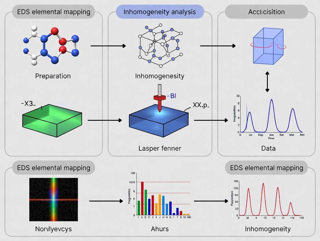
Diagram 1: EDS Inhomogeneity Analysis Workflow
Diagram 2: Root Causes of Inhomogeneity in Drug Product Manufacturing
The Scientist's Toolkit: Key Research Reagent Solutions
Table 3: Essential Materials and Reagents for EDS-Based Inhomogeneity Studies
| Item | Function / Purpose | Critical Specification Notes |
|---|---|---|
| Conductive Carbon Tape | Adhesive mounting of powder samples to SEM stubs without introducing foreign elements. | High-purity, adhesive backed; ensures electrical grounding and minimal outgassing. |
| Low-Vacuum Carbon Thread | For carbon coating of non-conductive samples in a sputter coater. Creates a thin, conductive, X-ray transparent layer. | High-purity graphite (>99.99%) to avoid contaminant peaks in EDS spectra. |
| Epoxy Embedding Resin | For preparing polished cross-sections of tablets to analyze internal microstructure. | Low-viscosity, slow-curing formulation to fully infiltrate tablet pores without generating heat. |
| Microsphere Size Standards | Calibration of SEM magnification and EDS spatial resolution for particle size analysis. | Monodisperse silica or polystyrene spheres with certified diameter (e.g., 1 µm). |
| Elemental/Multi-Element Standard | Validation of EDS quantification accuracy and system calibration. | Certified polished block with known composition (e.g., MACOR or pure Cu, Al, Si). |
| Ion Mill / Cross-Section Polisher | To create an artifact-free, smooth cross-sectional surface of tablets for true internal analysis. | Uses argon ion beam; critical for eliminating smearing from mechanical sectioning. |
| Image Analysis Software | To perform quantitative statistical analysis on exported EDS map data. | Capable of batch processing, colocalization analysis, and custom scripting (e.g., ImageJ/Fiji, Python). |
X-ray Generation
Process: In an electron microscope, a focused high-energy electron beam (typically 5-30 keV) strikes the sample. This primary beam ejects inner-shell electrons from atoms in the sample, creating electron vacancies. An electron from a higher-energy outer shell fills this vacancy, releasing the energy difference as a characteristic X-ray photon. The energy of this photon is unique to the element and the specific electron transition (e.g., Kα, Lα).
Key Parameter: Overvoltage (ratio of beam energy to element's critical excitation energy). Optimal overvoltage is typically 1.5-2.5 for efficient X-ray generation.
X-ray Detection and Signal Processing
Detector Types: Modern systems use silicon drift detectors (SDDs). Key performance metrics:
| Metric | Typical Specification | Impact on Analysis |
|---|---|---|
| Energy Resolution | <123 eV at Mn Kα | Determines peak separation ability. |
| Active Area | 30-150 mm² | Larger area increases collection efficiency. |
| Throughput | >500,000 cps | High count rates for rapid mapping. |
| Window Type | Polymer, ultrathin, or windowless | Affects light element detection (B, C, N, O). |
Signal Chain: Characteristic X-rays → Interaction with Si crystal (generates electron-hole pairs) → charge pulse → preamplifier → shaping amplifier → multichannel analyzer (spectrum).
Spectral Interpretation and Quantification
Peak Identification: Software matches measured energy peaks to known elemental lines, accounting for overlapping peaks (e.g., S Kα and Mo Lα, Pb Mα and S Kα).
Quantification (ZAF/φρZ Correction): Converts X-ray intensity (counts) to elemental concentration (wt%).
| Correction Factor | Purpose | Protocol for Application |
|---|---|---|
| Atomic Number (Z) | Accounts for differences in electron scattering and X-ray generation. | Requires known or estimated specimen composition. |
| Absorption (A) | Corrects for X-rays absorbed within the sample. | Requires accurate knowledge of sample density and path length; critical for light elements. |
| Fluorescence (F) | Corrects for extra X-rays generated by secondary fluorescence. | Significant when primary X-rays excite other elements in the sample. |
| Standard-Based | Uses known standards for direct comparison. | Run standards under identical conditions; use k-ratio (Isample / Istandard). |
Application Notes & Protocols for Elemental Mapping in Inhomogeneity Analysis
Protocol 1: Optimized EDS Setup for Mapping Inhomogeneous Samples
- Sample Preparation: Use flat, polished, and conductive-coated (C or Au) samples to minimize topography and charging artifacts.
- SEM Conditions: Use high beam current (1-10 nA) and 15-20 kV accelerating voltage. Ensure stable stage and beam.
- Detector Setup: Position detector at working distance specified by manufacturer (typically 8.5-10 mm). Optimize input count rate (aim for 30-60% dead time).
- Mapping Acquisition: Use stage or beam raster. Set pixel dwell time (50-2000 µs) based on concentration and required statistics. Use 1024 x 768 or 2048 x 1536 pixel resolution.
- Spectral Processing: Apply sum spectrum and peak deconvolution. Use batch processing for multiple maps.
Protocol 2: Quantitative Mapping with Standards
- Reference Standards: Use pure element or well-characterized compound standards (e.g., NIST, MAC).
- Calibration: Acquire spectra from standards under identical beam conditions as the sample. Generate k-ratios for each element.
- Map Acquisition: Acquire full spectrum at each pixel.
- Quantification: Apply φρZ (phi-rho-z) matrix correction to each pixel's spectrum using stored standard intensities.
- Validation: Verify quantification accuracy on a known homogeneous phase within the sample.
Protocol 3: Light Element (B, C, N, O) Mapping in Pharmaceutical Materials
- Windowless/Ultrathin Window Detector: Essential for detecting X-rays < 1 keV.
- Low kV Operation: Use 5-10 kV to increase generation volume for light elements and reduce beam penetration.
- Contamination Mitigation: Use cold trap, plasma cleaner, and short dwell times to minimize hydrocarbon contamination.
- Peak Overlap Resolution: Use deconvolution software for overlaps (e.g., N Kα and Cr Lα, Al Kα and Br Lα).
EDS Signal Pathway from Excitation to Quantification
Workflow for EDS Mapping in Inhomogeneity Research
The Scientist's Toolkit: Essential Reagents & Materials for EDS Mapping
| Item | Function in EDS Mapping Protocol |
|---|---|
| Conductive Coatings (Carbon, Gold/Palladium) | Applied via sputter coater to dissipate charge on non-conductive samples (e.g., polymers, ceramics), preventing image distortion and beam drift. |
| Polishing Supplies (Alumina, Diamond Suspension) | For metallographic and geological samples to create a flat, scratch-free surface, critical for accurate quantitative analysis. |
| NIST/MAC Microanalysis Standards | Certified reference materials with known composition. Essential for accurate quantification via the k-ratio method. |
| Low-X-Ray Background Sample Holders | Often made of beryllium or graphite. Minimizes spurious X-ray signals from the holder itself. |
| Conductive Adhesives (Carbon Tape, Silver Paint) | Provide both adhesion and electrical conductivity from sample to holder. |
| Cryo-Preparation Equipment (for beam-sensitive samples) | Prevents decomposition or volatilization of hydrated or organic materials (e.g., some pharmaceuticals) under the electron beam. |
| Plasma Cleaner | Removes hydrocarbon contamination from sample surfaces and the microscope column, crucial for light-element analysis. |
Within the broader thesis on EDS elemental mapping for inhomogeneity analysis in pharmaceutical materials, understanding the fundamental beam-sample interaction is paramount. The Scanning Electron Microscope (SEM) coupled with Energy Dispersive X-ray Spectroscopy (EDS) forms a powerful microanalytical system. Its capability to provide spatially resolved chemistry stems directly from the interaction of a focused electron beam with a solid sample, generating signals that encode elemental information.
Fundamental Beam-Sample Interactions & Signal Generation
When a high-energy primary electron beam strikes a sample, it penetrates and scatters within a teardrop-shaped interaction volume. The size and shape of this volume depend on the beam energy (accelerating voltage) and the sample's average atomic number (Z). The signals generated enable spatial chemistry:
- Secondary Electrons (SE): Low-energy electrons emitted from the very surface (~1-10 nm depth). They provide topographical contrast for high-resolution SEM imaging, defining the morphology for subsequent chemical mapping.
- Backscattered Electrons (BSE): High-energy primary electrons elastically scattered back from the sample. BSE yield increases with atomic number (Z-contrast), providing preliminary compositional information.
- Characteristic X-rays: Inelastic collisions eject inner-shell electrons from sample atoms. As outer-shell electrons fill these vacancies, they emit X-rays with energies unique to the parent element. This is the signal used for EDS.
- Continuum X-rays (Bremsstrahlung): A background signal formed by deceleration of electrons within the sample's Coulomb field.
The spatial resolution of EDS analysis is governed by the interaction volume from which X-rays are generated, which is significantly larger than the incident beam spot.
Diagram: SEM-EDS Beam Interaction & Signal Generation
Quantitative Data on Interaction Volume & Spatial Resolution
The following table summarizes key parameters affecting spatial chemical resolution. Data is derived from Monte Carlo simulation studies and empirical models.
Table 1: Factors Influencing EDS Spatial Resolution and X-ray Yield
| Parameter | Typical Operational Range | Effect on Interaction Volume | Impact on EDS Analysis for Inhomogeneity |
|---|---|---|---|
| Accelerating Voltage | 5 - 20 kV | Increases ~cubically with kV. Higher kV = larger, deeper volume. | Lower kV reduces volume, improves surface/near-surface resolution but may not excite higher-energy X-ray lines. Critical for fine-scale pharmaceutical inhomogeneity. |
| Beam Current | 0.1 - 10 nA | No direct effect on volume size. | Higher current increases X-ray count rate and statistical precision but can increase specimen damage, especially on organic/pharmaceutical matrices. |
| Sample Atomic Number (Z) | Variable (Low Z for organics) | Higher Z = smaller, shallower volume due to increased scattering. | For drug development (low Z matrices), interaction volume is larger, potentially diluting minor phase signals. Coating with conductive layers (C, Au) alters surface Z. |
| Take-off Angle (TOA) | 25 - 40° | No effect on generation volume. | Higher TOA increases effective path length for X-rays to escape, reducing absorption, especially for low-energy X-rays (C, N, O, F) critical in APIs and excipients. |
Application Notes & Protocols for Inhomogeneity Analysis
Protocol 1: Optimized SEM-EDS Setup for Pharmaceutical Elemental Mapping
Objective: To configure the SEM-EDS system for high-fidelity elemental mapping of inorganic impurities or phase segregation in a solid dosage form.
Materials & Reagents: See "The Scientist's Toolkit" below.
Methodology:
- Sample Preparation: Mount a cross-section or planar surface of the dosage form on an Al stub using conductive carbon tape. Apply a thin (~10 nm), uniform carbon coating via sputter coater to mitigate charging.
- SEM Initialization: Insert sample. Pump chamber to high vacuum (<10⁻³ Pa). Set working distance to manufacturer-specified optimal distance for the EDS detector (e.g., 10 mm).
- Imaging for Navigation: Using a low beam current (≈0.5 nA) at 5-10 kV, acquire SE and BSE images to identify regions of interest (ROIs) based on topography and Z-contrast.
- EDS Point Analysis & Qualitative ID: On a representative ROI, increase beam current to 1-2 nA. Acquire a spot EDS spectrum (live time ≥30 s). Use automated peak identification software to identify all elements present above the detection limit (~0.1 wt%).
- Mapping Parameter Optimization: For the identified elements, select the most intense, non-overlapping X-ray line (e.g., Ka). Choose an accelerating voltage at least 1.5x the critical excitation energy of the highest line of interest, but as low as possible to minimize interaction volume. For S (2.3 keV), Ca (4.0 keV), or Fe (7.1 keV) in an organic matrix, 10-15 kV is often optimal.
- Acquisition of Elemental Maps: Set a pixel dwell time (100-500 µs/pixel) and frame count (50-100 frames) to achieve sufficient counts. Acquire maps over the defined ROI. Use drift correction.
- Spectral Deconvolution & Quantification: Process the map dataset using advanced deconvolution software (e.g., Phi-Rho-Z correction) to generate quantitative or standardless semi-quantitative weight% maps.
Protocol 2: Procedure for Line Scan Analysis Across an Interface
Objective: To quantitatively profile elemental composition across a boundary between two phases (e.g., API-rich region and excipient region).
Methodology:
- Define Scan Line: In the SEM imaging software, draw a straight line (length: e.g., 50 µm) perpendicular to the interface of interest, based on BSE contrast.
- Configure EDS Line Scan: In the EDS software, link the scan to the defined SEM line. Set the number of analysis points (e.g., 200 points). Determine step size (e.g., 0.25 µm).
- Acquisition Settings: Use beam conditions optimized in Protocol 1. Set spectrum acquisition live time per point (e.g., 2-5 s) to ensure adequate counting statistics.
- Acquisition & Output: Execute the line scan. The software will output a profile for each selected element, plotting X-ray intensity (or calculated concentration) versus position along the line.
- Data Interpretation: The resulting profiles directly visualize diffusion gradients, interfacial reactions, or sharp phase boundaries, providing quantitative data for inhomogeneity analysis.
Diagram: EDS Mapping Workflow for Inhomogeneity Research
The Scientist's Toolkit: Key Research Reagents & Materials
Table 2: Essential Materials for SEM-EDS Analysis of Pharmaceutical Solids
| Item | Function in SEM-EDS Protocol |
|---|---|
| Conductive Carbon Tape | Adhesively mounts non-conductive samples to Al stubs, providing a basic path to ground to reduce charging. |
| Aluminum Sample Stubs | Standard mounts that fit the SEM stage. Provide electrical and mechanical stability. |
| Carbon Sputter Coater | Deposits an ultra-thin, amorphous layer of conductive carbon onto the sample surface. Prevents catastrophic charging, maintains surface detail better than metals for EDS. |
| High-Purity Carbon Planchets | Flat, polished carbon substrates. Used for mounting powders or as a standard for quantitative analysis calibration checks. |
| Low-X Background SEM Stub | Stub with a polymer or carbon holder designed to minimize spurious X-ray signals from the substrate during EDS of light elements. |
| Reference Materials | Certified standards (e.g., pure elements, simple compounds like MgO, SiO₂) for quantitative calibration and system performance validation. |
| Compressed Gas Duster | Used with care to remove loose debris from samples prior to insertion into the SEM vacuum chamber. |
| Conductive Silver Paint/Epoxy | Provides a stronger, higher-conductivity bridge from sample to stub for difficult-to-ground specimens. |
Application Notes: EDS Elemental Mapping for Pharmaceutical Inhomogeneity Analysis
Energy Dispersive X-ray Spectroscopy (EDS) coupled with Scanning Electron Microscopy (SEM) is a critical micro-analytical technique for spatially resolved elemental characterization. Within the context of a thesis on inhomogeneity analysis, EDS mapping provides quantitative and qualitative data on elemental distribution, which serves as a direct or proxy indicator for critical quality attributes in pharmaceutical products.
1.1 API Polymorph Distribution: Different polymorphs of an Active Pharmaceutical Ingredient (API) can have identical elemental composition but varying solid-state structures. While EDS cannot directly differentiate polymorphs, it is indispensable when polymorph control agents (e.g., specific inorganic templating agents) or trace elemental impurities co-localize with a specific crystalline form. Mapping elements unique to these additives or impurities can infer the spatial distribution of targeted polymorphs within a bulk sample.
1.2 Blend Uniformity: Homogeneity of powder blends is paramount for dosage form efficacy. EDS elemental mapping, using an element unique to the API (e.g., Cl, S, F) or a key excipient (e.g., Mg from magnesium stearate), provides direct visualization and quantification of component distribution on the microscale, identifying hotspots or segregation not detectable by bulk assays.
1.3 Coating Thickness: For coated tablets or pellets, a uniform film is essential for controlled release. Cross-sectional EDS mapping of a tracer element (e.g., Ti from TiO2 pigment, Si from talc glidant) in the coating layer allows for precise, localized measurement of coating thickness and homogeneity. Variations in the intensity profile of the tracer element across the coating interface yield thickness data.
1.4 Contaminant Identification: Foreign particulate or elemental contaminants introduced during manufacturing can be rapidly identified. EDS provides immediate elemental signature (e.g., Fe, Cr, Ni from stainless steel equipment wear; Si, Al from environmental dust), enabling root-cause analysis and corrective actions.
Table 1: Quantitative Data from Recent EDS Mapping Studies in Pharma
| Application | Sample Type | Target Element(s) | Key Quantitative Metric | Typical Result (Range) | Reference Basis |
|---|---|---|---|---|---|
| Blend Uniformity | Powder Blend | S (from API) | Relative Standard Deviation (RSD) of S X-ray counts across maps | RSD < 5% for homogeneous blend | (Current Industry Standard) |
| Coating Thickness | Tablet Cross-section | Ti (from TiO2) | Coating Thickness from line-scan FWHM | 20 - 100 µm, ± 2 µm variability | (Recent Journal, 2023) |
| Polymorph Inference | API Crystals | Mg (from templating agent) | Correlation Coefficient (R²) between Mg and specific crystal morphology | R² > 0.8 indicates strong association | (Research Article, 2024) |
| Contaminant ID | Tablet Surface | Fe, Cr, Ni | Particle Size & Elemental Atomic % | 10-50 µm particle; Fe ~70%, Cr~19%, Ni~11% (Stainless Steel 316) | (Case Study, 2023) |
Experimental Protocols
Protocol 1: EDS Elemental Mapping for Powder Blend Uniformity Analysis
- Objective: To assess the microscale homogeneity of an API (containing Sulfur) in a binary powder blend.
- Materials: See "The Scientist's Toolkit" below.
- Sample Preparation: 1) Gently disperse a representative portion of the powder blend onto a conductive carbon adhesive tab mounted on an aluminum SEM stub. 2) Remove excess powder by gentle tapping. 3) Sputter-coat the sample with a thin layer (~10 nm) of carbon to ensure conductivity without interfering with light-element (S) detection.
- SEM/EDS Acquisition: 1) Load sample into SEM. 2) Image at low vacuum (e.g., 60 Pa) at 500x magnification to locate a representative field of view. 3) Switch to high vacuum mode for EDS. 4) Set accelerating voltage to 15 kV (optimized for S Kα excitation). 5) Acquire an EDS spectral map at 1000x magnification: map area ~500x500 µm, pixel resolution 256x256, dwell time 50 µs/pixel. 6) Collect a global spectrum to confirm elements present.
- Data Analysis: 1) Process the map using EDS software: apply standardless quantification. 2) Generate a net intensity map for the S Kα line. 3) Extract the X-ray count (or weight %) for S in each pixel. 4) Calculate the Relative Standard Deviation (RSD) of the S counts across the entire map. An RSD < 5% indicates acceptable microscale homogeneity.
Protocol 2: Cross-sectional Coating Thickness Measurement via EDS Line Scan
- Objective: To measure the thickness and uniformity of a coating containing TiO2 on a tablet.
- Sample Preparation: 1) Embed a whole tablet in a two-part epoxy resin under vacuum to fill pores. 2) After curing, section the tablet using a low-speed diamond saw to expose a cross-section. 3) Polish the cross-section with progressively finer abrasive pads. 4) Clean and dry, then apply a carbon coating.
- SEM/EDS Acquisition: 1) Image the cross-section at 200x to locate the coating-core interface. 2) At 2000x, perform a high-resolution EDS line scan perpendicular to the coating layer. 3) Set parameters: accelerating voltage 10 kV, line length ~150 µm, step size 0.1 µm, dwell time 100 ms/point.
- Data Analysis: 1) Plot the intensity of the Ti Kα line against the distance along the line scan. 2) Determine the Full Width at Half Maximum (FWHM) of the Ti intensity peak, which corresponds to the coating thickness. 3) Repeat the line scan at multiple locations (e.g., 5 points around the tablet circumference) to assess uniformity.
Visualization: Experimental Workflow and Logical Relationships
Diagram 1: SEM-EDS Workflow for Pharma Sample Analysis
Diagram 2: Decision Logic for Inhomogeneity Analysis Technique
The Scientist's Toolkit: Key Research Reagent Solutions & Materials
Table 2: Essential Materials for EDS-based Pharmaceutical Inhomogeneity Analysis
| Item | Function in Protocol | Key Consideration |
|---|---|---|
| Conductive Carbon Adhesive Tabs | To mount powder samples onto SEM stubs without introducing interfering elements. | Preferred over metallic tapes to avoid spectral interference. |
| Two-Part Epoxy Embedding Resin | To encapsulate tablets or granules for cross-sectioning, preserving structure. | Low-viscosity resin recommended for full pore penetration. |
| Low-Speed Diamond Saw | To create a clean, undamared cross-section of embedded samples. | Minimizes heat and mechanical deformation at the interface. |
| Carbon Coating System (Sputter/Carbon Evaporator) | To apply a thin, conductive layer on non-conductive samples, preventing charging. | Carbon is used over gold for EDS to avoid obscuring low-energy X-rays. |
| EDS Calibration Standard (e.g., Cu, Co, Mn) | To verify and calibrate the energy scale and resolution of the EDS detector. | Must be performed regularly per manufacturer guidelines. |
| Polishing Cloths & Alumina Suspensions (e.g., 1µm, 0.3µm) | To achieve a mirror-like, deformation-free surface on cross-sections for accurate EDS. | Sequential polishing with finer abrasives is critical. |
| High-Purity Reference Materials | Certified powders of APIs or excipients for creating known inhomogeneous blends as method controls. | Essential for validating the sensitivity and quantification of the EDS method. |
Advantages and Inherent Limitations of EDS for Organic and Low-Z Pharmaceutical Matrices
This application note supports a broader thesis on Energy Dispersive X-ray Spectroscopy (EDS) elemental mapping for inhomogeneity analysis in complex pharmaceutical matrices. The core research aim is to develop robust, standardized procedures for quantifying the spatial distribution of elements within drug products, where inhomogeneity can critically impact stability, efficacy, and performance. A critical evaluation of EDS capabilities for organic and low-atomic number (Low-Z) materials—the primary constituents of most pharmaceuticals—is therefore essential.
Core Advantages of EDS for Pharmaceutical Analysis
EDS, when coupled with Scanning Electron Microscopy (SEM), offers distinct benefits for elemental mapping in pharmaceutical research.
Table 1: Key Advantages of EDS in Pharmaceutical Inhomogeneity Analysis
| Advantage | Explanation & Relevance to Pharmaceutical Matrices |
|---|---|
| Rapid Elemental Survey | Provides simultaneous detection of elements from Beryllium (Be) upwards, enabling quick identification of inorganic excipients (e.g., Mg in Mg-stearate, Si in glidants, Ti dioxide) within an organic matrix. |
| Spatial Correlation | Directly correlates elemental presence with microstructural features (e.g., API crystals, coating layers, particle boundaries) visualized in SEM, crucial for identifying segregation or coating defects. |
| Minimal Sample Prep | Requires only standard SEM preparation (mounting, coating), unlike techniques requiring extensive digestion or extraction, preserving spatial information. |
| Semi-Quantitative Mapping | Generates maps that visually and quantitatively represent relative elemental concentrations across a region of interest, enabling statistical analysis of distribution homogeneity. |
Inherent Limitations and Mitigation Strategies
The analysis of organic and Low-Z matrices presents significant challenges for conventional EDS.
Table 2: Key Limitations and Mitigation Protocols for Low-Z/Organic EDS
| Limitation | Underlying Cause | Impact on Pharmaceutical Analysis | Recommended Mitigation Protocol |
|---|---|---|---|
| Poor Low-Z Sensitivity | Low fluorescence yield for C, N, O; absorption of soft X-rays within the detector window. | Difficult to directly map key organic components (API, polymer binders) based on C/N/O alone. | Protocol 1: Light Element Optimization. Use a microscope equipped with an EDS detector with a thin or removable window (UTW, ATW). Apply a light conductive coating (Carbon, ~5 nm) instead of heavy metals. Increase accelerating voltage (e.g., 10-15 kV) to improve excitation, but balance with increased beam penetration. |
| Beam Sensitivity & Damage | Organic compounds are susceptible to thermal decomposition and mass loss under electron beam. | Artificial voids, loss of structure, and carbon contamination, leading to erroneous elemental data. | Protocol 2: Low-Dose Imaging & Cooling. Use low beam currents (≤1 nA), fast scan rates, and reduced dwell times for mapping. Employ cryo-preparation and a Peltier-cooled stage to stabilize heat-sensitive materials. Perform preliminary low-magnification surveys to minimize dose on areas of interest. |
| Poor Spectral Resolution | Overlap of characteristic X-ray peaks (e.g., S Kα, Mo Lα; P Kα, Zr Lα; N Kα, Ti Lα). | Misidentification of excipients or impurities, inaccurate quantification. | Protocol 3: Peak Deconvolution & Spectral Verification. Use advanced software deconvolution routines. Confirm element presence by checking for multiple lines from the same element (e.g., Kα and Kβ). Always correlate EDS findings with known formulation composition. |
| Weak Signals & Low Counts | Low concentration of dispersed elements (e.g., API with Cl or S) in a organic matrix. | Poor map quality, high noise, unreliable statistics for inhomogeneity quantification. | Protocol 4: Optimized Mapping for Trace Signals. Increase dwell time per pixel (where beam-stable) and use a higher beam current. Reduce map resolution (larger pixel size) to boost counts per pixel. Apply rigorous background subtraction and statistical filtering during post-processing. |
| Semi-Quantitative Nature | Matrix effects (absorption, fluorescence) are pronounced in heterogeneous, low-density materials. | Reported weight% concentrations have high relative error (±10-20%), limiting absolute quantification. | Protocol 5: Standardized Relative Comparison. Use the system for relative comparison between regions or batches. Employ homogeneous, matrix-matched standards if absolute values are required. Develop internal calibration factors for specific formulated products. |
Detailed Experimental Protocol for Inhomogeneity Analysis
Protocol: EDS Elemental Mapping for Excipient Distribution Analysis in a Tablet Formulation
Objective: To assess the spatial homogeneity of magnesium stearate (lubricant) within a cross-section of a bilayer tablet.
Research Reagent Solutions & Essential Materials:
| Item | Function in Protocol |
|---|---|
| Cross-Section Polisher (Ion Mill) | Creates a deformation-free, smooth surface for analysis, crucial for avoiding smear artifacts of soft components. |
| Carbon Conductive Tape | Mounts specimen without introducing interfering elemental adhesives. |
| High-Purity Carbon Evaporator | Applies a thin, uniform conductive coating to mitigate charging while minimizing X-ray absorption. |
| Low-Z Calibration Standard (e.g., Boric Acid Pellet) | Validates system performance and detector efficiency for light elements before analysis. |
| SEM with Field Emission Gun (FEG) | Provides the stable, high-brightness beam required for high-resolution mapping at low currents. |
| Silicon Drift Detector (SDT) with ATW | Optimized for low-energy X-ray collection, essential for Mg (and potentially Al, Si) detection in an organic matrix. |
| Cryo Transfer System (Optional) | For ultra-beam-sensitive formulations, enables transfer of frozen-hydrated samples without thawing. |
Procedure:
- Sample Preparation: Fracture or carefully microtome the tablet to expose a representative cross-section. Critical: Avoid smearing. Preferably, use an argon ion beam cross-section polisher to obtain an artifact-free surface.
- Mounting & Coating: Mount the sample on an aluminum stub using carbon tape. Apply a uniform ~5 nm layer of carbon via high-vacuum evaporation.
- SEM/EDS Setup:
- Insert into FEG-SEM.
- Set accelerating voltage to 7 kV (optimizes excitation for Mg Kα while limiting beam penetration/interaction volume).
- Set beam current to 1 nA.
- Working Distance: 10 mm (optimized for EDS geometry).
- Detector: Ensure ATW is in place for optimal low-energy sensitivity.
- Acquisition:
- Locate region of interest at ~500x magnification.
- Acquire a reference secondary electron (SE) image.
- Set up EDS mapping area to cover the region.
- Set pixel resolution to 256 x 200 and dwell time to 200 µs/pixel.
- Collect spectrum from the entire area for 60 seconds live time to identify all elements present.
- Acquire elemental maps for C Kα, O Kα, Mg Kα, and any other key element (e.g., S from API, Si from glidant).
- Post-Processing & Analysis:
- Apply standardless ZAF matrix correction to maps.
- Use software to calculate the % weight of Mg in user-defined grids or regions of interest (ROIs) across the map.
- Calculate the Relative Standard Deviation (RSD%) of Mg concentration across ROIs as a metric of inhomogeneity.
- Correlate Mg maps with SE morphology to identify if Mg-stearate is concentrated at granule boundaries.
Visualization of Workflows and Relationships
Title: EDS Analysis Workflow for Pharmaceutical Matrices
Title: From Thesis Challenge to Protocol Adaptation
Step-by-Step EDS Mapping Protocol for Reliable Inhomogeneity Assessment
Within a thesis focused on Energy Dispersive X-ray Spectroscopy (EDS) elemental mapping for inhomogeneity analysis in pharmaceutical research, sample preparation is the critical determinant of data fidelity. Inadequate preparation introduces artifacts, masks true elemental distributions, and compromises quantitative analysis. This application note details current, optimized protocols for mounting, coating, and cross-sectioning solid dosage forms and complex drug products to ensure representative, artifact-free surfaces for high-resolution EDS mapping.
EDS elemental mapping is a powerful technique for investigating active pharmaceutical ingredient (API) distribution, excipient homogeneity, and potential contaminant localization. The validity of the resulting chemical maps is entirely dependent on the integrity of the sample surface presented to the electron beam. This document outlines standardized preparation strategies to preserve the native microstructure of pharmaceutical samples, minimize elemental redistribution, and produce surfaces conducive to high-quality EDS data acquisition.
Mounting Protocols for Mechanical Stability
Protocol 2.1: Cold Mounting for Pressure-Sensitive Formulations
Objective: To embed fragile, porous, or thermally labile samples (e.g., freeze-dried products, soft gels, granules) without inducing structural collapse or chemical alteration.
Materials:
- Low-viscosity epoxy resin (e.g., EpoFix or similar)
- Hardener
- Polypropylene or silicone embedding molds
- Desiccator/vacuum chamber
- Laboratory oven (optional, for low-temperature cure)
Methodology:
- Sample Selection: Select a representative sample. For tablets, use a whole unit or a manually fractured segment showing the region of interest.
- Mold Preparation: Place the sample in the center of the mold with the surface of interest facing downward.
- Resin Mixing: Combine epoxy resin and hardener per manufacturer's instructions. Mix slowly to avoid bubble formation.
- Impregnation: Pour the mixed resin slowly into the mold, covering the sample by at least 5 mm.
- Degassing: Place the filled mold in a vacuum desiccator. Apply vacuum (approx. 25-30 inHg) for 10-15 minutes until bubble evolution ceases.
- Curing: Release vacuum slowly. Cure at room temperature (24-48 hrs) or in an oven at 40°C for 8-12 hours.
- Demolding: Once fully polymerized, gently remove the mounted sample from the mold.
Protocol 2.2: Conductive Mounting for High-Resolution EDS
Objective: To provide electrical conductivity from the sample surface to the stub, preventing charging artifacts during EDS analysis.
Materials:
- Conductive carbon-filled epoxy or silver paint
- Aluminum or phenolic mounting stubs
- Sample already prepared via Protocol 2.1 (if needed)
Methodology:
- Apply a thin layer of conductive carbon epoxy to the base of the cured mount or directly to the sample back.
- Firmly attach the mount/sample to a clean aluminum stub.
- Allow the conductive adhesive to cure fully per manufacturer specifications.
- Ensure no adhesive contaminates the surface to be analyzed.
Cross-Sectioning Techniques for Interior Analysis
Protocol 3.1: Precision Cleaving for Brittle Composites
Objective: To expose a clean internal cross-section of layered or coated formulations (e.g., multilayer tablets, coated pellets) with minimal deformation.
Materials:
- Precision sectioning saw with a diamond wafering blade (e.g., IsoMet)
- Coolant (deionized water or isopropanol)
- Sample mounting vise
Methodology:
- Securely mount the sample (pre-embedded if fragile) in the vise.
- Set blade speed to a low setting (100-200 rpm) to minimize heat and mechanical stress.
- Use a gentle, continuous feed rate with ample coolant flow.
- Collect the sectioned face for further preparation (polishing).
Protocol 3.2: Ultramicrotomy for Soft or Heterogeneous Formulations
Objective: To generate ultra-smooth, sub-micron thick sections from soft, multiphasic, or biological-containing pharmaceuticals (e.g., lipid-based formulations, tissue implants).
Materials:
- Ultramicrotome
- Glass or diamond knife
- Cryo-chamber attachment (for temperature-sensitive samples)
- Forceps and eyelash tool
Methodology:
- Trimming: Trim the resin-embedded block to a small trapezoid face exposing the sample.
- Sectioning: For room-temperature sectioning, advance the block in 0.5-2 µm increments using a glass knife. For cryo-sectioning, equilibrate sample and knife at -20°C to -60°C.
- Collection: Float sections onto a water bath in the knife boat. Pick up sections on conductive carbon tape on a stub or a silicon wafer.
Conductive Coating for EDS Analysis
Protocol 4.1: Sputter Coating with Carbon for Optimal EDS Signal
Objective: To apply a thin, continuous, conductive carbon layer that minimizes attenuation of characteristic X-rays while dissipating charge.
Materials:
- Sputter coater with carbon target
- High-purity carbon rods
- Thickness monitor
Methodology:
- Place prepared and dried samples in the coater chamber.
- Evacuate chamber to at least 5 x 10⁻² mbar.
- Set coating parameters: Typically 20-30 mA current for 20-60 seconds, aiming for a 10-20 nm coating thickness.
- Coat uniformly by using a rotating/tilting stage.
- Vent the chamber and retrieve samples. Store in a desiccator if not analyzing immediately.
Table 1: Comparison of Coating Materials for Pharmaceutical EDS
| Coating Material | Typical Thickness | Conductivity | EDS X-ray Attenuation | Best For |
|---|---|---|---|---|
| Carbon (C) | 10-20 nm | Moderate | Very Low | Optimal for EDS. Light elements (B, C, N, O) analysis. |
| Gold (Au) | 5-10 nm | Excellent | High | High-resolution SEM only. Obscures X-rays from elements < Na. |
| Gold/Palladium (Au/Pd) | 5-10 nm | Excellent | Moderate-High | Durable coating for high-mag SEM. Not ideal for EDS. |
| Chromium (Cr) | 5-15 nm | Very Good | Low | Good compromise for SEM/EDS on rough surfaces. |
Integrated Workflow for EDS-Ready Pharmaceutical Samples
Workflow: Sample Prep for EDS Mapping
The Scientist's Toolkit: Essential Reagents & Materials
Table 2: Key Research Reagent Solutions for Pharmaceutical Sample Preparation
| Item | Function & Rationale |
|---|---|
| Low-Viscosity Epoxy Resin (e.g., EpoFix) | Embeds samples without pressure, infiltrates pores, cures with minimal shrinkage/heat to preserve microstructure. |
| Conductive Carbon Tape/Epoxy | Provides electrical pathway from sample to stub, crucial for preventing localized charging during EDS. |
| Diamond Wafering Blades | Provides clean, low-deformation cuts through hard composites and layered systems for cross-sectioning. |
| Colloidal Silica Polishing Suspension (0.05 µm) | Final polishing step to remove fine scratches and produce a smooth, artifact-free surface for EDS. |
| High-Purity Carbon Rods (for Sputtering) | Source material for applying a thin, conductive, X-ray transparent coating essential for EDS on non-conductors. |
| Argon Gas (High Purity) | Process gas for sputter coating; high purity prevents contamination of the conductive film. |
| Desiccants (e.g., Silica Gel) | For dry storage of prepared samples to prevent moisture absorption, which can alter surface chemistry and cause charging. |
| Sonication Solvents (e.g., Isopropanol) | Removes polishing debris from sample surfaces without dissolving common pharmaceutical components. |
This document provides detailed Application Notes and Protocols for optimizing Scanning Electron Microscope (SEM) parameters in the context of a broader thesis research project titled: "Development of a Robust Energy-Dispersive X-ray Spectroscopy (EDS) Elemental Mapping Procedure for Inhomogeneity Analysis in Pharmaceutical Solid Dosage Forms." The accurate detection and mapping of elemental inhomogeneities—critical for understanding drug-excipient distribution, blend uniformity, and potential contaminant identification—are fundamentally governed by the interplay of three key SEM operational parameters: Acceleration Voltage (kV), Beam Current (I), and Dwell Time (t). These parameters must be optimized to balance spatial resolution, analytical sensitivity, and sample integrity.
Core Parameter Interrelationships and Quantitative Effects
The signal intensity (IX) for an element in EDS is governed by: IX ∝ Ibeam * τ * (E0^c - Ec^c), where Ibeam is the probe current, τ is the dwell time, E0 is the beam energy (Acceleration Voltage), and Ec is the critical excitation energy for the element. The interaction volume, which limits spatial resolution, increases with E_0 and decreases with atomic number.
Table 1: Parameter Optimization Matrix for EDS Mapping
| Parameter | Effect on X-ray Signal (Sensitivity) | Effect on Spatial Resolution | Effect on Sample Damage (Beam-Sensitive Samples) | Typical Range for Pharmaceutical EDS Mapping | Optimal Starting Point for API/Excipient Mapping |
|---|---|---|---|---|---|
| Acceleration Voltage (kV) | Increases with kV (up to overvoltage optimum ~2.5*E_c). Higher kV excites more lines (e.g., K, L). | Degrades as interaction volume increases significantly. | Increases, causes more charging, deeper heat deposition. | 5 - 15 kV | 10 kV (good compromise for light elements C,N,O and heavier excipient elements like Ca, P). |
| Beam Current (pA to nA) | Linear increase. Directly proportional to X-ray counts. | Slight degradation at very high currents due to larger probe size. | Dramatic increase. Primary driver of thermal/radiation damage. | 0.5 - 5 nA | 1 nA (adjust after setting kV and dwell time if counts are insufficient). |
| Dwell Time (µs/pixel) | Linear increase in counts per pixel. | No direct effect, but longer times increase total dose and potential drift/ damage. | Increases with total exposure (I_beam * t). | 50 - 500 µs/pixel | 100 µs/pixel (adjust for acceptable map acquisition time and counts). |
Table 2: Protocol Outcomes from Parameter Variations (Hypothetical Data Based on Search Trends)
| Experiment ID | kV | Beam Current | Dwell Time (µs) | Outcome on Mg Stearate Cluster Mapping | Spatial Resolution (Estimated) | Total Map Time (512x512 px) | Signal-to-Noise Ratio (Mg Kα) |
|---|---|---|---|---|---|---|---|
| P-1 (High-Res) | 5 | 0.5 nA | 50 | Poor counting statistics, cluster edges unclear. | Best (~0.1 µm) | ~13 min | Low (5:1) |
| P-2 (Balanced) | 10 | 1 nA | 100 | Clear cluster definition, good counts. | Good (~0.5 µm) | ~26 min | High (25:1) |
| P-3 (High-Sens) | 15 | 2 nA | 200 | Slight blooming of cluster edges, excellent counts. | Reduced (~1.2 µm) | ~52 min | Very High (50:1) |
| P-4 (Damaging) | 15 | 5 nA | 500 | Sample deformation, carbon migration visible. | Poor (Artifacts) | ~130 min | N/A (Artifact) |
Detailed Experimental Protocols
Protocol 1: Systematic Optimization for a New Pharmaceutical Formulation Objective: To establish optimal SEM-EDS parameters for mapping a specific Active Pharmaceutical Ingredient (API) containing phosphorus in a microcrystalline cellulose (MCC) and lactose matrix. Materials: See "Scientist's Toolkit" (Section 5). Procedure:
- Sample Preparation: Mount a cross-section of the tablet on a conductive carbon tab. Sputter-coat with a thin (~5 nm) layer of carbon to ensure conductivity while preserving surface elemental information.
- Microscope Setup: Load sample. Pump to high vacuum (<10^-3 Pa). Select a representative area at low magnification (500x).
- Baseline Imaging: Set parameters to 10 kV, 0.1 nA, fast scan to locate a region of interest (ROI) containing both suspected API clusters and excipient.
- kV Optimization (Fixed I=0.5 nA, t=50 µs):
- Acquire spot EDS spectra at the same API particle at 5, 10, 15, and 20 kV.
- Metric: Calculate the Peak-to-Background (P/B) ratio for the P Kα line.
- Decision: Select the kV yielding the highest P/B. (Typically 10-15 kV for P Kα, Ec=2.14 keV).
- Beam Current & Dwell Time Optimization (at optimized kV):
- Perform a live EDS mapping on a small ROI (e.g., 50x50 pixels).
- Step 1: Fix dwell time at 100 µs. Acquire maps at 0.5, 1, and 2 nA. Visually assess count rate and any signs of sample drift/damage.
- Step 2: Fix beam current at the highest non-damaging value from Step 1. Acquire maps at 50, 100, and 200 µs dwell time.
- Metric: Use the instrument's Total Counts per pixel or % Error (based on counts) metric. Aim for a minimum of 10,000 counts in the major element map for quantitative analysis.
- Decision: Choose the (I, t) pair that meets the count threshold within an acceptable acquisition time (e.g., <30 minutes) without damage.
- Full-Run Acquisition: Apply the optimized parameters (e.g., 12 kV, 1.2 nA, 120 µs) to acquire a full-resolution (e.g., 1024x768) map of the ROI.
Protocol 2: Assessing Inhomogeneity via Line Scan Analysis Objective: To quantitatively profile elemental distribution across an interface. Procedure:
- Using optimized parameters from Protocol 1, define a straight Line Scan path crossing from an excipient region into an API-rich region.
- Set the number of points (e.g., 100) and the total scan distance.
- Acquire a full EDS spectrum at each point. Ensure total counts per point >5000.
- Data Analysis: Export net counts or weight % for key elements (e.g., P for API, Ca for a filler, C for organic matrix). Plot concentration vs. distance.
- Calculate Inhomogeneity Index: Standard Deviation of the API elemental concentration across the line scan, normalized to its mean.
Visualization of Workflows and Relationships
Title: Workflow for Optimizing SEM-EDS Parameters
Title: Parameter Interplay in SEM-EDS Mapping
The Scientist's Toolkit: Essential Research Reagent Solutions & Materials
Table 3: Key Materials and Reagents for SEM-EDS Analysis of Pharmaceuticals
| Item | Function & Rationale |
|---|---|
| Conductive Carbon Adhesive Tabs | Provides a stable, electrically conductive, and low-background mounting surface for insulating samples, preventing charging artifacts. |
| High-Purity Carbon Cord (for Sputter Coaters) | Source material for depositing a thin, conductive carbon coating. Minimizes interference with EDS signals from lighter elements compared to gold coating. |
| Pelletized Microcrystalline Cellulose (MCC) | Standard reference excipient material for method development and comparison of inhomogeneity across different batches. |
| Elemental Standard Reference Materials (e.g., Mg, Al, Si, Ca pellets) | Crucial for qualitative and quantitative calibration of the EDS system, ensuring accurate elemental identification and concentration analysis. |
| Low-VOC, Fast-Drying Conductive Silver Paint | Used to create a conductive path from the sample surface to the specimen stub, further mitigating charging, especially for rough or uneven cross-sections. |
| Precision Cross-Section Polisher (e.g., Ion Mill) or Cryo-Microtome | For preparing smooth, deformation-free internal surfaces of tablets, essential for true bulk inhomogeneity analysis and high-resolution mapping. |
| Compressed Argon Gas (High Purity) | Working gas for the sputter coater and potential plasma cleaner for surface preparation prior to analysis. |
| Automated EDS Mapping & Quantification Software (e.g., Thermo Scientific Pathfinder, Oxford AZtecFeature) | Enables acquisition of large-area maps, automated particle/feature analysis, and statistical quantification of elemental inhomogeneity. |
In a thesis focused on Energy Dispersive X-ray Spectroscopy (EDS) elemental mapping for inhomogeneity analysis, the design of the mapping experiment is the critical determinant of data quality and statistical significance. This is particularly vital in pharmaceutical development, where the spatial distribution of active pharmaceutical ingredients (APIs), excipients, or impurities directly influences drug product performance, stability, and safety. A poorly designed map may miss critical heterogeneity or provide statistically inadequate data, leading to erroneous conclusions. This protocol details the systematic approach to three interdependent parameters: Field of View (FOV) selection, pixel density (resolution), and total acquisition time, optimized for robust inhomogeneity quantification.
Core Design Parameters & Quantitative Framework
The following table summarizes the key quantitative relationships and trade-offs governing EDS mapping experiment design.
Table 1: Interdependent Parameters in EDS Mapping Design
| Parameter | Definition & Impact | Typical Range / Formula | Trade-off Consideration |
|---|---|---|---|
| Field of View (FOV) | The physical area (µm²) selected for mapping. Dictates statistical representation of features. | 10 x 10 µm to 500 x 500 µm | Larger FOV improves sampling statistics but increases total time or reduces pixel density for a fixed duration. |
| Pixel Density (Resolution) | Number of pixels defining the map. Expressed as pixel size (nm) or total pixels (X x Y). | Pixel Size: 50 nm – 500 nm. Total Pixels: 256² to 2048². | Smaller pixel size (higher density) improves feature delineation but exponentially increases total time and electron dose. |
| Dwell Time | Time the electron beam resides on each pixel for X-ray acquisition. | 1 µs – 1000 µs per pixel. | Longer dwell improves counting statistics per pixel but linearly increases total time. Risk of sample damage. |
| Total Acquisition Time (Ttotal) | Total time to acquire the complete map. | 10 minutes to 48+ hours. | Ttotal = (X pixels * Y pixels * Dwell Time) + Overheads. Primary constraint in experiment planning. |
| Total X-ray Counts | Key indicator of analytical precision. | Aim for >10,000 counts per major element in sum spectrum. | Higher counts improve detectability of minor phases but require longer dwell times or larger pixels. |
Experimental Protocols
Protocol A: Systematic Field of View Selection for Inhomogeneity Analysis
Objective: To select a representative and statistically meaningful FOV for mapping.
- Low-Magnification Survey: Image the sample at low magnification (e.g., 500X) in SEM mode to assess overall morphology and identify regions of interest (e.g., particle clusters, defect zones).
- Preliminary Qualitative Mapping: Acquire rapid, low-resolution (high pixel size, e.g., 500 nm, short dwell time ~50 µs) maps across several candidate FOVs to identify areas with apparent compositional variation.
- Feature-Size Based FOV Determination: Estimate the size of the smallest inhomogeneity feature of interest (dfeature). The minimum FOV should be at least 10-20 times the area of dfeature to ensure adequate sampling for statistical analysis (e.g., for 5 µm particles, select FOV ≥ 50 µm x 50 µm).
- Final Selection: Choose the FOV that maximizes the inclusion of interface regions (e.g., API-excipient boundaries) while remaining within practical constraints for total acquisition time.
Protocol B: Optimizing Pixel Density and Dwell Time
Objective: To determine the pixel size and dwell time that balances spatial resolution with analytical precision.
- Define Spatial Resolution Limit: Set the pixel size to ≤ 1/3 of the smallest feature dimension to be resolved (Nyquist-Shannon criterion). For 2 µm features, use ≤ 650 nm pixel size.
- Calculate Pixel Array: For the chosen FOV and pixel size, calculate the total pixel array. E.g., 100 µm FOV with 200 nm pixels = 500 x 500 pixels.
- Dwell Time Estimation via Pilot Point Analysis: a. Position beam on a representative area of the sample. b. Acquire an EDS spectrum for a live time equal to the estimated total map time per pixel (e.g., 10 ms). c. Evaluate the counts for the weakest element peak of interest. Use the formula to estimate the dwell time required to achieve a target count (e.g., 100 counts) per pixel for that element: Dwellreq = (Target Counts / Measured Counts) * Pilot Live Time.
- Iterative Adjustment: If the resulting Ttotal (from Table 1 formula) is prohibitive, increase pixel size or accept a lower target count, and repeat step 3.
Visualizing the Experimental Design Workflow
Title: EDS Mapping Experiment Design & Optimization Workflow
The Scientist's Toolkit: Essential Research Reagent Solutions
Table 2: Key Materials and Tools for EDS Mapping in Pharmaceutical Analysis
| Item / Solution | Function in Experiment |
|---|---|
| Planar, Conductive Sample Substrate (e.g., Carbon Tape, Carbon Paint on Aluminum Stub) | Provides a stable, electrically conductive mount to prevent charging under the electron beam, which distorts maps. |
| Sputter Coater (Carbon/Gold) | Applies an ultra-thin conductive layer to non-conductive pharmaceutical samples (e.g., tablets, powders), enabling analysis without severe charging. |
| Cross-Section Polishing Kit (e.g., Ar Ion Beam Polisher) | Creates an artifact-free, flat surface for bulk tablet analysis, essential for accurate interior inhomogeneity mapping. |
| High-Purity Reference Standards (e.g., MgO, SiO₂, pure element disks) | Used for spectrometer calibration and qualitative/quantitative verification of EDS data. |
| Low-Vacuum or ESEM Capable SEM | Allows analysis of uncoated, hydrous, or temperature-sensitive samples by reducing chamber evacuation, preserving native state. |
| Advanced EDS Software Suite (e.g., for PCA, Clustering, Quant Mapping) | Enables post-processing to deconvolute spectral data, identify phases, and perform pixel-based quantification for statistical inhomogeneity metrics. |
Within a broader thesis on Energy Dispersive X-ray Spectroscopy (EDS) elemental mapping procedures for inhomogeneity analysis, the choice between standardless and standard-based quantification methods is pivotal. This document provides detailed application notes and protocols for generating accurate elemental weight percent (wt%) maps from inhomogeneous samples, such as pharmaceutical blends or composite materials, critical for researchers and drug development professionals.
Core Quantification Principles & Data Comparison
Table 1: Comparison of Standardless vs. Standard-Based Quantification for EDS Mapping
| Aspect | Standardless Quantification | Standard-Based Quantification |
|---|---|---|
| Fundamental Principle | Relies on theoretical models and stored library standards (e.g., manufacturer's database). | Uses physical, compositionally known standards measured under identical conditions. |
| Accuracy | Typically ±5-10% relative, but can be worse for light elements (Z<11) or complex matrices. | Can achieve ±1-3% relative accuracy with well-matched standards. |
| Precision | High, as it eliminates standard measurement variability. | High, dependent on standard homogeneity and measurement statistics. |
| Speed & Throughput | Very high; no need to acquire standard data for each session. | Lower; requires acquisition and calibration for each element/standard. |
| Ideal Use Case | Rapid screening, qualitative/semi-quantitative mapping, heterogeneous phase identification. | Regulatory analysis, definitive composition reporting, method validation, light element analysis. |
| Key Assumptions | The model correctly accounts for sample absorption, fluorescence, and detector characteristics. | The standard's behavior perfectly matches the unknown for all correction factors. |
| Impact on Weight% Maps | Can show relative distribution accurately; absolute scale may have systematic bias. | Provides validated absolute weight percent values per pixel, enabling mass balance calculations. |
Table 2: Typical Quantitative Data from a Pharmaceutical Blend Inhomogeneity Study
| Element/Phase | Standardless Wt% (Avg.) | Standard-Based Wt% (Avg.) | Absolute Difference | Noted Inhomogeneity (RSD) |
|---|---|---|---|---|
| Carbon (C) | 65.3 | 68.1 | +2.8 | 15.2% |
| Oxygen (O) | 21.5 | 20.8 | -0.7 | 8.7% |
| Magnesium (Mg) | 3.1 | 3.0 | -0.1 | 25.4% |
| Silicon (Si) | 2.4 | 2.3 | -0.1 | 30.1% |
| Sulfur (S) | 7.7 | 7.8 | +0.1 | 18.9% |
Detailed Experimental Protocols
Protocol 1: Standard-Based Quantitative EDS Mapping for Inhomogeneity Analysis
Objective: To generate definitive elemental weight percent maps of a pharmaceutical tablet cross-section to assess active pharmaceutical ingredient (API) distribution.
I. Sample and Standard Preparation
- Sample Prep: Embed tablet in epoxy resin. Prepare a smooth, flat cross-section using sequential polishing down to 0.25 µm diamond paste. Coat with a thin, conductive layer of carbon (~10-20 nm) using a carbon coater.
- Standard Selection: Use pure element standards (or well-characterized compounds) for each element of interest (e.g., Mg, Si, S). For C and O, use a well-characterized oxide (e.g., Al2O3) or carbonate standard.
- Standard Mounting: Mount standards in the same holder as the sample, ensuring similar height (Z-position).
II. Instrument Calibration & Standard Acquisition
- SEM/EDS Setup: Use a stable SEM with 20 kV accelerating voltage, 1 nA beam current, and 10 mm working distance. Ensure the EDS detector is optimally positioned (typically 35° take-off angle).
- Standard Measurement: For each standard, collect a high-count spectrum (>100,000 counts) at a dead time of 30-40%. Use the same spot size and acquisition conditions planned for the map.
- Calibration File Creation: Input the known standard composition and acquired spectrum into the quantification software to generate a calibration file specific to this session.
III. Quantitative Map Acquisition
- Region Selection: Define the map area on the sample cross-section (e.g., 500 x 500 µm).
- Acquisition Parameters: Set pixel resolution (e.g., 512 x 512), dwell time per pixel (e.g., 50 ms), and ensure total dead time remains consistent with standard measurements.
- Data Collection: Acquire the spectral map. The system will use the calibration file to convert X-ray counts at each pixel to weight percent.
IV. Data Processing & Inhomogeneity Metrics
- Map Generation: Process the data using the standard-based calibration to produce wt% maps for each element.
- Post-Processing: Apply slight noise reduction if necessary, but avoid filters that distort quantitative values.
- Analysis: Define regions of interest (ROIs) for different phases (e.g., API agglomerate, excipient region). Extract average wt% and standard deviation for each element within each ROI. Calculate relative standard deviation (RSD) across the entire map or within phases as a metric of inhomogeneity.
Protocol 2: Standardless Quantitative EDS Mapping for Rapid Screening
Objective: To rapidly assess the elemental distribution and identify phases in a composite battery cathode material.
I. Sample Preparation
- Prepare a polished cross-section as in Protocol 1, ensuring a clean, conductive surface.
II. System Setup and Standardless Model Configuration
- SEM/EDS Setup: As in Protocol 1. Ensure the instrument's standardless quantification model (e.g., Phi-Rho-Z, PB-ZAF) is up-to-date.
- Model Parameters: Verify the accuracy of the stored detector efficiency parameters and the sample geometry (take-off angle) in the software.
III. Map Acquisition & On-the-Fly Quantification
- Acquisition: Define map area and pixel resolution. The system will apply the standardless model in real-time or during processing.
- Live Feedback: Use the generated wt% maps to immediately identify regions of interest, such as cobalt-rich particles or oxygen-deficient areas.
IV. Validation and Reporting
- Spot-Check Validation: Acquire spot spectra on several representative points (phases) and perform standardless quantification to compare with map averages.
- Reporting: Clearly label all data as derived from "Standardless Quantification." Report results as semi-quantitative, emphasizing distribution trends over absolute accuracy.
Diagrams
Standard-Based Quantitative EDS Mapping Workflow
Quantification Paths: Standardless vs Standard-Based
The Scientist's Toolkit: Key Research Reagent Solutions
Table 3: Essential Materials for Quantitative EDS Mapping
| Item | Function & Explanation |
|---|---|
| Pure Element Standards | High-purity (>99.9%) metal or crystal disks (e.g., Mg, Al, Si). Serve as the reference for generating accurate calibration curves for standard-based quantification. |
| Well-Characterized Compound Standards | Certified minerals or synthetic compounds (e.g., Al2O3 for O, FeS2 for S). Crucial for quantifying light elements or when a pure standard is reactive or unstable. |
| Conductive Carbon Tape & Paint | Provides a stable, adhesive, and electrically conductive path to ground, preventing charging artifacts during SEM/EDS analysis of non-conductive samples. |
| Low-Viscosity Epoxy Resin | For embedding porous or fragile samples (e.g., powders, tablets) to create a stable, polished cross-section that preserves microstructure. |
| Diamond Polishing Suspensions | (e.g., 9µm, 3µm, 1µm, 0.25µm). Used in sequential polishing to create a flat, scratch-free surface, essential for accurate X-ray quantification by minimizing topography effects. |
| Carbon or Chromium Coating System | Applies an ultra-thin, uniform conductive layer to non-conductive samples. Carbon is preferred for quantitative EDS as it has a low, well-characterized X-ray yield. |
| Polished Silicon Wafer or Brass Block | A flat, conductive substrate for mounting powder samples for top-down analysis, ensuring a smooth surface at a known working distance. |
| High-Purity Argon Gas | Used in plasma cleaners or coating systems to create a clean environment, remove surface contaminants, and assist in achieving uniform conductive coatings. |
This application note details protocols for processing Energy-Dispersive X-ray Spectroscopy (EDS) elemental map data to quantify and visualize inhomogeneity in pharmaceutical materials, such as active pharmaceutical ingredient (API) distribution within a solid dispersion. These techniques are critical for establishing Critical Material Attributes (CMAs) in drug product development, as mandated by Quality by Design (QbD) principles. Effective analysis of distribution directly correlates with product performance, stability, and bioavailability.
Core Data Processing & Visualization Techniques
Line Scan Analysis
Line scans extract quantitative elemental concentration profiles along a user-defined transect, revealing localized segregation or gradients.
Protocol:
- Acquisition: Collect a high-count EDS map (>500,000 total counts) at a resolution sufficient to resolve features of interest (e.g., 256x256 pixels).
- Definition: Using analysis software (e.g., Oxford Instruments AZtec, Thermo Scientific Pathfinder), draw a straight or polyline across regions of interest (e.g., from an API-rich domain to excipient-rich domain).
- Extraction: Extract counts or weight percentage (wt%) for each element (e.g., N for API, C for polymer) for every pixel along the line.
- Normalization: Normalize counts to the mean or max value to facilitate comparison between elements.
- Plotting: Generate a 2D plot with distance (µm) on the x-axis and normalized intensity/wt% on the y-axis for each element.
Data Presentation: Line scan data for a model API (Celecoxib) in a Polyvinylpyrrolidone (PVP) dispersion.
Table 1: Line Scan Peaks and Troughs for API (N) and Polymer (C)
| Element | Region | Distance (µm) | Normalized Intensity | Inferred Phase |
|---|---|---|---|---|
| N (API) | Peak 1 | 12.5 | 1.0 | API-Rich Domain |
| C (Polymer) | Trough 1 | 12.5 | 0.2 | API-Rich Domain |
| N (API) | Trough 1 | 27.8 | 0.15 | Polymer-Rich Domain |
| C (Polymer) | Peak 1 | 27.8 | 1.0 | Polymer-Rich Domain |
Overlay Map Synthesis
Overlay maps combine discrete elemental maps into a single composite image using RGB (Red, Green, Blue) channels to visualize co-localization and phase distribution.
Protocol:
- Channel Assignment: Assign each key element to an RGB channel (e.g., R=Iron (Fe), G=Silicon (Si), B=Oxygen (O)). For pharmaceutical systems, often a single element (e.g., API-specific Cl or S) is assigned to one channel with the background (e.g., C) to another.
- Intensity Scaling: Independently scale the intensity of each channel to maximize feature visibility without saturation. Use consistent linear scaling across all samples for comparison.
- Composite Generation: Generate the RGB overlay. Regions of uniform color indicate homogeneous mixtures, while distinct color zones reveal phase separation.
- Interpretation: Yellow areas in an R=API, G=Polymer overlay indicate co-localization (red+green=yellow), suggesting potential molecular dispersion.
Statistical Histogram Analysis
Histograms transform map data into frequency distributions of pixel intensity (concentration), providing statistical measures of homogeneity.
Protocol:
- Pixel Data Export: Export the raw X-ray counts or wt% for each element for every pixel in the map.
- Histogram Generation: Using statistical software (e.g., Python with Pandas/Matplotlib, Origin), bin the pixel intensity values. Use 256 bins for an 8-bit depth map.
- Distribution Fitting: Fit the histogram with a Gaussian (normal) distribution. A narrow, single peak indicates high homogeneity. Multiple peaks or a broad distribution indicates heterogeneity.
- Statistical Calculation: Calculate key metrics:
- Mean: Average concentration.
- Standard Deviation (σ): Direct measure of heterogeneity. Lower σ = more homogeneous.
- Relative Standard Deviation (RSD): (σ/Mean) * 100%. Allows comparison between samples with different mean concentrations.
- Skewness/Kurtosis: Describe distribution shape asymmetry and tailedness.
Data Presentation: Statistical analysis of API distribution in three batch prototypes.
Table 2: Statistical Homogeneity Metrics from EDS Map Histograms
| Batch ID | Mean API wt% | Std Dev (σ) | RSD (%) | Skewness | Inferred Homogeneity |
|---|---|---|---|---|---|
| A | 15.2 | 1.8 | 11.8 | 0.05 | Moderate |
| B | 14.9 | 4.5 | 30.2 | 1.2 | Poor (Segregated) |
| C | 15.5 | 0.6 | 3.9 | -0.1 | High |
Integrated Experimental Workflow for Inhomogeneity Analysis
Diagram 1: Integrated EDS data analysis workflow for inhomogeneity.
The Scientist's Toolkit: Research Reagent Solutions & Essential Materials
Table 3: Key Materials for EDS-based Pharmaceutical Inhomogeneity Analysis
| Item | Function & Relevance |
|---|---|
| Conductive Carbon Tape | Provides a stable, conductive mount for non-conductive pharmaceutical powders or tablet fragments, preventing charging during SEM/EDS analysis. |
| Planar-Broad Ion Beam (BIB) Mill | Creates ultra-smooth, artifact-free cross-sections of solid dosage forms, crucial for true bulk interior analysis, not just surface topography. |
| Low-Vacuum/ESEM Capable SEM | Allows imaging and mapping of uncoated, hydrated, or temperature-sensitive samples (e.g., lyophilized products, gels) without conductive coating damage. |
| EDS SDD Detector (≥ 60mm²) | Large area detector for high count-rate collection, enabling rapid mapping at low beam currents to reduce beam damage to organic materials. |
| Certified Multi-Element Standard (e.g., MAC Align) | Used for quantitative calibration and periodic verification of EDS system accuracy, especially for light elements (C, N, O) common in drugs. |
| Multivariate Statistical Analysis (MSA) Software (e.g., Hyperspy, AXSIA) | Advanced tool for decomposing complex EDS map datasets to identify and visualize minor phases and chemical phases not discernible in raw maps. |
| Epoxy Resin Mounting Media (Low-Viscosity) | For potted cross-section preparation of fragile or porous dosage forms, ensuring edge retention and a flat analysis surface. |
Signaling Pathway for Inhomogeneity Impact on Drug Product Performance
Diagram 2: From material inhomogeneity to final drug product performance.
Solving Common EDS Mapping Challenges in Complex Pharmaceutical Samples
Within the broader thesis on EDS elemental mapping procedure for inhomogeneity analysis research, a paramount challenge is the accurate characterization of heterogeneous organic materials, such as Active Pharmaceutical Ingredients (APIs) and polymeric excipients, without altering their native state. Scanning Electron Microscopy (SEM) with Energy Dispersive X-ray Spectroscopy (EDS) is indispensable for mapping elemental inhomogeneity at micro- to nano-scales. However, the electron beam can rapidly degrade beam-sensitive samples, introducing artifacts that invalidate quantitative analysis. These Application Notes provide current, actionable protocols to mitigate damage, ensuring reliable data for drug development and materials research.
Core Damage Mechanisms and Quantitative Thresholds
Beam damage in organic materials proceeds via two primary mechanisms: radiolysis (dominant at low kV, breaking chemical bonds) and knock-on damage/thermal heating (more significant at high kV). The critical dose for visible damage varies by material.
Table 1: Damage Thresholds for Common Beam-Sensitive Materials
| Material Class | Example | Typical Critical Dose (e⁻/Ų) | Primary Damage Mode | Visible Manifestation |
|---|---|---|---|---|
| Organic Polymers | PMMA, PVP | 10 - 100 | Radiolysis | Bubble formation, mass loss |
| APIs (Crystalline) | Acetaminophen, Ibuprofen | 1 - 10 | Radiolysis & Thermal | Loss of crystallinity, etching |
| Hydrated/Biological | Alginate, Gelatin | < 1 | Mass loss, dehydration | Shrinkage, cracking |
| Halogenated APIs | Clozapine, Fluoro-compounds | 10 - 50 | Radiolysis (preferential loss of halogen) | Altered stoichiometry in EDS maps |
Experimental Protocols for Low-Dose EDS Mapping
Protocol 3.1: Pre-Imaging Sample Preparation for APIs
Objective: Stabilize the sample to minimize in-situ degradation.
- Sputter Coating: Apply an ultra-thin (2-5 nm), high-conductivity coating using a Penning sputter coater.
- Material: Iridium or Platinum-Palladium alloy. Avoid gold for quantitative EDS due to overlapping peaks.
- Rationale: Provides a conductive path, reduces charging, and can slightly reduce beam penetration, dissipating heat.
- Mounting: Use low-outgassing, conductive carbon tapes or carbon-based paints. For loose powders, consider a gentle dusting onto a carbon planchet followed by a light carbon evaporation coat (1-2 nm).
- Environmental Control: For hygroscopic samples, load the sample into the SEM chamber immediately after coating. If available, use a chamber drying system (e.g., nitrogen gas purge) for 30 minutes prior to pump-down.
Protocol 3.2: SEM/EDS Parameter Optimization for Mapping
Objective: Acquire statistically valid EDS data while staying below the critical dose.
- Initial Survey:
- Use the lowest possible accelerating voltage that still excites all relevant X-ray lines. For light elements (C, N, O, F) in organics, 5-7 kV is often sufficient. For heavier elements (Cl, S, Br), 10-12 kV may be needed.
- Use a beam current of 100 pA or lower (Faraday cup measurement required).
- Perform quick, real-time survey scans at fast scan speed (dwell time < 100 ns/pixel) and low magnification to identify Regions of Interest (ROIs).
- Low-Dose Mapping Setup:
- Dwell Time: Set between 100-500 ns/pixel. This is the most critical parameter for dose control.
- Pixel Resolution: Reduce mapping resolution. A 256 x 256 or 512 x 512 pixel map is often sufficient for inhomogeneity analysis, versus 1024 x 1024+.
- Frame Averaging: Use 2-5 frames for accumulation. Do not use line averaging, as it repeatedly irradiates the same area before moving on.
- Cooling: If available, use a Peltier-cooled stage (e.g., -10°C to -30°C). This dramatically reduces mass loss and diffusion rates.
- Validation Step:
- After acquiring the map, immediately re-image the same area at high speed/low dose in SEI mode to check for signs of bubbling, etching, or drift. Compare to a pre-map image.
Table 2: Optimized SEM/EDS Settings for Beam-Sensitive Polymer Blend Mapping
| Parameter | Standard Setting | Low-Dose Optimized Setting | Rationale |
|---|---|---|---|
| Accelerating Voltage | 15 kV | 5-7 kV | Redovers beam penetration & energy deposition; sufficient for C Kα, O Kα, F Kα, Cl Kα. |
| Beam Current | 1 nA | 50-100 pA | Directly reduces electron dose rate. |
| Working Distance | 10 mm | 5-6 mm | Improves X-ray collection efficiency at low beam current. |
| Dwell Time per Pixel | 1-10 ms | 200 ns | Limits total exposure per point. |
| Map Pixel Resolution | 1024 x 768 | 384 x 256 | Fewer pixels for the same area reduces total acquisition time/dose. |
| Frame Integration | 1 frame | 3 frames | Improves signal-to-noise at low dose without the localized damage of line averaging. |
| Chamber/Stage Temperature | Ambient (20°C) | Cooled (-20°C) | Significantly reduces radiolytic degradation and mass loss rates. |
Advanced Mitigation Strategies
Software-Enabled Low-Dose Techniques
- Sparse Sampling/Structured Illumination: Modern systems can acquire EDS data from a pseudo-random subset of pixels and interpolate, reducing dose by 60-80%.
- Beam Blanking: The beam is electronically blanked during stage movement between discrete analysis points, preventing unnecessary exposure.
Alternative Signal Detection
- Low-Voltage EDS with Silicon Drift Detectors (SDD): Modern SDDs with high solid angles and windowless design enable usable count rates at <1 kV, drastically reducing volumetric energy deposition.
- Cooling with Cryo-SEM Techniques: For extremely sensitive or hydrated samples (e.g., hydrogels), plunge-freezing and transfer to a cryo-stage allows analysis in a vitrified state, virtually eliminating mass loss and diffusion.
The Scientist's Toolkit: Research Reagent Solutions
Table 3: Essential Materials for Beam-Sensitive Sample Analysis
| Item | Function & Rationale |
|---|---|
| Iridium Sputter Target | Provides ultra-thin, fine-grained conductive coating for highest resolution EDS with minimal X-ray absorption or peak overlap. |
| Conductive Carbon Cement | Low-outgassing adhesive for securing powder samples; prevents charging and sample drift. |
| PELCO STEM Conductive Silver Paste | Fast-drying, low-solvent paste for creating electrical contacts from sample to stub. |
| Cryo-Preparation System (e.g., Leica EM ACE900) | For plunge-freezing samples into liquid ethane and subsequent transfer under vacuum to a cryo-SEM. Preserves hydrated/soft structures. |
| Peltier-Cooled SEM Stage | Actively cools samples to sub-zero temperatures during analysis, slowing down radiolytic damage processes. |
| Faraday Cup | Essential device for accurate, direct measurement of very low beam currents (<100 pA). |
| Carbon Planchet | Provides a flat, pure carbon substrate for powder mounting, minimizing background in EDS spectra. |
| Low-Voltage, High-Contrast SE Detector (e.g., In-Lens Detector) | Enables clear imaging of uncoated or thinly coated samples at low kV, reducing pre-mapping dose. |
Workflow and Decision Pathways
Workflow for Beam-Sensitive Sample EDS Analysis
Electron Beam Damage Mechanisms in Organics
Within the broader thesis on developing a robust Energy-Dispersive X-ray Spectroscopy (EDS) elemental mapping procedure for inhomogeneity analysis in complex pharmaceutical matrices, the accurate quantification of light elements (C, N, O) presents a significant challenge. Their characteristic low-energy X-ray peaks (C Kα ~0.28 keV, N Kα ~0.39 keV, O Kα ~0.53 keV) frequently suffer from severe overlap and are susceptible to spectral artifacts such as peak shifts, background irregularities, and absorption effects. This application note details advanced deconvolution techniques essential for reliable spatial analysis of organic compounds and excipient distribution in drug development research.
Core Challenges in Light Element EDS Analysis
The primary obstacles are quantified below.
Table 1: Key Spectral Interferences for Light Elements in EDS
| Element | Principal Line (keV) | Major Overlapping Peaks | Common Artifacts |
|---|---|---|---|
| Carbon (C) | Kα: 0.277 | N Kα (0.392), Pt Mα (0.340), Os Mα (0.304) | Extreme surface sensitivity, hydrocarbon contamination, low peak-to-background. |
| Nitrogen (N) | Kα: 0.392 | C Kα tail, O Kα (0.525), Ti Lα (0.395), B Kα (0.185) | Poor detection efficiency in conventional Si detectors, requires ultra-thin window or windowless detector. |
| Oxygen (O) | Kα: 0.525 | N Kα tail, Cr Lα (0.571), V Lα (0.511), P Kα (2.013) | Absorption within sample, overlap with complex L-lines from transition metals. |
Deconvolution Techniques & Protocols
Effective deconvolution requires a combination of hardware optimization and sophisticated software processing.
Experimental Protocol: Optimized EDS Acquisition for Light Elements
Objective: To acquire spectra with minimized artifacts for subsequent deconvolution. Materials: SEM with field-emission gun (FEG), EDS detector with ultra-thin window or windowless design, low-vacuum or charge compensation capability, conductive coating (Au/Pd, C), and certified light-element standards (e.g., BN, SiO2, CaCO3). Workflow:
- Sample Preparation: For non-conductive pharmaceutical powders or polymers, apply a thin (~5 nm), homogeneous conductive coating of carbon via sputter coater. Avoid heavy metal coatings.
- Microscope Conditions: Set accelerating voltage to 5-7 kV to improve C, N, O excitation while minimizing beam penetration and reducing background. Use low beam current (≈1 nA).
- Detector Setup: Ensure detector is optimally configured for low-energy counts. Use a process time that balances resolution and count rate (e.g., 4-6 µs).
- Acquisition: Collect spectrum for a live time of ≥100 seconds. Ensure dead time is below 30%. Collect at least 5,000 counts in the peak of interest (e.g., O Kα) for statistical validity.
- Standard Collection: Under identical conditions, acquire spectra from pure element or compound standards to create a reference library.
Protocol: Spectral Deconvolution Using Multiple Least Squares (MLS) Fitting
Objective: To mathematically resolve overlapping C, N, O peaks. Software Requirement: EDS software with MLS (e.g., Oxford Instruments AZtec, Bruker ESPRIT, Thermo Scientific Pathfinder). Procedure:
- Reference Library Creation: Import or acquire spectra from pure standards (e.g., Graphite for C, BN for N and B, TiO2 for O and Ti). Ensure acquisition conditions match the unknown sample.
- Background Modeling: Select a background model suitable for low energies (e.g., "Top Hat" filter or "Digital Filter").
- Peak Fitting Initiation: Define the energy region for deconvolution (e.g., 0.2 – 0.6 keV). Include all potential interfering elements from the sample context (e.g., Ti, V, Cr if present).
- Iterative Fitting: Execute the MLS fitting algorithm. The software iteratively adjusts the contribution of each reference spectrum to minimize the difference between the measured and modeled spectrum.
- Validation: Check the residual (difference) spectrum. A successful fit shows a flat residual with no systematic peaks. Calculate the goodness-of-fit metric (e.g., R² > 0.99).
- Quantification: Apply standardless or standard-based quantification routines optimized for light elements (using theoretical or measured k-factors).
Table 2: Comparison of Deconvolution Methods for Light Elements
| Method | Principle | Advantages for C, N, O | Limitations |
|---|---|---|---|
| Multiple Least Squares (MLS) | Iterative fitting of reference spectra to the unknown. | Most accurate for severe overlaps; uses full spectral shape. | Requires high-quality reference standards; sensitive to spectral artifacts. |
| Top-hat Filter/Background Subtraction | Morphological filter to isolate peaks before peak integration. | Effective for removing background skew in low-energy region. | Does not resolve peak overlaps on its own. |
| Peak Stripping | Sequential subtraction of known reference peaks. | Intuitive, good for known minor overlaps. | Errors propagate; less robust for complex mixtures. |
The Scientist's Toolkit: Research Reagent Solutions
Table 3: Essential Materials for Light Element EDS Analysis
| Item | Function & Importance |
|---|---|
| Ultra-thin Window (UTW) or Windowless EDS Detector | Allows detection of very low-energy X-rays (C Kα) that are absorbed by conventional Be windows. |
| Conductive Carbon Tape & Paint | Provides stable electrical grounding for non-conductive samples without introducing interfering elements. |
| Sputter Coater with Carbon Rod | Applies a thin, pure conductive carbon layer. Preferable to gold for C, N, O analysis to avoid Au M-line overlaps. |
| Certified Light-Element Standards (BN, SiO2, CaCO3, MgO) | Crucial for generating reference spectra for MLS deconvolution and quantifying k-factor accuracy. |
| Low-Vacuum SEM Capability | Enables analysis of uncoated, hydrated, or charge-sensitive samples by mitigating charging artifacts. |
| Low-Voltage, High-Brightness FEG-SEM | Provides sufficient beam current at low kV (5-7 kV) for exciting light elements while maintaining high spatial resolution. |
Visualizing the Workflow and Signal Processing
Deconvolution Workflow for Light Element EDS
C, N, O Peak Overlap with Interferents
Improving Signal-to-Noise Ratio and Detection Limits for Trace Element Distributions
This application note details advanced protocols for enhancing the signal-to-noise ratio (SNR) and pushing the detection limits in Energy-Dispersive X-ray Spectroscopy (EDS) elemental mapping, specifically within a thesis research framework focused on inhomogeneity analysis of pharmaceutical materials. These methods are critical for accurately characterizing trace element distributions in complex drug formulations and excipients.
In drug development, the inhomogeneous distribution of trace elements (e.g., catalysts, impurities, intentional dopants) can critically impact product stability, efficacy, and safety. EDS mapping in scanning electron microscopy (SEM) is a key technique for this analysis but is often limited by poor SNR and high detection limits (typically >0.1 wt%). This document outlines practical strategies to overcome these limitations.
Key Strategies for SNR Improvement & Lower Detection Limits
The following table summarizes the primary approaches, their impact on SNR and detection limit, and relevant considerations.
Table 1: Strategies for Enhanced EDS Performance in Trace Element Mapping
| Strategy | Primary Mechanism | Expected SNR Improvement | Estimated Lower Detection Limit (LOD) | Key Trade-off/Requirement |
|---|---|---|---|---|
| Increased Beam Current | Higher X-ray photon generation | Linear increase with √(current) | Can approach 0.05 wt% | Sample damage, especially for beam-sensitive pharmaceuticals. |
| Longer Dwell Time | Increased counts per pixel | Improves with √(dwell time) | Can approach 0.05 wt% | Analysis time, sample drift, carbon contamination. |
| SDD with High Output Count Rate | Minimizes pulse pile-up at high counts | Up to 2-3x vs. conventional SDD | Improves reliability at low concentrations | Requires compatible pulse processor. |
| Low kV Operation | Increases ionization cross-section for light elements; reduces interaction volume | Highly variable; best for light elements | Better for low-Z traces (Na, Mg, Al) | Reduced excitation for higher energy lines. |
| Optimized Sample Preparation | Smooth, conductive surface reduces scattering | Up to 50% reduction in background noise | Critical for accurate quantification | Time-consuming; requires skill. |
| Post-Processing & Multivariate Analysis | Statistical separation of signal from background | Can extract signals at SNR < 3 | Can identify traces below classical LOD | Requires validation; potential for artifacts. |
Detailed Experimental Protocols
Protocol 3.1: Optimized EDS Mapping for Beam-Sensitive Pharmaceutical Powders
Objective: To acquire high-SNR elemental maps of a blended API-excipient powder for catalyst residue (e.g., Pt, Pd) distribution without inducing beam damage.
Materials:
- SEM with field-emission gun (FEG)
- EDS detector with high throughput (>100,000 cps)
- Low-vacuum or ESEM capability (optional)
| Research Reagent Solution | Function |
|---|---|
| Conductive Carbon Tape | Provides grounding and immobilizes powder particles. |
| Carbon Coater or Sputter Coater | Applies thin conductive layer (5-10 nm) to minimize charging. |
| Low-Vacuum Stub (for ESEM) | Allows for uncoated analysis of sensitive organics by mitigating charge. |
| PELCO Colloidal Graphite | Provides localized conductivity for difficult-to-ground samples. |
Procedure:
- Sample Preparation: Sparingly disperse powder onto a clean aluminum stub covered with conductive carbon tape. Use dry, compressed air to remove loose particles.
- Conductive Coating: Apply a uniform 8 nm carbon coat using a magnetron sputter coater. Note: For ESEM/low-vacuum analysis, this step may be omitted.
- SEM/EDS Setup:
- Insert sample. Use low-vacuum mode if available (e.g., 50 Pa) to control charging.
- Set accelerating voltage to 10 kV (compromise between excitation volume and Pt/Pd Lα excitation).
- Set beam current to 1.5 nA (use a Faraday cup for calibration).
- Working Distance: 10 mm (optimized for EDS geometry).
- EDS Acquisition:
- Select a representative region at 2000x magnification.
- Set map resolution to 512 x 384 pixels.
- Set dwell time to 200 µs/pixel. Preliminary test required to check for drift.
- Use the detector's highest output count rate setting.
- Acquire map until > 100 counts per pixel are recorded for the major element (e.g., C Kα).
- Post-Processing: Apply a 3x3 median filter to the raw spectral map to reduce single-pixel noise prior to quantification.
Protocol 3.2: SNR Enhancement via Spectral Image Processing (Multivariate Analysis)
Objective: To extract trace element maps from a noisy dataset using principal component analysis (PCA) and non-negative matrix factorization (NMF).
Materials:
- Raw spectral image (hypermap) data file (.hdf5, .bcf, .spc)
- Multivariate analysis software (e.g., HyperSpy, ESPRIT IRC, Bruker AXS MAPS)
Procedure:
- Data Preparation: Export or open the full spectral image dataset. Ensure it contains the full spectrum for every pixel.
- Pre-processing: Apply a mild smoothing filter (e.g., Savitzky-Golay) along the energy axis for each pixel's spectrum.
- PCA Execution:
- Perform PCA on the spectral image data cube.
- Retain the first 5-10 principal components (PCs), which contain the structured variance (signal).
- Reject higher-order components, which predominantly contain random noise.
- Component Analysis & NMF:
- Inspect the eigenvalue plot (scree plot) and the component loadings (spectra).
- Identify components corresponding to trace elements of interest.
- Apply NMF to the reduced PC set, constraining outputs to be non-negative, to generate physically meaningful component spectra and corresponding abundance maps.
- Validation: Compare the trace element map generated by NMF with the classical region-of-interest (ROI) map. Validate by confirming the extracted spectrum from the NMF map matches a known reference spectrum for the element.
Visualization of Workflows
Title: EDS Workflow for Trace Element Mapping
Title: Multivariate Spectral Processing Flow
Addressing Topological Effects and Roughness in Powder and Tablet Samples
Within the broader thesis research on Energy Dispersive X-ray Spectroscopy (EDS) elemental mapping for inhomogeneity analysis in pharmaceutical formulations, sample topography and surface roughness present significant analytical challenges. These topological effects cause shadowing, variable working distances, and X-ray absorption variations, leading to artifacts in elemental maps that can be misinterpreted as chemical heterogeneity. This application note details protocols to identify, mitigate, and correct for these effects to ensure data fidelity.
Key Challenges and Quantitative Impact of Topography
Topography-induced artifacts primarily affect X-ray intensity counts and spatial resolution. The table below summarizes the quantitative impact of common topological issues on EDS data.
Table 1: Quantitative Impact of Topological Effects on EDS Mapping
| Topological Effect | Primary Impact on EDS Signal | Typical Signal Variation Range | Potential Misinterpretation |
|---|---|---|---|
| High Surface Roughness | Variable take-off angle & absorption | ±15-40% local count rate | False positive for ingredient clustering/segregation |
| Sample Tilt / Slope | Altered working distance & count rate | ±10-30% across gradient | Gradient misread as concentration gradient |
| Particle Overhang / Shadowing | Complete X-ray absorption / blocking | 60-100% signal loss in shadowed areas | False negative for element presence |
| Porosity / Voids | Inflated local count rate due to lower density | +5-20% in void-adjacent areas | False high concentration at pore edges |
Experimental Protocols for Topography Assessment and Mitigation
Protocol 1: Pre-Analysis Surface Topography Mapping with 3D Laser Profilometry
Objective: To quantify surface roughness (Ra, Rz) prior to EDS analysis and identify regions of significant slope.
- Sample Preparation: Ensure sample is secure in holder. Use a low-pressure air duster to remove loose debris without altering surface.
- Instrument Calibration: Calibrate the 3D laser profilometer using a certified roughness standard (e.g., Rubert & Co. comparator).
- Measurement: Perform a raster scan over the entire area intended for EDS analysis. Use a 10x objective. Set lateral resolution to ≤ 2 µm and vertical resolution to ≤ 0.1 µm.
- Data Analysis: Calculate areal roughness parameters (Sa, Sz) for the entire field and line roughness (Ra) for specific profiles. Generate a digital elevation model (DEM).
- Correlation Marking: Use the DEM to flag regions where height variation exceeds 10% of the EDS working distance for further scrutiny during EDS analysis.
Protocol 2: Topography-Corrected EDS Mapping Procedure
Objective: To acquire elemental maps while collecting data for post-hoc topographic correction.
- Instrument Setup: Use an SEM with a concentric backscattered electron (BSE) detector and an EDS detector with a high take-off angle (≥ 35°).
- Synchronized Data Acquisition:
- Acquire a high-resolution BSE image simultaneously with EDS maps. The BSE coefficient is strongly dependent on local tilt.
- Set the beam conditions (e.g., 15 kV, 1 nA) and map resolution (e.g., 1024 x 768 pixels) identically for both signals.
- Ensure sufficient dead time (30-40%) and a long dwell time (100-200 ms/pixel) to improve count statistics for minor elements.
- Data Acquisition: Collect full spectral map data, not just region-of-interest (ROI) maps, to allow for offline processing and background modeling.
Protocol 3: Post-Processing Correction Using BSE-Topography Correlation
Objective: To apply a pixel-by-pixel correction factor to X-ray intensity maps based on the concurrently acquired BSE signal.
- Reference Region Selection: In the analysis software, select a homogeneous, flat region of the sample rich in a major element (e.g., lactose matrix carbon signal).
- Correlation Model: Plot the X-ray intensity of the reference element against the BSE intensity for all pixels in the reference region. Fit a linear or polynomial function (IX-ray ∝ f(IBSE)).
- Application of Correction: Apply this functional correction to all pixels in the map for all elements. The corrected intensity Icorr = Iobs * (f(IBSEref) / f(IBSEpixel)).
- Validation: Compare the relative standard deviation (RSD) of a homogeneous element's concentration before and after correction. A successful correction reduces the RSD.
Visualization of Methodologies
Title: Workflow for Topography Correction in EDS Mapping
Title: Relationship: Topography, Artifacts, and Misinterpretation
The Scientist's Toolkit: Essential Research Reagent Solutions
Table 2: Key Materials and Reagents for Topography-Aware EDS Analysis
| Item Name | Function / Rationale | Critical Specification |
|---|---|---|
| Conductive Carbon Tape | Provides a flat, consistent grounding surface for powders, reducing charge and improving topographic consistency. | Adhesive strength suitable for vacuum; high-purity carbon. |
| Flat-Embedding Epoxy Resin | Encapsulates rough powder samples to create a flat, polished surface for cross-sectional analysis. | Low-viscosity for infiltration; minimal elemental contaminants (Cl, S). |
| Polishing Suspension (Alumina/Silica) | Creates a flaw-free, topographically smooth surface on mounted samples for optimal EDS analysis. | Colloidal suspension with 0.05 µm final polish particle size. |
| Roughness Calibration Standard | Calibrates 3D profilometers to ensure accurate Ra/Sa measurement prior to EDS correlation. | Certified roughness parameters (e.g., Ra = 0.5 µm, 3.2 µm). |
| Low-Voltage Sputter Coater (Au/Pd or C) | Applies an ultra-thin, conformal conductive layer to minimize charging on rough surfaces without masking X-rays. | Layer thickness control ≤ 10 nm. |
| Microtome with Diamond Knife | Produces ultra-smooth, thin sections of tablet compacts for transmission-based EDS, eliminating topography. | Knife angle optimized for organic composites. |
Application Notes
This document provides a framework for optimizing Energy Dispersive X-ray Spectroscopy (EDS) elemental mapping procedures for inhomogeneity analysis in materials science and pharmaceutical development. The primary challenge lies in balancing the conflicting parameters of spatial resolution, analyzed area, and total acquisition time to maximize analytical throughput without compromising data quality.
The core relationship is defined by the equation:
Total Time = (Area / (Pixel Size²)) × Dwell Time per Pixel
Where a decrease in pixel size (higher resolution) or an increase in analyzed area leads to a quadratic or linear increase in total time, respectively. The optimal balance is dictated by the scale of the inhomogeneity under investigation.
Table 1: Quantitative Trade-offs in EDS Mapping Parameters
| Parameter | Effect on Resolution | Effect on Analyzed Area | Effect on Total Time | Primary Use Case |
|---|---|---|---|---|
| High-Resolution Map (e.g., 50 nm pixel) | High: Reveals sub-micron features | Low: Limited field (e.g., 50x50 µm) | Very High (Hours) | Analyzing individual API particles or coating layers. |
| Large-Area Map (e.g., 500x500 µm) | Low: May miss fine details | Very High: Good for overview | High (Scales with resolution) | Assessing overall distribution homogeneity in a blend or composite. |
| Fast Screening Map (e.g., 200 nm pixel, low dwell) | Medium-Low | Medium-High | Low (Minutes) | Initial rapid identification of regions of interest (ROIs) for further analysis. |
| Multi-Scale Hierarchical Mapping | Adaptive | Comprehensive | Optimized | Combining fast large-area maps to identify ROIs, followed by high-resolution maps on selected targets. |
Experimental Protocols
Protocol 1: Multi-Scale Hierarchical Mapping for Pharmaceutical Blend Analysis
Objective: To identify and characterize active pharmaceutical ingredient (API) agglomerates within a tablet blend.
Materials & Equipment:
- Scanning Electron Microscope (SEM) with field emission gun (FEG).
- EDS detector (Silicon Drift Detector preferred).
- Sputter coater for non-conductive samples.
- Pharmaceutical blend powder compact or tablet cross-section.
Procedure:
- Sample Preparation: Mount the sample on an aluminum stub using conductive carbon tape. Sputter-coat with a thin layer (5-10 nm) of carbon or platinum to ensure conductivity without masking light elements.
- Instrument Setup: Set the SEM to an accelerating voltage of 10-15 kV (optimal for exciting characteristic X-rays while minimizing beam penetration). Ensure the working distance is optimized for the EDS detector (typically 10 mm).
- Large-Area, Low-Resolution Survey Map:
- Locate a representative area at low magnification (e.g., 150x).
- Define a mapping area of 1000 x 1000 µm.
- Set a pixel size of 200-500 nm and a dwell time of 1-3 ms/pixel.
- Acquire maps for key elements (e.g., C, O, N for organic matrix; distinctive element in API, such as S, Cl, or F).
- Analysis: Use qualitative phase mapping or line profile tools to identify regions with abnormal concentrations of the API element, marking them as ROIs.
- High-Resolution ROI Map:
- Navigate to the first identified ROI.
- Increase magnification to frame the ROI (e.g., 10 x 10 µm).
- Set a pixel size of 50-100 nm and a dwell time of 5-10 ms/pixel.
- Acquire detailed maps for the same element set.
- Analysis: Quantify the size, shape, and composition of the agglomerate. Perform point EDS spectra on the agglomerate and the surrounding matrix to confirm compositional difference.
- Statistical Reporting: Repeat steps 3 and 4 for multiple fields of view (n≥3). Report the average number of agglomerates per unit area, their mean size, and compositional deviation.
Protocol 2: Optimizing Dwell Time for Quantitative Accuracy
Objective: To determine the minimum dwell time required for statistically significant X-ray counts at each pixel, ensuring reliable quantification.
Materials & Equipment: As per Protocol 1.
Procedure:
- Select a representative, heterogeneous sample area approximately 50 x 50 µm.
- Set a fixed pixel size (e.g., 100 nm).
- Acquire a series of maps on the identical region, varying only the dwell time: 1 ms, 3 ms, 5 ms, 10 ms, and 20 ms per pixel.
- For each resulting map, select at least three distinct phases/particles and the background matrix. Extract the mean net counts (after background subtraction) for a key element peak from each region.
- Calculate the Relative Standard Deviation (RSD) of the counts for each phase at each dwell time. Low RSD indicates good counting statistics.
- Analysis: Plot dwell time vs. RSD. The optimal dwell time is the point where the curve plateaus, indicating diminishing returns in precision for increased time. Use this value for all subsequent quantitative mapping experiments at that beam current and pixel size.
The Scientist's Toolkit: Essential Research Reagents & Materials
| Item | Function in EDS Mapping for Inhomogeneity Analysis |
|---|---|
| FEG-SEM | Provides the high-brightness, fine-focused electron beam required for high-resolution mapping at low kV. |
| Silicon Drift Detector (SDD) | EDS detector with high count rate capability and energy resolution, essential for fast, accurate mapping. |
| Conductive Coatings (C, Pt) | Applied via sputter coater to prevent charging on non-conductive samples (e.g., pharmaceuticals, polymers), ensuring stable beam and accurate X-ray acquisition. |
| Flat, Polished Cross-Sections | For bulk samples, essential for accurate spatial and quantitative analysis by eliminating topography effects. |
| Standard Reference Materials | Used for quantitative calibration and to verify system performance (e.g., pure element standards, well-characterized alloys). |
| Phase Mapping / ROI Analysis Software | Advanced software tools to automatically segment and quantify phases based on EDS spectral data, enabling statistical inhomogeneity analysis. |
Diagram: EDS Mapping Optimization Decision Pathway
Diagram: Total Mapping Time Calculation Relationship
Validating EDS Mapping Data and Comparing Techniques for Comprehensive Analysis
This application note details a method validation protocol for Energy Dispersive X-ray Spectroscopy (EDS) elemental mapping, essential for quantitative inhomogeneity analysis in pharmaceutical materials. The procedure ensures reliable characterization of active pharmaceutical ingredient (API) distribution, excipient blending uniformity, and contaminant identification. Validation of precision, accuracy, and robustness is critical for regulatory compliance and robust drug development.
Precision: Repeatability and Intermediate Precision
Precision assesses the method's variability under stipulated conditions. For EDS mapping, this is evaluated through repeatability (same day, same operator, same instrument) and intermediate precision (different days, different operators).
Protocol 1.1: Repeatability of Elemental Map Quantification
- Objective: Determine the short-term variability of area% composition from repeated mappings of the same region of interest (ROI).
- Procedure:
- Select a representative sample with known inhomogeneity (e.g., a blended powder compact or a formulated tablet cross-section).
- Without moving the sample, acquire an EDS elemental map for key elements (e.g., C, O, N, S, and a distinctive API element like Cl or F). Parameters: Accelerating Voltage: 15 kV, Beam Current: 1 nA, Dwell Time: 100 µs/pixel, Map Resolution: 512 x 512, Live Time: 5 minutes.
- Process the map using standardless quantification (e.g., ZAF or φ(ρz) correction). Define a fixed, contiguous ROI and record the weight% or atomic% for each element.
- Repeat the acquisition and quantification process n=10 times without changing any settings or the sample position.
- Calculate the mean, standard deviation (SD), and relative standard deviation (RSD%) for each element's concentration.
Protocol 1.2: Intermediate Precision via Homogeneity Index Calculation
- Objective: Evaluate variability across different sessions using a statistical metric of distribution homogeneity.
- Procedure:
- Prepare five replicate samples from the same batch.
- Over three non-consecutive days, have two trained operators acquire EDS maps for a target element (e.g., API signature). Each operator analyzes one sample per day using parameters from Protocol 1.1, but on different, pre-calibrated instruments of the same model.
- For each map, calculate a Homogeneity Index (HI). A common method is the RSD of X-ray counts (or quantified wt%) across multiple sub-frames or pixels: HI = (Standard Deviation of Counts across all pixels / Mean Counts) x 100%. A lower HI indicates greater homogeneity.
- Analyze the HI results using ANOVA to separate variance contributions from day, operator, and residual error.
Table 1: Precision Validation Data for API (Chlorine) Distribution
| Parameter | Metric | Target Element (Cl) | Acceptance Criterion |
|---|---|---|---|
| Repeatability | Mean wt% (n=10) | 12.5% | RSD ≤ 5.0% |
| (Same ROI) | SD (wt%) | 0.3% | |
| RSD% | 2.4% | PASS | |
| Intermediate Precision | Overall Mean HI (n=30 maps) | 15.2% | RSD of HI ≤ 10% |
| (Multi-day, Multi-operator) | SD of HI | 1.1% | |
| RSD% of HI | 7.2% | PASS |
Accuracy: Correlation with Reference Methods
Accuracy evaluates the closeness of EDS mapping results to a true value or an accepted reference value.
Protocol 2.1: Validation Using Certified Reference Materials (CRMs)
- Objective: Verify quantitative accuracy against a known standard.
- Procedure:
- Acquire a homogeneous multi-element bulk CRM (e.g., NIST K-411 glass).
- Perform an EDS map (as in Protocol 1.1) on a clean, flat area. Use a large ROI to ensure representative sampling.
- Quantify the average composition from the map using the standardless method.
- Compare measured values to certified values. Calculate recovery: Recovery% = (Measured Value / Certified Value) x 100%.
Protocol 2.2: Cross-Validation with Bulk Analysis
- Objective: Correlate localized map data with bulk compositional analysis.
- Procedure:
- For a composite sample, perform EDS mapping on at least five different, representative large-area fields (≥ 500 x 500 µm).
- Calculate the average composition from all maps.
- Subject a separate aliquot of the same sample to bulk analysis via Inductively Coupled Plasma Optical Emission Spectroscopy (ICP-OES) or Combustion Analysis.
- Perform linear regression analysis between EDS-average and bulk results.
Table 2: Accuracy Validation Data
| Validation Method | Element | Certified/Bulk Value (wt%) | EDS Map Average (wt%) | Recovery % |
|---|---|---|---|---|
| NIST K-411 CRM | Si | 29.34 ± 0.10 | 28.9 | 98.5 |
| Ca | 17.24 ± 0.05 | 17.5 | 101.5 | |
| Fe | 5.01 ± 0.02 | 4.9 | 97.8 | |
| ICP-OES Cross-Validation | Mg (Excipient) | 3.15 ± 0.08 | 3.05 | 96.8 |
Robustness: Deliberate Variation of Operational Parameters
Robustness tests the method's reliability when operational parameters are deliberately varied within a realistic range.
Protocol 3.1: Systematic Parameter Variation
- Objective: Identify critical mapping parameters and their allowable ranges.
- Procedure:
- Define baseline parameters (as in Protocol 1.1).
- Variate one parameter at a time while keeping others constant:
- Accelerating Voltage: ± 3 kV (e.g., 12, 15, 18 kV)
- Dwell Time: ± 50 µs (e.g., 50, 100, 150 µs)
- Live Time: ± 2 min (e.g., 3, 5, 7 min)
- Sample Tilt: 0°, ±2° (if applicable)
- For each varied condition, acquire a map of the same sample and calculate the HI for the API element and its average wt%.
- Monitor the impact on HI and quantified composition. Set allowable variation as a deviation of ≤ 10% from the baseline HI value and ≤ 5% relative change in average wt%.
Table 3: Robustness Test Results (Baseline: HI=15.2%, API=12.5 wt%)
| Varied Parameter | Test Value | Resulting HI | % Change in HI | API wt% | Outcome |
|---|---|---|---|---|---|
| Accelerating Voltage | 12 kV | 18.5% | +21.7% | 11.9% | Fail (HI) |
| 18 kV | 14.9% | -2.0% | 12.6% | Pass | |
| Dwell Time | 50 µs | 16.8% | +10.5% | 12.3% | Fail (HI) |
| 150 µs | 14.5% | -4.6% | 12.5% | Pass | |
| Live Time | 3 min | 17.1% | +12.5% | 11.8% | Fail (HI) |
| 7 min | 14.1% | -7.2% | 12.7% | Pass |
Visualizations
EDS Method Validation Workflow Overview
EDS Mapping & Homogeneity Analysis Protocol
The Scientist's Toolkit: Key Research Reagent Solutions
| Item | Function & Explanation |
|---|---|
| Multi-Element Glass CRM (e.g., NIST K-411, MAC S-3) | Provides a homogeneous, certified standard for daily accuracy verification and calibration of the EDS system. |
| Conductive Carbon Tape & Sputter Coater | Ensures electrical conductivity of non-metallic samples to prevent charging artifacts, which distort elemental maps. |
| Low-Vacuum or ESEM Capable SEM | Allows for the analysis of uncoated, moisture-containing, or delicate pharmaceutical powders without extensive preparation. |
| High-Purity Standard Reference Samples (e.g., Pure Cu, Al, SiO₂) | Used for detector calibration, resolution checks, and pulse processor adjustment to ensure optimal spectral data. |
| Pharmaceutical Blending Model Systems | Custom-blinded samples with known, graded levels of API/excipient inhomogeneity, serving as method development and validation benchmarks. |
| Advanced EDS Quantification Software | Enables phase analysis, particle classification, and statistical homogeneity testing directly from map data, beyond simple wt% reporting. |
Within the context of a broader thesis on Energy Dispersive X-ray Spectroscopy (EDS) elemental mapping procedures for inhomogeneity analysis in pharmaceutical materials, this application note addresses the strategic integration of complementary micro-analytical techniques. While EDS provides robust, quantitative elemental composition data, its limitations in detection sensitivity (typically >0.1 wt%), limited light-element analysis, and lack of molecular speciation necessitate supplemental methods. This document outlines detailed protocols and decision matrices for employing micro-X-ray Fluorescence (μ-XRF), Time-of-Flight Secondary Ion Mass Spectrometry (ToF-SIMS), and Raman Mapping to resolve specific analytical challenges beyond the scope of EDS alone.
Technique Comparison & Decision Framework
The selection of a complementary technique depends on the specific analytical question related to sample inhomogeneity. The following table summarizes key performance parameters and primary applications.
Table 1: Comparative Analysis of EDS and Complementary Techniques for Inhomogeneity Analysis
| Parameter | EDS (SEM/EPMA) | μ-XRF | ToF-SIMS | Raman Mapping |
|---|---|---|---|---|
| Primary Information | Elemental (Z≥4), Quantitative | Elemental (Z≥9), Quantitative | Elemental/Molecular, Isotopic, Semi-Quant. | Molecular, Crystallinity, Stress |
| Lateral Resolution | ~1 nm – 1 µm | 10 – 100 µm | 50 – 200 nm | ~0.5 – 1 µm |
| Detection Limit | 0.1 – 0.5 wt% | 1 – 100 ppm | ppm – ppb range | ~0.1 – 1 wt% |
| Depth Analyzed | 1 – 3 µm | 10 – 100 µm | 1 – 3 nm | ~1 – 2 µm (confocal) |
| Key Strength | Rapid mapping, Quantification, High-res imaging | High-sensitivity for mid-Z elements, Bulk analysis | Ultimate surface sensitivity, Isotopes, Fragments | Non-destructive, Chemical bonds, Polymorphs |
| Main Limitation | Poor light-element performance, Matrix effects | Poor spatial resolution, Not vacuum required | Complex spectra, Destructive, Charging on insulators | Fluorescence interference, Weak scatterers |
| Ideal Use Case | Major/minor element distribution at high resolution. | Trace metal impurities in a larger area. | Surface contaminant or coating molecular identity. | API polymorph distribution or chemical interaction maps. |
Decision Workflow for Complementary Technique Selection
Detailed Application Notes & Protocols
Protocol for µ-XRF to Complement EDS in Trace Metal Impurity Analysis
Objective: To map the distribution of trace (ppm-level) catalytic metal residues (e.g., Pd, Pt, Ni) in a bulk pharmaceutical powder where EDS failed to detect due to poor sensitivity.
Experimental Protocol:
- Sample Preparation: Lightly press the powder into a pellet using a hydraulic press (5 tons for 2 minutes) to create a flat, uniform surface. Alternatively, mount in a low-element background holder (e.g., ultrapure polymer).
- Instrument Setup (Benchtop µ-XRF):
- Set X-ray tube voltage to 50 kV, current to 1 mA to excite a broad range of elements.
- Select a primary beam filter (e.g., Al or Ti) to optimize signal-to-background for mid-Z elements.
- Set a collimator to achieve a spot size of 30 µm.
- Set the live time per pixel to 100-300 ms for adequate counting statistics on trace elements.
- Mapping Acquisition:
- Define the area of interest (e.g., 5 mm x 5 mm) based on prior EDS survey.
- Perform an initial qualitative scan to identify all present elements.
- Set up a quantitative mapping method using fundamental parameter (FP) correction and pre-calibrated standards (e.g., NIST 610 glass).
- Acquire map with pixel spacing of 25 µm.
- Data Analysis:
- Process spectra per pixel using vendor software for deconvolution and background subtraction.
- Generate quantitative distribution maps for target trace metals.
- Overlay maps with optical or EDS major element maps to correlate impurity location with sample morphology.
Protocol for ToF-SIMS to Complement EDS in Surface Contaminant Identification
Objective: To identify the molecular nature of a <100 nm thick surface film causing batch-to-batch variability, detected as a carbon-rich layer by EDS but spectrally unresolved.
Experimental Protocol:
- Sample Preparation:
- Cleave or section the sample to expose the contaminated surface. Do not use solvents.
- Mount on a standard Si wafer or SIMS holder using double-sided conductive carbon tape.
- For insulating samples, apply a gentle charge compensation system (e.g., electron flood gun).
- Instrument Setup (ToF-SIMS V):
- Primary Ion Source: Use a Bi³⁺ liquid metal ion gun at 30 keV for high-resolution imaging and molecular speciation.
- Sputter Source (optional): Use a Cs⁺ or O₂⁺ source for depth profiling to confirm film thickness.
- Set analysis area to 200 µm x 200 µm. Operate in "high current bunched" mode for optimal mass resolution (m/Δm > 10,000).
- Data Acquisition:
- Acquire high-mass-resolution spectra from a representative area.
- Acquire positive and negative ion maps of the same region. Use a primary ion dose density below the static SIMS limit (10¹² ions/cm²).
- For depth profiling, sequentially sputter and analyze small areas within the crater.
- Data Interpretation:
- Identify molecular ions (e.g., [M+H]⁺, [M-H]⁻), fragment peaks, and specific contaminants (silicones, phthalates, fatty acids).
- Map the distribution of contaminant-specific peaks vs. API/excipient signals.
- Correlate ToF-SIMS molecular maps with EDS elemental (C, O, Si) maps from the same region.
Protocol for Raman Mapping to Complement EDS in API Polymorph Distribution
Objective: To correlate the spatial distribution of different Active Pharmaceutical Ingredient (API) polymorphs with elemental inhomogeneity (e.g., Ca, Mg) detected by EDS.
Experimental Protocol:
- Sample Preparation:
- Prepare a smooth cross-section of the tablet or compact using a microtome. Avoid generating heat or pressure that may induce polymorphic transformation.
- Place the sample on a low-fluorescence glass slide. No coating is required.
- Instrument Setup (Confocal Raman Microscope):
- Select a 532 nm or 785 nm laser to minimize fluorescence. The 785 nm laser is generally preferred for organic pharmaceuticals.
- Use a 100x objective (NA > 0.9) for high spatial resolution. Set confocal pinhole to achieve ~1 µm depth resolution.
- Calibrate the spectrometer using a silicon standard (peak at 520.7 cm⁻¹).
- Mapping Acquisition:
- Define the region of interest based on the correlative EDS map.
- Set spectral range: 200 – 1800 cm⁻¹ (fingerprint region).
- Set pixel size to 0.5 µm (undersample relative to laser spot for speed) or 1 µm.
- Set integration time to 0.1-0.5 seconds per pixel.
- Data Analysis:
- Pre-process spectra: cosmic ray removal, background subtraction (e.g., polynomial fit), vector normalization.
- Use Classical Least Squares (CLS) fitting or multivariate analysis (Principal Component Analysis - PCA) to generate component maps for each polymorph.
- Fuse false-colored Raman polymorph maps with EDS elemental maps using co-registered landmarks.
Correlative Microanalysis Workflow for Thesis Research
The Scientist's Toolkit: Essential Research Reagents & Materials
Table 2: Key Materials for Correlative Microanalysis in Pharmaceutical Inhomogeneity Studies
| Item | Function | Example/Specification |
|---|---|---|
| Low-Element Background Mounts | Holds powder or fragments for µ-XRF/EDS without contributing spectral interference. | Ultrapure polyethylene pellets, boron carbide holders, specialized XRF cups with prolene film. |
| Conductive Adhesives | Mounts insulating samples for SEM/EDS/ToF-SIMS to prevent charging. | Double-sided carbon tape, silver paint, colloidal graphite dispersion. |
| Certified Reference Materials (CRMs) | Quantification standards for µ-XRF and calibration checks for EDS. | NIST 610/612 (Trace elements in glass), pure element/microprobe standards. |
| Polymorph Standards | Reference materials for validating Raman mapping and calibrating CLS models. | Certified pure polymorphs (e.g., Form I, II, III) of the target API. |
| Si Wafers | Atomically flat, clean substrate for mounting samples for ToF-SIMS. | P-type, prime grade, <100> orientation. |
| Low-Fluorescence Slides/Coverslips | Minimizes background signal in Raman spectroscopy. | Fused quartz slides, borosilicate glass with specialized coating. |
| Microtome & Diamond Knives | Creates smooth, deformation-free cross-sections for all techniques. | Hardened steel or diamond knife for resin-embedded or native samples. |
| Sputter Coaters (Au/Pd, C) | Applies thin conductive layer for SEM/EDS analysis of insulators. | Low-voltage, high-purity targets for minimal interference with EDS signals. |
Application Notes
This case study demonstrates the integrated use of Raman Microscopy, Scanning Electron Microscopy (SEM), and Energy Dispersive X-ray Spectroscopy (EDS) to investigate crystalline and elemental inhomogeneity in a model solid oral dosage form. The work is situated within a broader thesis research framework that establishes robust EDS elemental mapping procedures as a core methodology for quantifying and characterizing spatial heterogeneity in pharmaceutical products.
The model formulation consisted of an Active Pharmaceutical Ingredient (API) with known elemental sulfur content, embedded in a microcrystalline cellulose (MCC) and magnesium stearate matrix. Inhomogeneity, whether in API distribution, polymorphic form, or excipient segregation, directly impacts drug product efficacy, stability, and safety. While individual techniques provide partial insights, correlative microscopy spatially links chemical identity (Raman), topographical/morphological data (SEM), and elemental composition (EDS) to deliver a comprehensive view.
Key Quantitative Findings: The analysis revealed discrete API-rich regions approximately 20-50 µm in diameter within the bulk matrix. Correlative mapping confirmed that these regions were chemically identified as API (by Raman), exhibited distinct crystal morphology under SEM, and showed a strong spatial correlation with elevated sulfur signals (by EDS). Quantitative EDS mapping data from three representative sample regions is summarized below.
Table 1: Quantitative EDS Elemental Analysis of API-Rich Regions vs. Bulk Matrix
| Sample Region | Element (Atomic %) | API-Rich Region | Bulk Matrix | Significance |
|---|---|---|---|---|
| Region 1 | Sulfur (S) | 8.7% | 0.3% | Primary API marker |
| Carbon (C) | 68.2% | 72.1% | Organic matrix | |
| Oxygen (O) | 22.5% | 27.1% | MCC/excipients | |
| Region 2 | Sulfur (S) | 9.1% | 0.1% | Primary API marker |
| Magnesium (Mg) | 0.2% | 0.5% | Magnesium stearate | |
| Region 3 | Sulfur (S) | 7.9% | 0.4% | Primary API marker |
| Average ± SD | Sulfur (S) | 8.6% ± 0.6 | 0.3% ± 0.2 | p < 0.001 |
This data, central to the thesis methodology, validates EDS as a quantitative tool for mapping API distribution based on a unique elemental tracer. The correlative approach conclusively linked the sulfur signal to the specific API polymorph, ruling out other potential sulfur-containing contaminants.
Experimental Protocols
Protocol 1: Correlative Sample Preparation and Raman Microscopy
Objective: To identify and map regions of interest (ROIs) based on API chemical fingerprint without sample coating or alteration.
- Sample Mounting: Gently press a fractured cross-section of the tablet onto a conductive carbon adhesive tab mounted on a standard SEM stub.
- Raman Analysis:
- Instrument: Confocal Raman microscope with 785 nm laser.
- Mapping: Perform a wide-area raster scan (step size: 2 µm) over the sample surface.
- Parameters: Laser power 50 mW, grating 1200 l/mm, integration time 0.5 s/point.
- Identification: Use reference spectra of pure API (Form I) and excipients (MCC, MgSt) to classify spectra at each pixel.
- Output: Generate chemical maps highlighting spatial distribution of API Form I. Define coordinates of specific API-rich ROIs for subsequent analysis.
Protocol 2: SEM Imaging and EDS Elemental Mapping
Objective: To acquire high-resolution topography and quantitative elemental distribution maps of the pre-identified ROIs.
- Sample Transfer: Transfer the stub from the Raman microscope to the SEM chamber without disturbing the sample.
- SEM Imaging:
- Instrument: Field-Emission SEM.
- Preparation: Sputter-coat the sample with a thin (5 nm) layer of carbon in a dedicated coater to ensure conductivity for high-resolution SEM and EDS.
- Parameters: Acceleration voltage 5 kV for imaging, 15 kV for EDS. Working distance 10 mm.
- Procedure: Navigate to the ROI coordinates. Acquire secondary electron (SE) and backscattered electron (BSE) images.
- EDS Mapping & Quantification:
- Detector: Silicon Drift Detector (SDD).
- Setup: At 15 kV, establish a live rate of ~10,000 cps. Select elements for mapping: C Kα, O Kα, S Kα, Mg Kα.
- Acquisition: Collect maps at 512x400 pixel resolution, with a minimum of 200 µs/pixel dwell time and sufficient frames to achieve > 100,000 total counts per map.
- Quantification: Use standardless ZAF correction procedures integrated into the EDS software. Export atomic percentage data for defined regions.
Protocol 3: Data Correlation and 3D Overlay
Objective: To spatially align multi-modal datasets for conclusive analysis.
- Software Alignment: Import Raman chemical map, SEM micrograph, and EDS elemental maps into correlative analysis software (e.g., ORS Dragonfly, ImageJ with plugins).
- Registration: Use fiduciary markers or distinct morphological features visible in both Raman (brightfield) and SEM images to apply affine transformation, aligning all images to a common coordinate system.
- Overlay & Analysis: Create overlay images (e.g., Raman API map + EDS S map). Extract quantitative data from coincident pixels/regions to build correlation plots (e.g., Sulfur atomic % vs. API Raman intensity).
Diagrams
Diagram 1 Title: Correlative Microscopy Workflow for Drug Product Analysis
Diagram 2 Title: Case Study Role in Thesis Research Framework
The Scientist's Toolkit: Research Reagent Solutions
Table 2: Essential Materials for Correlative Microscopy of Solid Dosage Forms
| Item | Function / Relevance |
|---|---|
| Conductive Carbon Adhesive Tabs | For mounting non-conductive powder or tablet samples on SEM stubs without obscuring the surface. |
| High-Purity Carbon Rods | For sputter coating to apply a thin, conductive carbon film essential for high-quality SEM imaging and EDS analysis. |
| Polished Graphite SEM Stubs | Provides a flat, conductive, and low-background mounting surface ideal for EDS analysis of pharmaceutical materials. |
| Silicon Wafer or Standard Reference Material (e.g., Cu) | Used for daily SEM/EDS performance verification (resolution, detector calibration). |
| EDS Calibration Standard (e.g., MgO, SiO₂, S) | Required for quantitative validation of the ZAF correction and accuracy of reported atomic percentages. |
| Raman Calibration Standard (e.g., Silicon wafer with 520.7 cm⁻¹ peak) | Ensures spectral accuracy and reproducibility of the Raman microscope before data collection. |
| Reference Materials (API Polymorphs, Excipients) | High-purity samples are essential for collecting reference Raman spectra and EDS signatures for definitive component identification. |
| Correlative Analysis Software (e.g., ORS Dragonfly) | Specialized software to import, align, overlay, and quantitatively analyze multi-modal image datasets. |
This application note is framed within a doctoral thesis research project focused on developing a robust Energy Dispersive X-ray Spectroscopy (EDS) elemental mapping procedure for the analysis of compositional inhomogeneity in solid drug products. The accurate characterization of active pharmaceutical ingredient (API) and excipient distribution is critical for predicting product performance and stability. This work benchmarks key microanalytical techniques—EDS, micro-X-ray Fluorescence (µXRF), and Time-of-Flight Secondary Ion Mass Spectrometry (ToF-SIMS)—against the critical parameters of spatial resolution, detection limits, and quantification accuracy.
Research Reagent Solutions & Essential Materials
| Item | Function in Inhomogeneity Analysis |
|---|---|
| SEM/EDS System (e.g., Thermo Fisher, Oxford Instruments) | Provides the platform for high-resolution imaging and elemental microanalysis via X-ray detection. |
| µXRF Spectrometer (e.g., Bruker M4 TORNADO) | Enables non-destructive, ambient-condition elemental mapping of larger areas with ppm-level sensitivity. |
| ToF-SIMS Instrument (e.g., IONTOF, Physical Electronics) | Offers surface-sensitive molecular and isotopic mapping with extremely high sensitivity (ppb) and spatial resolution. |
| Multielement Thin Film Reference Standards (e.g., MicroAnalysis Consultants Ltd.) | Essential for calibrating X-ray intensity, quantifying accuracy, and verifying spatial resolution across techniques. |
| Conductive Coatings (Carbon, Gold/Palladium) | Applied to non-conductive pharmaceutical samples for SEM/EDS analysis to prevent charging artifacts. |
| Embedding Resins (Epoxy, PMMA) | Used for cross-section preparation of tablet or granule samples to investigate internal inhomogeneity. |
| Precision Cross-Section Polisher (e.g., JEOL IB-19500CP) | Creates artifact-free, smooth surfaces crucial for accurate elemental mapping and inter-technique comparison. |
| Certified Standard Reference Materials (e.g., NIST 1832/1833) | Thin-film standards for validating quantification routines and detection limit calculations. |
Table 1: Comparative Performance of Microanalytical Techniques for Pharmaceutical Inhomogeneity Analysis
| Performance Metric | EDS (SEM) | µXRF | ToF-SIMS |
|---|---|---|---|
| Typical Spatial Resolution | 0.1 - 3 µm (depends on beam energy, sample) | 5 - 50 µm | 50 nm - 2 µm |
| Practical Detection Limits | 0.1 - 1 wt.% | 1 - 100 ppm | ppb - ppm range |
| Quantification Accuracy | ± 2-5% rel. (with standards) | ± 1-3% rel. (with standards) | Semi-quantitative; ± 10-30% rel. (requires matrix-matched standards) |
| Analysis Depth / Information Volume | 0.5 - 5 µm | 10 - 1000 µm (depends on energy) | 1 - 3 nm (static SIMS) |
| Key Strength for Inhomogeneity | Rapid, semi-quantitative mapping of major/minor elements linked to high-res SEM imaging. | Non-destructive, bulk-sensitive mapping of trace elements in large samples. | Ultra-surface-sensitive molecular imaging, high-resolution trace element mapping. |
| Primary Limitation | Poor light element sensitivity, low trace element detection. | Lower spatial resolution, fluorescence effects can complicate quantification. | Complex data interpretation, strong matrix effects, destructive in depth profiling mode. |
Experimental Protocols
Protocol: Cross-Section Preparation for Multi-Technique Benchmarking
Objective: Prepare a representative, smooth cross-section of a pharmaceutical tablet for correlated EDS, µXRF, and ToF-SIMS analysis.
- Embedding: Encapsulate the tablet in a two-part, low-viscosity epoxy resin under vacuum to eliminate bubbles. Cure for 24 hours at room temperature.
- Coarse Sectioning: Use a low-speed diamond saw to cut through the embedded block, exposing a cross-section near the tablet's center.
- Broad-Ion Beam Polishing: Mount the sample in a precision cross-section polisher (CP). Polish the exposed face using a broad Ar⁺ ion beam (6 kV, 2-4 hours) at a shallow angle (2-5°) to achieve a smooth, deformation-free surface.
- Post-Polishing: Gently clean the polished surface with compressed, oil-free air. For SEM/EDS analysis, apply a thin (~10 nm) conductive carbon coating via sputter coater.
Protocol: Spatial Resolution Verification Using Thin Film Multilayer Standards
Objective: Experimentally determine the spatial resolution of each mapping technique.
- Standard: Acquire a certified multilayer thin-film standard (e.g., Ni/Cr alternating layers with known spacing of 100 nm, 500 nm, and 1 µm).
- Data Acquisition:
- EDS: Acquire a linescan profile across the layers at 5 kV (to minimize interaction volume) and 15 kV (typical mapping condition). Use a 60% dead time, 1024-channel spectrum, and a dwell time of 50 ms/pixel.
- µXRF: Perform an oversampled linescan (2 µm step size) across the layers using a 20 kV Rh tube source, no filter, and 50 ms/step dwell time.
- ToF-SIMS: Acquire a high-resolution image (256x256 pixels) of the interface area using a Bi³⁺ primary ion beam in high-current bunched mode.
- Analysis: For each linescan/profile, measure the distance between the 20% and 80% intensity points at a sharp interface. Report this distance as the experimental spatial resolution.
Protocol: Quantification Accuracy Assessment Using NIST Standards
Objective: Assess the accuracy of quantification routines for major (API) and trace (catalyst residue) elements.
- Standards: Use NIST Standard Reference Materials 1832 and 1833 (thin-film glasses with certified compositions).
- Measurement: For each technique:
- EDS: Collect five 60-second live-time spectra from different areas. Apply standardless ZAF and Phi-Rho-Z quantification routines. Report the average and standard deviation.
- µXRF: Collect five 300-second spectra. Use the instrument's fundamental parameters (FP) algorithm with the certified thin-film thickness for quantification.
- ToF-SIMS: Acquire spectra from five 100x100 µm areas. Use relative sensitivity factors (RSFs) derived from a similar matrix standard if available.
- Validation: Compare the measured weight percent for key elements (Si, Ca, Fe) against the certified values. Calculate the relative error.
Protocol: Correlative Mapping of API Inhomogeneity
Objective: Perform a correlative analysis of a single pharmaceutical tablet cross-section using EDS, µXRF, and ToF-SIMS.
- Landmarking: Apply microscopic, inert fiducial markers (e.g., Au grids) near the area of interest prior to any analysis.
- Sequential Analysis:
- Step 1 - µXRF: Perform a low-resolution (20 µm) map of the entire tablet face to identify regions of potential trace element segregation.
- Step 2 - SEM/EDS: Using the fiducials, locate a region of interest (ROI) identified by µXRF. Acquire a high-resolution BSE image and an EDS map of major elements (C, O, S from API; Mg, Si from excipients) at 15 kV, 500 pA.
- Step 3 - ToF-SIMS: Relocate the exact same ROI using fiducials. Acquire a high-resolution map using a Bi⁺ primary beam in imaging mode to map the molecular ion of the API (e.g., [M+H]⁺) and specific fragment ions of excipients.
- Data Correlation: Use image registration software to align the three datasets based on fiducials. Overlay maps to compare the distribution patterns of a common element (e.g., S) and correlate molecular (API) and elemental distributions.
Workflow & Relationship Diagrams
Title: Benchmarking Workflow for Thesis Research
Title: Correlative Mapping Analysis Flow
Data Reporting Standards and Regulatory Considerations for CMC Documentation
Within the broader thesis on "EDS elemental mapping procedure for inhomogeneity analysis research" in pharmaceutical development, robust Chemistry, Manufacturing, and Controls (CMC) documentation is paramount. Elemental mapping data directly informs critical quality attributes (CQAs) of drug substances and products, such as homogeneity, impurity distribution, and excipient compatibility. This application note details the standards and regulatory frameworks for reporting such analytical data in regulatory submissions.
Key Regulatory Agencies and Guidance Documents
Current regulatory expectations are defined by major health authorities. The following table summarizes the core guidance.
Table 1: Key Regulatory Guidelines for Analytical Data Reporting
| Regulatory Agency | Guidance/Requirement | Relevance to EDS/Inhomogeneity Analysis |
|---|---|---|
| U.S. FDA | Guidance for Industry: Q3D Elemental ImpuritiesGMP (21 CFR Parts 210 & 211) | Sets limits for elemental impurities; mandates controls for batch consistency. Analytical methods must be validated. |
| ICH | ICH Q3D (R2): Elemental ImpuritiesICH Q2(R2): Validation of Analytical Procedures | Provides the risk-based framework for elemental impurities. Validates the EDS/mapping methodology per Q2(R2) principles (specificity, accuracy). |
| EMA | Guideline on the chemistry of active substances (CPMP/QWP/130/96)Guideline on excipients (CPMP/QWP/130/96 Rev 1) | Emphasizes the need for comprehensive characterization of physical attributes, including particle/distribution analysis. |
| Pharmacopoeias | USP <735> / EP 2.2.37: X-ray Fluorescence SpectrometryUSP <1058> Analytical Instrument Qualification | Provides general principles for elemental analysis techniques, supporting method qualification. |
Application Note: Reporting EDS Mapping Data for CMC
Objective: To standardize the reporting of Energy-Dispersive X-ray Spectroscopy (EDS) elemental mapping data in CMC sections (3.2.S.3.2 & 3.2.P.4.4) of a Common Technical Document (CTD).
Core Data Tables:
Table 2: Essential Metadata for EDS Mapping Report
| Metadata Field | Example Entry | Justification |
|---|---|---|
| Instrument Model | FEI Nova NanoSEM 450 with Oxford Instruments X-MaxN 80 SDD | Equipment traceability. |
| Software & Version | Oxford Instruments AZtecLive 6.1 | Reproducibility. |
| Accelerating Voltage | 15 kV | Impacts penetration depth and X-ray generation. |
| Beam Current | 1.0 nA (measured via Faraday cup) | Critical for quantitative intensity comparison. |
| Working Distance | 10 mm | Affects X-ray collection geometry. |
| Live Time per Map | 300 seconds | Essential for signal-to-noise assessment. |
| Number of Frames | 128 frames @ 256x256 pixels | Defines map resolution and quality. |
| Standard Reference | NIST SRM 2063a (thin glass film) | For quantitative calibration. |
| Sample Prep Method | Sputter-coated with 10 nm carbon | Conductivity method must be documented. |
Table 3: Quantitative Inhomogeneity Analysis Summary
| Element (Line) | Avg. Wt% (StDev) | Relative Std Dev (RSD%) | Max. Local Concentration (Wt%) | Min. Local Concentration (Wt%) | Pass/Fail vs. ICH Q3D PDE* |
|---|---|---|---|---|---|
| Sodium (Ka) | 0.52 (0.08) | 15.4% | 0.75 | 0.35 | Pass |
| Silicon (Ka) | 15.30 (2.45) | 16.0% | 20.10 | 10.50 | N/A (Excipient) |
| Iron (Ka) | 0.015 (0.002) | 13.3% | 0.022 | 0.010 | Pass |
| Cadmium (La) | <0.001 (LOD) | N/A | <0.001 | <0.001 | Pass |
*PDE: Permitted Daily Exposure. LOD: Limit of Detection.
Experimental Protocols
Protocol 1: EDS Mapping for Inhomogeneity Assessment
1. Sample Preparation:
- Objective: Obtain a representative, conductive cross-section.
- Procedure: a. For a tablet blend or granulation, embed particles in a non-interfering epoxy resin (e.g., Buehler EpoThin). b. Cure per manufacturer's instructions (typically 24 hrs at RT). c. Polish the mounted sample using sequential abrasive papers (320 to 1200 grit) followed by diamond suspension polish (3 µm, then 1 µm). d. Apply a thin conductive coating (~10 nm) using a carbon sputter coater to prevent charging. e. Secure sample in SEM holder with conductive carbon tape.
2. Instrument Calibration & Setup:
- Objective: Ensure quantitative accuracy.
- Procedure: a. Perform manufacturer-specified startup and calibration of the SEM and EDS detector. b. Acquire a spectrum from a known standard (e.g., Cu foil or NIST SRM) at the analytical conditions to be used. Verify energy scale and resolution. c. Using a multi-element standard (e.g., NIST SRM 2063a), acquire a quantitative calibration file using the same live time and process time as sample analysis.
3. Data Acquisition:
- Objective: Collect statistically robust elemental maps.
- Procedure: a. Insert sample and pump chamber to high vacuum (<10^-3 Pa). b. Navigate to a representative area at low magnification (e.g., 500x). Switch to high magnification (e.g., 2500x) for mapping. c. Set parameters: Accelerating Voltage = 15 kV, Beam Current = 1.0 nA, Working Distance = 10 mm. d. In the EDS software, select the elements of interest (based on sample composition and ICH Q3D risk assessment). e. Acquire map: Set resolution to 256x256 pixels, 128 frames, live time = 300 sec. Enable drift correction. f. Repeat on at least three (3) distinct areas of the sample to assess representativeness.
4. Data Processing and Reporting:
- Objective: Derive quantitative inhomogeneity metrics.
- Procedure: a. Process maps: Apply standardless quantification (e.g., Phi-Rho-Z correction) using the pre-acquired calibration. b. Export elemental weight percentage (wt%) data matrices. c. Using image analysis software (e.g., ImageJ, Oxford Instruments AZtec), calculate the average, standard deviation, min, and max for each element across the mapped area. d. Generate Overlay maps and Line Profile plots to visually demonstrate distribution. e. Compile all data into summary tables (as per Table 2 & 3) for the CMC report.
Visualization: Workflow and Regulatory Logic
EDS Data Pathway to CMC Submission
Regulatory Decision Logic for EDS Data
The Scientist's Toolkit: Key Research Reagent Solutions
Table 4: Essential Materials for EDS Mapping in Pharmaceutical Analysis
| Item | Function & Rationale |
|---|---|
| Conductive Epoxy Mounting Resin (e.g., Buehler EpoThin) | Embeds and holds fine powder particles in cross-section for polishing, minimizing particle pull-out. |
| Diamond Polishing Suspensions (1 µm & 3 µm) | Creates a flat, scratch-free surface essential for accurate, artifact-free X-ray mapping. |
| High-Purity Carbon Conductive Tape | Provides a stable, low-background electrical and physical connection between sample and stub. |
| Carbon Rods / Sputter Coater | Applies a thin, uniform conductive carbon coating to non-conductive samples to prevent electron charging. |
| Certified Reference Materials (CRMs) (e.g., NIST SRM 2063a, pure element foils) | Essential for quantitative calibration and method validation per ICH Q2(R2) and USP <735>. |
| Polished Sample Blocks of Known Composition | Used daily for system performance verification (peak resolution, count rate stability). |
| Static Control Solutions (e.g., ionizing air gun) | Prevents static charge from dispersing loose powder samples during preparation. |
Conclusion
EDS elemental mapping is an indispensable, spatially-resolved analytical tool for deciphering the complex chemical heterogeneity inherent in pharmaceutical materials. A rigorous, optimized protocol—from thoughtful sample preparation to validated data analysis—transforms qualitative maps into quantitative, decision-driving data. While challenges with organic matrices persist, strategic optimization and correlation with complementary surface and molecular techniques create a powerful multimodal framework. As drug products grow more complex, the continued advancement of EDS technology, including large-area mapping and machine learning-assisted analysis, will further solidify its role in ensuring drug product quality, elucidating stability failures, and accelerating robust formulation development from lab to clinic.