Imaging Flow Cytometry: A High-Throughput Platform for Quantifying Stem Cell Morphology and Function
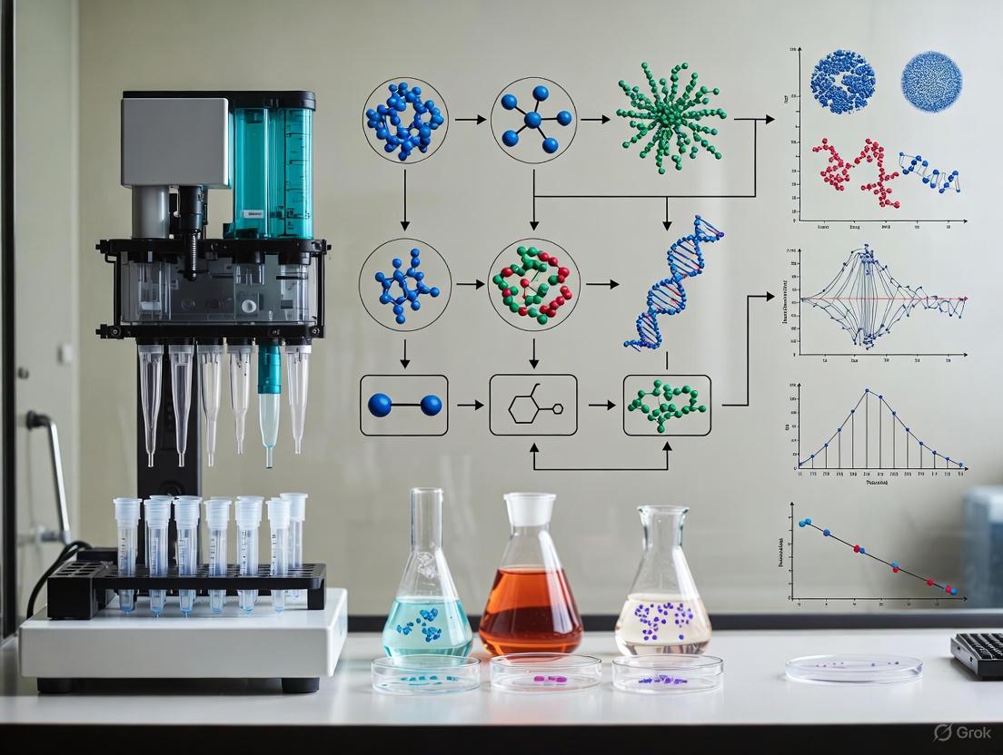
This article explores the transformative role of imaging flow cytometry (IFC) in stem cell research, a technology that merges the high-throughput capabilities of conventional flow cytometry with the detailed morphological...
Imaging Flow Cytometry: A High-Throughput Platform for Quantifying Stem Cell Morphology and Function
Abstract
This article explores the transformative role of imaging flow cytometry (IFC) in stem cell research, a technology that merges the high-throughput capabilities of conventional flow cytometry with the detailed morphological analysis of microscopy. Aimed at researchers, scientists, and drug development professionals, we cover foundational principles, from distinguishing stem cells via specific markers to advanced applications in organoid and therapy development. The content provides a methodological guide for implementation, a troubleshooting framework for common technical challenges, and a comparative analysis of IFC against other imaging and 'omics techniques. By synthesizing current applications and future directions, this resource aims to equip scientists with the knowledge to leverage IFC for accelerating phenotypic drug discovery and the clinical translation of stem cell therapies.
Unveiling Stem Cell States: Core Principles of Morphological Analysis with IFC
Imaging flow cytometry (IFC) represents a transformative technological advancement that successfully integrates the high-throughput, multiparametric analysis of conventional flow cytometry with the high-resolution, morphological detail of fluorescence microscopy. This synergy enables the quantitative analysis of complex cellular processes—such as heterogeneous stem cell differentiation, rare cell population identification, and subcellular event detection—at unprecedented speed and resolution. This Application Note details the core principles of IFC, provides validated protocols for stem cell morphology research, and presents quantitative data and pathway analyses to guide researchers in leveraging this powerful technology for advanced biomedical discovery.
The fundamental challenge in single-cell analysis has long been the trade-off between statistical power and morphological detail. Conventional flow cytometry offers high-throughput, multiparametric analysis of thousands of cells per second but lacks the ability to provide visual confirmation of cellular morphology and subcellular structure [1] [2]. Conversely, microscopy provides high-resolution spatial information but is typically low-throughput and susceptible to operator bias [3]. IFC bridges this critical gap by capturing high-resolution images of individual cells in flow, enabling simultaneous quantification of fluorescent markers and detailed morphological analysis [2] [3].
The value of IFC is particularly pronounced in stem cell research, where populations are often heterogeneous and rare subpopulations with distinct morphological features—such as very small embryonic-like stem cells (VSELs)—can be critically important. IFC allows for the identification and characterization of these rare cells (often with a frequency of <0.01%) based on both marker expression and distinct morphological profiles, a task that is challenging with either technology alone [1].
Technical Principles and Capabilities
System Architecture and Workflow
An IFC system integrates four core components to achieve its unique analytical capabilities:
- Fluid System: Utilizes microfluidic channels and sheath fluid to hydrodynamically focus cells into a single-file stream, ensuring stable, aligned passage through the detection zone [2] [3].
- Optical System: Comprises lasers and optical filters to excite fluorescently labeled cells and isolate specific emission wavelengths [2].
- Imaging System: Employs a high-precision camera (e.g., CCD) and objective lens to capture high-resolution images of each cell as it passes through the detection area. Some advanced systems use techniques like fluorescence imaging via radiofrequency-tagged emission (FIRE) [2] [3].
- Electronic System: Converts optical signals into electrical data for processing, analysis, and storage [2].
Quantitative Performance Metrics
Modern IFC platforms demonstrate impressive performance characteristics, as summarized in the table below.
Table 1: Performance Metrics of Advanced IFC Systems
| Platform / Technology | Max Throughput (cells/sec) | Spatial Resolution | Key Application Strengths |
|---|---|---|---|
| Light-Field Flow Cytometer (LFC) [4] | 5,750 | 400-600 nm in X, Y, Z (3D) | Volumetric visualization of 3D subcellular structures |
| Sheathless Microfluidic IFC [5] | 60,000 (fluorescence)400,000 (bright-field) | ~500 nm (lateral) | Sub-cellular localization of phase-separated compartments |
| High-Resolution Epi-Fluorescence Platform [4] | Varies | 337 nm (X), 291 nm (Y), 542 nm (Z) | Multi-color 3D imaging of organelles |
Application Protocols for Stem Cell Research
Protocol: Analysis of Rare Stem Cell Populations
This protocol is adapted for identifying and characterizing rare very small embryonic-like stem cells (VSELs) in heterogeneous populations, which can have frequencies as low as 0.0025% in murine adult spleen [1].
Research Reagent Solutions
Table 2: Essential Reagents for Rare Stem Cell Analysis
| Reagent / Material | Function | Example Application |
|---|---|---|
| Viability Dye | Exclusion of non-viable cells | Distinguishing live/dead cells to improve analysis accuracy |
| Lineage Marker Antibodies | Negative selection | Creating a "dump" channel for unwanted cells |
| Stem Cell Marker Antibodies | Positive identification | Staining for specific stem cell surface antigens (e.g., on VSELs) |
| Magnetic Cell Separation Kits | Pre-enrichment | Increasing relative frequency of rare cells before IFC analysis |
| Intracellular Staining Kits | For internal markers | Permeabilization and staining of intracellular antigens |
Step-by-Step Procedure:
Sample Preparation and Staining:
- Isolate mononuclear cells from tissue (e.g., spleen, bone marrow) using density gradient centrifugation.
- Optionally, pre-enrich target cells via magnetic cell separation to increase the relative frequency of rare cells [1] [6].
- Resuspend cells in appropriate staining buffer.
- Stain with viability dye to exclude dead cells.
- Incubate with fluorescently conjugated antibodies against lineage markers (for negative selection) and specific stem cell surface markers.
- For intracellular markers, perform fixation and permeabilization followed by staining with antibodies against intracellular antigens.
Instrument Setup and Data Acquisition:
- Calibrate the IFC instrument using appropriate fluorescence calibration beads.
- Establish a core acquisition template including brightfield, darkfield, and fluorescence channels.
- Set the flow rate to minimize coincidence events (typically <10% coincidence) [6].
- Acquire a sufficient number of events based on Poisson statistics. For a population representing 0.0025%, acquiring several million events is necessary to obtain a low coefficient of variation (CV) for reliable detection [1] [6].
Data Analysis and Gating Strategy:
- Step 1: Focused Cell Gate. Select cells that are in focus using gradient RMS morphology-based feature.
- Step 2: Single Cell Gate. Exclude aggregates and doublets using features like Aspect Ratio and Area.
- Step 3: Viable Cell Gate. Select viability dye-negative cells.
- Step 4: Lineage-Negative Gate. Exclude cells positive for lineage markers.
- Step 5: Morphological and Marker Analysis. Analyze the remaining cells for small size and high nuclear-to-cytoplasmic ratio, and positivity for specific stem cell markers. The final population can be identified and its morphological features quantified.
Protocol: Monitoring Stem Cell Asymmetry and Division
IFC is uniquely capable of quantifying asymmetric cell division—a critical process in stem cell biology—by simultaneously measuring protein polarization and morphological changes in large cell populations [1].
Step-by-Step Procedure:
Cell Labeling:
- Transfert or transduce stem cells with constructs expressing fluorescently tagged proteins of interest (e.g., cell fate determinants).
- Alternatively, use immunofluorescence to stain fixed cells for polarized proteins and mitotic markers.
Stimulation and Fixation:
- Culture cells under conditions that promote asymmetric division.
- At appropriate time points, harvest and fix cells.
IFC Acquisition and Analysis:
- Acquire data on the IFC instrument, ensuring to capture a statistically significant number of dividing cells.
- Use spot counting or protein polarization algorithms to quantify the distribution of key proteins between daughter cells.
- Correlate protein asymmetry with morphological features of division.
Data Analysis and Computational Integration
The high-information-content data generated by IFC requires sophisticated computational tools for full exploitation. Modern analysis involves:
- Morphometric Feature Extraction: Automated calculation of >100 quantitative features per cell, including size, shape, texture, and fluorescence intensity [2].
- Machine Learning and Deep Learning: Convolutional neural networks (CNNs) can be trained to automatically classify cell types, morphological states, and subcellular patterns from IFC images, enabling unbiased, high-content analysis [7].
- 3D Volumetric Visualization: Advanced systems like the Light-Field Flow Cytometer (LFC) enable 3D reconstruction of subcellular structures from single camera exposures, providing volumetric data on organelle morphology and distribution [4].
Imaging flow cytometry effectively bridges the technological gap between high-throughput flow cytometry and high-content microscopy, creating a powerful platform for stem cell research. By providing statistically robust, multiparametric data coupled with visual validation, IFC enables researchers to decipher cellular heterogeneity, identify rare stem cell subsets, and analyze complex subcellular processes with unprecedented clarity. As IFC technology continues to evolve with improvements in speed, resolution, and computational analysis, its role in advancing fundamental stem cell biology and translational drug development is poised for significant expansion.
Stem cells are widely known for their unique capabilities of prolonged self-renewal and differentiation into specific cell types, offering significant promise for regenerative medicine, disease modeling, and drug discovery. A critical step in leveraging their potential is the accurate verification of their pluripotent status. Flow cytometry has emerged as a powerful, high-throughput tool that provides rapid, quantitative, and multi-parameter analysis of stem cells at single-cell resolution, enabling researchers to characterize the expression of specific cell surface and intracellular markers that define the undifferentiated state. This application note details optimized protocols for the flow cytometric characterization of human induced pluripotent stem cells (iPSCs), framed within the broader context of imaging flow cytometry and stem cell morphology research [8].
Key Markers for Stem Cell Characterization
The pluripotent state of stem cells is defined by the expression of a specific set of markers. These can be broadly categorized into cell surface antigens and intracellular transcription factors. Their homogeneous, high expression is a hallmark of bona fide iPSCs, while decreased expression often indicates spontaneous differentiation [9] [8]. The following table summarizes the core markers used for assessing human iPSC pluripotency.
Table 1: Key Undifferentiated Stem Cell Markers for Flow Cytometry
| Marker Name | Marker Type | Expression Localization | Typical Expression in Undifferentiated iPSCs | Primary Function |
|---|---|---|---|---|
| TRA-1-60 | Carbohydrate antigen | Cell Surface | >90% | Pluripotency-associated glycoprotein [8] |
| SSEA-4 | Glycolipid | Cell Surface | >90% | Pluripotency-associated glycolipid [8] |
| OCT-3/4 | Transcription Factor | Intracellular (Nuclear) | >85% | Master regulator of self-renewal and pluripotency [9] |
| SOX2 | Transcription Factor | Intracellular (Nuclear) | >85% | Core transcription factor maintaining pluripotency [9] |
| NANOG | Transcription Factor | Intracellular (Nuclear) | >85% | Key factor for ground-state pluripotency [9] |
Experimental Workflow for iPSC Characterization
The complete process, from cell culture to data analysis, is outlined below. Adherence to this workflow is critical for generating reliable and reproducible data on stem cell pluripotency.
Basic Protocol 1: iPSC Culture and Collection for Flow Cytometry Analysis
Objective: To harvest high-quality, single-cell suspensions from human iPSC cultures.
Materials:
- Cultured human iPSCs
- Appropriate dissociation reagent (e.g., Accutase, EDTA)
- Flow cytometry staining buffer (PBS containing 1-2% FBS or BSA)
Procedure:
- Culture Maintenance: Maintain iPSCs in their standard culture conditions (e.g., on feeder layers or in feeder-free conditions). Ensure cultures are in a log-phase growth state and have a high density of undifferentiated colonies before harvesting.
- Cell Dissociation:
- Aspirate the culture medium and wash the cells gently with PBS without calcium and magnesium.
- Add an appropriate volume of pre-warmed dissociation reagent to cover the cell layer.
- Incubate at 37°C for the time required to dissociate the colonies into a single-cell suspension (typically 3-7 minutes). Monitor under a microscope to avoid over-digestion.
- Cell Quenching and Washing:
- Neutralize the dissociation reagent by adding 2-3 volumes of staining buffer.
- Gently pipette the cell suspension to break any remaining clumps and transfer it to a conical tube.
- Centrifuge at 300 x g for 5 minutes. Carefully aspirate the supernatant.
- Cell Washing and Counting:
- Resuspend the cell pellet in 3-5 mL of staining buffer and pass the suspension through a 40 μm cell strainer to remove aggregates.
- Centrifuge again at 300 x g for 5 minutes and aspirate the supernatant.
- Resuspend the cell pellet in a small volume of buffer and count the cells using an automated cell counter or hemocytometer [10].
- Aliquot Cells: Aliquot 0.5 - 1 x 10^6 cells per staining tube (or well of a V-bottom plate). Proceed immediately to staining or place the cell pellet on ice.
Basic Protocol 2: Staining for Extracellular and Intracellular Markers
Objective: To specifically label cell surface and intracellular pluripotency markers for detection by flow cytometry.
Materials:
- Fluorochrome-conjugated antibodies (e.g., against TRA-1-60, SSEA-4, OCT-3/4, SOX2, NANOG)
- Live/Dead viability dye (optional)
- Fixation buffer (e.g., 4% paraformaldehyde in PBS)
- Permeabilization buffer (e.g., 90% methanol or commercial saponin-based buffers)
- Flow cytometry staining buffer
Procedure:
- Viability Staining (Optional but Recommended):
- Resuspend the cell pellet in staining buffer containing a viability dye (e.g., a fixable viability dye). Incubate for 15-30 minutes on ice, protected from light.
- Wash the cells with 2 mL of staining buffer and centrifuge at 300 x g for 5 minutes. Aspirate the supernatant [10].
Extracellular (Cell Surface) Staining:
- Resuspend the cell pellet in 100 μL of staining buffer containing pre-titrated, fluorochrome-conjugated antibodies against surface markers (e.g., TRA-1-60, SSEA-4) [8].
- Vortex gently and incubate for 30 minutes on ice, protected from light.
- Wash the cells with 2 mL of staining buffer and centrifuge at 300 x g for 5 minutes. Aspirate the supernatant completely.
Fixation and Permeabilization:
- Fixation: Resuspend the cell pellet in 100 μL of fixation buffer. Incubate for 20 minutes at room temperature, protected from light.
- Wash: Add 2 mL of staining buffer, centrifuge at 300 x g for 5 minutes, and aspirate the supernatant.
- Permeabilization: Resuspend the cell pellet in 100 μL of ice-cold permeabilization buffer. Incubate for 15-30 minutes on ice, protected from light. For transcription factors, methanol-based buffers are often more effective [9].
Intracellular Staining:
- Add 2 mL of staining buffer to the tube and centrifuge at 300 x g for 5 minutes. Aspirate the supernatant.
- Resuspend the cell pellet in 100 μL of permeabilization buffer containing pre-titrated, fluorochrome-conjugated antibodies against intracellular markers (e.g., OCT-3/4, SOX2, NANOG).
- Vortex gently and incubate for 30-60 minutes on ice, protected from light.
- Wash the cells twice with 2 mL of permeabilization buffer, centrifuging each time.
- Perform a final wash with 2 mL of staining buffer.
Resuspension for Acquisition: Resuspend the final cell pellet in 200-500 μL of staining buffer. Keep samples on ice and protected from light until acquisition.
Basic Protocol 3: Flow Cytometry Acquisition and Data Analysis
Objective: To acquire fluorescence data and quantitatively analyze marker expression.
Materials:
- Flow cytometer (equipped with appropriate lasers and filters)
- Compensation beads (for multicolor panel setup)
- Flow cytometry analysis software (e.g., FlowJo, FCS Express)
Procedure:
- Instrument Setup:
- Start up and perform quality control on the flow cytometer using calibration beads.
- Create an acquisition template that includes forward scatter (FSC), side scatter (SSC), and all fluorescence channels corresponding to your antibody panel.
- Set up voltage and compensation settings using single-stained compensation beads or control cells [10].
Data Acquisition:
- Pass the resuspended stained cells through the cytometer. Adjust the flow rate to a medium or low setting to ensure data quality, especially for delicate stem cells.
- Collect a minimum of 10,000 events per sample that fall within the live, single-cell gate.
- Save the data as FCS files.
Data Analysis (Basic Protocol 4):
- Gating Strategy: Import FCS files into analysis software. The sequential gating strategy is visualized below.
- Quantification: After gating on the population of interest, visualize the fluorescence intensity for each marker. The percentage of positive cells and the Median Fluorescence Intensity (MFI) should be reported. A high-quality, undifferentiated iPSC culture should show a homogeneous population with >85-90% positivity for key pluripotency markers [9].
The Scientist's Toolkit: Essential Research Reagents
Successful characterization relies on a suite of validated reagents and instruments. The table below lists essential solutions for flow cytometric analysis of stem cells.
Table 2: Essential Research Reagent Solutions for Stem Cell Flow Cytometry
| Item | Function/Application | Example Products/Types |
|---|---|---|
| Validated Antibody Panels | Specific detection of pluripotency markers; ensures reproducibility. | Pre-conjugated antibodies against TRA-1-60, SSEA-4, OCT-3/4, SOX2, NANOG [9]. |
| Viability Dyes | Discrimination between live and dead cells; critical for data accuracy. | Fixable Viability Dyes (e.g., Zombie dyes, LIVE/DEAD kits) [10]. |
| Fixation/Permeabilization Kits | Preserve cell structure and allow antibody access to intracellular targets. | Commercial buffers (e.g., Foxp3/Transcription Factor Staining Buffer Sets) [9]. |
| Flow Cytometer | Instrument for multi-parameter, high-throughput cell analysis. | Attune NxT, iQue Advanced Flow Cytometry systems; instruments with 3+ lasers [10] [11]. |
| Analysis Software | Quantitative data analysis, visualization, and population gating. | FlowJo, FCS Express, ModFit LT [10]. |
This application note provides a cost-effective and optimized platform for defining the pluripotency status of human iPSCs using flow cytometry. The detailed protocols for cell preparation, staining, and analysis enable researchers to reliably quantify the expression of critical surface and intracellular markers. Integrating this flow cytometric data with insights from imaging flow cytometry and morphological studies provides a powerful, multi-faceted approach for the rigorous characterization of stem cells, which is fundamental for their successful application in regenerative medicine and drug discovery.
Stem cells, characterized by their capacities for self-renewal and multipotency, are fundamental to developmental biology, regenerative medicine, and therapeutic discovery [8]. A primary challenge in stem cell research is the inherent heterogeneity within stem cell populations; even under clonal conditions, these populations often contain subpopulations at different stages of the cell cycle, states of commitment, or phases of differentiation. Traditional single-parameter analysis methods, which focus on individual markers or physical characteristics, are insufficient to resolve this complexity. They risk averaging out critical rare cells or missing subtle but biologically significant transitions entirely.
Multiparametric analysis, particularly through advanced flow cytometry (FC) and imaging flow cytometry (IFC), provides a powerful solution. By simultaneously quantifying dozens of parameters—from cell surface and intracellular proteins to morphological features and functional assays—researchers can deconstruct heterogeneity into its constituent cell states [12] [8]. This approach is indispensable for accurately identifying and isolating rare stem cells, such as hematopoietic stem cells (HSCs) or cancer stem cells (CSCs), and for gaining a systems-level understanding of cell fate decisions. This Application Note details the protocols and analytical frameworks for applying multiparametric analysis to stem cell research, providing a standardized yet flexible pathway for high-resolution investigation.
Key Instrumentation and Core Principles
The transition to high-parameter analysis has been driven by synergistic advances in three key technological areas: instrumentation, reagents, and data analysis software [12].
Evolution of Flow Cytometry Platforms
Modern cytometers have evolved significantly from basic analyzers to sophisticated multiparametric platforms:
- Spectral Flow Cytometry: Utilizes full spectral fingerprinting of fluorophores, greatly improving fluorescence resolution and enabling the use of more overlapping dyes [2].
- Mass Cytometry (CyTOF): Replaces fluorescent tags with heavy metal isotopes and uses time-of-flight mass spectrometry for detection. This virtually eliminates spectral overlap, allowing for the simultaneous measurement of over 40 parameters [2] [13].
- Imaging Flow Cytometry (IFC): Integrates the high-throughput capability of conventional FC with high-resolution morphological imaging. IFC captures multiple images of each cell, providing quantitative data on subcellular localization, cell shape, and structure, thereby bridging the gap between population statistics and microscopy [2] [8]. Cutting-edge systems, such as those based on optical time-stretch (OTS) imaging, can achieve real-time throughput exceeding 1,000,000 events per second, facilitating the analysis of rare cells in large populations [14].
Fluorescent Reagents and Probes
The expansion of multiparametric panels relies on a diverse library of fluorochromes with distinct excitation and emission profiles. Key developments include:
- Novel Fluorochromes: The commercial availability of antibodies conjugated to violet (e.g., Pacific Blue, Alexa 405) and red-excitable (e.g., APC, HiLyte 750) dyes has been crucial for polychromatic cytometry [12].
- Tandem Dyes: These dyes rely on fluorescence resonance energy transfer (FRET) to create new spectral signatures (e.g., PE-Cy5, PE-Cy7). However, they can be prone to instability and lot-to-lot variation, requiring careful validation [12].
- Quantum Dots: Semiconductor nanocrystals with bright, narrow emission peaks, which are highly suitable for complex panels [12].
- Functional Assay Kits: A wide array of kits is available for multiparametric analysis of cell health, including assays for viability, apoptosis (e.g., Click-iT TUNEL), mitochondrial health, DNA damage (e.g., phosphorylated H2AX), and cell proliferation (e.g., Click-iT EdU) [15].
From Gating to High-Dimensional Analysis
The massive datasets generated require specialized computational tools that move beyond traditional sequential gating.
- Dimensionality Reduction: Algorithms like t-SNE and UMAP project high-dimensional data into two-dimensional maps where visually distinct clusters represent cells with similar expression profiles [16] [13].
- Automated Clustering: Unsupervised algorithms such as PhenoGraph and FlowSOM automatically identify and quantitate cell populations within the data without prior manual gating, revealing novel or unexpected subtypes [13].
- Integrated Software Platforms: Commercial software like FlowJo incorporates many of these tools, providing workflows for preprocessing, clustering, and visualization, thereby making high-dimensional analysis more accessible [16].
Table 1: Key Platforms for Multiparametric Cell Analysis
| Platform | Key Principle | Typical Max Parameters | Primary Advantage |
|---|---|---|---|
| Spectral Flow Cytometry [2] | Full spectrum emission capture | 30-40+ | Superior fluorescence resolution; minimizes spillover |
| Mass Cytometry (CyTOF) [2] [13] | Heavy metal isotope tagging & mass spectrometry detection | 40-50+ | Virtually no spectral overlap |
| Imaging Flow Cytometry (IFC) [2] [8] | High-speed cellular imaging in flow | 10+ (with morphological data) | Adds spatial and morphological context |
| Optical Time-Stretch IFC [14] | Ultra-high-speed line-scan imaging | N/A | Extreme throughput (>1 million cells/sec) |
Experimental Protocols
The following protocols provide a framework for multiparametric analysis of stem cell populations, from sample preparation to data acquisition.
Protocol 1: Sample Preparation and Viability Staining for Solid Tissues
This protocol, adapted from guidelines for preparing human lung tissue for dendritic cell analysis, is applicable to various solid tissue-derived stem cells (e.g., mesenchymal stem cells) [17].
Reagents and Equipment:
- Collagenase IV (Sigma, #C5138)
- DNAse I (Roche, #10104159001)
- Fetal Bovine Serum (FBS)
- RPMI 1640 medium
- 1x RBC Lysis Buffer (eBioscience, #00-4333-57)
- Ficoll-Paque (GE Healthcare, #17-1440-02)
- Live/Dead viability stain (e.g., Image-iT DEAD Green [15])
- Sterile scissors, 70 µm cell strainer, 50 mL conical tubes, water bath, centrifuge.
Step-by-Step Procedure:
- Preparation: Prepare digestion buffer: RPMI 1640 with 10% FBS, 0.2 mg/mL Collagenase IV, and 0.05 mg/mL DNAse I. Warm to 37°C.
- Tissue Mincing: Transfer the tissue sample (approx. 1-2 cm³) to a tube or petri dish with 0.5 mL digestion buffer. Using sterile scissors, mince the tissue into fine pieces (1-2 mm³).
- Enzymatic Digestion: Transfer the minced tissue and buffer to a six-well plate, adding more digestion buffer for a total of 4-5 mL per well. Incubate for 30-60 minutes at 37°C.
- Single-Cell Suspension: Gently pipette the mixture up and down 6-8 times with a 10 mL serological pipette to dissociate any remaining tissue.
- Filtration and Washing: Pass the suspension through a 70 µm cell strainer into a 50 mL tube. Rinse the well with PBS and pass it through the same strainer. Centrifuge at 365 × g for 5 min at room temperature (RT) and aspirate the supernatant.
- Density Gradient Centrifugation: Resuspend the cell pellet in 40 mL of PBS. Carefully layer it over 10 mL of RT Ficoll-Paque in a new 50 mL tube. Centrifuge at 1800 × g for 25 min at RT with low acceleration and no brake.
- Harvest Mononuclear Cells: Carefully collect the mononuclear cell layer at the PBS-Ficoll interface and transfer it to a new 50 mL tube. Top up with PBS to 50 mL.
- Red Blood Cell Lysis: Centrifuge at 365 × g for 5 min at 4°C. Aspirate supernatant, resuspend pellet in 1-2 mL of RBC lysis buffer, and incubate for 5-10 min at RT. Top up with PBS and centrifuge again.
- Viability Staining: Resuspend the final cell pellet in a suitable buffer and stain with a live/dead viability dye according to the manufacturer's instructions (e.g., HCS LIVE/DEAD Green Kit [15]) before proceeding to surface or intracellular staining.
Protocol 2: Staining and Acquisition for a High-Parameter Panel
This protocol outlines the steps for staining cells with a complex antibody panel and acquiring data on a spectral or mass cytometer.
Reagents and Equipment:
- Pre-conjugated antibodies for target markers
- Fc receptor blocking antibody (e.g., anti-CD16/32)
- Fixation/Permeabilization buffers (for intracellular targets)
- Cell staining buffer (PBS with 1-2% FBS)
- U-bottom 96-well plates
- Flow cytometer (spectral, mass, or imaging)
Step-by-Step Procedure:
- Cell Counting and Plating: Count the single-cell suspension and aliquot 1-5 × 10^6 cells per U-bottom well. Centrifuge at 400 × g for 5 min and thoroughly aspirate the supernatant.
- Fc Receptor Blocking: Resuspend the cell pellet in 100 µL of staining buffer containing an Fc block. Incubate for 10-15 minutes at 4°C.
- Surface Staining: Without washing, add a pre-titrated cocktail of antibodies against surface antigens (e.g., CD34, CD45, CD90, CD105 for MSCs) in a minimal volume (50-100 µL). Vortex gently and incubate for 30 minutes at 4°C in the dark.
- Wash: Add 150 µL of staining buffer, centrifuge, and aspirate the supernatant. Repeat this wash step once more.
- Fixation and Permeabilization (if needed): For intracellular transcription factors (e.g., NANOG, OCT4), resuspend cells in a fixation/permeabilization buffer (e.g., eBioscience FoxP3 Fix/Perm buffer) and incubate for 30-60 min at 4°C. Wash twice with 1x permeabilization buffer.
- Intracellular Staining: Resuspend the fixed/permeabilized cells in a cocktail of antibodies against intracellular targets in permeabilization buffer. Incubate for 30-60 min at 4°C in the dark. Wash twice with permeabilization buffer, then once with standard staining buffer.
- Data Acquisition: Resuspend the final cell pellet in staining buffer containing a DNA dye (e.g., Hoechst 33342) if required. Filter the cells through a 35-70 µm mesh-top tube immediately before acquiring data on the cytometer. For mass cytometry, resuspend in Cell-ID Intercalator-Ir [13]. Adjust the concentration to achieve an optimal event rate for your instrument.
The following workflow diagram summarizes the key stages of a multiparametric stem cell analysis experiment.
Diagram 1: Experimental workflow for multiparametric stem cell analysis, covering sample preparation, staining, and data acquisition.
Data Analysis and Interpretation
The transformation of raw, high-parameter data into biological insight requires a structured analytical workflow.
Data Preprocessing and Quality Control
The initial steps ensure data quality and integrity.
- Data Normalization: For mass cytometry, signal drift over time is corrected using normalization beads spiked into each sample [13].
- Data Cleaning: Removal of technical artifacts is critical. This includes gating for single cells based on pulse area vs. height characteristics to exclude doublets or clumps, and gating out dead cells based on viability staining [18] [13].
- Compensation: In fluorescence-based cytometry, spectral overlap between channels must be corrected using compensation matrices, often calculated automatically by software from single-stain controls [12].
High-Dimensional Analysis Workflow
After preprocessing, data is analyzed using a combination of automated and guided approaches.
- Dimensionality Reduction: The preprocessed and concatenated data from all samples is fed into algorithms like t-SNE, UMAP, or viSNE [16] [13]. These tools map high-dimensional data onto a 2D scatter plot where each dot is a cell, and distances between dots approximate their phenotypic similarity.
- Automated Clustering: Simultaneously, clustering algorithms like PhenoGraph or FlowSOM are used to partition the data into distinct subpopulations [13]. Each cluster is assigned a unique ID.
- Cluster Annotation and Interpretation: The resulting clusters are then visualized on the t-SNE/UMAP layout. The biological identity of each cluster is determined by inspecting the median expression of known markers (e.g., CD34, CD133, OCT4) across all cells within that cluster.
- Statistical Comparison: The abundance of each cluster or the expression levels of key markers can be compared between experimental conditions (e.g., healthy vs. diseased, untreated vs. drug-treated) using statistical tests to identify biologically significant changes.
Table 2: Common Marker Panels for Stem Cell Identification
| Stem Cell Type | Key Positive Markers | Key Negative Markers | Functional/Rare Population Markers |
|---|---|---|---|
| Hematopoietic Stem Cells (HSCs) [8] | CD34, CD133, CD90, c-Kit | CD38, Lineage markers (CD3, CD19, etc.) | Aldehyde dehydrogenase (ALDH) activity, Hoechst Side Population [12] |
| Mesenchymal Stem Cells (MSCs) [8] | CD105, CD73, CD90 | CD45, CD34, CD11b | |
| Embryonic Stem Cells (ESCs) [8] | Transcription factors: OCT4, SOX2, NANOG | - | |
| Cancer Stem Cells (CSCs) [8] | CD44, CD133, EpCAM | - | Aldehyde dehydrogenase (ALDH) activity |
The following diagram illustrates the core logic of progressing from raw data to biological insight.
Diagram 2: High-dimensional data analysis workflow, showing parallel paths of dimensionality reduction and automated clustering that converge for biological interpretation.
The Scientist's Toolkit: Research Reagent Solutions
Successful multiparametric analysis relies on a suite of reliable reagents and tools. The following table details essential items for a typical experiment.
Table 3: Essential Reagents and Kits for Multiparametric Stem Cell Analysis
| Reagent/KIT | Specific Example | Primary Function in the Protocol |
|---|---|---|
| Viability Stain | HCS LIVE/DEAD Green Kit [15] | Distinguishes live from dead cells based on membrane integrity; critical for data cleaning. |
| Fc Block | Anti-CD16/32 antibody (e.g., 2.4G2) [13] | Blocks non-specific antibody binding to Fc receptors on cells, reducing background. |
| Surface Marker Antibodies | Conjugates against CD34, CD45, CD90, CD105, CD133 [8] | Identifies and defines cell populations based on cell surface phenotype. |
| Transcription Factor Staining Buffer Set | eBioscience FoxP3 / Transcription Factor Staining Buffer Set [13] | Permeabilizes cells for intracellular staining of nuclear proteins like OCT4 and NANOG. |
| Cell Proliferation Assay | Click-iT EdU HCS Assay [15] | Quantifies DNA synthesis and cell cycle activity using a click chemistry-based method. |
| DNA Damage/ Apoptosis Assay | HCS DNA Damage Kit / Click-iT TUNEL Assay [15] | Detects double-strand DNA breaks (γH2AX) or DNA fragmentation (TUNEL) as indicators of genotoxic stress or apoptosis. |
| Mitochondrial Health Assay | HCS Mitochondrial Health Kit [15] | Simultaneously measures mitochondrial membrane potential (MitoHealth stain) and cytotoxicity (Dead Green stain). |
| Cell Demarcation Stain | HCS CellMask Deep Red Stain [15] | Stains the entire cytoplasm, enabling accurate cytoplasmic segmentation and morphological analysis in IFC/HCS. |
Multiparametric analysis represents a paradigm shift in stem cell research, moving the field beyond the limitations of single-parameter analysis. By leveraging advanced cytometric platforms, a growing arsenal of fluorescent reagents, and sophisticated computational tools, researchers can now deconstruct the inherent heterogeneity of stem cell populations with unprecedented resolution. The protocols and frameworks outlined in this Application Note provide a practical foundation for implementing these powerful techniques. As instrumentation continues to advance—with ever-increasing parameter numbers and throughput—and analytical algorithms become more intelligent and accessible, multiparametric analysis is poised to unlock deeper insights into stem cell biology, driving innovations in regenerative medicine and therapeutic development.
The advent of stem cell-derived organoids has revolutionized biomedical research by providing sophisticated three-dimensional (3D) in vitro models that closely mimic the complex architecture and functionality of human organs [19]. These patient-derived models preserve genetic and phenotypic features, enabling more physiologically relevant studies in disease modeling, drug screening, and personalized medicine [19]. However, the transition from traditional two-dimensional (2D) cultures to complex 3D organoid systems has created an urgent need for advanced analytical technologies that can provide high-throughput, quantitative, and multidimensional data.
Imaging Flow Cytometry (IFC) emerges as a powerful solution to this analytical challenge, bridging the critical gap between conventional flow cytometry and microscopy. IFC integrates the high-throughput capabilities of flow cytometry with high-resolution morphological imaging, enabling multiparametric analysis at the single-cell level within 3D organoid systems [2]. This technology allows researchers to simultaneously quantify molecular markers while capturing detailed morphological information from thousands of cells within organoids, providing unprecedented insights into cellular heterogeneity, structural organization, and functional dynamics in a statistically robust manner.
The convergence of organoid technology with IFC platforms is particularly valuable for pharmaceutical research, where it enhances the predictive power of preclinical drug development while addressing ethical concerns through animal-free testing strategies [19]. This application note provides comprehensive methodologies and protocols for implementing IFC in the quantitative analysis of stem cell-derived organoids, framed within the broader context of advanced stem cell morphology research.
Technical Principles and Advantages of IFC
How Imaging Flow Cytometry Works
IFC operates on an integrated system that combines fluidics, optics, imaging, and electronic components to deliver simultaneous multiparametric and morphological data [2]. The core technological components include:
- Fluid System: Utilizes microfluidic channels and sheath fluid mechanisms to align cells into a single-file stream, ensuring stable flow through the detection zone for consistent analysis [2].
- Optical System: Comprises laser sources and optical filters that generate and isolate excitation/emission signals from fluorescently labeled cells and organoids [2].
- Imaging System: Incorporates high-precision cameras (e.g., CCD) or fluorescence imaging via radiofrequency-tagged emission (FIRE) to capture high-resolution cellular images during flow [2].
- Electronic Systems: Process optical signals into electrical data for downstream analysis, enabling real-time data acquisition and processing [2].
Table 1: Comparative Analysis of Cell Analysis Technologies
| Feature | Traditional Flow Cytometry | Microscopy | Imaging Flow Cytometry |
|---|---|---|---|
| Throughput | High (thousands of cells/sec) | Low | High (hundreds to thousands of cells/sec) |
| Morphological Data | Limited to scatter parameters | Comprehensive | Quantitative morphological data |
| Statistical Power | High | Limited | High |
| Spatial Context | None | Preserved | Partially preserved |
| Analysis Automation | Automated | Often manual | Automated with AI assistance |
| Subcellular Resolution | No | Yes | Yes |
Key Advantages for Organoid Analysis
IFC offers several distinct advantages for the analysis of complex 3D organoid systems:
- Morpho-Functional Integration: Unlike conventional flow cytometry that lacks detailed morphological analysis, IFC provides both quantitative molecular data and morphological images simultaneously, offering a more comprehensive perspective for organoid characterization [2].
- High-Throughput 3D Analysis: IFC maintains the high-throughput capability of traditional flow cytometry while incorporating 3D structural information, enabling statistically robust analysis of organoid heterogeneity [2].
- Single-Cell Resolution within Organoids: By dissociating organoids into single-cell suspensions, IFC enables detailed analysis of cellular heterogeneity and subpopulation dynamics while retaining morphological context [19] [2].
- Reduced Analytical Bias: Advanced IFC software automates image processing and multidimensional data integration, minimizing human bias—a significant advantage over manual microscopy-based analyses [2].
Experimental Design and Workflow
The successful application of IFC for organoid analysis requires careful experimental planning and optimization. The following workflow diagram illustrates the complete experimental pipeline from organoid generation to data analysis:
Organoid Generation and Quality Control
The foundation of successful IFC analysis begins with robust organoid generation and rigorous quality control:
- Stem Cell Source Selection: Utilize human pluripotent stem cells (hPSCs), including embryonic stem cells (hESCs) or induced pluripotent stem cells (hiPSCs), selected based on research objectives. Patient-specific hiPSCs offer particular advantages for personalized medicine applications [19].
- Directed Differentiation: Implement established protocols for directing stem cell differentiation toward target tissues (e.g., cerebral, hepatic, intestinal organoids) using specific morphogen gradients and signaling pathway modulators [19].
- Quality Assessment: Perform comprehensive quality control including:
- Pluripotency marker verification (OCT4, NANOG, SOX2) for starting populations [20]
- Tissue-specific marker expression confirmation
- Morphological assessment of 3D structure integrity
- Karyotyping and genetic stability monitoring
Table 2: Key Markers for Organoid Characterization by IFC
| Organoid Type | Pluripotency Markers | Early Lineage Markers | Maturation Markers | Functional Assays |
|---|---|---|---|---|
| Cerebral | OCT4, NANOG | SOX1, PAX6 | MAP2, NeuN, GFAP | Calcium imaging, Synaptic activity |
| Hepatic | OCT4, TRA-1-60 | AFP, HNF4α | Albumin, CYP3A4 | Albumin secretion, LDL uptake |
| Intestinal | TRA-1-81, SOX2 | CDX2, Villin | MUC2, Lysozyme | Barrier function, Enzyme activity |
| Renal | SSEA-4 | WT1, PAX2 | AQP2, Nephrin | Filtration assays |
| Cardiac | TRA-1-60 | NKX2.5, TNNT2 | cTnT, MYH6 | Beating analysis, Calcium transients |
Sample Preparation Protocol
Proper sample preparation is critical for successful IFC analysis of organoids:
Organoid Dissociation Protocol
Purpose: To generate single-cell suspensions from 3D organoids while maintaining cell viability and antigen integrity for IFC analysis.
Materials:
- Advanced DMEM/F-12 medium
- Enzyme-free dissociation buffer or gentle cell dissociation reagent
- Trypsin-EDTA (0.25%) for robust tissues
- DNase I (1 mg/mL in PBS)
- FBS-containing medium for enzyme inhibition
- Cell strainers (40μm and 100μm)
- Viability dye (e.g., propidium iodide or 7-AAD)
Procedure:
- Harvesting: Collect organoids from culture matrix using gentle pipetting or enzymatic matrix degradation if embedded in Matrigel.
- Washing: Wash organoids 3x with PBS to remove residual matrix and culture media.
- Primary Dissociation: Incubate organoids with appropriate dissociation reagent:
- For cerebral organoids: Enzyme-free dissociation buffer, 15-20 minutes at 37°C
- For hepatic/intestinal organoids: Trypsin-EDTA, 5-10 minutes at 37°C
- Mechanical Disruption: Gently pipet organoids up and down 10-15 times using wide-bore pipet tips.
- Enzyme Neutralization: Add 2 volumes of FBS-containing medium to neutralize proteolytic enzymes.
- Filtration: Pass cell suspension sequentially through 100μm and 40μm cell strainers.
- DNase Treatment: Add DNase I (final concentration 10μg/mL) to prevent cell clumping.
- Washing: Centrifuge at 300xg for 5 minutes and resuspend in staining buffer.
- Viability Assessment: Mix 10μL cell suspension with viability dye and count using hemocytometer.
Quality Control Parameters:
- Target viability: >85%
- Single-cell yield: >1×10^6 cells per organoid
- Clump index: <5% doublets/triplets
Staining and Labeling Strategies
Comprehensive immunolabeling is essential for multiparametric IFC analysis:
Surface and Intracellular Marker Staining Protocol
Purpose: To simultaneously label extracellular and intracellular markers for comprehensive phenotyping of organoid-derived cells.
Materials:
- Flow cytometry staining buffer (PBS + 2% FBS)
- Fixation buffer (4% paraformaldehyde in PBS)
- Permeabilization buffer (0.2% Triton X-100 in PBS)
- Blocking buffer (5% normal serum in staining buffer)
- Primary antibodies against target antigens
- Fluorochrome-conjugated secondary antibodies (if needed)
- DAPI or Hoechst 33342 for DNA staining
- Fc receptor blocking reagent (optional)
Procedure:
- Cell Counting and Aliquoting: Adjust cell concentration to 1×10^7 cells/mL in staining buffer.
- Fc Receptor Blocking: Incubate cells with Fc block (optional) for 10 minutes at 4°C.
- Surface Staining:
- Add optimized concentrations of antibodies against surface markers
- Incubate for 30 minutes at 4°C in the dark
- Wash twice with 2mL staining buffer, centrifuge at 300xg for 5 minutes
- Fixation: Resuspend cells in 4% PFA, incubate 20 minutes at room temperature
- Permeabilization:
- Centrifuge at 400xg for 5 minutes, remove supernatant
- Resuspend in 0.2% Triton X-100, incubate 10 minutes at room temperature
- Intracellular Staining:
- Add blocking buffer, incubate 30 minutes at room temperature
- Add antibodies against intracellular targets, incubate 45 minutes at room temperature
- Wash twice with permeabilization buffer
- Nuclear Staining: Resuspend in staining buffer containing DAPI (1μg/mL)
Critical Optimization Steps:
- Antibody titration for optimal signal-to-noise ratio
- Compensation controls for spectral overlap
- Fluorescence-minus-one (FMO) controls for gating
- Isotype controls for nonspecific binding assessment
IFC Data Acquisition and Analysis
Instrument Setup and Optimization
Proper instrument configuration is essential for high-quality IFC data:
- Laser Power Optimization: Balance between signal intensity and potential photobleaching
- Image Resolution Settings: Select appropriate magnification (20x or 40x) based on cellular features of interest
- Flow Rate Calibration: Adjust sample flow rate to ensure optimal image clarity while maintaining throughput
- Focus Maintenance: Implement automated focus routines for consistent image quality
- Quality Control Beads: Run daily QC with calibration beads to ensure instrument performance
Data Analysis Workflow
IFC generates complex multidimensional data requiring sophisticated analysis approaches:
Quantitative Morphological Analysis
IFC enables extraction of sophisticated morphological parameters that provide insights into cellular state and function:
- Basic Morphometric Features: Area, perimeter, aspect ratio, circularity
- Texture Analysis: Contrast, entropy, granularity
- Spatial Features: Nuclear-to-cytoplasmic ratio, organelle distribution
- Intensity Distribution: Signal heterogeneity within cellular compartments
Table 3: Key Morphological Features Extractable by IFC
| Feature Category | Specific Parameters | Biological Significance | Application Example |
|---|---|---|---|
| Size Parameters | Area, Diameter, Perimeter | Cell growth, cycle status, differentiation | Increased size in senescent cells |
| Shape Descriptors | Circularity, Aspect Ratio, Irregularity | Cellular activation, structural polarization | Neurite outgrowth in neural organoids |
| Texture Features | Contrast, Entropy, Granularity | Subcellular organization, vesicle content | Granularity changes in secretory cells |
| Spatial Relationships | N/C Ratio, Centroid Position | Cellular maturity, polarity establishment | Apical-basal polarization in epithelia |
| Intensity Distribution | CV, Skewness, Kurtosis | Protein distribution heterogeneity | Transcription factor gradients |
Artificial Intelligence and Machine Learning Applications
The integration of AI with IFC data analysis represents a transformative advancement for organoid research:
- Convolutional Neural Networks (CNNs): Automate cell classification and segmentation with accuracy up to 97.5%, significantly outperforming traditional methods [21].
- Dimensionality Reduction Algorithms: t-SNE and UMAP enable visualization of high-dimensional IFC data in two-dimensional space, revealing hidden population structures.
- Unsupervised Clustering: Algorithms like PhenoGraph identify novel cell subpopulations without prior biological assumptions.
- Predictive Modeling: Machine learning classifiers can predict differentiation outcomes, drug responses, or disease states based on morphological signatures.
Applications in Stem Cell and Organoid Research
Characterization of Organoid Heterogeneity
Organoids inherently exhibit considerable cellular heterogeneity that mirrors their in vivo counterparts. IFC enables quantitative assessment of this heterogeneity through:
- Differentiation Efficiency Quantification: Simultaneous measurement of multiple lineage markers at single-cell resolution provides precise quantification of differentiation efficiency and identification of contaminating cell types.
- Developmental Staging: Multiparametric analysis enables reconstruction of developmental trajectories and identification of transitional cell states during organoid maturation.
- Quality Control Metrics: Establishment of quantitative benchmarks for organoid quality assessment, enabling comparison across different batches and protocols.
Drug Screening and Toxicity Assessment
The pharmaceutical applications of organoid-IFC platforms are particularly promising:
- High-Content Screening: IFC enables multiparametric assessment of drug effects, capturing both molecular changes and morphological alterations in response to compound treatment [19].
- Mechanistic Toxicology: Comprehensive analysis of organoid responses to toxic compounds, including specific cell death pathways, stress responses, and adaptive mechanisms.
- Personalized Medicine Applications: Patient-derived organoids screened with IFC can predict individual responses to anticancer therapies and other treatments, enabling personalized therapeutic strategies [19].
Disease Modeling and Pathophysiological Studies
IFC-enhanced organoid analysis provides unique insights into disease mechanisms:
- Patient-Specific Modeling: Organoids derived from patient-specific hiPSCs retain disease-specific phenotypes that can be quantitatively characterized using IFC [19].
- Infection Models: Analysis of host-pathogen interactions in organoid systems, including cellular entry mechanisms, intracellular replication, and host response pathways.
- Genetic Engineering Assessment: Evaluation of CRISPR/Cas9 and other genome editing outcomes in organoid systems through multiplexed phenotypic analysis.
Research Reagent Solutions
Table 4: Essential Research Reagents for IFC Analysis of Organoids
| Reagent Category | Specific Products | Application | Key Considerations |
|---|---|---|---|
| Dissociation Reagents | Enzyme-free dissociation buffer, Accutase, Trypsin-EDTA | Organoid dissociation to single cells | Gentle enzymes preserve surface antigens |
| Viability Markers | Propidium iodide, 7-AAD, DAPI, LIVE/DEAD fixable dyes | Discrimination of live/dead cells | Fixable dyes compatible with intracellular staining |
| Extracellular Antibodies | CD markers, TRA-1-60, TRA-1-81, EpCAM | Surface antigen profiling | Direct conjugation with bright fluorophores recommended |
| Intracellular Antibodies | OCT4, NANOG, SOX2, lineage-specific transcription factors | Pluripotency and differentiation assessment | Require permeabilization; validate for IFC |
| Nuclear Stains | DAPI, Hoechst 33342, SYTOX dyes | DNA content analysis, cell cycle | Concentration optimization critical for image quality |
| Functional Probes | CellTracker dyes, MitoTracker, Fluo-4 AM | Metabolic activity, mitochondrial function, calcium flux | Loading conditions require optimization |
| Blocking Reagents | Normal serum, Fc receptor blockers, BSA | Reduction of nonspecific binding | Species-matched to secondary antibodies |
Troubleshooting and Optimization
Successful IFC analysis of organoids requires addressing several common challenges:
- High Background Signal: Increase blocking buffer concentration (10-15% serum), optimize antibody concentrations, increase wash steps, or switch to monoclonal antibodies to reduce cross-reactivity [20].
- Weak Immunofluorescence Signal: Reduce blocking buffer concentration, increase antibody concentration, ensure proper permeabilization for intracellular targets, and verify appropriate filter sets [20].
- Cell Aggregation: Optimize dissociation protocol, include DNase treatment, use appropriate cell strainers, and maintain single-cell suspension through gentle agitation.
- Spectral Overlap: Implement proper compensation controls, select fluorophores with minimal spectral overlap, and utilize spectral unmixing algorithms when available.
- Low Throughput: Optimize cell concentration, adjust flow rates, and implement automated sample loading for increased processing capacity.
Future Perspectives
The integration of IFC with organoid technology continues to evolve with several promising directions:
- Multi-omics Integration: Correlation of IFC morphological data with transcriptomic, proteomic, and epigenomic profiles from the same cells.
- Dynamic Monitoring: Development of live-cell compatible IFC protocols for tracking organoid development and responses over time.
- 3D Imaging Capabilities: Advancement of IFC technologies to preserve and analyze larger 3D structures without complete dissociation.
- Standardized Analytical Frameworks: Establishment of community standards and reference datasets for reproducible organoid analysis.
- Clinical Translation: Implementation of IFC-based quality control metrics for organoids destined for therapeutic applications.
The synergistic combination of organoid models with IFC technology represents a powerful platform for advancing our understanding of human development, disease mechanisms, and therapeutic interventions. By providing high-content, high-throughput quantitative analysis of these sophisticated 3D models, IFC enables researchers to extract maximum biological insights while maintaining statistical robustness—a critical advancement for the field of stem cell research and regenerative medicine.
From Theory to Practice: Implementing IFC in Your Stem Cell Research Pipeline
Imaging flow cytometry (IFC) represents a revolutionary convergence of conventional flow cytometry and quantitative microscopy, enabling high-throughput, multi-parameter analysis while preserving rich morphological information at the single-cell level [2]. Within stem cell research, where subtle morphological changes often reflect critical changes in cell state, potency, or early differentiation, IFC provides a powerful tool to quantify these features across large populations [22] [8]. This application note provides a detailed, step-by-step protocol for preparing, staining, and acquiring stem cell samples on an IFC platform, framed specifically within the context of stem cell morphology research.
Technical Principles and Stem Cell Relevance
An IFC system integrates four core components: a fluidics system to hydrodynamically focus cells into a single-file stream; an optical system (lasers and filters) for illumination and signal collection; an imaging system (typically a high-precision camera) to capture high-resolution images of each cell; and an electronic system for signal processing and data acquisition [2]. Unlike conventional flow cytometry, which only records fluorescence intensity and light scatter, IFC captures multidimensional images, allowing for the analysis of cell size, shape, texture, and the precise subcellular localization of fluorescent markers [22]. For heterogeneous stem cell populations, this means that cell types and transitional states can be discriminated not just by biomarker presence, but by their morphological signatures and the spatial organization of proteins and organelles [22] [8].
Sample Preparation and Staining Protocol for Stem Cells
Proper sample preparation is critical for obtaining high-quality, morphologically accurate IFC data. The following protocol is optimized for adherent mesenchymal stem cells (MSCs) but can be adapted for other stem cell types.
Coating and Seeding of Adherent Stem Cells
Many stem cells, including MSCs, require surface coating for optimal adherence and morphological preservation.
- Coating Solution Preparation: Prepare a 0.1% (w/v) solution of poly-D-lysine or poly-L-lysine in sterile deionized water [23] [24].
- Coating Process: Place sterilized #1.5 glass coverslips or directly use the surface of a culture dish. Add enough coating solution to cover the surface completely. Incubate for 1 hour at room temperature [23].
- Seeding: Remove the coating solution, rinse the surface three times with sterile water, and allow it to dry completely. Seed cells at a density of 60-80% confluence. This density is optimal for maintaining natural cell architecture and preventing morphological artifacts caused by over-crowding [25]. Incubate cells for at least 6 hours in growth media to facilitate adherence before any treatment or fixation [23].
Fixation and Permeabilization
Fixation preserves cellular morphology at a specific time point. The choice of fixative can impact epitope recognition and overall morphology.
- Fixative Selection: Gently wash cells once with phosphate-buffered saline (PBS).
- For most stem cell markers (transcription factors, cytoskeletal proteins): Use 4% paraformaldehyde (PFA) in PBS for 10-20 minutes at room temperature [23] [24].
- For better preservation of membrane-associated antigens: Ice-cold 100% methanol can be used for 5 minutes at room temperature. Note that methanol fixation typically eliminates the need for a separate permeabilization step [23].
- Washing: After fixation, aspirate the fixative and wash the cells three times with PBS (5 minutes per wash) [23].
- Permeabilization: For intracellular targets when using PFA fixation, permeabilize the cells with 0.1-0.5% Triton X-100 in PBS for 5 minutes at 4°C. For membrane-associated proteins, consider milder detergents like Tween-20 or saponin to better preserve membrane structures [23].
Immunofluorescence Staining
This protocol outlines indirect immunofluorescence, which offers signal amplification and is widely used.
- Blocking: Incubate cells with a blocking buffer for 30-45 minutes at room temperature to minimize non-specific antibody binding. A common and effective buffer is 1-5% normal serum (from the same species as the secondary antibody) with 0.1-0.3% Triton X-100 in PBS [23] [24].
- Primary Antibody Incubation: Prepare the primary antibody in dilution buffer (e.g., PBS with 1% BSA and 0.1% Triton X-100). Incubate the samples with the antibody solution either for 2 hours at room temperature or overnight at 4°C in a humidified chamber [23] [24].
- Washing: Wash the samples three times with a wash buffer (e.g., PBS with 0.1% Triton X-100) to remove unbound primary antibody.
- Secondary Antibody Incubation: Incubate with fluorochrome-conjugated secondary antibodies, diluted in blocking or dilution buffer (typically 1:500-1:1000), for 1 hour at room temperature in the dark [23] [24].
- Counterstaining and Mounting (for validation): For initial validation using microscopy, counterstain nuclei with DAPI (1 µg/ml for 2-5 minutes) and mount with an anti-fade mounting medium [24]. For IFC analysis, cells are typically resuspended in a suitable buffer like PBS, and DAPI can be added directly to the suspension.
Table 1: Critical Reagents for Stem Cell Staining and IFC Analysis
| Reagent / Material | Function / Purpose | Example / Notes |
|---|---|---|
| Poly-D/L-Lysine | Coating material to enhance cell adhesion to glass surfaces. | Essential for proper spreading and morphology of many stem cell types [24] [25]. |
| Paraformaldehyde (PFA) | Crosslinking fixative. Preserves cellular morphology and protein localization. | Standard for most applications; use 2-4% in PBS [23] [24]. |
| Methanol | Precipitating fixative. Preserves some antigens and permeabilizes simultaneously. | Ideal for certain membrane targets; use ice-cold [23]. |
| Triton X-100 | Detergent for permeabilizing cellular membranes post-PFA fixation. | Allows antibodies access to intracellular targets [23]. |
| Normal Serum | Blocking agent to reduce non-specific antibody binding. | Should match the host species of the secondary antibody [23]. |
| Fluorochrome-Conjugated Secondary Antibodies | Detection of primary antibodies for visualization. | Select bright, photostable fluorophores compatible with IFC laser lines [24]. |
| DAPI | DNA-binding dye for nuclear counterstaining. | Enables cell counting, cell cycle analysis, and nuclear morphological assessment [22] [24]. |
Data Acquisition on an IFC Platform
Sample Preparation for Acquisition
After staining, adherent cells must be detached and resuspended into a single-cell suspension.
- For cells stained in dishes or on coverslips, use a gentle cell dissociation reagent (e.g., enzyme-free dissociation buffer or trypsin/EDTA, with caution) to lift the cells.
- Resuspend the cell pellet in a suitable IFC running buffer (e.g., PBS with 0.1-1% BSA).
- Filter the suspension through a cell strainer (e.g., 35-70 µm nylon mesh) to remove aggregates that can clog the fluidics system and cause erroneous imaging.
Instrument Setup and Data Collection
Commercial IFC platforms like the ImageStreamX Mark II or the BD FACSDiscover S8 have specific setup procedures, but general principles apply.
- Startup and Quality Control: Perform instrument startup and quality control using manufacturer-recommended calibration beads to ensure optical alignment and fluidics are stable.
- Software Configuration: Create an experiment in the acquisition software. Define the channels to be acquired (e.g., brightfield, darkfield, side scatter, and fluorescence channels for each fluorophore used).
- Set Acquisition Criteria: Establish a threshold on a parameter like brightfield area or a fluorescence channel to trigger image acquisition only on cell-like events, ignoring small debris.
- Acquisition and Data Storage: Run the sample at an appropriate concentration and flow rate. For high-resolution morphological analysis, a lower flow rate is often preferable as it produces sharper images. Acquire a statistically significant number of cells (typically 10,000-50,000 events per sample). Data files, which can become very large (easily gigabytes), are saved in the manufacturer's proprietary format for subsequent analysis [26].
Table 2: Key IFC Instrument Parameters and Considerations for Stem Cell Analysis
| Parameter | Consideration for Stem Cell Morphology | Typical Settings/Options |
|---|---|---|
| Flow Rate/Speed | Higher speed reduces image resolution. Use lower speeds for detailed morphological analysis. | 1,000 - 5,000 cells/second [26]. |
| Magnification | Determines the level of subcellular detail. Higher magnification resolves organelles but reduces field of view. | 20x, 40x, or 60x objectives available on platforms like ImageStream [22]. |
| Lasers & Channels | Must match the excitation spectra of the fluorophores used. | Configurable lasers (e.g., 488 nm, 561 nm, 640 nm) with multiple emission detection channels [2]. |
| Spatial Resolution | The ability to distinguish fine structural details. | Commercial systems can resolve subcellular features down to ~0.2 µm [22]. |
| Throughput | The number of cells that can be processed in a given time. | Varies by instrument; modern IFCs can process 1,000-15,000 cells/second [26]. |
Workflow Visualization
The following diagram summarizes the end-to-end experimental workflow for IFC analysis of stem cells, from culture to data acquisition.
This application note provides a foundational protocol for applying IFC to stem cell morphology research. By following this detailed workflow—from optimized sample preparation and staining to informed data acquisition—researchers can robustly capture the quantitative morphological and spatial data that IFC uniquely offers. This enables deeper investigation into stem cell heterogeneity, differentiation states, and functional adaptations, accelerating discovery in basic research and therapeutic development.
Optimization of Blocking and Staining Protocols to Maximize Signal-to-Noise Ratio
In the context of imaging flow cytometry stem cell morphology research, the quality of input data is the cornerstone of valid biological interpretation [27]. Fluorescently-conjugated antibodies provide a powerful tool for specific target measurement, but the data quality is ultimately limited by non-specific interactions that increase background noise. For researchers investigating subtle morphological changes in stem cells, such as the differentiation of mesenchymal stem cells (MSCs) or the heterogeneity of hematopoietic stem cells (HSCs), maximizing the signal-to-noise ratio through optimized blocking and staining protocols is not merely beneficial—it is essential for detecting authentic biological signals above assay noise [27] [28]. This application note provides detailed methodologies to minimize these unwanted effects, thereby increasing specificity and sensitivity in stem cell imaging applications.
Strategic Planning for Stem Cell Imaging
Successful optimization requires understanding the primary sources of non-specific binding in flow cytometry:
- Fc Receptor-Mediated Binding: Fc receptors provide a natural binding partner for immunoglobulins independent of variable domain specificity [27]. This is particularly problematic in hematopoietic stem cell research due to the prevalence of Fc receptor expression in the immune system [29].
- Low-Affinity Fab Binding: Non-specific binding can occur through the Fab region when antibody concentrations are too high [29].
- Fluorophore-Cell Interactions: Certain cell types can bind directly to fluorochromes rather than the antibodies themselves [29]. This has been documented with PE-Cy5 conjugates binding to cells expressing CD205 and with Brilliant Blue 700, which contains a cyanine tandem dye [29].
- Dye-Dye Interactions: Polymer dyes like Brilliant dyes, NovaFluors, and Qdots are prone to dye-dye interactions, potentially leading to signal skews and misassignment of signals to different markers [27].
Key Considerations for Stem Cell Research
When working with stem cells, consider these specialized requirements:
- Cell Type Variations: Stem cells often exhibit unique surface marker profiles that may require customized blocking approaches.
- Rare Cell Populations: For rare stem cell populations, enhanced sensitivity is crucial for accurate identification and characterization.
- Viability Concerns: Stem cells may be more sensitive to fixation and permeabilization methods, requiring optimized protocols to maintain morphology and antigen integrity.
Research Reagent Solutions
Table 1: Essential Reagents for Optimized Blocking and Staining
| Reagent Category | Specific Examples | Function & Application |
|---|---|---|
| Fc Blocking Reagents | Normal serum (species-matched), Purified IgG, Commercial Fc Block (anti-CD16/CD32) | Saturates Fc receptors to prevent non-specific antibody binding; crucial for hematopoietic cells [27] [29]. |
| Serum Blockers | Mouse serum, Rat serum | Provides general protein blocking; use serum from the same species as your antibodies [27]. |
| Dye Stabilizers | Tandem stabilizer, Brilliant Stain Buffer, CellBlox | Prevents dye-dye interactions and tandem dye degradation; essential for panels containing SIRIGEN "Brilliant" or NovaFluor dyes [27]. |
| Specialized Blockers | True-Stain Blocker, Oligo-Block (phosphorothioate-oligodeoxynucleotides) | Minimizes binding of fluorochromes to monocytes; addresses non-antibody mediated fluorochrome binding [29]. |
| General Protein Blockers | BSA, FBS | Reduces background from non-specific protein interactions in staining buffers [30]. |
Experimental Protocols
Basic Protocol 1: Optimized Surface Staining for Stem Cell Imaging
This protocol provides an optimized approach for reducing non-specific interactions when analyzing surface markers on stem cells [27].
Materials
- Mouse serum (Thermo Fisher, cat. no. 10410 or equivalent)
- Rat serum (Thermo Fisher, cat. no. 10710C or equivalent)
- Tandem stabilizer (BioLegend, cat. no. 421802)
- Brilliant Stain Buffer (Thermo Fisher, cat. no. 00‐4409‐75) or BD Horizon Brilliant Stain Buffer Plus (BD Biosciences, cat. no. 566385)
- FACS buffer (PBS with 0.5-1% BSA and 2-5mM EDTA, with optional 0.1% sodium azide)
- Sterilin clear microtiter plates, 96-well V-bottom (Fisher Scientific, cat. no. 1189740)
- Centrifuge
- 20- and 200-µl multichannel pipettes and tips
- Imaging flow cytometer
Procedure
Prepare Blocking Solution Table 2: Blocking Solution Formulation
Reagent Dilution Factor Volume for 1-ml Mix Mouse serum 3.3 300 µl Rat serum 3.3 300 µl Tandem stabilizer 1000 1 µl Sodium azide (10%)* 100 10 µl FACS buffer Remaining volume 389 µl *Sodium azide may be omitted for short-term use [27].
Cell Preparation
- Dispense cells into V-bottom, 96-well plates for staining. Standardize cell numbers to reduce batch effects.
- Centrifuge 5 min at 300 × g, 4°C or room temperature, and remove supernatant.
Blocking Incubation
- Resuspend cells in 20 µl blocking solution.
- Incubate 15 min at room temperature in the dark.
Prepare Surface Staining Master Mix Table 3: Surface Staining Master Mix
Reagent* Dilution Factor Volume for 1-ml Mix Tandem stabilizer 1000 1 µl Brilliant Stain Buffer 3.3 300 µl Antibody 1 As appropriate Determined by titration Antibody 2 As appropriate Determined by titration FACS buffer Remaining volume To 1 ml total *Sodium azide may be added for long-term storage. Brilliant Stain Buffer Plus may be used in place of Brilliant Stain Buffer, reducing the volume by 4× [27].
Staining Procedure
- Add 100 µl surface staining mix to each sample and mix by pipetting.
- Incubate 1 hr at room temperature in the dark.
- Wash with 120 µl FACS buffer.
- Centrifuge 5 min at 300 × g, 4°C or room temperature. Discard supernatant.
- Repeat the wash with 200 µl FACS buffer.
- Resuspend samples in FACS buffer containing tandem stabilizer at 1:1000 dilution.
- Acquire the samples on your imaging flow cytometer.
Basic Protocol 2: Intracellular Staining for Stem Cell Markers
For stem cell research, intracellular staining is often essential for evaluating transcription factors, cytokines, and other internal markers.
Additional Materials
- Fixation buffer (2-4% paraformaldehyde in PBS)
- Permeabilization buffer (PBS with 0.1-0.5% saponin or 0.1% Triton X-100)
- Intracellular antibodies
Procedure
Surface Staining
- Complete Basic Protocol 1 steps 1-10 for surface staining.
- After the final wash, proceed to fixation.
Fixation
- Resuspend cells in 100 µl fixation buffer.
- Incubate 20 min at room temperature in the dark.
- Wash with 200 µl FACS buffer.
Permeabilization
- Resuspend cells in 100 µl permeabilization buffer.
- Incubate 10 min at room temperature.
Intracellular Blocking and Staining
- Prepare intracellular staining mix in permeabilization buffer containing antibodies against intracellular targets.
- Add 100 µl intracellular staining mix to each sample.
- Incubate 30-60 min at room temperature in the dark.
- Wash twice with 200 µl permeabilization buffer.
- Wash once with 200 µl FACS buffer.
- Resuspend in FACS buffer for acquisition.
Workflow Diagram: Optimized Staining Protocol
Advanced Optimization Strategies
Titration and Controls
Antibody Titration: For each antibody, perform titration experiments to determine the optimal concentration that provides the best signal-to-noise ratio [30]. Using excessive antibody increases non-specific binding, while insufficient antibody yields weak signals.
Essential Controls:
- Unstained Controls: Determine autofluorescence levels [30].
- FMO Controls: Critical for accurate gating in multicolor panels, especially for dim markers [30].
- Compensation Controls: Required for multicolor experiments to correct for spectral spillover [30].
- Biological Controls: Include known positive and negative samples where possible [30].
Special Considerations for Stem Cell Imaging
Morphological Preservation: For stem cell morphology research, ensure fixation methods preserve cellular structure without introducing artifacts. Test different paraformaldehyde concentrations (1-4%) and fixation times.
Viability Assessment: Use fixable viability dyes to exclude dead cells, which exhibit higher non-specific binding and autofluorescence [30].
Rare Event Analysis: For rare stem cell populations, consider increasing cell acquisition numbers and using high-specificity blocking to enhance detection sensitivity.
Troubleshooting Common Issues
Table 4: Troubleshooting Blocking and Staining Problems
| Problem | Potential Causes | Solutions |
|---|---|---|
| High background across all channels | Insufficient Fc blocking, excessive antibody, dead cells | Increase blocking reagent concentration, titrate antibodies, use viability dye, add additional wash steps [29] [30]. |
| Specific high background in one channel | Fluorophore-specific binding, spectral overlap | Try alternative fluorophores, use True-Stain Blocker or Oligo-Block, adjust compensation [29]. |
| Poor signal for specific markers | Inadequate antigen retrieval, antibody degradation | Optimize permeabilization, verify antibody performance, check antigen accessibility [30]. |
| Unusual staining patterns | Fluorochrome-antibody interactions, dye degradation | Test with isoclonal controls (mixture of labeled and unlabeled antibody), use fresh tandem dyes with stabilizers [29]. |
Optimized blocking and staining protocols are fundamental for maximizing the signal-to-noise ratio in imaging flow cytometry studies of stem cell morphology. By systematically addressing Fc-mediated binding, non-specific antibody interactions, and fluorophore-specific artifacts, researchers can significantly enhance data quality and reliability. The protocols presented here provide a foundation for obtaining high-quality results in stem cell research, particularly when investigating subtle morphological changes associated with differentiation, senescence, or functional heterogeneity. As stem cell research increasingly incorporates artificial intelligence and automated image analysis [21] [31], the importance of standardized, optimized staining protocols becomes even more critical for generating reproducible, quantitatively accurate data.
Application Note: Label-Free Cell Cycle Analysis via Machine Learning
Imaging flow cytometry (IFC) combines the high-throughput capabilities of conventional flow cytometry with single-cell imaging, enabling detailed morphological analysis without requiring fluorescent stains [32]. This label-free approach facilitates non-destructive monitoring of cells while avoiding potentially confounding effects of fluorescent stains and maximizing available fluorescence channels for other biological questions [32]. For stem cell research, this technology is particularly valuable as it allows repeated analysis of precious samples without fixation or staining-induced artifacts.
Table 1: Performance Metrics of Label-Free Cell Cycle Analysis in Jurkat Cells
| Analysis Type | Cell Type | Correlation/Accuracy | Assessment Method |
|---|---|---|---|
| DNA Content Prediction | Fixed Jurkat | Pearson's r = 0.896 ± 0.007 | Comparison to PI staining [32] |
| Mitotic Phase Classification | Fixed Jurkat | Prophase: 55.4±7.0%, Metaphase: 50.2±17.2% | Comparison to MPM2 antibody staining [32] |
| DNA Content with Blocking Agent | Nocodazole-treated | Pearson's r = 0.894 ± 0.032 | Detection of G2/M increase [32] |
| Live Cell DNA Content | Live Jurkat | Pearson's r = 0.786 ± 0.010 | Comparison to DRAQ5 staining [32] |
Experimental Protocol: Label-Free Cell Cycle Analysis
Materials and Reagents:
- Imaging flow cytometer (e.g., ImageStream platform)
- Cell culture materials for Jurkat cells or stem cells of interest
- Appropriate cell culture media
- Fixation reagents (if using fixed cells)
- fluorescent stains for ground truth validation (optional)
Procedure:
Cell Preparation and Acquisition:
- Harvest cells in appropriate growth phase
- For fixed cells: fix with recommended fixative (e.g., formaldehyde)
- Acquire brightfield and darkfield images using IFC (15-30,000 cells recommended)
- Tile individual cell images into 15 × 15 montages (up to 225 cells per montage)
Image Processing and Feature Extraction:
- Load montages into CellProfiler software
- Segment cells in brightfield images using contrast between cells and flow media
- Extract 213 morphological features from segmented brightfield and full darkfield images
- Categorize features into: size and shape, granularity, intensity, radial distribution, and texture
Machine Learning Implementation:
- Train supervised machine learning algorithms using ground truth data
- For DNA content prediction: Use regression ensemble (least squares boosting)
- For mitotic phase classification: Apply random undersampling to compensate for class imbalance
- Validate predictions using cross-validation (10-fold recommended)
Data Analysis:
- Predict DNA content using regression model
- Classify mitotic phases using classification algorithm
- Apply Watson pragmatic curve fitting to estimate percentage of cells in G1/S/G2M phases
- Compare results to traditional staining methods for validation
Application Note: Protein Localization and Spatial Analysis
IFC enables discrimination of cell states based on localization information that was previously indistinguishable using conventional flow cytometry [22]. This capability is particularly valuable for stem cell research where protein localization often determines cell fate decisions. The technology has been applied to investigate intracellular survival and differentiation signals triggered by external stimuli, and to monitor DNA damage responses such as γH2AX foci formation [22].
Research Reagent Solutions
Table 2: Essential Reagents for Protein Localization Studies
| Reagent Category | Specific Examples | Function/Application | Considerations |
|---|---|---|---|
| DNA Binding Dyes | DRAQ5, DyeCycle stains, Hoechst 33342 | Live-cell DNA staining, cell cycle analysis | Varying toxicity; compatibility with live cells [33] |
| Fixation Reagents | Paraformaldehyde, Ethanol | Cell preservation and permeabilization | Affects fluorescent protein preservation [34] |
| Permeabilization Agents | Triton X-100, NP-40 | Enable antibody access to intracellular targets | Concentration critical for membrane integrity [34] |
| Primary Antibodies | Phospho-specific, organelle markers | Target specific proteins and modifications | Validation for IFC essential [22] |
| Secondary Antibodies | Fluorophore-conjugated | Signal amplification and detection | Spectral overlap considerations [33] |
Experimental Protocol: Protein Localization Analysis
Materials and Reagents:
- Appropriate fluorescent antibodies or probes
- Fixation and permeabilization reagents
- Imaging flow cytometer with multiple fluorescence channels
- Cell preparation materials
Procedure:
Cell Preparation and Staining:
- Harvest cells and prepare single-cell suspension
- Fix cells with appropriate fixative (paraformaldehyde recommended for fluorescent proteins)
- Permeabilize with 0.1% Triton X-100 or NP-40
- Incubate with primary antibodies targeting proteins of interest
- Apply fluorophore-conjugated secondary antibodies
- Counterstain with DNA dye if needed
Image Acquisition:
- Set up imaging flow cytometer with appropriate laser lines
- Configure fluorescence channels for each marker
- Acquire images at sufficient resolution (40x or 60x recommended)
- Collect minimum of 10,000 cells per condition
Spatial Analysis:
- Use IDEAS software or CellProfiler for image analysis
- Apply co-localization algorithms for protein interaction studies
- Quantify protein distribution using radial distribution and texture features
- Analyze subcellular patterns using morphological profiling
Data Interpretation:
- Correlate localization patterns with functional states
- Compare across experimental conditions
- Validate findings with complementary techniques
Application Note: Immunological Synapse Characterization
The immunological synapse represents the crucial communication interface between immune cells, and IFC enables high-throughput morphological analysis of this dynamic structure [35]. Recent advances combine IFC with artificial intelligence through frameworks like scifAI (single-cell imaging flow cytometry AI) for preprocessing, feature engineering, and explainable, predictive machine learning on IFC data [36]. For stem cell research in immunology, this enables quantitative analysis of cell-cell interactions and signaling events.
Table 3: Immunological Synapse Analysis Performance
| Analysis Parameter | Method | Performance/Scale | Application Context |
|---|---|---|---|
| Cell Classification | scifAI Framework | 9 distinct cell interaction classes | Human T-B cell conjugates [36] |
| Dataset Scale | Multichannel IFC | Over 2.8 million images | Primary human cells from multiple donors [36] |
| Annotation Reproducibility | Expert Immunologist | Cohen's kappa 0.84-0.95 | Intra- and inter-rater variability [36] |
| Cell Conjugate Identification | Morphological Gating | High-throughput of tens of thousands of cells | Primary human T cells with APCs [35] |
Experimental Protocol: Immunological Synapse Analysis
Materials and Reagents:
- Primary human T cells and antigen-presenting cells (APCs)
- Staphylococcus aureus enterotoxin B (SEB) or other antigens
- Fluorescent antibodies for CD3, MHCII, F-actin, P-CD3ζ
- Cell culture materials and media
Procedure:
Synapse Induction:
- Isolate primary human T cells from peripheral blood
- Prepare Raji cells or other APCs loaded with SEB
- Co-culture T cells and APCs at appropriate ratios (typically 1:1)
- Incubate for 45 minutes to allow synapse formation
Staining and Acquisition:
- Fix cells if required for downstream analysis
- Stain with multichannel panel: brightfield, F-actin, MHCII, CD3, P-CD3ζ
- Acquire images using IFC with appropriate settings
- Remove dead, deformed, unfocused, or cropped cells using multi-step pipeline
Image Analysis and Classification:
- Apply gating strategy to identify T cell/APC couples
- Use scifAI framework for automated classification
- Categorize cells into nine classes: singlets, doublets, multiplets
- Further classify by cell type, interactions, and TCR signaling presence
- Filter out artifacts ('T cell w/ small B-LCL' and 'no cell-cell interaction')
Machine Learning Application:
- Implement feature engineering pipelines
- Train models including logistic regression, SVM, random forest, XGBoost
- Apply deep learning models (ResNet18, ResNet34) for complex patterns
- Use explainable AI components for biological insight
Functional Correlation:
- Correlate morphological features with functional readouts
- Predict T cell cytokine production based on synaptic features
- Characterize mode of action of therapeutic antibodies
These application notes and protocols demonstrate how imaging flow cytometry provides powerful solutions for analyzing cellular machinery in stem cell research, enabling researchers to decode complex biological processes through high-throughput, image-based single-cell analysis.
Harnessing Machine Learning for Automated Classification and Analysis of Stem Cell Morphological States
The integration of imaging flow cytometry (IFC) with machine learning (ML) algorithms is revolutionizing the quantitative analysis of stem cell morphology, enabling high-throughput, objective assessment of cellular states. This paradigm is critical for regenerative medicine and drug discovery, where subtle morphological features can indicate differentiation, senescence, or therapeutic potential. This protocol details the application of IFC and ML for the automated classification of mesenchymal stem cell (MSC) morphological states, providing a framework to achieve accuracy rates up to 97.5% in distinguishing critical biological processes. We present standardized methodologies for sample preparation, high-throughput image acquisition, feature extraction, and the implementation of convolutional neural networks (CNNs) for robust, non-invasive analysis.
Imaging flow cytometry (IFC) represents a cutting-edge cellular analysis tool that merges the high-throughput capabilities of conventional flow cytometry with the high-resolution morphological detail of microscopy [2]. This synergy allows for the simultaneous multi-parametric analysis of thousands of individual cells while capturing high-resolution images that reveal cell size, shape, intracellular granularity, and subcellular structure [37] [2]. For stem cell research, particularly involving mesenchymal stem cells (MSCs), this technological advance is pivotal. MSCs are multipotent cells with significant therapeutic potential in regenerative medicine, immunomodulation, and anticancer therapy, but their inherent heterogeneity and dynamic nature present substantial characterization challenges [38].
Traditional manual analysis of MSC cultures is plagued by subjective assessments, a lack of standardized criteria, and low throughput, which negatively impacts data reproducibility and reliability [38]. Machine learning, particularly deep learning models like convolutional neural networks (CNNs), automates the extraction, analysis, and interpretation of morphological data from IFC. These algorithms can identify complex, subtle patterns that are often imperceptible to the human eye, providing an unbiased and scalable solution for quality control and experimental analysis [38]. By framing this within the context of a broader thesis on IFC stem cell morphology, this document provides detailed Application Notes and Protocols to harness these technologies, enabling researchers to precisely classify MSC states based on morphological signatures.
Technological Foundations
Imaging Flow Cytometry (IFC): Principles and Advantages
IFC is an advanced bioanalytical instrument whose core structure consists of four integrated systems [2]:
- Fluid System: Utilizes microfluidic channels and sheath fluid to align cells into a single-file stream, ensuring they pass through the detection zone one by one.
- Optical System: Comprises lasers and optical filters to excite fluorescently labeled cells and isolate specific emission signals.
- Imaging System: Employs a high-precision camera (e.g., CCD) or similar technology (e.g., FIRE) to capture high-resolution images of each cell as it passes the detector.
- Electronic System: Converts the captured optical signals into electrical data for subsequent computational processing and analysis.
The unique value of IFC for stem cell morphology research stems from its ability to provide morpho-functional integration. Unlike conventional flow cytometry, which lacks imaging capability, IFC simultaneously delivers quantitative fluorescence data and quantitative morphological information on a cell-by-cell basis [37] [2]. This allows for the direct visualization of morphological features, facilitating rapid identification of cell types and the detection of subtle abnormal features within large populations. Furthermore, IFC enables new research frontiers, such as the study of subcellular dynamics and organelle localization, which are crucial for understanding stem cell state and function [37].
Machine Learning in Image Analysis
Artificial intelligence (AI) methods, particularly machine learning, utilize mathematical models to semi-automatically or fully automatically extract, analyze, and interpret image data. For IFC data analysis, CNNs are the most widely employed architecture, accounting for approximately 64% of studies in MSC image analysis [38]. These models are trained on large, annotated datasets to identify complex patterns and relationships, adjusting their parameters over time to improve accuracy.
The primary applications of ML in MSC image analysis include [38]:
- Cell Classification (20% of studies): Categorizing cells into predefined groups (e.g., undifferentiated vs. differentiated).
- Segmentation and Counting (20%): Identifying individual cells and their boundaries within an image.
- Differentiation Assessment (32%): Determining the lineage (e.g., osteogenic, chondrogenic, adipogenic) into which a stem cell is differentiating.
- Senescence Analysis (12%): Identifying cells that have ceased dividing and entered a state of arrested growth.
The advantages of these methods include the automation of image analysis, the elimination of subjective biases, high data processing speed and scalability, and the ability to perform dynamic, non-invasive monitoring of live cells without the need for fixation and staining [38].
Experimental Protocols
Protocol 1: Sample Preparation and Staining for IFC of MSCs
This protocol describes the procedure for preparing MSC samples for IFC analysis to capture morphological and phenotypic data.
- Objective: To harvest, label, and prepare MSC cultures for high-throughput imaging and analysis on an IFC platform.
Materials:
- Mesenchymal stem cells (human or animal source)
- Standard cell culture reagents (growth medium, trypsin/EDTA, PBS)
- Fluorescently conjugated antibodies (see Table 1: Research Reagent Solutions)
- Viability dye (e.g., NIR Zombie [39])
- Fixation and Permeabilization buffer kit (e.g., FoxP3/transcription factor staining buffer kit [39])
- FACs buffer (PBS with 1% FCS, 0.01% NaN₃, and 2mM EDTA)
- Brilliant Buffer Plus (BD Biosciences) [39]
Methodology:
- Cell Harvesting: Culture MSCs to the desired confluency. Gently wash the monolayer with PBS and detach cells using a standard trypsin/EDTA protocol. Inactivate trypsin with complete medium and collect cells by centrifugation.
- Viability Staining: Resuspend the cell pellet in PBS. Incubate with a viability dye (e.g., NIR Zombie) for 15 minutes at room temperature to label dead cells [39]. Wash once with FACs buffer.
- Fc Blocking: Incubate cells with an Fc receptor blocking agent for 5-30 minutes at 4°C to reduce non-specific antibody binding [39].
- Surface Staining: Stain cells with fluorescently conjugated antibodies against surface markers (e.g., CD73, CD90, CD105) diluted in FACs buffer supplemented with Brilliant Buffer Plus for 20-30 minutes at 4°C [39]. Protect from light.
- Intracellular Staining (Optional): For intracellular targets (e.g., transcription factors, cytokines), fix and permeabilize cells using a commercial kit (e.g., FoxP3/transcription factor staining buffer kit) according to the manufacturer's instructions. Subsequently, stain with antibodies against intracellular targets [39].
- Final Resuspension and Filtration: Wash cells thoroughly and resuspend in an appropriate volume of FACs buffer. Filter the cell suspension through a 70 μm filter to remove aggregates prior to running on the IFC [39].
Protocol 2: IFC Data Acquisition and Pre-processing
This protocol covers the setup for acquiring cell images on an IFC instrument and the critical pre-processing steps required for downstream ML analysis.
- Objective: To acquire high-quality single-cell images using an IFC system and prepare the data for machine learning.
Materials:
- Prepared MSC sample (from Protocol 1)
- Imaging Flow Cytometer (e.g., Amnis ImageStream, Thermo Fisher Attune CytPix, BD FACSDiscover S8)
- Data processing software (e.g., SpectroFlo, IDEAS, or equivalent)
Methodology:
- Instrument Setup and Calibration: Power on the IFC instrument and lasers. Perform daily quality control and calibration using manufacturer-specified calibration beads to ensure optimal laser alignment and signal detection.
- Data Acquisition: Create an experiment template within the instrument software. Define the channels to be acquired based on the fluorophores used. For morphological analysis, brightfield and darkfield channels are essential. Adjust the flow rate to ensure optimal image clarity; a lower rate may yield higher resolution. Acquire a minimum of 10,000-50,000 cell events per sample to ensure robust statistical power for ML training.
- Data Export: Export the acquired data for each cell, including all fluorescence channels and morphological images, in a standard format (e.g., .TIF or .PNG for images, .FCS for data).
- Data Pre-processing for ML: This is a critical step for building effective models.
- Data Transformation: Apply an arcsinh transformation to the fluorescence intensity data to stabilize variance and bring the data onto a more Gaussian-like scale. Common cofactors are 5 for mass cytometry data and 6000 for spectral flow cytometry data [39].
- Cell Segmentation: Use the IFC analysis software or a dedicated algorithm to identify individual cells within the acquired images, defining the cellular boundaries (masks).
- Feature Extraction: Calculate a set of quantitative features for each cell. This can include:
- Morphological Features: Cell area, diameter, perimeter, aspect ratio, and texture.
- Fluorescence Features: Intensity and spatial distribution of each fluorophore.
- Advanced Features: Haralick texture features, Zernike moments, or custom descriptors.
- Data Normalization: Normalize the extracted features to a common scale (e.g., Z-score normalization) to prevent features with large variances from dominating the model training.
Protocol 3: Implementing Machine Learning for Classification
This protocol outlines the workflow for training and validating a machine learning model, such as a CNN, to classify MSC morphological states.
- Objective: To develop, train, and validate a supervised ML model for automated classification of MSC states (e.g., undifferentiated, senescent, differentiated).
Materials:
- Pre-processed and feature-extracted IFC dataset (from Protocol 2)
- Programming environment (e.g., Python with TensorFlow/Keras, PyTorch, or R)
- Access to computational resources (GPU recommended for deep learning)
Methodology:
- Dataset Preparation: Split the pre-processed dataset into three subsets:
- Training Set (~70%): Used to train the model.
- Validation Set (~15%): Used to tune hyperparameters during training.
- Test Set (~15%): Used for the final, unbiased evaluation of model performance.
- Model Selection and Architecture:
- For image-based classification, a Convolutional Neural Network (CNN) is typically the best choice. A simple starting architecture may include:
- Input layer (accepting pre-processed cell images or feature vectors)
- 2-3 Convolutional and Pooling layers for feature hierarchy learning
- Fully connected (Dense) layers
- Output layer with a softmax activation function for class probability
- Alternatively, for pre-extracted feature data, other algorithms like Support Vector Machines (SVMs) or Random Forests can be effective.
- For image-based classification, a Convolutional Neural Network (CNN) is typically the best choice. A simple starting architecture may include:
- Model Training: Train the selected model on the training set. Use the validation set to monitor performance and apply techniques like early stopping to prevent overfitting. The model's parameters are iteratively adjusted to minimize the prediction error (loss).
- Model Evaluation: Evaluate the final model on the held-out test set. Report standard performance metrics, including Accuracy, Precision, Recall, F1-Score, and the Confusion Matrix. State-of-the-art models can achieve accuracies up to 97.5% for specific classification tasks in MSC analysis [38].
- Deployment and Prediction: Use the trained model to predict the classes of new, unlabeled IFC data. The output provides a quantitative and objective assessment of the distribution of MSC states within a population.
- Dataset Preparation: Split the pre-processed dataset into three subsets:
The following workflow diagram illustrates the integrated process from sample to analysis:
Data Presentation and Analysis
Quantitative Performance of ML Models in MSC Analysis
The table below summarizes the performance of AI/ML methods as identified in a recent scoping review of 25 studies on MSC image analysis [38]. These metrics demonstrate the effectiveness of automated analysis.
| Biological Task | Prevalence in Studies | Reported AI Model Performance (Accuracy) | Primary AI Algorithm Used |
|---|---|---|---|
| Differentiation Assessment | 32% | Up to 97.5% | Convolutional Neural Network (CNN) |
| Cell Classification | 20% | High (Specific metrics vary) | CNN |
| Segmentation & Counting | 20% | High (Specific metrics vary) | CNN |
| Senescence Analysis | 12% | High (Specific metrics vary) | Machine Learning |
Key Research Reagent Solutions
Essential reagents and materials for conducting IFC and ML analysis of MSCs are listed below.
| Reagent / Material | Function / Purpose | Example |
|---|---|---|
| Fluorescently Conjugated Antibodies | Labeling specific cell surface (e.g., CD73, CD90, CD105) or intracellular markers to identify phenotype and state. | CD90-APC, CD105-FITC |
| Viability Dye | Distinguishing live cells from dead cells to ensure analysis is performed on a healthy population. | NIR Zombie Dye [39] |
| Fixation & Permeabilization Buffer Kit | Enabling intracellular staining by making the cell membrane permeable to antibodies while preserving cell structure. | FoxP3/Transcription Factor Staining Buffer Kit [39] |
| Brilliant Buffer | Reducing fluorescence spillover between dyes in multicolor panels, improving signal resolution. | Brilliant Buffer Plus [39] |
| IFC System | The core instrument for high-throughput cellular image acquisition. | Amnis ImageStream, Attune CytPix, BD FACSDiscover S8 [2] |
| Programming Framework | Providing the environment and libraries for developing and training machine learning models. | Python with TensorFlow/PyTorch [38] |
The Scientist's Toolkit: Visualization and Data Interpretation
High-Dimensional Data Analysis
Following IFC acquisition and ML classification, high-dimensional data analysis algorithms are crucial for visualizing and interpreting complex datasets. Techniques such as t-SNE (t-distributed Stochastic Neighbor Embedding) and UMAP (Uniform Manifold Approximation and Projection) reduce dozens of measured parameters per cell into a two-dimensional map, where clusters of cells with similar properties are plotted closer together [39]. This allows researchers to visualize the heterogeneity within an MSC population and see how different classified states (e.g., differentiated vs. undifferentiated) relate to each other in a low-dimensional space. FlowSOM is another powerful clustering algorithm that uses an unsupervised approach to identify and visualize unique cellular subsets automatically [39]. The following diagram illustrates this analytical workflow:
Signaling Pathways in MSC Differentiation
Understanding the molecular mechanisms behind the morphological changes observed by IFC and classified by ML is fundamental. MSC differentiation into various lineages is governed by specific signaling pathways. For instance, the Wnt/β-catenin and PI3K/AKT/mTOR pathways are critically involved in regulating MSC differentiation and have been targeted in anticancer strategies using MSCs [38]. Monitoring the activation of these pathways, potentially through phospho-specific antibodies and IFC, can provide a molecular validation of the morphologically defined cell states. The diagram below provides a simplified overview of these key pathways:
Solving the Puzzle: A Troubleshooting Guide for High-Fidelity IFC Data
In the field of imaging flow cytometry (IFC) stem cell research, the quality of fluorescent staining is paramount for accurate data interpretation. Weak fluorescence signals can obscure critical morphological details, while high background staining can lead to false positives and compromised data, particularly when characterizing rare stem cell populations [22] [8]. This application note systematically addresses these common pitfalls, providing targeted troubleshooting strategies, detailed protocols, and optimized workflows to enhance assay sensitivity and specificity in stem cell morphology studies. The integration of high-resolution imaging with flow cytometry makes IFC particularly vulnerable to these issues, as it captures multidimensional information including cellular morphology and the spatial arrangement of proteins, nucleic acids, and organelles [22] [2].
Troubleshooting Weak Fluorescence Signals
Weak or absent fluorescence signals represent a frequent challenge in IFC stem cell applications, potentially leading to missed detection of critical stem cell markers or morphological features. The table below outlines common causes and evidence-based solutions.
Table 1: Troubleshooting Guide for Weak Fluorescence Signals
| Cause | Solution | Application Note |
|---|---|---|
| Suboptimal Antibody Concentration | Titrate antibodies to determine optimal concentration; use pretitrated test sizes when available [40]. | For stem cell surface markers (e.g., CD34, CD133), titrate in the specific stem cell model being studied, as expression levels can vary [8]. |
| Target Inaccessibility | Use appropriate fixation and permeabilization methods for target location; prevent surface antigen internalization by processing cells on ice [41]. | For intracellular transcription factors (e.g., Nanog, Oct4), verify permeabilization buffer compatibility; vigorous detergents (Triton X-100) are needed for nuclear antigens [8] [41]. |
| Insufficient Target Expression | Pre-treat cells to induce target expression (e.g., cell stimulation); use bright fluorochromes for low-abundance targets [41]. | For cytokine detection in stem cells, use protein transport inhibitors (Brefeldin A) to trap secreted proteins intracellularly [40] [41]. |
| Instrument Configuration Issues | Ensure correct laser and filter combinations; verify laser alignment using calibration beads [41]. | Regular instrument calibration is crucial for IFC to maintain high-resolution morphological imaging capabilities [2]. |
| Fluorochrome Degradation | Protect samples from light exposure; limit fixation exposure time, especially for tandem dyes [41]. | Tandem dyes are particularly susceptible to breakdown from fixatives and prolonged light exposure, causing erroneous signals [27]. |
Special Considerations for Stem Cell Markers
Stem cell characterization often relies on detecting low-abundance surface and intracellular markers. Conjugated antibodies are recommended for direct labeling over primary-secondary antibody pairs to reduce background [41]. When assessing very low-density intracellular targets in rare stem cell populations, techniques like Single-Cell Westerns may complement IFC experiments [41].
Resolving High Background Staining
High background fluorescence can obscure specific signals and complicate the accurate identification of stem cell subpopulations. The following table summarizes major causes and resolution strategies.
Table 2: Troubleshooting Guide for High Background Staining
| Cause | Solution | Application Note |
|---|---|---|
| Cellular Autofluorescence | Use fresh cells or briefly fixed samples; include unstained controls; employ viability dyes to exclude dead cells [41]. | Autofluorescence is particularly problematic in tissue-derived stem cells (e.g., mesenchymal stem cells) due to processing [8] [41]. |
| Fc Receptor-Mediated Binding | Block Fc receptors using species-appropriate normal sera; increase blocker concentration or incubation time if needed [27] [41]. | Fc receptor blocking is crucial for hematopoietic stem cell analysis where Fc receptor expression is prevalent [27]. |
| Non-Specific Antibody Binding | Increase wash volume, frequency, and duration; titrate antibodies to optimal dilution; use detergents judiciously [41]. | For problematic intracellular targets, alcohol permeabilization (methanol/acetone) can reduce background vs. detergents, though it may diminish PE/APC signals [41]. |
| Dye-Dye Interactions | Use Brilliant Stain Buffer or Plus for polymer dyes; employ CellBlox for NovaFluors [27] [40]. | Dye-dye interactions create correlated emission patterns that skew population representation in multicolor panels [27]. |
| Compensation and Spillover Issues | Ensure proper compensation using bright single-stain controls; redesign panels to minimize spillover spreading [41] [42]. | Spillover spreading significantly affects dim marker detection; use panel builder tools to optimize fluorophore combinations [41]. |
Spectral Considerations for Imaging Flow Cytometry
IFC presents unique challenges for background reduction due to its sensitivity to spectral characteristics. Formalin/PFA fixatives can produce fluorescent background at green wavelengths; using fluorophores in the red or infrared range can minimize this overlap [43]. The enhanced sensitivity of IFC systems like the ImageStreamX Mark II necessitates rigorous background control measures to fully leverage their high-resolution capabilities [22].
Optimized Staining Protocols for Stem Cell Research
Surface Staining Protocol with Enhanced Blocking
This optimized protocol incorporates comprehensive blocking to minimize non-specific binding while preserving stem cell surface marker integrity.
Materials:
- Research Reagent Solutions:
- FACS Buffer: Ca/Mg-free PBS with 0.5-1% BSA or 1-2% FBS [42]
- Blocking Solution: Combination of normal sera (e.g., rat and mouse serum), tandem stabilizer, and sodium azide in FACS buffer [27]
- Surface Staining Master Mix: Antibody cocktail with Brilliant Stain Buffer (up to 30% v/v) and tandem stabilizer [27]
Procedure:
- Prepare single-cell suspension from stem cell cultures (1-10 million cells/mL) in V-bottom 96-well plates [42].
- Centrifuge at 300 × g for 5 minutes at 4°C or room temperature; discard supernatant [27].
- Resuspend cell pellet in 20 μL blocking solution; incubate 15 minutes at room temperature in the dark [27].
- Prepare surface staining master mix according to Table 2 without adding cells.
- Add 100 μL surface staining mix to each sample; mix gently by pipetting.
- Incubate 60 minutes at room temperature in the dark (or 37°C for 10 minutes for certain chemokine receptors) [40].
- Wash with 120 μL FACS buffer; centrifuge at 300 × g for 5 minutes; discard supernatant.
- Repeat wash with 200 μL FACS buffer.
- Resuspend in FACS buffer containing tandem stabilizer (1:1000 dilution) [27].
- Acquire data on imaging flow cytometer; for cell sorting, use 35 μm filter cap tubes to remove aggregates [42].
Combined Surface and Intracellular Staining Protocol
For stem cell markers requiring intracellular detection (e.g., transcription factors, cytokines).
Procedure:
- Complete surface staining protocol (steps 1-7 above).
- Fix cells using appropriate fixative (e.g., 4% formaldehyde for 15 minutes at room temperature); avoid prolonged fixation (>30 minutes) to preserve epitopes [41].
- Permeabilize cells using buffer appropriate for target location:
- Apply additional blocking step with species-appropriate normal serum for 15 minutes [27].
- Add intracellular antibody cocktail prepared in permeabilization buffer.
- Incubate 30-60 minutes at room temperature in the dark.
- Wash twice with permeabilization buffer, then once with FACS buffer.
- Resuspend in FACS buffer with tandem stabilizer for acquisition.
Stem Cell Staining Workflow
Panel Design and Experimental Optimization
Fluorochrome Selection and Panel Design
Effective panel design is crucial for maximizing signal-to-noise ratio in stem cell IFC applications:
- Match Fluorochrome Brightness to Antigen Density: Use bright fluorochromes (e.g., PE, APC) for low-abundance stem cell markers (e.g., transcription factors); employ less bright fluorochromes for highly expressed antigens [41].
- Distribute Fluorophores Across Lasers: Minimize spillover by selecting fluorophores with distant emission spectra and dividing across multiple laser lines [42].
- Utilize Panel Building Tools: Leverage online resources (e.g., FluoroFinder, Spectra Viewer) to visualize emission spectra and assess spillover potential [41] [42].
- Account for Tandem Dye Limitations: Reserve tandem dyes for surface staining only due to their size and sensitivity to fixation [41].
Essential Experimental Controls
Proper controls are fundamental for data interpretation in stem cell IFC:
- Unstained Cells: Assess autofluorescence and serve as universal negative [42].
- Single-Stain Controls: Required for compensation; should be as bright or brighter than experimental samples [41] [42].
- Fluorescence-Minus-One (FMO) Controls: Critical for accurate gating, especially for rare stem cell populations and dim markers [41] [42].
- Biological Controls: Include appropriate reference samples (e.g., unstimulated cells, wild-type controls) [42].
- Viability Staining: Essential for excluding dead cells, particularly relevant for processed tissue samples or activated stem cells [40] [41].
Signal Quality Troubleshooting Guide
Optimizing fluorescence signal strength and minimizing background are essential for obtaining reliable data in imaging flow cytometry studies of stem cell morphology. The integrated approaches presented here—combining rigorous blocking protocols, strategic panel design, appropriate controls, and systematic troubleshooting—provide a comprehensive framework for enhancing assay performance. As IFC continues to evolve with advanced machine learning applications and higher-parameter capabilities [22] [2], these fundamental optimization principles will remain critical for extracting meaningful biological insights from stem cell populations at single-cell resolution.
Intracellular staining is an indispensable technique for elucidating cellular function and signaling pathways at the single-cell level. Within the specialized context of imaging flow cytometry stem cell morphology research, mastering this technique becomes particularly critical. Imaging flow cytometry (IFC) merges the high-throughput capabilities of conventional flow cytometry with high-resolution morphological imaging, enabling researchers to simultaneously quantify intracellular proteins and analyze subcellular localization within complex stem cell populations [2] [37]. The integrity of such multidimensional analysis hinges on optimized staining protocols that preserve cellular morphology while providing specific access to intracellular targets.
This application note provides a comprehensive framework for intracellular staining, focusing on the critical triad of fixation, permeabilization, and antibody validation. We detail standardized protocols and quantitative validation methods tailored specifically for stem cell research, where accurate intracellular protein detection is essential for understanding differentiation, pluripotency, and signaling pathways. By integrating these methodologies with advanced IFC platforms, researchers can achieve unprecedented insights into stem cell biology, from basic molecular mechanisms to therapeutic applications.
Theoretical Foundation of Intracellular Staining
The Fixation and Permeabilization Imperative
Intracellular staining requires strategic sample preparation to maintain cellular integrity while allowing antibody access to internal targets. Fixation stabilizes cellular structures by cross-linking proteins or precipitating macromolecules, preserving the cellular state at the moment of fixation. Subsequent permeabilization disrupts lipid membranes to create pores that enable antibodies to reach intracellular epitopes. The choice of fixatives and detergents must be optimized based on the target protein's subcellular localization and the epitope's sensitivity to chemical treatments [44] [45].
For stem cell research, fixation conditions must balance structural preservation with epitope integrity. Over-fixation can mask epitopes through excessive cross-linking, while under-fixation may fail to preserve delicate morphological details that IFC aims to capture. The intracellular target's localization—whether cytoplasmic, nuclear, or within specific organelles—dictates the optimal permeabilization strategy, with milder detergents sufficient for cytoplasmic targets and harsher agents required for nuclear antigens [44].
Antibody Validation Specifics for Intracellular Targets
Antibody validation presents unique challenges for intracellular targets compared to surface markers. The fixation and permeabilization steps can alter protein conformation, potentially destroying epitopes or creating new non-specific binding sites. Consequently, antibodies must be validated under the exact same fixation and permeabilization conditions used in experimental protocols [46]. Furthermore, many intracellular targets in stem cells (such as transcription factors and signaling proteins) may exist in multiple phosphorylation states or have closely related isoforms, demanding exceptional antibody specificity.
Recommended validation strategies include:
- Genetic approaches: Knockout or knockdown cells to demonstrate specific signal reduction.
- Orthogonal methods: Correlation with mRNA/protein expression data across multiple cell types.
- Overexpression systems: Ectopic expression of tagged targets to confirm detection capability.
- Independent antibodies: Multiple clones targeting different epitopes of the same protein showing concordant staining patterns [46].
Leveraging community-validated resources such as the Human Cell Differentiation Molecules (HCDM) workshop findings can provide a starting point for selecting antibodies with proven performance characteristics [46].
Comprehensive Protocols for Intracellular Staining
Sample Preparation and Viability Staining
Proper sample preparation establishes the foundation for successful intracellular staining and subsequent IFC analysis.
- Prepare a single-cell suspension using gentle dissociation methods appropriate for your stem cell population. For embryoid bodies or organoids, this may require enzymatic digestion with accutase or collagenase [44].
- Transfer suspension to 96-well plates or polystyrene round-bottom tubes. Centrifuge at approximately 200 × g for 5 minutes at 4°C [44].
- Wash cells with ice-cold suspension buffer (PBS with 5-10% fetal calf serum). Determine total cell number and check viability, aiming for ≥90% viability [44].
- Stain with viability dye (e.g., Fixable Viability Dyes) according to manufacturer's instructions. DNA-binding dyes like DAPI or 7-AAD are unsuitable for fixed cells. Incubate in the dark at 4°C, then wash twice with buffer [44] [45].
- Optional surface staining: If analyzing both surface and intracellular markers, stain surface antigens at this stage before fixation [45].
Critical considerations for stem cells: Maintain cells at 4°C throughout to prevent internalization of surface markers and preserve physiological state. Use recommended cell concentration of 0.5–1 × 10^6 cells/mL to avoid clogging the IFC system [44].
Fixation Strategies
Different fixation methods preserve different classes of intracellular antigens with varying efficiency. Selection should be guided by target characteristics and compatibility with downstream IFC analysis.
Table 1: Fixation Methods for Intracellular Antigens
| Fixative | Concentration | Incubation | Compatible Targets | Considerations for Stem Cell Research |
|---|---|---|---|---|
| Paraformaldehyde (PFA) | 1-4% | 15-20 min on ice | Most proteins, cell structure | Preserves light scatter properties; ideal for morphology studies |
| Methanol | 90% | 10 min at -20°C | Phospho-proteins, transcription factors | Can destroy some epitopes; alters light scatter |
| Acetone | 100% | 10-15 min on ice | Cytoskeletal antigens | Also permeabilizes; not compatible with plastic tubes |
Standard PFA Fixation Protocol:
- After surface staining/viability dye, centrifuge cells and discard supernatant.
- Resuspend cell pellet in 100-200 µL of IC Fixation Buffer (1-4% PFA).
- Incubate 20-60 minutes at room temperature, protected from light.
- Add 2 mL of wash buffer and centrifuge at 400-600 × g for 5 minutes.
- Discard supernatant and proceed to permeabilization [45].
For stem cell transcription factor analysis (e.g., Nanog, Oct4), commercial fixation/permeabilization combinations such as the Foxp3/Transcription Factor Staining Buffer Set often provide superior results [45].
Permeabilization Methodologies
Permeabilization creates controlled openings in cellular membranes to grant antibodies access to intracellular compartments. The optimal detergent and concentration depend on the target localization.
Table 2: Permeabilization Agents and Applications
| Detergent | Concentration | Incubation | Mechanism | Optimal Targets |
|---|---|---|---|---|
| Saponin | 0.1-0.5% | 10-15 min at RT | Creates pores in membranes | Cytoplasmic proteins, signaling molecules |
| Tween-20 | 0.1-0.5% | 10-15 min at RT | Mild detergent | Cytosolic antigens, soluble nuclear proteins |
| Triton X-100 | 0.1-1% | 10-15 min at RT | Dissolves membranes | Nuclear proteins, cytoskeletal components |
| NP-40 | 0.1-1% | 10-15 min at RT | Dissolves membranes | Nuclear antigens, chromatin-associated proteins |
Standard Permeabilization Protocol:
- After fixation and washing, resuspend cell pellet in 100 µL of permeabilization buffer.
- Incubate for 10-15 minutes at room temperature, protected from light.
- Add 2 mL of permeabilization buffer and centrifuge at 400-600 × g for 5 minutes.
- Discard supernatant. Repeat wash step [44] [45].
Note: Acetone fixation simultaneously permeabilizes cells, making separate permeabilization unnecessary. Harsh detergents like Triton X-100 partially dissolve nuclear membranes and are suitable for nuclear antigen staining, while mild detergents like saponin enable antibody access without complete membrane dissolution, making them suitable for antigens in the cytoplasm or on the cytoplasmic face of the plasma membrane [44].
Antibody Staining and Blocking
Non-specific antibody binding poses particular challenges in permeabilized cells, necessitating strategic blocking steps.
- Fc receptor blocking: Resuspend cell pellet in blocking buffer (e.g., 2-10% normal serum from the secondary antibody host species, human IgG, or specific Fc block reagents). Incubate 30-60 minutes in the dark at 4°C [44].
- Intracellular antibody staining: Without washing, add directly conjugated primary antibodies diluted in permeabilization buffer. Use titrated antibody concentrations determined in validation experiments. Incubate 20-60 minutes at room temperature, protected from light [45].
- Washing: Add 2 mL of permeabilization buffer and centrifuge at 400-600 × g for 5 minutes. Discard supernatant and repeat wash step.
- Optional secondary staining: If using unconjugated primary antibodies, repeat staining process with fluorochrome-conjugated secondary antibodies.
- Resuspension: Resuspend stained cells in an appropriate volume of flow cytometry staining buffer for acquisition on IFC [45].
Critical considerations: Always include controls—unstained cells, fluorescence minus one (FMO) controls, and isotype controls—to establish background fluorescence and gating boundaries. For IFC, consider fluorochrome brightness and potential spectral overlap carefully to ensure optimal signal detection across channels [2].
Antibody Validation for Intracellular Targets
Comprehensive Validation Strategies
Antibody performance must be rigorously established under actual experimental conditions, as fixation and permeabilization can dramatically alter epitope accessibility and antibody binding characteristics.
The validation workflow begins with screening in genetically modifiable cell lines, proceeds through specificity confirmation, and culminates in application-specific testing. Genetic approaches (knockout/knockdown) provide the most definitive evidence of specificity, while orthogonal methods correlate staining patterns with independent protein or mRNA expression data [46]. Community resources such as the Human Cell Differentiation Molecules (HCDM) workshops offer valuable validation data for many antibodies, particularly against common targets [46].
Fixation-Specific Antibody Performance
Antibody clones demonstrate markedly different performance characteristics following various fixation and permeabilization methods. The table below illustrates how clone-specific effects must be empirically determined.
Table 3: Antibody Clone Performance Following Fixation/Permeabilization (Selected Examples)
| Antigen | Clone | Live Cells (Before Fixation) | After IC Fixation and Perm Wash | After IC Fixation/Methanol |
|---|---|---|---|---|
| CD3 (Human) | OKT3 | +++ | ++ | + |
| UCHT1 | +++ | +++ | +++ | |
| SK7 | +++ | ++ | +++ | |
| CD4 (Human) | OKT4 | +++ | + | +/- |
| RPA-T4 | +++ | +++ | + | |
| SK3 | +++ | +++ | + | |
| CD8 (Human) | OKT8 | +++ | +/- | - |
| RPA-T8 | +++ | +++ | +/- | |
| SK1 | +++ | +++ | +/- | |
| CD19 (Mouse) | 1D3 | +++ | ++ | - |
| MB19-1 | ++ | - | - |
This data, adapted from Thermo Fisher Scientific, demonstrates that some clones (e.g., UCHT1 for CD3) maintain strong performance across fixation methods, while others (e.g., OKT8 for CD8) show dramatic method-dependent performance loss [47]. Such clone-specific effects underscore the necessity of validating each antibody under exact experimental conditions rather than assuming consistent performance across fixation methods.
Quantitative Flow Cytometry for Intracellular Targets
Principles and Standardization
Quantitative flow cytometry (QFCM) enables precise measurement of the absolute number of specific molecules on or within individual cells, transforming flow cytometry from a qualitative to a quantitative technique. For intracellular staining applications in stem cell research, this allows precise quantification of transcription factor expression, signaling molecule activation, or organelle abundance at the single-cell level [48].
QFCM utilizes fluorescence calibration standards to convert median fluorescence intensity (MFI) into quantitative units such as Molecules of Equivalent Soluble Fluorochrome (MESF) or Antibody Binding Capacity (ABC) [48] [49]. This standardization enables:
- Direct comparison of results across different instruments and laboratories
- Longitudinal monitoring of biomarker expression in therapeutic development
- More accurate assessment of low-abundance intracellular targets
- Improved reproducibility in multicenter studies [48] [50]
Implementation with Calibration Beads
Several commercial bead systems facilitate quantitative fluorescence measurements:
Table 4: Quantitative Flow Cytometry Calibration Bead Kits
| Bead Kit | Manufacturer | Type | Key Features | Compatible Fluorochromes |
|---|---|---|---|---|
| Quantibrite | BD Biosciences | Direct | 4 PE fluorescence levels for ABC calculation | PE-conjugated antibodies |
| Quantum Simply Cellular | Bangs Labs | Direct | 5 populations with defined antibody capacity | Various conjugates |
| QIFKIT | Agilent | Indirect | 6 bead populations with defined mAb quantities | Requires secondary detection |
| Quantum MESF | Bangs Labs | Direct/Indirect | Generates MESF standard curve | Multiple fluorophores available |
Standard Quantification Protocol:
- Prepare calibration beads according to manufacturer instructions, using the same antibody conjugates and concentrations as experimental samples.
- Acquire bead samples using the same instrument settings as experimental samples.
- Generate a standard curve by plotting fluorescence channel values against the vendor-provided fluorochrome molecules per bead.
- Acquire experimental samples and interpolate cellular fluorescence values using the standard curve to determine MESF or ABC values [48].
For stem cell applications, quantitative approaches have been particularly valuable in characterizing differentiation markers, quantifying cell cycle regulators, and monitoring signaling pathway activation through phosphorylation events [48] [50].
Advanced Applications in Imaging Flow Cytometry
Integration with Stem Cell Morphology Research
Imaging flow cytometry represents a revolutionary advancement for stem cell research by combining the statistical power of high-throughput analysis with rich morphological information. For intracellular staining applications, IFC enables simultaneous quantification of protein expression and assessment of subcellular localization, organelle morphology, and cellular health [2] [37].
Key advantages of IFC for intracellular staining include:
- Morpho-functional integration: Simultaneous analysis of protein expression and cellular morphology
- Subcellular resolution: Precise localization of targets to specific cellular compartments
- Multiplexed analysis: Correlation of multiple intracellular markers with morphological features
- Rare cell detection: Identification and characterization of rare subpopulations based on both marker expression and morphological criteria [2]
Specialized IFC Workflow for Intracellular Targets
The integrated workflow highlights how intracellular staining protocols are adapted for IFC applications. Following standard staining procedures, IFC acquisition captures both fluorescence intensity and high-resolution morphological data for each cell. Subsequent analysis leverages morphometric parameters and, increasingly, machine learning algorithms to classify cells based on complex patterns that integrate protein expression with morphological features [37].
Advanced IFC applications in stem cell research include:
- Cell cycle analysis: Correlation of cell cycle regulator expression with morphological changes
- Protein localization: Quantitative analysis of transcription factor nucleocytoplasmic shuttling
- Signal transduction: Monitoring phosphorylation and signaling events in subcellular compartments
- Stem cell heterogeneity: Resolving functionally distinct subpopulations based on multidimensional profiles [37]
Essential Research Reagent Solutions
Table 5: Key Research Reagents for Intracellular Staining
| Reagent Category | Specific Products | Function | Application Notes |
|---|---|---|---|
| Fixation Buffers | Intracellular Fixation Buffer (00-8222) | Preserves cellular structure | Compatible with most intracellular targets |
| Foxp3/Transcription Factor Buffer Set (00-5523) | Combined fixation/permeabilization | Optimal for nuclear antigens | |
| Permeabilization Buffers | Permeabilization Buffer (10X) (00-8333) | Creates membrane pores | Use at 1X working concentration |
| Saponin-based Permeabilization | Mild permeabilization | Reversible; suitable for delicate epitopes | |
| Viability Dyes | Fixable Viability Dyes eFluor series | Distinguishes live/dead cells | Compatible with fixation; multiple laser options |
| Blocking Reagents | Normal Serum (Goat, Mouse, Rat) | Reduces non-specific binding | Match to secondary antibody host species |
| Fc Block (anti-CD16/CD32) | Blocks Fc receptor binding | Critical for hematopoietic stem cells | |
| Calibration Standards | Quantum Simply Cellular Beads | Quantitative flow cytometry | Converts MFI to ABC values |
| Quantibrite PE Beads | PE quantification | Specifically for PE-conjugated antibodies |
Mastering intracellular staining techniques represents a critical competency for modern stem cell research, particularly as the field increasingly adopts multidimensional analytical approaches like imaging flow cytometry. The interplay between optimized fixation/permeabilization strategies, rigorously validated antibodies, and quantitative assessment methods creates a foundation for reliable intracellular protein detection. When implemented within advanced IFC platforms, these methodologies enable unprecedented resolution of stem cell heterogeneity, function, and molecular organization at the single-cell level.
As stem cell research progresses toward more complex model systems and therapeutic applications, standardized intracellular staining protocols will play an increasingly vital role in ensuring reproducibility and biological relevance. By adhering to the comprehensive framework presented here—encompassing theoretical principles, practical protocols, validation strategies, and quantitative standardization—researchers can maximize the value of intracellular staining in elucidating the fundamental mechanisms governing stem cell behavior.
In imaging flow cytometry stem cell morphology research, data integrity is paramount. Non-specific binding, primarily through Fc receptors (FcRs) and dye-dye interactions, poses a significant threat to data accuracy by increasing background fluorescence and causing misinterpretation of marker expression. These artifacts are particularly problematic in stem cell research, where populations are often rare and phenotypes subtle. The following application note provides detailed, evidence-based protocols and strategic guidance to mitigate these confounding factors, ensuring the high-quality data required for robust scientific discovery and drug development.
Understanding and Blocking Fc Receptor-Mediated Binding
Fc receptors are expressed on various immune and stem cells and can bind the constant (Fc) region of antibodies, independent of the antigen-binding site. This non-specific interaction leads to false-positive signals and can obscure true phenotypic markers.
Strategic Selection of Blocking Reagents
The choice of blocking reagent is critical and depends on the sample origin (human or mouse) and the specific antibodies used in the panel. A 2025 study systematically evaluated five different FcR blocking reagents for their impact on B cell receptor (BCR) immunoglobulin heavy chain (IgH) isotype staining, providing key quantitative insights for protocol design [51].
The table below summarizes the performance of various FcR blocking reagents:
Table 1: Comparative Performance of FcR Blocking Reagents
| Blocking Reagent | Cell Type Tested | Impact on Class-Switched B Cells | Impact on IgG Subclass Detection | Effect of Post-Block Wash |
|---|---|---|---|---|
| Normal Mouse Serum [51] | Human PBMCs | No significant effect | No significant effect | Not required |
| Commercial Blocker (Reagent 2) [51] | Human PBMCs | Not specified | Reduced detection of IgG1+ and IgG4+ cells | Partial restoration of detection |
| Commercial Blocker (Reagent 3) [51] | Human PBMCs | Not specified | Slightly increased IgG2+; no effect on IgG3 | Fully restored detection |
| Commercial Blocker (Reagent 4) [51] | Human PBMCs | Not specified | Reduced detection of IgG1+ and IgG4+ cells | Partial restoration of detection |
| Human AB Serum [51] | Human PBMCs | Impaired detection of IgA1+ and IgA2+ cells | Reduced detection of all IgG subclasses | Interference persists |
| Purified Human IgG [29] | Human monocytes/macrophages | Effective reduction of background | Effective reduction of background | Not specified |
| FcR Block (αCD16/αCD32) [29] [52] | Human/mouse immune cells | Effective block of FcγR | Effective block of FcγR | Not required |
A key recommendation from this study is to avoid human-derived FcR blocking reagents, such as Human AB Serum, in experiments that include BCR IgH staining, as they contain immunoglobulins that act as decoy targets and significantly reduce the signal of IgH isotypes, even after washing. Instead, non-human alternatives like normal mouse serum are preferable for such applications [51].
Optimized Protocol for Fc Receptor Blocking
The following protocol is optimized for high-parameter flow cytometry of human cells, such as stem cell populations, and can be adapted for murine samples [27].
Materials:
- FACS Buffer (PBS without Ca2+/Mg2+, 0.5-1% BSA or 2-5% serum, 0.5-5 mM EDTA, 0.1% sodium azide) [53] [52]
- Mouse Serum (for panels using mouse-derived antibodies) [27]
- Rat Serum (for panels using rat-derived antibodies) [27]
- Purified Human or Mouse IgG [29]
- Tandem Stabilizer (e.g., BioLegend, cat. no. 421802) [27]
- V-bottom 96-well plates
- Centrifuge
Workflow:
- Preparation: Prepare a blocking solution containing 10% (v/v) serum from the host species of your staining antibodies (e.g., mouse and/or rat serum) and Tandem Stabilizer (1:1000 dilution) in FACS buffer [27].
- Cell Suspension: Wash and resuspend cells in FACS buffer. Dispense into a V-bottom 96-well plate (e.g., 5 x 10^5 cells in 50 µL) [53].
- Blocking: Add an equal volume of blocking solution (e.g., 50 µL) to the cell suspension. Incubate for 15 minutes at room temperature in the dark [27]. Note: For whole blood, this step may be omitted due to the high concentration of serum already present [53].
- Staining: Without washing, add the pre-titrated, fluorochrome-conjugated antibody cocktail directly to the cells. Incubate for 30-60 minutes at 4°C in the dark [53] [52].
- Washing: Wash cells twice with 150-200 µL of FACS buffer to remove unbound antibodies.
- Acquisition: Resuspend the cell pellet in FACS buffer containing tandem stabilizer (1:1000) and acquire on the flow cytometer [27].
Visual Guide to Fc Receptor Blocking The following diagram illustrates the mechanism of Fc receptor-mediated non-specific binding and how blocking reagents prevent it.
Mitigating Dye-Dye Interactions and Fluorophore-Specific Binding
With the rise of high-parameter spectral flow cytometry and novel synthetic dyes, interactions between fluorophores themselves have become a major source of non-specific signal.
Types of Dye-Related Artifacts
- Dye-Dye Interactions: Polymer dyes (e.g., Brilliant Violet/UltraViolet series) and quantum dots are prone to aggregation and hydrophobic interactions when multiple reagents from the same family are used simultaneously. This can lead to energy transfer between dyes, creating false signals or quenching [27] [29].
- Tandem Dye Degradation: Tandem dyes (e.g., PE-Cy7, APC-Cy7) consist of a donor fluorophore coupled to an acceptor fluorophore. They are susceptible to breakdown, especially when fixed or exposed to light. This degradation causes the emission spectrum to shift towards that of the donor fluorophore, leading to signal misassignment [27].
- Direct Fluorophore-Cell Binding: Certain fluorophores, particularly cyanine-based tandems (e.g., Cy5, PE-Cy5), can bind directly to specific cell types, such as monocytes, via non-antibody-mediated interactions [29].
Strategic Use of Mitigation Reagents
The table below summarizes key reagents and their roles in preventing dye-related artifacts:
Table 2: Reagents for Mitigating Dye-Dye and Dye-Cell Interactions
| Reagent | Primary Function | Recommended For |
|---|---|---|
| Brilliant Stain Buffer (BSB) [27] | Prevents aggregation and interaction between polymer-based dyes (e.g., Brilliant Violet/UltraViolet). | Panels containing two or more Brilliant dyes. |
| Brilliant Stain Buffer Plus (BSB+) [27] | Enhanced formulation of BSB; allows for 4x reduction in volume used. | All panels containing Brilliant dyes for improved workflow. |
| Tandem Stabilizer [27] | Stabilizes tandem fluorophores, reducing their degradation and preventing signal bleed. | Panels containing tandem dyes (e.g., PE-Cy7, APC-Cy7). |
| CellBlox [27] | Specifically designed to prevent non-specific binding of NovaFluor dyes. | Panels containing NovaFluor dyes. |
| True-Stain Blocker [29] | Minimizes the binding of fluorochromes (especially cyanine dyes) to monocytes. | Panels where monocyte background is high. |
| "Oligo-Block" (Phosphorothioate-oligodeoxynucleotides) [29] | Blocks the binding of PE-Cy5 and other cyanine tandems to monocytes. | A specific solution for historically problematic tandems. |
Optimized Staining Protocol to Minimize Dye Interactions
This protocol integrates blocking for both FcR and dye-dye interactions in a single workflow.
Materials:
- All materials from Protocol 1.1
- Brilliant Stain Buffer (BSB) or Brilliant Stain Buffer Plus (BSB+)
- Surface staining antibody cocktail
Workflow:
- Block Cells: Follow Steps 1-3 of the Fc Receptor Blocking protocol.
- Prepare Staining Master Mix: Prepare the surface antibody cocktail in a mixture containing:
- Stain: Add the staining master mix directly to the pre-blocked cells. Mix gently by pipetting.
- Incubate: Incubate for 60 minutes at room temperature in the dark. Note: Staining in the dark and using stabilizers is crucial for tandem dye integrity.
- Wash and Acquire: Wash cells twice with 200 µL FACS buffer. Resuspend in FACS buffer with tandem stabilizer (1:1000) and acquire immediately [27].
Visual Guide to Mitigating Dye-Dye Interactions The following diagram illustrates the sources and solutions for dye-dye interactions in flow cytometry panels.
The Scientist's Toolkit: Essential Reagents for blocking in Stem Cell Research
Table 3: Essential Research Reagent Solutions
| Reagent / Product | Function | Application Note |
|---|---|---|
| Normal Mouse Serum [51] [27] | Blocks Fc receptors on human cells when using mouse-derived antibodies. | Inexpensive and effective; ideal for panels including BCR IgH isotypes. |
| Purified Human IgG [29] | High-specificity FcR blocker for human cells; does not interfere with anti-mouse secondary antibodies. | Recommended over serum for reduced lot-to-lot variation and avoidance of cell-activating compounds. |
| BD Horizon Brilliant Stain Buffer Plus [27] | Prevents polymer dye interactions in complex panels. | Allows for reduced volume compared to original Brilliant Stain Buffer. |
| BioLegend Tandem Stabilizer [27] | Protects tandem dyes from degradation, preserving spectral integrity. | Critical for long staining protocols or when samples cannot be acquired immediately. |
| StarBright Dyes (Bio-Rad) [54] [55] | Novel, bright fluorescent dyes with narrow emission profiles and high stability. | Do not require special staining buffers and can be fixed without signal loss. |
| F(ab')₂ Fragment Antibodies [55] | Antibody fragments lacking the Fc region, eliminating the need for FcR blocking. | Streamlines protocols and provides the most specific solution for Fc-mediated binding. |
| Posibeads (Cell Signaling Technology) [55] | Bead-based controls coated with linker peptides to verify antibody conjugate function. | Serves as an internal control for experiments monitoring CAR expression or other engineered receptors. |
In stem cell morphology research, where precise phenotyping is critical, controlling for non-specific binding is not a mere optimization but a fundamental requirement. A strategic combination of the right blocking reagents—selected based on the sample type and panel composition—is essential. By rigorously applying the protocols and principles outlined here, researchers can significantly enhance the specificity, sensitivity, and reproducibility of their imaging flow cytometry data, thereby unlocking more reliable insights into stem cell biology.
In the field of stem cell research, imaging flow cytometry (IFC) has emerged as a powerful tool that combines the high-throughput, multi-parametric analysis of conventional flow cytometry with the high-resolution morphological imaging of microscopy [2] [8]. This integration is particularly valuable for assessing the complex morphological characteristics and subtle functional states of stem cells, which are essential for verifying pluripotency and lineage commitment [8]. However, the accuracy of IFC in stem cell morphology research is highly dependent on two critical factors: optimal sample preparation that preserves cellular integrity and precise management of flow rates to prevent analytical artifacts. This application note provides detailed methodologies and best practices to manage these variables effectively, ensuring the generation of high-quality, reproducible data within the context of stem cell research.
Sample Preparation for Optimal Cell Integrity
Proper sample preparation is the foundation for preserving inherent biological characteristics and minimizing artifacts in IFC. The overarching principle is to maintain high cell viability and a homogeneous single-cell suspension, as dead cells and clumps are primary sources of background signal and inaccurate data [56].
Buffer and Reagent Selection
The choice of buffers and reagents is application-specific and crucial for preserving epitopes and cellular morphology. The table below summarizes key reagents and their recommended uses.
Table 1: Selection Guide for Flow Cytometry Buffers and Reagents
| Reagent Type | Primary Function | Recommended Use Cases | Examples & Key Considerations |
|---|---|---|---|
| Flow Cytometry Staining Buffer [57] | Antibody and cell dilution; wash steps for surface staining. | General surface marker staining for any cell type (e.g., human, mouse). | Contains fetal bovine serum and sodium azide; scalable for tubes and plates. |
| Intracellular Fixation & Permeabilization Buffer Set [57] | Fixation and permeabilization for staining cytoplasmic proteins and secretory pathway proteins (e.g., cytokines). | Staining intracellular markers; not recommended for intranuclear targets. | Use with any cell type; allows for staining of chemokines and cytokines. |
| Foxp3 / Transcription Factor Staining Buffer Set [57] | Optimized for staining transcription factors and nuclear proteins (e.g., Foxp3, Ki-67). | Simultaneous nuclear and cytoplasmic staining; analysis of stem cell transcription factors. | Essential for nuclear proteins like those involved in maintaining pluripotency. |
| Cell Lysis Buffers (e.g., Cal-Lyse, High-Yield Lysis) [57] | Lysis of erythrocytes in whole blood or bone marrow samples. | Preparing samples from whole blood for leukocyte analysis. | Formulated for use post-monoclonal antibody staining; some contain fixatives. |
| Super Bright Complete Staining Buffer [57] | Prevents non-specific polymer interactions between multiple Super Bright dye-conjugated antibodies. | Experiments using more than one Super Bright dye-conjugated antibody. | Helps prevent data from appearing under-compensated. |
Protocol: Staining of Induced Pluripotent Stem Cells (iPSCs) for Pluripotency Markers
The following protocol, adapted from Saware et al. (2025), is optimized for evaluating the pluripotent status of human iPSCs by measuring the expression of both surface and intracellular undifferentiated stem cell markers via flow cytometry [9]. This is a critical quality control step in stem cell research.
Basic Protocol 1: iPSC Culture and Collection
- Culture: Maintain human iPSCs under standard, feeder-free or feeder-dependent culture conditions appropriate for the specific cell line.
- Collection: Gently dissociate the iPSC colonies using a cell dissociation reagent (e.g., EDTA or a gentle enzyme). Critical: Avoid using trypsin or other strongly digestive proteases if the epitopes of interest are sensitive to damage; mechanical dislodging is an alternative but can cause clumping [56].
- Quenching: Neutralize the dissociation reagent with a complete culture medium.
- Wash: Centrifuge the cell suspension and wash the pellet with PBS. Note: For adherent cell lines harvested with trypsin, an overnight recovery period post-thaw can improve epitope expression [56].
- Filter: Pass the cell suspension through a cell strainer (e.g., 40 µm) to obtain a single-cell suspension and remove aggregates.
Basic Protocol 2: Staining for Extracellular and Intracellular Markers
- Viability Staining (Optional but Recommended): Resuspend the cell pellet in a viability dye (e.g., DAPI, 7-AAD) diluted in PBS. Incubate for 5-15 minutes on ice, protected from light. Centrifuge and wash with Flow Cytometry Staining Buffer [57] [56].
- Surface Staining:
- Resuspend the cell pellet in Flow Cytometry Staining Buffer.
- Add fluorochrome-conjugated antibodies against surface pluripotency markers (e.g., TRA-1-60, TRA-1-81, SSEA-4). Titrate all antibodies beforehand to ensure an optimal signal-to-noise ratio [56].
- Incubate for 20-30 minutes on ice, protected from light.
- Wash cells twice with Flow Cytometry Staining Buffer.
- Fixation and Permeabilization:
- Fix cells using the Fixation Buffer from the Intracellular Fixation & Permeabilization Buffer Set or a similar product. Critical: Vortex or mix the sample while adding the fixative to ensure good penetration and reduce clumping [56].
- Incubate for 20-60 minutes at room temperature or overnight at 4°C.
- Centrifuge and permeabilize the cell pellet using the 1X Permeabilization Buffer from the same kit.
- Intracellular Staining:
- Add fluorochrome-conjugated antibodies against intracellular pluripotency markers (e.g., Nanog, Oct-3/4) directly to the cell suspension in permeabilization buffer.
- Incubate for 30-60 minutes on ice or at room temperature, protected from light.
- Wash cells twice with 1X Permeabilization Buffer, then resuspend in Flow Cytometry Staining Buffer for acquisition.
Basic Protocol 3 & 4: Flow Cytometry Acquisition and Data Analysis
- Acquisition: Analyze the sample on a flow cytometer or IFC system. Use unstained and single-stained controls for compensation.
- Analysis: A bona fide iPSC population should exhibit high, homogeneous expression of the undifferentiated stem cell markers [9]. Use sequential gating to first exclude debris and doublets, then exclude dead cells (via viability dye), and finally analyze the expression of surface and intracellular markers.
Managing Flow Rates and Detection Artifacts
The management of flow rate is critical in IFC, as it directly impacts image quality, data accuracy, and the detection of rare cell populations. Suboptimal flow rates can introduce significant artifacts, particularly when analyzing small particles like synaptosomes or stem cell-derived vesicles, which also provides instructive lessons for stem cell analysis [58].
The Challenge of Coincidence and Swarm Detection
A primary artifact related to flow rate is coincidence (or "swarm detection"), where multiple particles are present in the laser path simultaneously and are recorded as a single event [58]. This leads to:
- Under-counting of the true particle number.
- False positive signals for double-positive events, as a single event may appear to co-express markers from two separate particles.
- Inaccurate morphological data, as the image captured is of an aggregate, not a single cell.
Strategies for Artifact Mitigation
- Sample Dilution: The most straightforward method to reduce coincidence is to dilute the sample sufficiently. Empirical testing is required to find the concentration where the event rate plateaus, indicating minimal coincidence [58].
- Optimal Triggering Parameter: For conventional flow cytometry of submicron particles, Forward Scatter (FSC) triggering is often inadequate due to the low scattering efficiency of biological particles. Fluorescence triggering is a superior alternative, where the threshold is set on a specific fluorescent signal (e.g., a ubiquitous surface marker like CD45 for leukocytes, or a stem cell-specific marker) to ensure only particles of interest are recorded [58].
- Buffer Composition: The buffer used can influence particle aggregation. For sensitive preparations, using sucrose/EDTA/tris (SET) buffer has been shown to reduce aggregation compared to standard PBS [58].
Table 2: Troubleshooting Guide for Common Sample Preparation and Analysis Artifacts
| Artifact/Problem | Potential Causes | Solutions & Preventive Measures |
|---|---|---|
| High Background/Non-specific Staining | Dead cells; over-titrated antibody; inappropriate fixative. | Use viability dye to gate out dead cells [56]; titrate antibodies [56]; use fixative-compatible fluorophores (e.g., Alexa Fluor, Brilliant Violet) [56]. |
| Cell Clumping/Aggregation | Over-concentrated sample; release of DNA from damaged cells; inadequate dissociation. | Filter sample before analysis [56]; use DNase during dissociation [56]; optimize enzymatic dissociation protocol [56]; ensure proper dilution during acquisition [58]. |
| Loss of Epitope/Antigenicity | Harsh enzymatic dissociation (e.g., trypsin); inappropriate fixation/permeabilization. | Use gentler proteases (e.g., Dispase, Collagenase) [56]; allow overnight recovery post-thaw/harvest [56]; validate fixation protocol for specific antibody clones [56]. |
| Coincidence (Swarm Detection) | Sample concentration too high; flow rate too fast. | Dilute sample until event rate plateaus; use lower sample pressure/flow rate; employ fluorescence triggering [58]. |
Workflow Diagram: Integrated IFC Analysis of Stem Cells
The following diagram illustrates a comprehensive workflow for the preparation, analysis, and data interpretation of stem cells using Imaging Flow Cytometry, integrating the protocols and best practices outlined in this document.
The Scientist's Toolkit: Essential Reagent Solutions
The following table catalogs key reagents and materials essential for successful flow cytometry and IFC experiments in stem cell research.
Table 3: Research Reagent Solutions for Stem Cell Flow Cytometry
| Category | Item | Function/Application |
|---|---|---|
| Viability Assessment | Viability Dyes (DAPI, 7-AAD, PI) | Distinguish live from dead cells during analysis to reduce background staining [56]. |
| Cell Dissociation | Gentle Proteases (Dispase, Collagenase) | Harvest adherent stem cells while preserving surface epitopes [56]. |
| Cell Isolation | DNase | Reduces cell clumping by digesting DNA released from damaged cells [56]. |
| Antibody Panels | Titrated Fluorochrome-Conjugated Antibodies | Ensure optimal signal-to-noise ratio for specific stem cell markers (e.g., SSEA-4, OCT-4) [9] [56]. |
| Data Analysis | Cell Type Naming Algorithms (CytoPheno) | Automates unbiased phenotyping of post-clustered cytometry data using Cell Ontology references [59]. |
| Reference Materials | Protein Ontology (PRO), Cell Ontology (CL) | Standardized vocabularies for marker and cell type naming, enabling data reproducibility and integration [59]. |
Imaging flow cytometry presents a powerful platform for advancing stem cell research by providing multidimensional data that links cell phenotype with morphological state. The reliability of this technology is contingent upon rigorous sample preparation that preserves cell integrity and the implementation of acquisition parameters, such as optimized flow rates and dilution, that minimize analytical artifacts. By adhering to the detailed protocols and best practices outlined in this application note—from gentle cell dissociation and validated staining procedures to careful management of flow dynamics—researchers can ensure the generation of high-quality, statistically significant data. This rigorous approach is fundamental for accurate stem cell characterization, ultimately supporting advancements in regenerative medicine, disease modeling, and drug discovery.
Benchmarking IFC: Validation Against Omics Data and Comparison with Other Technologies
Imaging Flow Cytometry (IFC) has emerged as a powerful analytical technique that combines the high-throughput capabilities of conventional flow cytometry with the detailed morphological information derived from digital microscopy. In stem cell research, understanding the relationship between cellular morphology and underlying molecular states is crucial for deciphering differentiation processes, identifying novel subtypes, and ensuring therapeutic efficacy. This application note details integrated methodologies for correlating IFC-derived morphological data with transcriptomic and proteomic profiles, creating a comprehensive framework for validating cell identity and function within stem cell populations. The approach leverages single-cell multi-omics technologies to establish definitive links between morphological phenotypes and their corresponding molecular signatures, thereby enhancing the reliability of IFC data interpretation in pharmaceutical development and basic research.
Experimental Design and Workflow
The fundamental premise of this validation approach involves the parallel acquisition and integrated analysis of morphological, transcriptomic, and proteomic data from the same cellular populations. The workflow has been specifically optimized for human-induced pluripotent stem cells (iPSCs) and their derivatives, which represent a fundamental model system in cardiovascular research and regenerative medicine [60] [61]. A critical consideration in experimental design is the inherent trade-off between molecular profiling quality and morphological preservation, particularly when working with sensitive cell types like cardiomyocytes.
Table 1: Key Experimental Considerations for Multi-Modal Single-Cell Analysis
| Experimental Factor | Challenge | Recommended Solution |
|---|---|---|
| Cell Size Compatibility | Adult cardiomyocytes (>100 μm) incompatible with standard IFC/microfluidic systems (∼40 μm) [60] | Use iPSC-derived CMs, neonate CMs, or single-nuclei sequencing alternatives |
| Viability Preservation | Mechanical stress during sorting damages fragile cells [60] | Implement large-particle FACS (500 μm nozzle) or gentle centrifugation protocols |
| Molecular Capture | Trade-off between RNA quality (scRNA-seq) and nuclear content (snRNA-seq) [60] | scRNA-seq for protein-coding genes; snRNA-seq for non-coding RNA and frozen samples |
| Spatial Context | Loss of architectural information in dissociated cells [60] | Incorporate spatial transcriptomics (Visium, seqFISH) for tissue localization |
The sequential workflow begins with sample preparation that maintains both cellular integrity and molecular fidelity, proceeds through correlated data acquisition, and culminates in computational integration. For morphological analysis, the IFC gating strategy must be rigorously standardized to ensure reproducible identification of cellular subpopulations based on morphological parameters [62] [63].
Methodologies
Imaging Flow Cytometry Protocol for Stem Cell Morphology
Principle: IFC simultaneously measures morphological, proteomic, and functional parameters at single-cell resolution in high-throughput mode. The following protocol is optimized for stem cell analysis with emphasis on morphological feature extraction.
Materials:
- Imaging flow cytometer (e.g., ImageStreamX Mk II)
- High-quality antibodies for surface and intracellular markers
- Viability dyes (e.g., DAPI for viability assessment)
- Cell preparation reagents maintaining viability
Procedure:
- Sample Preparation: Harvest stem cells using gentle dissociation protocols to preserve membrane integrity and surface antigens. For iPSC-CMs, use minimal enzymatic digestion times [61].
- Staining: Implement antibody panels for relevant stem cell markers (e.g., OCT4, NANOG for pluripotency; TNNT2, ACTN2 for cardiomyocytes). Include viability staining.
- Data Acquisition: Acquire a minimum of 10,000 cellular events per sample using consistent laser powers and camera settings across experiments.
- Morphological Gating Strategy [63]:
- Singlets Selection: Plot FSC Area vs. FSC Aspect Ratio to gate single cells, excluding doublets and aggregates.
- Focus Quality: Use FSC gradient RMS histogram to select in-focus cells (high gradient values).
- Viability/Nuclear Quality: Gate cells with intact nuclei using DAPI aspect ratio (excluding high aspect ratio events indicating fragmentation).
- Morphological Feature Extraction: Calculate morphological descriptors (size, shape, texture, intensity) for subsequent correlation with omics data.
Troubleshooting Tip: For cells with low surface marker expression (e.g., nucleolin), use sensitive max pixel intensity analysis rather than area measurements [63].
Integrated Mechanical Measurement and Gene Expression
Principle: This specialized protocol correlates nanomechanical properties with transcriptional profiles in single cells, revealing biophysical markers of cellular state.
Materials:
- Atomic Force Microscope (AFM) with live-cell chamber
- Multiplexed RT-qPCR system with pre-designed gene panels
- Lysis buffer compatible with both RNA preservation and prior mechanical measurements
Procedure:
- Mechanical Characterization: Measure stiffness and viscoelastic properties of individual cells using AFM with appropriate indentation parameters.
- Cell Tracking: Implement a precise tracking system to maintain identity of each mechanically characterized cell.
- Immediate Lysis: Following mechanical testing, immediately transfer individual cells to lysis buffer with RNase inhibitors.
- Gene Expression Analysis: Perform multiplexed RT-qPCR using panels targeting relevant pathways (metastatic genes, cytoskeletal genes, stemness markers) [64].
Application Note: In metastatic studies, this approach revealed that pro-metastatic gene expression patterns (EMT, extracellular matrix remodeling) correlated more strongly with cellular softness than cytoskeletal gene expression [64]. For stem cell applications, adapt gene panels to assess pluripotency, differentiation, and maturation markers.
Single-Cell RNA Sequencing for Transcriptomic Profiling
Principle: scRNA-seq enables comprehensive transcriptional profiling of individual cells, allowing identification of distinct subpopulations within heterogeneous stem cell cultures.
Materials:
- Single-cell partitioning system (10X Genomics, Fluidigm C1, or droplet-based platforms)
- Single-cell RNA sequencing kit
- Viability dye for cell sorting
Procedure:
- Cell Preparation: Create high-viability single-cell suspensions (>90% viability) using optimized dissociation protocols.
- Cell Partitioning: Load cells into appropriate single-cell partitioning system. For large cells like cardiomyocytes, use systems compatible with larger cell sizes (iCELL8) or nuclei sorting (snRNA-seq) [60].
- Library Preparation: Perform reverse transcription, amplification, and library preparation according to platform-specific protocols.
- Sequencing: Sequence libraries to appropriate depth (typically 50,000-100,000 reads per cell).
Considerations: scRNA-seq captures more protein-coding genes and mitochondrial RNAs, while snRNA-seq provides better detection of non-coding RNAs and is compatible with frozen specimens [60]. Choose based on research priorities and sample availability.
Spatial Transcriptomics for Architectural Context
Principle: Spatial transcriptomics preserves the architectural context of cells within tissues while capturing transcriptional profiles.
Materials:
- Spatial transcriptomics platform (Visium, slide-seq, or seqFISH)
- Fresh frozen tissue samples
- Appropriate fixation and permeabilization reagents
Procedure:
- Tissue Preparation: Snap-freeze tissues in optimal cutting temperature (OCT) compound and section at appropriate thickness (10-20 μm).
- Spatial Barcoding: Transfer sections to spatial barcoding slides containing position-barcoded oligo-dT primers.
- On-Slide cDNA Synthesis: Perform reverse transcription directly on slides to maintain spatial information.
- Library Preparation and Sequencing: Harvest barcoded cDNA, prepare libraries, and sequence.
Application: This approach has revealed spatial organization of transcriptional programs in developing, normal, and diseased hearts [60], providing crucial contextual information for interpreting IFC-derived morphological data.
Data Integration and Analytical Framework
The core challenge in correlating IFC data with omics profiles lies in the computational integration of multimodal datasets. While direct physical linkage of measurements from the same cell is ideal (as in the AFM/RT-qPCR approach), most applications require statistical integration of data from parallel samples.
Table 2: Data Integration Strategies for Correlated Analysis
| Integration Approach | Methodology | Applications |
|---|---|---|
| Physical Linkage | Direct measurement of morphology/mechanics and omics from same cell [64] | Gold standard; limited throughput |
| Statistical Correlation | Computational alignment of morphological and transcriptional clusters from parallel samples | High-throughput applications; requires large cell numbers |
| Multi-Omic Integration | Simultaneous measurement of transcriptome and proteome with subsequent morphological correlation [60] | Comprehensive molecular profiling |
| Satial Mapping | Registration of IFC data with spatial transcriptomics [60] | Tissue context preservation |
Dimensionality reduction techniques (PCA, UMAP, t-SNE) applied to combined datasets can reveal clusters driven by both morphological and molecular features. Research has demonstrated that combining mechanical and gene expression data improves cell clustering separation compared to either data type alone [64].
Visualization of Workflows
Workflow for Multi-Modal Single-Cell Analysis
Data Correlation and Biomarker Discovery Framework
Research Reagent Solutions
Table 3: Essential Research Reagents for IFC-Omics Integration
| Reagent/Category | Specific Examples | Function in Workflow |
|---|---|---|
| Cell Viability Markers | DAPI, Propidium Iodide, Calcein AM | Distinguish live/dead cells for quality control in IFC and sequencing |
| Lineage Tracing Antibodies | OCT4, NANOG (pluripotency); TNNT2, ACTN2 (cardiomyocytes) [61] | Identify differentiation status and cell identity during IFC analysis |
| Extracellular Matrix Components | Gelatin Methacryloyl (GelMA) [61] | Provide biomimetic 3D environment for enhanced cellular maturation |
| Single-Cell Partitioning Systems | 10X Genomics, Fluidigm C1, ICELL8 [60] [65] | Enable single-cell transcriptomic/proteomic profiling |
| Spatial Transcriptomics Platforms | Visium, slide-seq, seqFISH [60] | Preserve architectural context during transcriptomic analysis |
| Multi-Omic Assay Kits | Proximity Extension Assay, CITE-seq [60] | Simultaneous measurement of proteins and mRNA in single cells |
| Photoinitiators for Hydrogels | Phenyl-2,4,6-trimethyl-benzoyl phosphinate (LAP) [61] | Enable photopolymerization of 3D culture environments |
Application in Stem Cell Research
The integration of IFC with multi-omics technologies has particular significance in stem cell research and drug development. In the context of iPSC-derived cardiomyocytes (iPSC-CMs), which typically exhibit an immature fetal phenotype, this approach enables rigorous assessment of maturation status through correlated analysis of morphological features and molecular profiles [61]. Research demonstrates that 3D culture systems combining GelMA hydrogels with endothelial cell co-cultures enhance cardiomyocyte maturation, as evidenced by increased expression of cardiac markers (TNNT2, ACTN2, MYL2, MYH7, CX43) and improved functional properties [61].
For pharmaceutical applications, this validated framework supports more accurate cardiotoxicity screening by establishing connections between drug-induced morphological changes and specific molecular pathways. The correlation of IFC data with transcriptomic and proteomic profiles creates robust biomarkers for assessing compound effects on stem cell derivatives, ultimately enhancing drug safety profiling.
The integration of Imaging Flow Cytometry with single-cell multi-omics technologies represents a transformative approach for validating morphological data with comprehensive molecular profiles. The methodologies detailed in this application note provide researchers with a structured framework for implementing these correlated analyses in stem cell research and drug development contexts. As single-cell technologies continue to advance, with improvements in spatial resolution, multi-omic capture efficiency, and computational integration algorithms, the precision and scope of morphology-function correlation will further expand, enabling deeper insights into stem cell biology and more predictive toxicological assessments.
Stem cell research presents a unique analytical challenge, demanding both the quantitative, high-throughput data of population studies and the qualitative, high-resolution insight of single-cell morphology. Conventional flow cytometry (FC) and standard microscopy have historically served these respective needs, yet this bifurcated approach creates a critical information gap. It obscures the relationship between a stem cell's molecular phenotype and its physical, morphological state—a relationship crucial for understanding self-renewal, lineage commitment, and heterogeneity. Imaging Flow Cytometry (IFC) emerges as a transformative technology that bridges this divide [2] [37]. By integrating the statistical power of FC with the detailed imaging capabilities of microscopy, IFC enables a holistic view of stem cell populations. This application note provides a structured, comparative analysis of these technologies and details practical protocols for leveraging IFC in stem cell morphology research, framing them within the context of a broader thesis on deciphering stem cell fate.
Technology Comparison
The following tables provide a quantitative and qualitative comparison of the three core technologies, highlighting the unique position of IFC.
Table 1: Quantitative Technical Specifications
| Parameter | Conventional Flow Cytometry | Imaging Flow Cytometry (IFC) | Standard Microscopy (Epifluorescence) |
|---|---|---|---|
| Throughput | Very High (10,000 - 100,000+ cells/sec) [8] | High (up to 5,750 cells/sec) [66] | Low (typically 10s-100s of cells per experiment) |
| Spatial Resolution | N/A (No imaging) | 400-600 nm (3D, Light-Field IFC) [66] | ~200 nm (Lateral, diffraction-limited) |
| Dimensionality | 0D (Spectral intensity only) | 2D & 3D (Volumetric data) [66] [67] | 2D & 3D (with confocal/z-stacking) |
| Multiplexing Capacity | Very High (Up to 40+ parameters) [2] | High (Up to 12 fluorescence channels) [22] | Moderate (Limited by filter sets and fluorophore overlap) |
| Rare Event Detection | Statistical, based on marker expression | Direct visual identification and quantification [1] | Possible but labor-intensive and statistically weak |
Table 2: Qualitative Functional Analysis
| Functional Capability | Conventional Flow Cytometry | Imaging Flow Cytometry (IFC) | Standard Microscopy |
|---|---|---|---|
| Morphological Analysis | Indirect (via FSC/SSC) | Direct (Size, shape, texture, granularity) [2] [37] | Direct (High-resolution) |
| Subcellular Localization | No | Yes (Nucleus vs. cytoplasm, organelle distribution) [37] [22] | Yes (Excellent) |
| Spatial Context | No | Yes (Cell-cell interactions, immune synapses) [37] [1] | Yes (Superior, in situ) |
| Data Objectivity | Subject to gating strategy subjectivity [68] | Enhanced by image-based validation of gates [2] | Can be subjective and biased in manual analysis |
| Cell Sorting Capability | Yes (FACS) | Yes (Image-activated sorting, e.g., BD FACSDiscover S8) [22] | No (or very low-throughput) |
Experimental Protocols for Stem Cell Research
The following protocols are designed for the analysis of human mesenchymal stem cells (MSCs), a common model in therapeutic development, and can be adapted for other stem cell types.
Protocol 1: IFC for Assessing Stem Cell State and Heterogeneity
Objective: To simultaneously quantify pluripotency marker expression, analyze cell cycle status, and assess morphological heterogeneity within an MSC population.
Workflow Overview:
Detailed Methodology:
- Cell Preparation:
- Harvest MSCs using standard trypsinization and resuspend in a suitable buffer (e.g., PBS + 0.5% BSA) at a concentration of 1-5 x 10^6 cells/mL.
- Fixation and Permeabilization: To allow for intracellular staining, fix cells with a 4% paraformaldehyde solution for 15 minutes at room temperature. Then, permeabilize with ice-cold 90% methanol for 30 minutes on ice. This is critical for cell cycle and nuclear marker analysis.
- Staining:
- Prepare a master mix of antibodies in a staining buffer. A typical panel for this experiment could include:
- Surface Pluripotency Marker: CD90-APC (e.g., 1:100 dilution)
- Intracellular Pluripotency Marker: Nanog-Pacific Blue (e.g., 1:50 dilution)
- DNA Stain: DAPI (1 µg/mL) for cell cycle analysis.
- Incubate the cell suspension with the antibody master mix for 60 minutes in the dark at 4°C.
- Wash cells twice with staining buffer to remove unbound antibody.
- Prepare a master mix of antibodies in a staining buffer. A typical panel for this experiment could include:
- Data Acquisition on IFC:
- Use an IFC instrument such as the ImageStreamX Mark II or BD FACSDiscover S8.
- Set up the instrument according to manufacturer guidelines, ensuring lasers for 405 nm (Pacific Blue, DAPI), 488 nm (FITC), and 640 nm (APC) are activated.
- Create a compensation matrix using single-stained controls to correct for fluorescence spillover.
- Acquire a minimum of 10,000 cellular events per sample at 40x or 60x magnification to ensure sufficient resolution for morphological detail.
- Data Analysis:
- Use IDEAS or similar software for initial analysis.
- Gating Strategy: Create a series of bivariate plots (density or contour plots with outliers) [68]. First, gate single cells using aspect ratio vs. area of the brightfield channel. Then, identify CD90+/Nanog+ populations.
- Morphological Feature Extraction: On the gated populations, calculate features for each cell: Cell Area (Brightfield), Nuclear Circularity (DAPI), and Cytoplasmic Texture (Brightfield).
- Cell Cycle Analysis: Use the DAPI intensity histogram on the CD90+/Nanog+ gate to distinguish G0/G1, S, and G2/M phases.
- Machine Learning Integration: Export feature data and images for analysis in CellProfiler or a Python/R environment. Train a classifier (e.g., Random Forest) to identify subpopulations based on combined marker expression and morphological features [37] [22].
Protocol 2: Monitoring Early Lineage Commitment via Protein Localization
Objective: To detect the early nuclear translocation of transcription factors (e.g., RUNX2 for osteogenesis) as a hallmark of lineage-specific differentiation, prior to full phenotypic commitment.
Workflow Overview:
Detailed Methodology:
- Differentiation Induction:
- Seed MSCs and upon reaching 70% confluence, switch the culture medium to an osteogenic induction medium (containing dexamethasone, β-glycerophosphate, and ascorbic acid). Maintain control cells in standard growth medium.
- Sample Preparation and Staining:
- At daily intervals (e.g., Days 0, 1, 3, 5, 7), harvest a subset of induced and control cells.
- Fix and permeabilize as described in Protocol 1.
- Stain with an anti-RUNX2 primary antibody (e.g., mouse anti-RUNX2) for 60 minutes, followed by a secondary antibody conjugated to AF488 (e.g., goat anti-mouse AF488) for 45 minutes in the dark. Co-stain with DAPI to identify the nucleus.
- IFC Data Acquisition:
- Acquire data for all samples using consistent instrument settings to enable cross-comparison.
- Quantitative Image Analysis:
- The key to this assay is the "Similarity Score" feature in IFC analysis software.
- Gate for single, intact cells (DAPI-positive).
- For each cell, the software calculates the Pearson's correlation coefficient between the RUNX2 (AF488) signal and the nuclear (DAPI) signal masks. A high similarity score indicates the protein is predominantly nuclear; a low score indicates it is cytoplasmic.
- Plot the distribution of similarity scores over time. A statistically significant shift towards higher scores in the induced population, but not the control, indicates RUNX2 nuclear translocation and the activation of the osteogenic program [37] [1].
The Scientist's Toolkit: Research Reagent Solutions
Table 3: Essential Reagents for IFC-based Stem Cell Analysis
| Reagent / Material | Function / Application | Example(s) |
|---|---|---|
| Fluorophore-Conjugated Antibodies | Labeling specific stem cell surface and intracellular markers for phenotyping. | CD90-APC, CD105-PE, Nanog-Pacific Blue, RUNX2 (primary + AF488 secondary) [69] [8] |
| DNA Staining Dyes | Cell cycle analysis, nuclear identification, and viability assessment. | DAPI, Hoechst 33342, Propidium Iodide (PI) [37] |
| Fixation & Permeabilization Kits | Preserve cell structure and allow intracellular antibody access. | Commercially available buffers (e.g., FoxP3/Transcription Factor Staining Buffer Set) |
| Viability Dyes | Distinguish live from dead cells to exclude artifacts from apoptosis/necrosis. | Fixable Viability Dye eFluor 506, Propidium Iodide (for post-fixation) |
| Cell Sorting Matrix | Maintain cell viability and integrity during image-activated sorting. | Pre-filtered FBS-containing buffer or proprietary sorting media. |
| Calibration Beads | Instrument calibration, ensuring day-to-day performance consistency. | Fluorescent microspheres of various sizes (e.g., 200 nm - 4 μm) [66] |
In the field of stem cell research and therapy, defining Critical Quality Attributes (CQAs) is essential for ensuring the safety, efficacy, and consistency of cellular products. CQAs are the physical, chemical, biological, or microbiological properties that must be maintained within specific limits to guarantee product quality [70]. Among these attributes, standardized morphological metrics have emerged as crucial, non-invasive indicators of stem cell state, function, and therapeutic potential. The integration of imaging flow cytometry (IFC), which combines the high-throughput capabilities of conventional flow cytometry with the morphological detail of microscopy, provides a powerful platform for quantifying these morphological CQAs [2]. This application note details standardized protocols for the morphological assessment of stem cell CQAs using IFC, framed within the broader context of manufacturing stem cell-based therapeutics.
Critical Morphological CQAs in Stem Cell Manufacturing
Stem cell morphology serves as a primary, non-invasive indicator of cellular state, reflecting underlying biological processes critical for therapeutic applications. The table below summarizes key morphological CQAs and their significance.
Table 1: Key Morphological Critical Quality Attributes (CQAs) in Stem Cell Cultures
| CQA Category | Specific Morphological Metrics | Biological and Therapeutic Significance | Associated Risks if Out-of-Spec |
|---|---|---|---|
| Cell Morphology and Phenotype | Cell and nuclear size, shape, and circularity; presence and length of pseudopods; texture and granularity [71] [2]. | Indicator of cellular health, senescence, and pluripotency maintenance. Consistent morphology as a population indicates homeostatic replication potential [71]. | Loss of proliferation capacity, reduced viability, and unintended differentiation, compromising therapeutic efficacy [71]. |
| Differentiation Potential & Lineage Fidelity | Morphological shifts during differentiation (e.g., cell shrinkage, elongation, cytoskeletal reorganization) [70]. | Ability to commit to target lineages; early indicator of off-target differentiation. | Generation of incorrect or heterogeneous cell products, leading to potential inefficacy or adverse effects [70]. |
| Senescence and Aging | Increased cell size, cytoplasmic granularity, and multinucleation [71]. | Reflects replicative aging and loss of proliferative capacity. | Reduced expansion potential and impaired immunomodulatory function in therapies [71]. |
| Genetic and Molecular Stability | Morphological anomalies linked to karyotypic abnormalities [70]. | Non-invasive proxy for genetic integrity, which is crucial for safety. | Risk of oncogenic transformation or functional failure post-transplantation [70]. |
Quantitative studies have demonstrated the direct impact of these morphological attributes on therapeutic potential. For instance, in mesenchymal stem cells (MSCs), the percentage of pseudopod area was shown to be a critical metric. One study found that MSCs maintained a pseudopod area of 3.2%-3.8% during early passages (P3-P5) but exhibited a marked increase to 5.7%-10.7% in later passages (P6-P10), which correlated with a loss of homeostatic features and a reduction in the expression of characteristic surface markers (CD73, CD90, CD105) post-transplantation [71].
Experimental Protocol: IFC-Based Morphological Analysis of MSCs
This protocol provides a detailed methodology for acquiring and analyzing morphological CQAs from human Mesenchymal Stem Cells (MSCs) using Imaging Flow Cytometry.
Principle
The protocol leverages IFC to capture high-resolution images of individual cells in flow. Subsequent computational analysis, including machine learning algorithms, quantifies key morphological descriptors such as size, shape, and texture. These metrics are correlated with cellular quality, such as senescence state and differentiation potential [71] [2].
Materials and Equipment
Table 2: Essential Research Reagent Solutions and Equipment
| Item Name | Function/Description | Example/Catalog Note |
|---|---|---|
| Imaging Flow Cytometer | Instrument for high-throughput, image-based single-cell analysis. | Systems such as ImageStream (Luminex), Attune CytPix (Thermo Fisher), or BD FACSDiscover S8 [2]. |
| Cell Preparation Reagents | For gentle detachment and suspension of adherent stem cells. | Enzyme-free dissociation buffers or Accumax [72]. |
| Viability Stain | To exclude dead cells from analysis. | Propidium Iodide (PI) or 7-Aminoactinomycin D (7-AAD). |
| MSC Culture Medium | Defined medium for the expansion of MSCs. | Commercial media such as CiMS (Nipro) or Cellartis (Takara) [71]. |
| Fixative | To preserve cell morphology for later analysis if live-cell analysis is not required. | 4% Paraformaldehyde (PFA) in PBS [72]. |
| AI-Based Morphology Software | Software for automated extraction of morphological features from cell images. | Platforms like Cell Pocket (Shimadzu) or custom Convolutional Neural Networks (CNNs) [70] [71]. |
| Compensation Beads | Essential for multicolor experiments to correct for spectral overlap in fluorescence channels. | Anti-antibody coated beads for setting compensation [73]. |
Step-by-Step Procedure
Cell Sample Preparation and Harvesting:
- Culture MSCs according to established guidelines, carefully recording the population doubling level (PDL) or passage number [71] [74].
- At the desired passage, gently harvest cells using a minimal volume of a non-enzymatic dissociation reagent to preserve surface markers and cellular integrity.
- Resuspend the cell pellet in an appropriate staining buffer (e.g., PBS with 1-2% FBS). Pass the cell suspension through a 35-40 µm cell strainer to ensure a single-cell suspension and prevent clogging the instrument.
- If performing live-dead discrimination, stain cells with a viability dye like PI or 7-AAD according to manufacturer instructions.
IFC Instrument Setup and Data Acquisition:
- Power on the IFC instrument and associated computer. Launch the data acquisition software.
- Align the fluidics and optics of the instrument according to the manufacturer's standard operational procedure.
- Create a new experiment template. Select brightfield and darkfield (side scatter) as core channels for label-free morphological analysis [2].
- If using fluorescent markers (e.g., for viability or specific antigens), configure the corresponding laser excitation lines and emission filters. For multicolor panels, refer to panel design guidelines in Section 3.4.
- Critical Step: Run an unstained control sample to set the photomultiplier tube (PMT) voltages and establish the background signal levels.
- If using fluorescence, run single-stained controls (either cells or compensation beads) for each fluorophore to set up fluorescence compensation and correct for spectral overlap [73].
- Load the prepared sample and begin data acquisition. Aim to collect a minimum of 10,000-50,000 focused, single-cell events to ensure robust statistical analysis.
Morphometric Feature Extraction using AI:
- Export the acquired cell images for analysis.
- Using specialized morphology software (e.g., Cell Pocket [71] or a custom CNN [70]), perform automated image analysis.
- The software should be trained or configured to identify and quantify features such as:
- Cell and nuclear area and diameter
- Cell circularity or aspect ratio
- Cellular texture and granularity (from darkfield images)
- Pseudopod presence and length [71]
- Export the quantitative data for all cells and all features into a spreadsheet or statistical software package.
Data Analysis and CQA Determination:
- Perform statistical analysis (e.g., mean, median, standard deviation) on the extracted morphological features for the cell population.
- Compare the distributions of key metrics (e.g., pseudopod area) against established benchmarks for different passages or culture conditions [71].
- Use machine learning classifiers (e.g., Support Vector Machines) to build models that can classify cell state (e.g., early vs. late passage) based on the multidimensional morphological data [70].
The following workflow diagram illustrates the complete experimental process from cell preparation to data analysis.
Panel Design for Multicolor IFC Experiments
When combining morphological analysis with fluorescent marker detection, careful panel design is critical.
- Know Your Instrument: Understand the available lasers and filter configurations on your IFC [73].
- Fluorophore Selection: Match fluorophore brightness to antigen expression level. Use bright fluorophores (e.g., PE, APC) for low-abundance markers and dimmer ones for highly expressed antigens [73] [75].
- Minimize Spectral Overlap: Choose fluorophores with minimal emission spectrum overlap to reduce compensation errors. Tools like fluorophore excitation/emission spectrum viewers are essential [73].
- Compensation Controls: Always include single-stain controls for each fluorophore used to set accurate compensation during analysis [73].
Data Analysis Pathway: From Raw Images to CQAs
The transformation of raw cellular images into actionable CQA decisions involves a multi-step computational pathway, increasingly powered by Artificial Intelligence (AI). The diagram below outlines this analytical workflow.
Key Analysis Steps:
- Image Preprocessing: Acquired images are first processed to select only those in sharp focus and to correct for background illumination variations [2].
- AI-Powered Segmentation: Convolutional Neural Networks (CNNs) are highly effective at precisely identifying the boundaries of the whole cell and the nucleus, a process known as segmentation [70]. This is a critical step for accurate feature measurement.
- Morphometric Feature Extraction: From the segmented images, a suite of quantitative features is calculated for each cell. This creates a high-dimensional data set describing the morphology of the entire population.
- Machine Learning Classification: Supervised machine learning models, such as Support Vector Machines (SVMs) or Random Forests, are trained on this morphological data. These models learn to classify cells or entire samples based on their quality state (e.g., early vs. late passage) with high accuracy, often exceeding 90% [70] [71]. This allows for the automated and objective assignment of CQAs.
The standardization of morphological metrics as CQAs represents a significant advancement in the quest for robust and reproducible stem cell manufacturing. Imaging flow cytometry provides the technological bridge, enabling the quantitative capture of subtle phenotypic changes that are invisible to traditional methods. The integration of AI and machine learning for image analysis is transformative, moving from subjective, manual observation to objective, data-driven decision-making [70]. By implementing the protocols and frameworks outlined in this application note, researchers and drug development professionals can enhance process control, improve product consistency, and ultimately accelerate the clinical translation of safe and effective stem cell therapies.
Imaging Flow Cytometry (IFC) is an advanced cellular analysis tool that integrates the high-throughput, quantitative capabilities of conventional flow cytometry with the high-resolution morphological imaging of microscopy [2]. This combination is particularly powerful for stem cell research and therapy, where understanding cell morphology, heterogeneity, and functional state is crucial for clinical translation. IFC enables researchers to acquire not only fluorescence intensity data but also high-precision localization data and morphological features of individual stem cells at a scale of tens of thousands of cells per sample [22]. This multifaceted analytical capability provides a robust platform for characterizing stem cell products, tracking their fate post-administration, and generating the quantitative data required for regulatory submissions of cell-based therapies. By offering a more comprehensive view of the cellular state than either flow cytometry or microscopy alone, IFC bridges a critical technological gap in the development and monitoring of stem cell therapies.
Key Technical Advantages of IFC for Stem Cell Analysis
IFC offers several distinct advantages that make it uniquely suited for the characterization and tracking of therapeutic stem cells, addressing key challenges in the field.
- Morpho-Functional Integration: Unlike conventional flow cytometry, which lacks detailed morphological analysis, IFC provides both quantitative fluorescence data and high-resolution images of cells, offering a more comprehensive perspective for cell analysis [2]. This allows for the correlation of cell surface marker expression with critical morphological features.
- High-Throughput Single-Cell Resolution: IFC inherits the high-throughput capability of flow cytometry, enabling the analysis of thousands to hundreds of thousands of individual cells per sample [14]. This statistical power is essential for detecting rare stem cell subpopulations and assessing population heterogeneity, a common challenge in stem cell products [8].
- Analysis of Subcellular Localization: IFC can discriminate cell states based on the localization of proteins, nucleic acids, and organelles, which is difficult to assess accurately using conventional flow cytometry [22]. This is vital for monitoring processes like differentiation, activation of signaling pathways, and DNA damage response.
- Objective, Automated Analysis with Machine Learning: Advanced software in IFC automates image processing and multi-dimensional data integration, minimizing human bias [2]. The large volumes of single-cell image data acquired are well-suited for automated classification using machine learning, enabling more precise and reproducible analysis [22].
Table 1: Core Capabilities of Imaging Flow Cytometry in Stem Cell Research
| Capability | Technical Description | Relevance to Stem Cell Therapy |
|---|---|---|
| Multiparametric Phenotyping | Simultaneous measurement of multiple cell surface and intracellular markers. | Definitive identification and purity assessment of stem cell populations (e.g., MSCs, HSCs) [8]. |
| Morphometric Analysis | Quantitative analysis of cell size, shape, nuclear-to-cytoplasmic ratio, and intracellular granularity. | Monitoring differentiation status, detecting abnormal cells, and assessing cell health [2] [22]. |
| Protein Localization Analysis | Detection of subcellular distribution and translocation of proteins (e.g., transcription factors). | Elucidating activation of signaling pathways (e.g., NF-κB, Wnt/β-catenin) and mechanistic studies [22]. |
| Cell Cycle Analysis | Discrimination of cell cycle phases (G0/G1, S, G2/M) based on DNA content and nuclear morphology. | Unraveling the proliferative capacity and quiescence of stem cells [22] [8]. |
| High-Throughput Imaging | Capture of tens of thousands of cellular images at high speed (>>1,000 events/second) [14]. | Robust statistical analysis of heterogeneous stem cell populations and detection of rare events. |
Application Notes: IFC in the Stem Cell Therapy Workflow
Characterization of Stem Cell Products for Release
For regulatory compliance, stem cell-based therapeutics must be thoroughly characterized before patient administration. IFC provides a multi-parameter profile of the cell product, confirming the identity, purity, viability, and potency of the therapeutic dose.
- Identity and Purity: IFC can simultaneously verify the expression of positive markers (e.g., CD73, CD90, CD105 for Mesenchymal Stem Cells) and absence of negative markers, providing a purity assessment [8]. The imaging capability allows for the visual confirmation that markers are appropriately localized on the cell surface.
- Viability and Apoptosis: Beyond simple viability dyes, IFC can detect subtle, early morphological features of apoptosis, such as membrane blebbing, chromatin condensation, and nuclear fragmentation, providing a more sensitive assessment of product quality [2].
- Potency Assay Development: IFC can be used to develop and qualify potency assays. For example, the ability of MSCs to inhibit T-cell proliferation—a key immunomodulatory potency metric—can be assessed by co-culture experiments followed by IFC analysis of T-cell division and immune synapse formation [22].
Tracking Stem Cell Fate and Engraftment
Monitoring the fate of administered stem cells is a significant challenge in clinical therapy. IFC, when applied to patient samples, can help track these cells and their progeny.
- Detection of Rare Cells: IFC's high-throughput imaging is ideal for identifying and characterizing rare circulating stem cells or their derivatives in peripheral blood or bone marrow aspirates [8] [76].
- Analysis of Differentiation Status: Post-administration, IFC can be used to analyze cells retrieved from tissues (e.g., via biopsy) to determine if they have maintained their stemness or begun differentiating into target lineages (e.g., osteogenic, chondrogenic), using a combination of marker expression and morphological analysis [8].
Monitoring Tumorigenicity and Safety
A critical safety concern for stem cell therapies is the potential for tumor formation. IFC can contribute to safety assessments by enabling the detection of aberrant cells.
- Detection of Karyotypic Abnormalities: IFC combined with fluorescence in situ hybridization (FISH) enables high-throughput detection of chromosomal abnormalities, such as translocations, in large cell populations [22]. This can be used to screen for genomic instability in master cell banks or final products.
- Analysis of DNA Damage Response: Monitoring DNA damage markers, such as γH2AX foci formation, using IFC provides a quantitative measure of genotoxic stress or pre-malignant changes within a stem cell population [22].
Experimental Protocols
Protocol 1: Multimodal Characterization of Human Mesenchymal Stem Cells (MSCs)
This protocol details the use of IFC for the comprehensive analysis of MSC identity, viability, and morphology, generating data suitable for regulatory filings.
I. Research Reagent Solutions
Table 2: Essential Reagents for MSC Characterization by IFC
| Reagent / Material | Function / Specificity | Example |
|---|---|---|
| Fluorochrome-conjugated Antibodies | Labeling of cell surface and intracellular markers for phenotyping. | Anti-CD73, CD90, CD105 (positive); Anti-CD34, CD45 (negative); Isotype controls [8] [77]. |
| Viability Dye | Discrimination of live/dead cells. | Propidium Iodide (PI), 7-AAD, or fixable viability dyes [77]. |
| Fixation/Permeabilization Buffer | Cell preservation and intracellular antigen access. | BD Cytofix/Cytoperm or similar commercial kits [77]. |
| DNA Stain | Nuclear visualization and cell cycle analysis. | DAPI, Hoechst 33342. |
| Wash/Staining Buffer | Antibody dilution and cell washing. | Phosphate Buffered Saline (PBS) with 1-2% Fetal Bovine Serum (FBS). |
II. Step-by-Step Procedure
- Sample Preparation: Harvest MSCs and create a single-cell suspension. Determine cell count and viability.
- Viability Staining: Resuspend ~1x10^6 cells in buffer containing a viability dye. Incubate for 15-20 minutes at room temperature (RT), protected from light.
- Fc Receptor Blocking (Optional): To reduce non-specific antibody binding, incubate cells with an Fc block reagent for 10 minutes on ice [77].
- Cell Surface Staining: Add a pre-titrated cocktail of fluorochrome-conjugated antibodies against MSC markers (e.g., CD73, CD90, CD105) and hematopoietic lineage markers (e.g., CD34, CD45). Incubate for 30 minutes on ice, protected from light.
- Washing: Wash cells twice with cold wash buffer to remove unbound antibody.
- Fixation and Permeabilization: If intracellular staining is required, fix and permeabilize cells using a commercial kit according to the manufacturer's instructions [77].
- Intracellular Staining (Optional): For nuclear transcription factors or intracellular cytokines, incubate with the appropriate antibodies after permeabilization.
- Nuclear Staining: Add a DNA stain like DAPI to the final cell suspension for nuclear imaging.
- IFC Data Acquisition: Resuspend cells in a suitable sheath fluid or buffer. Acquire data on the IFC instrument (e.g., ImageStreamX Mark II, BD FACSDiscover S8). Aim to collect a minimum of 10,000 cellular events per sample for robust statistical analysis [22].
- Data Analysis: Use IFC analysis software (e.g., IDEAS) to:
- Gate on focused, single cells using brightfield area and aspect ratio.
- Gate viable cells (viability dye negative).
- Quantify the percentage of cells positive for specific markers.
- Extract morphometric features (cell/nuclear diameter, circularity, texture).
Protocol 2: Monitoring DNA Damage Response in Stem Cells
This protocol uses IFC to detect γH2AX foci, a sensitive marker of DNA double-strand breaks, as part of safety and tumorigenicity assessments.
I. Research Reagent Solutions
- Primary Antibody: Phospho-specific antibody against Histone H2AX (Ser139), clone JBW301.
- Secondary Antibody (if needed): Fluorochrome-conjugated antibody specific to the primary antibody host species.
- DNA Damage Inducer (Positive Control): Etoposide or Hydrogen Peroxide.
- Fixation/Permeabilization Buffer: BD Cytofix/Cytoperm or similar.
- DNA Stain: DAPI.
II. Step-by-Step Procedure
- Treatment: Treat stem cells with a DNA-damaging agent (e.g., 50 µM Etoposide for 2 hours) for a positive control. Include an untreated control.
- Fixation: Harvest cells and fix with 4% paraformaldehyde for 15 minutes at RT.
- Permeabilization: Permeabilize cells with ice-cold 90% methanol for 30 minutes on ice. This step is crucial for antibody access to the nucleus.
- Staining: Wash cells and incubate with anti-γH2AX antibody for 1-2 hours at RT. If using an unconjugated primary antibody, wash and then incubate with a fluorochrome-conjugated secondary antibody for 30 minutes at RT, protected from light.
- Nuclear Staining: Counterstain with DAPI.
- IFC Data Acquisition: Acquire data on the IFC instrument. Use a high magnification (e.g., 60x) if available to resolve individual foci.
- Data Analysis: Use spot counting or intensity analysis algorithms in the IFC software to quantify the number and intensity of γH2AX foci per cell. Compare the distribution of foci-positive cells between treated and untreated groups [22].
Instrumentation and Data Analysis
Commercially Available IFC Platforms
Several commercial IFC platforms are suitable for stem cell research, each with unique strengths:
- ImageStreamX Mark II (Cytek Biosciences): A high-end system capable of acquiring up to 12 channels of fluorescence images, ideal for complex multicolor panels and detailed subcellular analysis [22].
- BD FACSDiscover S8 Cell Sorter (BD Biosciences): This system integrates IFC with cell sorting, enabling the sorting of cells based on specific morphological features or protein localization patterns [2] [22]. This is invaluable for isolating pure stem cell subpopulations for downstream functional assays or transplantation.
- High-Throughput OTS-IFC Systems: Emerging systems using optical time-stretch (OTS) technology can achieve real-time throughput exceeding 1,000,000 events per second, opening possibilities for the analysis of extremely rare cells or very large sample sizes [14].
Data Analysis and Machine Learning
The high-content data generated by IFC requires robust analytical approaches.
- Morphometric Feature Extraction: IFC software automatically calculates hundreds of quantitative features for each cell, including size, shape, texture, and fluorescence intensity [2].
- Machine Learning Integration: The large volumes of single-cell image data are well-suited for automated classification using machine learning. This allows for the objective identification of stem cell states, differentiation stages, or abnormal cells without researcher bias [22] [8]. Tools like CellProfiler can be linked to IFC data for custom analysis pipelines [22].
Table 3: Key Quantitative Parameters for Stem Cell Analysis by IFC
| Parameter Category | Specific Readouts | Application in Stem Cell Analysis |
|---|---|---|
| Fluorescence Intensity | Median pixel intensity, max pixel intensity, total fluorescence. | Quantification of marker expression level (e.g., pluripotency factors). |
| Morphometric | Cell area, diameter, perimeter, circularity, nuclear area. | Assessment of cell size, shape, and differentiation-induced morphological changes. |
| Textural | Gradient RMS, contrast. | Analysis of internal complexity, granulation, and nuclear texture. |
| Co-localization | Bright Detail Similarity, Pearson's Correlation Coefficient. | Measurement of protein-protein interactions or organelle proximity. |
| Spot Counting | Spot count, spot area. | Quantification of γH2AX foci, micronuclei, or specific mRNA transcripts (FISH). |
Conclusion
Imaging flow cytometry has firmly established itself as an indispensable tool in the stem cell researcher's arsenal, providing unparalleled quantitative insights into cell morphology, heterogeneity, and function at a single-cell resolution. By integrating the high-throughput capability of flow cytometry with rich image-based data, IFC enables the rigorous characterization essential for both basic research and the development of clinical-grade therapies. The future of IFC is intrinsically linked to advancements in machine learning for automated image analysis, its integration with multi-omics datasets to build predictive models of cell behavior, and the ongoing development of international standards and critical quality attributes. As the field progresses, IFC is poised to play a pivotal role in accelerating phenotypic drug discovery, enhancing the quality control of stem cell-derived products, and ultimately, translating groundbreaking stem cell research into effective clinical applications that improve human health.
References
- 1. Imaging Flow Cytometry: Coping with Heterogeneity in ... [https://pmc.ncbi.nlm.nih.gov/articles/PMC3524563/]
- 2. Imaging flow cytometry: from high - resolution ... [https://www.frontiersin.org/journals/bioengineering-and-biotechnology/articles/10.3389/fbioe.2025.1580749/full]
- 3. Imaging flow cytometry: from high - resolution morphological ... [https://pmc.ncbi.nlm.nih.gov/articles/PMC12098090/]
- 4. Light-field flow cytometry for high-resolution, volumetric ... [https://pmc.ncbi.nlm.nih.gov/articles/PMC10912605/]
- 5. High-throughput multiparametric imaging flow cytometry [https://www.sciencedirect.com/science/article/pii/S2211124721001388]
- 6. Rare Cell Detection... | Thermo Fisher Scientific - RU Flow Cytometry [https://www.thermofisher.com/ru/ru/home/life-science/cell-analysis/flow-cytometry/flow-cytometry-learning-center/flow-cytometry-resource-library/flow-cytometry-methods/flow-cytometry-rare-cell-analysis.html]
- 7. Diagnostic Potential of Imaging Flow Cytometry [https://www.sciencedirect.com/science/article/pii/S0167779918300039]
- 8. Flow Cytometry: a versatile tool for stem cell research [https://www.sciencedirect.com/science/article/abs/pii/S0006291X25017498]
- 9. Optimized, Efficient Measurement of the Expression ... [https://pubmed.ncbi.nlm.nih.gov/40028708/]
- 10. Flow Cytometry Application Notes [https://www.thermofisher.com/us/en/home/life-science/cell-analysis/flow-cytometry/flow-cytometry-learning-center/flow-cytometry-resource-library/flow-cytometry-application-notes.html]
- 11. Application Note: Utilizing advanced flow cytometry and ... [https://www.biotechniques.com/cell-and-tissue-biology/sartorius_ipsccgt_utilizing-advanced-flow-cytometry-and-live-cell-imaging-to-evaluate-ipsc-pluripotency-during-cell-line-selection-and-differentiation-procedures/]
- 12. Advances in Complex Multiparameter Flow Cytometry ... [https://pmc.ncbi.nlm.nih.gov/articles/PMC2728153/]
- 13. A Beginner's Guide To Analyzing and Visualizing Mass ... [https://pmc.ncbi.nlm.nih.gov/articles/PMC5765874/]
- 14. Imaging flow cytometry with a real-time throughput beyond ... [https://www.nature.com/articles/s41377-025-01754-9]
- 15. Assays for Multiparametric Analysis of Cell Health [https://www.thermofisher.com/us/en/home/references/newsletters-and-journals/bioprobes-journal-of-cell-biology-applications/bioprobes-issues-2011/bioprobes-65-july-2011/assays-for-multiparametric-analysis-of-cell-health.html]
- 16. Advanced flow cytometry data analysis ... [https://www.flowjo.com/advanced_analysis.html]
- 17. Guidelines for preparation and flow cytometry analysis of ... [https://pmc.ncbi.nlm.nih.gov/articles/PMC11739683/]
- 18. Data analysis in flow cytometry [https://www.abcam.com/en-us/technical-resources/guides/flow-cytometry-guide/data-analysis-in-flow-cytometry]
- 19. Stem Cells and Organoids: A Paradigm Shift in Preclinical ... [https://pmc.ncbi.nlm.nih.gov/articles/PMC12299584/]
- 20. Protocol for immunocytochemistry of stem cells [https://www.reprocell.com/blog/protocol-for-immunocytochemistry-of-stem-cells]
- 21. Current Trends and Future Opportunities of AI-Based ... [https://www.mdpi.com/2313-433X/11/10/371]
- 22. Imaging Flow Cytometry as a Molecular Biology Tool [https://pmc.ncbi.nlm.nih.gov/articles/PMC12524540/]
- 23. ICC/IF Protocol [https://www.antibodies.com/applications/immunofluorescence/immunofluorescence-protocol]
- 24. Fluorescent (ICC) Immunocytochemistry Protocol of Cells ... [https://www.rndsystems.com/resources/protocols/protocol-preparation-and-fluorescent-icc-staining-cells-coverslips]
- 25. Sample Preparation for ICC IF Experiments [https://www.novusbio.com/support/sample-preparation-for-icc-if-experiments?srsltid=AfmBOoqBgaQfKlqU1spwR-f9VOI7pOrKFtwF0hqiXkMRpKZ-HXOgRDKS]
- 26. Recent Technologies on 2D and 3D Imaging Flow Cytometry [https://www.mdpi.com/2073-4409/13/24/2073]
- 27. Optimization of the Blocking and Signal Preservation ... [https://pmc.ncbi.nlm.nih.gov/articles/PMC12462741/]
- 28. Quantitative phase imaging with temporal kinetics predicts ... [https://www.nature.com/articles/s41467-025-61846-3]
- 29. Optimizing Flow Cytometry Experiments-Part 2 How To Block ... [https://expertcytometry.com/optimizing-flow-cytometry-experiments-part2-how-to-block-samples-sample-blocking/]
- 30. Flow Cytometry: A Comprehensive Guide to Staining Protocols [https://www.clyte.tech/post/how-to-do-flow-cytometry-a-comprehensive-guide-to-staining-protocols?srsltid=AfmBOoocbA6mdy3oUu0-sKSRmoQLVi1PI3g235tyhfgZa4SIRq1Zndrk]
- 31. Senescence screening of mesenchymal stromal cells by ... [https://www.sciencedirect.com/science/article/pii/S1746809425000606]
- 32. Label-free cell cycle analysis for high-throughput imaging ... [https://www.nature.com/articles/ncomms10256]
- 33. Cell Cycle Assays for Flow Cytometry [https://www.thermofisher.com/us/en/home/life-science/cell-analysis/flow-cytometry/flow-cytometry-assays-reagents/cell-cycle-assays-flow-cytometry.html]
- 34. Cell cycle analysis with flow cytometry and propidium iodide [https://www.abcam.com/en-us/technical-resources/protocols/flow-cytometry-with-propidium-iodide-staining]
- 35. Qualitative and Quantitative Analysis of the Immune ... [https://pubmed.ncbi.nlm.nih.gov/30663655/]
- 36. Explainable machine learning for profiling the ... [https://www.nature.com/articles/s41467-023-43429-2]
- 37. Imaging Flow Cytometry as a Molecular Biology Tool [https://pubmed.ncbi.nlm.nih.gov/41096530/]
- 38. Current Trends and Future Opportunities of AI-Based ... [https://pmc.ncbi.nlm.nih.gov/articles/PMC12564989/]
- 39. High-dimensional data analysis algorithms yield comparable ... [https://pmc.ncbi.nlm.nih.gov/articles/PMC7682594/]
- 40. 7 Tips for Optimizing Your Flow Cytometry Experiments [https://www.bdbiosciences.com/en-us/learn/science-thought-leadership/blogs/optimizing-tips-flow-cytometry-experiments]
- 41. Flow Cytometry Troubleshooting Guide [https://www.novusbio.com/support/support-by-application/flow-cytometry/troubleshooting?srsltid=AfmBOopUu-Fb7RLi_WEQ-EGzABNM-hV4aNV50JD818ss_3oKYcon0ZOy]
- 42. Protocols and Guidelines - McGovern Medical School [https://med.uth.edu/imm/flow-cytometry-services/protocols-and-guidelines/]
- 43. High background in immunohistochemistry [https://www.abcam.com/en-us/technical-resources/troubleshooting/high-background-in-immunohistochemistry]
- 44. Flow Cytometry Protocol | Abcam [https://www.abcam.com/en-us/technical-resources/protocols/flow-cytometry-for-intracellular-and-extracellular-targets]
- 45. Staining Intracellular Antigens for Flow Cytometry [https://www.thermofisher.com/us/en/home/references/protocols/cell-and-tissue-analysis/protocols/staining-intracellular-antigens-flow-cytometry.html]
- 46. Antibody Validation for Flow Cytometry [https://blog.addgene.org/antibody-validation-for-flow-cytometry]
- 47. Antibody Fixation Considerations [https://www.thermofisher.com/us/en/home/life-science/cell-analysis/cell-analysis-learning-center/cell-analysis-resource-library/ebioscience-resources/antibody-fixation-considerations.html]
- 48. Quantitative Flow Cytometry: Applications & Benefits [https://kcasbio.com/blogs/understanding-quantitative-flow-cytometry/]
- 49. Quantitative Flow Cytometry - PubMed - NIH [https://pubmed.ncbi.nlm.nih.gov/11770285/]
- 50. Quantitative Flow Cytometry Measurements | NIST [https://www.nist.gov/programs-projects/quantitative-flow-cytometry-measurements]
- 51. Strategies for Flow Cytometric Profiling of BCR IgH Isotypes [https://pmc.ncbi.nlm.nih.gov/articles/PMC12486338/]
- 52. A Practical Guide to Flow Cytometry[v1] [https://www.preprints.org/manuscript/202508.1781/v1]
- 53. Blocking of FC-Receptor Binding [https://cyto.med.ucla.edu/lab-information/lab-protocols/blocking-of-fc-receptor-binding]
- 54. Unlock more colors in flow cytometry with new StarBright ... [https://www.news-medical.net/health/Unlock-more-colors-in-flow-cytometry-with-new-StarBright-dyes.aspx]
- 55. Latest Developments in Flow Cytometry [https://fluorofinder.com/latest-developments-in-flow-cytometry/]
- 56. Flow Cytometry Sample Preparation: Best Practices [https://kcasbio.com/blogs/sample-preparation-for-flow-cytometry-best-practices/]
- 57. Flow Cytometry Sample Preparation Buffers and Reagents [https://www.thermofisher.com/us/en/home/life-science/cell-analysis/flow-cytometry/sample-preparation-assays-for-flow-cytometry.html]
- 58. Critical Analysis of Particle Detection Artifacts in Synaptosome ... [https://pmc.ncbi.nlm.nih.gov/articles/PMC6565374/]
- 59. Automated descriptive cell type naming in flow and mass ... [https://www.nature.com/articles/s41598-025-12153-w]
- 60. Applications of Single-Cell Omics Technologies for Induced ... [https://pmc.ncbi.nlm.nih.gov/articles/PMC11867907/]
- 61. Integration of co-culture conditions and 3D gelatin ... [https://www.frontiersin.org/journals/bioengineering-and-biotechnology/articles/10.3389/fbioe.2025.1576824/full]
- 62. Protocol for assessing and visualizing cell microaggregate ... [https://www.sciencedirect.com/science/article/pii/S2666166725000048]
- 63. 2.13. Imaging Flow Cytometry Data Analysis [https://bio-protocol.org/exchange/minidetail?id=2184628&type=30]
- 64. Correlating mechanical and gene expression data on the ... [https://www.sciencedirect.com/science/article/pii/S2589004223004704]
- 65. Current and Future Perspectives of Single-Cell Multi-omics ... [https://pmc.ncbi.nlm.nih.gov/articles/PMC11974510/]
- 66. Light-field flow cytometry for high-resolution, volumetric ... [https://www.nature.com/articles/s41467-024-46250-7]
- 67. Review: tomographic imaging flow cytometry - PubMed [https://pubmed.ncbi.nlm.nih.gov/34514484/]
- 68. How To Analyze FACS Data And Prepare Flow Cytometry ... [https://expertcytometry.com/analyze-facs-data-prepare-cytometry-figures-scientific-papers/]
- 69. Evaluation of a 12-Color Flow Cytometry Panel to Study ... [https://pmc.ncbi.nlm.nih.gov/articles/PMC11742174/]
- 70. AI‐Driven Quality Monitoring and Control in Stem Cell Cultures [https://pmc.ncbi.nlm.nih.gov/articles/PMC12336434/]
- 71. A Novel Approach for Determining the Critical Quality ... [https://pmc.ncbi.nlm.nih.gov/articles/PMC10021496/]
- 72. Quantifying human pluripotent stem cell attributes with ... [https://stemcellres.biomedcentral.com/articles/10.1186/s13287-025-04668-w]
- 73. Designing a multicolor flow cytometry panel [https://www.abcam.co.jp/primary-antibodies/multicolor-flow-cytometry-panel-design]
- 74. Defining minimal criteria for peer-reviewed reporting of ... [https://www.sciencedirect.com/science/article/pii/S1465324925007443]
- 75. Mastering Multi-Color Panel Design in Flow Cytometry [https://www.elabscience.com/resources/flow-cytometry/1882?srsltid=AfmBOopVrEQCmgeMDoWm2_sRNCjRFwGZXUGHqlwp2tnzrwhVh6b8kCeT]
- 76. Review Technologic advances in flow cytometry [https://www.sciencedirect.com/science/article/abs/pii/S0009898125004462]
- 77. Flow Cytometry Protocols [https://www.bdbiosciences.com/en-us/resources/protocols/flow-cytometry]