FMO Controls in Stem Cell Flow Cytometry: A Complete Guide for Accurate Multicolor Panel Design
This article provides a comprehensive resource for researchers and drug development professionals on the critical role of Fluorescence Minus One (FMO) controls in stem cell flow cytometry.
FMO Controls in Stem Cell Flow Cytometry: A Complete Guide for Accurate Multicolor Panel Design
Abstract
This article provides a comprehensive resource for researchers and drug development professionals on the critical role of Fluorescence Minus One (FMO) controls in stem cell flow cytometry. It covers foundational principles, detailing how FMO controls enable accurate discrimination of positive and negative cell populations in multicolor panels by accounting for background fluorescence spread. The guide offers step-by-step methodological protocols for implementing FMO controls in stem cell immunophenotyping, practical troubleshooting solutions for common issues, and explores advanced applications in automated gating and clinical data analysis. By synthesizing current best practices and emerging methodologies, this resource aims to enhance experimental reproducibility and data reliability in stem cell research and therapeutic development.
Understanding FMO Controls: The Foundation of Accurate Stem Cell Analysis
Defining FMO Controls and Their Critical Role in Multicolor Flow Cytometry
FMO Controls: The FAQs
What is an FMO control?
A Fluorescence Minus One (FMO) control is a critical sample used in multicolor flow cytometry experiments. It contains all the fluorescently-labeled antibodies from your full staining panel, except for one, which is intentionally omitted. This "left-out" parameter is the one you are controlling for. FMO controls serve as a specialized negative control that accounts for the background signal and spectral spillover from all other fluorochromes in your panel, allowing you to accurately determine where to set the boundary between positive and negative cells for that specific marker [1].
Why are FMO controls non-negotiable in multicolor flow cytometry, especially for stem cell research?
In multicolor experiments, a phenomenon called "fluorescence spillover" or "spectral overlap" is unavoidable. The emitted light from one fluorochrome can be detected in the channel of another, causing a false positive signal or spreading the negative population, making it difficult to identify truly positive cells [2] [1]. This is particularly critical in stem cell research, where scientists often investigate rare populations, like hematopoietic stem cells (HSCs), defined by complex combinations of markers (e.g., lin-CD34+CD38-CD45RA-CD90+CD49f+) [3]. For these rare cells with dimly expressed markers, FMO controls are essential to:
- Accurately identify low-abundance or dim populations.
- Validate gating strategies and provide scientific evidence for gate placement [1].
- Prevent misinterpretation of data that could lead to erroneous conclusions in high-stakes research and drug development.
When must I use an FMO control?
You should prepare an FMO control for every fluorophore in your multicolor panel in the following situations [1] [4]:
- For any multicolor flow cytometry experiment (typically >4 colors).
- When analyzing dimly or variably expressed antigens (e.g., activation markers, CD34+ subsets).
- When the expression level of a marker is unknown.
- When setting gates for complex populations in high-dimensional panels, such as advanced immunophenotyping [5].
What is the difference between an FMO control and an isotype control?
While both are controls, they serve different purposes. The table below outlines the key differences.
| Feature | FMO Control | Isotype Control |
|---|---|---|
| Purpose | Corrects for spectral spillover from other fluorochromes in the panel; defines positive/negative boundary. | Theoretically estimates non-specific antibody binding via the Fc region. |
| Composition | All antibodies in the panel except one. | An antibody of the same isotype but irrelevant specificity, with the same fluorochrome. |
| Primary Use | Essential for accurate gating in multicolor panels. | Use is debated; less critical when using high-quality, titrated antibodies and Fc receptor blocking [1] [4]. |
How do I properly design and use an FMO control?
- Preparation: For each marker in your N-color panel, you need a separate FMO control. This means an N-color panel requires N different FMO controls. Each control contains the full antibody cocktail minus one specific antibody [4].
- Staining: The FMO control should be stained using the same cell type, same viability, and identical protocol (blocking, incubation time, washing) as your experimental samples [1].
- Acquisition: Run the FMO controls on the flow cytometer, ideally during your experiment setup.
- Gating: When analyzing your full-stained experimental sample, use the corresponding FMO control plot to set the gate for the "left-out" marker. The gate should be placed so that nearly all cells (typically >99.5%) in the FMO control are considered negative [1].
Troubleshooting Guide: Solving Common FMO Issues
| Problem | Possible Cause | Solution |
|---|---|---|
| High background in FMO control | Non-specific antibody binding or poor panel design. | Ensure proper Fc receptor blocking [4]. Re-titrate antibodies and use the brightest fluorophores for dim targets [2] [6]. |
| FMO control does not resolve positive population | The omitted marker is very bright and its spillover is minimal. | While the FMO is still valuable, also use a biological negative control (cells known not to express the marker) for confirmation. |
| Inconsistent FMO results between experiments | Day-to-day variation in staining or instrument settings. | Implement rigorous standardization of staining protocols, and use standardized beads for daily instrument quality control [5]. |
| Weak or no signal in experimental sample | The antibody concentration is too low, or the target is not expressed. | Titrate all antibodies to determine the optimal concentration for a strong signal-to-noise ratio [4] [6]. |
The Scientist's Toolkit: Essential Research Reagent Solutions
The following table details key materials and reagents critical for performing reliable FMO-controlled flow cytometry experiments in stem cell research.
| Item | Function in FMO Experiments |
|---|---|
| High-Quality Monoclonal Antibodies | Well-validated, specific antibodies are the foundation of any panel. Using bright fluorophores (e.g., PE, APC) for dim targets like some stem cell markers is crucial [3] [2]. |
| Viability Dye (Fixable) | Distinguishes live from dead cells. Dead cells non-specifically bind antibodies, increasing background. This must be included in your FMO controls [4] [6]. |
| Fc Receptor Blocking Reagent | Blocks non-specific binding of antibodies to Fc receptors on immune cells, a common source of background stain [4]. |
| Compensation Beads | Used to create consistent and bright single-stain controls for calculating compensation, which is a prerequisite for accurate FMO interpretation [4]. |
| Cell Strainers (e.g., 35-70µm) | Ensures a single-cell suspension before sorting or analysis, preventing clogs and data artifacts [7]. |
Experimental Protocol: Implementing FMO Controls in Stem Cell Research
This protocol outlines the key steps for using FMO controls when analyzing human hematopoietic stem and progenitor cells (HSPCs), a common application in stem cell research [3].
Sample Preparation
Isolate mononuclear cells from your source (e.g., mobilized peripheral blood, bone marrow) using density gradient centrifugation. For frozen samples, thaw cells properly and assess viability using a fixable viability dye. Critical: The same cell preparation must be used for your experimental samples and all FMO controls [3] [4].
Antibody Titration and Panel Design
Before the full experiment, titrate every antibody on your target cells to determine the optimal concentration for the best signal-to-noise ratio [4]. Design your panel by matching bright fluorophores (e.g., PE, APC) to dim markers (e.g., CD90, CD49f on HSCs) and dimmer fluorophores to highly expressed markers [3] [2].
Staining and Control Setup
- Experimental Sample: Stain with the full panel (e.g., lin-CD34-CD38-CD45RA-CD90-CD49f for LT-HSCs) [3].
- FMO Controls: Prepare one tube for each fluorophore. For example, your "FMO-CD90" tube would contain all antibodies except the anti-CD90.
- Other Essential Controls: Include an unstained control and single-stain compensation controls (beads or cells) for each fluorophore [4] [7].
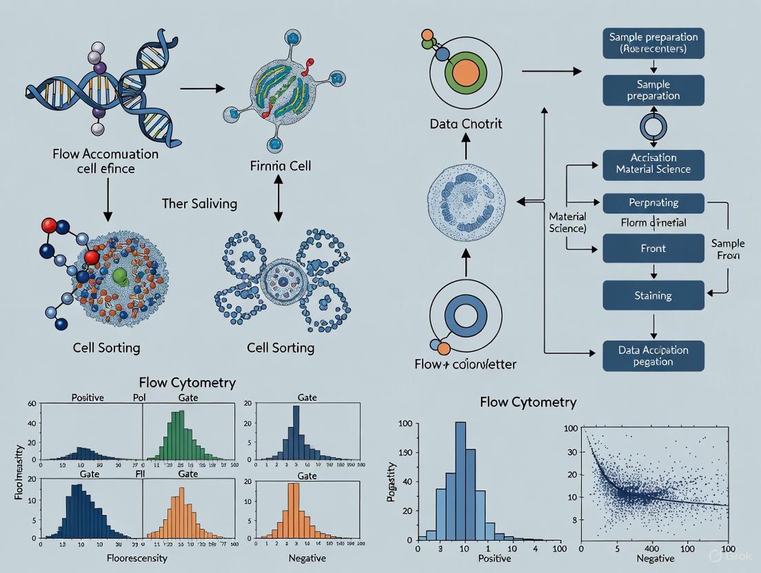
Data Acquisition and Gating Strategy
The following workflow diagram visualizes the logical process of using FMO controls for accurate gating in flow cytometry data analysis.
The Critical Next Step in Your Research
Mastering FMO controls is a fundamental skill for producing publication-quality data in multicolor flow cytometry. As emphasized by Maecker et al. and cited in expert literature, "in experiments of >4 colors, the major source of background staining tends to be fluorescence spillover. Because of this, the use of FMO controls has become both popular and prudent" [1]. By integrating these controls into your standard workflow, you lay the groundwork for robust, reproducible, and defensible science, which is paramount in both basic stem cell research and clinical drug development.
Why Spectral Overlap Necessitates FMO Controls in Complex Stem Cell Panels
Frequently Asked Questions
What is the fundamental reason FMO controls are needed in spectral flow cytometry? While spectral flow cytometry uses full-spectrum detection and unmixing algorithms to separate signals, the measurement is not perfect. Spectral overlap and electronic noise create a "spreading error" in the data. An FMO control contains all the fluorophores in your panel except one, which provides a realistic view of the background and spreading error specifically in the channel of the omitted antibody. This allows you to set a gating boundary that accurately distinguishes true positive signals from this background [8] [9].
Are FMO controls still necessary with the advanced unmixing of spectral cytometers? Yes. The unmixing process in spectral cytometry relies on having high-quality single-stain controls to build a reference library of each fluorophore's signature. However, the unmixed data can still contain background and spreading error, especially for dimly expressed markers. FMO controls are considered a superior gating control because they account for the cumulative spread from all other fluorophores in the panel, providing the most accurate picture for setting positive/negative boundaries [8] [9].
Can I use beads instead of cells for my FMO controls? No. FMO controls must be prepared using the same cell type as your experimental samples. This is critical because the autofluorescence and background marker expression levels in the control cells must perfectly match those in your test sample. Using beads or a different cell line will not accurately reflect the experimental background and can lead to incorrect gating [9].
My stem cells are highly autofluorescent. How does this affect my controls? High autofluorescence, common in cells like mesenchymal stem cells, significantly increases background signal. During the compensation process, this can even lead to counterintuitive "negative fluorescence values" in some channels. For autofluorescent cells, it is essential to use FMO controls to establish correct positivity thresholds and to display your data using biexponential transformations to properly visualize the compensated populations [10].
Troubleshooting Guides
Issue 1: Poor Separation Between Positive and Negative Populations
| Potential Cause | Recommended Solution |
|---|---|
| Excessive spectral spillover | Re-design the panel to use fluorophores with more distinct emission spectra. Use a spectra viewer tool to minimize overlap [11]. |
| Antibody concentration is too high | Titrate every antibody to find the optimal concentration that maximizes the signal-to-noise ratio (Stain Index) [8]. |
| High cellular autofluorescence | Use an unstained control from the same cell type and treatment to measure autofluorescence. The spectral unmixing algorithm can use this to improve resolution [8] [12]. |
| Inadequate Fc receptor blocking | Use an Fc receptor blocking reagent prior to staining to reduce non-specific antibody binding [8] [11]. |
Issue 2: High Background Fluorescence in Unstained Control
| Potential Cause | Recommended Solution |
|---|---|
| Cell death or poor health | Use a viability dye to gate out dead cells, which are prone to non-specific binding [13] [11]. |
| Fixation or permeabilization issues | Optimize fixation time and formaldehyde concentration; over-fixation can increase autofluorescence. For intracellular targets, consider alcohol permeabilization if detergents cause high background [11]. |
| Insufficient washing | Increase the number or volume of washes after staining steps, especially when using unconjugated primary antibodies [11]. |
| Old or over-fixed cells | Use fresh cells wherever possible, as autofluorescence increases when cells age or are fixed for extended periods [11]. |
Essential Research Reagent Solutions
The following reagents are critical for successful multicolor flow cytometry experiments in stem cell research.
| Item | Function in the Experiment |
|---|---|
| Fc Receptor Blocking Reagent | Binds to Fc receptors on cells (e.g., macrophages, monocytes) to prevent non-specific antibody binding, reducing background [8] [11]. |
| Fixable Viability Dye | Distinguishes live from dead cells. Dead cells bind antibodies non-specifically and must be excluded from analysis [13] [11]. |
| Compensation Beads | Antibody-capture beads used to generate consistent and bright single-stain controls for setting compensation or building the unmixing matrix in spectral cytometry [13] [11]. |
| UltraComp eBeads | A specific type of compensation bead that can be used with most fluorophore-conjugated antibodies to create compensation controls [11]. |
| FMO Control Cells | The same cell type as the experimental sample, stained with all antibodies except one, used to accurately set gates for positive populations [9]. |
| Single-Stain Control Cells/Beads | Samples stained with only one antibody each, used to generate the spectral unmixing matrix in spectral flow cytometry [8]. |
Experimental Protocols and Workflows
Detailed Protocol: Preparing FMO Controls for a Multicolor Panel
This protocol outlines the steps to create FMO controls, which are critical for establishing accurate gates in complex stem cell panels.
- Prepare a Master Mix: Calculate the total volume of antibody cocktail needed for your full-panel stained sample. Create a master mix containing all fluorophore-conjugated antibodies from your panel except for the one(s) you are controlling for [14].
- Aliquot for FMO Control: Add the appropriate volume of the "minus one" master mix to a separate tube containing your cells. This tube will become your FMO control.
- Complete the Full Stain: To the remaining master mix, add the omitted antibody. Then add this complete cocktail to your main sample tube [14].
- Stain and Process in Parallel: Incubate the full-panel sample and all FMO control tubes together under identical conditions (time, temperature, light protection). Wash and, if applicable, fix all samples simultaneously [8].
- Acquire Data: Run the samples on your flow cytometer. When analyzing, use the FMO control to set the positive gate for the channel of the omitted antibody.
Diagram 1: FMO control preparation workflow.
Workflow: A Systematic Gating Strategy for Stem Cell Analysis
A robust gating hierarchy is essential to cleanly identify your target stem cell population before analyzing marker expression.
- Debris Exclusion: Create a plot of Forward Scatter (FSC) vs. Side Scatter (SSC). Draw a gate (R1) around the cell population of interest, excluding low-FSC debris and noise [13].
- Singlets Selection: Create a plot of FSC-H vs. FSC-A (or FSC-W). Apply gate R1. Single cells will form a diagonal population; draw a gate (R2) around them to exclude doublets and multiplets [13].
- Viability Gating: Create a plot of your viability dye vs. FSC. Apply gate R2. Draw a gate (R3) around the viability dye-negative population (live cells) [13].
- Lineage or Marker Gating: For stem cell isolation, it is useful to stain with a broad marker like CD45 to gate out contaminating leukocytes or other lineage-specific markers. Create a plot of FSC vs. CD45 (or another marker). Apply gate R3. Draw a gate (R4) around the positive or negative population of interest [13].
- Functional Marker Analysis with FMO: Now create necessary fluorophore analysis plots (e.g., FITC vs. PE). Apply all previous gates (R1-R4). Use the corresponding FMO control to accurately set the boundary for positive cells in the channel of interest [13] [8].
Diagram 2: Sequential gating strategy for flow cytometry analysis.
Understanding the Core Concept: Spillover Spreading
The necessity of FMO controls is rooted in a phenomenon known as spillover spreading. In a perfect system, a population of negative cells would appear as a tight cluster. However, in reality, the combined effect of spectral overlap and electronic measurement error causes this negative population to spread out [9]. The more fluorophores you add to a panel, the more this spreading accumulates. An unstained control cannot account for this multi-fluorophore effect. An FMO control, because it contains all antibodies except one, shows you the exact amount of spread in the omitted channel caused by all the other colors in your panel. This allows you to place your gate just beyond the edge of this spread, ensuring that events called "positive" are truly positive and not just background artifacts.
A precise gating strategy is the foundation of accurate flow cytometry data in stem cell research.
Selecting the appropriate negative control is a critical step in flow cytometry experiment design, directly impacting the reliability of your data. This guide will help you understand the distinct roles of Fluorescence Minus One (FMO), isotype, and unstained controls, and how to apply them correctly in your stem cell research.
Understanding the Core Types of Negative Controls
Each negative control serves a unique purpose in experimental validation and data interpretation. The following table summarizes their primary functions and applications.
| Control Type | Primary Function | Best Used For |
|---|---|---|
| FMO Control | Defines positive/negative gating boundaries in multicolor panels by accounting for background fluorescence spread from all other fluorophores in the panel [13] [15]. | Setting gates for markers with low expression or continuous expression; multicolor panel validation; identifying spreading error [13] [16] [17]. |
| Isotype Control | Assesses background from non-specific antibody binding (e.g., to Fc receptors) [15] [18]. | Verifying staining specificity; troubleshooting high background staining. It is not for setting positive/negative gates [19] [20] [21]. |
| Unstained Control | Measures inherent cellular autofluorescence and instrument background [15] [18]. | Setting photomultiplier tube (PMT) voltages; identifying autofluorescence issues; establishing a baseline fluorescence profile [11] [18]. |
The logical relationship between these controls and their specific applications within an experimental workflow can be visualized as a decision pathway.
Troubleshooting Guide: Common Scenarios & Solutions
Problem 1: Indistinct Cell Populations in Multicolor Stem Cell Panels
You cannot clearly separate positive and negative populations for a key marker (e.g., CD34) in your hematopoietic stem cell panel.
- Recommended Control: FMO Control.
- Solution: Prepare an FMO control that contains all antibodies in your panel except the one against your target marker (e.g., anti-CD34). During analysis, use this FMO sample to set the upper boundary of the negative population on the CD34 channel. Any signal in your fully stained sample that exceeds this boundary should be considered positive [13] [15]. This is crucial for accurately identifying stem cell populations.
Problem 2: High Background Staining Despite Optimized Protocol
Your data shows a consistently high fluorescent signal across multiple channels, making it difficult to identify true negatives.
- Recommended Controls: Isotype Control and Unstained Control.
- Solution:
- First, use the unstained control to quantify the level of cellular autofluorescence [11].
- Second, use the isotype control to identify the contribution of non-specific antibody binding. If the isotype control signal is significantly higher than the unstained control, it indicates issues with Fc receptor binding or other non-specific interactions [15] [18].
- Based on this, incorporate an Fc receptor blocking step and re-titrate your antibodies to improve the signal-to-noise ratio [15] [11].
Problem 3: Incorrectly Set Gates Leading to False Positives
You are seeing an unexpectedly high frequency of double-positive cells for two markers that are not known to be co-expressed on your stem cell population.
- Recommended Control: FMO Controls for both channels.
- Solution: This pattern often indicates spreading error, which compensation alone cannot fully correct [22] [17]. Use the relevant FMO controls to accurately set the quadrants for both parameters. This ensures that the "positivity" you see is due to actual antibody binding and not fluorescence spillover from other bright markers in the panel [13].
Frequently Asked Questions (FAQs)
Can I use an isotype control instead of an FMO control to set my gates?
No. This is a common misconception. Isotype controls are only useful for assessing nonspecific antibody binding and should not be used to distinguish positive from negative cells or to set positive gates [19] [20] [21]. Isotype controls do not account for the spreading error caused by the other fluorophores in your multicolor panel, which is the primary reason for using an FMO control [20].
When is it absolutely necessary to use an FMO control?
FMO controls are essential in the following situations in stem cell research [13] [16]:
- When analyzing markers with low expression levels.
- When gating for continuously expressed markers (e.g., activation markers like PD-1, HLA-DR) that do not form a clear, distinct negative population.
- When validating a new multicolor flow cytometry panel.
- Whenever you encounter ambiguous populations that could be caused by spreading error.
My unstained control has high fluorescence. What does this mean?
High fluorescence in an unstained control indicates significant autofluorescence [15] [11]. This can be caused by the cell type itself (some stem cells are inherently autofluorescent), the physiological state of the cells, or the use of fixatives. If autofluorescence is high, consider using fluorophores excited by different laser wavelengths that avoid autofluorescence peaks (e.g., avoiding ~488 nm excitation) to improve detection sensitivity [15] [11].
How do I properly design and use single-stain compensation controls?
- Brightness: The positive control must be at least as bright as your brightest experimental sample [20].
- Match: The fluorophore must exactly match the one used in your experiment, paying special attention to tandem dyes (e.g., PE-Cy7), which can have lot-to-lot variability [13] [20].
- Treatment: Compensation controls must be treated identically to experimental samples (e.g., same fixation/permeabilization) as these processes can alter fluorophore spectra [22] [19].
- Carrier: While compensation beads are a good tool, using single-stained cells is often preferable because the fluorophore emission spectrum can sometimes differ when bound to a bead versus a cell [22] [19].
The Scientist's Toolkit: Essential Research Reagent Solutions
The table below lists key reagents and their critical functions for implementing robust negative controls in your experiments.
| Research Reagent | Function in Flow Cytometry Controls |
|---|---|
| Compensation Beads | Synthetic beads that bind antibody conjugates, providing a bright, consistent signal for calculating spillover and compensation matrices [13] [20]. |
| Fc Receptor Blocking Reagent | Reduces nonspecific antibody binding, a key source of background that is detected by isotype controls [15] [11]. |
| Viability Dye (e.g., PI, 7-AAD) | Allows exclusion of dead cells during analysis, which are a major source of autofluorescence and nonspecific binding [13] [15] [11]. |
| Antibody Capture Beads | Used similarly to compensation beads; ensure they are compatible with your specific antibody conjugates for reliable single-stain controls [19]. |
Experimental Protocol: Implementing FMO Controls in Clinical-Grade Stem Cell Analysis
The following workflow, derived from high-throughput clinical data analysis, ensures reliable and reproducible gating for complex panels [16].
Panel Design and Sample Preparation
- For a 12-color T cell or regulatory T cell panel, set up one FMO control for every marker with continuous expression (e.g., PD-1, HLA-DR, CTLA-4).
- Collect each fully stained sample and its corresponding FMO controls in the same plate to minimize technical variation.
- Stain cells following your standard protocol, ensuring the FMO control contains all fluorophore-conjugated antibodies except the one being controlled.
Data Acquisition and Transformation
- Acquire data on the flow cytometer, collecting a sufficient number of events for robust analysis.
- Use software (e.g., R package
flowCore) to apply bi-exponential transformation and a spillover compensation matrix to the raw FCS files [16].
Automated Gating via OpenCyto Pipeline
This protocol uses an automated gating template to ensure consistency, mirroring the manual gating process.
- Step 1: Pre-defined hierarchical gating strategies are implemented in an OpenCyto gating template [16].
- Step 2: For one-dimensional (1D) gating, the cut-off point for the negative population is determined from the FMO control using a probability density function and slope analysis [16].
- Step 3: This calculated cut-off point is transferred to the fully stained samples to gate the positive and negative populations accurately.
- Step 4: To adopt the "0.5% rule" common in manual gating, the
adjustandtoleranceparameters of the density function are fine-tuned [16].
Quality Control and Validation
- Filter 1: Any automated gating result generated without a corresponding FMO control is automatically flagged as a "failure" [16].
- Filter 2: Cluster identification (e.g., for CD3+ populations) is monitored. If the coordinates of the gated population are significantly different from pre-calculated cluster centroids, the gating is flagged for review or re-gating with an alternative parameter set [16].
This standardized, control-based pipeline demonstrates that automated analysis can achieve precision and accuracy comparable to traditional manual gating, even in large-scale clinical studies [16].
Frequently Asked Questions (FAQs)
FAQ 1: What defines a "rare event" in stem cell research, and how many cells do I need to acquire?
In flow cytometry, a cell population is generally considered "rare" when it has a frequency of 0.01% or less of the total parent population [23]. Stem cells and their specific progenitors often fall into this category.
The number of events you need to acquire for statistically significant data depends on the desired precision, which is governed by Poisson statistics. To keep the Coefficient of Variation (CV) below 5%, you must acquire a substantial number of total events [23] [24]. The table below summarizes the total events required to detect populations of varying frequencies at different CVs.
Table 1: Total Events Required for Rare Cell Detection
| Desired CV | Frequency 0.01% | Frequency 0.1% | Frequency 1% |
|---|---|---|---|
| 5% | 4 million events | 400,000 events | 40,000 events |
| 10% | 1 million events | 100,000 events | 10,000 events |
| 20% | 250,000 events | 25,000 events | 2,500 events |
For a population at 0.01%, acquiring one million events yields a CV of 10%, while four million events are needed to achieve a more robust CV of 5% [23].
FAQ 2: Why is my stem cell marker signal so dim or undetectable?
Weak or no fluorescence signal from dim markers can result from several factors [25]:
- Insufficient Antibody: The antibody concentration may be too low, or the antibody itself may not be validated for flow cytometry.
- Poor Permeabilization: For intracellular stem cell markers (e.g., transcription factors), inadequate fixation and/or permeabilization will prevent antibody access.
- Fluorochrome Choice: A dim fluorochrome (e.g., FITC) was paired with a low-abundance target. Always use the brightest fluorochrome (e.g., PE) for your dimmest markers [25].
- Instrument Settings: The laser and photomultiplier tube (PMT) settings on the flow cytometer may not be optimized for the fluorochrome being used.
FAQ 3: My staining looks non-specific; how can I reduce high background?
High background can be caused by non-specific antibody binding, often through Fc receptors on cells like monocytes [25] [15]. To reduce this:
- Fc Receptor Blocking: Use an Fc receptor blocking reagent or normal serum prior to staining [25] [26].
- Titrate Antibodies: Optimize the concentration of every antibody to find the best signal-to-noise ratio [15] [26].
- Remove Dead Cells: Dead cells cause non-specific binding and autofluorescence. Always use a viability dye and gate out dead cells [25] [15].
- Increase Wash Steps: Perform additional washes between antibody incubations to remove unbound antibody [25].
FAQ 4: What is the single most important control for accurately gating dimly expressed stem cell markers?
The Fluorescence Minus One (FMO) control is critical for setting gates for dim markers in a multicolor panel [13] [26]. This control contains all the fluorophore-labeled antibodies in your panel except for one. It reveals the background fluorescence "spread" into the channel of the omitted antibody, allowing for correct gate placement to distinguish true positive from negative cells [15]. Using an unstained control alone often sets the gate too high, misclassifying negative cells as positive [13].
Troubleshooting Guide
Problem: Inability to Resolve a Rare Stem Cell Population
| Possible Cause | Recommendation | Experimental Protocol |
|---|---|---|
| Insufficient events acquired | Calculate and acquire the necessary total events based on the expected frequency and desired CV (see Table 1). | Use Poisson statistics: r = (100/CV)², where r is the number of target cells needed. The total events to acquire = r / expected frequency [24]. |
| High background masking the rare population | Implement a "dump channel" to exclude unwanted cells and use FMO controls for precise gating. | In your panel, include markers for common cell types you wish to exclude (e.g., lineage markers) all conjugated to the same bright fluorochrome. Also, include a viability dye in this channel to exclude dead cells [24]. |
| Sample loss during preparation | Minimize processing steps. Consider "no-lyse/no-wash" or "lyse/no-wash" protocols. | For rare cells in whole blood, use a fixative-free lysing solution (e.g., Invitrogen High-Yield Lyse Solution) that does not require a subsequent wash step, thereby minimizing cell loss [24]. |
| Instrument sensitivity and clogging | Ensure the flow cytometer is clean and well-maintained. Use high-speed acquisition settings if available. | Prior to running samples, perform a cleaning cycle if the instrument has been idle. For persistent clogs, run 10% bleach for 5-10 minutes followed by distilled water for 5-10 minutes as per manufacturer instructions [25]. |
Problem: Weak Staining of Dim Intracellular Markers
| Possible Cause | Recommendation | Experimental Protocol |
|---|---|---|
| Inadequate permeabilization | Ensure you are using the correct permeabilization reagent and technique for your target. | For transcription factors, use ice-cold 90% methanol. Chill cells on ice first, then add methanol drop-wise to the cell pellet while gently vortexing to prevent hypotonic shock [25]. |
| Fluorochrome is too dim or too large | Switch to a brighter, smaller fluorochrome conjugate. | Avoid large tandem dyes or synthetic dye complexes for intracellular staining. Use bright, low molecular weight fluorochromes like PE or Alexa Fluor dyes for better penetration [25] [27]. |
| Antibody concentration is suboptimal | Titrate the antibody to find the optimal concentration for your specific cell type. | Perform a titration assay by staining cells with a series of antibody dilutions (e.g., 1:50, 1:100, 1:200). Calculate the stain index (Mean Positive - Mean Negative) / (2 × SD of Negative) and select the dilution with the highest index [26]. |
| Target protein is soluble or secreted | Use a Golgi-blocking step during stimulation to allow intracellular protein accumulation. | Treat cells with Brefeldin A for 4-6 hours prior to harvest and staining. This inhibits protein transport, allowing for better detection of cytoplasmic cytokines and other soluble factors [27]. |
The Scientist's Toolkit: Essential Research Reagent Solutions
Table 2: Key Reagents for Stem Cell Flow Cytometry
| Reagent / Material | Function | Example & Notes |
|---|---|---|
| Viability Dyes | Distinguish live from dead cells to reduce background. | Fixable Viability Dyes (e.g., eFluor, Zombie dyes): Required for intracellular staining as they withstand fixation/permeabilization [25]. |
| Fc Receptor Blocking Reagent | Reduces non-specific antibody binding. | Purified IgG or commercial Fc block; incubate with cells for 10-15 minutes prior to antibody staining [15] [26]. |
| Bright Fluorochrome-Conjugated Antibodies | Detect low-abundance (dim) markers. | PE and Brilliant Violet 421: These are typically brighter than FITC and should be reserved for your dimmest markers like many stem cell transcription factors [25]. |
| Compensation Beads | Generate consistent single-stain controls for accurate compensation. | Anti-mouse/anti-rat Igκ beads; bind to most antibodies, providing a uniform positive population for setting compensation on the cytometer [15]. |
| Methanol (Ice-cold) | Effective permeabilization agent for nuclear transcription factors. | Use 90% methanol, ice-cold. Critical for intracellular staining of targets like Nanog or Oct4 [25]. |
| Magnetic Cell Separation Kits | Pre-enrich rare stem cell populations to reduce acquisition time. | Kits for positive or negative selection (e.g., Lin- depletion) can increase the relative frequency of your target population, making flow analysis more efficient [23]. |
Experimental Workflows and Visualization
Logical Gating Strategy for Rare and Dim Stem Cell Populations
The following diagram outlines a rigorous gating strategy to resolve a rare stem cell population expressing a dim marker. This workflow emphasizes the use of doublet discrimination, viability staining, dump channel exclusion, and FMO-controlled gating for the final, dim target.
The Critical Role of the FMO Control
This diagram visually contrasts how gating is performed using an unstained control versus an FMO control, demonstrating how the FMO control accounts for fluorescence spread and enables accurate identification of dim positive cells.
FAQs on FMO Controls in Stem Cell Research
What is the primary purpose of an FMO control in multicolor flow cytometry? An FMO control is a sample stained with all the fluorophore-labeled antibodies in your panel except for one. Its primary purpose is to determine the correct placement of the positive/negative gate for the omitted fluorophore by accurately revealing the background fluorescence and signal "spread" caused by spectral overlap from all the other colors in the panel [13]. This is particularly critical for accurately identifying dimly expressed markers and precisely defining rare cell populations, such as hematopoietic stem cells (HSCs) [13] [28].
Why is an FMO control superior to an unstained or isotype control for setting gates? An unstained control only shows a cell's autofluorescence, while an isotype control helps assess non-specific antibody binding. However, neither accounts for the fluorescence spillover from multiple other fluorochromes used in a complex panel. The FMO control is the only control that captures this combined background signal, providing a true baseline for distinguishing positive from negative cells in a multicolor experiment [15]. Using an unstained control can lead to a false positive population being gated, whereas the FMO control correctly identifies the boundary [15].
For which markers in a panel are FMO controls most critical? FMO controls are most critical for:
- Dimly expressed markers: When the target antigen has low expression levels [13].
- Rare cell populations: When identifying small subpopulations, such as HSCs, where accurate gating is paramount [28].
- Channels with significant spillover: When the fluorophore of interest is in a channel that receives spillover from several other bright fluorochromes [13] [15].
Do I need an FMO control for every single fluorophore in my panel? While it is highly recommended to set up an FMO control for every fluorophore-conjugated antibody during panel design and optimization, it may not be practical for every experiment. For well-established panels and for populations with clear, bright separation between positive and negative cells, it may be sufficient to run FMO controls only for the most critical and problematic markers [13] [15].
Troubleshooting Guide: Resolving Common FMO and Gating Issues
| Problem | Possible Causes | Recommended Solutions |
|---|---|---|
| High background in FMO control | Excessive antibody concentration; non-specific antibody binding; dead cells in sample [29]. | Titrate all antibodies to find optimal signal-to-noise ratio [15]; include a viability dye and Fc receptor blocking step prior to staining [15]; increase wash steps after staining [29]. |
| Poor separation between positive and negative populations | Marker is dimly expressed; fluorophore is too dim for the target; excessive spectral overlap [29]. | Switch to a brighter fluorophore for the dim marker [29]; re-evaluate panel design to minimize spillover into the critical channel; use the FMO control to rigorously define the negative population [13]. |
| Inconsistent gating between experiments | Day-to-day instrument performance variation; subjective manual gating [28]. | Standardize instrument setup using CS&T or other calibration beads daily [3]; where possible, use automated gating algorithms to reduce analyst subjectivity [28]. |
| Unable to resolve rare population | Insufficient event count; high background masking the population [3]. | Acquire a significantly higher number of events to adequately capture rare cells [3]; use a pre-enrichment step (e.g., MACS) prior to staining for FACS [13]; use FMO controls to set tight, accurate gates. |
Essential Experimental Protocols
1. Standardized Staining Protocol for Immunophenotyping This protocol is adapted for use with lyophilized reagent plates, which enhance reproducibility, but can be modified for liquid reagents [28].
- Sample Preparation: Isolate your cell sample (e.g., PBMCs, bone marrow). Use a viability dye, such as fixable viability dye eFluor 506, to stain live cells prior to fixation [28].
- Fc Receptor Blocking: Incubate cells with an Fc receptor blocking reagent for 10-15 minutes on ice to reduce non-specific antibody binding [15].
- Surface Staining: Add pre-titrated antibody cocktails (either liquid or reconstituted lyophilized reagents) to the cell pellet. Vortex gently and incubate for 30 minutes in the dark at 4°C [28].
- Fixation and Permeabilization: For intracellular targets (e.g., transcription factors), fix cells with 4% formaldehyde immediately after staining, then permeabilize with ice-cold 90% methanol or a saponin-based buffer [29].
- Intracellular Staining: Centrifuge cells to remove permeabilization buffer, then incubate with intracellular antibodies for 30-60 minutes at 4°C in the dark [3].
- Data Acquisition: Resuspend cells in wash buffer and acquire data on the flow cytometer. Use standardized instrument settings and calibration beads to ensure consistency [28] [3].
2. Application: Isolating Human Hematopoietic Stem Cells (HSCs) The following workflow details how FMO controls are integral to the precise isolation of rare LT-HSCs [3]:
- Sample: Use mobilized peripheral blood from leukapheresis.
- Lineage Depletion: Label cells with a cocktail of biotin-conjugated antibodies against lineage markers (CD2, CD3, CD14, CD16, CD19, CD56, CD235a) and remove them using magnetic separation.
- Staining: Stain the lineage-negative cells with a panel including CD34, CD38, CD45RA, CD90, and CD49f, along with a viability dye.
- FMO Controls: Prepare FMO controls for every fluorochrome in this panel to accurately gate on the dim but critical markers (e.g., CD90, CD49f).
- Gating Strategy:
- Exclude debris based on FSC-A vs. SSC-A.
- Exclude doublets using FSC-H vs. FSC-A.
- Select viable cells (viability dye negative).
- Gate on lineage-negative cells.
- Identify CD34+ CD38- population.
- Further gate on CD45RA- CD90+ cells.
- The final, highly enriched LT-HSC population is defined as CD49f+ [3].
The logical sequence of this gating strategy is visualized below.
The Scientist's Toolkit: Key Research Reagent Solutions
| Item | Function | Example Application |
|---|---|---|
| Lyophilized Antibody Panels | Pre-configured, multi-color antibody cocktails in 96-well plates. Improve standardization by reducing pipetting errors and ensuring reagent stability [28]. | Standardized immunophenotyping of major immune cell subsets in PBMCs (e.g., T cells, B cells, monocytes) [28]. |
| Fixable Viability Dyes | Cell-impermeant dyes that covalently bind to amines in dead cells. Allow for exclusion of dead cells during analysis, which is critical for reducing background [3] [15]. | Used in all staining protocols to improve data quality by gating out dead cells that cause non-specific binding. |
| Compensation Beads | Synthetic beads that bind antibodies. Used to create single-color controls for calculating spectral compensation matrix, independent of biological sample [13] [15]. | Setting up compensation controls for every fluorochrome in a panel, especially when a specific biological positive control is not available. |
| Fc Receptor Blocking Reagent | Blocks Fc receptors on immune cells to prevent non-specific binding of antibodies, thereby reducing background staining [15]. | Essential when staining samples high in monocytes, macrophages, or when using cells from homogenized tissues. |
| CD34 MicroBead Kit | Magnetic-activated cell sorting (MACS) kit for positive selection of CD34+ cells. Pre-enriches target population prior to FACS, improving sort efficiency and recovery of rare cells [3]. | Pre-enrichment of hematopoietic stem and progenitor cells (HSPCs) from mobilized peripheral blood or bone marrow [3]. |
Implementing FMO Controls: A Step-by-Step Protocol for Stem Cell Research
A foundational guide for ensuring accurate data interpretation in multicolor flow cytometry panels for stem cell research.
In the realm of stem cell research, accurately identifying and isolating specific cell populations, such as pluripotent stem cells, is crucial. When using multicolor flow cytometry panels, Fluorescence Minus One (FMO) controls are indispensable tools for setting correct gates and distinguishing true positive signals from background. This guide provides detailed protocols for designing and implementing FMO controls in your experiments.
What Is an FMO Control and Why Is It Essential?
An FMO control is a sample stained with all the fluorophore-conjugated antibodies in your multicolor panel except for one. [13] [15]
- Purpose: It measures the "spread" or "bleed" of background signal into the channel of the omitted antibody caused by the combined spectral overlap from all the other fluorophores in your panel. [13] [15]
- Critical Use: FMO controls allow you to determine the precise cutoff between negative and positive cell populations, which is particularly important for:
The diagram below illustrates the logical relationship between your full staining panel and the corresponding FMO controls you need to create.
How to Design an FMO Control: A Step-by-Step Protocol
Step 1: Prepare the Staining Master Mix
Create a master mix containing all antibodies from your full panel. [13] Then, for each FMO control, prepare an aliquot of this master mix and remove the specific antibody for which the FMO is being made.
Step 2: Stain the Cells
Use the same cell type (e.g., PBMCs, stem cell lines) as your experimental samples. The cells used for FMO controls should have a known expression profile for the marker of interest, ideally containing both positive and negative populations. [13] [15] Stain the FMO control sample with the prepared "minus one" antibody mix.
Step 3: Process and Acquire Data
Treat the FMO control samples identically to your fully stained samples throughout all subsequent steps: incubation, washing, fixation, and data acquisition on the flow cytometer. [13]
Step 4: Set the Gate
During analysis, plot the fluorescence intensity for the channel of the omitted antibody. The negative population in the FMO control defines the upper limit of background signal. Set your positivity gate just above this population to accurately identify true positive cells in your fully stained sample. [13] [15]
FMO Controls vs. Other Negative Controls
FMO controls are often confused with other types of negative controls. The table below clarifies their distinct purposes.
| Control Type | Description | Primary Function | Recommended for Gating? |
|---|---|---|---|
| FMO Control | Contains all antibodies in the panel except one. [13] [15] | Measures spillover spreading from all other fluorophores into the channel of interest. [13] [15] | Yes, essential for defining positive/negative boundaries in multicolor panels. [13] [19] |
| Isotype Control | An antibody with the same isotype but irrelevant specificity. | Assesses non-specific antibody binding via Fc receptors. [15] | No, not for gating. Use to check background from non-specific binding. [19] [15] |
| Unstained Control | Cells with no antibody staining. | Measures cellular autofluorescence and instrument noise. | No, insufficient for multicolor panels as it does not account for spillover. [15] |
The Scientist's Toolkit: Essential Reagents for FMO Controls
| Reagent / Material | Function in FMO Controls |
|---|---|
| Viability Dye (e.g., Fixable Viability Stain, PI, 7-AAD) | Distinguishes live from dead cells. Dead cells increase background and non-specific binding. [13] [11] [15] |
| Fc Receptor Blocking Reagent | Reduces non-specific antibody binding, providing a cleaner background signal. [11] [15] |
| Compensation Beads / Cells | Used to create single-stain controls for calculating the compensation matrix. Best practice is to use cells, as beads can sometimes have different spectral properties. [19] |
| Cell Preparation | Fresh or properly frozen-thawed cells (e.g., PBMCs, stem cell lines) with high viability. [5] [11] |
FAQs and Troubleshooting
How many FMO controls do I need for my panel?
In an ideal setting, you should create one FMO control for every fluorophore-antibody conjugate in your panel. [13] [15] For large panels (e.g., 20+ colors), this may not be feasible due to cost or cell number limitations. In such cases, prioritize creating FMO controls for:
- Dim markers
- Markers expressed on rare cell populations
- Channels with high levels of spillover spreading [13]
Can I use an isotype control instead of an FMO control?
No. Isotype controls are not a substitute for FMO controls. [19] Isotype controls help identify problems with background staining from non-specific antibody binding but do not account for the spreading error caused by the spectral overlap of multiple fluorophores in your panel. [19] [15] Relying on isotype controls for gating in multicolor experiments can lead to inaccurate data interpretation.
My FMO control shows a very bright background. What could be wrong?
A bright background in your FMO control can stem from several issues:
- High Cell Death: An increase in dead cells leads to elevated autofluorescence and non-specific antibody binding. Always include a viability dye and gate on live cells. [11] [15]
- Insufficient Fc Blocking: Phagocytic cells can bind antibodies non-specifically via Fc receptors. Ensure you are using an appropriate Fc receptor blocking reagent. [11] [15]
- Antibody Concentration Too High: An overly high antibody titer can increase non-specific binding. Titrate all antibodies to find the optimal signal-to-noise ratio. [11] [15]
- Poorly Compensated Sample: Errors in the compensation matrix will cause incorrect spreading of signal into adjacent channels. Ensure your single-stain controls are set up correctly. [11]
Are FMO controls necessary for spectral flow cytometry?
Yes. While spectral flow cytometry uses full-spectrum fingerprinting and unmixing algorithms instead of traditional compensation, background signal and spreading error remain a concern. FMO controls are still critical for validating your gating strategy and ensuring the accurate detection of dimly expressed markers, even on spectral cytometers. [30] [19]
FAQs on Parallel Staining Controls
What are the essential control samples required for accurate flow cytometry analysis?
Proper controls are fundamental for validating your flow cytometry results and are considered parallel, non-experimental samples. The core set of controls is detailed in the table below.
| Control Type | Purpose | Key Consideration |
|---|---|---|
| Unstained Control [31] | Measures cellular autofluorescence and background instrument signal. | Use the same cell population as your experimental sample. |
| Isotype Control [31] [13] | Assesses non-specific, Fc receptor-mediated antibody binding. | Must match the primary antibody's host species, isotype, and fluorophore conjugation [32]. |
| Positive Control [31] [33] | Verifies that the staining protocol works correctly. | Use a cell line or population known to express the target antigen. |
| Fluorescence Minus One (FMO) Control [13] [32] | Determines the correct gating boundary by showing the background signal from all other fluorophores in a panel. | Critical for multicolor panels and low-abundance targets [13]. |
| Compensation Control [13] | Allows the instrument to calculate and correct for spectral overlap between fluorophores. | Use compensation beads or cells stained with a single fluorophore for each color in the panel. |
Why is an FMO control critical in multicolor stem cell panels?
The Fluorescence Minus One (FMO) control is indispensable in multicolor flow cytometry because it accounts for spectral spillover spreading, a phenomenon where the emission from one fluorophore spreads into the detector of another, increasing the background signal of the negative population [13] [32]. This is especially important when analyzing stem cells for lowly expressed markers, as it allows for the most accurate placement of positive/negative gates, preventing the misclassification of false-positive cells [13]. An FMO control contains all the fluorophore-conjugated antibodies in your full panel except for one, whose gating boundary you are trying to define [13].
How do I troubleshoot high background signal in my control samples?
High background or non-specific binding in controls can obscure your results. The table below outlines common causes and solutions.
| Problem | Possible Cause | Recommended Solution |
|---|---|---|
| High background in isotype control | Fc receptor binding on cells (e.g., monocytes). | Block Fc receptors prior to staining using BSA, specific blocking reagents, or normal serum [31] [33]. |
| General high background | Insufficient washing steps leaving unbound antibody. | Increase the number or volume of wash steps between antibody incubations [31] [34]. |
| Antibody concentration is too high. | Titrate all antibodies to determine the optimal concentration for the best signal-to-noise ratio [31] [32]. | |
| Presence of dead cells. | Use a viability dye (e.g., PI, 7-AAD, or a fixable dye) to gate out dead cells during analysis [31] [13]. |
What are the best practices for preparing parallel control samples?
To ensure your controls provide reliable data, adhere to the following protocols:
- Use the Same Cell Source: Controls must be prepared from the same cell suspension as your experimental samples (e.g., the same stem cell culture or primary tissue isolate) to ensure biological relevance [33].
- Maintain Identical Processing: All samples and controls must be processed in parallel. This includes identical treatment during fixation, permeabilization, washing, incubation times, and buffer compositions [31] [35].
- Titrate All Reagents: Antibodies and fluorescent dyes should be carefully titrated on your specific cell type to find the concentration that provides the best stain index, maximizing signal while minimizing background [32].
- Handle Cells Gently: Centrifuge cells at low speeds (300–400g), keep samples on ice or at 4°C, and avoid vortexing vigorously to maintain cell viability and reduce debris [35].
Experimental Protocols for Key Controls
Protocol 1: Staining for FMO Controls
Objective: To create a control that accurately defines the positive gate for a specific marker in a multicolor panel.
Materials:
- Cell suspension from the same source as experimental sample.
- All antibodies from the full staining panel.
- Staining buffer (e.g., PBS with 1% BSA).
- Centrifuge and vortexer.
Method:
- Prepare the Tube: Aliquot the same number of cells used for the full stain into a separate tube.
- Add Antibodies: Add every antibody from your multicolor panel except for one—the antibody for which you are defining the gate.
- Incubate and Wash: Follow the same incubation and washing protocol as your experimental sample.
- Resuspend and Analyze: Resuspend the cells in staining buffer and acquire on the flow cytometer.
- Set the Gate: Use the FMO control to set the boundary between negative and positive cells for the omitted antibody [13].
Protocol 2: Preparation of Compensation Controls
Objective: To generate single-color stained controls for the instrument to calculate spillover compensation.
Materials:
- Compensation beads (recommended for consistency) or a cell suspension.
- Each individual antibody-fluorophore conjugate from your panel.
- Staining buffer.
Method:
- Aliquot Beads/Cells: Set up one tube for each fluorophore used in your panel.
- Stain Tubes: Add a small volume of each antibody to its corresponding tube. Incubate as per your staining protocol.
- Wash and Resuspend: Wash the beads or cells and resuspend in buffer.
- Acquire Data: Run each single-color control on the cytometer and use the software to build the spillover (compensation) matrix [13]. When using tandem dyes, use the same antibody lot for compensation as for your experiment due to potential lot-to-lot variability [13].
Research Reagent Solutions
The following table lists essential materials for establishing robust parallel staining procedures.
| Item | Function | Application Note |
|---|---|---|
| Fc Receptor Blocking Reagent | Blocks non-specific antibody binding to Fc receptors on cells. | Critical for reducing false positives when working with immune cells [33]. |
| Compensation Beads | Uniform particles that bind antibodies, used to create consistent single-color controls. | Provide a clean, uniform population for setting compensation, often superior to cells [13] [35]. |
| Viability Dye (Fixable) | Distinguishes live from dead cells; fixable dyes withstand permeabilization. | Allows gating out of dead cells that cause non-specific binding, crucial for intracellular staining [31] [13]. |
| DNase I & EDTA | Prevents cell clumping by digesting released DNA and chelating calcium. | Adding to wash buffers improves cell suspension quality and prevents clogging of the instrument [35]. |
| Titrated Antibodies | Antibodies whose optimal concentration has been pre-determined for a specific cell type. | Using a titrated concentration is key to maximizing the signal-to-noise ratio [33] [32]. |
Workflow for Parallel Sample Preparation
The diagram below illustrates the logical sequence for preparing your experimental and parallel control samples to ensure a rigorously controlled experiment.
Advanced Troubleshooting Guide
For persistent issues, consult this advanced guide.
| Observed Issue in Controls | Root Cause | Advanced Solution |
|---|---|---|
| Weak or no signal in positive control. | Inadequate fixation/permeabilization for intracellular targets. | For methanol permeabilization, chill cells on ice and add ice-cold methanol drop-wise while vortexing [31]. |
| A weakly expressed target was paired with a dim fluorochrome. | Pair your lowest abundance targets with the brightest fluorophores (e.g., PE) [31]. | |
| FMO control background is too high for analysis. | Significant spectral overlap from other fluorophores in the panel. | Re-design the panel to avoid pairing fluorochromes with major spillover on the same cell type. Use a spillover spread matrix for guidance [36]. |
| Compensation is difficult or inaccurate. | The positive signal for the single-color control is too weak. | Use an antibody for a highly expressed marker conjugated to the same fluorophore to boost signal intensity [13]. |
In stem cell flow cytometry research, accurately identifying and isolating specific cell populations is paramount. A cornerstone of this process is the Fluorescence Minus One (FMO) control, a critical tool for establishing precise gates in multicolor panels. FMO controls account for background fluorescence and spectral spillover, enabling researchers to distinguish true positive signals from background, thereby ensuring the data's validity and reproducibility in drug development and scientific research [13] [9].
FAQs: Understanding FMO Controls
1. What is an FMO control, and why is it essential in multicolor flow cytometry?
A Fluorescence Minus One (FMO) control is a sample stained with all the fluorophore-conjugated antibodies in a multicolor panel except for one. This control is crucial because it reveals the "spread" of the background signal in the channel of the omitted antibody caused by spectral overlap from all the other fluorophores in the panel [13] [37] [9]. Without an FMO control, this background spread can be mistaken for a true positive signal, leading to inaccurate data interpretation and conclusions [38].
2. When is it absolutely necessary to use an FMO control?
FMO controls are particularly critical in several scenarios [13] [9] [38]:
- Dimly expressed markers: When the target antigen has low expression levels.
- Rare cell populations: When identifying infrequent cell subsets.
- Continuous expression patterns: When there is no clear separation between positive and negative populations.
- Panel validation: During the development and optimization of any new multicolor antibody panel.
- Critical markers: For intracellular targets like cytokines (e.g., IFN-γ, TNF) or transcription factors (e.g., FoxP3) [39].
3. How does an FMO control differ from an unstained or isotype control?
These controls address different aspects of experimental background and are not interchangeable.
- Unstained Control: Consists of cells with no fluorescent staining. It measures cellular autofluorescence and is used for setting photomultiplier tube (PMT) voltages [15] [39].
- Isotype Control: An antibody of the same isotype but irrelevant specificity, conjugated to the same fluorophore. It aims to measure non-specific antibody binding. However, its utility for setting positive gates is controversial, as it often does not accurately match the background staining of the specific antibody and does not account for spectral spillover [38] [39] [40].
- FMO Control: Specifically measures the combined effects of autofluorescence, non-specific binding, and, most importantly, spectral spillover from other fluorochromes in the panel [9] [38].
4. Can I use beads or a different cell type for my FMO controls?
No. FMO controls are affected by the autofluorescence and marker expression profile of the specific cells under investigation. Therefore, you must use the same type of cells as your experimental samples (e.g., the same stem cell line or primary cell preparation) to generate meaningful FMO controls [9].
5. Do I need to run an FMO control for every marker in every experiment?
While it is recommended to run FMOs for every marker during the initial panel development and validation, it may not be practical for every subsequent experiment. Once a panel is validated, researchers often continue running FMOs only for the most difficult-to-gate markers (e.g., dim or continuously expressed) with each batch of fully stained samples [9].
Troubleshooting Guide: FMO Controls in Practice
Problem 1: Indistinct Positive and Negative Populations
- Symptoms: The positive population appears as a "smear" with no clear separation from the negative cells, even in the FMO control.
- Solutions:
- Verify Antibody Titration: Ensure all antibodies have been optimally titrated to achieve the best signal-to-noise ratio [15].
- Check Cell Viability: An increase in dead cells can elevate non-specific staining. Re-gate your analysis on viable cells using a viability dye [13] [41] [15].
- Review Compensation: Improper compensation can distort population distributions. Re-check compensation using single-stained controls or compensation beads [13] [15].
Problem 2: High Background in FMO Control
- Symptoms: The FMO control shows an unexpectedly high level of fluorescence, making it difficult to set a meaningful positive gate.
- Solutions:
- Fc Receptor Blocking: Cells of myeloid origin or stressed cells can express Fc receptors that bind antibodies non-specifically. Incubate cells with an Fc receptor blocking reagent prior to staining [15] [39].
- Titrate Antibodies: High antibody concentrations are a common cause of non-specific binding and background. Re-titrate the antibodies in your panel [15].
- Wash Steps: Ensure thorough washing after staining steps to remove unbound antibody [39].
Problem 3: Inconsistent FMO Results Between Experiments
- Symptoms: The gate position defined by the FMO control varies significantly from one experiment to another.
- Solutions:
- Control Cell Source: Use a consistent and well-characterized source of cells for your FMO controls [9].
- Standardize Staining Protocol: Ensure staining procedures (incubation time, temperature, buffer composition) are rigorously followed every time [39].
- Reagent Lot Consistency: For antibodies labeled with tandem fluorophores (e.g., PE-Cy7), use the same antibody lot for experiments and their corresponding FMO controls, as tandem dyes can have lot-to-lot variability [13].
The following table summarizes these common issues and their solutions for quick reference.
| Problem | Possible Causes | Recommended Solutions |
|---|---|---|
| Indistinct positive/negative populations | Poor antibody titration, low cell viability, improper compensation | Titrate antibodies; use viability dye; re-check compensation with single-stained controls [13] [41] [15] |
| High background in FMO control | Fc receptor binding, high antibody concentration, insufficient washing | Use Fc receptor blocking; titrate antibodies; ensure thorough washing steps [15] [39] |
| Inconsistent FMO results | Varying cell sources, non-standardized protocols, reagent lot variation | Use consistent control cells; standardize staining protocol; use same antibody lots for experiments and FMOs [13] [9] [39] |
Experimental Protocol: Implementing FMO Controls
Detailed Methodology for FMO Control Setup
This protocol outlines the steps for preparing an FMO control as part of a multicolor flow cytometry panel for stem cell analysis.
1. Principle An FMO control is prepared in parallel with the fully stained sample. It contains all fluorophore-conjugated antibodies except the one of interest, allowing researchers to visualize the background signal and accurately set the boundary for positive cell detection [13] [37].
2. Reagents and Materials
- Single-cell suspension of your stem cell sample
- All fluorophore-conjugated antibodies in your multicolor panel
- Staining buffer (e.g., 1% BSA in PBS)
- Viability dye (e.g., propidium iodide, 7-AAD, or a fixable viability dye)
- Flow cytometry tubes
- Centrifuge
3. Step-by-Step Procedure
- Prepare Cells: Aliquot the same number of cells (e.g., 3-5 x 10^5) into two tubes: one for the full stain and one for the FMO control [39].
- Stain with Viability Dye: If required by your protocol, stain both tubes with the viability dye.
- Add Antibodies:
- Incubate and Wash: Follow your standard staining procedure for incubation (typically 30 minutes on ice) and washing. A recommended method to minimize cell loss is to perform one high-volume wash (e.g., with 4 mL of buffer) followed by centrifugation [39].
- Resuspend and Acquire: Resuspend the cell pellets in an appropriate volume of buffer (e.g., 300 µL) and run on the flow cytometer [39].
4. Data Analysis and Gate Setting
- Apply your initial gating strategy (viable cells, singlets) to both samples [13] [42] [41].
- Display the data for the channel corresponding to the omitted antibody in the FMO control.
- Set the positive gate boundary so that nearly all events (typically 98-99%) in the FMO control sample fall below it (negative) [9] [38].
- Apply this gate to the fully stained sample to identify the true positive population.
The workflow below illustrates the core concepts of preparing and using an FMO control.
The Scientist's Toolkit: Essential Research Reagent Solutions
The following table lists key reagents and materials required for implementing a robust FMO control strategy in flow cytometry.
| Research Reagent / Material | Function in FMO Controls & Flow Cytometry |
|---|---|
| Fluorophore-Conjugated Antibodies | Target-specific probes for detecting cell surface and intracellular markers. Form the core of the multicolor panel. |
| Viability Dyes (e.g., Propidium Iodide, 7-AAD, Fixable Viability Dyes) | Distinguish live cells from dead cells, which exhibit high autofluorescence and non-specific binding, ensuring analysis is performed on viable cells. |
| Fc Receptor Blocking Reagent | Reduces non-specific antibody binding to Fc receptors on certain cell types (e.g., macrophages, stem cells), lowering background signal. |
| Compensation Beads | Synthetic beads that bind antibodies, used to create single-stained controls for accurate calculation of spectral overlap compensation. |
| Staining Buffer (e.g., 1% BSA-PBS) | The medium for antibody staining and washing steps; helps maintain cell viability and reduce non-specific background. |
| Biological Control Cells | A well-characterized cell sample (e.g., knockout cells) that biologically lacks the marker of interest, serving as an additional negative control. |
Advanced Gating Strategy: Integrating FMO into a Hierarchical Workflow
A robust gating strategy is sequential, using a hierarchy of gates to progressively refine the population of interest. The FMO control is applied at the final stage of phenotypic analysis. The following diagram outlines this integrated, step-by-step process.
This hierarchical approach ensures that the final analysis of marker expression is performed on a clean, well-defined population of viable, single stem cells, with the FMO control providing the critical benchmark for accurate gate placement [13] [42] [41].
Experimental Protocol: Isolation of Human HSCs from Mobilized Peripheral Blood
This protocol details the prospective purification of human LT-HSCs (Long-Term Hematopoietic Stem Cells) and MPPs (Multipotent Progenitors) from mobilized peripheral blood (mPB) after leukapheresis using Fluorescence-Activated Cell Sorting (FACS) [3].
Sample Preparation and CD34+ Enrichment
- Source: Use mobilized peripheral blood (mPB) leukapheresis products from donors treated with granulocyte colony-stimulating factor (G-CSF).
- Cell Isolation: Isolate mononuclear cells from fresh or frozen mob LPs using density gradient centrifugation (e.g., Ficoll).
- CD34+ Purification: Perform initial enrichment of CD34+ cells using a magnetic cell sorting system (MACS) per manufacturer's instructions. This step significantly enriches the target population prior to FACS.
- Cell Staining: Resuspend the enriched CD34+ cell pellet in a suitable buffer (e.g., PBS with 2% FBS). Add a viability dye (e.g., Fixable Viability Dye) to exclude dead cells. Incubate with a pre-titrated antibody cocktail for surface marker staining. Use a stain buffer (e.g., Horizon Brilliant Stain Buffer) to minimize fluorochrome interactions.
- Antibody Cocktail: The core antibody panel should include specific clones against the following markers [3] [43]:
- Lineage (Lin) Cocktail: Antibodies to exclude mature cells (e.g., anti-CD2, CD3, CD14, CD16, CD19, CD56, CD235a).
- CD34 (e.g., clone 8G12)
- CD38 (e.g., clone HB7)
- CD45RA (e.g., clone HI100)
- CD90/Thy1 (e.g., clone 5E10)
- CD49f (e.g., clone GoH3) – for highest purity of LT-HSCs.
Gating Strategy for FACS Sorting
The following diagram illustrates the sequential gating strategy to identify and isolate pure LT-HSC and MPP populations from the stained sample.
HSC Marker Panel Specification
The table below summarizes the key cell surface markers used to define primitive human hematopoietic stem and progenitor cells (HSPCs) and their biological significance [3] [43] [44].
Table 1: Key Markers for Human Hematopoietic Stem and Progenitor Cell Isolation
| Marker | Expression on HSCs | Biological Role / Significance |
|---|---|---|
| CD34 | Positive | Phosphoglycoprotein expressed on primitive hematopoietic stem and progenitor cells; about 0.2–3% of nucleated bone marrow cells are CD34+ [3]. |
| CD38 | Negative | Ectoenzyme and receptor; its absence defines a more primitive compartment with long-term repopulating capacity [3] [43]. |
| CD90 (Thy1) | Positive | Further enriches for HSCs within the CD34+CD38− compartment; associated with self-renewal and engraftment potential [3] [43]. |
| CD45RA | Negative | Isoform of CD45; its absence helps distinguish long-term HSCs from multipotent progenitors (MPPs) that are CD45RA+ [3] [43]. |
| CD49f | Positive | Integrin subunit; expression marks human HSCs with significantly increased engraftment potential, enabling ultra-high-resolution purification [3]. |
| Lineage (Lin) | Negative | A cocktail of antibodies against mature cell markers (e.g., CD2, CD3, CD14, CD16, CD19, CD56). Used to deplete mature hematopoietic cells [3] [43]. |
| ALDH | High Activity | Aldehyde dehydrogenase enzyme activity; a functional marker of primitive stem/progenitor cells. Normal HSCs are ALDHbright, while leukemic stem cells (LSCs) in AML are typically ALDHlow [44]. |
Frequently Asked Questions (FAQs) & Troubleshooting
Panel Design and Experimental Setup
Q1: Why is a lineage cocktail used in an HSC panel? A lineage cocktail is crucial for "lineage depletion." It uses antibodies against markers specific to mature blood cells (e.g., T cells, B cells, monocytes, granulocytes). By gating on Lin– cells, you effectively remove differentiated cells from your analysis, allowing you to focus on the primitive, undifferentiated stem and progenitor compartment [3] [43].
Q2: Are there CD34-negative HSCs? Yes, emerging research has identified a rare population of human cord blood-derived CD34– HSCs that reside at the apex of the HSC hierarchy. These can be purified using markers like CD133 and GPI-80. However, the vast majority of transplantable human HSCs in mobilized peripheral blood and bone marrow are found within the CD34+CD38– compartment, which remains the standard for prospective isolation [45].
Q3: What is the role of FMO controls in this panel? FMO controls are essential for accurately setting gates, especially for markers with continuous expression like CD90 and CD49f. In a complex panel, fluorescence "spillover" from other dyes can cause false-positive signals. An FMO control contains all antibodies except the one of interest, allowing you to distinguish true positivity from background spread signal and thus correctly identify CD90+CD49f+ LT-HSCs [3].
Common Technical Issues and Solutions
The table below outlines common problems encountered during HSC flow cytometry experiments, their potential causes, and recommended solutions [46].
Table 2: Troubleshooting Guide for HSC Flow Cytometry
| Problem | Possible Cause | Recommendation |
|---|---|---|
| Weak or No Signal | Low target expression paired with a dim fluorochrome. | Always use the brightest fluorochrome (e.g., PE) for the lowest density target (like CD90). Use dimmer fluorochromes (e.g., FITC) for high-density targets [46]. |
| Inadequate instrument settings. | Ensure the laser and PMT settings on the flow cytometer are compatible with the fluorochromes used. Check with compensation beads and properly align the instrument [46]. | |
| High Background / Non-Specific Staining | Presence of dead cells. | Use a viability dye to gate out dead cells, which often bind antibodies non-specifically. For fixed cells, use a fixable viability dye [46]. |
| Non-specific binding via Fc receptors. | Block cells with Fc receptor blocking reagent, Bovine Serum Albumin, or normal serum from the host species of the primary/secondary antibody prior to staining [46]. | |
| Too much antibody. | Titrate antibodies to find the optimal concentration. CST recommends dilutions optimized for 10^5-10^6 cells [46]. | |
| Low Purity / Poor Resolution of Populations | Incorrect gating strategy. | Follow a sequential, hierarchical gating strategy as shown in Section 1.2. Use appropriate controls (FMO, viability, isotype) to define population boundaries correctly. |
| High flow rate during sorting. | When high resolution is critical (e.g., for DNA content or rare cell sorting), ensure samples are run at the lowest flow rate setting. High flow rates increase coefficients of variation (CVs), leading to a loss of resolution [46]. | |
| Antibody Works in Other Apps but Not Flow | Antibody not validated for flow cytometry. | Check the product data sheet to confirm the antibody is recommended for flow cytometry. Antibodies approved only for immunofluorescence may require extensive optimization via titration for flow [46]. |
The Scientist's Toolkit: Research Reagent Solutions
The table below lists essential materials and reagents used for the prospective isolation of human HSCs, based on the protocol outlined in Section 1 [3].
Table 3: Essential Reagents for Human HSC Isolation via FACS
| Reagent / Equipment | Function / Application | Example (Catalog Number & Company) |
|---|---|---|
| CD34 MicroBead Kit | Immunomagnetic enrichment of CD34+ cells from a heterogeneous cell suspension prior to FACS. | CD34 MicroBead Kit UltraPure, human (130-100-453, Miltenyi Biotec) [3]. |
| Lineage Cocktail Antibodies | To negatively select against mature lineage-committed cells (Lin–). | e.g., anti-CD14, CD16, CD19, CD2, CD235a, CD3, CD56 (Various catalog numbers, Thermo Fisher Scientific) [3]. |
| Fluorochrome-conjugated mAbs | Direct staining of cell surface markers for detection and sorting by FACS. | Anti-Human CD34 (345804, BD), CD38 (656646, BD), CD45RA (304132, BioLegend), CD90 (561557, BD), CD49f (551129, BD) [3]. |
| Fixable Viability Dye | To distinguish and exclude dead cells from the analysis and sort. | Fixable Viability Dye (65-0866-14, Thermo Fisher Scientific) [3]. |
| Brilliant Stain Buffer | To prevent fluorochrome complexing and interaction in panels containing multiple "Brilliant" dyes (e.g., Brilliant Violet 421). | Horizon Brilliant Stain Buffer (563794, BD) [3]. |
| Cell Sorter | Instrument for high-speed, high-purity isolation of defined cell populations based on fluorescence and light scatter. | FACSAria III Cell Sorter (BD Biosciences) [3]. |
FAQs on Control Integration
What is the fundamental purpose of combining FMO, viability, and compensation controls? Using these controls together is essential for accurate data interpretation in multicolor flow cytometry. FMO controls help define positive and negative populations and correctly set gates, especially for dimly expressed markers or in the presence of significant spillover spreading [11]. Viability dyes allow for the exclusion of dead cells, which exhibit high autofluorescence and non-specific antibody binding, thereby reducing background signal [47]. Compensation controls correct for the spectral spillover of fluorochromes into adjacent detectors [48] [49]. When integrated, these controls work synergistically to ensure that the final data reflects true biological variation rather than experimental artifacts.
When should I use compensation beads instead of cells for single-stain controls? Compensation beads are particularly useful in the following situations identified in your research [48]:
- When sample material is limited for setting up controls.
- When working with poorly expressed markers that lack a clear distinction between positive and negative cell populations.
- When creating large multicolor panels to establish accurate single-color compensation.
- For standardizing controls across multiple experimental plates or large studies to save precious sample. However, a recent 2023 study highlights a critical caution: bead-based compensation does not always perfectly match cell-based compensation and can sometimes alter data interpretation. It is recommended to evaluate which beads and fluorochromes are most accurately compensated for your specific experiment [49].
My viability stain shows high background; what could be the cause? High background in viability staining can often result from procedural issues. According to troubleshooting guides, you should ensure that the dye is properly titrated to find the optimal concentration and that all washing steps post-staining are sufficient to remove any unbound dye [11]. Furthermore, using a fixable viability dye that is compatible with your subsequent fixation and permeabilization steps (if used) is crucial, as incompatibility can lead to increased background [50].
How do I validate my FMO control if I have a very dim population? For dim populations, the FMO control is especially critical. It establishes the upper boundary of the negative population, allowing you to set gates that accurately capture the dim positive cells [11] [47]. When analyzing dim markers, it is essential to collect a sufficient number of events (the guide recommends over 5,000 positive events for compensation calculations, which is a good benchmark) to ensure statistical significance and reliable identification of the population [11].
Troubleshooting Guide
The table below outlines common issues, their potential sources, and recommended solutions related to the integration of FMO, viability, and compensation controls.
| Issue Observed | Potential Source | Recommended Solution |
|---|---|---|
| High background in FMO channels | Dead cells causing non-specific binding [11]. | Incorporate a viability dye and gate on live cells during analysis [47]. |
| Poor compensation matrix | Single-stain control too dim or does not match experimental sample brightness [47]. | Ensure single-stain controls are at least as bright as the experimental sample. Re-titrate antibodies or use bright compensation beads [48]. |
| Inconsistent spillover spreading | Fluorophores with extensive emission spectrum overlap [11]. | Redesign panel to use fluorochromes with minimal spectral overlap. Use an online spectral viewer during panel design [11]. |
| Viability dye staining all cells | Incorrect dye concentration or over-fixation of cells [11]. | Titrate the viability dye and optimize fixation time and concentration [11]. |
| Failed separation of positive and negative populations | Inadequate use of FMO controls for gate setting [47]. | Use the FMO control, not the unstained control, to set gates for dim markers and in multicolor experiments [11] [47]. |
Experimental Protocol: Control Setup for Stem Cell Immunophenotyping
This protocol provides a detailed methodology for setting up integrated controls for the analysis of human hematopoietic stem cells (HSCs), a context where purity and accuracy are paramount [3].
Materials:
- Single-cell suspension of human mobilized peripheral blood (mPB) or bone marrow [3].
- Fluorophore-conjugated antibodies for your stem cell panel (e.g., CD34, CD38, CD45RA, CD90) [3] [51].
- Compensation beads (e.g., UltraComp eBeads [48] [3]).
- Fixable Viability Dye (e.g., Ghost Dye v450 [5]).
- Fc Receptor Blocking Reagent [52].
- Staining buffer (PBS with 2-5% FCS) [3] [5].
- Flow cytometer with appropriate lasers and filters.
Procedure:
- Sample Preparation: Isolate mononuclear cells from your source (e.g., mPB) using density gradient centrifugation (e.g., Ficoll-Paque). Determine cell count and viability using trypan blue exclusion on an automated cell counter [3] [5].
- Fc Receptor Blocking: Resuspend the cell pellet (e.g., 2x10^6 cells) in staining buffer and add an Fc receptor blocking reagent. Incubate for 10-15 minutes on ice to prevent non-specific antibody binding [52].
- Viability Staining: Without washing, add the pre-titrated fixable viability dye to the cell suspension. Incubate for 20 minutes at room temperature in the dark [5].
- Cell Staining:
- Experimental Sample: Add the full antibody panel to the viability-stained cells.
- FMO Controls: For each fluorophore in your panel, prepare a separate tube containing all antibodies except one.
- Unstained Control: Aliquot cells that will receive no antibodies.
- Incubate all tubes for 30 minutes on ice or at 4°C in the dark [3] [5].
- Compensation Bead Staining: While cells are incubating, prepare compensation controls. Aliquot compensation beads into separate tubes. Stain each bead aliquot with a single antibody from your panel, matching the same antibody-fluorophore conjugate used in the experimental stain. Incubate for 20-30 minutes at 4°C in the dark [48].
- Washing and Fixation: Wash all cell and bead samples twice with cold staining buffer. After the final wash, resuspend the cell pellets in a suitable fixation buffer (e.g., 1% PFA) if required for downstream applications. Resuspend beads in staining buffer [52] [5].
- Data Acquisition: Run the samples on the flow cytometer. Acquire the unstained cells, single-stain beads, and FMO controls first to set up voltages, compensation matrices, and gating strategies. Finally, acquire the experimental samples.
Research Reagent Solutions
The table below lists key reagents essential for implementing the controls discussed in this guide.
| Item | Function in Control Integration | Example Product |
|---|---|---|
| Compensation Beads | Provide consistent, bright positive populations for setting single-color compensation matrices, especially when cell numbers are low [48]. | UltraComp eBeads [48] [3] |
| Fixable Viability Dyes | Distinguish live from dead cells; the "fixable" property allows them to withstand subsequent staining and fixation steps without leaking [3] [47]. | Ghost Dye V450 [5] |
| Fc Receptor Blocking Reagent | Reduces non-specific antibody binding, lowering background and improving the clarity of FMO controls [52]. | Purified anti-FcR antibodies or normal serum [52] |
| Stain Buffer | Specialized buffer used during antibody staining to help maintain fluorescence and reduce non-specific background [3]. | Horizon Brilliant Stain Buffer [3] |
Experimental Workflow for Integrated Controls
The diagram below outlines the logical workflow for integrating FMO, viability, and compensation controls in a flow cytometry experiment.
In flow cytometry, particularly in stem cell research, accurately distinguishing rare positive populations from background signal is a fundamental challenge. The "0.5% Rule" represents a critical guideline for establishing quantitative cut-offs when identifying low-frequency cell populations. This technical guide addresses the implementation of this rule within the context of Fluorescence Minus One (FMO) controls, providing researchers with standardized methodologies and troubleshooting approaches to ensure data integrity in characterizing rare stem cell subsets.
Understanding Diagnostic Test Cut-offs and FMO Controls
Theoretical Foundations of Cut-off Determination
Establishing a cut-off value for a diagnostic test, including flow cytometry, requires balancing sensitivity and specificity. The cut-off value determines the rates of true positive, true negative, false positive, and false negative test results [53]. In a perfect test, both sensitivity and specificity would equal 1, but in practice, enhancing sensitivity typically occurs at the expense of specificity and vice versa [53].
Several statistical criteria exist for determining the most appropriate cut-off value:
- Youden's Index: Maximizes the sum of sensitivity and specificity
- Minimum Distance: Selects the point on the ROC curve with minimum distance from the left-upper corner
- Equal Sensitivity-Specificity: The point where sensitivity equals specificity [53]
For rare population detection in flow cytometry, the empirical method using FMO controls has become the gold standard, as it specifically addresses the spectral overlap issues inherent in polychromatic panels.
The Critical Role of FMO Controls
FMO controls are essential for accurate gating in multicolor flow cytometry experiments, particularly when:
- Identifying rare cell populations (<1% frequency)
- Resolving populations with continuous or dim marker expression
- Setting boundaries for positive versus negative populations in high-dimensional panels
- Validating findings in stem cell immunophenotyping
Table 1: Comparison of Cut-off Determination Methods
| Method | Principle | Best Application | Limitations |
|---|---|---|---|
| FMO Controls | Measures background fluorescence from all markers except one | Rare population detection, dim expression | Requires additional sample, increased cost |
| Youden's Index | Maximizes (sensitivity + specificity) | Balanced error minimization | Requires known disease status |
| Empirical Quantile | Uses quantile of negative control distribution | Emerging diseases with limited data | Less precise with small sample sizes |
| Extreme Value Theory | Fits Pareto distribution to upper tail of negatives | High specificity targets | Complex implementation |
Establishing the 0.5% Rule: Methodological Framework
Experimental Design and Panel Optimization
Proper panel design is foundational to successful implementation of the 0.5% rule. The transition from low- to high-dimensional cytometry requires a fundamental change in experimental approach [54]. Key considerations include:
- Marker Selection: Prioritize markers based on biological relevance and expression levels
- Fluorophore Brightness: Pair the brightest fluorochromes with the lowest density targets and the dimmest fluorochromes with the highest density targets [55]
- Spectral Overlap: Utilize fluorescence spectra viewers to minimize spillover
- Validation: Ensure all markers have been validated in the target population [54]
For stem cell research, specific surface markers enable isolation of true hematopoietic stem cells. Long-term repopulating HSCs can be defined as lin-CD34+CD38-CD45RA-CD90+CD49f+ using fluorescence-activated cell sorting [3].
Step-by-Step Protocol for FMO Implementation
Sample Preparation
- Process peripheral blood mononuclear cells via Ficoll-Paque separation
- Use fresh cells whenever possible; if using frozen PBMCs, ensure proper thawing techniques
- Resuspend cells to 1×10^7 cells/mL in FACS buffer [5]
Control Staining
- Prepare full staining panel with all fluorochrome-conjugated antibodies
- Prepare FMO controls containing all antibodies except the target marker
- Include unstained and viability dye controls
- Use compensation beads for single-color controls
Antibody Titration
- Plate 2.5×10^5 cells in U-bottom 96-well plate
- Create serial antibody dilutions (typically from 1:12.5 to 1:200)
- Incubate with antibody dilutions for 30 minutes on ice
- Fix cells with 1% paraformaldehyde or commercial fixation buffers [5]
Data Acquisition and Analysis
- Acquire data using standardized instrument settings
- Collect sufficient events for rare populations (typically >1 million events for <0.5% populations)
- Analyze FMO controls to establish background fluorescence
- Set gates to include ≤0.5% of events in the FMO control
Troubleshooting Guide: Common FMO Implementation Challenges
FAQ: Addressing Specific Experimental Issues
Q: What causes consistently high background in FMO controls, making the 0.5% rule difficult to apply?
A: High background fluorescence typically results from:
- Excessive antibody concentrations: Titrate all antibodies to determine optimal concentrations [55]
- Insufficient Fc receptor blocking: Block with bovine serum albumin or normal serum [55]
- Dead cell accumulation: Use viability dyes to exclude dead cells [55]
- Cell autofluorescence: Certain cell types naturally exhibit higher autofluorescence [55]
Table 2: Troubleshooting FMO Control Background Issues
| Problem | Possible Causes | Solutions |
|---|---|---|
| High Background | Excessive antibody, dead cells, autofluorescence | Titrate antibodies, use viability dyes, shift to red fluorophores |
| Weak Signal | Low target expression, poor permeabilization, dim fluorochrome | Optimize treatment, validate permeabilization, use bright fluorophores |
| Population Spillover | Spectral overlap, improper compensation | Redesign panel, check compensation with single stains |
| Day-to-Day Variability | Instrument drift, reagent lot changes | Standardize protocols, use calibration beads, batch experiments |
Q: How do we validate a 0.5% cut-off for novel stem cell populations where no reference standard exists?
A: Validation requires a multi-faceted approach:
- Biological replication: Consistent population identification across multiple donors
- Functional assays: Correlate phenotypic identification with functional stem cell assays
- Independent markers: Confirm population identity using alternative surface markers
- Statistical support: Apply extreme value theory for high-specificity targets [56]
Q: What are the specific challenges in applying the 0.5% rule to spectral flow cytometry?
A: Spectral cytometry introduces unique considerations:
- Increased panel complexity: 30-color panels require rigorous optimization [5]
- Unmixing algorithms: Verify that population separation isn't an artifact of unmixing
- Reference controls: Include same-stain controls across batches to normalize data [5]
- Data quality assessment: Monitor CVs and staining index for each marker
Advanced Applications in Stem Cell Research
High-Dimensional Immunophenotyping
Modern spectral flow cytometry enables unprecedented resolution of stem cell heterogeneity. Advanced panels measuring over 50 lymphocyte and monocyte populations provide comprehensive immunophenotyping capabilities [5]. When applying the 0.5% rule in these high-dimensional spaces:
- Implement clustering algorithms to identify rare populations automatically
- Use visualization tools (t-SNE, UMAP) to confirm population identities
- Apply statistical methods to distinguish true populations from background [54]
Metabolic Profiling Integration
Recent advances enable combined immunophenotyping and metabolic profiling at single-cell resolution. Standardized spectral flow cytometry panels can profile eight key metabolic pathways simultaneously with immune markers [57]. This approach reveals:
- Distinct metabolic programs between resident and infiltrating myeloid cells
- Functionally divergent metabolic states in naive, effector, and tissue-resident memory T cells
- NAD(P)H autofluorescence for label-free detection of glycolytic activity [57]
Research Reagent Solutions for Stem Cell Flow Cytometry
Table 3: Essential Reagents for FMO-Controlled Stem Cell Analysis
| Reagent Category | Specific Examples | Function & Application |
|---|---|---|
| Core HSC Markers | CD34, CD38, CD45RA, CD90, CD49f | Defining long-term repopulating hematopoietic stem cells [3] |
| Lineage Exclusion | CD14, CD16, CD19, CD2, CD3, CD56 | Excluding differentiated lineages from stem cell gates [3] |
| Viability Stains | Fixable Viability Dye eFluor, PI, 7-AAD | Distinguishing live/dead cells to improve resolution |
| Instrument Calibration | BD CS&T Beads, UltraComp eBeads | Standardizing instrument performance across experiments |
| Separation Media | Ficoll-Paque, Leucosep tubes | PBMC isolation from blood and mobilization products |
| Spectral Panel Design | Fluorescence SpectraViewer, Panel Design Tools | Planning multicolor panels minimizing spillover [58] |
The 0.5% rule represents a critical quality standard in flow cytometry-based stem cell research, ensuring accurate identification of rare populations through proper FMO control implementation. As cytometry technologies advance toward higher parameter panels, the fundamental principles of rigorous control samples, appropriate statistical thresholds, and validation through functional assays remain essential for reliable data interpretation. By integrating traditional gating strategies with modern computational approaches, researchers can confidently apply the 0.5% rule to push the boundaries of stem cell characterization and discovery.
Troubleshooting FMO Controls: Solving Common Problems in Stem Cell Analysis
Addressing High Background and Fluorescence Spread Issues
In stem cell flow cytometry research, high background fluorescence and fluorescence spread are major technical challenges that can compromise data integrity. High background signal obscures the detection of true positive cells, particularly for low-abundance antigens critical for identifying stem cell populations. Fluorescence spread, or spreading error (SE), is an increase in background noise caused by the spectral overlap of multiple fluorochromes in a multicolor panel, which can lead to the misidentification of cell populations and erroneous data interpretation in high-dimensional analyses [59]. Addressing these issues is fundamental for the accurate phenotyping and isolation of stem cells.
Troubleshooting Guides
Guide 1: Resolving High Background Fluorescence
High background fluorescence can mask true positive signals and is often caused by autofluorescence, non-specific antibody binding, or suboptimal sample preparation.
Table: Troubleshooting High Background Fluorescence
| Cause of Issue | Recommended Solution | Key Controls & Reagents |
|---|---|---|
| Cellular Autofluorescence | Use fresh or briefly fixed cells [11]. Employ viability dyes to exclude dead cells [11] [15]. Consider spectral flow cytometry for autofluorescence unmixing [60]. | Unstained cells [11] [15]; Viability dyes (e.g., PI, 7-AAD, Zombie Aqua) [11] [59] [15]. |
| Non-Specific Antibody Binding | Titrate all antibodies to find the optimal signal-to-noise ratio [11] [15]. Use Fc receptor (FcR) blocking reagents prior to staining [11] [15]. | Titration assay; FcR blocking reagents. |
| Poor Compensation | Ensure single-stained compensation controls are brighter than the experimental sample and contain sufficient events (>5,000) [11]. | Single-stained controls (cells or compensation beads) [11] [15]. |
| Spillover Spreading | Optimize panel design to use fluorochromes with minimal spectral overlap. For complex panels, leverage a multicolor panel builder tool [11]. | Fluorescence-minus-one (FMO) controls [15] [13]. |
Guide 2: Managing Fluorescence Spread and Spillover
Spreading error is a key source of variability in polychromatic flow cytometry. It manifests as a broadening of the negative population, reducing the ability to distinguish dimly positive cells [59] [61].
- Panel Design is Critical: The most effective way to manage spillover is during panel planning. Assign the brightest fluorochromes to low-density markers and dimmer fluorochromes to highly expressed antigens [11]. Use tools like spectral viewers to select fluorochromes with minimal emission spectrum overlap [11] [61].
- Apply Data Transformation: Using biexponential transformation of raw flow cytometry data can dramatically improve visualization and mitigate the impact of spreading error in downstream analysis [59].
- Utilize FMO Controls: FMO controls are essential for accurate gating. They contain all fluorochromes in the panel except one, providing a true representation of background and spillover spreading into that channel, which is crucial for setting gates for dim markers or rare stem cell populations [15] [13].
Frequently Asked Questions (FAQs)
Q1: What are the most effective strategies for minimizing background fluorescence when working with sensitive stem cell populations? A multi-pronged approach is most effective. First, always use a viability dye to exclude dead cells, which exhibit high autofluorescence and non-specific binding [11] [15]. Second, titrate all antibodies and use Fc receptor blocking to minimize non-specific antibody binding [11] [15]. Finally, for intracellular staining of stem cell markers, ensure fixation and permeabilization protocols are optimized, as excessive fixation can increase background [11].
Q2: How do FMO controls differ from isotype controls, and when should each be used in stem cell research? FMO and isotype controls serve distinct purposes. An FMO control contains all antibodies in your panel except one and is used to accurately set gates for that specific channel by accounting for spillover spreading from all other fluorochromes [15] [13]. This is crucial for identifying dimly positive stem cell populations. An isotype control helps assess non-specific binding of a specific antibody but should not be used for gating [15]. It must match the primary antibody's species, isotype, and conjugation.
Q3: My high-parameter panel is suffering from severe spreading error. What steps can I take to resolve this without completely abandoning my panel? Before rebuilding the panel, first ensure your compensation is accurate by using bright, single-stained controls with enough positive events [11]. Apply a biexponential transformation to your data, as this can dramatically improve the visualization and mitigate the effects of spreading error [59]. If the issue persists, consider transitioning to spectral flow cytometry if available, as it uses full-spectrum collection and unmixing algorithms that are better suited for resolving complex panels and minimizing spreading error [60].
Q4: Can the source of my cells impact background fluorescence? Yes. The cell source and handling procedures significantly impact background. Cryopreserved cells can have higher autofluorescence and altered antigen expression compared to fresh cells [11]. Additionally, if using adherent cells dissociated with trypsin, be aware that trypsinization can cleave surface antigens, leading to weaker signals or altered background [11]. Always including an unstained control from the same cell source is critical for assessing this contribution.
Research Reagent Solutions
Table: Essential Reagents for Managing Background and Spread
| Reagent / Tool | Primary Function | Application Note |
|---|---|---|
| Viability Dyes | Distinguishes live from dead cells to reduce non-specific signal and autofluorescence from dead cells. | Use a fixable viability dye for experiments involving intracellular staining or fixation [15]. |
| Fc Receptor Blocking Reagent | Blocks non-specific binding of antibodies to Fc receptors on immune cells. | Essential when working with phagocytic cells like monocytes or macrophages present in stem cell cultures [11] [15]. |
| Compensation Beads | Provide a uniform and consistent particle for setting up single-stained compensation controls. | More reliable than using cells for compensation, especially for low-expression markers [15] [13]. |
| Brightly Conjugated Antibodies | Maximizes signal for low-abundance antigens, improving the signal-to-noise ratio. | Assign the brightest fluorochromes (e.g., PE, APC) to the least abundant stem cell markers in your panel [11]. |
| Multicolor Panel Builder Tool | Software to visualize spectral overlap and optimize fluorochrome combinations during panel design. | Critical for minimizing spillover spreading in high-parameter panels [11] [61]. |
Optimizing Antibody Titration to Improve Signal-to-Noise Ratio
Troubleshooting Guides
Troubleshooting Poor Signal-to-Noise Ratio
| Problem | Possible Causes | Recommended Solutions |
|---|---|---|
| Weak or No Signal | Antibody concentration too low [62] | Perform antibody titration to find optimal concentration [63] [62]. |
| Low target expression paired with a dim fluorochrome [64] | Use the brightest fluorochrome (e.g., PE) for the lowest density target [64]. | |
| Inadequate fixation/permeabilization (intracellular targets) [64] | Optimize fixation and permeabilization protocol for your specific target [64]. | |
| High Background | Antibody concentration too high [64] [62] | Titrate antibody to reduce non-specific binding to low-affinity targets [63] [62]. |
| Non-specific Fc receptor binding [64] [15] | Block Fc receptors prior to staining using Fc blocking reagents or normal serum [64] [15] [8]. | |
| Presence of dead cells [64] | Use a viability dye (e.g., PI, 7-AAD, fixable dyes) and gate out dead cells [13] [64] [15]. | |
| Poor Population Resolution | Suboptimal antibody titer [62] | Calculate the Stain Index to determine the concentration giving the best positive/negative separation [63] [8]. |
| Spectral overlap (spillover) spreading [13] | Use properly titrated antibodies to minimize spillover and use FMO controls for accurate gating [13] [9] [15]. | |
| Unaccounted autofluorescence [15] | Analyze unstained cells to assess autofluorescence; use bright or red-shifted fluorochromes [64] [15]. |
Frequently Asked Questions (FAQs)
Q1: Why is antibody titration critical in multicolor flow cytometry panels? Antibody titration is the process of finding the reagent concentration that provides the best separation (highest signal-to-noise ratio) between a positive signal and the background [63] [62]. Using an optimal concentration ensures reliable and reproducible results by maximizing the specific signal while minimizing non-specific binding and reducing spillover spreading into other detectors, which is crucial for the integrity of multicolor experiments [62].
Q2: How do I determine the optimal antibody concentration? The optimal concentration is identified by staining a known number of cells with a series of antibody dilutions and calculating the Stain Index (SI) for each dilution [63] [8]. The SI is calculated as (Median Fluorescence Intensity of positive population - Median Fluorescence Intensity of negative population) / (2 × Standard Deviation of the negative population) [63]. The dilution that yields the highest SI is the optimal titer [63].
Q3: Can I use the vendor's recommended antibody concentration? Vendor-recommended concentrations are a good starting point, but they are not always optimal for your specific experimental conditions [63]. The recommended concentration may be higher than necessary, leading to increased background and costs [63]. Titrating antibodies under your actual staining conditions (cell type, buffer, protocol) is essential for achieving the best data quality [63] [62].
Q4: What is the relationship between antibody titration and FMO controls? Both techniques are essential for panel optimization but serve different purposes. Antibody titration ensures you are using the right amount of each antibody to maximize the signal-to-noise ratio for that specific reagent [63]. FMO controls are then used to accurately set gates for positive populations by accounting for the background fluorescence spread caused by all other fluorophores in the panel [13] [9]. Properly titrated antibodies reduce spillover, making FMO controls more effective.
Q5: How often should I re-titrate my antibodies? You should re-titrate antibodies whenever there is a change that could affect binding, such as using a new cell type, a new lot of antibody, or significant changes to the staining protocol (e.g., buffer, incubation time) [63] [62].
Experimental Protocols
Detailed Antibody Titration Protocol
This protocol is adapted from best practices for flow cytometry assay optimization [63] [62].
1. Materials
- Flow Staining Buffer (1x PBS)
- V-bottom 96-well plates
- Multichannel pipette
- Centrifuge with plate adapters
- Antibody to be titrated
- Cell sample (e.g., PBMCs) expressing the target antigen
2. Antibody Dilution Preparation
- Resuspend the antibody stock and note its concentration.
- In a 96-well plate, prepare a series of 2-fold serial dilutions of the antibody in staining buffer. An 8-12 point dilution series is recommended [62].
- A typical starting point is 1000 ng/test for antibodies with concentration in mg/mL, or 2x the vendor's recommended volume per test [62].
3. Cell Staining
- Prepare a single-cell suspension of your cells. Use the same cell type and preparation method as in your final experiment.
- Resuspend cells in staining buffer at a concentration of 2 × 10⁶ cells/mL [62].
- Add 100 µL of cell suspension (200,000 cells) to each well of the titration plate.
- Pipette to mix and incubate for 20 minutes at room temperature in the dark.
- Centrifuge the plate at 400 × g for 5 minutes, decant the supernatant, and blot on a paper towel.
- Wash the cells by resuspending in 200 µL of staining buffer, then repeat the centrifugation and decanting step.
- Resuspend the stained cell pellets in a suitable volume of buffer for acquisition on the flow cytometer.
4. Data Acquisition and Analysis
- Acquire data on the flow cytometer using consistent instrument settings.
- For each dilution, identify the positive and negative cell populations.
- Record the Median Fluorescence Intensity (MFI) of both the positive and negative populations.
- Calculate the Stain Index (SI) for each dilution using the formula:
SI = (MFI_positive - MFI_negative) / (2 × SD_negative)[63]. - Plot the Stain Index against the antibody concentration. The optimal titer is the concentration at which the SI is at its maximum, just before the point where background noise begins to increase [63].
Titration and FMO Control Workflow
The Role of FMO Controls in Gating
The Scientist's Toolkit: Research Reagent Solutions
| Reagent | Function in Titration & SNR Optimization |
|---|---|
| Flow Staining Buffer | Provides the appropriate ionic and pH environment for antibody binding; may contain proteins like BSA to reduce non-specific binding [62]. |
| Fc Receptor Blocking Reagent | Critical for reducing background. Blocks Fc receptors on cells (e.g., monocytes) to prevent non-specific antibody binding, thereby improving the signal-to-noise ratio [64] [15] [8]. |
| Viability Dye | Allows for the identification and subsequent gating-out of dead cells, which exhibit high autofluorescence and non-specific binding, dramatically improving data clarity [13] [64] [15]. |
| Compensation Beads | Used to generate single-stain controls for calculating compensation or creating a spectral unmixing matrix, which corrects for fluorophore spectral overlap [13] [15]. |
| Reference Control Cells | Cells with a known negative or positive expression of the target antigen. Essential for accurately calculating the Stain Index during titration and validating staining specificity [15] [8]. |
| Serial Dilution Plates | (e.g., V-bottom 96-well plates) enable efficient and consistent preparation of multiple antibody dilutions simultaneously, which is the foundation of a robust titration experiment [62]. |
Dealing with Dim Markers and Low-Abundance Stem Cell Populations
Detecting dim markers and low-abundance stem cell populations presents significant challenges in flow cytometry. These rare cell types often exhibit weak fluorescence signals that can be obscured by background noise, spillover spreading, and non-specific antibody binding. This technical guide provides targeted troubleshooting strategies and FAQs to help researchers optimize experimental protocols for accurately identifying and analyzing these elusive populations, with particular emphasis on the critical role of FMO controls in stem cell research.
Troubleshooting Guides
Weak or No Signal from Dim Markers
Problem: Inconsistent or absent detection of low-expression markers on stem cell populations.
Potential Causes and Solutions:
| Potential Cause | Recommended Solution | Technical Notes |
|---|---|---|
| Suboptimal Antibody Concentration [11] [65] | Titrate antibodies to find the separating concentration that provides the best signal-to-noise ratio. | Calculate the Stain Index (SI) = (Meanpositive - Meannegative) / (2 x SD_negative); use the concentration yielding the highest SI [65]. |
| Inappropriate Fluorophore Choice [11] [66] [67] | Pair low-abundance targets with the brightest fluorophores (e.g., PE, APC). Use dimmer fluorophores (e.g., FITC) for highly expressed antigens [66]. | Bright fluorophores: PE, APC. Dim fluorophores: FITC, Pacific Blue [66]. |
| Photobleaching [11] [68] | Protect all fluorescent reagents and stained samples from light during every step of the procedure. | Tandem dyes are especially light-sensitive and may also be affected by extended fixation [11]. |
| Inefficient Staining Protocol [11] [66] | For intracellular targets, verify that fixation and permeabilization methods are appropriate for the target location. | Use mild detergents (e.g., Saponin) for cytoplasmic targets; vigorous detergents (e.g., Triton X-100) or methanol for nuclear antigens [11]. |
High Background Obscuring Dim Populations
Problem: Excessive background fluorescence masks the weak signal from rare stem cell populations.
Potential Causes and Solutions:
| Potential Cause | Recommended Solution | Technical Notes |
|---|---|---|
| Dead Cells and Debris [68] [66] [15] | Use a viability dye (e.g., 7-AAD, PI, DAPI) to gate out dead cells during analysis. | Dead cells bind antibodies non-specifically and are highly autofluorescent [15]. |
| Fc Receptor-Mediated Binding [11] [66] [15] | Incubate cells with an Fc receptor blocking reagent prior to antibody staining. | Crucial for phagocytic cells like macrophages and cell lines such as THP-1 [15]. |
| Excessive Antibody [11] [66] | Titrate antibodies to avoid using excess concentration, which increases non-specific binding. | Follow manufacturer recommendations and perform titration with your specific cell type [66]. |
| Spillover Spreading [11] [65] | Optimize panel design to minimize spectral overlap. Use tools like panel builders and spectra viewers. | Spillover spreading is caused by measurement errors from multiple fluorochromes and significantly affects dim marker detection [11]. |
| Autofluorescence [11] [68] [66] | Use unstained cells to assess autofluorescence. Switch to red-shifted fluorophores (e.g., APC) which are less affected. | Cell over-fixation can increase autofluorescence [68]. |
Poor Resolution of Rare Populations
Problem: Inability to clearly distinguish a small, low-abundance population from the main cell population.
Potential Causes and Solutions:
| Potential Cause | Recommended Solution | Technical Notes |
|---|---|---|
| Inadequate Gating Strategy [69] [15] [65] | Use FMO controls to accurately set gates for dim markers and define positive/negative boundaries. | The FMO control contains all antibodies except the one of interest, revealing background from spillover [69]. |
| Suboptimal Instrument Settings [11] [65] | Perform a voltage walk using dimly fluorescent beads to determine the Minimum Voltage Requirement (MVR) for each detector. | Running below MVR loses dim population resolution; running above MVR provides no advantage and increases background [65]. |
| Cell Doublets [67] | Exclude doublets by gating on single cells in an FSC-H vs. FSC-A plot. | Two cells stuck together can be misidentified as a single, rare event with abnormal marker expression [67]. |
Frequently Asked Questions (FAQs)
Q1: Why are FMO controls especially critical for detecting low-abundance stem cell populations?
FMO controls are essential because they account for spillover spreading, a phenomenon where fluorescence from other dyes in the panel spreads into the detector of your dim marker of interest [69] [15]. For a rare population, this background spread can make a truly negative population appear faintly positive, leading to overestimation of the population size. Using an unstained control to set gates does not account for this spillover. The FMO control provides a true background reference for a multicolor experiment, allowing you to set gates that accurately discriminate between negative and positive cells, which is paramount for rare cell analysis [15] [65].
Q2: How do I choose the right viability dye for my stem cell experiment?
The choice depends on your sample processing and staining protocol:
- For live, unfixed cells: Use cell-impermeable dyes like Propidium Iodide (PI), 7-AAD, or DRAQ7 [11] [15]. These are excluded from live cells and only enter dead cells.
- For experiments requiring fixation: Use fixable viability dyes (e.g., eFluor dyes). These dyes covalently bind to cellular amines before fixation, allowing the viability information to be retained even after the cells are fixed and permeabilized [66]. Always include the viability dye in your staining panel and gate out positive cells during analysis.
Q3: What is the single most important step in panel design for dim markers?
The most critical step is the judicious allocation of fluorophores based on antigen density [66] [65] [67]. Always pair your lowest-abundance (dim) target with the brightest fluorophore available on your cytometer (e.g., PE or APC). Conversely, assign your highest-abundance (bright) target with a dimmer fluorophore (e.g., FITC). This strategy maximizes the signal for your hard-to-detect marker while minimizing the spillover spreading caused by overly bright fluorophores on highly expressed antigens, which can obscure dim signals in other channels.
Q4: My antibody is validated for flow cytometry, but I'm still not getting a signal on my stem cells. What should I check?
First, verify that your fixation and permeabilization methods are appropriate for your target's subcellular location if staining intracellularly [11] [66]. Second, confirm that the expression of the target itself is induced in your stem cells; some markers require specific stimulation or culture conditions to be expressed [11]. Third, ensure you are using freshly isolated cells whenever possible, as freezing and thawing can damage cell surface antigens and reduce signal [66].
Experimental Workflows & Visualization
Workflow for Optimizing Detection of Dim Stem Cell Populations
Relationship Between Controls and Data Interpretation
The Scientist's Toolkit: Essential Research Reagents
| Reagent | Function in Experiment | Key Consideration for Dim Markers |
|---|---|---|
| Bright Fluorophore-Conjugated Antibodies (e.g., PE, APC) [66] [67] | Detection of low-abundance antigens. | Essential for pairing with dim markers to maximize signal over background. |
| Fixable Viability Dyes [66] [65] | Distinguishes live from dead cells for subsequent gating. | Allows fixation without loss of viability information; critical for excluding dead cells that cause high background. |
| Fc Receptor Blocking Reagent [11] [15] | Reduces non-specific antibody binding. | Minimizes false positives by preventing antibody binding via Fc receptors instead of specific antigen recognition. |
| Compensation Beads [11] [15] | Used to create single-stain controls for accurate compensation. | Provide a consistent and uniform population for calculating spillover, superior to using rare cell samples. |
| Permeabilization Buffers [11] [66] | Allows antibodies to access intracellular targets. | Must be matched to target location: mild (Saponin) for cytoplasm, strong (Triton X-100) for nuclear targets. |
FAQ: Optimizing Your Flow Cytometry Instrument
What is the core purpose of optimizing PMT voltages? The goal is to maximize the separation between a negative and a positive signal (the staining index or separation index) while ensuring that bright fluorescent signals do not exceed the upper limit of the PMT's linear detection range [70] [71]. Correct optimization ensures you can resolve dim populations without compressing the bright ones.
Why is laser alignment considered a fixed parameter in most experiments? For most users, laser alignment is a core function of the instrument's quality control and is typically checked and maintained by trained facility staff. It is not a routine user-setting because it requires specialized knowledge and tools [69].
How do FMO controls relate to instrument settings? Fluorescence Minus One (FMO) controls are essential for accurate gating in multicolor panels. They account for background signal and fluorescence spread caused by spillover from other fluorochromes in the panel [69] [9] [15]. While they do not directly set PMT voltages, they are used to establish correct positive/negative population boundaries after voltages and compensation have been set.
My stem cell population is very rare. How does PMT voltage affect my ability to detect it? For rare populations, such as stem cells, optimal PMT voltage is critical. Too low a voltage will fail to resolve dim positive cells from background noise. Too high a voltage can cause bright populations to go off-scale and may artificially spread the negative population, making it harder to identify true, dim positives [70] [72]. A properly optimized voltage ensures maximum sensitivity for detecting these rare events.
Troubleshooting Guide: PMT Voltages and Signal Issues
| Problem | Possible Causes | Recommendations |
|---|---|---|
| Weak or No Signal | PMT voltage set too low [71] [73]. | Perform a voltage titration (voltration) with a stained control sample to find the optimal voltage [70] [74]. |
| Laser power is low or laser is misaligned [75]. | Check instrument quality control reports; consult facility staff for laser alignment checks [69]. | |
| Fluorochrome is dim or incompatible with laser [73]. | Verify that the fluorochrome's excitation matches the laser wavelength [75] [73]. | |
| High Background in Negative Populations | PMT voltage set too high [70] [71]. | Re-optimize voltage using unstained cells or FMO controls to define the negative population [70] [15]. |
| Excessive spectral overlap (spillover) [69] [15]. | Ensure compensation is correctly calculated and applied using single-stain controls [69] [75]. | |
| Presence of dead cells or cellular debris [73] [15]. | Gate out debris and doublets; use a viability dye to exclude dead cells [75] [15]. | |
| Poor Resolution of Dim Populations | Suboptimal PMT voltage [70]. | Use the "Peak 2" voltration method or calculate the separation index with your target cells to find the voltage that best distinguishes dim positives from negatives [70] [74]. |
| Insufficient antibody concentration [15]. | Titrate antibodies to find the concentration that provides the best signal-to-noise ratio [15]. | |
| Inconsistent Results Between Runs | Day-to-day variation in PMT voltage settings [70]. | Use standardized fluorescent beads to set and monitor consistent target values for your voltages across experiments [74]. |
Experimental Protocol: PMT Voltage Optimization (Voltration)
A critical step in assay development is empirically determining the Minimum Voltage Requirement (MVR) for each PMT detector. The following protocol, adaptable for stem cell samples, ensures maximum sensitivity and linearity [70] [71] [74].
Method 1: Using Antibody-Capture Beads or Cells
This method uses the Staining Index (SI) or Separation Index and is ideal for optimizing a specific assay.
- Prepare Samples: Create two samples:
- Negative Control: Unstained cells or unstained compensation beads.
- Positive Sample: Cells stained with a bright, titrated antibody for the target detector, or brightly stained compensation beads.
- Acquire Data Over a Voltage Series: Run both samples on the flow cytometer, acquiring data over a wide range of voltage settings (e.g., from 50 mV to 650 mV in 50 mV increments) for the detector being optimized.
- Calculate the Staining Index: For each voltage setting, calculate the Staining Index (SI) using the following formula. The goal is to find the voltage that maximizes the SI.
- Determine Optimal Voltage (MVR): Plot the calculated SI values against the PMT voltage. The point where the SI plateaus or is maximized is the Minimum Voltage Requirement (MVR). This voltage provides the best separation for your specific assay [70].
Method 2: The Peak 2 Method (Using Beads)
This method uses the Coefficient of Variation (CV) and is excellent for initial instrument setup.
- Obtain Beads: Use dimly fluorescent beads (e.g., "Peak 2" from an 8-peak bead set).
- Acquire Data: Run the beads over a series of voltage settings.
- Plot and Analyze CV: For each voltage, plot the CV of the dim bead population. As voltage increases, the CV will decrease and then plateau. The inflection point where the CV stops improving is the optimal voltage for detector sensitivity [70] [74].
The table below summarizes key findings from a study comparing these methods on an Attune NxT flow cytometer in the BL1 (FITC) channel [70].
Table: Comparison of MVR Determined by Different Methods and Samples
| Sample Composition | Staining Index (SI) | Alternative Staining Index (Alt SI) | Voltration Index (VI) |
|---|---|---|---|
| AbC Antibody-Capture Beads (unstained & stained) | 400 mV | 400 mV | 400 mV |
| CYTO-TROL Lymphocytes (unstained & stained) | 425 mV | 450 mV | 450 mV |
| AbC Beads (stained) + CYTO-TROL Cells (unstained) | 450 mV | 450 mV | 450 mV |
The Scientist's Toolkit: Essential Research Reagents
Table: Key Materials for Flow Cytometry Optimization and Controls
| Reagent / Material | Function and Importance |
|---|---|
| Compensation Beads [69] [15] | Synthetic beads that bind antibodies, providing a consistent and cell-free negative and positive population for calculating spillover and compensation. |
| Multiplex Bead Sets (e.g., 8-peak beads) [74] | A mixture of beads with varying fluorescence intensities. Used for instrument performance tracking, PMT voltage optimization (Peak 2 method), and monitoring sensitivity over time. |
| Viability Dye [73] [15] | A cell-impermeable dye (e.g., 7-AAD, Propidium Iodide) or a fixable viability dye. Critical for identifying and gating out dead cells, which are a major source of non-specific binding and high background. |
| Fc Receptor Blocking Reagent [73] [15] | Used to block Fc receptors on cells like monocytes and macrophages, preventing non-specific antibody binding and reducing background signal. |
| Single-Stain Controls [69] [75] | Samples (cells or beads) stained with a single antibody-fluorochrome conjugate. Essential for accurately calculating compensation in multicolor experiments. |
| FMO Controls [69] [9] [15] | Samples stained with all antibodies in a panel except one. The gold standard for setting gates and identifying positive populations in multicolor flow cytometry, as they account for spillover spread. |
Workflow for Instrument Setup and Gating
The following diagram illustrates the logical sequence of steps for proper instrument setup, leading to confident data analysis using the appropriate controls.
Workflow for PMT Voltage Optimization (Voltration)
This diagram details the specific process of optimizing the voltage for a single detector using the voltration method.
Troubleshooting Guides
FAQ 1: How can I resolve weak or no fluorescence signal in my flow cytometry data?
Weak or absent signals can stem from multiple sources related to your reagents, sample preparation, or instrument setup.
| Possible Cause | Recommended Solution |
|---|---|
| Suboptimal antibody concentration | Titrate antibody concentrations for your specific cell type and experimental conditions, even for validated antibodies [11]. |
| Low antigen expression with dim fluorophore | Pair low-density antigens with the brightest fluorochromes available (e.g., PE) [11] [76]. |
| Inaccessible target | Verify protein location and use appropriate fixation/permeabilization methods. Keep cells on ice during surface staining to prevent antigen internalization [11]. |
| Incompatible laser/filter setup | Confirm the instrument's laser and filter combination matches your fluorochrome's excitation and emission spectra [11] [76]. |
| Fluorochrome photobleaching | Protect samples from excessive light exposure during staining and processing [11]. |
| Inadequate fixation/permeabilization | For intracellular targets, ensure the fixation and permeabilization protocol is appropriate for the target's subcellular location [76]. |
FAQ 2: What are the primary causes of high background fluorescence, and how can I reduce it?
High background can obscure true positive signals and is often manageable through optimized sample and reagent handling.
| Possible Cause | Recommended Solution |
|---|---|
| Autofluorescence | Use fresh cells or cells fixed for short periods. Include unstained control cells to assess autofluorescence levels [11]. |
| Dead cells | Incorporate a viability dye (e.g., PI, 7-AAD, DAPI) to gate out dead cells that exhibit non-specific binding [11] [76]. |
| Fc receptor-mediated binding | Use Fc receptor blocking reagents to prevent non-specific antibody binding [11]. |
| Excessive antibody | Titrate antibodies to find the optimal concentration and increase wash steps or duration [11] [76]. |
| Poor compensation | Ensure compensation controls are brighter than the experimental sample and verify spillover compensation is correct [11]. |
| Spillover spreading | Redesign your panel using tools like a spectra viewer to select fluorochromes with minimal spectral overlap [11]. |
FAQ 3: How do I strategically assign fluorochromes to antigens in a multicolor panel?
Strategic fluorochrome assignment is critical for minimizing spillover and maximizing detection sensitivity. The key principle is to match the brightness of the fluorochrome with the expression level of the target antigen.
- High-Abundance Antigens: Assign dimmer fluorochromes (e.g., FITC, PerCP) [76].
- Low-Abundance Antigens: Assign your brightest fluorochromes (e.g., PE, APC) [11] [76].
- Spectral Overlap: Prioritize placing highly expressed antigens in fluorescent channels with minimal spillover into channels used for detecting low-abundance antigens [77].
The table below summarizes this strategy and other key considerations.
| Assignment Principle | Application | Rationale |
|---|---|---|
| Antigen Density Matching | Assign bright fluorophores (PE, APC) to low-density antigens (e.g., CD25). Assign dim fluorophores (FITC) to high-density antigens (e.g., CD8). | Maximizes signal-to-noise ratio for detecting poorly expressed targets [76]. |
| Spectral Spillover Management | Place antigens with high expression in channels that have minimal spillover into the channels of dimmer, critical markers. | Reduces spillover spreading, which can obscure dim positive populations [11] [77]. |
| Tandem Dye Considerations | Use tandem dyes (e.g., PE-Cy7) for surface staining only. Note that they can be dissociated by fixation and light exposure. | Ensures fluorescent stability and prevents loss of signal [11]. |
Experimental Protocols
Protocol 1: Quantitative Antigen Density Measurement Using Calibration Beads
This protocol allows for the quantification of antigen density (molecules per cell) on your cell samples, which is crucial for informed panel design [77].
Workflow Overview
Materials
- Research Reagent Solutions:
- Monoclonal Antibodies with known Fluorophore-to-Protein (F:P) ratios [77].
- Fluorescence Quantitation Beads: e.g., BD Quantibrite PE beads and custom beads for other fluorophores like APC, PE-Cy7, BV421 [77].
- Viability and Lineage Markers: e.g., CD45 antibody to exclude hematopoietic cells [77].
- Fixation/Permeabilization Buffers: As required for intracellular targets (see Protocol 2) [11].
Methodology
- Panel Development: Develop a multicolor flow panel that includes antibodies for:
- Tumor Cell Identification: Use a combination of markers (e.g., viability dye, CD45 for lineage exclusion, tissue-specific markers) to precisely gate on your target cell population [77].
- Antigen Quantification: Include the antibodies against your targets of interest, conjugated to fluorophores with known or determined F:P ratios [77].
- Instrument Setup: Run the fluorescence quantitation beads according to the manufacturer's instructions. This calibrates the flow cytometer, establishing a linear relationship between fluorescence intensity and the number of fluorophore molecules [77].
- Sample Staining and Acquisition:
- Stain your cells with the pre-titrated, saturating concentration of your antibody panel [77].
- Acquire data for both the stained cells and the calibration beads on the flow cytometer.
- Data Analysis:
- Gating: Identify your population of interest (e.g., viable tumor cells) using the backbone markers [77].
- Bead Calibration: Use the bead data to generate a standard curve for each fluorophore, converting the Median Fluorescence Intensity (MFI) of your stained sample to Antibodies Bound per Cell (ABC), and subsequently to antigen molecules per cell, using the known F:P ratio [77].
Protocol 2: Intracellular Staining for Phospho-Proteins and Nuclear Antigens
This protocol is essential for analyzing intracellular targets, such as signaling proteins or nuclear transcription factors, which is common in stem cell research [11] [76].
Workflow Overview
Materials
- Research Reagent Solutions:
Methodology
- Surface Staining First: Perform staining for cell surface markers first on live, unfixed cells. Keep samples on ice to prevent antigen internalization [11].
- Fixation: Add fixative (e.g., 4% methanol-free formaldehyde) directly to the cell pellet. Fix for no longer than 30 minutes to prevent epitope damage [11] [76].
- Permeabilization: Centrifuge the fixed cells and resuspend the pellet in an appropriate permeabilization buffer. Incubate according to protocol. Cells must be kept in permeabilization buffer throughout the intracellular staining steps to prevent membrane regeneration [11].
- Intracellular Staining: Add the antibody for your intracellular target directly to the cells in permeabilization buffer. Incubate, then wash with permeabilization buffer before final resuspension in flow cytometry buffer for acquisition [11].
The Scientist's Toolkit
| Research Reagent / Tool | Function in Panel Design & Troubleshooting |
|---|---|
| Spectra Viewer | An online tool to plot and compare fluorescence emission spectra of different fluorochromes to check for spectral overlap and compatibility with your instrument's lasers and filters [58]. |
| Multicolor Panel Builder | Software that assists in building a multicolor panel, often including features to visualize spillover and recommend fluorophore assignments based on your instrument configuration and markers [11] [58]. |
| Fc Receptor Blocker | A reagent used to block non-specific binding of antibodies to Fc receptors on cells, thereby reducing background staining [11]. |
| Viability Dye | A dye (e.g., PI, 7-AAD, DAPI, fixable viability dyes) used to distinguish and gate out dead cells, which are a major source of non-specific antibody binding and high background [11] [76]. |
| Calibration/Quantitation Beads | Microbeads with a known number of fluorophore molecules used to calibrate the flow cytometer, enabling quantitative measurements of antigen density (molecules/cell) [77]. |
| Compensation Beads | Antibody capture beads used to create consistent and accurate single-stained controls for setting fluorescence compensation, which is critical for resolving spectral overlap in multicolor experiments [11]. |
Related FAQs
Why is viability staining particularly crucial for the analysis of cryopreserved stem cell products? Cryopreservation can significantly compromise cell membrane integrity. Viability staining allows researchers to identify and gate out dead cells during flow cytometry analysis. This is vital because dead cells exhibit high levels of non-specific antibody binding and autofluorescence, which can obscure specific staining and lead to inaccurate interpretation of data, especially critical when characterizing rare stem cell populations [78] [79].
My flow cytometry data shows high background fluorescence. How can my sample processing method be contributing to this? High background can stem from multiple sources in sample processing. The presence of dead cells is a major contributor, as they bind antibodies non-specifically. Using a viability dye to exclude them is recommended [79]. Additionally, incomplete red blood cell lysis can leave debris, and using overly concentrated antibodies can cause off-target binding. For intracellular staining, inadequate fixation or the use of old Triton X-100 can also increase background [79].
What are the key considerations when transitioning to an intracellular staining protocol after surface marker staining? The fixation step is critical. It must be performed immediately after treatment with a high enough concentration of methanol-free formaldehyde to inhibit enzyme activity and preserve cell structure [79]. The subsequent permeabilization method (e.g., saponin, Triton X-100, or ice-cold methanol) must be compatible with the intracellular target and the surface epitopes you have already stained, as fixation can sometimes compromise the detection of certain surface markers [79].
How does the choice of fluorochrome relate to sample processing for intracellular targets? For detecting weakly expressed intracellular targets, it is essential to use a bright fluorochrome (e.g., PE). Furthermore, the physical size and conformation of the fluorochrome must be considered. Larger synthetic dyes may not efficiently penetrate the cell and nuclear membranes, leading to weak or failed staining [79].
Troubleshooting Guide
The following table outlines common issues, their potential causes related to sample processing, and recommended solutions.
| Problem | Possible Causes | Recommendations |
|---|---|---|
| Weak or no fluorescence signal | Inadequate fixation and/or permeabilization [79]. | For intracellular targets, ensure proper fixation and permeabilization protocol is followed. Use methanol-free formaldehyde and add ice-cold methanol drop-wise while vortexing [79]. |
| A weakly expressed target was paired with a dim fluorochrome [79]. | Use the brightest fluorochrome (e.g., PE) for the lowest density targets [79]. | |
| High background and/or non-specific staining | Presence of dead cells [79]. | Use a viability dye (e.g., PI, 7-AAD, or a fixable dye) to gate out dead cells [79]. |
| Off-target binding to Fc receptors [79]. | Block cells with BSA, Fc receptor blocking reagents, or normal serum prior to staining [79]. | |
| Too much antibody used [79]. | Titrate antibodies to use the optimal concentration. | |
| High variability in results from day to day | Lack of standardization in sample processing steps, such as fixation or permeabilization times [80]. | Establish and strictly follow a Standard Operating Procedure (SOP) for all sample preparation steps [81]. |
| Inconsistent viability assessment of cryopreserved products [78]. | Carefully select and validate a single viability assay for cryopreserved samples, as results can vary between methods [78]. | |
| Suboptimal cell scatter properties | Poorly fixed or permeabilized sample preparation [79]. | Closely follow optimized protocols, such as introducing ice-cold methanol drop-wise to the cell pellet while vortexing to avoid hypotonic shock [79]. |
Viability Assay Comparison
Selecting an appropriate viability assay is essential for accurate data interpretation. Different methods offer various advantages and complexities. The following table summarizes key assays as evaluated in cellular therapy products [78] [82].
| Assay | Principle | Key Considerations |
|---|---|---|
| Manual Trypan Blue | Dye exclusion by viable cells [78]. | Simple and cost-effective, but subjective and has a narrow dynamic range. Lacks audit-proof documentation [78]. |
| Flow Cytometry (7-AAD/PI) | Nucleic acid staining in dead cells with compromised membranes [78]. | Objective, high-throughput, and allows multiparameter analysis. Enables viability gating on specific cell subsets [78]. |
| Image-based (e.g., Cellometer) | Fluorescent staining (Acridine Orange/PI) with automated counting [78]. | Provides rapid and accurate measurements, enhancing efficiency and reproducibility over manual methods [78]. |
| Automated (e.g., Vi-Cell BLU) | Trypan Blue exclusion with automated analysis [78]. | Improves reproducibility of the trypan blue method through automation and advanced imaging [78]. |
Experimental Workflow for Sample Processing
The diagram below outlines a general workflow for processing samples for surface and intracellular staining, highlighting key decision points and potential impacts on data quality.
The Scientist's Toolkit: Research Reagent Solutions
| Reagent | Function in Sample Processing |
|---|---|
| Methanol-free Formaldehyde | A cross-linking fixative that preserves cellular structure and intracellular antigens while preventing the loss of proteins that can occur with methanol-containing formulations [79]. |
| Saponin / Triton X-100 | Detergents used for permeabilization. They create pores in the lipid membranes, allowing antibodies to access intracellular targets. The choice depends on the target antigen [79]. |
| Ice-cold Methanol | A precipitating fixative and permeabilizer. It is commonly used for cell cycle analysis (DNA staining) and certain intracellular targets. Cells must be chilled before drop-wise addition to prevent hypotonic shock [79]. |
| Viability Dyes (7-AAD, PI, Fixable Dyes) | Nucleic acid-binding dyes excluded by live cells. 7-AAD and PI are for non-fixed cells, while fixable viability dyes are stable after fixation, allowing dead cell exclusion in intracellular staining protocols [78] [79]. |
| Fc Receptor Blocking Reagent | Used to block Fc receptors on cells like monocytes to prevent non-specific antibody binding, thereby reducing background signal and improving data accuracy [79]. |
| Bovine Serum Albumin (BSA) | Often used as a protein block to reduce non-specific staining in flow cytometry protocols [79]. |
Beyond Manual Gating: FMO Controls in Automated Analysis and Clinical Validation
Validating Automated Gating Algorithms with FMO Controls
Frequently Asked Questions (FAQs)
What is the primary advantage of using FMO controls in automated gating validation?
FMO controls provide an objective, data-driven standard for setting positive/negative boundaries in multicolor panels, which is crucial for training and validating automated algorithms. Unlike unstained controls, FMO controls account for fluorescence spread from all other fluorophores in the panel into the channel of interest. This is particularly critical for validating automated gating of rare cell populations, continuously expressed markers with low signal shifts, or in high-color panels (6+ colors) where spectral overlap is significant. By providing a consistent reference for gate placement, FMOs reduce the subjectivity that can occur when different researchers validate the same automated output [16] [13] [83].
My automated gating results don't match my manual analysis. How can FMO controls help troubleshoot this?
Discrepancies between manual and automated gating often arise from differing interpretations of where to set positive/negative boundaries. To troubleshoot:
- Use FMO Controls as a Common Reference: Process your FMO control samples through both your manual workflow (e.g., in FlowJo) and your automated pipeline. Compare where the gate is set in each case. A 2019 study demonstrated that an automated pipeline using FMO controls for gate placement could achieve a high degree of accuracy comparable to manual gating on large-scale clinical datasets [16].
- Inspect Specific Markers: Focus on markers with continuous expression or low-intensity signals. Use the corresponding FMO control to establish an unambiguous cutoff (e.g., the "0.5% rule" used in manual gating) and verify that your algorithm correctly implements this threshold [16] [83].
- Check for Population Drift: In large batch analyses, instrument drift or biological variability can cause population positions to shift. Automated tools like the recently developed ElastiGate software can use FMO-informed training gates and elastically adjust them to follow these local shifts in new data, improving consistency [84].
Can I validate an automated gating tool without using FMO controls?
While it is technically possible, omitting FMO controls significantly weakens the validation, especially for complex panels. Without FMO controls, you lack a robust metric to determine if the algorithm is correctly identifying dim populations or properly accounting for spectral spillover. Validation then relies more heavily on manual gating as a "gold standard," which itself can be subjective and introduce variability of up to 78% between analysts [85]. Using FMOs provides an objective biological and technical benchmark, making the validation process more rigorous and reproducible [16] [15].
How many FMO controls are needed to properly validate an automated gating pipeline?
You need one FMO control for every fluorophore-conjugated antibody in your panel that is used to define key populations, especially those with low expression or continuous patterns. For example, a 12-color T-cell panel may require FMO controls for markers like PD1, HLA-DR, and CTLA-4 to set accurate gates for these continuously expressed markers in memory T-cell subsets [16]. It is not always necessary to create an FMO for every single marker; the decision should be based on the importance of the population and the clarity of its separation [83] [15].
Troubleshooting Guide
| Problem | Potential Cause | Solution |
|---|---|---|
| High False Positives in Automated Gating | Algorithm gate boundaries are too permissive, incorrectly classifying background spread as positive signal. | Use the FMO control to recalibrate the cutoff. Train the algorithm to set the positive gate based on the 99.5th percentile (or similar density-based threshold) of the FMO control population [16]. |
| Inconsistent Results Across Batches | Technical or biological sample variation is not being accounted for by a rigid automated gate. | Implement a more adaptive automated gating tool. Newer methods like ElastiGate can use a single FMO-informed gate from a training sample and elastically deform it to fit data variations in subsequent samples, maintaining accuracy [84]. |
| Poor Algorithm Performance on Rare Cells | The algorithm cannot reliably distinguish a small positive population from background noise. | Provide the FMO control as a reference to define the "negative" profile with high precision. This helps the algorithm identify the subtle signal shift that defines the rare population [16] [83]. |
| Disagreement between Automated and Manual Gates | Subjectivity in manual gating or inconsistent application of rules by the algorithm. | Use the FMO control as an objective arbiter. Align both manual and automated gate placements to the same FMO-derived standard, for example, by applying the "0.5% rule" to the FMO control in both analyses [16]. |
Experimental Protocol: Validating an Automated Gating Pipeline with FMO Controls
This protocol outlines a method to validate the performance of an automated gating algorithm against traditional manual gating, using FMO controls as a critical reference standard. The following workflow visualizes the key steps in this validation process.
Materials and Reagents
- Biological Sample: Fresh or preserved single-cell suspension (e.g., PBMCs, stem cells).
- Stained Panel: Full staining panel with all fluorophore-conjugated antibodies.
- FMO Controls: A set of tubes, each containing all antibodies in the panel except one. Critical for markers with low or continuous expression [16] [15].
- Compensation Controls: Single-stained beads or cells for each fluorophore in the panel [13] [15].
- Viability Dye: e.g., Propidium Iodide (PI) or 7-AAD, to exclude dead cells [86] [87] [15].
- Flow Cytometer: Capable of measuring all parameters in the panel.
- Analysis Software:
Step-by-Step Procedure
Sample Preparation and Staining:
- Prepare your main stained sample(s) according to your optimized protocol.
- In parallel, prepare the complete set of FMO controls. Each FMO control must contain all antibodies except one, with the cell type and staining conditions identical to the full stain [13] [15].
- Acquire all data files (full stain and FMO controls) on the flow cytometer.
Manual Gating with FMO Controls:
- Process the data using manual gating software like FlowJo.
- Perform sequential gating to isolate viable, single cells based on FSC/SSC, doublet exclusion (FSC-A vs. FSC-H), and viability dye [86] [87].
- For the target population (e.g., a specific stem cell subset), use the corresponding FMO control to set the gate for the missing marker. A common method is the "0.5% rule," where the gate is set to include only the top 0.5% of negative population events from the FMO control [16].
- Export the population frequencies and median fluorescence intensities from the manual analysis.
Automated Gating Pipeline Execution:
- Process the same FCS files through your automated gating pipeline.
- The pipeline should be designed to incorporate FMO controls. For instance, it can use density estimation on the FMO control to determine a cutoff point, which is then transferred to gate the fully stained sample [16].
- Implement quality control filters within the pipeline. For example, flag samples where pre-defined clustering parameters fail to correctly identify core populations like CD3+ cells, and attempt re-gating with alternative parameters [16].
- Export the results in the same format as the manual analysis.
Validation and Comparison:
- Compare the population frequencies obtained from the automated and manual methods.
- Calculate statistical metrics to quantify the level of agreement, as detailed in the table below.
Key Validation Metrics from Literature
The following table summarizes quantitative performance metrics from published studies that utilized FMO controls in automated gating validation, providing a benchmark for your own work.
| Metric | Description | Benchmark from Literature |
|---|---|---|
| F1 Score | Harmonic mean of precision and recall; measures gating accuracy. | > 0.9 average F1 score reported for ElastiGate software vs. manual gating across various assays [84]. |
| Coefficient of Variation (CV) | Measures reproducibility and robustness of the gating method. | Automated gating with FMO controls can demonstrate comparable CVs to traditional manual gating in large-scale datasets [16]. |
| Correlation Coefficient | Measures the linear relationship between cell frequencies from two methods. | High correlation (e.g., R² > 0.95) can be achieved between automated and manual frequencies when using a robust FMO-based pipeline [16]. |
| Cosine Similarity | Measures the similarity in patterns, useful for time-course data. | Used to evaluate the similarity of fold-change trends over time from automated vs. manual gating, showing high concordance [16]. |
The Scientist's Toolkit: Research Reagent Solutions
| Item | Function in Validation |
|---|---|
| Fluorescence Minus One (FMO) Controls | The gold-standard control for defining positive/negative boundaries in multicolor flow cytometry; essential for training and benchmarking automated algorithms [16] [88] [15]. |
| Compensation Beads | Uniform particles used with single-stained antibodies to calculate a spillover matrix and correct for spectral overlap in both manual and automated data analysis [13] [15]. |
| Viability Dye (e.g., Fixable Viability Dye) | Allows for the identification and exclusion of dead cells, which exhibit higher autofluorescence and non-specific antibody binding, thereby improving analysis accuracy [86] [87] [15]. |
| Open-Source R Packages (flowCore, OpenCyto) | Provide a flexible computational framework to build custom automated gating pipelines that can integrate FMO controls for gate placement, as demonstrated in clinical-scale studies [16]. |
| FlowJo Software | Industry-standard software for manual flow cytometry analysis. It is commonly used as the comparator ("ground truth") when validating the output of new automated gating tools [16] [85]. |
This technical support center provides focused guidance for researchers in stem cell and drug development fields on selecting and validating tissue samples for flow cytometry, with an emphasis on Fluorochrome-Matched One (FMO) controls. The integrity of your cellular samples—whether fresh frozen or fixed—is foundational to achieving reliable, reproducible data in characterizing stem cell populations, such as dental stem cells (DSCs) and their immunomodulatory markers.
Troubleshooting Guides
Guide 1: Selecting Between Fixed and Fresh Frozen Samples
The choice between Formalin-Fixed Paraffin-Embedded (FFPE) and fresh frozen (FF) tissue significantly impacts downstream flow cytometry results, including the accuracy of your FMO controls.
- Problem: Inconsistent staining and high background fluorescence in flow cytometry, complicating FMO interpretation.
Solution: Select the appropriate tissue preservation method based on your experimental goals.
Recommendations:
- Choose Fresh Frozen Tissue when: Your primary goal is the detection of native proteins, intact DNA/RNA for molecular genetic analysis (e.g., PCR, qPCR, next-generation sequencing), or biochemical analysis of protein activity [89] [90]. This is often the preferred source for high-quality flow cytometry and FMO control creation.
- Choose FFPE Tissue when: Your study requires long-term, room-temperature storage, access to vast historical archives (biobanks), or detailed analysis of cellular and tissue morphology (histology) [89] [90]. Be aware that proteins in FFPE tissue are denatured, which can affect antibody binding [89].
Guide 2: Optimizing FMO Controls for Fixed Samples
Using FFPE tissues for flow cytometry introduces specific challenges that FMO controls must account for.
- Problem: High autofluorescence and non-specific antibody binding in FFPE-derived cell suspensions, which can obscure true positive signals and compromise FMO utility.
- Solution: Implement enhanced sample preparation and validation protocols.
- Antigen Retrieval: Perform optimized antigen retrieval to break protein cross-links formed during formalin fixation. Validate that this process does not destroy the epitopes of your target antigens [89].
- Titration is Critical: Antibody titration on FFPE-derived cell samples is non-negotiable. The optimal concentration may differ from that used on fresh or frozen cells.
- Validate FMO Performance: Confirm that your FMO controls effectively capture the level of background and spillover fluorescence specific to your FFPE sample preparation workflow. A well-validated FMO is essential for correct gating [91].
Guide 3: Addressing FMO and Panel Performance in Complex Stem Cell Assays
As multiparametric panels expand to characterize complex immunomodulatory properties of DSCs, FMO controls become increasingly critical [92].
- Problem: Technical variability and inconsistent results in a 10-color flow cytometry assay for DSC immunophenotyping.
- Solution: A systematic approach to panel design and instrument optimization.
- Instrument Optimization: Adjust instrument settings, such as photomultiplier tube (PMT) voltages, to optimally resolve dim populations while keeping bright signals within the dynamic range. These settings must be maintained over time for reproducibility [91].
- Antibody Validation: Rigorously validate all antibody reagents for your specific application, including clone selection, fluorochrome pairing, and staining protocols. The quality of antibodies directly impacts the reliability of your FMO controls and main panels [91].
- FMO-Based Gating: Use FMO controls to establish precise gates for positive populations, especially for dimly expressed markers like PD-L1, IDO, and TGF-β1, which are relevant to DSC immunomodulation [92].
Frequently Asked Questions (FAQs)
What is the core purpose of an FMO control in stem cell flow cytometry?
An FMO control is a tube containing your cell sample stained with all antibodies in your panel except one. Its primary purpose is to accurately distinguish true positive signal from background fluorescence and spectral spillover in that channel, enabling correct gating decisions for multicolor experiments [91]. This is crucial for accurately identifying stem cell populations and characterizing their expression of key markers.
Can I use FFPE tissues for DNA- or RNA-based sequencing downstream of flow cytometry studies?
While fresh frozen tissue remains the "gold standard" for nucleic acid integrity, FFPE tissues can be used for Next Generation Sequencing (NGS) with careful optimization [93] [94]. Studies show a high concordance (>94%) in mutation detection between matched FFPE and FF samples [93]. However, the formalin fixation process causes DNA fragmentation and cross-linking, which can affect data quality. Using FFPE-optimized DNA/RNA extraction and library preparation protocols is essential for success [94].
Our fresh frozen stem cell samples thawed during a freezer failure. Can they be saved?
The viability of thawed samples is highly time- and temperature-sensitive. Immediate refreezing is not recommended. If the samples are still cold but have thawed, process them into single-cell suspensions immediately and either:
- Proceed directly with staining and analysis.
- Re-preserve the cells using a controlled freezing protocol with a cryoprotectant like DMSO. Always document the incident, as sample integrity may be compromised, which could affect experimental outcomes and FMO control baselines [89] [90].
How does tissue processing affect flow cytometry results for solid tissues like dental pulp?
Successful flow cytometry requires a single-cell suspension. Tissue samples like dental pulp, periodontal ligament, or lymph nodes must be disaggregated using mechanical or enzymatic methods. The chosen method must preserve cell viability and antigenicity. It is advisable to validate that your processing method does not cleave or alter the surface markers you intend to study, as this would directly impact staining and FMO control accuracy [91].
Data Presentation
| Metric | Fresh Frozen Tissue | FFPE Tissue | Notes |
|---|---|---|---|
| DNA/RNA Integrity | High (Gold Standard) | Moderate to Low | DNA in FFPE is fragmented and cross-linked [89]. |
| Protein Antigenicity | Preserved in native state | Denatured; may require retrieval | Native proteins are vital for many biochemical assays [89]. |
| Mutation Concordance | Baseline | >94.0% | Concordance rate for variants in a 22-gene NGS panel for colorectal cancer [93]. |
| Key Advantages | Optimal for molecular genetics (PCR, NGS) and native protein analysis [89]. | Superior morphology, stable at room temperature, vast biobank archives [89]. |
Experimental Protocols
Protocol 1: Standard Workflow for Processing Fresh Frozen Tissue for Flow Cytometry
Application: Preparation of single-cell suspensions from fresh frozen dental stem cell tissues (e.g., pulp, periodontal ligament) for immunophenotyping and FMO control creation.
Reagents:
- Phosphate Buffered Saline (PBS)
- Tissue Dissociation Enzyme (e.g., Collagenase)
- Cell Culture Media (with serum)
- DNase I (optional, to reduce clumping)
- Cryopreservation Medium (if not processing immediately)
Methodology:
- Rapid Thawing: Quickly thaw the frozen tissue sample in a 37°C water bath until just a small ice crystal remains.
- Washing: Transfer the tissue to a tube containing cold, complete media. Centrifuge to remove residual cryoprotectant.
- Mechanical Disaggregation: Mince the tissue into the finest possible pieces using sterile surgical blades or scissors.
- Enzymatic Digestion: Incubate the minced tissue with a suitable dissociation enzyme (concentration and time must be optimized for the specific tissue type) at 37°C with agitation.
- Termination & Filtration: Neutralize the enzyme with complete media. Pass the cell suspension through a 70μm cell strainer to obtain a single-cell suspension.
- Cell Counting and Viability Assessment: Perform a cell count using a hemocytometer and viability stain (e.g., Trypan Blue). A viability of >80% is generally desirable for flow cytometry.
- Proceed to Staining: The single-cell suspension is now ready for antibody staining and the creation of FMO controls.
Protocol 2: A Systematic Workflow for Designing and Validating a Multicolor Panel with FMO Controls
Application: Developing a robust flow cytometry assay for characterizing immunomodulatory markers on dental stem cells.
Reagents:
- Single-cell suspension of DSCs
- Antibody panel (validated clones)
- Flow Cytometry Staining Buffer
- Viability Dye
- Compensation Beads
Methodology:
- Panel Design: Select antibodies and fluorochromes based on marker density and antigen abundance. Consider the instrument's laser and detector configuration. Prioritize bright fluorochromes for dim markers [91].
- Antibody Titration: For each antibody, perform a titration experiment on target cells to determine the optimal concentration that provides the best signal-to-noise ratio.
- Staining Protocol:
- Distribute the required number of cells into tubes.
- Add viability dye.
- Wash cells and resuspend in staining buffer.
- Add pre-titrated antibody cocktails to sample tubes. Prepare FMO controls by adding cocktails that are missing one antibody each.
- Incubate in the dark at 4°C for 20-30 minutes.
- Wash cells twice and resuspend in staining buffer for acquisition.
- Compensation Setup: Use compensation beads stained singly with each fluorochrome in the panel to calculate spillover compensation matrices on the flow cytometer.
- Acquisition & Gating:
- Run the compensation controls first to set the compensation matrix.
- Run the FMO controls. Use these to set the boundaries for positive/negative populations when gating the fully stained samples.
- Finally, acquire data from the fully stained experimental samples.
Mandatory Visualization
Diagram 1: Sample Selection and FMO Workflow
Diagram 2: FMO Control Logic in Analysis
The Scientist's Toolkit
Table 2: Research Reagent Solutions for DSC Flow Cytometry
| Item | Function | Application Note |
|---|---|---|
| Fluorochrome-Conjugated Antibodies | To tag specific cell surface/intracellular markers for detection. | For DSCs, common positive markers include CD73, CD90, CD105; negative markers include CD34, CD45 [92]. |
| FMO Controls | To establish background fluorescence and define positive/negative gates in multicolor panels. | Critical for accurately interpreting expression levels of immunomodulatory markers like PD-L1 [92] [91]. |
| Viability Dye | To distinguish live cells from dead cells during analysis. | Excluding dead cells reduces non-specific binding and improves data quality. |
| Cell Dissociation Enzymes | To break down tissue matrix and create single-cell suspensions. | Collagenase is commonly used for dissociating dental tissues like pulp and ligament [91]. |
| Ultra-Low Temperature Freezer (-80°C) | For long-term preservation of fresh frozen tissues and cell stocks. | Prevents degradation of nucleic acids and native proteins; requires backup power [89] [90]. |
FMO Controls in High-Throughput Clinical Flow Cytometry
FAQs and Troubleshooting Guides
What is the primary purpose of an FMO control?
An FMO (Fluorescence Minus One) control is a sample stained with all antibodies in a multicolor panel except for one. Its primary purpose is to accurately define positive and negative cell populations by accounting for fluorescence spillover from all other fluorochromes into the detector channel of the missing antibody [16] [15] [69].
Unlike an unstained control, an FMO control measures the combined background signal caused by spectral spillover and autofluorescence, providing a true negative reference for setting gates in multicolor experiments [15]. This is crucial for correctly identifying dimly expressed markers and rare cell populations [16].
My FMO control shows high background. What could be the cause?
High background in an FMO control can stem from several sources. The table below outlines common causes and recommended solutions.
| Possible Cause | Recommendation |
|---|---|
| High sample autofluorescence [15] [95] | Analyze unstained cells to check autofluorescence. Use fluorochromes emitting in red-shifted channels (e.g., APC) or brighter dyes in autofluorescence-prone channels [95]. |
| Non-specific antibody binding or Fc Receptor (FcR) binding [15] [95] | Block Fc receptors prior to staining using specific blocking reagents, especially for phagocytic cells like monocytes [15]. |
| Poorly titrated antibodies [15] | Titrate all antibodies to determine the optimal concentration that provides the best signal-to-noise ratio [15]. |
| Presence of dead cells [15] [95] | Use a cell-impermeable viability dye (e.g., 7-AAD, Propidium Iodide) or a fixable viability dye to gate out dead cells during analysis [15] [95]. |
How do I use an FMO control to set my gates correctly?
To set gates using an FMO control, follow these steps [15]:
- Run your FMO control sample on the flow cytometer using the same instrument settings as your fully stained experimental samples.
- Display the data for the channel corresponding to the missing antibody.
- Set the positive/negative gate so that nearly all events (typically 99% or more) in the FMO control fall below the gate threshold (negative). This gate accounts for the background spread from all other fluorophores.
- Apply this gate to your fully stained experimental sample. Events that appear positive in the experimental sample but were negative in the FMO control are considered specifically stained.
The diagram below illustrates this gating logic and its outcome.
Using an unstained control to set the gate can lead to an overestimation of the positive population, as it fails to account for spillover spreading [15].
In a high-throughput setting, is automated FMO gating feasible?
Yes, automated gating pipelines that incorporate FMO controls are not only feasible but are highly effective for analyzing large-scale clinical datasets. One study demonstrated an automated pipeline that could process thousands of samples with precision comparable to manual gating [16].
The workflow involves using open-source R packages (like flowCore and OpenCyto) to create gating templates. The cut-off points for gating are determined from the FMO controls and are then transferred to the fully stained samples. This process mimics the manual "0.5% rule" often used by technicians [16]. Quality control filters are essential in this pipeline to flag samples where target populations are incorrectly identified, ensuring data reliability [16].
When should I use an FMO control instead of an isotype control?
FMO and isotype controls serve different purposes. The table below compares their uses.
| Control Type | Purpose | Best Used For |
|---|---|---|
| FMO Control | Defining positive/negative boundaries by accounting for spectral spillover spread [69]. | Accurately gating populations, especially for dimly expressed markers or in complex multicolor panels (>4 colors) [16] [15]. |
| Isotype Control | Assessing background from non-specific antibody binding (e.g., FcR binding) [69]. | Estimating non-specific binding of a specific antibody; not recommended for setting positive gates [69]. |
For most multicolor panels, FMO controls are the gold standard for establishing accurate gating boundaries. Biological controls, such as unstimulated samples in stimulation assays, can also be highly appropriate for distinguishing positive and negative expression [69].
Experimental Protocol: Implementing FMO Controls for an Automated Gating Pipeline
This protocol is adapted from a high-throughput clinical study analyzing T cell populations [16].
Panel Design and Sample Preparation
- Panel Design: Two 12-color panels for T effector/memory and regulatory T cells were used. FMO controls were established for markers with continuous expression (e.g., PD1, HLA-DR, CTLA-4) [16].
- Sample Staining: Blood samples were stained as previously described [16]. Each fully stained sample was acquired alongside more than 10 FMO control samples to prevent technical variation [16].
Data Acquisition and Transformation
- Instrumentation: A Beckman Coulter CytoFLEX S flow cytometer was used. Regular performance monitoring with beads is recommended to minimize technical variation [16].
- Data Preprocessing: FCS files were transformed using a bi-exponential function and compensated using a spillover matrix with the R package
flowCore[16].
Central Manual Gating (For Reference & Validation)
Manual gating was performed with FlowJo for result validation [16]:
- Gate CD45+ leukocytes.
- Exclude doublets using FSC-A vs FSC-W.
- Gate CD3+ T cells, then subdivide into CD4+ and CD8+ populations.
- For continuous markers (e.g., PD1), apply the "0.5% rule" using the respective FMO control to set the gate for each subset [16].
Automated Gating Setup with FMO Controls
The automated pipeline was built using open-source R packages and in-house modules [16].
- Gating Template: Pre-defined hierarchical gating strategies were implemented in
OpenCytogating templates [16]. - 1D Gating from FMOs: For FMO controls, a probability density function was estimated to determine a cut-off point based on slopes. This cut-off was transferred to fully stained samples. The "adjust" and "tolerance" parameters were tuned to adopt the "0.5% rule" [16].
- 2D Gating: Populations like CD3+ cells were gated using
flowClustwith pre-calculated parameters (number of clusters, mean vector, covariance matrix) [16].
Quality Control in High-Throughput Analysis
Implementing robust QC filters is critical for success in an automated workflow [16]:
- Filter 1: Flag samples where automated gating failed due to missing negative controls from FMOs.
- Filter 2: Monitor if target populations (e.g., CD3+) are correctly identified by comparing their coordinates to pre-calculated cluster centroids. In the cited study, this identified errors in 10.7% of T effector/memory samples and 6.8% of regulatory T cell samples, which were then re-gated [16].
The following table summarizes key quantitative findings from a large-scale clinical study that successfully implemented automated gating with FMO controls [16].
| Metric | T Effector/Memory Panel | Regulatory T Cell Panel |
|---|---|---|
| Total Samples Analyzed | 1,698 samples | 1,908 samples |
| Automated Gating Failure Rate (CD3+ mis-identification) | 182 samples (10.7%) | 129 samples (6.8%) |
| Gating Method for Continuous Markers | "0.5% rule" applied via FMO controls | "0.5% rule" applied via FMO controls |
| Comparison to Manual Gating | Comparable precision and accuracy | Comparable precision and accuracy |
The Scientist's Toolkit: Key Research Reagent Solutions
| Item | Function in FMO Experiments |
|---|---|
| Compensation Beads [15] [69] | Synthetic beads that bind to conjugated antibodies, providing a reliable and consistent signal for setting compensation in multicolor panels without using precious cellular material. |
| Cell Viability Dyes [15] [95] | Dyes like 7-AAD, Propidium Iodide, or fixable viability dyes distinguish dead cells, which exhibit autofluorescence and non-specific binding, from live cells for cleaner analysis. |
| Fc Receptor Blocking Reagent [15] [95] | Reduces non-specific antibody binding to Fc receptors on cells like monocytes and macrophages, lowering background staining. |
| Single-Stain Controls [69] | Samples (beads or cells) stained with a single fluorochrome, used to calculate compensation values and correct for spectral spillover into other detectors. |
| Open-Source R Packages (flowCore, OpenCyto, flowClust) [16] | Enable the construction of automated, reproducible gating pipelines that can incorporate FMO controls for high-throughput data analysis. |
Validating a flow cytometry panel for the simultaneous analysis of Regulatory T Cells (Tregs) and Hematopoietic Stem Cells (HSCs) presents unique challenges, primarily due to the overlapping immunophenotypes and the rarity of these cell populations within complex samples like bone marrow. In the context of stem cell research, where accurate identification is paramount, Fluorescence Minus One (FMO) controls are not merely a best practice but an essential component of rigorous experimental design. FMO controls provide a precise method for gating and accurately discriminating positive and negative cell populations in a multicolor experiment by accounting for fluorescence spread and background caused by the other fluorophores in the panel [13] [15]. This case study details a standardized protocol for panel validation and provides a targeted troubleshooting guide for common issues encountered in this specific experimental setting.
Experimental Protocols
Key Immunophenotyping Markers for Treg and HSC Analysis
The following table summarizes the core markers used to identify Tregs and HSCs, which form the basis of a multicolor flow cytometry panel.
Table 1: Essential Surface and Intracellular Markers for Treg and HSC Identification
| Cell Type | Key Markers | Function/Purpose | Expression |
|---|---|---|---|
| Tregs | CD4, CD25, FOXP3, CD127low | Definitive identification of regulatory T cell population [96] [97] | Surface (CD4, CD25, CD127) & Nuclear (FOXP3) |
| HSCs | CD34, CD45, CD90, CD38low | Identification of primitive hematopoietic stem cells [98] | Surface |
| Viability | Fixable Viability Dye (e.g., 7-AAD) | Critical for excluding dead cells to reduce background [13] [99] [15] | N/A |
Step-by-Step Staining and FMO Control Preparation
Protocol: Cell Staining and FMO Control Setup for Treg/HSC Panel
This protocol assumes a 6-color panel: Fixable Viability Dye, CD4, CD25, CD127, FOXP3, and CD34.
- Sample Preparation: Isolate mononuclear cells from bone marrow or peripheral blood using a standard Ficoll density gradient protocol. Count cells and ensure high viability.
- Fc Receptor Blocking: Resuspend the cell pellet in Fc receptor blocking reagent and incubate for 10-15 minutes on ice to minimize non-specific antibody binding [15] [11].
- Viability Staining: Stain cells with a fixable viability dye as per manufacturer's instructions. Using a cell-impermeable dye like 7-AAD or propidium iodide on unfixed cells allows discrimination of live/dead cells [13] [15].
- Surface Staining:
- Divide cells into the required number of tubes. For the full panel and its corresponding FMO controls, you will need:
- Full Stain Tube: All 6 antibodies.
- FMO CD25 Tube: All antibodies except anti-CD25.
- FMO FOXP3 Tube: All antibodies except anti-FOXP3.
- Unstained Control: Cells only, no antibodies.
- Compensation Controls: Beads or cells stained singly with each fluorophore.
- Add the appropriate antibody cocktails to each tube. Vortex gently and incubate for 20-30 minutes in the dark at 4°C.
- Wash cells twice with cold Flow Cytometry Staining Buffer.
- Divide cells into the required number of tubes. For the full panel and its corresponding FMO controls, you will need:
- Intracellular Staining (for FOXP3):
- Fix and permeabilize cells using a commercial FoxP3 / Transcription Factor Staining Buffer Set, following the manufacturer's protocol. Note that formaldehyde fixation can sometimes compromise the detection of certain surface epitopes, so a validated protocol is crucial [99].
- For the FMO FOXP3 tube, add an isotype control or leave out the antibody during the intracellular staining step. Add anti-FOXP3 antibody to all other tubes.
- Incubate for 30-60 minutes in the dark at 4°C.
- Wash twice with permeabilization buffer, then resuspend in staining buffer for acquisition.
- Data Acquisition: Acquire data on a flow cytometer. It is critical to use the lowest practical flow rate to ensure stability and low CVs, especially for cell cycle or DNA content analysis, which can also improve resolution in complex immunophenotyping [99].
Visualizing the FMO Control Gating Strategy
The following diagram illustrates the logical workflow for using FMO controls to establish accurate positive and negative populations, which is critical for validating a Treg and HSC panel.
Troubleshooting Guides & FAQs
This section addresses specific, high-impact problems researchers face when validating and running this complex panel.
Frequently Asked Questions
Q1: Why is my FMO control critical for gating on CD25 and FOXP3, but less so for a marker like CD4?
- A: The expression level of a marker dictates the necessity of an FMO control. CD4 is highly and uniformly expressed, resulting in two well-separated positive and negative populations. In contrast, CD25 and FOXP3 can have low or variable expression. The FMO control accurately captures the background fluorescence spread from all other fluorophores into the CD25 or FOXP3 detector, allowing you to set a gate that correctly distinguishes dim positive signals from the negative population [15].
Q2: I am detecting a weak FOXP3 signal despite a validated antibody. What could be the issue?
- A: Weak intracellular signals often stem from suboptimal fixation and permeabilization. Ensure you are using a validated intracellular staining kit designed for transcription factors like FOXP3. Additionally, the fluorophore-antibody pairing matters greatly. For low-density targets like FOXP3, always pair them with the brightest fluorophore in your panel (e.g., PE) [99] [11].
Q3: After adding the CD34 antibody, I notice increased background in other channels. What should I do?
- A: This indicates a compensation issue. First, ensure your single-stain compensation controls are set up correctly using beads or cells and are treated identically to your experimental samples. High background can also result from spectral overlap (spillover). Use a multicolor panel builder tool to check if the emission spectrum of your CD34 fluorophore is spilling into adjacent detectors and consider redesigning your panel with brighter fluorophores or different channel assignments to minimize this overlap [13] [11].
Structured Troubleshooting Guide
Table 2: Troubleshooting Common Flow Cytometry Issues in Treg/HSC Panels
| Problem | Possible Causes | Recommended Solutions |
|---|---|---|
| Weak or No Signal | Inadequate fixation/permeabilization [99]. Antibody concentration too low or target not induced [11]. Fluorophore paired with low-density target is too dim [99]. | Titrate antibodies to find optimal concentration [15]. Use brightest fluorophore (e.g., PE) for lowest abundance targets (e.g., FOXP3) [99] [11]. Verify fixation/permeabilization protocol is appropriate for the target. |
| High Background / Poor Resolution | High dead cell count [99] [11]. Fc receptor-mediated nonspecific binding [15]. Inadequate compensation [13]. Antibody concentration too high [99]. | Always use a viability dye to exclude dead cells [13] [15]. Implement an Fc receptor blocking step prior to surface staining [15]. Re-run compensation with bright, single-stain controls. Titrate antibodies to optimize signal-to-noise ratio [15]. |
| Low Frequency of Target Cells | Gating strategy is too exclusive/stringent. Actual biological frequency is low (e.g., HSCs are rare). Cell loss during processing. | Use FMO controls to verify gating boundaries are correct [15]. Ensure high cell number at the start of staining. Pre-enrich target cells prior to FACS to improve the frequency of the population of interest [13]. |
| High Coefficient of Variation (CV) | Flow rate set too high [99]. Nozzle clog on the sorter. Poorly resuspended sample. | Run samples at the lowest possible flow rate [99]. Check instrument alignment and unclog nozzle if necessary. Ensure sample is homogenous before acquisition. |
The Scientist's Toolkit: Key Research Reagent Solutions
Success in this validation workflow is dependent on using the right tools. The following table lists essential reagents and their critical functions.
Table 3: Essential Reagents for Treg and HSC Flow Cytometry
| Reagent / Material | Function / Application | Key Considerations |
|---|---|---|
| Fc Receptor Blocking Reagent | Blocks non-specific binding of antibodies to Fc receptors on monocytes, macrophages, etc., reducing background [15] [11]. | Essential for staining in whole blood or bone marrow. Should be used before surface antibody incubation. |
| Fixable Viability Dye | Distinguishes live from dead cells; dead cells are highly autofluorescent and cause nonspecific binding [13] [15]. | Must be used on unfixed cells prior to fixation. Choose a dye compatible with your instrument's lasers and filters. |
| FOXP3 Staining Buffer Set | Provides optimized buffers for fixation and permeabilization to allow intracellular access to the FOXP3 transcription factor. | Using a dedicated, validated kit is crucial for consistent results. Standard methanol permeabilization can destroy some epitopes. |
| Compensation Beads | Uniformly sized beads that bind antibodies, used to create single-stain controls for accurate compensation calculations [15]. | More consistent than using cells for compensation. Ensure the beads bind the antibody isotype you are using. |
| CD4+ CD25+ Regulatory T Cell Isolation Kit | For pre-enrichment of Tregs from a bulk population prior to staining or sorting, improving purity and analysis of rare cells [100]. | Helps overcome the challenge of low Treg frequency (5-10% of CD4+ T cells) in peripheral blood [96] [97]. |
| Bright Fluorophore-Conjugated Antibodies (e.g., PE) | Critical for detecting low-abundance targets like FOXP3 and CD25 [99] [11]. | Always assign the brightest fluorophore in your panel to the marker with the lowest expression density. |
Visualizing the Experimental Workflow
The following diagram provides a high-level overview of the complete end-to-end workflow for validating and executing the Treg/HSC flow cytometry panel, integrating the key steps of staining, controls, and analysis.
Establishing Assay Reproducibility and Inter-Laboratory Consistency
Fluorescence Minus One (FMO) controls are essential experimental tools in multicolor flow cytometry, designed to accurately determine the boundaries between positive and negative cell populations. In the context of stem cell research, where characterizing rare populations like hematopoietic stem cells is common, FMO controls enable researchers to account for background signal spread caused by spectral overlap from all other fluorochromes in the panel. This is particularly critical for establishing reproducible assays across different laboratories, as it provides a standardized method for gate placement that minimizes analyst subjectivity and technical variability.
The core function of FMO controls involves preparing a sample containing all antibodies in the staining panel except one, allowing researchers to visualize the background fluorescence and spread in the channel of the omitted antibody. This approach is especially valuable for evaluating markers with continuous expression or low expression levels, where distinguishing true positive signals from background can be challenging. By implementing FMO controls consistently across experiments and laboratories, researchers can significantly enhance data reproducibility and inter-laboratory consistency in stem cell flow cytometry studies.
Frequently Asked Questions (FAQs)
Q1: Why are FMO controls particularly important for stem cell research using flow cytometry?
FMO controls are crucial in stem cell research because they enable precise identification of rare cell populations, which is fundamental to characterizing stem cell subsets. In multicolor flow cytometry panels, the emission spectra of fluorophores can overlap, causing background fluorescence spread that leads to false-positive events. FMO controls help distinguish true positive signals from background by displaying what the staining looks like in the absence of one specific antibody. This is especially critical when evaluating markers with continuous expression patterns or low expression levels commonly encountered in stem cell immunophenotyping. Furthermore, for automated analysis of high-throughput clinical data, FMO controls provide objective cut-off points for population discrimination, enhancing reproducibility across laboratories [13] [16].
Q2: How do I properly design and implement FMO controls for multicolor panels?
Designing effective FMO controls requires strategic planning:
- Create a separate FMO control for each fluorophore-labeled antibody in your staining panel
- Include all antibodies except the one being evaluated in each FMO control
- Use the same antibody lots for FMO controls as in your experimental samples, especially when working with tandem dyes which show higher lot-to-lot variability
- Ensure the signal intensity of control samples is at least as bright as in test samples
- Consider using compensation beads instead of stained cells for compensation controls when appropriate
For stem cell research focusing on populations like ALDHbr cells, FMO controls should be set up for all relevant markers including viability dyes and lineage markers according to standardized staining schemes [13] [101].
Q3: What are the common pitfalls when using FMO controls, and how can I avoid them?
Common pitfalls and their solutions include:
- Inadequate cellular events: Collect sufficient events (typically >5,000 positive events) for accurate calculation of median fluorescence intensity [102]
- Lot inconsistency: Use the same antibody lot for FMO controls and experimental samples to maintain consistency [13]
- Improper compensation: Ensure single-stain controls are brighter than corresponding signals in experimental samples [102]
- Incorrect gating: Use FMO controls specifically for setting gates rather than relying on isotype controls alone [69]
- Sample degradation: Process samples within 36 hours of extraction to prevent increased debris that affects FMO accuracy [103]
Q4: How do FMO controls differ from other flow cytometry controls?
Different controls serve distinct purposes in flow cytometry:
| Control Type | Purpose | Limitations |
|---|---|---|
| FMO Control | Determines background signal spread due to spectral overlap in multicolor panels; sets gates for positive/negative population discrimination | Does not account for non-specific antibody binding; requires separate control for each fluorochrome |
| Isotype Control | Assesses non-specific binding of antibodies through Fc receptors or other non-antigen-specific interactions | Difficult to match exact background of test antibody; does not account for fluorescence spillover from other channels |
| Single-Stain Control | Measures spillover of one fluorochrome's emission into other detectors; calculates compensation | Does not show combined background effects of full panel |
| Biological Control | Uses known negative cell populations within sample to set positive/negative boundaries | May not be available for all markers or experimental conditions |
FMO controls are particularly valuable for multicolor panels because they account for the combined effects of all fluorochromes on the background in a specific detector, providing the most accurate method for setting gates in complex staining panels [13] [69].
Q5: What specific steps enhance inter-laboratory consistency when using FMO controls?
To enhance inter-laboratory consistency:
- Implement standardized operating procedures for FMO control preparation and analysis
- Use the "0.5% rule" for gating continuous markers, where gates are set to include ≤0.5% of events in the FMO control [16]
- Establish quality control filters for automated gating pipelines that flag results without proper negative controls
- Regularly monitor instrument performance using calibration beads to minimize technical variation
- For multicenter studies, centralize FMO control analysis or establish strict protocols for local analysis
- In stem cell assays like the ALDHbr assay, follow published gating strategies consistently across laboratories [101] [16]
Quantitative Validation of FMO-Enhanced Reproducibility
Robust statistical analysis demonstrates that FMO controls significantly enhance gating consistency in both manual and automated analysis approaches. Implementation of FMO controls in large-scale clinical flow cytometry studies has shown excellent correlation between traditional manual gating and automated methods when appropriate FMO controls are incorporated.
Table 1: Performance Metrics of FMO-Enhanced Automated Gating in Clinical Datasets
| Panel Type | Sample Size | CV with FMO Controls | Incorrect Population ID Rate | Re-gating Success with Secondary Parameters |
|---|---|---|---|---|
| T effector/memory panel | 1,698 samples | Significantly reduced | 10.7% (182/1698) initially | 100% with alternative clustering parameters |
| Regulatory T cell panel | 1,908 samples | Significantly reduced | 6.8% (129/1908) initially | 100% with alternative clustering parameters |
The data demonstrates that automated gating pipelines incorporating FMO controls can analyze large-scale clinical datasets with precision and accuracy comparable to traditional manual gating. The coefficient of variation (CV) for cell population frequencies is significantly reduced when FMO controls are properly implemented, indicating enhanced reproducibility. For the small percentage of samples where initial automated gating incorrectly identified populations (CD3+ populations in 10.7% of T effector/memory panels and 6.8% of regulatory T cell panels), application of alternative pre-calculated parameters successfully corrected these inaccuracies [16].
Experimental Protocols
Protocol 1: Standard FMO Control Setup for Stem Cell Panels
Purpose: To establish properly compensated FMO controls for multicolor stem cell flow cytometry panels.
Materials:
- Single-cell suspension of interest (e.g., cord blood mononuclear cells)
- Antibody panel for stem cell characterization
- Staining buffer (PBS with 1-2% FBS)
- Fc receptor blocking reagent
- Viability dye (e.g., 7-AAD, fixable viability dyes)
- Flow cytometry tubes
Procedure:
- Prepare a single-cell suspension at 1×10⁶ cells/mL in staining buffer.
- Aliquot cells into separate tubes for each FMO control plus fully stained samples.
- Add Fc receptor blocking reagent to all tubes and incubate for 10-15 minutes on ice.
- For each FMO control, add the complete antibody panel except for one specific antibody.
- For the fully stained sample, add the complete antibody panel.
- Incubate for 30 minutes in the dark at 4°C.
- Wash cells twice with staining buffer and resuspend in appropriate volume for acquisition.
- For intracellular staining, fix and permeabilize cells according to manufacturer instructions before antibody staining.
- Acquire data on flow cytometer, ensuring sufficient events are collected for statistical relevance (>5,000 events for target populations).
Technical Notes: Always use the same antibody lots for FMO controls as for experimental samples. For tandem dyes, this is critical due to lot-to-lot variability. Include a viability dye to exclude dead cells during analysis, as they contribute to non-specific binding and background fluorescence [13] [101] [102].
Protocol 2: Automated Gating with FMO Controls for High-Throughput Applications
Purpose: To implement a standardized automated gating pipeline utilizing FMO controls for consistent analysis across multiple laboratories.
Materials:
- Flow cytometry standard (FCS) files from fully stained samples and FMO controls
- R statistical environment with flowCore, OpenCyto, and flowClust packages
- Pre-defined gating templates
- Compensation matrices
Procedure:
- Transform FCS files using bi-exponential function and apply compensation using spillover matrix.
- Implement pre-defined hierarchical gating strategies in OpenCyto gating templates.
- For one-dimensional gating, process FMO controls by estimating probability density function and determining cut-off points based on slopes.
- Transfer these cut-off points from FMO controls to fully stained samples for population gating.
- For two-dimensional gating (e.g., CD3+ populations, Treg populations), use flowClust with pre-calculated parameters.
- Apply quality control filters:
- Flag results without negative controls as "failure"
- Monitor correct population identification by comparing coordinates to pre-calculated cluster centroids
- Reformate results into FlowJo workspace files for consistent visualization across laboratories.
Technical Notes: Implement the "0.5% rule" for continuous markers by tuning "adjust" and "tolerance" parameters of density functions. This approach mimics manual gating practices where gates are set to include ≤0.5% of events in the FMO control [16].
Workflow Visualization
FMO Control Implementation Workflow: This diagram illustrates the sequential process for implementing FMO controls to achieve inter-laboratory consistency, highlighting critical steps that require strict standardization across facilities.
Research Reagent Solutions
Table 2: Essential Reagents for FMO-Controlled Stem Cell Flow Cytometry
| Reagent Category | Specific Examples | Function in FMO Experiments | Quality Considerations |
|---|---|---|---|
| Viability Dyes | 7-AAD, PI, fixable viability dyes | Distinguish live/dead cells; dead cells increase background | Compatibility with fixation; photostability |
| Fc Blocking Reagents | Human FcR Blocking Reagent, normal serum | Reduce non-specific antibody binding | Species-specific; concentration optimization |
| Compensation Beads | Antibody capture beads | Create consistent single-stain controls | Lot-to-lot consistency; binding capacity |
| Cell Stabilization | TransFix, Cyto-Chex | Presample antigen expression during transport | Validation required for specific applications |
| Lysis Solutions | Ammonium chloride, FACSLyse | Remove red blood cells from bone marrow/peripheral blood | Impact on population distribution and debris |
| Tandem Dyes | PE-Cy7, APC-Cy7 | Expand panel multiplexing capacity | Lot-to-lot variability; light sensitivity |
Proper selection and consistent use of these reagents across laboratories is fundamental to achieving reproducible FMO-controlled experiments. For stem cell research, particular attention should be paid to viability dyes and Fc blocking reagents, as stem cell samples often have limited cell numbers and viability concerns [101] [103] [102].
Advanced Applications in Automated Analysis
The integration of FMO controls with automated analysis pipelines represents the cutting edge of reproducible flow cytometry in multi-center trials. These approaches combine the objectivity of computational algorithms with the biological relevance of FMO-based gate setting.
In practice, automated gating pipelines heavily relying on FMO controls have successfully analyzed large-scale clinical datasets comprising thousands of samples. These pipelines use FMO controls to extract negative control populations, then transfer these boundaries to fully stained samples, effectively mimicking the manual gating process while eliminating analyst subjectivity. This approach is particularly valuable for pharmaceutical development and clinical trials where consistent analysis across multiple sites and timepoints is essential for reliable data interpretation.
For stem cell applications, automated FMO-controlled gating has demonstrated particular utility in monitoring stem cell populations over time, with cosine similarity scores showing high concordance between manual and automated analysis of longitudinal samples. This confirms that FMO-enhanced automated gating can reliably track stem cell population dynamics, a critical capability for therapeutic monitoring and potency assays [16].
Frequently Asked Questions (FAQs)
Q1: What are the most common data errors that affect ML model performance in integrated FMO-ML workflows? Data errors such as missing values, incorrect labels, noisy measurements, and out-of-distribution values are particularly detrimental. These errors originate in early stages of the pipeline (e.g., from quantum chemical calculations or experimental measurements) and propagate, harming downstream predictive tasks like classifying stem cell subtypes. Traditional data cleaning methods are often insufficient as they don't consider how these errors propagate through interconnected pipeline stages [104].
Q2: How can I identify which specific data points in my training set are most responsible for poor model predictions? The framework of data attribution can be used to pinpoint problematic data. Methods like influence functions can trace a model's prediction back to its training data, identifying points most responsible for a given error. Furthermore, Data Shapley provides a principled framework to equitably quantify the value of each training datum to the predictor's performance, effectively highlighting outliers and corruptions [104].
Q3: My ML pipeline's performance degrades over time, even though the model was initially accurate. What could be causing this? This is often a symptom of data drift, where the underlying data distribution slowly changes over time. In the context of FMO and stem cell research, this could mean that the feature relationships learned from initial quantum chemical calculations are no longer representative of new experimental data. Sudden changes can also occur if a feature used in the model stops correlating with the true causal feature it was indirectly representing [105].
Q4: What should I do if my flow cytometry data shows high background or non-specific staining, potentially corrupting the features for ML? High background can stem from several sources. You should:
- Block cells with Bovine Serum Albumin or Fc receptor blocking reagents to prevent non-specific antibody binding.
- Ensure you are using the recommended antibody dilution and perform additional wash steps.
- Use a viability dye to gate out dead cells, which can cause non-specific staining [106]. Always include appropriate controls (unstained, isotype) to diagnose the issue [106].
Q5: How can I improve the resolution of distinct cell populations from flow cytometry for more reliable ML features? To resolve distinct phases or populations:
- Ensure your samples are run at the lowest flow rate setting on your cytometer, as high flow rates increase coefficients of variation (CVs) and reduce resolution [106].
- For cell cycle analysis, confirm sufficient staining with DNA dyes like Propidium Iodide.
- In panel design, use bright fluorochromes (e.g., PE) for low-density targets and dimmer fluorochromes (e.g., FITC) for high-density targets to maximize signal clarity [106].
Troubleshooting Guides
Troubleshooting Data Quality in FMO-ML Pipelines
| Problem | Possible Cause | Recommended Solution |
|---|---|---|
| Ambiguous Model Errors | Entanglement of input variables in wide data; "changing anything changes everything" [105]. | Decouple source directories for pipeline steps; use representative data subsets for faster iteration [107] [105]. |
| Pipeline Reruns Unnecessarily | Reuse of the same source_directory for multiple pipeline steps [107]. |
Use the source_directory parameter to point to an isolated directory for each step [107]. |
| Low ML Model Accuracy | Presence of impactful data errors (e.g., mislabeled cells, incorrect feature values) [104]. | Use Confident Learning to characterize and identify label errors in datasets by estimating the joint distribution between noisy and clean labels [104]. |
| Difficulty Identifying Causal Features | High dimensionality and entanglement in wide data, common with multi-parameter flow cytometry [105]. | Employ feature selection techniques; use data attribution methods like influence functions to understand model behavior [104]. |
Troubleshooting Flow Cytometry Experimental Issues
| Problem | Possible Cause | Recommended Solution |
|---|---|---|
| Weak or No Fluorescence Signal | Laser/PMT settings incompatible with fluorochrome [106]. | Verify laser wavelength and PMT settings match the fluorochrome's excitation/emission spectra [106]. |
| Weakly expressed target paired with a dim fluorochrome [106]. | Pair the brightest fluorochrome (e.g., PE) with the lowest density target [106]. | |
| High Background Staining | Non-specific antibody binding via Fc receptors [106]. | Block cells with BSA, Fc receptor blockers, or normal serum prior to staining [106]. |
| Presence of dead cells [106]. | Use a viability dye (e.g., PI, 7-AAD, or fixable dyes) to gate out dead cells during analysis [106]. | |
| Suboptimal Cell Scatter Properties | Incorrect instrument settings [106]. | Load settings using a control sample or from a previously optimized experiment [106]. |
| Flow cell clog [106]. | Unclog by running 10% bleach for 5-10 min, followed by distilled water for 5-10 min [106]. | |
| High Variability Day-to-Day | Non-biological factors like fluorophore batch variation or environmental conditions [5]. | Implement rigorous antibody titration and panel optimization; use stability analyses to control for time and temperature [5]. |
Experimental Protocols
Protocol 1: Prospective Isolation of Human Hematopoietic Stem Cells for Generating High-Quality Data
This protocol outlines the purification of human HSPC subpopulations, a critical first step in generating reliable data for ML models [3].
1. Sample Preparation
- Use mobilized peripheral blood (mPB) from leukapheresis products as the cell source.
- Isolate nucleated cells via Ficoll-Paque density gradient centrifugation using Leucosep tubes.
- Centrifuge for 15 minutes at 800g at 21°C with no brake. Collect the buffy coat containing PBMCs.
- Wash cells with PBS and centrifuge for 5 minutes at 400g. Resuspend in freezing medium (90% HI-FCS, 10% DMSO) and cryopreserve [3].
2. CD34+ Cell Enrichment
- Thaw PBMCs and wash with pre-warmed RPMI medium containing 10% HI-FCS.
- Use magnetic cell separation (MACS) with a CD34 MicroBead Kit for positive selection to highly enrich CD34+ cells [3].
3. Antibody Staining for FACS
- Plate approximately 2x10^6 cells per well in a 96-well U-bottom plate.
- Centrifuge and flick off supernatant.
- Prepare an antibody master mix in FACS wash (PBS with 2.5% FCS and 0.1% sodium azide). Critical antibodies for LT-HSC isolation include:
- Lineage Cocktail: CD2, CD3, CD14, CD16, CD19, CD56, CD235a
- HSC Enrichment Panel: CD34, CD38, CD45RA, CD90, CD49f
- Incubate cells with the antibody mix for 30 minutes on ice. Protect from light.
- Wash cells and resuspend in FACS wash containing a fixable viability dye.
- Fix cells (e.g., with 1% paraformaldehyde) if not sorting immediately [3].
4. Fluorescence-Activated Cell Sorting (FACS)
- Use a high-parameter sorter (e.g., FACSAria III).
- Define the LT-HSC population as lin⁻/CD34⁺/CD38⁻/CD45RA⁻/CD90⁺/CD49f⁺.
- Sort this population directly into collection tubes for immediate downstream molecular analysis (e.g., RNA sequencing) to feed into ML pipelines [3].
Protocol 2: Designing and Optimizing a High-Parameter Spectral Flow Cytometry Panel
This protocol supports the creation of complex, high-dimensional datasets for ML analysis of immune and stem cell populations [5].
1. Antibody Titration
- Plate 2.5x10^5 cells in a U-bottom 96-well plate.
- In a separate plate, create a series of antibody dilutions (e.g., from 1:12.5 to 1:200).
- Centrifuge the cell plate, flick off supernatant, and add 25 µL of each antibody dilution to the cell pellets.
- Incubate for 30 minutes on ice.
- Add a viability dye (e.g., Ghost Dye V450), incubate 20 minutes at RT.
- Fix cells using a commercial fixation buffer set or 1% PFA [5].
2. Full Panel Staining and Acquisition
- Plate 2x10^6 cells per well. Centrifuge and remove supernatant.
- Add the titrated, optimized antibody cocktail to the cell pellet.
- Incubate, wash, and acquire data on a spectral flow cytometer.
- Critical: Include single-stained controls for every fluorophore in the panel to generate a spectral library for unmixing [5].
3. Data Analysis and Quality Control
- Use specialized software for spectral unmixing.
- Analyze the data to quantify over 50 lymphocyte and monocyte populations.
- To ensure panel robustness and minimize non-biological variability, perform stability analyses across different days and operators [5].
Research Reagent Solutions
Essential materials and reagents for conducting integrated FMO and flow cytometry studies.
| Item | Function | Example (from Search Results) |
|---|---|---|
| CD34 MicroBead Kit | Magnetic beads for positive selection and enrichment of CD34+ hematopoietic stem and progenitor cells (HSPCs) from complex mixtures like mPB or bone marrow [3]. | CD34 MicroBead Kit UltraPure human (Miltenyi Biotec) [3]. |
| Fluorochrome-conjugated Antibodies | Antibodies tagged with fluorescent dyes used to detect specific cell surface markers (CD antigens) for cell population identification and sorting via FACS [3]. | Anti-Human CD34 [8G12] (BD Biosciences); Anti-Human CD90/Thy1 [5E10] (BD Biosciences) [3]. |
| Viability Dye | Distinguishes live from dead cells during flow cytometry analysis, preventing false positives from non-specific staining in dead cells [3] [5]. | Fixable Viability Dye eFluor 506 (Thermo Fisher); Ghost Dye V450 [3] [5]. |
| FACS Instrument | High-speed cell sorter that uses lasers and detectors to identify and physically isolate specific cell populations based on their fluorescence and light-scattering properties [3]. | FACSAria III Cell Sorter (BD Biosciences) [3]. |
| Spectral Flow Cytometer | Advanced cytometer that captures the full emission spectrum of fluorophores, allowing for greater multiplexing (more colors) in a single sample compared to conventional cytometry [5]. | 3-laser (V-B-R) spectral analyzer as used in the 30-colour immunophenotyping panel [5]. |
Workflow Visualization
Integrated FMO-ML-Cytometry Pipeline
Data Error Identification & Debugging
Conclusion
FMO controls represent an indispensable component of rigorous stem cell flow cytometry, providing the necessary framework for accurate population discrimination in complex multicolor panels. Their proper implementation addresses critical challenges in spectral overlap, background fluorescence, and rare cell population identification that are particularly relevant in stem cell research. As flow cytometry advances toward higher parameter panels and increased automation, the principles of FMO controls remain fundamental to ensuring data validity. The integration of FMO methodologies into automated gating pipelines and high-throughput clinical applications promises to enhance reproducibility in stem cell characterization, ultimately supporting more reliable biomarker discovery and therapeutic development. Future advancements will likely focus on streamlining FMO implementation in large-scale studies while maintaining the rigorous standards they provide for data interpretation.
References
- 1. Why You Need To Use FMO Controls For All Multicolor Flow ... [https://expertcytometry.com/why-use-fmo-controls-multicolor-flow-cytometry-experiment/]
- 2. Designing a multicolor flow cytometry panel [https://www.abcam.co.jp/primary-antibodies/multicolor-flow-cytometry-panel-design]
- 3. FACS-Based Assessment of Human Hematopoietic Stem ... [https://pmc.ncbi.nlm.nih.gov/articles/PMC12429214/]
- 4. Flow Cytometry: A Comprehensive Guide to Staining Protocols [https://www.clyte.tech/post/how-to-do-flow-cytometry-a-comprehensive-guide-to-staining-protocols?srsltid=AfmBOorI1Hovbsxi308gnqnbY66-1D7s5DBEddzpNquC9URvO6YRf3YU]
- 5. Advanced immunophenotyping of lymphocyte and ... [https://www.frontiersin.org/journals/immunology/articles/10.3389/fimmu.2025.1577206/full]
- 6. Flow Cytometry Troubleshooting Guide [https://www.cellsignal.com/learn-and-support/troubleshooting/flow-cytometry-troubleshooting-guide?srsltid=AfmBOor0MR05KNeNz4LlRi2GIS5tBf9L6hOcA9HWCB9AeEzBqV5VJxKf]
- 7. Sample Guidelines & Protocols | Flow Cytometry and Cell ... [https://www.uthsc.edu/research/institutional-cores/fccs/protocols.php]
- 8. Spectral Flow Cytometry Panel Controls and Sample ... [https://www.thermofisher.com/us/en/home/life-science/cell-analysis/flow-cytometry/flow-cytometry-learning-center/flow-cytometry-resource-library/flow-cytometry-methods/spectral-flow-cytometry-panel-controls-sample-preparation.html]
- 9. FMO Controls - McGovern Medical School - UTHealth Houston [https://med.uth.edu/imm/flow-cytometry-services/protocols-and-guidelines/fmo-controls/]
- 10. Auto Fluorescent Cells, Including Mesenchymal Stem Cells ... [https://www.auctoresonline.org/article/auto-fluorescent-cells-including-mesenchymal-stem-cells-are-indeed-amenable-to-flow-cytometric-analyses]
- 11. Flow Cytometry Troubleshooting Guide [https://www.bio-techne.com/applications/flow-cytometry/flow-cytometry-troubleshooting-guide]
- 12. Deep metabolic profiling of immune cells by spectral flow ... [https://pmc.ncbi.nlm.nih.gov/articles/PMC12268576/]
- 13. Considerations for Flow Cytometry Gating | FACS Analysis [https://www.stemcell.com/considerations-for-facs-gating.html]
- 14. FMO Protocol | Flow Cytometry Core - Brody School of Medicine [https://medicine.ecu.edu/flow-core/fmo-protocol/]
- 15. Recommended controls for flow cytometry [https://www.abcam.com/en-us/technical-resources/guides/flow-cytometry-guide/recommended-controls-for-flow-cytometry]
- 16. High-Throughput Analysis of Clinical Flow Cytometry Data by ... [https://pmc.ncbi.nlm.nih.gov/articles/PMC6448119/]
- 17. Quick Tips for Better Flow Cytometry Data [https://www.bdbiosciences.com/en-us/learn/science-thought-leadership/blogs/flow-cytometry-data-quick-tips]
- 18. Flow Cytometry Controls [https://www.fortislife.com/resources/antibody-resources/flow-cytometry-controls]
- 19. How to Identify Problems with Flow Cytometry Experiment ... [https://voices.uchicago.edu/ucflow/2020/12/03/how-to-identify-problems-with-flow-cytometry-experiment-design-bad-data-part-3/]
- 20. Flow Cytometry – DartLab [https://geiselmed.dartmouth.edu/dartlab/services/flow-cytometry/]
- 21. The 3 Most Common Flow Cytometry Fallacies [https://bitesizebio.com/35460/flow-cytometry-fallacies/]
- 22. Identifying and fixing errors in flow data [https://www.colibri-cytometry.com/post/identifying-and-fixing-errors-in-flow-data]
- 23. Flow Cytometry Rare Cell Detection and Analysis Guide [https://www.thermofisher.com/us/en/home/life-science/cell-analysis/flow-cytometry/flow-cytometry-learning-center/flow-cytometry-resource-library/flow-cytometry-methods/flow-cytometry-rare-cell-analysis.html]
- 24. Rare-Event Detection Using Flow Cytometry [https://www.thermofisher.com/us/en/home/references/newsletters-and-journals/bioprobes-journal-of-cell-biology-applications/bioprobes-71/bioprobes-71-flow-cytometry-rare-event-detection.html]
- 25. Flow Cytometry Troubleshooting Guide [https://www.cellsignal.com/learn-and-support/troubleshooting/flow-cytometry-troubleshooting-guide?srsltid=AfmBOooNDCV7MOKVdRNu67JfIkamUQcKslPIxYenqtCyhuPKaa7NElAv]
- 26. Spectral Flow Cytometry Panel Controls and Sample ... [https://www.thermofisher.com/sa/en/home/life-science/cell-analysis/flow-cytometry/flow-cytometry-learning-center/flow-cytometry-resource-library/flow-cytometry-methods/spectral-flow-cytometry-panel-controls-sample-preparation.html]
- 27. Flow cytometry troubleshooting [https://www.abcam.com/en-us/technical-resources/applications/flow-cytometry/troubleshooting]
- 28. Standardizing Flow Cytometry Immunophenotyping ... [https://www.nature.com/articles/srep20686]
- 29. Flow Cytometry Troubleshooting Guide [https://www.cellsignal.com/learn-and-support/troubleshooting/flow-cytometry-troubleshooting-guide?srsltid=AfmBOoptWLTi14jqnyK0hRRkTQjanh9CWqhOT01rm8YGxj7ELDwYq0w5]
- 30. Spectral Flow Cytometry: The Current State and Future of ... [https://pmc.ncbi.nlm.nih.gov/articles/PMC12193525/]
- 31. Flow Cytometry Troubleshooting Guide [https://www.cellsignal.com/learn-and-support/troubleshooting/flow-cytometry-troubleshooting-guide?srsltid=AfmBOor7doKSUhefZ-5jylj-b--iI9n6JaMtkXSobcwq35CKVaRwKDuo]
- 32. Principles of Advanced Flow Cytometry: A Practical Guide [https://pmc.ncbi.nlm.nih.gov/articles/PMC10802916/]
- 33. Sample Preparation Tips for Accurate Flow Cytometry ... [https://www.labx.com/resources/sample-preparation-tips-for-accurate-flow-cytometry-results/5521]
- 34. Flow Cytometry Issues and Solutions [https://www.curiox.com/blog/flow-cytometry-issues-and-solutions/]
- 35. Flow Cytometry Staining Protocols for High-Quality Data [https://www.technologynetworks.com/cell-science/infographics/flow-cytometry-staining-protocols-for-high-quality-data-404722]
- 36. A user's guide to multicolor flow cytometry panels for ... [https://www.sciencedirect.com/science/article/pii/S0003269721001111]
- 37. The Fluorescence Minus One (FMO) Control [https://nanocellect.com/blog/the-fluorescence-minus-one-control/]
- 38. 4 Gating Controls Your Flow Cytometry Experiment Needs To ... [https://expertcytometry.com/gating-controls-flow-cytometry-experiment-improve-reproducibility/]
- 39. Flow Cytometry Core: Methods | Academic Institute [https://www.houstonmethodist.org/academic-institute/research/cores/flow-cytometry/methods/]
- 40. 4 Common Gating Controls For Better Consistency And ... [https://www.linkedin.com/pulse/4-common-gating-controls-better-consistency-tim-bushnell]
- 41. Flow Cytometry Gating for Beginners [https://www.ptglab.com/news/blog/flow-cytometry-gating-for-beginners/?srsltid=AfmBOopBfcETXfjft7iXy9jYTs8GgR0xroNde3hHVXRl1iglOucq1w5q]
- 42. Comprehensive Guide to Gating Strategies in Flow Cytometry [https://www.bosterbio.com/blog/post/comprehensive-guide-to-gating-strategies-in-flow-cytometry?srsltid=AfmBOor-A9j5zC17S9l1-Z60a_cLblxuvm3g69qudRB5M_C4qPBIUtq3]
- 43. CD34+CD38−CD90+CD45RA− hematopoietic stem cell ... [https://pmc.ncbi.nlm.nih.gov/articles/PMC3255253/]
- 44. Normal Hematopoietic Stem Cells within the AML Bone ... [https://journals.plos.org/plosone/article?id=10.1371/journal.pone.0078897]
- 45. Human CD34-negative hematopoietic stem cells [https://www.sciencedirect.com/science/article/pii/S0301472X21000382]
- 46. Flow Cytometry Troubleshooting Guide [https://www.cellsignal.com/learn-and-support/troubleshooting/flow-cytometry-troubleshooting-guide?srsltid=AfmBOoquZFHW7EbDulHS832_N74iqKHtC2iq4cN0lhdccEbddrsKz-CV]
- 47. Flow Cytometry Controls [https://www.antibodies.com/applications/flow-cytometry/flow-cytometry-controls]
- 48. Flow Cytometry Compensation Beads [https://www.thermofisher.com/us/en/home/life-science/cell-analysis/flow-cytometry/flow-cytometry-calibration/flow-cytometry-compensation-tools.html]
- 49. Side-by-Side Comparison of Compensation Beads Used in ... [https://pmc.ncbi.nlm.nih.gov/articles/PMC10759156/]
- 50. Flow Cytometry Support—Troubleshooting [https://www.thermofisher.com/us/en/home/technical-resources/technical-reference-library/cell-analysis-support-center/flow-cytometry-support/flow-cytometry-support-troubleshooting.html]
- 51. Methodologic considerations on how to identify human ... [https://www.sciencedirect.com/science/article/pii/S0301472X25000207]
- 52. Flow Cytometry: A Comprehensive Guide to Staining Protocols [https://www.clyte.tech/post/how-to-do-flow-cytometry-a-comprehensive-guide-to-staining-protocols?srsltid=AfmBOorSGyfnCMFZCg3DzMG0cPugHpqoEvPhJSmukhlx6SeGK7-o7EbF]
- 53. On determining the most appropriate test cut-off value [https://pmc.ncbi.nlm.nih.gov/articles/PMC5082211/]
- 54. Making the most of high‐dimensional cytometry data - PMC [https://pmc.ncbi.nlm.nih.gov/articles/PMC8453896/]
- 55. Flow Cytometry Troubleshooting Guide [https://www.cellsignal.com/learn-and-support/troubleshooting/flow-cytometry-troubleshooting-guide?srsltid=AfmBOopJVuftc8BEVsTkQq_tDRnyXuLteKwU4XtP3odq-uswo33QJns0]
- 56. Estimating cutoff values for diagnostic tests to achieve target ... [https://bmcmedresmethodol.biomedcentral.com/articles/10.1186/s12874-023-02139-5]
- 57. Deep metabolic profiling of immune cells by spectral flow ... [https://www.sciencedirect.com/science/article/pii/S2589004225011551]
- 58. Flow Cytometry Support | Thermo Fisher Scientific - US [https://www.thermofisher.com/us/en/home/technical-resources/technical-reference-library/cell-analysis-support-center/flow-cytometry-support.html]
- 59. Background fluorescence and spreading error are major ... [https://pmc.ncbi.nlm.nih.gov/articles/PMC6175173/]
- 60. Spectral Flow Cytometry [https://www.bdbiosciences.com/en-us/learn/applications/multicolor-flow-cytometry/spectral-flow-cytometry]
- 61. Newsletter: Background Fluorescence [https://fluorofinder.com/newsletter-background-fluorescence/]
- 62. The Power of Reagent Titration in Flow Cytometry - PMC [https://pmc.ncbi.nlm.nih.gov/articles/PMC11506663/]
- 63. Importance of Antibody Titration in Flow cytometry [https://bitesizebio.com/22374/importance-of-antibody-titration-in-flow-cytometry/]
- 64. Flow Cytometry Troubleshooting Guide [https://www.cellsignal.com/learn-and-support/troubleshooting/flow-cytometry-troubleshooting-guide?srsltid=AfmBOoqL66czX3PBJHaZjeZmoX6DkxO_ZSW_gDOsEIZ1j6vx6AWhBfGl]
- 65. Best Practices for Multiparametric Flow Cytometry [https://www.thermofisher.com/us/en/home/references/newsletters-and-journals/bioprobes-journal-of-cell-biology-applications/bioprobes-79/best-practices-multiparametric-flow-cytometry.html]
- 66. Flow Cytometry Troubleshooting Guide [https://www.cellsignal.com/learn-and-support/troubleshooting/flow-cytometry-troubleshooting-guide?srsltid=AfmBOoqRZntMd6iW53gLSlR1rzQmKR3wUxpuPlinDuEGwum-bAYEnmWM]
- 67. Immunophenotyping by flow cytometry [https://www.abcam.com/en-us/technical-resources/guides/flow-cytometry-guide/immunophenotyping-by-flow-cytometry]
- 68. Flow Cytometry Troubleshooting Guide [https://fluorofinder.com/flow-cytometry-troubleshooting/]
- 69. Flow Cytometry Controls - Tips for Better Data [https://www.bdbiosciences.com/en-eu/learn/science-thought-leadership/blogs/flow-cytometry-controls-tips-for-better-data]
- 70. Evaluation of Different Techniques for PMT Optimization ... [https://www.thermofisher.com/us/en/home/references/newsletters-and-journals/bioprobes-journal-of-cell-biology-applications/bioprobes-78/photomultiplier-tube-pmt-optimization-attune-nxt-flow-cytometer.html]
- 71. PMT Voltage Optimization | McGovern Medical School [https://med.uth.edu/imm/flow-cytometry-services/protocols-and-guidelines/pmt-voltage-optimization/]
- 72. Flow Cytometry: a versatile tool for stem cell research [https://www.sciencedirect.com/science/article/abs/pii/S0006291X25017498]
- 73. Flow Cytometry Troubleshooting Guide [https://www.cellsignal.com/learn-and-support/troubleshooting/flow-cytometry-troubleshooting-guide?srsltid=AfmBOoqFV0j5IxOcl5nGxSeJj9lere3Kd9IUu41mbTXQehUChqs3V-co]
- 74. How To Set And Monitor Optimal Voltages For A Flow ... [https://expertcytometry.com/set-monitor-optimal-voltages-flow-cytometry-experiment/]
- 75. Separating Stem Cells By Flow Cytometry [https://pmc.ncbi.nlm.nih.gov/articles/PMC3076188/]
- 76. Flow Cytometry Troubleshooting Guide [https://www.cellsignal.com/learn-and-support/troubleshooting/flow-cytometry-troubleshooting-guide?srsltid=AfmBOooJFfRA9kWDSP11TWiDGMqD8Wez5BxaxyjTIfLyTmjiG6wpgJ0S]
- 77. Antigen density quantification of cell-surface immunotherapy ... [https://pmc.ncbi.nlm.nih.gov/articles/PMC10684814/]
- 78. Assessment and Comparison of Viability Assays for ... [https://pmc.ncbi.nlm.nih.gov/articles/PMC10872314/]
- 79. Flow Cytometry Troubleshooting Guide [https://www.cellsignal.com/learn-and-support/troubleshooting/flow-cytometry-troubleshooting-guide?srsltid=AfmBOopYsVnIXixX3gJ_VVNe7iAUpkN7WA2Ge3jD45q-KxaL6rX17PDu]
- 80. Flow Cytometry Troubleshooting Guide [https://www.sigmaaldrich.com/US/en/technical-documents/technical-article/protein-biology/flow-cytometry/flow-cytometry-troubleshooting-guide?srsltid=AfmBOoprZeWnPd3XpJbCR3AOtEyEDAldARdwBub2brt1HY5oYnc6WNQS]
- 81. Cytometry in Stem Cell Research and Therapy - PMC [https://pmc.ncbi.nlm.nih.gov/articles/PMC4293101/]
- 82. Assessment and comparison of viability assays for cellular ... [https://www.sciencedirect.com/science/article/pii/S1465324923011003]
- 83. What are FMO Controls? 3 Recommendations [https://www.bosterbio.com/blog/post/what-are-fmo-controls-3-recommendations?srsltid=AfmBOopZSRDkjg_gl37WcvgxB4SBh9oingDM9t_G00HFXGZ1NbA3cpuf]
- 84. Automation of flow cytometry data analysis with elastic ... [https://www.nature.com/articles/s41598-025-99118-1]
- 85. An Introduction to Automated Flow Cytometry Gating Tools ... [https://pmc.ncbi.nlm.nih.gov/articles/PMC4515551/]
- 86. Comprehensive Guide to Gating Strategies in Flow Cytometry [https://www.bosterbio.com/blog/post/comprehensive-guide-to-gating-strategies-in-flow-cytometry?srsltid=AfmBOopIjpCeOFR1I9l62USyKYHQOMPAmXmgFrmdnqMsZ1q0tNXORWzK]
- 87. Flow Cytometry Gating for Beginners [https://www.ptglab.com/news/blog/flow-cytometry-gating-for-beginners/?srsltid=AfmBOoogSJqbyS3s2lfmQOrDgUKqSL5wK6wXw8CJxzLu6YBZIwON8NiT]
- 88. What Is A Fluorescence Minus One, or FMO Control [https://expertcytometry.com/fluorescence-minus-one-fmo-control/]
- 89. FFPE vs Frozen Tissue Samples [https://www.biochain.com/blog/ffpe-vs-frozen-tissue-samples/]
- 90. FFPE vs. Fresh Frozen Tissue: Which is Better? [https://superiorbiodx.com/blog/ffpe-vs-frozen-tissue/]
- 91. ICCS Quality & Standards Committee Modules [https://www.cytometry.org/web/quality.php]
- 92. Flow Cytometry Illuminates Dental Stem Cells: a Systematic ... [https://pmc.ncbi.nlm.nih.gov/articles/PMC12316807/]
- 93. Comparison of Fresh Frozen Tissue With Formalin-Fixed ... [https://pmc.ncbi.nlm.nih.gov/articles/PMC7083147/]
- 94. Fresh frozen vs FFPE samples for next-generation ... [https://www.lexogen.com/blog/fresh-frozen-vs-ffpe-samples-for-next-generation-sequencing-studies/]
- 95. Flow Cytometry Troubleshooting Guide [https://www.cellsignal.com/learn-and-support/troubleshooting/flow-cytometry-troubleshooting-guide?srsltid=AfmBOopPiuPfDCmFapkTMPrIYevbC9X0I6FNUGsJVBjYF4QbkXPRn6Aq]
- 96. Regulatory T cell therapies: from patient data to biological ... [https://www.frontiersin.org/journals/immunology/articles/10.3389/fimmu.2025.1675114/full]
- 97. Regulatory T cell therapy for Graft-versus-Host Disease [https://www.nature.com/articles/s41409-025-02553-x]
- 98. Regulation of hematopoietic and leukemia stem cells by ... [https://pmc.ncbi.nlm.nih.gov/articles/PMC9666425/]
- 99. Flow Cytometry Troubleshooting Guide [https://www.cellsignal.com/learn-and-support/troubleshooting/flow-cytometry-troubleshooting-guide?srsltid=AfmBOoqr_0Z0kygt5AUUb39FylNcH5ecuy8chvviIjq_42pMuERhefUL]
- 100. Protocols & Troubleshooting Guides [https://www.rndsystems.com/resources/protocols-troubleshooting-guides]
- 101. Setting Up Controls and Gating for ALDHbr Assay Analysis [https://www.stemcell.com/setting-up-controls-and-gating-for-aldhbr-assay-analysis.html]
- 102. Flow Cytometry Troubleshooting Guide [https://www.novusbio.com/support/support-by-application/flow-cytometry/troubleshooting?srsltid=AfmBOorhS_kOufrybx4c3NRRMx84VFDwAqiAJVUL25JBMSh2d4D5rZPk]
- 103. FAQs (Knowledge Base) – Cytognos S.L. [https://kb.cytognos.com/faqs]
- 104. Navigating Data Errors in Machine Learning Pipelines [https://navigating-data-errors.github.io/]
- 105. Chapter 9 Troubleshooting and Testing Pipelines [https://ppml.dev/troubleshooting-code.html]
- 106. Flow Cytometry Troubleshooting Guide [https://www.cellsignal.com/learn-and-support/troubleshooting/flow-cytometry-troubleshooting-guide?srsltid=AfmBOooMF-0COxzVRq1yaKP93TaN8IQln6roKB0Kl8PgrvycIZKLXs9S]
- 107. Troubleshooting ML pipelines - Azure Machine Learning [https://learn.microsoft.com/en-us/azure/machine-learning/how-to-debug-pipelines?view=azureml-api-1]