Flow Cytometry vs. Imaging Flow Cytometry in Stem Cell Research: A Guide to Techniques, Applications, and Optimization
This article provides a comprehensive comparison of flow cytometry and imaging flow cytometry for researchers and drug development professionals working with stem cells.
Flow Cytometry vs. Imaging Flow Cytometry in Stem Cell Research: A Guide to Techniques, Applications, and Optimization
Abstract
This article provides a comprehensive comparison of flow cytometry and imaging flow cytometry for researchers and drug development professionals working with stem cells. It covers the foundational principles of both technologies, explores their specific methodological applications in stem cell characterization and organoid analysis, and offers practical troubleshooting advice. A direct comparative analysis outlines the strengths and limitations of each technique to guide technology selection, empowering scientists to effectively leverage these powerful tools for advancing stem cell biology and therapeutic development.
Understanding the Core Principles: From Flow Cytometry to Imaging Flow Cytometry
The High-Throughput Power of Conventional Flow Cytometry
In the field of stem cell research, where analyzing heterogeneous cell populations is routine, conventional flow cytometry establishes itself as an indispensable tool through one paramount attribute: unparalleled throughput. This technique can analyze tens of thousands of cells per second, providing statistically robust data from millions of cells in minutes [1]. For researchers and drug development professionals racing against time in projects ranging from basic phenotype characterization to large-scale drug screening, this speed is not merely convenient—it is foundational. While imaging flow cytometry offers compelling morphological insights, conventional flow cytometry delivers the quantitative precision and statistical power required for large-scale studies, making it the preferred choice for applications where high-throughput analysis is the primary objective [1].
The core principle of conventional flow cytometry is hydrodynamic focusing, which directs a single-cell suspension to pass individual cells through one or more laser beams at high speed [1]. As each cell intersects the laser, it scatters light and may emit fluorescence from conjugated probes. These optical signals are captured by an array of detectors and converted into quantitative, multi-parameter data for each cell [1]. This process, while information-rich, intentionally forgoes spatial context to achieve its remarkable analytical speed.
Throughput Comparison: Conventional Flow vs. Imaging Flow Cytometry
The selection between conventional and imaging flow cytometry often involves a fundamental trade-off between the throughput and statistical power of conventional systems and the rich morphological and spatial information provided by imaging platforms [1]. Understanding the scale of this difference is critical for experimental design.
The table below summarizes the key operational differences that directly impact throughput and application suitability.
Table 1: Performance Comparison Between Conventional and Imaging Flow Cytometry
| Feature | Conventional Flow Cytometry | Imaging Flow Cytometry (Typical Systems) |
|---|---|---|
| Throughput | High (10,000+ events/second) [1] | Low to Medium (1 - 10,000 events/second) [1] [2] |
| Data Type | Quantitative fluorescence intensity & light scatter [1] | Quantitative fluorescence intensity plus brightfield & morphological images [1] |
| Information Gained | Phenotype, cell count, protein expression level [1] | Phenotype, morphology, cell-cell interactions, subcellular localization [1] |
| Spatial Context | Lost [1] | Preserved [1] |
| Best For | High-throughput screening, cell counting, bulk phenotyping [1] | Rare event analysis, studies requiring morphological data, co-localization [1] |
This throughput disparity exists because imaging systems must capture, process, and store high-resolution images for each cell, which is a computationally intensive and time-consuming process. It is worth noting that research into next-generation imaging flow cytometry, such as optofluidic time-stretch (OTS) IFC, aims to overcome these barriers, with some experimental systems reporting throughputs exceeding 1,000,000 events per second [2]. However, as of late 2025, these technologies are not yet the commercial standard, whose throughput remains significantly lower than that of conventional flow cytometers [1] [3].
Experimental Protocols: Leveraging High-Throughput Capabilities
Protocol for High-Throughput Immunophenotyping of Stem Cell Populations
This protocol is designed for the rapid characterization of surface marker expression in a heterogeneous stem cell culture, typical in projects assessing differentiation efficiency or purity.
1. Sample Preparation:
- Create a single-cell suspension from your stem cell culture using enzymatic (e.g., Accutase) or mechanical dissociation methods [4].
- Critical Step: Filter the suspension through a 35-70 µm cell strainer to ensure a monodisperse sample and prevent clogging the instrument. Adjust cell concentration to 1-10 x 10^6 cells/mL in a FACS buffer (e.g., PBS with 1-2% FBS) [3].
2. Staining:
- Aliquot cells into a V-bottom or U-bottom 96-well plate, enabling simultaneous processing of dozens of samples for high-throughput screening [5].
- Add pre-titrated fluorescently conjugated antibodies against target markers (e.g., CD34, CD45, SSEA-4). Include viability dye (e.g., DAPI or propidium iodide) and appropriate isotype controls [3].
- Incubate for 20-30 minutes in the dark at 4°C.
3. Acquisition on a Conventional Flow Cytometer:
- Resuspend stained cells in a sufficient volume of buffer and load onto the instrument.
- Key to High-Throughput: Utilize the system's auto-sampler for 96-well or 384-well plates to run multiple samples unattended [5].
- Set a pre-defined acquisition volume or time to standardize the number of events collected per sample. For population statistics, acquiring 10,000-100,000 events per sample is typically sufficient.
4. Data Analysis:
- Use the cytometer's software to create a sequential gating strategy: first, gate on cells using FSC-A vs. SSC-A, then single cells using FSC-H vs. FSC-A, and finally, on viable cells (viability dye negative) [3].
- Analyze marker expression on the population of interest. The quantitative data allows for precise calculation of the percentage of positive cells and the intensity of marker expression [1].
Protocol for High-Throughput Cell Cycle Analysis
This method leverages the speed of conventional flow cytometry to generate robust cell cycle distribution data from large sample sets.
1. Sample Preparation and Fixation:
- Harvest and wash cells as in Step 3.1.
- Fix cells in 70% ethanol added drop-wise to cell pellet while vortexing gently. Fixed cells can be stored at -20°C for weeks, enabling batched analysis of large experiments [3].
2. Staining:
- Pellet fixed cells and thoroughly resuspend in a staining solution containing a DNA-binding dye, such as Propidium Iodide (PI, 50 µg/mL) or DAPI, and RNase (100 µg/mL) to remove RNA interference.
- Incubate for 15-30 minutes at room temperature in the dark [3].
3. High-Speed Acquisition:
- Load samples onto the flow cytometer. The simplicity of the signal (fluorescence from a single dye) allows the instrument to operate at its highest acquisition rates, often analyzing over 10,000 cells per second [1].
- Acquire a minimum of 20,000 events per sample to ensure accurate modeling of the G0/G1, S, and G2/M phases.
4. Data Analysis:
- Gate for single cells based on pulse processing (PI-Width vs. PI-Area).
- Use the software's cell cycle modeling algorithm (e.g., Watson pragmatic) to quantify the percentage of cells in each phase of the cell cycle.
Essential Research Reagent Solutions
The high-throughput power of conventional flow cytometry is enabled by a robust ecosystem of specialized reagents. The table below details key materials essential for conducting the experiments described in this guide.
Table 2: Key Reagent Solutions for High-Throughput Flow Cytometry
| Reagent / Material | Function | Example & Note |
|---|---|---|
| Fluorochrome-Conjugated Antibodies | Tagging target cellular proteins for detection. | Panel design is key. New dyes like BD Horizon RY743 allow more parameters [6]. StarBright Dyes from Bio-Rad provide bright, consistent conjugates [6]. |
| Viability Dyes | Distinguishing live from dead cells. | DAPI, Propidium Iodide. Critical for data accuracy, as dead cells can cause non-specific antibody binding. |
| DNA Staining Kits | Quantifying DNA content for cell cycle analysis. | Kits containing Propidium Iodide or DAPI with RNase are standard for robust, high-throughput cell cycle assays. |
| Cell Preparation Buffers | Maintaining cell integrity and reducing background. | FACS Buffer (PBS + 1-2% FBS). A defined buffer system is essential for staining reproducibility across many samples. |
| Standardized Dried Panels | Enhancing workflow reproducibility. | BD Horizon Chroma Dried Panels are pre-mixed, dried antibody cocktails that reduce pipetting steps and variability in high-volume testing [6]. |
| Compensation Beads | Correcting for spectral overlap between fluorochromes. | UltraBright Compensation Beads. Essential for any multi-color experiment to ensure data purity. |
| Specialized Controls (e.g., Posibeads) | Verifying antibody function. | CST's Posibeads are coated with a peptide sequence to serve as a positive control for linker antibodies, confirming conjugate functionality [6]. |
Workflow and Data Analysis
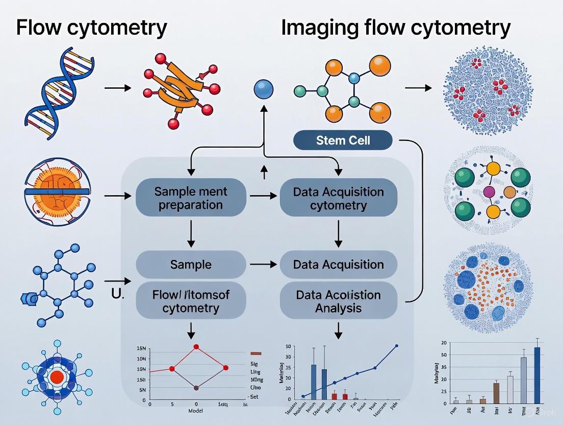
The following diagram illustrates the streamlined, high-throughput workflow of conventional flow cytometry, which is fundamental to its speed advantage.
Figure 1: High-Throughput Workflow of Conventional Flow Cytometry. This streamlined process, from sample preparation in multi-well plates to automated data analysis, enables the rapid generation of quantitative statistical data from thousands of cells per second.
In contrast, imaging flow cytometry incorporates a more complex image capture and processing step, which creates a bottleneck and limits its overall throughput.
Figure 2: Imaging Flow Cytometry Workflow with Inherent Throughput Limitation. The critical step of capturing and processing high-resolution images for each cell, while providing rich spatial data, inherently restricts the speed of analysis compared to conventional flow cytometry.
Conventional flow cytometry remains the undisputed champion for high-throughput cellular analysis in stem cell research and drug development. Its ability to provide robust, quantitative data from millions of cells in a short time is irreplaceable for large-scale phenotyping, screening, and bioprocessing applications. While imaging flow cytometry adds a valuable layer of morphological detail, its lower throughput makes it a complementary rather than a replacement technology. The choice between them is strategic: when the research question demands statistical power and speed for analyzing vast cell populations, the high-throughput power of conventional flow cytometry is the most effective tool for the task.
In the field of stem cell research, the precise analysis of cellular identity, function, and heterogeneity is paramount. Traditional flow cytometry (FC) has long been an indispensable tool, offering high-throughput, quantitative analysis of cell populations based on fluorescent markers [1] [7]. However, a significant limitation has been its inability to provide morphological context, subcellular localization, and visual confirmation of cellular events. Imaging Flow Cytometry (IFC) emerges as a transformative technology that integrates the high-throughput capabilities of conventional flow cytometry with the high-content morphological information of microscopy [8] [9]. This guide provides an objective comparison of these techniques, framing their performance within the context of stem cell research, a field where understanding subtle cellular changes is critical for applications from basic biology to regenerative medicine.
The fundamental difference lies in the nature of the data produced. While conventional FC generates quantitative scatter and fluorescence intensity plots, IFC captures high-resolution images of every cell as it flows through the detection system [8]. This allows researchers not only to quantify marker expression but also to observe cell size, shape, nuclear morphology, and the precise subcellular distribution of proteins—information often lost in traditional flow cytometry [9] [4]. For stem cell scientists, this capability is invaluable for assessing differentiation status, identifying rare progenitor cells, and detecting nuanced morphological changes indicative of cellular stress or transformation.
Technical Comparison: How IFC Enhances Conventional Flow Cytometry
The core architecture of an IFC system builds upon that of a conventional flow cytometer but incorporates critical imaging components. Both systems consist of a fluidics system to hydrodynamically focus cells into a single-file stream, an optical system with lasers to excite fluorescent probes, and detectors to capture the resulting signals [8] [9]. The pivotal distinction is the imaging system: conventional FC uses photomultiplier tubes (PMTs) to detect light intensity, whereas IFC employs a high-precision camera, such as a charge-coupled device (CCD) or CMOS sensor, to capture a detailed image of each cell [8] [10]. Some advanced systems, like the BD FACSDiscover S8, utilize novel technologies such as focusless imaging, while others, like the Thermo Fisher Attune CytPix, leverage acoustic focusing for high-speed morphological imaging [8] [11].
The following table summarizes the key operational differences between the two technologies:
Table 1: Fundamental Operational Differences Between Flow Cytometry and Imaging Flow Cytometry
| Feature | Conventional Flow Cytometry | Imaging Flow Cytometry |
|---|---|---|
| Primary Data Output | Quantitative fluorescence and scatter intensity values [1] | Quantitative values + high-resolution cellular images [8] |
| Morphological Information | Indirectly inferred from scatter signals [1] | Directly visualized (size, shape, texture, structure) [9] |
| Spatial Context | Lost; single-cell suspension [4] | Preserved at the single-cell level (subcellular localization) [1] |
| Throughput | Very High (can exceed 20,000 cells/second) [10] | Moderate to High (typically 1,000 - 15,000 cells/second) [2] [10] |
| Cell Sorting Capability | Yes (FACS - Fluorescence-Activated Cell Sorting) [8] | Limited; available in newer, specialized systems [10] |
| Data Volume | Relatively low (numerical data files) [1] | Very high (multi-gigabyte to terabyte image datasets) [10] |
The workflow for IFC begins with standard cell preparation and fluorescent labeling, similar to conventional FC. The labeled cells are then introduced into the fluidic system and focused into a core stream. As each cell passes through the laser interrogation point, it is illuminated, and the resulting scattered and fluorescent light is collected. In IFC, this light is then projected onto the camera, capturing a multi-spectral image that is processed and stored for subsequent analysis [8] [9].
Application in Stem Cell Research: A Comparative Look at Experimental Data
The integration of morphological context makes IFC particularly powerful for stem cell research. Key applications where IFC provides distinct advantages over conventional FC include:
- Pluripotency and Differentiation Assessment: Confirming the localization of transcription factors like Oct4 or Nanog in the nucleus is crucial for validating pluripotent stem cells. Conventional FC can quantify total expression levels but cannot distinguish nuclear from cytoplasmic localization. IFC visually confirms this subcellular distribution, providing greater confidence in the pluripotent status of a culture [10] [12].
- Analysis of Rare Stem Cell Populations: While conventional FC is excellent for identifying known populations based on surface markers, IFC adds a layer of verification through morphology. For instance, when isolating very rare multilineage-differentiating stress enduring (MUSE) cells or hematopoietic stem cells (HSCs), IFC can be used to visually confirm cell size and granularity, reducing false positives and improving isolation purity [7].
- Monitoring Cell State and Health: IFC enables label-free analysis of cellular morphology, which can be used to monitor apoptosis, necrosis, and other changes in cell health without additional staining. Subtle morphological changes during early differentiation or in response to drugs can be detected and quantified, offering insights beyond what is possible with standard viability dyes in conventional FC [10] [4].
- Characterizing Cancer Stem Cells (CSCs): The "stemness" of cancer cells may represent a cell state rather than a fixed cell type [7]. IFC is ideally suited to investigate this hypothesis by correlating marker expression with dynamic morphological states and cell-cycle positions, providing a more nuanced understanding of tumor heterogeneity than conventional FC alone.
The following table compares the performance of both technologies in a hypothetical experiment designed to characterize differentiating neural stem cells (NSCs), a common scenario in stem cell research and drug development.
Table 2: Performance Comparison in a Neural Stem Cell Differentiation Assay
| Experimental Parameter | Conventional Flow Cytometry Data | Imaging Flow Cytometry Data |
|---|---|---|
| % β-III-Tubulin+ Cells (Neuronal Marker) | 45% | 48% |
| % GFAP+ Cells (Astrocyte Marker) | 38% | 35% |
| Analysis of Marker Localization | Not Available | Confirms β-III-Tubulin is cytoplasmic and GFAP is filamentous/cellular processes. |
| Morphology of Positive Cells | Not Available | Quantifies neurite outgrowth in β-III-Tubulin+ cells; classifies astrocyte morphology. |
| Detection of Double-Positive Cells | 5% (could be autofluorescence, debris, or true doublets) [10] | 4%; images confirm true cellular co-expression vs. cell clumps. |
| Throughput (Cells Analyzed in 5 min) | ~500,000 cells | ~60,000 cells |
Detailed Experimental Protocol: Utilizing IFC for Stem Cell Analysis
The following is a generalized protocol for analyzing stem cell differentiation using Imaging Flow Cytometry, highlighting steps where it diverges from conventional flow cytometry practices.
Aim: To quantify and morphologically characterize the differentiation of human induced Pluripotent Stem Cells (iPSCs) into a neuronal lineage.
Materials and Reagent Solutions: Table 3: Essential Research Reagents and Materials
| Reagent/Material | Function in the Experiment |
|---|---|
| Human iPSC Culture | The starting cell population for differentiation. |
| Neuronal Differentiation Media | Directs cell fate from pluripotency to neuronal lineages. |
| Antibody: Anti-β-III-Tubulin (conjugated to e.g., AF488) | Labels newly formed neurons; primary fluorescent marker. |
| Antibody: Anti-GFAP (conjugated to e.g., PE) | Labels astrocytes; secondary fluorescent marker. |
| Nuclear Stain (e.g., DAPI or Hoechst) | Identifies and segments the nucleus in acquired images. |
| Cell Dissociation Enzyme (Accutase) | Generates a single-cell suspension from adherent cultures. |
| Phosphate Buffered Saline (PBS) / Buffers | For washing and resuspending cells. |
| Fixation Buffer (e.g., 4% PFA) | Preserves cell morphology and fixes internal antigens. |
| Permeabilization Buffer (e.g., Triton X-100) | Allows intracellular antibodies (like β-III-Tubulin) to access their targets. |
Methodology:
- Differentiation and Harvesting: Differentiate human iPSCs toward a neuronal lineage using a standardized protocol over 14-21 days. Harvest cells using a gentle dissociation enzyme like Accutase to create a single-cell suspension, minimizing clumping.
- Fixation and Permeabilization: Fix cells with 4% Paraformaldehyde (PFA) for 15 minutes at room temperature to preserve morphology. Pellet cells by centrifugation, wash with PBS, and then permeabilize with ice-cold 100% methanol or a detergent-based buffer for intracellular staining.
- Immunostaining: Resuspend the cell pellet in blocking buffer for 30 minutes to reduce non-specific antibody binding. Incubate with directly conjugated primary antibodies (e.g., Anti-β-III-Tubulin-AF488 and Anti-GFAP-PE) for 1 hour at room temperature or overnight at 4°C. Include a nuclear stain (DAPI) in the final wash or resuspension buffer.
- Data Acquisition on IFC: Resuspend the stained sample in an appropriate sheath fluid for the IFC instrument (e.g., Amnis ImageStreamX, Thermo Fisher Attune CytPix, or BD FACSDiscover S8). Start with a low flow rate to ensure high image clarity, and acquire a minimum of 10,000-50,000 cell images per sample. Include brightfield, darkfield (side scatter), and all fluorescence channels in the acquisition template.
- Data Analysis: Use the IFC's proprietary software (e.g., IDEAS or INSPIRE) for analysis. The steps typically include:
- Image Preprocessing: Compensation for fluorescence spillover, similar to conventional FC.
- Cell Gating: Create a hierarchy of gates. First, gate on single cells using a plot of Aspect Ratio vs. Area of the brightfield image to exclude doublets and debris.
- Focus Gating: Select cells that are in sharp focus using a feature like Gradient RMS.
- Morphometric and Fluorescence Analysis: For the focused, single-cell population, apply fluorescence intensity thresholds to identify β-III-Tubulin+ and GFAP+ populations. Then, use morphological features (e.g., cell area, diameter, circularity) and advanced image-based algorithms (e.g., spot counters for punctate staining, or texture analysis) to quantitatively describe the positive populations.
The following diagram illustrates the core logical workflow of an IFC system, from cell introduction to image-based analysis, which is fundamental to the protocol above.
Diagram 1: Simplified Workflow of an Imaging Flow Cytometer.
The Evolving Landscape: Throughput, AI, and the Future
A traditional trade-off has been IFC's lower throughput compared to conventional FC. However, this is rapidly changing with technological innovations. Optical time-stretch (OTS) imaging flow cytometry has demonstrated real-time throughput exceeding 1,000,000 events per second with sub-micron resolution, shattering previous limits of around 1,000-10,000 eps [2]. This breakthrough addresses the primary bottleneck of IFC, making million-cell-scale morphological screening a tangible reality for large-scale stem cell biomanufacturing and drug screening.
Furthermore, the analysis of the vast, complex image datasets generated by IFC is being revolutionized by Artificial Intelligence (AI) and machine learning [8] [12]. Convolutional Neural Networks (CNNs) can be trained to automatically identify, classify, and analyze stem cells based on their morphological features with high accuracy, moving beyond the manual gating strategies of traditional FC [12]. For instance, AI models can predict iPSC colony formation with over 90% accuracy or track differentiation trajectories in real-time, providing an objective and powerful tool for quality control in stem cell cultures [12]. The following diagram contrasts the fundamental analytical principles of the two technologies.
Diagram 2: Contrasting Analytical Approaches of FC and IFC.
The choice between conventional flow cytometry and imaging flow cytometry is not a matter of superiority, but of strategic application. Conventional FC remains the tool of choice for extremely high-throughput tasks, such as rapid immunophenotyping of millions of cells or high-speed sorting for bulk population isolation. Its strength lies in its speed, simplicity, and powerful quantitative capabilities for well-defined markers.
In contrast, Imaging Flow Cytometry is the superior choice when the research question demands visual confirmation, morphological detail, or subcellular spatial information. For stem cell researchers, this translates to critical applications like validating pluripotency markers' nuclear localization, analyzing complex differentiation outcomes, characterizing rare cell populations with higher confidence, and employing label-free morphology for quality control. As throughput increases and AI-powered analysis becomes more accessible, IFC is poised to become an even more central technology in the stem cell scientist's toolkit, providing a more holistic and profound understanding of cellular systems.
In the field of stem cell research, the choice of analytical instrumentation is pivotal, influencing the resolution, depth, and statistical power of experimental outcomes. Flow Cytometry (FC) and Imaging Flow Cytometry (IFC) represent two powerful yet fundamentally different approaches to single-cell analysis. For researchers and drug development professionals, understanding the core technological distinctions between these platforms—specifically their detection systems, the nature of their data output, and their operational throughput—is essential for selecting the right tool for specific applications, from phenotyping heterogeneous stem cell populations to tracking rare differentiation events. This guide provides an objective, data-driven comparison of these technologies, framing their capabilities within the context of modern stem cell research.
Core Principles and Instrumentation
At its heart, conventional Flow Cytometry is a laser-based technology that measures optical and fluorescence characteristics of cells as they pass in a single-file stream through one or more laser beams [1]. It provides high-speed, quantitative data on protein expression and cellular light scattering but lacks the ability to capture morphological images [1] [8].
Imaging Flow Cytometry represents an evolution of this technology, combining the high-throughput, single-cell analysis of traditional FC with the detailed imaging capabilities of microscopy [2] [8]. IFC captures high-resolution images of each cell as it flows through the detection system, thereby preserving spatial and morphological information that is lost in conventional FC [1] [8]. The fundamental components of an IFC system include a fluidics system to hydrodynamically focus the cells, an optical system with lasers and filters, and a critical imaging system, often based on a high-precision camera like a Charge-Coupled Device (CCD), to capture the cellular images [8].
The following diagram illustrates the core workflow and components of an Imaging Flow Cytometry system, highlighting the integration of imaging into the traditional flow cytometry pipeline.
Comparative Analysis: Detectors, Data, and Throughput
The divergence in the core principles of FC and IFC directly drives differences in their detector technology, the data they generate, and their maximum operational speeds. These factors are critical for experimental design in stem cell research, where balancing statistical significance with rich, morphological data is often necessary.
Detector Technology
- Conventional Flow Cytometry: Traditional flow cytometers use an array of Photomultiplier Tubes (PMTs) or avalanche photodiodes to capture light scatter and fluorescence signals [13]. These detectors are highly sensitive to photon emissions but generate numerical intensity values without any spatial context.
- Spectral Flow Cytometry: An advanced form of conventional FC, spectral flow cytometry uses an array of detectors to capture the full emission spectrum of all fluorophores used in a panel, which is then unmixed to differentiate individual signals [13] [14].
- Imaging Flow Cytometry: IFC primarily relies on a high-precision camera, typically a CCD or CMOS sensor, to capture a digital image of each cell [8]. Some next-generation IFC systems are exploring innovative detectors like event-based cameras for rapid, initial cell detection, which can then trigger a slower, more sensitive interferometric camera for detailed analysis of rare cells [15]. This approach mimics the asynchronous processing of the human retina for higher efficiency.
Data Output and Content
The type of detector fundamentally shapes the data output, which is the most significant differentiator for researchers.
- Flow Cytometry Data Output: FC generates quantitative, numerical data. The output consists of listmode files containing intensity values for forward scatter (FSC, approximating cell size), side scatter (SSC, indicating internal complexity), and multiple fluorescence channels for each cell [1]. This data is typically visualized and analyzed using bivariate plots (dot plots, histograms) and requires a gating strategy to identify cell populations based on these quantitative thresholds [13] [16].
- Imaging Flow Cytometry Data Output: IFC produces quantitative data plus high-resolution images for every cell. This means that in addition to intensity values for FSC, SSC, and fluorescence, researchers get a visual snapshot of each cell [1] [8]. The data output is therefore multidimensional, encompassing:
- Morphological Data: Quantitative metrics on cell size, shape, nuclear morphology, and texture [8].
- Spatial Information: The subcellular location of fluorescent markers (e.g., nuclear translocation, cytoplasmic distribution) [1] [17].
- Interaction Data: The ability to visualize and quantify cell-cell interactions or the formation of clusters [1].
Throughput and Speed
Throughput, measured in events per second (eps), is a key practical consideration, especially for large-scale stem cell studies.
- Flow Cytometry Throughput: Conventional FC is the undisputed leader in pure analysis speed. Modern analytical flow cytometers can easily analyze tens of thousands of cells per second, making them ideal for rapidly screening large cell populations [1].
- Imaging Flow Cytometry Throughput: Traditional IFC systems, which use CCD or CMOS sensors, have a significantly lower throughput, typically in the range of 1 to several hundred cells per second [1]. This is due to limitations in camera framerate and the massive data volume of images. However, groundbreaking technologies are shattering this barrier. Optical Time-Stretch (OTS) imaging has enabled IFC systems to achieve real-time throughput exceeding 1,000,000 events per second with sub-micron resolution [2]. While this represents the cutting edge, it highlights the rapidly evolving nature of IFC throughput capabilities.
Table 1: Key Technological Comparisons Between Flow Cytometry and Imaging Flow Cytometry
| Feature | Flow Cytometry (Conventional) | Imaging Flow Cytometry (Traditional) | Imaging Flow Cytometry (Advanced OTS) |
|---|---|---|---|
| Primary Detector | Photomultiplier Tubes (PMTs) [13] | CCD/CMOS Camera [8] | High-Speed Digitizer & FPGA [2] |
| Data Type | Quantitative fluorescence & scatter intensity [1] | Quantitative intensity + High-resolution images [1] [8] | Quantitative intensity + High-resolution images [2] |
| Spatial Context | Lost | Preserved (subcellular localization) [1] | Preserved (subcellular localization) [2] |
| Morphological Insight | Indirect (from FSC/SSC) | Direct and quantitative [8] | Direct and quantitative [2] |
| Typical Throughput | 10,000+ events/second [1] | 1 - 100 events/second [1] | >1,000,000 events/second [2] |
| Best For | High-throughput screening, bulk phenotyping, cell sorting [1] | Rare event analysis, morphological studies, co-localization [1] | Large-scale analysis requiring both high speed and morphological detail [2] |
Experimental Protocols and Data Analysis
High-Throughput IFC for Clinical Sample Analysis
A landmark 2025 study demonstrated IFC with a real-time throughput beyond 1,000,000 eps, setting a new benchmark for the technology [2].
- Objective: To develop an IFC system capable of high-speed imaging, data capture, and online analysis at unprecedented rates for applications like identifying malignancies in clinical samples [2].
- Methodology: The system integrated optical time-stretch (OTS) imaging, microfluidic cell focusing, and online image processing via a Field-Programmable Gate Array (FPGA). Cells flowing at speeds up to 15 m/s were illuminated by temporally stretched laser pulses. The interaction with the cell encoded spatial information into the pulse's spectrum, which was then detected, digitized, and processed in real-time [2].
- Data Analysis Workflow: The massive data stream (up to ~4.8 GB/s) was processed on the FPGA using real-time algorithms to analyze and filter data, significantly reducing its volume for storage. This allowed for continuous capture and analysis of over 1 million cell images per second [2].
A Semi-Automated IFC Workflow for Internalization Studies
A 2025 technical note detailed a protocol perfectly suited for studying the uptake of biomolecules by stem cells, a common requirement in drug delivery and functional studies [17].
- Objective: To create a semi-automated pipeline for quantifying the internalization of fluorescently labeled compounds into cells, overcoming the limitations of confocal microscopy (low throughput) and conventional FC (no spatial information) [17].
- Methodology: Cells were treated with fluorescent compounds (e.g., antisense oligonucleotides) and analyzed on an Amnis FlowSight IFC. The protocol used IDEAS software to apply custom image masks that automatically discriminate fluorescence signal on the plasma membrane from signal in the cytoplasmic compartment [17].
- Data Analysis Workflow: An internalization coefficient was calculated, and a novel parameter, signal distribution entropy, was introduced to quantify the uniformity of the compound's distribution inside the cell. This was supported by in-house FluoSta software for automated statistical analysis and visualization [17].
The data processing workflows for conventional and imaging flow cytometry are structurally distinct, as summarized below.
The Scientist's Toolkit: Essential Research Reagents and Materials
The following table details key reagents and materials used in a typical IFC internalization study, as described in the experimental protocol above [17].
Table 2: Key Research Reagent Solutions for IFC Internalization Assays
| Item | Function/Application | Example from Protocol |
|---|---|---|
| Antisense Oligonucleotides | Fluorescently labeled model compounds to study uptake and delivery mechanisms. | FAM-labeled PS- and PS/LNA-modified ASOs [17]. |
| Cell Staining Buffer | A buffer used to dilute antibodies and maintain cell viability during staining procedures. | BioLegend Cell Staining Buffer [17]. |
| Fixation Buffer | Preserves cell structure and fluorescence at a specific time point, halting biological processes. | BioLegend Fixation Buffer [17]. |
| Propidium Iodide (PI) | A vital dye that stains dead cells, allowing for their exclusion from analysis. | 0.1 µg/mL working solution [17]. |
| Permeabilization Wash Buffer | Allows antibodies or other reagents to access intracellular targets by making the cell membrane permeable. | BioLegend Intracellular Staining Permeabilization Wash Buffer [17]. |
| Antibodies for Surface Markers | Used to identify specific cell types or states within a heterogeneous population (e.g., stem cell markers). | Anti-CD4 Alexa Fluor 700 [17]. |
| Microfluidic Chips | The consumable chips through which cells are hydrodynamically focused and flow for analysis. | ChipShop microfluidic chip (Width: 100 μm, Height: 37 μm) [17]. |
Flow Cytometry and Imaging Flow Cytometry are complementary technologies, each with a distinct and powerful role in the stem cell researcher's arsenal. The choice is fundamentally a trade-off between the unparalleled throughput and quantitative power of conventional FC and the rich morphological and spatial context provided by IFC.
For high-speed screening, immunophenotyping of large sample sets, or any application where statistical power from millions of cells is the primary goal, conventional flow cytometry remains the optimal choice. However, when the research question hinges on visualizing subcellular events, confirming the internalization of therapeutics, analyzing complex morphology, or identifying rare cells based on structural features, Imaging Flow Cytometry is indispensable. The emergence of ultra-high-throughput IFC systems promises to blur these lines further, potentially enabling the simultaneous acquisition of high-speed statistics and high-content imaging. For researchers in stem cell biology and drug development, a clear understanding of these technological differences ensures the selection of the most appropriate and impactful tool for their scientific inquiries.
Why Stem Cell Research Demands Advanced Cytometry Tools
Stem cell research represents one of the most promising frontiers in modern biology, with profound implications for regenerative medicine, drug development, and our understanding of fundamental biological processes. The progress in this field is intrinsically linked to technological advancements in cellular analysis. Among these, cytometry tools—encompassing both flow cytometry and imaging cytometry—have become indispensable. This guide provides an objective comparison of these technologies, focusing on their performance in stem cell research applications.
Table of Contents
- Introduction to Cytometry in Stem Cell Biology
- Flow Cytometry vs. Imaging Cytometry: A Technical Comparison
- Experimental Data and Performance Comparison
- Detailed Experimental Protocols
- Key Research Reagent Solutions
- Future Directions: Integrating Cytometry with Predictive Modeling
Cytometry, the measurement of cell characteristics, is fundamental to stem cell research because it enables scientists to identify, characterize, and isolate rare stem cell populations from complex heterogeneous mixtures. The functional quality of stem cells, such as Hematopoietic Stem Cells (HSCs), critically influences the safety and efficacy of stem cell therapies [18]. Traditionally, flow cytometry has been the workhorse technique, using fluorescently tagged antibodies to quantify marker expression on thousands of cells per second. However, a paradigm shift is underway. While snapshot techniques like flow cytometry have driven the field forward, there is a growing need for dynamic, time-resolved analysis that can predict future stem cell function based on past cellular kinetics [18]. This evolution has given rise to advanced image cytometry, which combines the quantitative power of flow cytometry with the rich morphological and spatial context of microscopy.
Flow Cytometry vs. Imaging Cytometry: A Technical Comparison
The choice between flow and imaging cytometry is not about superiority, but about selecting the right tool for the specific biological question. They offer complementary strengths and trade-offs.
Core Principles and Workflows
The following diagram illustrates the fundamental operational differences between the two techniques.
Comparative Analysis of Techniques
The table below summarizes the key operational differences and capabilities of each technology, crucial for planning stem cell research experiments.
| Feature | Flow Cytometry | Imaging Cytometry |
|---|---|---|
| Throughput | High (10,000+ events/second) [19] | Low to Medium (1-100 events/second) [1] |
| Cell State | In suspension [4] | In culture environment [4] |
| Data Type | Quantitative fluorescence intensity [1] | Quantitative fluorescence + morphology + spatial context [4] [1] |
| Spatial Context | Lost | Preserved [4] |
| Cell Morphology | Not available [4] | Detailed analysis available [4] [1] |
| Subcellular Localization | Not available | Excellent (e.g., nuclear translocation) [4] [1] |
| Cell Sorting (FACS) | Yes [19] | No [1] |
| Long-Term Live Cell Tracking | Challenging [19] | Excellent (same replicate over time) [4] |
| Best For | High-throughput screening, immunophenotyping, rare cell detection, cell sorting [19] [1] | Rare/complex events, morphological analysis, cell signaling, co-localization studies [4] [1] |
Experimental Data and Performance Comparison
Objective performance data is essential for selecting the appropriate cytometric method. The following tables consolidate findings from key stem cell research studies.
Correlation of Stem Cell Counting Methods
A comparative study assessed parameters for timing the onset of peripheral blood stem cell apheresis, highlighting the precision of different methods [20].
| Measurement Method | Comparison with Flow Cytometry (CD34+) | Precision for Apheresis Timing |
|---|---|---|
| Flow Cytometry (CD34+) | Gold Standard (r=1.0) | Precise determination |
| Haematology Analyser (IMI) | Correlation: r = 0.46 (P < 0.05) [20] | Does not allow precise determination [20] |
| Haematology Analyser (HPC) | Correlation: r = 0.44 (P < 0.05) [20] | Does not allow precise determination [20] |
Conclusion: The haematology analyser-based method, while faster, does not allow the precise determination of absolute haematopoietic stem cell numbers and cannot replace flow cytometry for monitoring peripheral blood stem cell counts [20].
Revealing Heterogeneity with Kinetic Imaging
A 2025 study using Quantitative Phase Imaging (QPI) and machine learning to analyze single HSCs ex vivo revealed unprecedented diversity that snapshot analysis cannot resolve [18].
| Kinetic Parameter Measured by QPI | Observed Heterogeneity in HSCs | Significance |
|---|---|---|
| Proliferation Rate | 12.5% of HSCs produced >20 cells in 96h; 21.9% produced <4 cells [18] | Identifies subpopulations with vastly different expansion capacities |
| Cell Dry Mass | 10.9% of HSCs produced cells >200 pg; 17.2% produced cells <100 pg [18] | Links physical properties to functional potential |
| Division Patterns | 91.3% normal division; 8.21% interrupted cytokinesis; 0.48% abnormal division [18] | Uncovers rare biological events critical for quality control |
Detailed Experimental Protocols
To ensure reproducibility, here are detailed methodologies for key experiments cited in this guide.
Objective: To precisely determine the optimal time for peripheral blood stem cell apheresis by quantifying CD34+ hematopoietic stem cells.
Materials:
- Peripheral blood samples
- Fluorescently conjugated anti-human CD34 antibody
- Isotype control antibody
- Flow cytometer with 488 nm laser
- Lysing solution (if using whole blood)
Procedure:
- Sample Preparation: Collect peripheral blood in anticoagulant tubes. Prepare a single-cell suspension.
- Staining: Aliquot 100 µL of sample. Add the recommended volume of CD34 antibody and isotype control to respective tubes. Incubate for 15-20 minutes in the dark at room temperature.
- Lysis and Washing: If using whole blood, add lysing solution to remove red blood cells. Wash cells with PBS and resuspend in a suitable buffer for analysis.
- Flow Cytometry Acquisition: Acquire a minimum of 50,000 events on the flow cytometer. Use the isotype control to set the negative population and establish a fluorescence gate for CD34+ cells.
- Data Analysis: Calculate the absolute count of CD34+ cells. The optimal time for apheresis is typically guided by a pre-defined threshold of CD34+ cells per µL of blood.
Objective: To classify functional HSC diversity based on temporal kinetic features without labels.
Materials:
- Pure phenotypic HSCs (e.g., murine CD201+CD150+CD48−KSL cells or human Lin-CD34+CD38-CD45RA-CD90+CD201+ cells)
- Single-HSC ex vivo expansion culture system [18]
- U-bottom 96-well plate
- Quantitative Phase Imaging (QPI) system (e.g., ptychographic)
Procedure:
- Cell Sorting and Culture: Sort a single HSC into each well of a 96-well U-bottom plate containing expansion medium.
- Time-Lapse QPI: Place the culture plate in the QPI system. Monitor cell expansion for up to 96 hours with time-lapse imaging, ensuring environmental control (37°C, 5% CO2).
- Feature Extraction: For each tracked cell, extract kinetic parameters from the image data, including:
- Dry Mass: A measure of cell biomass.
- Sphericity: Roundness of the cell.
- Velocity: Cell movement speed.
- Division Gap: Time between first and second divisions.
- (A total of 11 parameters were used in the cited study [18])
- Dimensionality Reduction and Clustering: Perform Uniform Manifold Approximation and Projection (UMAP) on the extracted parameters from all cell images. Use clustering algorithms (e.g., K-means) to identify distinct cell populations based solely on kinetic features.
- Functional Correlation: Correlate the identified kinetic clusters with functional outcomes like long-term proliferation capacity and stem cell marker expression (e.g., via flow cytometry at endpoint).
Key Research Reagent Solutions
The following table details essential materials and their functions for cytometry-based stem cell experiments.
| Research Reagent / Material | Function in Experiment |
|---|---|
| Anti-CD34 Antibody | Fluorescently conjugated antibody for identifying and quantifying hematopoietic stem cells via flow cytometry [20]. |
| Anti-CD201 & Anti-CD90 Antibodies | Critical for isolating a highly pure population of phenotypic long-term HSCs in both human and murine models [18]. |
| Sheath Fluid | A physiological buffer used in flow cytometers to hydrodynamically focus the sample stream, ensuring single-cell analysis [19]. |
| Viability Dye (e.g., Propidium Iodide) | A DNA-binding dye used to assess cell viability by distinguishing live cells from necrotic/dead cells with compromised membranes [21]. |
| Single-HSC Expansion Medium | A specialized culture medium formulated to support the proliferation of HSCs from a single cell while maintaining stemness [18]. |
| Fluorescent Bead Standards | Used for quality control, calibration, and standardization of flow cytometers to ensure comparable, reproducible measurements across instruments and time [22]. |
Future Directions: Integrating Cytometry with Predictive Modeling
The future of stem cell research lies in moving from static identification to dynamic prediction. As demonstrated by the QPI study, integrating live-cell imaging with machine learning allows researchers to forecast future stem cell status based on past temporal dynamics [18]. This predictive power is poised to fundamentally alter the landscape of stem cell biology and therapy development.
Quantitative modeling, including machine learning, has great potential to predict the outcome of biological processes. However, it requires vast amounts of high-quality data, precisely of the kind generated by advanced cytometry tools [23]. The synergy between ever-more sophisticated cytometry and powerful computational models will continue to be a major driver of innovation, enabling the quantitative prediction of stem cell functional quality at the single-cell level.
Methodological Applications in Stem Cell Research: From Phenotyping to Organoids
Stem Cell Identification and Characterization Using Surface and Intracellular Markers
In stem cell research and therapy, the precise identification and characterization of stem cells is paramount. This process fundamentally relies on detecting specific surface and intracellular protein markers that define cellular identity, potency, and differentiation status [7] [24]. Flow Cytometry (FC) has long been the cornerstone technique for this multiplexed, quantitative analysis, enabling the rapid measurement of multiple cellular parameters simultaneously [7]. However, a technological evolution is underway with the emergence of Imaging Flow Cytometry (IFC), which integrates the high-throughput, quantitative capabilities of conventional FC with the rich morphological and spatial context of microscopy [8] [9]. This guide provides a comparative analysis of conventional flow cytometry and imaging flow cytometry, objectively evaluating their performance in stem cell marker analysis to inform method selection for research and drug development.
Core Technologies: Principles and Data Output
Conventional Flow Cytometry
Flow cytometry operates by hydrodynamically focusing a single-cell suspension into a stream, where each cell passes through one or more laser beams [1]. Detectors measure light scattering (indicating cell size and internal complexity) and fluorescence from labeled antibodies, generating high-throughput, quantitative data on protein expression for millions of cells [1]. Its primary strengths are quantitative precision, high statistical power, and unparalleled speed, analyzing tens of thousands of cells per second [1].
Imaging Flow Cytometry
Imaging flow cytometry represents an advanced fusion of flow cytometry and fluorescence microscopy. While it maintains the core fluidic and optical systems of conventional FC, its defining feature is an integrated imaging system—typically a high-precision camera or similar technology—that captures high-resolution images of each cell as it flows through the detection area [8] [9]. This allows IFC to provide all the quantitative fluorescence data of conventional FC, augmented with detailed information on cell morphology, subcellular structure, and the precise location of markers within the cell [1] [9].
Table 1: Core Technical Comparison between Flow Cytometry and Imaging Flow Cytometry.
| Feature | Flow Cytometry | Imaging Flow Cytometry |
|---|---|---|
| Throughput | High (10,000+ events/second) [1] | Low to Medium (1-100 events/second for conventional IFC); up to 1,000,000+ eps with advanced OTS-IFC [1] [2] |
| Primary Data | Quantitative fluorescence intensity & light scatter [1] | Quantitative fluorescence intensity & high-resolution morphological images [8] |
| Spatial Context | Lost | Preserved [1] |
| Morphological Insight | Limited to scatter parameters | Detailed (size, shape, nuclear morphology, subcellular localization) [1] [9] |
| Best Applications | High-throughput screening, immunophenotyping, rare population detection in large samples, cell sorting [1] | Rare/complex event analysis, co-localization studies, cell signaling (e.g., nuclear translocation), morphological analysis [1] |
Comparative Experimental Data in Stem Cell Applications
Discriminating Mesenchymal Stem Cells (MSCs) from Fibroblasts
A critical challenge in MSC therapy is distinguishing therapeutic MSCs from contaminating fibroblasts, which share similar morphology and some surface markers. A 2021 multiplex flow cytometry study systematically analyzed a panel of 14 surface markers across MSCs derived from bone marrow, adipose tissue, Wharton's jelly, and placental tissue, using fibroblasts as a negative control [25]. The research identified tissue-specific marker combinations for robust discrimination, which can be validated and explored further using IFC.
Table 2: Surface Markers for Differentiating MSCs from Fibroblasts (Flow Cytometry Data) [25].
| MSC Source | Markers with Higher Expression in MSCs | Markers Useful for Discrimination from Fibroblasts |
|---|---|---|
| Adipose Tissue | CD105, CD106, CD146, CD271 [25] | CD79a, CD105, CD106, CD146, CD271 [25] |
| Bone Marrow | CD105, CD106, CD146 [25] | CD105, CD106, CD146 [25] |
| Wharton's Jelly | CD105 [25] | CD14, CD56, CD105 [25] |
| Placental Tissue | CD105, CD146 [25] | CD14, CD105, CD146 [25] |
Identifying Surface Markers for Neural Lineage Cells (Neuropoiesis)
A combined surface and intracellular antigen analysis protocol was developed to define surface markers for human neuronal differentiation (neuropoiesis) [26]. The method involved staining live cells for surface marker candidates, followed by fixation, permeabilization, and co-staining with established intracellular lineage markers (e.g., nestin, MAP2, doublecortin, TUJ1). Subsequent flow cytometric co-expression analysis identified a combinatorial surface code (CD49f-/CD200high) that successfully enriched for neuronal cells from human induced pluripotent stem cell (iPSC) derivatives via Fluorescence-Activated Cell Sorting (FACS) [26]. This workflow is ideally suited for IFC, which could simultaneously verify the intracellular marker presence and neuronal morphology.
Characterizing Podoplanin in Skeletal Stem/Progenitor Cells
A study comparing flow cytometry (LSR II) and live-cell large field-of-view (LFOV) imaging for detecting the skeletal stem cell (SSC) marker podoplanin (PDPN) highlights the trade-offs between the techniques. While flow cytometry provided high-throughput, quantitative data on PDPN prevalence, the LFOV imaging systems (EVOS FL Auto 2 and Cell X automation platform) enabled the tracking of individual colony-founding connective tissue progenitors (CTPs) over time, linking initial PDPN expression to functional colony-forming potential [27].
Essential Methodologies and Protocols
Standard Protocol for Surface Marker Staining for Flow/Imaging Cytometry
The following is a generalized protocol for surface antigen staining, applicable to both conventional FC and IFC [28].
Required Materials:
- Cells in a single-cell suspension
- Staining medium (e.g., PBS with 0.1% BSA)
- Fluorophore-conjugated antibodies specific to target surface antigens
- Flow cytometer or imaging flow cytometer
One-Step Staining Procedure [28]:
- Harvesting and Washing: Gently trypsinize adherent cells or collect suspension cells. Wash cells by centrifuging at 300 × g for 5 minutes at 4°C and discarding the supernatant.
- Antibody Incubation: Resuspend the cell pellet in 5 μL of a pre-titrated, optimally diluted fluorescently-labeled antibody. Flick the tube to mix and incubate on ice for 25-30 minutes, protected from light.
- Washing: Add 10 mL of cold staining medium to the cells. Centrifuge at 300 × g for 5 minutes at 4°C and carefully discard the supernatant to remove unbound antibody.
- Resuspension and Analysis: Resuspend the final cell pellet in 0.5 mL of staining medium. Filter the suspension through a cell strainer cap or FACS tube to remove clumps before running on the cytometer.
Critical Controls: Include a negative control (unstained cells) and an isotype control (cells stained with a non-specific antibody of the same isotype and fluorophore) to account for autofluorescence and non-specific binding, respectively [28].
Combined Staining of Surface and Intracellular Antigens
For co-detection of surface and intracellular markers, a sequential staining protocol is required [26]:
- Surface Staining: First, stain live cells for surface antigens following the standard protocol above.
- Fixation and Permeabilization: After the final wash, gently fix the cells (e.g., with a formaldehyde-based fixative) and then treat with a permeabilization buffer (e.g., containing saponin or Triton X-100).
- Intracellular Staining: Incubate the fixed and permeabilized cells with fluorescently-labeled antibodies against the intracellular target (e.g., transcription factors, cytoskeletal proteins).
- Washing and Analysis: Wash the cells to remove excess antibody and resuspend for analysis on a flow or imaging flow cytometer.
The Scientist's Toolkit: Essential Research Reagent Solutions
Table 3: Key Reagents and Materials for Stem Cell Marker Analysis by Cytometry.
| Item | Function/Application | Example Use-Case |
|---|---|---|
| Fluorophore-Conjugated Antibodies | Specific detection of surface and intracellular markers. | Anti-CD44-APC for identifying human T cells [24]; Anti-CD105 for characterizing MSCs [25]. |
| Biotinylated Antibodies & Streptavidin Conjugates | Signal amplification for low-abundance targets via two-step staining [28]. | Detecting weakly expressed cytokine receptors on stem cells. |
| Fixation & Permeabilization Buffers | Cell preservation and enabling intracellular antibody access. | Co-detection of surface CD markers and intracellular transcription factors like Nanog in pluripotent stem cells [26]. |
| Viability Dyes | Distinguishing live from dead cells in analysis, crucial for accurate interpretation. | Excluding dead cells from analysis of primary bone marrow samples [24]. |
| Isotype Controls | Differentiating specific antibody binding from non-specific background signal [28]. | Validating the specificity of a novel stem cell marker antibody. |
| Cell Strainer Tubes | Removing cell clumps to prevent instrument clogging and ensure single-cell data. | Preparing single-cell suspensions from disaggregated solid tissues like tumors [7]. |
Workflow and Pathway Visualization
The following diagram illustrates the strategic decision-making process for selecting between flow cytometry and imaging flow cytometry based on research objectives, summarizing the core trade-off of throughput versus information.
The choice between conventional flow cytometry and imaging flow cytometry is not a matter of one technology being superior to the other, but rather of selecting the appropriate tool for the specific biological question [1]. Conventional flow cytometry remains the undisputed champion for high-throughput, quantitative analysis of large cell populations, offering unmatched speed and statistical power for applications like immunophenotyping and large-scale screening [1] [7]. In contrast, imaging flow cytometry provides a powerful, information-rich alternative when the research question requires visual validation, detailed morphological data, or insight into subcellular localization and cell-cell interactions [1] [8] [9]. For a comprehensive stem cell characterization strategy, the technologies are highly complementary; flow cytometry can rapidly identify a population of interest, which can then be sorted and subjected to deeper investigation using imaging flow cytometry, thereby combining statistical confidence with spatial and morphological resolution [1].
Fluorescence-Activated Cell Sorting (FACS) for Isolating Rare Stem Cell Populations
The isolation of pure, viable populations of rare stem cells is a cornerstone of modern regenerative medicine, cell therapy, and fundamental developmental biology research. Among the technologies available for this task, Fluorescence-Activated Cell Sorting (FACS) has established itself as a powerful and high-throughput method for the prospective isolation of stem cells based on their specific surface marker profiles. Within the broader context of cellular analysis techniques, FACS represents a specialized application of flow cytometry that adds a physical sorting capability, enabling researchers to separate cells of interest from a heterogeneous mixture for downstream molecular analysis or functional assays [29]. This guide objectively compares the performance of FACS with its emerging alternatives—particularly imaging flow cytometry and spectral flow cytometry—for the specific challenge of isolating rare stem cell populations, providing researchers with the experimental data and protocols necessary to make an informed technological choice.
Technical Comparison: FACS vs. Emerging Cytometry Technologies
The choice of cellular analysis and sorting technology involves significant trade-offs between throughput, information depth, and resolution. The table below summarizes the key performance characteristics of FACS, imaging flow cytometry, and spectral flow cytometry for stem cell research.
Table 1: Performance Comparison of Cell Sorting and Analysis Technologies for Stem Cell Research
| Feature | FACS (Conventional) | Imaging Flow Cytometry (IFC) | Spectral Flow Cytometry |
|---|---|---|---|
| Throughput | High (can analyze >10,000 cells/sec) [1] | Low to Medium (1-100 events/sec) [1] | High (comparable to conventional FACS) [30] |
| Primary Strength | High-speed sorting; robust quantitative data [1] | Morphological and subcellular insight; spatial context [1] [31] | High-parameter panel resolution; unmixing of overlapping fluorophores [32] |
| Spatial Context | Lost [1] | Preserved (captures high-resolution images) [1] [8] | Lost [30] |
| Data Type | Fluorescence intensity and light scatter [33] | Fluorescence intensity + high-resolution cellular images [31] [8] | Full emission spectrum for each fluorophore [30] [32] |
| Best Suited For | High-throughput sorting based on phenotype; bulk phenotyping [1] | Analysis of complex events requiring visual verification (e.g., morphological changes, protein localization) [1] [31] | Ultra-high-parameter immunophenotyping (>30 colors); using fluorophores with highly overlapping spectra [32] |
| Spillover Correction | Compensation [30] | Compensation or spectral unmixing (varies by platform) | Spectral Unmixing [30] [32] |
The core trade-off is clear: FACS offers unparalleled speed and sorting precision for high-throughput applications, while imaging and spectral flow cytometry provide deeper layers of information—morphological and spectral, respectively—often at the cost of speed or simplicity [1] [30]. For isolating rare stem cells like HSCs, where the primary requirement is to separate a phenotypically defined, rare population from a complex suspension with high purity and viability, FACS remains the gold standard.
Experimental Protocol: FACS-Based Isolation of Human Hematopoietic Stem Cells
The following detailed protocol for isolating human long-term hematopoietic stem cells (LT-HSCs) from mobilized peripheral blood illustrates a standard, reproducible FACS workflow [33].
Sample Preparation and Pre-enrichment
- Source Material: Use mobilized leukapheresis products (mob LPs) from donors treated with granulocyte colony-stimulating factor (G-CSF). These are a common source due to their high yield of CD34+ hematopoietic stem and progenitor cells (HSPCs) [33].
- Cell Isolation: Isolate mononuclear cells from fresh or frozen mob LPs using density gradient centrifugation (e.g., Ficoll-Paque).
- Magnetic Pre-enrichment: To increase sorting efficiency, positively select CD34+ cells using magnetic-activated cell sorting (MACS) with an anti-human CD34 MicroBead kit. This step significantly enriches the target population before FACS, saving instrument time and improving purity [33].
Antibody Staining and Panel Design
- Viability Staining: Resuspend the pre-enriched cells in PBS and stain with a fixable viability dye (e.g., Fixable Viability Dye eFluor 506) to exclude dead cells during sorting.
- Surface Marker Staining: Stain the cells with a cocktail of fluorophore-conjugated monoclonal antibodies against the following surface markers, used to define LT-HSCs and exclude lineage-committed cells and progenitors [33]:
- Lineage (Lin) Cocktail: A mixture of antibodies against CD2, CD3, CD14, CD16, CD19, CD56, CD235a to exclude mature lymphoid, myeloid, and erythroid cells.
- CD34: Marker for hematopoietic stem and progenitor cells.
- CD38: Absent on primitive HSCs.
- CD45RA: Absent on long-term HSCs.
- CD90 (Thy1): Expressed on LT-HSCs.
- CD49f: An integrin that further enriches for engrafting LT-HSCs.
- Incubation: Incubate the staining cocktail with the cells for 20-30 minutes on ice in the dark, then wash with a buffer like autoMACS Rinsing Solution.
FACS Sorting and Gating Strategy
The sorting strategy is designed to sequentially gate on the population of highest purity, defined as Lin-CD34+CD38-CD45RA-CD90+CD49f+ [33].
- Instrument Setup: Use a high-speed sorter (e.g., FACSAria III). Perform instrument calibration using CS&T or Accudrop beads and compensate for spectral overlap using compensation beads (e.g., UltraComp eBeads) stained with single colors.
- Gating Hierarchy:
- Viable Singlets: Gate on cells based on forward scatter (FSC) area vs. height to exclude doublets, then select viability dye-negative cells.
- Lineage Negative: Select the Lin- population from the viable singlets.
- HSPC Enrichment: From the Lin- gate, select CD34+CD38- cells.
- LT-HSC Isolation: From the CD34+CD38- gate, sequentially select cells that are CD45RA- and then CD90+CD49f+.
This refined gating strategy allows for the prospective purification of a highly enriched population of human LT-HSCs for functional assays like transplantation or single-cell RNA sequencing [33].
The Scientist's Toolkit: Essential Reagents for FACS Isolation of HSCs
The successful isolation of rare stem cells via FACS is dependent on a carefully selected set of reagents and instruments. The following table details key materials required for the human LT-HSC protocol.
Table 2: Essential Research Reagents and Materials for HSC Isolation
| Item | Specific Example | Function in the Protocol |
|---|---|---|
| Flow Cytometer/Cell Sorter | BD FACSAria III | High-speed, multi-parameter cell sorter for physically isolating the target population. |
| Magnetic Cell Sorter | Miltenyi Biotec autoMACS | For initial pre-enrichment of CD34+ cells, improving sort efficiency. |
| CD34 MicroBead Kit | Miltenyi Biotec CD34 MicroBead Kit UltraPure human | Antibody-conjugated magnetic beads for positive selection of CD34+ HSPCs. |
| Anti-Human CD34 mAb | BD Bioscience 345804 (Clone 8G12) | Fluorescently conjugated antibody for identifying hematopoietic stem/progenitor cells. |
| Anti-Human CD38 mAb | BD Bioscience 656646 (Clone HB7) | Antibody to exclude CD38+ committed progenitor cells. |
| Anti-Human CD90 mAb | BD Bioscience 561557 (Clone 5E10) | Antibody to identify the LT-HSC population within the CD34+CD38- compartment. |
| Anti-Human CD49f mAb | BD Bioscience 551129 (Clone GoH3) | Antibody against integrin alpha-6, a marker that further enriches for engrafting LT-HSCs. |
| Lineage Cocktail mAbs | Various clones from Thermo Fisher (e.g., CD3, CD14, CD19) | A mixture of antibodies to exclude mature, lineage-committed cells from the analysis. |
| Fixable Viability Dye | Thermo Fisher Scientific 65-0866-14 | Critical for distinguishing and excluding dead cells, which can non-specifically bind antibodies. |
| Software | BD FACSDiva (acquisition), FlowJo (analysis) | For instrument operation, sort setup, and post-acquisition data analysis. |
The isolation of rare stem cell populations remains a technically demanding but essential procedure in biomedical research. FACS continues to be the most effective tool for high-throughput, high-purity sorting based on well-defined phenotypic markers, as demonstrated by its central role in isolating human LT-HSCs for therapeutic applications. However, the emergence of powerful alternative technologies necessitates a strategic choice.
- Choose FACS when the experimental goal requires the high-speed physical separation of a phenotypically defined cell population with high purity and viability, particularly for downstream functional assays like transplantation [33].
- Choose Imaging Flow Cytometry when the biological question involves morphological analysis, protein localization, or the verification of complex cellular events that cannot be captured by fluorescence intensity alone [1] [31] [8].
- Choose Spectral Flow Cytometry when the research demands the deepest possible immunophenotyping with panels of 30+ colors, requiring the resolution of many overlapping fluorophores that are incompatible with conventional cytometers [30] [32].
Ultimately, these technologies are not mutually exclusive but are complementary. A powerful modern workflow may involve using spectral cytometry for deep, high-resolution phenotyping to refine sorting panels, followed by high-speed FACS to isolate the newly defined populations for functional validation.
Analyzing Stem Cell-Derived Organoids and 3D Models
The emergence of sophisticated stem cell-derived organoids and 3D models has revolutionized biomedical research by providing unprecedented physiological relevance for studying human development, disease mechanisms, and drug responses. These complex microtissues self-organize to mimic the cellular heterogeneity, architecture, and functionality of human organs, offering a critical bridge between traditional two-dimensional cell cultures and in vivo models [34] [35]. As these advanced models become more prevalent in research and drug development, the technologies for analyzing them must similarly evolve to extract meaningful, high-content data at single-cell resolution.
Within this context, flow cytometry technologies serve as indispensable tools for characterizing the complex cellular composition of organoids. While conventional flow cytometry provides high-throughput, quantitative analysis of cell populations based on biomarker expression, imaging flow cytometry represents an evolutionary advancement that combines the statistical power of flow cytometry with detailed morphological information [10]. This comparison guide examines the performance characteristics, applications, and limitations of both technologies specifically for analyzing stem cell-derived organoids and 3D models, providing researchers with experimental data and methodologies to inform their technology selection.
Technology Comparison: Conventional vs. Imaging Flow Cytometry
Performance Characteristics and Capabilities
Table 1: Technical Comparison of Flow Cytometry Technologies for Organoid Analysis
| Performance Metric | Conventional Flow Cytometry | Imaging Flow Cytometry |
|---|---|---|
| Throughput | High (>20,000 cells/sec) [10] | Moderate (1,000-15,000 cells/sec) [10] |
| Spatial Information | None | Detailed cellular morphology and subcellular localization [10] |
| Data Content per Cell | Multiplexed biomarker intensity | High-content morphological features + biomarker intensity [10] |
| Data Volume | Moderate | Large (gigabytes to terabytes per experiment) [10] |
| Key Strengths | High-throughput population analysis, cell sorting | Analysis of protein condensation, co-localization, cell morphology [10] |
| Sample Requirements | Single-cell suspensions from dissociated organoids | Single-cell suspensions, requires precise flow control for imaging [10] |
Analytical Applications for Organoid Research
The choice between conventional and imaging flow cytometry depends heavily on the research questions being addressed:
Conventional flow cytometry excels in high-throughput applications requiring quantitative analysis of cell populations within organoids. For example, the CelltypeR pipeline demonstrates its utility in reproducibly identifying and quantifying major brain cell types (astrocytes, radial glia, and neurons) in midbrain organoids through optimized antibody panels and computational analysis [36]. This approach enables tracking cell type proportions across organoid differentiation timecourses and facilitates fluorescence-activated cell sorting (FACS) for downstream transcriptional analysis.
Imaging flow cytometry provides unique capabilities for analyzing morphological features and subcellular phenomena that conventional flow cytometry cannot detect. This includes identifying protein condensation or diffusion within cells, monitoring co-localization of proteins, and detecting subtle changes in cellular morphology that may indicate drug response or disease phenotypes [10]. These capabilities are particularly valuable when analyzing complex processes such as different phases of mitosis or subcellular structural changes in response to experimental conditions.
Experimental Protocols for Organoid Analysis
Workflow for Comprehensive Organoid Characterization
The following diagram illustrates the integrated experimental workflow for analyzing organoids using complementary technologies:
Detailed Methodologies
Protocol 1: Cell Type Quantification in Brain Organoids Using Conventional Flow Cytometry (CelltypeR Pipeline)
- Organoid Generation: Human iPSCs are differentiated into midbrain organoids using established protocols, typically spanning 60-90 days with specific morphogen patterning to generate relevant neuronal subtypes [36] [34].
- Single-Cell Dissociation: Organoids are collected and enzymatically dissociated using papain-based neural tissue dissociation systems or accutase to generate single-cell suspensions while maintaining cell viability [36].
- Antibody Staining: Cells are incubated with a optimized antibody panel targeting cell-type specific markers (e.g., GFAP for astrocytes, NES for radial glia, RBFOX3 for neurons) in flow cytometry buffer containing PBS with 1-2% FBS [36].
- Flow Cytometry Analysis: Stained cells are analyzed using conventional flow cytometers equipped with appropriate lasers and detectors. The CelltypeR pipeline incorporates computational steps for dataset alignment, unsupervised clustering optimization, and statistical comparison of cell type proportions across experimental conditions [36].
- Fluorescence-Activated Cell Sorting (FACS): Defined cell populations (e.g., astrocytes, radial glia, neurons) can be isolated using FACS for downstream applications such as single-cell RNA sequencing to characterize transcriptional states [36].
Protocol 2: Morphological Analysis of Organoid-Derived Cells Using Imaging Flow Cytometry
- Sample Preparation: Single-cell suspensions from dissociated organoids are prepared similarly to conventional flow cytometry protocols, with emphasis on maintaining cell integrity and minimizing debris [10].
- Staining Strategy: Multiplexed fluorescence staining targeting both lineage markers and subcellular structures (e.g., nuclear, cytoplasmic, and membrane markers) to enable comprehensive morphological analysis [10].
- Image Acquisition: Cells are processed through imaging flow cytometers such as ImageStream systems, which utilize time-delay-and-integration (TDI) technology to capture high-quality images of flowing cells at rates up to 5,000 cells/second [10].
- Multispectral Imaging: Multiple color channels (up to 12) are captured simultaneously using dichroic-filter stacks, allowing comprehensive phenotypic characterization [10].
- Image Analysis: Computational algorithms extract both intensity-based and morphological features from cell images, including texture, shape, and subcellular distribution patterns. Machine learning approaches can be applied for automated classification of cell states based on morphological criteria [10].
Research Reagent Solutions for Organoid Analysis
Table 2: Essential Reagents and Materials for Organoid Flow Cytometry Analysis
| Reagent/Material | Function | Application Examples |
|---|---|---|
| Extracellular Matrix (Matrigel) | Provides 3D scaffold for organoid growth and differentiation [34] | Supports development of complex organoid structures including lumen formation |
| Tissue Dissociation Enzymes | Generates single-cell suspensions from 3D organoids for flow analysis | Papain-based systems for neural organoids; accutase for epithelial organoids |
| Fluorescent Antibody Panels | Cell surface and intracellular marker detection | CelltypeR panel for brain cell types; EpCAM/CD49f for mammary lineages [36] [37] |
| Viability Dyes | Exclusion of non-viable cells from analysis | Critical for accurate quantification of cell populations from dissociated organoids |
| Cell Sorting Collection Media | Maintains cell viability during FACS | Enables downstream transcriptional or functional analysis of sorted populations |
Comparative Performance in Research Applications
Case Study: Characterization of Brain Organoid Cell Types
In the analysis of human iPSC-derived midbrain organoids, conventional flow cytometry with the CelltypeR pipeline successfully identified and quantified the major brain cell types, including astrocytes, radial glia, and neurons [36]. This approach enabled researchers to track changes in cell type proportions during organoid differentiation and isolate specific neuronal populations expressing markers associated with substantia nigra dopaminergic neurons vulnerable in Parkinson's disease [36]. The high-throughput nature of conventional flow cytometry allowed for comprehensive statistical analysis across multiple organoids and time points.
Case Study: Analysis of Protein Localization and Morphological Features
Imaging flow cytometry provides unique capabilities for analyzing subcellular localization and morphological features that are lost in conventional flow cytometry. For example, in drug screening applications, imaging flow cytometry can monitor changes in the position of fluorescence within a cell, enabling detection of protein translocation events that may indicate drug efficacy [10]. Similarly, in cancer research, imaging flow cytometry can identify rare cell populations based on morphological criteria that would be indistinguishable using conventional biomarker approaches alone.
The analysis of stem cell-derived organoids and 3D models requires sophisticated technologies that can resolve cellular heterogeneity while providing statistically robust data. Conventional flow cytometry offers superior throughput and well-established analytical pipelines for quantitative cell population analysis, making it ideal for applications requiring statistical power and cell sorting capabilities. Imaging flow cytometry sacrifices some throughput for rich morphological information, enabling researchers to capture spatial and subcellular data that is increasingly recognized as critical for understanding complex biological processes in organoids.
Forward-looking research strategies will likely incorporate both technologies in complementary roles: using conventional flow cytometry for high-throughput screening and population analysis, while employing imaging flow cytometry for deeper investigation of morphological phenotypes and subcellular events. As organoid technology continues to advance, generating more complex and physiologically relevant models, the parallel evolution of flow cytometry technologies will ensure researchers have the analytical tools needed to fully leverage these powerful model systems in basic research and drug development.
Cell Cycle and Proliferation Analysis for Assessing Self-Renewal Capacity
In stem cell research, accurately assessing self-renewal capacity—the ability of a stem cell to divide and generate more identical stem cells—is fundamental for advancing regenerative medicine and therapeutic applications. The analysis of cell cycle and proliferation serves as a critical experimental readout for this potential. The choice of analytical technology significantly influences the depth and reliability of this assessment. This guide provides an objective comparison between two cornerstone technologies: conventional flow cytometry (FC) and imaging flow cytometry (IFC), framing their performance within the context of stem cell research. As highlighted in a recent comparative analysis, the decision between these techniques is not about superiority but about selecting the right tool for the specific research question, as they offer complementary strengths in throughput and information depth [1].
Technology Comparison: Flow Cytometry vs. Imaging Flow Cytometry
The core distinction between these technologies lies in the type of data they capture. Conventional flow cytometry is a high-throughput, laser-based technology that measures optical properties—light scatter and fluorescence intensity—of cells in a fluid stream as they pass single-file by detectors. It provides robust, quantitative data on protein expression and cellular characteristics for thousands of cells per second [1] [38]. In contrast, imaging flow cytometry integrates the high-throughput capability of flow cytometry with the detailed morphological insight of microscopy. It captures high-resolution images of each cell as it flows through the system, simultaneously providing quantitative fluorescence data and spatial information [8] [1].
Table 1: Core Technical Characteristics and Data Output
| Feature | Conventional Flow Cytometry | Imaging Flow Cytometry |
|---|---|---|
| Primary Data | Quantitative fluorescence and light scatter intensity [38] | Quantitative fluorescence intensity + high-resolution cell images [8] |
| Throughput | Very High (10,000+ events/second) [1] | Lower to Medium (1 - 10,000 events/second), technology-dependent [2] [1] |
| Spatial Context | Lost; cells are analyzed as individual data points without visual context [1] | Preserved; enables analysis of subcellular localization and cell-cell interactions [8] [1] |
| Key Strength | High-speed, statistical analysis of large cell populations [38] | Morpho-functional integration; adds visual validation to quantitative data [8] |
| Best Suited For | High-throughput screening, bulk phenotyping, analyzing heterogeneous populations for rare cells [1] [38] | Analyzing complex processes like cell signaling, co-localization, and morphological changes [8] [1] |
Table 2: Performance in Stem Cell and Proliferation Analysis
| Aspect | Conventional Flow Cytometry | Imaging Flow Cytometry |
|---|---|---|
| Proliferation Assays | Excellent for DNA content analysis (cell cycle phase distribution) via dye incorporation (e.g., BrdU) [39]. | Capable of DNA content analysis; can additionally visualize mitotic figures and nuclear morphology [8]. |
| Self-Renewal Capacity | Indirect assessment via cell cycle status and marker expression in population studies. | Enhanced assessment by linking marker expression (e.g., Oct-4, Nanog) to specific morphological states of stemness [40]. |
| Multiparameter Analysis | Spectral systems can analyze 40+ parameters, ideal for deep immunophenotyping [32]. | Lower parameter count than high-end spectral cytometers, but each parameter is linked to a spatial context. |
| Rare Event Analysis | High statistical power for detecting rare populations within large samples [38]. | Superior for rare events requiring visual confirmation, such as identifying specific cell divisions or morphological anomalies [1]. |
Experimental Data and Application in Stem Cell Research
Supporting experimental data from recent studies illustrates how these technologies are applied to investigate self-renewal and proliferation.
Assessing Pluripotency Transcription Factor Function
A 2025 study investigating the self-renewal function of Oct-4 in embryonic stem (ES) cells utilized flow cytometry to analyze the expression of pluripotency markers like Sox2, Nanog, and SSEA-1. When Oct-4 was genetically deleted, these markers decreased, but were preserved upon introduction of the EWS-Oct-4 fusion protein, demonstrating its ability to replace Oct-4's core function. Flow cytometry provided the quantitative data on marker expression levels that underpinned these findings [40].
Evaluating the Impact of Pro-Aging and Anti-Aging Factors
Research on the anti-aging hormone α-Klotho provides a clear example of proliferation analysis. Scientists administered the DNA label BrdU to young and old mice, with and without Klotho supplementation. Flow cytometry was then used to quantify BrdU-positive proliferating cells in tissues with varying regenerative capacities. The data showed that Klotho deficiency significantly reduced BrdU incorporation in intestinal crypt cells, kidney tubular cells, and even cardiomyocytes, a decline that was reversed with soluble Klotho supplementation. This quantitative proliferation data was key to concluding that Klotho ameliorates age-related declines in cellular turnover [39].
Technical Advancements in High-Throughput Imaging
A groundbreaking 2025 study demonstrated an IFC system using optical time-stretch (OTS) imaging that shattered previous throughput barriers. This system achieved a real-time throughput exceeding 1,000,000 events per second with a spatial resolution of 780 nm, enabling the clear imaging of cells flowing at speeds up to 15 m/s. The system was validated by successfully identifying malignant cells in clinical colorectal samples, showcasing the potential of high-speed morphological analysis for diagnostic and research applications [2].
Experimental Protocols for Self-Renewal Analysis
Below are detailed methodologies for key experiments cited in this guide, adaptable for both FC and IFC.
Protocol: Cell Cycle Analysis via DNA Content
This classic protocol distinguishes cells in different phases of the cell cycle [38].
- Cell Harvesting and Fixation: Harvest stem cells (e.g., ES cells or HSCs) and create a single-cell suspension. Fix cells in cold 70% ethanol for at least 2 hours.
- Staining: Pellet cells and resuspend in a staining solution containing a DNA-binding fluorescent dye, such as Propidium Iodide (PI, 50 µg/mL) or DAPI, along with RNase A (100 µg/mL) to degrade RNA.
- Incubation: Incubate the cell suspension for 30-45 minutes at room temperature in the dark.
- Data Acquisition and Analysis: Analyze samples on a flow cytometer. For IFC, brightfield and fluorescent images of the nucleus are also captured. The fluorescence intensity is plotted on a histogram; the peaks representing G0/G1, S, and G2/M phases are quantified using analysis software.
Protocol: Proliferation Tracking via BrdU Incorporation
This method identifies cells that have actively synthesized DNA during a specific period [39].
- Pulse-Labeling: Add BrdU (or an analog like EdU) to the culture medium of living stem cells for a defined pulse (e.g., 1-2 hours). In vivo, BrdU is injected into the animal.
- Cell Harvesting and Fixation: Harvest cells and fix them, typically using a paraformaldehyde-based fixative.
- Denaturation and Permeabilization: Treat cells with acid or DNase to denature the DNA and expose the incorporated BrdU. Permeabilize cells with a detergent like Triton X-100.
- Antibody Staining: Incubate cells with a fluorescently conjugated anti-BrdU antibody.
- Data Acquisition and Analysis: Analyze by FC to quantify the percentage of BrdU-positive cells. With IFC, the analysis is enhanced by visually confirming the nuclear localization of the BrdU signal, ensuring it is not surface-bound or artifactual.
Protocol: Pluripotency Marker Analysis
This protocol assesses the expression of key proteins associated with an undifferentiated, self-renewing state [40].
- Cell Preparation: Create a single-cell suspension of stem cells.
- Staining for Surface Markers (e.g., SSEA-1): Resuspend cells in a buffer containing a fluorescent antibody against the surface marker. Incubate, wash, and analyze. For intracellular markers, proceed to step 3.
- Fixation and Permeabilization: Fix cells (e.g., with paraformaldehyde) and then permeabilize them (e.g., with saponin-based buffers) to allow antibodies to enter the cell.
- Staining for Intracellular Markers (e.g., Oct-4, Nanog, Sox2): Incubate permeabilized cells with fluorescent antibodies against the target transcription factors.
- Data Acquisition and Analysis: Use FC to quantify fluorescence intensity, reflecting protein expression levels. IFC adds a critical layer by confirming the nuclear localization of transcription factors like Oct-4 and Nanog, which is essential for their function.
Visualizing Key Signaling Pathways
The following diagrams, generated using Graphviz DOT language, illustrate signaling pathways critical for stem cell self-renewal, as identified in the research.
Klotho Pathway in Aging
This diagram illustrates the mechanism by which the anti-aging hormone α-Klotho promotes cell proliferation, as identified in a 2025 study [39]. The research demonstrated that Klotho acts through the Sirt1-CHK2 pathway. Upregulation of Sirt1 inhibits (represented by a blocked arrow) the DNA-damage response kinase CHK2. This inhibition leads to reduced expression of the cell cycle inhibitor p21, thereby promoting cell cycle progression and ultimately enhancing cellular proliferation and tissue regeneration.
Oct-4 in Stem Cell Self-Renewal
This diagram outlines the core regulatory network maintaining embryonic stem cell self-renewal, based on a 2025 study [40]. The transcription factor Oct-4 is a master regulator essential for this process. The study found that the EWS-Oct-4 fusion protein can functionally replace Oct-4. Both proteins activate a core pluripotency network, which in turn upregulates key markers like Sox2 and Nanog. This sustained network activity is critical for maintaining the undifferentiated, self-renewing state of stem cells.
The Scientist's Toolkit: Essential Research Reagents
Table 3: Key Reagent Solutions for Stem Cell Proliferation Analysis
| Reagent / Solution | Function / Application |
|---|---|
| BrdU (Bromodeoxyuridine) | A thymidine analog incorporated into DNA during synthesis (S-phase); detected with specific antibodies to label proliferating cells [39]. |
| Anti-BrdU Antibody | Fluorescently conjugated antibody used to detect and quantify incorporated BrdU via flow cytometry [39]. |
| DNA Staining Dyes (e.g., Propidium Iodide, DAPI) | Fluorescent dyes that intercalate with DNA, allowing for quantification of DNA content and discrimination of G0/G1, S, and G2/M cell cycle phases [38]. |
| Pluripotency Marker Antibodies (e.g., anti-Oct-4, anti-Nanog, anti-Sox2, anti-SSEA-1) | Antibodies targeting key transcription factors and surface proteins to identify and characterize undifferentiated, self-renewing stem cells via immunophenotyping [40]. |
| Phospho-Histone H3 (Ser10) Antibody | Marker for mitosis (M-phase); allows for specific quantification of cells actively undergoing division [39]. |
| Cell Viability Dyes (e.g., DAPI, 7-AAD) | Impermeant dyes used to exclude dead cells from analysis, ensuring data accuracy by gating on live cells. |
| Fixation & Permeabilization Buffers | Chemical solutions to preserve cell structure (fixation) and rupture membranes to allow intracellular antibody access (permeabilization), crucial for staining nuclear proteins like Oct-4 [40]. |
The transition from conventional flow cytometry (FC) to imaging flow cytometry (IFC) represents a significant evolution in cellular analysis tools, particularly for stem cell research and drug development. While conventional FC provides high-throughput, multi-parameter quantification of cell populations, it lacks the ability to capture morphological details and spatial relationships between cellular components [8] [1]. IFC bridges this critical gap by integrating the high-throughput capabilities of flow cytometry with the high-resolution imaging power of microscopy [8] [31]. This enables researchers to not only quantify cellular phenotypes but also visualize and quantify fundamental biological processes—such as immunological synapse formation, protein localization, and DNA damage response—in a high-throughput manner [31]. This guide objectively compares the performance of these technologies in advanced applications, providing experimental data and protocols to inform research decisions.
Technical Comparison: Flow Cytometry vs. Imaging Flow Cytometry
The core difference between these technologies lies in their data acquisition systems. Conventional flow cytometry uses a series of optical filters and photomultiplier tubes (PMTs) to measure fluorescence intensity, following a "one detector–one fluorophore" approach [32]. In contrast, imaging flow cytometry incorporates a high-precision camera (e.g., CCD) or similar technology to capture high-resolution images of each cell as it flows through the detection system [8]. This fundamental distinction creates a trade-off: conventional FC offers unparalleled speed for analyzing thousands of cells per second, while IFC provides rich morphological and spatial data at a lower throughput [1].
Table 1: Core Technical Characteristics of Flow Cytometry and Imaging Flow Cytometry
| Feature | Conventional Flow Cytometry | Imaging Flow Cytometry |
|---|---|---|
| Throughput | High (10,000+ events/second) [1] | Low to Medium (1-100 events/second for traditional IFC; up to 1,000,000 eps with advanced OTS-IFC) [1] [2] |
| Primary Data Output | Quantitative fluorescence and light scatter intensity [1] | Quantitative fluorescence intensity + high-resolution brightfield and fluorescence images [8] [1] |
| Spatial Context | Lost [1] | Preserved [1] |
| Morphological Information | Limited to derived parameters (e.g., FSC, SSC) | Detailed data on size, shape, nuclear morphology, and subcellular structure [8] [1] |
| Key Strength | High-throughput screening, bulk phenotyping, statistical power [1] [38] | Rare event analysis, co-localization studies, morphological assessment [1] [31] |
For advanced applications in stem cell research, the spatial and morphological information provided by IFC is often critical. For instance, confirming the nuclear localization of a transcription factor or analyzing the complex structure of an immunological synapse is not possible with conventional FC alone [1] [31].
Comparative Analysis of Key Advanced Applications
The following section provides a detailed, data-driven comparison of how conventional FC and IFC perform in three critical application areas.
Protein Localization and Intracellular Signaling
Understanding the subcellular distribution of proteins is fundamental to stem cell biology, particularly for studying signaling pathways that govern pluripotency, differentiation, and response to external stimuli.
IFC Performance: IFC excels at quantifying the translocation of proteins between cellular compartments, such as the nucleus and cytoplasm [31]. It can capture high-resolution images that clearly delineate these compartments for thousands of individual cells, enabling robust statistical analysis of signaling dynamics in heterogeneous stem cell populations. This allows researchers to investigate intracellular survival and differentiation signals triggered by external stimuli at a single-cell level [31].
Conventional FC Limitation: Standard FC cannot distinguish the subcellular origin of a fluorescent signal. A cell with a protein localized entirely in the nucleus may yield the same total fluorescence intensity as a cell where the same protein is diffusely distributed in the cytoplasm, leading to a potential loss of critical biological information [1].
Table 2: Experimental Data for Protein Localization Analysis
| Technology | Measurable Parameter | Experimental Readout | Reference Application |
|---|---|---|---|
| Imaging Flow Cytometry | Nuclear to Cytoplasmic Ratio | Quantification of transcription factor translocation in response to differentiation signals [31] | Analysis of signal transduction in stem cells [31] |
| Conventional Flow Cytometry | Total Cellular Fluorescence | Combined fluorescence intensity from all cellular compartments, without spatial discrimination | Not applicable for translocation studies |
Immunological Synapse Formation
The immunological synapse is a specialized, transient interface between an immune cell (e.g., a T-cell) and a target cell. Analyzing its formation is crucial for immunology and cancer research, including the study of CAR-T cell therapies.
IFC Performance: IFC is uniquely capable of monitoring immune synapse formation among interacting cells within large populations [31]. It can capture images of cell conjugates and analyze these interactions at the molecular level, providing both quantitative and visual data on synapse quality and stability. This is invaluable for assessing the functional efficacy of therapeutic cells.
Conventional FC Limitation: Conventional FC disrupts cell-cell interactions during hydrodynamically-focused flow. While it can detect the formation of conjugates based on increased size and complexity (via FSC and SSC), it cannot provide any visual confirmation or detailed morphological analysis of the synapse structure itself [8] [1].
DNA Damage Response (DDR)
Monitoring DNA damage, such as the formation of γH2AX foci, is essential for studying genotoxic stress, cancer biology, and stem cell genomic integrity.
IFC Performance: IFC has been successfully applied to monitor DNA damage responses, specifically the formation of γH2AX foci, in a high-throughput format [31]. The technology can identify and count discrete nuclear foci against the background of diffuse nuclear staining, a task that requires spatial resolution.
Conventional FC Limitation: Conventional FC can only measure the total increase in γH2AX phosphorylation within a cell's nucleus. It cannot distinguish between a general, diffuse increase in signal and the presence of distinct, quantifiable DNA repair foci, which often hold more specific biological meaning [31].
Table 3: Experimental Data for DNA Damage Response Analysis
| Technology | Assay Type | Key Readout | Information Depth |
|---|---|---|---|
| Imaging Flow Cytometry | γH2AX Foci Assay | Number and intensity of discrete nuclear foci per cell [31] | High (Spatial distribution within nucleus) |
| Conventional Flow Cytometry | Phospho-γH2AX Intensity | Total median fluorescence intensity (MFI) of γH2AX signal per cell | Low (Bulk cellular measurement) |
Experimental Protocols for Imaging Flow Cytometry
To implement the advanced applications discussed, the following general protocols can be adapted.
Protocol for Analyzing Protein Localization
This protocol outlines the steps to study transcription factor translocation using IFC.
- Cell Stimulation and Fixation: Treat stem cells with the desired stimulus (e.g., growth factor, differentiation agent). After the appropriate incubation time, fix the cells with a cross-linking fixative like 4% paraformaldehyde (PFA) to preserve protein localization.
- Permeabilization and Staining: Permeabilize the fixed cells using a buffer containing a mild detergent (e.g., 0.1% Triton X-100) to allow antibodies access to intracellular targets. Incubate with a primary antibody specific to the transcription factor of interest, followed by a fluorescently-labeled secondary antibody. Include a fluorescent DNA stain (e.g., DAPI) to define the nuclear region.
- IFC Data Acquisition: Acquire data on an imaging flow cytometer, ensuring that the brightfield, nuclear (DAPI), and transcription factor (e.g., FITC) channels are activated.
- Image Analysis: Using IFC analysis software (e.g., IDEAS), create a mask based on the DAPI signal to define the nuclear area. Create a second mask for the entire cell or the cytoplasmic region (cell mask minus nuclear mask). Calculate the similarity score between the transcription factor image and the nuclear mask. A high similarity score indicates nuclear localization, while a low score indicates cytoplasmic retention.
The workflow for this protocol is summarized in the following diagram:
Protocol for Immunological Synapse Detection
This protocol is used to identify and quantify conjugates between immune cells and target cells.
- Cell Conjugate Formation: Co-culture effector cells (e.g., T-cells) with target cells (e.g., cancer cells) at a defined ratio. Centrifuge briefly to encourage contact and incubate for a set time to allow synapse formation.
- Fluorescent Labeling: Pre-label the two cell populations with different, spectrally distinct fluorescent cell membrane dyes (e.g., CellTracker Green and CellTracker Deep Red) before co-culture.
- Fixation and Staining: Fix the co-culture gently to avoid disrupting conjugates. If required, stain for additional proteins of interest (e.g., actin, phosphorylated kinases) using fluorescent antibodies.
- IFC Data Acquisition and Gating: Run the sample on the IFC. The initial gating strategy involves identifying focused single cells, then selecting the population that is double-positive for both cell tracker dyes, which represents the cell conjugates.
- Image-Based Verification and Analysis: Manually inspect images of double-positive events to confirm the presence of a true synapse, characterized by a tight interface between the two cells. Further analysis can quantify protein polarization to the contact site using similarity or gradient scoring metrics.
Protocol for DNA Damage Foci Quantification
This protocol details the steps for detecting γH2AX foci as a marker of DNA double-strand breaks.
- Cell Treatment and Fixation: Treat cells with a DNA-damaging agent (e.g., ionizing radiation, chemotherapeutic drug). After incubation, fix cells with 4% PFA.
- Permeabilization and Immunostaining: Permeabilize cells with ice-cold 70% ethanol or a detergent-based buffer. Block cells to reduce non-specific binding, then incubate with a primary antibody specific for phospho-histone H2AX (Ser139), followed by a fluorescent secondary antibody. Counterstain nuclei with DAPI.
- IFC Data Acquisition: Acquire a statistically significant number of cell images on the IFC.
- Foci Identification and Counting: Use the IFC analysis software to identify nuclei based on the DAPI signal. Within the nuclear mask, apply a spot detection algorithm to identify bright, discrete puncta of γH2AX staining that are significantly brighter than the background nuclear fluorescence. The output is the number of foci per cell.
Successful execution of advanced IFC applications requires a carefully selected set of reagents and tools.
Table 4: Key Research Reagent Solutions for Advanced IFC Applications
| Reagent/Material | Function | Example Application |
|---|---|---|
| Phospho-Specific Antibodies | Detect post-translationally modified proteins (e.g., γH2AX) that are key markers of signaling activity or DNA damage. | DNA Damage Response (γH2AX) [31] |
| Transcription Factor Antibodies | Target and visualize intracellular nuclear proteins; require cell permeabilization. | Protein Localization Studies [31] |
| Cell Membrane Dyes (e.g., CellTracker) | Fluorescently label distinct cell populations prior to co-culture to track interactions. | Immunological Synapse Detection [31] |
| DNA Stains (e.g., DAPI) | Define the nuclear region for morphometric and co-localization analysis. | Essential for all nuclear-based assays [31] |
| Cross-linking Fixative (e.g., PFA) | Preserve protein localization and cellular morphology by cross-linking proteins. | Standard step for intracellular staining [31] |
| Permeabilization Buffer | Solubilize cell membranes to allow antibodies to access intracellular epitopes. | Required for all non-surface target staining [31] |
| IFC with Analysis Software | Instrumentation and software for image acquisition, feature extraction, and automated analysis (including machine learning). | All Applications [8] [31] |
The comparative data and protocols presented herein clearly demonstrate that the choice between conventional flow cytometry and imaging flow cytometry is application-dependent. For pure, high-throughput quantification of cell surface phenotypes or total intracellular protein levels, conventional FC remains a powerful tool. However, for advanced applications in stem cell research and drug development that require spatial context—such as quantifying protein translocation, immune synapse quality, or DNA damage foci—IFC provides indispensable data that is simply unattainable with conventional methods [1] [31].
The future of IFC is tightly linked to computational advancements. The high-content data generated by IFC is ideally suited for analysis by machine learning (ML) and deep learning algorithms [8] [31]. These tools can automate the classification of cell states based on subtle morphological features, vastly increasing the speed, objectivity, and power of IFC-based assays. As these technologies continue to converge, IFC is poised to become an even more central technology for unraveling the complexities of stem cell biology and accelerating therapeutic discovery.
Troubleshooting and Optimizing Your Cytometry Experiments for Stem Cells
In the evolving landscape of stem cell research, the choice between flow cytometry and imaging flow cytometry is fundamentally influenced by the quality of single-cell suspensions derived from whole blood. While technological advancements in cytometers have enabled unprecedented multiparametric analysis, the integrity of biological data remains contingent on optimized sample preparation protocols. Current challenges in clinical and research settings include the need for rapid processing to prevent RNA degradation, preservation of fragile cell populations like granulocytes, and maintaining transcriptional stability during storage and transport. This guide objectively compares leading sample preparation methodologies, providing experimental data and detailed protocols to help researchers select the optimal approach for their specific application in stem cell research.
Comparative Analysis of Sample Preparation Methods
The transition from whole blood to high-quality single-cell suspensions can be achieved through several methodological pathways, each with distinct advantages and limitations for downstream flow cytometry and imaging flow cytometry applications.
Method 1: Traditional Density Gradient Centrifugation
Protocol Overview: This conventional approach involves layering diluted whole blood over a density gradient medium (e.g., Ficoll-Paque) followed by centrifugation. Peripheral blood mononuclear cells (PBMCs) are collected from the interface ring, washed, and either analyzed immediately or cryopreserved in freezing media containing DMSO [41].
- Key Steps:
- Dilute whole blood with phosphate-buffered saline (PBS).
- Carefully layer the diluted blood over Ficoll-Paque in a centrifuge tube.
- Centrifuge at 400-800 × g for 20-30 minutes at room temperature with the brake disengaged.
- Aspirate the PBMC layer from the interface and transfer to a new tube.
- Wash cells with PBS containing 0.04% bovine serum albumin (BSA) [42].
- Pass cells through 40 µm cell strainers to remove aggregates.
- Perform cell counting and viability assessment (e.g., using Acridine Orange/Propidium Iodide) [42].
- Cryopreserve cells in freezing media (e.g., 90% FBS/10% DMSO) if not used immediately.
Method 2: Direct Whole Blood Fixation with the 10x Genomics Flex Protocol
Protocol Overview: This method stabilizes blood samples at the point of collection by direct fixation, bypassing the need for immediate cell isolation. Fixed samples can be stored or transported before PBMC or leukocyte enrichment, and are compatible with downstream probe-based single-cell RNA sequencing assays [43].
- Key Steps:
- Collect blood and add it to a fixation buffer.
- For PBMC isolation: Incubate samples at 4°C overnight or for up to 1 week.
- For total leukocyte isolation (including granulocytes): Incubate at room temperature overnight or at 4°C for 1 week.
- (Optional) Resuspend the fixed sample in 50% glycerol and store at –80°C for up to one month.
- After storage, proceed with post-fixation processing and cell isolation, which includes red blood cell (RBC) lysis and washing steps.
- Cells are then ready for downstream single-cell assays [43].
Method 3: Simplified Whole Blood Cryopreservation (SENSE and CryoSCAPE)
Protocol Overview: These methods involve single-step cryopreservation of whole blood, drastically reducing hands-on time at the clinical site. The SENSE method uses a freezing solution of 80% Fetal Bovine Serum (FBS) and 20% DMSO [41], while CryoSCAPE uses 15% DMSO in CryoStor preservation media [44]. Granulocyte depletion and RBC lysis are performed post-thaw at the research site.
- Key Steps:
- Combine whole blood with freezing media at a 1:1 ratio.
- Mix thoroughly and transfer to a controlled-rate freezing container.
- Store at –80°C or in liquid nitrogen for long-term preservation.
- Upon thawing in a 37°C water bath, remove RBCs using lysis buffer.
- Incubate with DNase I to reduce cell clumping.
- Deplete granulocytes and dead cells using fluorescence-activated cell sorting (FACS) or magnetic bead depletion [44].
- Wash and resuspend cells for analysis.
The following workflow diagram summarizes the key decision points in creating single-cell suspensions from whole blood for stem cell research:
Performance Comparison of Sample Preparation Methods
The following tables summarize key experimental data from comparative studies, providing a quantitative basis for method selection.
Table 1: Cell Viability and Yield Metrics
| Method | Reported Cell Viability (%) | Cell Yield per mL of Blood | Key Cell Population Observations |
|---|---|---|---|
| Traditional Density Gradient | 91 ± 1.64 [41] | ~1 million PBMCs [43] | Standardized recovery of PBMCs; potential loss of granulocyte subsets. |
| Direct Whole Blood Fixation | Comparable to traditional method [43] | ~1 million PBMCs; ~2 million total leukocytes [43] | Effectively preserves fragile granulocytes; population proportions maintained. |
| SENSE Cryopreservation | 86.3 ± 1.51 [41] | 22,353 cells from 3-4.5 mL WB (post-processing) [41] | Higher T-cell enrichment; enables deeper characterization of T-cell subtypes [41]. |
| CryoSCAPE Cryopreservation | High (specific % not stated) [44] | High-quality data from 213,282 cells (CWB) vs. 272,837 cells (PBMC) [44] | Highly correlated cell frequencies (R=0.966) with traditional method [44]. |
Table 2: Technical and Practical Considerations
| Method | Hands-on Time at Clinical Site | Compatible Storage Duration | Suitability for Multi-site Trials |
|---|---|---|---|
| Traditional Density Gradient | High (~1 hour/sample for isolation) [43] | Long-term after cryopreservation | Low (requires specialized equipment and training) [44] |
| Direct Whole Blood Fixation | Low (5-15 minutes/sample) [43] | Up to 1 week at 4°C; 1 month at -80°C [43] | High (minimal equipment needed) |
| SENSE/CryoSCAPE Cryopreservation | Low (single-step freeze) [41] [44] | Long-term at -80°C or in LN₂ [41] [44] | High (enables participation of underserved sites) [44] |
The Scientist's Toolkit: Essential Research Reagent Solutions
Successful sample preparation relies on a core set of reagents and materials. The following table details key solutions and their functions in the workflow.
Table 3: Key Reagents for Sample Preparation
| Reagent/Material | Function | Example Application Note |
|---|---|---|
| Density Gradient Medium (e.g., Ficoll-Paque) | Separates mononuclear cells from granulocytes and erythrocytes based on density during centrifugation. | Foundation of the traditional PBMC isolation protocol [41]. |
| Cryoprotectant (DMSO) | Prevents ice crystal formation during freezing, protecting cellular integrity and viability. | Used in both traditional PBMC cryopreservation (10% final concentration) and whole blood cryopreservation (7.5-10% final concentration) [41] [44]. |
| Serum (e.g., FBS) | Provides proteins and nutrients that stabilize cell membranes during the freezing process. | A component of the freezing media in the SENSE protocol (40% FBS final concentration) [41]. |
| RBC Lysis Buffer | Lyses red blood cells without significantly harming nucleated white blood cells, thereby enriching the sample for analysis. | Critical step in post-thaw processing for whole blood cryopreservation methods and in traditional isolation from whole blood [42] [44]. |
| DNase I Solution | Degrades extracellular DNA released by dead cells, reducing cell clumping and improving sample flow. | Used in the CryoSCAPE workflow after thawing to prevent clumping caused by lysed granulocytes [44]. |
| Fixation Buffer | Chemically cross-links and stabilizes cellular biomolecules, halting biological activity and preserving the cellular state at the time of fixation. | Core component of the direct whole blood fixation protocol, enabling flexible storage [43]. |
The evolution of sample preparation methods from traditional density gradient centrifugation to direct fixation and simplified cryopreservation addresses critical bottlenecks in stem cell research. The choice of method involves a careful trade-off between immediate data quality, logistical constraints, and specific research goals.
For studies where preservation of rare or fragile cell populations like granulocytes is paramount, direct fixation or whole blood cryopreservation methods are superior. When highest cell viability is the absolute priority and resources permit immediate processing, traditional density gradient separation remains a robust option. For large-scale, multi-center clinical trials where standardization and logistical simplicity are critical, direct fixation and simplified cryopreservation methods like SENSE and CryoSCAPE offer a democratized path to high-quality single-cell data.
As imaging flow cytometry continues to advance, integrating high-resolution morphological imaging with high-throughput analysis [8] [2], the foundational step of obtaining pristine single-cell suspensions from whole blood will only grow in importance. Selecting and optimizing the right sample preparation protocol is the first and most crucial step in ensuring the biological fidelity of all subsequent analyses.
Antibody Titration and Panel Design for High-Parameter Assays
In stem cell research, the precise characterization of cellular phenotypes is paramount. Flow cytometry has long been a cornerstone technology for this purpose, enabling the quantitative analysis of cell surface and intracellular markers across vast populations of cells [7]. Its evolution into high-parameter platforms allows researchers to probe dozens of targets simultaneously, offering unprecedented resolution of complex stem cell populations, from hematopoietic and mesenchymal stem cells to induced pluripotent stem cells (iPSCs) and cancer stem cells [7]. However, the emergence of imaging flow cytometry presents a powerful alternative, combining the high-throughput, quantitative strengths of conventional flow cytometry with the rich morphological and spatial data of microscopy [3]. The choice between these techniques profoundly influences experimental design, particularly the critical processes of antibody titration and panel design. These pre-analytical steps are not mere technicalities; they are fundamental to data quality, determining the signal-to-noise ratio, the accuracy of population identification, and the overall validity of experimental findings [45] [46]. This guide objectively compares the application of these practices in high-parameter flow and imaging flow cytometry, providing a framework for optimizing assays in stem cell research and drug development.
The decision between flow cytometry and imaging flow cytometry hinges on the specific biological questions being asked. The table below summarizes their core operational characteristics, highlighting their complementary strengths and limitations.
Table 1: Technical Comparison of Flow Cytometry and Imaging Flow Cytometry
| Feature | Flow Cytometry | Imaging Flow Cytometry |
|---|---|---|
| Throughput | High (10,000+ events/second) [1] | Low to Medium (1-100 events/second) [1] |
| Primary Data Output | Quantitative fluorescence intensity and light scatter [38] | Quantitative fluorescence intensity plus high-resolution cell images [3] |
| Spatial Context | Lost; cells analyzed in suspension [4] | Preserved; subcellular localization and cell-cell interactions can be quantified [4] [1] |
| Cell Morphology | Indirectly inferred from scatter [4] | Directly measured (size, shape, texture) [1] |
| Best For | High-throughput screening, immunophenotyping, rare population detection (statistical) [7] [1] | Analysis of rare/complex events, nuclear translocation, co-localization, morphological changes [4] [3] |
| Impact on Titration/Panel Design | Focus on minimizing spectral overlap to resolve many colors [45] | Balance of spectral overlap and spatial distinction; may use same-channel markers if spatially separate [3] |
For stem cell research, this comparison is critical. Flow cytometry is ideal for rapidly quantifying differentiation efficiency by tracking the emergence of lineage-specific markers in a heterogeneous culture [7]. Conversely, imaging flow cytometry is superior for confirming the nuclear localization of pluripotency factors like Oct4 or Nanog, or for observing morphological changes during reprogramming to iPSCs [4] [3].
Antibody Titration: Principles and Protocols
Antibody titration is the process of determining the optimal concentration of a fluorescently conjugated antibody to achieve the best possible separation between a positive signal and background noise [46]. This is not a one-time exercise but a necessary validation step for each new antibody clone, lot, and sample type [46] [47]. Using too little antibody results in a weak, poorly resolved signal, while excess antibody leads to increased background, nonspecific binding, and heightened spillover spreading, which is particularly detrimental in high-parameter panels [45] [46].
The fundamental metric for determining the optimal titer is the Stain Index (SI), which quantifies the separation between positive and negative populations [45]. It is calculated as follows: SI = (Mean Fluorescence Intensity of Positive cells – Mean Fluorescence Intensity of Negative cells) / (2 × Standard Deviation of the Negative population) [45]. The optimal antibody concentration is typically at the "separating concentration," which provides the highest SI, not the "saturating concentration" [45].
Generalized Titration Protocol
The following protocol, synthesizing best practices from the literature, can be adapted for both flow and imaging flow cytometry [45] [46] [3].
Materials:
- Fluorochrome-conjugated antibody to be titrated
- Cell sample (e.g., PBMCs or a relevant cell line known to express the target antigen)
- Staining buffer (e.g., 1× PBS with protein stabilizer)
- V-bottom 96-well plate
- Centrifuge with plate adapters
Method:
- Prepare Antibody Dilutions: Calculate the stock concentration of the antibody. In a 96-well plate, perform a series of 2-fold serial dilutions in staining buffer. An 8-12 point dilution series is recommended, starting from the manufacturer's recommended concentration or, for antibodies quantified by mass, beginning at 1000 ng/test [46].
- Prepare Cells: Harvest and resuspend cells at a concentration of 2 × 10^6 cells/mL in staining buffer. Aliquot 100 μL of cell suspension (200,000 cells) into each well of the titration plate.
- Stain Cells: Add the pre-prepared antibody dilutions to the cell aliquots, pipetting to mix. The final volume should be consistent with your standard staining protocol (e.g., 250 μL) [46].
- Incubate and Wash: Incubate for 20 minutes at room temperature in the dark. Centrifuge the plate at 400× g for 5 minutes, decant the supernatant, and resuspend the cell pellet in staining buffer. Repeat this wash step twice [46].
- Acquire Data: Resuspend the final cell pellet in a suitable volume of buffer and acquire data on your flow or imaging flow cytometer.
- Analyze and Calculate: For each dilution, plot the fluorescence intensity and calculate the Stain Index. The dilution that yields the maximum SI is the optimal titer for that antibody.
Table 2: Expected Outcomes in Antibody Titration
| Antibody Concentration | Impact on Signal | Impact on Background | Resultant Stain Index |
|---|---|---|---|
| Too Low | Weak, incomplete staining | Low | Suboptimal |
| Optimal (Separating) | Bright, specific staining | Low | Highest |
| Too High (Saturating) | Bright, specific staining | Increased (non-specific binding) | Reduced |
The following diagram illustrates the logical workflow and decision-making process for antibody titration.
Diagram 1: Antibody Titration Workflow
Technique-Specific Titration Considerations
- Flow Cytometry: The primary challenge is spillover spreading, where the signal from a bright fluorophore spills into other detectors, compressing the dynamic range and obscuring dim populations [45]. Proper titration is the first line of defense against this. Using the separating concentration, rather than the saturating one, conserves antibody and significantly reduces spillover, thereby improving resolution in other channels [45].
- Imaging Flow Cytometry: Titration is equally critical but introduces additional constraints. The cameras on systems like the ImageStream have a lower dynamic range (e.g., 12-bit) compared to conventional flow cytometers [3]. An over-titrated antibody can easily saturate the camera pixels, leading to a complete loss of quantitative data for that channel and potentially "blooming" into adjacent pixels in the image. Furthermore, because signal intensity for a given laser is controlled by laser power (affecting all channels on that laser), careful titration is needed to balance dim and bright markers excited by the same laser [3].
Panel Design for High-Parameter Assays
Designing a high-parameter panel is an iterative process that requires strategic planning to minimize spectral overlap and maximize data quality.
Core Principles of Panel Design
- Match Fluorophore Brightness to Antigen Density: Pair bright fluorophores (e.g., PE, Brilliant Violet 421) with low-abundance antigens (e.g., many cytokine receptors or transcription factors). Conversely, dim fluorophores (e.g., FITC, some Alexa Fluor dyes) should be paired with highly expressed antigens (e.g., CD45, CD4) [45] [3].
- Minimize Spectral Overlap: Use tools like spectral viewers or the Invitrogen Flow Cytometry Panel Builder to choose fluorophores with minimal emission spectrum overlap [45]. This reduces the complexity of compensation and decreases spillover spreading.
- Account for Co-expression: Avoid using fluorophores with significant spillover on the same detector for markers that are co-expressed on the same cell population. The combined spillover can make it impossible to resolve the true expression of either marker [45].
- Incorporate Essential Controls: No panel is complete without the proper controls.
- Viability Dye: Dead cells bind antibodies non-specifically. A viability dye is mandatory for their exclusion [45].
- Fluorescence Minus One (FMO) Controls: These controls contain all antibodies in the panel except one. They are essential for accurately setting gates, especially for dimly expressed or continuously expressed markers, as they reveal the background contributed by all other dyes in the channel of interest [45].
- Compensation Controls: Single-stained samples are required for calculating spectral overlap compensation [45] [3].
Panel Design for Imaging Flow Cytometry
While the above principles apply, imaging flow cytometry offers a unique advantage: the ability to use spatially distinct markers conjugated to the same fluorophore [3]. For instance, a nuclear marker and a cell surface marker could theoretically be labeled with the same dye if the analysis software can distinguish the fluorescent signal based on its location within the cell image. This can effectively increase the multiplexing capacity of a panel without requiring more laser lines or detectors. The panel design must then consider not only spectral separation but also the spatial context of the target proteins.
The Scientist's Toolkit: Essential Reagents and Materials
Successful high-parameter cytometry relies on a suite of essential reagents and tools.
Table 3: Key Research Reagent Solutions for Cytometry
| Item | Function | Technical Considerations |
|---|---|---|
| Fluorophore-Conjugated Antibodies | Specific detection of cellular targets. | Critical to validate for selectivity using knockout/knockdown or orthogonal methods [47]. |
| Viability Dye | Distinguishes live from dead cells to exclude artifacts from non-specific binding. | Viability gating dramatically alters population statistics [45]. |
| Cell Staining Buffer | Medium for antibody dilution and washing. | Often contains proteins (e.g., BSA) to block non-specific binding. |
| Fc Receptor Blocking Solution | Blocks non-specific antibody binding via Fc receptors on monocytes, macrophages, etc. | Essential for reducing background in certain cell types [46]. |
| Compensation Beads | Highly uniform particles used to create single-stained controls for compensation. | Provide a consistent negative and positive population for calculation [45]. |
| Spectral Viewer / Panel Builder Tool | Online software to visualize fluorophore spectra and design panels. | Simplifies the selection of fluorophores to minimize spillover (e.g., Invitrogen Panel Builder) [45]. |
Experimental Data and Comparative Analysis
The impact of optimized titration and panel design is quantifiable. A well-executed titration directly improves the Stain Index, which translates to clearer population resolution. In multiparametric flow cytometry, using a voltage "walk" to determine the Minimum Voltage Requirement (MVR) for each detector, combined with antibody titration, ensures that dim signals are resolved without pushing bright signals off-scale [45].
The difference between techniques is also evident in data output. The following diagram contrasts the fundamental workflow and data generation pathways of flow cytometry and imaging flow cytometry.
Diagram 2: Data Output Pathways
In stem cell research, this translates to concrete applications. For example, when analyzing hematopoietic stem cells (HSCs), flow cytometry can rapidly quantify the percentage of CD34+CD38- cells in a sample [7]. Imaging flow cytometry, on the other hand, could be used on the same sample to visually confirm that the CD34 signal is localized to the cell membrane and to simultaneously assess cell size and granularity through morphological features, providing an additional layer of validation [4] [3].
The choice between high-parameter flow cytometry and imaging flow cytometry for stem cell research is not a matter of selecting a superior technology, but of aligning the tool with the experimental objective. Flow cytometry remains the gold standard for high-throughput, deeply multiplexed quantitative analysis, where statistical power and speed are paramount. Imaging flow cytometry provides a powerful complementary technology when the biological question demands spatial, morphological, or subcellular context. Ultimately, the reliability of data generated by either platform is fundamentally rooted in rigorous, upfront optimization. Meticulous antibody titration and strategic panel design are non-negotiable prerequisites for achieving high-quality, reproducible results that drive discovery in stem cell biology and therapeutic development.
Addressing Weak Fluorescence Signals and High Background
In stem cell research, the accurate detection of cellular markers is often compromised by weak fluorescence signals and high background noise. These challenges can obscure critical biological information, leading to inaccurate cell population identification and flawed experimental conclusions. This guide provides a performance comparison between conventional flow cytometry (FC) and imaging flow cytometry (IFC) in addressing these issues, with a focus on applications in stem cell research and drug development.
Technical Comparison: Flow Cytometry vs. Imaging Flow Cytometry
The core principles of conventional FC and IFC lead to fundamental differences in their susceptibility to fluorescence intensity fluctuations and background interference.
Fundamental Operational Differences
- Conventional Flow Cytometry analyzes cells in suspension as they pass single-file through a laser beam. It quantifies light scatter and fluorescence intensity but lacks spatial context [4] [1]. This makes it highly sensitive to fluctuations in fluorophore concentration and intensity [48].
- Imaging Flow Cytometry combines the high-throughput capability of FC with the detailed imaging of microscopy. It captures high-resolution images of each cell, preserving spatial and morphological information directly in the culture environment [4] [8]. This allows for the analysis of subcellular localization and cell morphology, which can be critical for distinguishing specific cell types or assessing cellular health [1].
Comparative Strengths and Limitations
Table 1: Comparison of Flow Cytometry and Imaging Flow Cytometry
| Feature | Flow Cytometry | Imaging Flow Cytometry |
|---|---|---|
| Cell State | In suspension [4] | In culture environment [4] |
| Throughput | Very High (10,000+ events/second) [48] [1] | Low to Medium (1 - 100 events/sec) to Very High (1,000,000+ with advanced systems) [1] [2] |
| Spatial Context | Lost [4] | Preserved [4] [1] |
| Fluorescence Localization | Not available [4] | Available and quantifiable [4] [1] |
| Cell Morphology | Not available [4] | Detailed analysis possible [4] [1] |
| Sensitivity to Intensity Fluctuations | High [48] | Lower, due to additional data dimensions (e.g., morphology, spatial data) [48] |
| Label-free Capability | Limited [4] | Yes, often with AI assistance [4] |
Quantitative Performance Benchmarking
Experimental data demonstrates how IFC's multi-parameter approach mitigates the challenges of weak signals and background.
Robustness Against Signal Fluctuations
A study comparing the coefficient of variation (CV) for fluorescence intensity and fluorescence lifetime revealed that fluorescence lifetime exhibits smaller variation both between objects and within pixel values of each object. This indicates that FLIM flow cytometry is more robust against factors affecting fluorescence intensity variations compared to intensity-based imaging flow cytometry [48].
Resolution and Sensitivity in Analyzing Complex Samples
- Extracellular Vesicle (EV) Characterization: A benchmark study comparing a conventional flow cytometer (FACS Aria III), a high-resolution flow cytometer (Apogee A60), and an imaging flow cytometer (ImageStream X Mk II) demonstrated that the highly sensitive platforms were better suited for characterizing small particles like EVs. The imaging and high-resolution cytometers provided superior detection and quantification capabilities for these challenging samples [49].
- High-Speed, High-Resolution IFC: Advanced systems utilizing optical time-stretch (OTS) imaging can achieve throughputs exceeding 1,000,000 events per second with sub-micron spatial resolution (~0.78 µm). This high resolution is critical for distinguishing true weak signals from background noise in complex samples like whole blood [2].
Experimental Protocols for Signal Optimization
Protocol: Fluorescence Lifetime Imaging Flow Cytometry
Fluorescence Lifetime Imaging (FLIM) bypasses intensity-based limitations by measuring the average time a fluorophore remains in the excited state, which is largely independent of concentration and intensity [48].
- Sample Preparation: Stain cells with environmentally sensitive dyes (e.g., for metabolic state like NAD(P)H) or use intrinsic autofluorescence [50].
- Instrument Setup: Utilize a system equipped for frequency-domain or time-domain FLIM. This may involve a modulated continuous-wave laser or a pulsed laser with high-speed detection [48].
- Data Acquisition: For high-throughput FLIM flow cytometry, systems can use dual intensity-modulated beam arrays with complementary modulation frequencies. Acquire fluorescence decay curves at high speeds (e.g., over 10,000 cells per second) [48].
- Data Analysis: Fit the decay curves to exponential models or use phasor analysis to calculate fluorescence lifetime. This lifetime value is used to distinguish cell subpopulations or monitor environmental changes, unaffected by intensity fluctuations [48] [50].
Diagram 1: FLIM Flow Cytometry Workflow. This diagram illustrates the process of using fluorescence lifetime, rather than intensity, for robust cellular analysis.
Protocol: High-Throughput Morphological Analysis for Background Discrimination
This IFC protocol uses morphological features to distinguish specific cellular events from background debris or non-specific staining.
- Sample Preparation: Prepare a single-cell suspension from stem cell cultures. Stain with fluorescent antibodies for target markers (e.g., pluripotency factors) and a nuclear dye [3].
- Data Acquisition: Run samples on an IFC system (e.g., ImageStream, Attune CytPix). Acquire brightfield, darkfield (side scatter), and fluorescent channel images for each cell [3] [8].
- Image Analysis and Gating:
- Focus and Singlets: Gate cells based on image sharpness and aspect ratio to exclude out-of-focus events and cell doublets.
- Morphological Gating: Use features like cell area, nuclear diameter, and nuclear/cytoplasmic ratio to distinguish viable stem cells from debris and apoptotic cells.
- Location-Based Analysis: For a transcription factor marker, create a feature that measures the fluorescence intensity within the nuclear region (identified by the nuclear dye). A high nuclear-to-cytoplasmic intensity ratio confirms correct subcellular localization, reducing false positives from background fluorescence in the cytoplasm [3].
- Validation: Visually inspect image galleries of the gated populations to confirm the accuracy of the analysis [4].
Diagram 2: IFC Morphological Gating Strategy. A sequential gating strategy uses image-based features to isolate a clean population of single, live cells for analysis.
The Scientist's Toolkit: Key Research Reagent Solutions
The following reagents and tools are essential for implementing the protocols described and optimizing signal-to-noise ratios.
Table 2: Essential Reagents and Tools for Cytometry Experiments
| Item | Function/Application | Considerations for Signal & Background |
|---|---|---|
| Recombinant Antibodies [51] | Highly specific binding to target antigens with minimal lot-to-lot variation. | Lack Fc portion, reducing non-specific binding to Fc receptors and lowering background. |
| Environment-Sensitive Dyes [50] | Report on cellular microenvironment (e.g., metabolism, pH, ions) via fluorescence lifetime or intensity shifts. | Lifetime is concentration-independent, overcoming a major source of intensity fluctuation. |
| Nuclear Stains [3] | Identify nuclear region for spatial analysis in IFC. | Critical for creating features like nuclear/cytoplasmic ratio to validate protein localization. |
| Cell Viability Dyes | Distinguish live cells from dead cells and debris. | Excluding dead cells (which non-specifically bind antibodies) drastically reduces background. |
| Single-Stained Controls [3] | Samples stained with a single fluorophore each. | Essential for calculating spectral compensation in both FC and IFC to eliminate crosstalk. |
| Time Tagger Ultra [50] | Data acquisition unit for timestamped single-photon counting. | Enables high-precision FLIM by recording the precise arrival time of every photon. |
For stem cell researchers confronting weak signals and high background, the choice between conventional flow cytometry and imaging flow cytometry hinges on the biological question. Conventional flow cytometry remains the tool of choice for the highest-throughput, purely quantitative population analysis. However, imaging flow cytometry provides a superior solution for experiments where signal ambiguity is a concern, as it offers robust, multi-parameter data that integrates fluorescence intensity with spatial, morphological, and lifetime information. This allows for precise discrimination of true positive events from background, making it an indispensable technology for advancing the rigor and reliability of stem cell characterization and drug development workflows.
Critical Steps for Intracellular Staining and Fixation
Intracellular staining is an indispensable technique in single-cell analysis, enabling researchers to probe beyond the cell surface to examine cytoplasmic proteins, cytokines, transcription factors, and signaling intermediates. Within the context of stem cell research, this capability is paramount for investigating pluripotency networks, differentiation trajectories, and functional responses. The core challenge lies in successfully making the cell's interior accessible to antibodies without compromising structural integrity or antigenicity, a process achieved through carefully optimized fixation and permeabilization steps [52] [53].
The choice between standard flow cytometry and imaging flow cytometry further shapes protocol requirements. While standard flow cytometry excels in high-throughput, quantitative analysis of antigen expression across vast cell populations, imaging flow cytometry merges this statistical power with visual confirmation of subcellular localization—a critical advantage for verifying nuclear translocation of transcription factors or analyzing organelle-specific events in stem cells [54]. This guide provides a comparative analysis of fixation and permeabilization methods, supported by experimental data, to inform reliable experimental design in stem cell and drug development research.
Core Principles: Fixation and Permeabilization
The process of intracellular staining hinges on two sequential steps: fixation and permeabilization. Fixation stabilizes cellular structures by cross-linking or precipitating proteins, thereby locking intracellular antigens in place. Permeabilization then creates pores in the lipid membranes, allowing fluorescently-labeled antibodies to access their intracellular targets [52] [53] [55].
The order and execution of these steps are critical. For co-staining of surface and intracellular markers, surface staining is typically performed first on live or fixed-but-not-permeabilized cells, followed by fixation, permeabilization, and finally, intracellular staining [52] [55]. It is vital to note that certain surface antigen epitopes may be masked or altered by fixation, and some fluorophores (especially protein-based dyes like PE and APC) can be denatured by harsh permeabilization agents like methanol [52].
Types of Fixatives
- Cross-linking Fixatives (e.g., Paraformaldehyde - PFA): These reagents preserve cellular architecture by creating covalent bonds between proteins. PFA is excellent for maintaining cell morphology and is often preferred for studying post-translational modifications like phosphorylation. However, excessive cross-linking can mask epitopes, requiring careful optimization of concentration and incubation time [52] [53].
- Precipitating Fixatives (e.g., Methanol, Acetone): These solvents dehydrate cells and precipitate proteins, simultaneously fixing and permeabilizing the cell. While they can disrupt morphology and destroy some epitopes, they are highly effective for certain nuclear and cytoskeletal targets and can "unmask" epitopes inaccessible after aldehyde fixation [52] [55].
Types of Permeabilization Agents
- Detergents (e.g., Saponin, Triton X-100, Tween-20): Saponin creates reversible pores by complexing with cholesterol in membranes and is considered mild, making it suitable for labile cytoplasmic antigens and compatible with protein-based fluorophores. Strong detergents like Triton X-100 dissolve lipids more aggressively, providing better access to nuclear targets but posing a greater risk to epitope integrity and light scatter properties [52] [55].
- Alcohols (e.g., Methanol): Often used as a combined fixative/permeabilization agent, methanol is strong enough to access most intracellular compartments, including the nucleus. Its key drawback is the denaturation of protein-based fluorophores, meaning it cannot be used if surface antibodies conjugated to PE or APC are applied prior to permeabilization [52].
The diagram below illustrates the decision pathway for selecting the appropriate protocol based on research goals.
Comparative Analysis of Fixation and Permeabilization Methods
Different research questions and target antigens demand specific fix/perm strategies. The table below summarizes the key characteristics, advantages, and limitations of common approaches.
Table 1: Comparison of Common Intracellular Staining Methods
| Method | Mechanism | Best For | Advantages | Limitations |
|---|---|---|---|---|
| PFA + Saponin [52] [55] | Cross-linking fixative + mild detergent | Cytoplasmic proteins, cytokines; when using protein fluorophores (PE/APC). | Good morphology; compatible with sensitive epitopes and fluorophores. | Less effective for nuclear targets; permeabilization is reversible. |
| PFA + Triton X-100 [52] [55] | Cross-linking fixative + strong detergent | Nuclear antigens, transcription factors. | Strong access to nuclear targets. | Can damage epitopes and morphology; not for protein fluorophores. |
| Methanol [52] [55] | Precipitating fixative & permeabilizer | Nuclear targets, phospho-proteins, robust cytoskeletal antigens. | Excellent nuclear access; can unmask some epitopes; simple one-step. | Denatures protein fluorophores; alters light scatter; can destroy some epitopes. |
| Commercial Buffer Kits [53] [56] | Proprietary formulations (often aldehyde + detergent) | Standardized cytokine, chemokine staining. | Optimized and consistent; user-friendly. | Can be expensive; may not be ideal for all targets (e.g., not for nuclear staining [53]). |
| "Dish Soap" Protocol [57] | Formaldehyde + dish detergent (Fairy/Dawn) | Simultaneous detection of transcription factors and fluorescent proteins. | Low cost; enables multi-target staining (GFP+FoxP3). | Novel method; may require in-house validation. |
Experimental Performance Data
Independent comparisons of fix/perm buffers reveal significant performance differences. One study evaluating five different buffers for staining the transcription factor FoxP3 in T regulatory cells found substantial variation in the resolution of the target population and the impact on surface marker staining [56].
Table 2: Experimental Buffer Performance in FoxP3 Staining [56]
| Buffer Tested | FoxP3+ Population Resolution | Impact on Surface Marker (CD45) Staining | Overall Suitability |
|---|---|---|---|
| BD Pharmingen FoxP3 Buffer Set | Distinct, well-resolved | Minimal loss | Good to optimal |
| Proprietary FCSL Buffer Set | Not reported | Significant decrease | Not ideal |
| BioLegend FoxP3 Fix/Perm Buffer Set | Poor resolution | Not reported; other data shows reduced CD25 [56] | Poor |
Furthermore, the use of alcohols like methanol can dramatically alter light scatter profiles, complicating the identification of cell populations based on size and granularity. As demonstrated in one study, 50% and 100% methanol treatment caused a clear loss in light scatter resolution compared to formaldehyde fixation alone [56]. This underscores the necessity of including proper controls and consistency within a study.
Detailed Experimental Protocols
Below are detailed methodologies for key fix/perm protocols cited in the comparative analysis.
This is a common "do-it-yourself" protocol effective for many intracellular targets.
Materials:
- Single-cell suspension (1x10⁶ cells/tube)
- Ice-cold 4% Paraformaldehyde (PFA) in PBS
- Ice-cold 90% Methanol
- Flow cytometry staining buffer (e.g., PBS with 0.5-1% BSA)
Procedure:
- Fixation: After surface staining (if applicable), wash cells in PBS and pellet by centrifugation (~300g for 5 min). Resuspend pellet in 100 µL of ice-cold 4% PFA. Gently vortex and incubate for 20 minutes at room temperature.
- Wash: Add PBS, centrifuge, and discard the supernatant to remove residual PFA.
- Permeabilization: Ensure the cell pellet is pre-chilled. Remove PBS and resuspend in 100 µL of ice-cold 90% methanol. Gently vortex and incubate for 15 minutes on ice.
- Wash: Wash cells in staining buffer to remove methanol.
- Intracellular Staining: Proceed with intracellular antibody staining in staining buffer.
Note: Methanol denatures protein-based fluorophores (PE, APC). Do not use this permeabilization method if such fluorophores were used in prior surface staining steps [52].
This novel, low-cost protocol is designed for challenging simultaneous detection, such as transcription factors alongside fluorescent proteins like GFP.
Materials:
- Fixative: 2% Formaldehyde, 0.05% Fairy dish soap, 0.5% Tween-20 in PBS.
- Permeabilization Buffer: 0.05% Fairy dish soap in PBS.
- FACS Buffer: PBS with 2.5% FBS and 2mM EDTA.
Procedure:
- Surface Staining: Perform surface staining as normal on live cells. Wash and pellet cells.
- Fixation: Resuspend cell pellet in 200 µL of Fixative. Incubate for 30 minutes at room temperature in a fume hood.
- Wash: Centrifuge and remove supernatant appropriately for formaldehyde waste.
- Permeabilization: Resuspend in 100 µL of Permeabilization Buffer. Incubate for 15-30 minutes at room temperature.
- Wash and Stain: Wash twice in FACS Buffer. Stain with intracellular antibodies in FACS Buffer overnight at 4°C.
- Acquisition: Wash twice and acquire samples on a flow cytometer.
Commercial kits offer a standardized and convenient workflow, validated for specific applications like cytokine staining.
Materials:
- Intracellular Fixation & Permeabilization Buffer Set (e.g., Thermo Fisher, cat. no. 88-8824-00)
- Flow Cytometry Staining Buffer
Procedure:
- Surface Staining & Fixation: Complete surface staining and viability staining. Wash cells. Resuspend pellet in Fixation Buffer and incubate for 20-60 minutes at room temperature.
- Wash: Centrifuge and discard supernatant.
- Permeabilization & Intracellular Staining: Resuspend cells in Permeabilization Buffer containing the intracellular antibodies. Incubate for 30 minutes at room temperature.
- Wash: Wash cells once in Permeabilization Buffer.
- Resuspension and Acquisition: Wash cells once in Flow Cytometry Staining Buffer, then resuspend in the same buffer for acquisition.
The Scientist's Toolkit: Essential Reagents and Materials
Successful intracellular staining relies on a set of core reagents. The following table lists essential items and their functions.
Table 3: Essential Reagents for Intracellular Staining
| Reagent / Material | Function / Application | Notes |
|---|---|---|
| Paraformaldehyde (PFA) [52] [55] | Cross-linking fixative. Standard concentration is 1-4%. | Ice-cold recommended; requires proper waste disposal. |
| Methanol [52] [55] | Precipitating fixative and permeabilizer. | Must be ice-cold; denatures protein fluorophores. |
| Saponin [52] [55] | Mild detergent for permeabilization. | Permeabilization is reversible; must be included in all wash and antibody buffers. |
| Triton X-100 [52] [55] | Strong detergent for nuclear permeabilization. | Can be harsh on epitopes and morphology. |
| Fc Receptor Blocking Reagent [55] | Reduces non-specific antibody binding. | Critical for primary cells like PBMCs; can be serum or anti-CD16/CD32 antibodies. |
| Brefeldin A / Monensin [58] | Protein transport inhibitors that block cytokine secretion. | Essential for intracellular cytokine staining (ICS). |
| Fixable Viability Dye [53] [55] | Distinguishes live from dead cells prior to fixation. | Amine-reactive dyes; must be used before fixation. |
Workflow Integration and Best Practices
A robust intracellular staining experiment extends beyond the fix/perm steps. The overall workflow, from stimulation to data acquisition, must be carefully planned and controlled.
Workflow Visualization
The following diagram outlines the key stages of a typical intracellular staining experiment, highlighting critical decision points and steps.
Essential Controls and Optimization Tips
- Critical Controls: For reliable data, include an unstimulated control (to establish background), fluorescence-minus-one (FMO) controls (for accurate gating, especially with dim populations), and single-stain controls (for compensation) [58].
- Antibody Titration: Always titrate antibodies under your specific experimental conditions. Over-staining increases background, while under-staining misses low-abundance targets [58].
- Consistency is Key: Maintain consistent gating strategies, stimulation conditions, and buffer lots throughout a study to minimize technical variability [56] [58].
- Epitope and Fluorophore Compatibility: Verify that your fix/perm method is compatible with your antibody clones and fluorophores. Methanol destroys epitopes for some clones and denatures protein-based fluorophores [52] [56].
The critical steps of fixation and permeabilization form the foundation of successful intracellular staining. The optimal protocol is not universal but depends on a balance between the target antigen's location and sensitivity, the required preservation of morphology or fluorescent proteins, and the compatibility with other reagents in the panel.
As stem cell research continues to delve into complex signaling networks and heterogeneous differentiation cultures, the choice between high-throughput flow cytometry and spatially informative imaging flow cytometry will increasingly dictate protocol stringency. By applying a data-driven approach to selecting and optimizing fix/perm methods—whether traditional, commercial, or innovative—researchers can ensure the generation of robust, reliable, and insightful data for both basic research and drug development.
Utilizing Viability Stains to Improve Data Accuracy
In stem cell research, accurate viability assessment is not merely a preliminary step but a fundamental requirement for ensuring data integrity. The presence of dead cells in a sample can severely compromise experimental outcomes by non-specifically binding antibodies, releasing cellular debris that causes inaccuracies, and ultimately leading to misinterpretation of results [59] [60]. Viability stains serve as critical tools to identify and exclude these compromised cells, thereby improving the precision of downstream analysis. The choice between conventional flow cytometry and imaging flow cytometry introduces distinct considerations for viability staining applications, each with unique advantages and limitations for characterizing precious stem cell populations. This guide provides an objective comparison of these technologies and the viability staining methods optimized for each, supported by experimental data to inform researchers in selecting the most appropriate approach for their specific experimental needs in stem cell research and drug development.
Technology Comparison: Flow Cytometry vs. Imaging Flow Cytometry
Understanding the core differences between conventional flow cytometry and imaging flow cytometry is essential for selecting the appropriate viability assessment strategy. While both technologies analyze cells in suspension using fluorescent labeling, their capabilities and outputs differ significantly.
Conventional Flow Cytometry is a high-throughput, single-cell analysis technology that measures optical and fluorescence characteristics of cells as they pass in a fluid stream through lasers. It provides multi-parametric data on physical properties like size (forward scatter) and granularity (side scatter), along with fluorescence intensity from labeled cellular components [61]. However, it does not capture visual images of the cells, meaning that morphological context is lost during analysis.
Imaging Flow Cytometry combines the high-throughput, quantitative capabilities of conventional flow cytometry with digital microscopy. This technology captures high-resolution images of each cell as it flows through the system, allowing for simultaneous quantification of fluorescence intensity and morphological analysis of cellular and subcellular structures [62]. This capability is particularly valuable for distinguishing true cellular events from artifacts and for confirming the subcellular localization of staining patterns.
The table below summarizes the key technical differences relevant to viability staining applications:
Table 1: Technical Comparison of Flow Cytometry Modalities for Viability Assessment
| Feature | Conventional Flow Cytometry | Imaging Flow Cytometry |
|---|---|---|
| Image Capture | No visual images | High-resolution images of every cell |
| Throughput | Very high (tens of thousands of cells/second) | High (up to thousands of cells/second) |
| Morphological Analysis | Indirect via light scatter | Direct visualization and quantification |
| Sensitivity to Debris | High (cannot distinguish intact cells from debris) | Low (can visually confirm cell integrity) |
| Multiplexing Capability | High (multiple fluorescence parameters) | High (multiple fluorescence parameters plus morphology) |
| Data Complexity | Numerical data (FCS files) | Numerical data plus image libraries |
| Best for Viability | Rapid assessment of large populations | Complex samples, apoptotic vs. necrotic distinction |
Figure 1: Technology Selection Workflow for Viability Analysis
Comparative Performance Data Across Viability Assays
Recent studies have directly compared the performance of various viability assessment methods, providing valuable insights for method selection in stem cell research. The accuracy and precision of these assays become particularly important when working with sensitive cell populations like stem cells, where maintaining population integrity is paramount.
A comprehensive 2023 study compared multiple viability assays on fresh and cryopreserved cellular products, including peripheral blood stem cell (PBSC) apheresis products and cultured engineered T-cell products [59]. The research evaluated manual trypan blue exclusion, flow cytometry-based assays using 7-AAD or propidium iodide (PI), image-based assays using acridine orange/propidium iodide (AO/PI) staining, and the Vi-Cell BLU analyzer. The findings revealed that while all methods provided accurate and reproducible viability measurements for fresh cellular products, cryopreserved products exhibited significant variability among the tested assays [59]. This highlights the importance of method validation for specific sample types.
Another 2025 study comparing fluorescence microscopy (FM) and flow cytometry (FCM) for assessing cytotoxicity in response to bioactive glass exposure found a strong correlation between the two methods (r = 0.94, R² = 0.8879, p < 0.0001) [61]. However, FCM demonstrated superior precision, particularly under high cytotoxic stress conditions. In the most extreme test condition (<38 μm particles at 100 mg/mL), FCM measured viability at 0.2% and 0.7% at 3 and 72 hours respectively, while FM assessments were 9% and 10% under the same conditions [61]. This suggests flow cytometry may offer greater sensitivity in detecting early viability loss.
Table 2: Quantitative Comparison of Viability Assay Performance
| Assay Method | Typical Accuracy vs. Standard | Precision (Reproducibility) | Sample Throughput | Best Application Context |
|---|---|---|---|---|
| Manual Trypan Blue | High (with experience) | Moderate (operator-dependent) | Low | Quick assessment, minimal equipment |
| Flow Cytometry (7-AAD/PI) | Very High [59] | Excellent (CV <5%) [59] | Very High | Large sample numbers, high throughput |
| Imaging Flow Cytometry | High with morphological confirmation [62] | Excellent | High | Complex samples, morphological detail needed |
| Automated Cell Counters | High [59] | Excellent | High | Routine testing, standardized processes |
| Fluorescence Microscopy | High [61] | Good (operator-dependent) | Moderate | Limited samples, visual confirmation needed |
Detailed Experimental Protocols for Viability Staining
Flow Cytometry Protocol with 7-AAD/PI Staining
The following protocol is adapted from established methods for dead cell discrimination in flow cytometry [59] [63]:
Materials Required:
- Propidium Iodide Staining Solution or 7-AAD Viability Staining Solution
- Flow Cytometry Staining Buffer (azide- and protein-free PBS recommended)
- Antibodies for surface marker staining (if performing immunophenotyping)
- 12 × 75 mm round-bottom tubes or 96-well plates
Procedure:
- Prepare single-cell suspension: Create a homogeneous single-cell suspension of stem cells at a concentration of 1-5 × 10⁶ cells/mL in flow cytometry staining buffer.
- Surface staining (optional): If immunophenotyping is required, stain cells with fluorochrome-conjugated antibodies targeting cell surface markers (e.g., CD34, CD45 for hematopoietic stem cells) for 20 minutes at 4°C in the dark.
- Wash cells: Centrifuge at 400-600 × g for 5 minutes and discard supernatant.
- Viability staining: Resuspend cell pellet in an appropriate volume of staining buffer. Add 5 μL of propidium iodide (PI) or 7-AAD staining solution per 100 μL of cell suspension.
- Incubate: Incubate for 5-15 minutes on ice or at room temperature. Do not wash cells after staining as these dyes require continuous presence in buffer for accurate dead cell identification [63].
- Acquire data: Analyze samples by flow cytometry within 4 hours of staining. Store samples at 2-8°C and protect from light until acquisition.
Critical Notes: Neither PI nor 7-AAD are compatible with intracellular staining protocols as they cannot penetrate intact cell membranes. For assays requiring subsequent intracellular staining, use fixable viability dyes instead [63].
Imaging Flow Cytometry Protocol with Fixable Viability Dyes
Fixable viability dyes (FVDs) are preferred for imaging flow cytometry as they covalently cross-link to cellular proteins, allowing samples to undergo fixation and permeabilization procedures without loss of staining intensity [63].
Materials Required:
- Fixable Viability Dye (eFluor series or equivalent, selected based on laser availability)
- Phosphate-buffered saline (PBS), azide- and protein-free
- Flow Cytometry Staining Buffer
- Antibodies for surface and intracellular markers (if required)
Procedure:
- Prepare cells: Create a single-cell suspension of stem cells and wash twice in azide- and protein-free PBS.
- Stain with FVD: Resuspend cells at 1-10 × 10⁶/mL in azide- and protein-free PBS. Add 1 μL of FVD per 1 mL of cells and vortex immediately.
- Incubate: Incubate for 30 minutes at 2-8°C protected from light.
- Wash cells: Wash cells 1-2 times with Flow Cytometry Staining Buffer to remove unbound dye.
- Surface staining: If required, stain cells with antibodies against cell surface markers for 20 minutes at 4°C in the dark.
- Fix and permeabilize: For intracellular staining, fix and permeabilize cells according to standard protocols. The FVD staining will remain stable through this process.
- Intracellular staining (optional): Stain with antibodies against intracellular targets.
- Acquire data: Analyze by imaging flow cytometry, collecting sufficient events for statistical analysis (typically 10,000-50,000 events).
Figure 2: Viability Staining Workflow for Flow Cytometry
The Scientist's Toolkit: Essential Reagents for Viability Staining
Table 3: Key Research Reagent Solutions for Viability Assessment
| Reagent / Material | Primary Function | Application Notes |
|---|---|---|
| Propidium Iodide (PI) | DNA-binding dye excluded by live cells; stains dead cells | Incompatible with intracellular staining; must remain in buffer during acquisition [63] |
| 7-AAD | DNA-binding dye with preferential affinity for GC regions | Better for multicolor panels than PI due to spectral properties [59] |
| Fixable Viability Dyes | Amine-reactive dyes that covalently bind to dead cells | Compatible with intracellular staining; multiple laser options available [63] |
| DAPI | DNA-binding dye for dead cell identification | Can penetrate live cells at high concentrations; use with counterstains [60] |
| Trypan Blue | Traditional dye exclusion method for dead cells | Simple, cost-effective; suitable for manual counting but subjective [59] [64] |
| Calcein AM | Live cell stain converted by intracellular esterases | Requires intact membranes and enzyme activity; labels viable cells [63] |
| Flow Cytometry Staining Buffer | Maintains cell viability during staining procedures | Azide- and protein-free versions recommended for viability dyes [63] |
Advanced Techniques and Future Directions
Label-Free Viability Assessment Using Quantitative Phase Imaging
Emerging technologies are challenging conventional staining-based approaches to viability assessment. Quantitative phase imaging (QPI) methods, particularly spatial light interference microscopy (SLIM), enable label-free viability determination by measuring biophysical properties of cells. This approach utilizes deep learning to compute viability markers from quantitative phase images, achieving approximately 95% accuracy in identifying live and dead cells without exogenous labels [62].
The significant advantage of this methodology is its nondestructive nature, allowing for long-term investigation of the same cell population without the risk of stain-induced toxicity. Studies comparing stained and unstained cell populations have revealed that chemical viability reagents can themselves decrease cell viability, highlighting a critical limitation of conventional approaches [62]. While currently more aligned with microscopy platforms, the integration of similar biophysical measurements into imaging flow cytometry represents a promising future direction for the field.
Addressing the Challenges of Cryopreserved Samples
Cryopreserved stem cell products present particular challenges for viability assessment. Research has demonstrated that while various viability assays generate consistent results for fresh cellular products, cryopreserved products exhibit significant variability among different assays [59]. Furthermore, different cell populations within heterogeneous samples show varying susceptibility to the freeze-thaw process. In cryopreserved PBSC apheresis products, T cells and granulocytes demonstrated decreased viability compared to other cell populations after thawing [59].
This evidence suggests that a single universal viability assay may not be appropriate for all sample types. Rather, researchers should perform careful assay selection and validation specifically for their sample type and experimental conditions, particularly when working with cryopreserved stem cell products where accurate viability assessment is crucial for determining transplant doses or experimental endpoints.
The strategic application of viability stains is fundamental to ensuring data accuracy in both conventional and imaging flow cytometry. While conventional flow cytometry offers superior throughput for large-scale screening applications, imaging flow cytometry provides invaluable morphological context for complex samples or when distinguishing between apoptosis and necrosis. The experimental data presented demonstrates that method selection should be guided by specific research needs, sample type, and required throughput. As the field advances, label-free approaches based on quantitative phase imaging and artificial intelligence offer promising alternatives that eliminate potential stain-induced artifacts. By carefully selecting and implementing appropriate viability staining protocols, researchers can significantly improve data quality in stem cell research, leading to more reliable conclusions and advancements in therapeutic development.
A Direct Comparison: Validating Your Choice of Technology
In the field of stem cell research, the choice of analytical instrumentation is pivotal, centering on a fundamental compromise between the throughput (the number of cells analyzed per second) and the information content (the richness and type of data collected per cell) [4] [1]. Conventional flow cytometry offers high-speed, multi-parameter analysis of cells in suspension, making it a workhorse for high-throughput screening. In contrast, imaging flow cytometry (IFC) merges the statistical power of flow cytometry with the detailed morphological context of microscopy [8]. This guide provides an objective comparison of these technologies, framing their performance within the specific demands of stem cell research, such as the need to identify rare stem cell populations, characterize differentiation states, and monitor complex cellular events.
Technology Comparison: Performance Benchmarks
The core trade-off between these technologies is quantified by their operational performance. The following tables summarize key metrics and information types relevant to stem cell analysis.
Table 1: Throughput and Performance Specifications
| Feature | Conventional Flow Cytometry | High-Parameter Spectral Flow Cytometry | Imaging Flow Cytometry (IFC) | High-Speed IFC (Optical Time-Stretch) |
|---|---|---|---|---|
| Typical Throughput | 10,000 - 100,000 events/second [32] [38] | Up to 10,000 events/second [65] | 100 - 1,000 events/second [2] [66] | >10,000 - 1,000,000 events/second [2] [66] |
| Spatial Resolution | Not applicable (no images) | Not applicable (no images) | ~700 nm - 1.4 µm [2] [66] | 780 nm [2] |
| Multiplexing Capacity | ~10-20 markers [32] | Up to 40-50 markers [32] | Limited by fluorescence channels, but provides spatial data | Limited by imaging modality |
| Key Strength | High-throughput quantification, cell sorting [4] [38] | Deep immunophenotyping, minimal spectral overlap [32] | Morphological detail, subcellular localization [4] [1] | Ultra-high-speed imaging [2] |
Table 2: Information Content and Application in Stem Cell Research
| Information Type | Conventional Flow Cytometry | Imaging Flow Cytometry |
|---|---|---|
| Cell Phenotype | Quantitative fluorescence intensity for protein expression [1] | Quantitative fluorescence intensity plus morphological context [8] |
| Cell Morphology | Indirect via light scatter [1] | Direct measurement of size, shape, and nuclear morphology [4] [1] |
| Spatial Context | Lost (cells in suspension) [4] | Preserved; subcellular protein localization, cell-cell interactions [1] [8] |
| Stem Cell Application | High-throughput screening of surface markers, identification of major populations [38] | Analysis of colony morphology, differentiation status via morphology, and rare event detection (e.g., asymmetric division) [1] [66] |
Experimental Protocols for Technology Benchmarking
To objectively compare the capabilities of flow cytometry and IFC, researchers can employ the following experimental protocols using standardized samples.
Protocol 1: High-Throughput Analysis of Heterogeneous Cell Populations
This protocol is designed to stress-test the throughput and population discrimination capabilities of each system [2] [38].
- Sample Preparation: Prepare a single-cell suspension from a heterogeneous source, such as peripheral blood mononuclear cells (PBMCs) or a mixed culture of differentiated and undifferentiated stem cells. Dilute the sample to an appropriate concentration for the instrument's linear range.
- Staining: Label the cells with a panel of fluorescently conjugated antibodies against common surface markers (e.g., CD34, CD45, CD90 for stem cells).
- Data Acquisition:
- Flow Cytometry: Set the flow rate to the instrument's "high" setting. Acquire data for a predetermined time (e.g., 1 minute) or until 1,000,000 events are collected. Record the actual events per second (eps) reported by the software.
- Imaging Flow Cytometry: Set the flow rate to the maximum setting that still provides high-quality, blur-free images. Acquire data for the same duration as the flow cytometer. Record the eps.
- Analysis: Use the software to gate on specific cell populations based on scatter and fluorescence. Compare the percentage of each population identified by both instruments and the statistical confidence of the data, particularly for rare subsets.
Protocol 2: Characterizing Morphological Changes During Differentiation
This protocol evaluates the ability of IFC to provide information that is inaccessible to conventional flow cytometry [4] [66].
- Sample Preparation: Culture stem cells and induce differentiation down a specific lineage (e.g., cardiomyocytes, neurons) over a time course (e.g., days 0, 3, 7).
- Staining: Fix the cells at each time point. Stain with a fluorescent dye for a cytoskeletal marker (e.g., Phalloidin for F-actin) and a nuclear stain (e.g., DAPI).
- Data Acquisition:
- Flow Cytometry: Acquire data on cell size (FSC) and complexity (SSC). The fluorescence intensity of the stains can be quantified.
- Imaging Flow Cytometry: Acquire images of thousands of cells at each time point.
- Analysis:
- Flow Cytometry: Analyze shifts in FSC/SSC profiles and fluorescence intensity histograms.
- Imaging Flow Cytometry: Use integrated software to calculate morphological features (e.g., cell diameter, circularity, nuclear/cytoplasmic ratio). Create scatter plots of these parameters to visually separate populations based on morphology. Manually review image galleries to confirm the automated analysis.
The Scientist's Toolkit: Essential Research Reagents
The following reagents are critical for performing the experiments described above and for general stem cell analysis using cytometry.
Table 3: Key Research Reagent Solutions for Cytometry
| Reagent / Solution | Function | Example Application in Stem Cell Research |
|---|---|---|
| Fluorochrome-conjugated Antibodies | Tag specific cell surface or intracellular proteins for detection and quantification. | Immunophenotyping of stem cell markers (e.g., CD34, SSEA-4, OCT4) [32]. |
| Viability Dyes | Distinguish live cells from dead cells, improving analysis accuracy. | Excluding dead cells from analysis after cell sorting or during differentiation time courses. |
| Cell Fixation and Permeabilization Buffers | Preserve cell structure and allow antibodies to access intracellular targets. | Staining for transcription factors (e.g., NANOG, SOX2) or other intracellular markers. |
| Functional Assay Kits (e.g., Cell Cycle, Apoptosis) | Probe dynamic cellular processes. | Assessing stem cell proliferation status or response to drug treatments. |
| Reference Size Beads | Calibrate instruments for size measurement and ensure day-to-day reproducibility. | Standardizing instrument performance for longitudinal studies. |
Technology Selection Workflow
The following diagram illustrates the decision-making process for selecting the appropriate technology based on research goals.
The choice of cellular analysis technology is a critical strategic decision in biomedical research, particularly in the advanced field of stem cell studies. Stem cell research demands tools capable of not only identifying distinct cellular populations but also providing deep insights into their functional state, morphological characteristics, and spatial organization. For decades, flow cytometry (FC) has been the cornerstone of single-cell analysis, offering unparalleled statistical power for high-throughput assays. Meanwhile, imaging flow cytometry (IFC) has emerged as a powerful hybrid technology that integrates the quantitative capabilities of conventional FC with the detailed morphological context of microscopy [1] [67]. This comparative guide objectively analyzes these two techniques, focusing on their data output, preservation of spatial information, and optimal applications within stem cell research, providing researchers with a framework to select the most appropriate tool for their specific experimental needs.
Technical Comparison: Flow Cytometry vs. Imaging Flow Cytometry
The fundamental difference between these technologies lies in their approach to data acquisition. Flow cytometry operates by hydrodynamically focusing a cell suspension into a single-file stream. As each cell passes through one or more laser beams, it scatters light and emits fluorescence. Detectors capture these signals, generating quantitative, multi-parametric data for each cell at remarkably high speeds of up to tens of thousands of cells per second [1]. However, this process sacrifices spatial context; the cell is reduced to a set of numerical values representing signal intensity, with no visual information about its morphology or the subcellular distribution of targets [1] [4].
Imaging flow cytometry retains the core principle of analyzing cells in a fluidic stream but introduces a crucial innovation: the capture of high-resolution images of each cell as it passes through the detection point. By employing technologies like time-delay integration (TDI) cameras, IFC systems can collect multichannel images (brightfield, darkfield, and multiple fluorescence channels) while maintaining a high throughput of up to 5,000 cells per second in commercial systems like the ImageStream [67] [3]. This capability transforms the data output from purely numerical to a rich combination of quantitative intensity data and qualitative, high-content image data, preserving the spatial relationships that are lost in conventional FC [1] [8].
Table 1: Core Technical Characteristics and Data Output
| Feature | Flow Cytometry | Imaging Flow Cytometry |
|---|---|---|
| Primary Data Type | Quantitative fluorescence and light scatter intensity [1] | Quantitative fluorescence intensity plus high-resolution cellular images [1] [67] |
| Spatial Context | Lost; cells are analyzed as single points of data [1] [4] | Preserved; enables analysis of cell morphology, subcellular localization, and cell-cell interactions [1] [3] |
| Throughput | Very High (10,000+ events/second) [1] [2] | Low to Medium (1 - 10,000 events/second), with advanced systems reaching >1,000,000 eps [1] [2] |
| Morphological Insight | Indirect, inferred from scatter signals [4] | Direct, from high-resolution images of size, shape, and nuclear morphology [1] [67] |
| Key Technological Driver | Photomultiplier Tubes (PMTs) for signal detection [68] | High-speed CCD/CMOS cameras or optical time-stretch imaging [67] [2] [68] |
Analysis of Spatial Context and Applications
The presence or absence of spatial information is the most significant differentiator between these technologies and directly dictates their best applications.
Spatial Context in Stem Cell Research
In conventional FC, the spatial arrangement of cells is disrupted during the creation of a single-cell suspension, and all information about a cell's shape, the internal distribution of proteins, or its physical interaction with neighboring cells is lost [4]. IFC bridges this gap by capturing this spatial information during flow. This is critically important in stem cell research for several reasons:
- Subcellular Localization: IFC can quantitatively measure the translocation of transcription factors (e.g., Nanog, Oct4) from the cytoplasm to the nucleus, a key event in pluripotency and differentiation [1] [3]. This is achieved by calculating the similarity in the spatial distribution of fluorescent signals between the nuclear stain and the transcription factor.
- Morphological Analysis: Stem cells often undergo distinct morphological changes during differentiation. IFC can automatically quantify parameters like cell size, nuclear-to-cytoplasmic ratio, and cellular complexity, providing non-invasive markers for identifying different stages of stem cell commitment [1] [8].
- Analysis of Cell-Cell Interactions: IFC can identify and analyze rare events, such as immunological synapses between cells or the formation of stem cell niches in co-culture systems, by preserving the spatial context of conjugates [1].
Best Applications Workflow
The following diagram illustrates the decision-making workflow for selecting the appropriate cytometry technology based on research goals.
Diagram 1: Technology Selection Workflow for Cell Analysis. This flowchart guides researchers in choosing between flow cytometry and imaging flow cytometry based on their specific experimental requirements.
Table 2: Application-Based Comparison for Stem Cell Research
| Research Goal | Flow Cytometry | Imaging Flow Cytometry |
|---|---|---|
| High-Throughput Phenotyping | Ideal for immunophenotyping large stem cell populations and quantifying surface marker expression (e.g., CD34, CD133) [1]. | Excellent for verifying population purity and identifying heterogeneity based on combined marker expression and morphology [4]. |
| Tracking Stem Cell Differentiation | Indirect, via shifts in marker expression profiles over time. | Direct, by correlating marker expression with morphological changes (e.g., cytoskeletal rearrangement) in real time [4] [8]. |
| Analyzing Complex Signaling Events | Limited to measuring total fluorescence intensity of phosphorylated proteins. | Superior for quantifying nuclear translocation of transcription factors, protein co-localization, and organelle reorganization (e.g., mitochondrial dynamics) [1] [67]. |
| Rare Event Detection | Capable of detecting rare cells (e.g., cancer stem cells) with high statistical confidence due to high speed. | Excellent for detecting, visualizing, and validating rare cells (e.g., circulating tumor cells) based on both marker expression and distinctive morphology, reducing false positives [1] [15]. |
| 3D Culture & Microenvironment Analysis | Requires dissociation, which disrupts native spatial context and cell-cell interactions [4]. | Can analyze partially dissociated clusters to preserve some structural information and study cell-cell interactions within a population [1]. |
Experimental Protocols and Data Analysis
Detailed Protocol for a Nuclear Translocation Assay
A common application of IFC in stem cell biology is the quantitative analysis of transcription factor translocation, which can be critical for assessing pluripotency or differentiation onset. The following protocol outlines the key steps for such an assay [3].
- Cell Preparation and Stimulation: Culture stem cells under conditions that promote a specific signaling event (e.g., induce differentiation). Include appropriate control groups (unstimulated cells).
- Staining:
- Fixation and Permeabilization: Fix cells with 2–5% formaldehyde to preserve protein localization, followed by permeabilization using a detergent like Triton X-100 or saponin to allow intracellular antibody access.
- Antibody Incubation: Stain cells with a fluorescently conjugated antibody against the target transcription factor (e.g., Anti-Oct4). Co-stain with a nuclear dye (e.g., DAPI or Hoechst). Titrate all antibodies and dyes carefully to avoid signal saturation on the CCD camera [3].
- Data Acquisition on IFC:
- Prepare a concentrated cell suspension (20-30 million cells/mL in ~50 µL) to ensure efficient data collection [3].
- On the IFC instrument (e.g., ImageStream), acquire images using a 40x or 60x objective. Collect brightfield, the nuclear channel, and the channel for the transcription factor fluorophore.
- Acquire single-stained controls for each fluorophore for spectral compensation, which is performed at the pixel level in IFC data analysis [3].
- Image Analysis with Morphological Features:
- Using analysis software (e.g., IDEAS), identify focused single cells based on brightfield and gradient root mean square (RMS) features.
- Create a Masks: Define image masks for the nucleus (based on the nuclear dye) and the whole cell (based on brightfield or transcription factor signal).
- Calculate Similarity Score: Apply a feature like the "Similarity Score" (a log-transformed Pearson's correlation coefficient) to quantify the degree of overlap between the nuclear mask and the transcription factor image. A high score indicates strong nuclear localization, while a low score indicates cytoplasmic retention [3].
The Role of Artificial Intelligence
The high-content data generated by IFC is increasingly analyzed with artificial intelligence (AI) and machine learning. These tools can automate the identification of complex cellular phenotypes that are difficult to define with traditional gating strategies. For instance, researchers can train a convolutional neural network (CNN) to classify different stages of stem cell differentiation based on morphological features alone or in combination with fluorescence markers [67] [69]. The process involves training an AI model using a set of manually annotated cell images, which then learns to recognize and classify complex morphological patterns in new datasets with high accuracy and throughput [69] [70].
Essential Research Reagent Solutions
The following table details key reagents and materials essential for successful cytometry experiments in stem cell research.
Table 3: Key Research Reagents for Cytometry Experiments
| Reagent/Material | Function in Experiment | Key Consideration |
|---|---|---|
| Fluorochrome-conjugated Antibodies | To specifically tag surface and intracellular markers of interest (e.g., SSEA-4, Tra-1-60) for detection. | Titration is critical. Assign bright fluorochromes to low-abundance markers. Beware of spectral overlap [3] [69]. |
| Viability Dyes | To exclude dead cells from analysis, which can cause non-specific antibody binding. | Use a fixable viability dye for experiments requiring intracellular staining and fixation. |
| Nuclear Dyes | To identify and create a mask for the nucleus for morphological and localization analysis (e.g., DAPI, Hoechst). | Carefully titrate to avoid signal saturation, which can bleed into other channels and compromise compensation [3]. |
| Fixation/Permeabilization Kits | To preserve cell structure and allow antibodies to access intracellular targets. | Optimization is required for different cell types. Over-fixation can mask epitopes or increase autofluorescence. |
| Cell Sorting Collection Media | To collect sorted cells for downstream culture or -omics analysis. | Use media supplemented with high serum or specific factors to maintain stem cell viability and pluripotency post-sort. |
| Reference Beads | For instrument calibration, laser alignment, and ensuring day-to-day reproducibility. | Essential for longitudinal studies to maintain consistent data quality. |
The study of stem cells, with their unique capabilities for self-renewal and differentiation, requires tools that can deeply characterize rare and heterogeneous populations [71]. For decades, flow cytometry has been a cornerstone technology in stem cell research, enabling the identification and isolation of stem cells based on specific surface and intracellular markers [71]. However, conventional flow cytometry faces inherent limitations in its ability to resolve complex cellular phenotypes due to spectral overlap between fluorochromes, restricting panel size to typically 10-20 parameters [32] [72]. This bottleneck has driven the development of advanced technologies capable of higher-dimensional analysis.
Spectral flow cytometry (SFC) and mass cytometry (MC) represent two revolutionary approaches that have transformed multidimensional single-cell analysis [73] [14]. Both technologies enable the simultaneous detection of over 40 parameters from individual cells, facilitating deep immunophenotyping and the discovery of novel cell subsets [32] [73]. SFC achieves this by capturing the full emission spectrum of fluorochromes across a wide wavelength range, followed by computational unmixing to resolve individual signals [32] [72]. In contrast, MC replaces fluorochromes with heavy metal isotopes and detects cells using time-of-flight mass spectrometry, effectively eliminating spectral overlap entirely [73] [14].
This comparison guide objectively examines the performance characteristics, experimental applications, and practical considerations of these two platforms within the context of stem cell research and drug development. By providing structured experimental data and detailed methodologies, we aim to equip researchers with the information necessary to select the appropriate technology for their specific multidimensional analysis needs.
Technical Comparison: Fundamental Principles and Performance Metrics
Core Technological Differences
The fundamental difference between spectral flow cytometry and mass cytometry lies in their detection systems and labeling strategies. Conventional flow cytometry employs optical filters to direct specific wavelength ranges to detectors, approximating a "one detector–one fluorophore" approach [32]. SFC enhances this principle by using a prism or diffraction grating to scatter emitted light, which is then captured by an array of highly sensitive detectors (on average 40 detectors), collecting the entire fluorescence spectrum of each fluorophore [32]. This full-spectrum approach enables more precise resolution of fluorophores with overlapping emission peaks.
Mass cytometry operates on a completely different principle, utilizing antibodies conjugated to stable heavy metal isotopes rather than fluorochromes [73]. Cells are introduced into a plasma, ionized, and the metal isotopes are quantified based on their mass-to-charge ratios using time-of-flight mass spectrometry [73] [14]. This approach virtually eliminates spectral overlap, as metal isotopes have distinct mass signatures unlike the broad emission spectra of fluorochromes.
Performance Comparison and Experimental Data
Direct comparative studies provide valuable insights into the practical performance of these technologies. A 2023 study systematically evaluated SFC and MC using panels containing 21 common markers for identifying innate myeloid cell populations in blood [73]. The results demonstrated strong correlation in population identification (Pearson's ρ=0.99) and marker staining resolution (ρ=0.55), indicating general concordance between the platforms.
Table 1: Key Performance Metrics of Spectral Flow Cytometry vs. Mass Cytometry
| Parameter | Spectral Flow Cytometry | Mass Cytometry | Experimental Context |
|---|---|---|---|
| Throughput | ~20,000 events/second [73] | ~300 events/second [73] | Cell acquisition rates |
| Sensitivity | ~40 molecules/cell [73] | 400-500 molecules/cell [73] | Detection limit for low-abundance proteins |
| Measured Parameters | Up to 50+ simultaneously [32] [72] | Up to 50+ simultaneously [73] | Maximum panel size demonstrated |
| Intra-measurement Variability | Median CV: 42.5% [73] | Median CV: 68.0% [73] | Technical reproducibility (p<0.0001) |
| Cell Recovery Rate | Median 53.1% [73] | Median 26.8% [73] | Sample efficiency post-processing |
| Acquisition Time | Median 16 minutes [73] | Median 159 minutes [73] | Time for comparable sample processing |
The significantly longer acquisition times and lower cell recovery rates associated with mass cytometry represent important practical considerations for experimental design, particularly when working with precious or limited samples such as primary stem cell populations [73].
Application in Stem Cell Research and Clinical Translation
Stem Cell Characterization and Analysis
Both SFC and MC have proven invaluable in stem cell research, where identifying and characterizing rare populations is paramount. Flow cytometry enables the detection of stem cell-specific markers, assessment of proliferation capacity, and analysis of intracellular proteins, all at single-cell resolution [71]. The high-parameter capabilities of SFC and MC are particularly suited to dissecting the heterogeneity within stem cell populations and identifying novel subpopulations with distinct functional properties.
For hematopoietic stem cells (HSCs), comprehensive immunophenotyping panels have been developed that surpass the capabilities of conventional flow cytometry [71] [72]. Similarly, mesenchymal stem cells (MSCs) and embryonic stem cells can be thoroughly characterized using these high-dimensional approaches [71]. The ability to simultaneously measure cell surface markers, intracellular transcription factors, and signaling proteins provides a systems-level view of stem cell states and differentiation pathways.
Clinical and Pharmaceutical Applications
In clinical diagnostics and pharmaceutical development, both technologies have demonstrated significant utility. SFC has been particularly impactful in minimal residual disease (MRD) detection in hematologic malignancies, with validated panels achieving sensitivities below 0.02% in acute myeloid leukemia and 0.001% in B-cell acute lymphoblastic leukemia [72]. The technology's ability to incorporate numerous lineage and disease-specific markers into single-tube assays is especially valuable for post-treatment samples with limited cellularity [72].
In the realm of immunotherapy development, both platforms enable comprehensive immune monitoring and biomarker discovery. For CD19-directed CAR-T cell therapies, SFC has been used to monitor treatment efficacy, track CAR-T cell kinetics, and identify phenotypic signatures associated with favorable outcomes [72]. Similarly, MC has been employed to delineate complex immune responses and cellular interactions following therapeutic interventions [74].
Table 2: Research Reagent Solutions for High-Dimensional Cytometry
| Reagent Type | Specific Examples | Function/Application | Technology Compatibility |
|---|---|---|---|
| Fluorescent Dyes | Spark, Spark PLUS [32] | Small organic fluorophores for antibody conjugation | Spectral Flow Cytometry |
| Metal Tags | Lanthanide isotopes [73] | Heavy metal labels for antibody conjugation | Mass Cytometry |
| Tandem Dyes | BD Horizon BYG, CY3 [32] | Fluorophore-protein complexes with large Stokes shifts | Spectral Flow Cytometry |
| Viability Markers | Cisplatin-based markers [73] | Distinguish live/dead cells based on membrane integrity | Both Technologies |
| Barcoding Reagents | Palladium isotopes [73] | Sample multiplexing for experimental normalization | Primarily Mass Cytometry |
| Antibody Clones | CD34, CD45, CD90 [71] | Stem cell marker identification and characterization | Both Technologies |
Experimental Protocols and Methodologies
Standardized Workflow for Comparative Studies
To ensure valid comparisons between SFC and MC platforms, standardized experimental protocols are essential. The following workflow is adapted from a 2023 comparative study that directly evaluated both technologies for innate myeloid cell analysis [73]:
Sample Preparation Protocol:
- Sample Collection: Collect fresh citrate-anticoagulated peripheral blood from donors within 2 hours of collection.
- PBMC Isolation: Isolate peripheral blood mononuclear cells (PBMCs) using Ficoll-Paque Plus gradient centrifugation (520g for 5 minutes).
- Cell Counting: Quantify PBMC concentration using an automated hematological analyzer.
- Aliquot Preparation: Divide 5×10^6 PBMCs for staining with both SFC and MC antibody panels.
Antibody Panel Design:
- Marker Selection: Choose 21 common markers shared between both panels for direct comparison.
- Clone Consistency: Use identical antibody clones whenever possible (achieved for 15/21 markers in reference study).
- Antibody Titration: Titrate all antibody reagents for optimal signal-to-noise ratios according to established guidelines.
- Fluorophore/Metal Assignment: Assign dim markers to bright fluorochromes/metal tags and vice-versa.
Staining Procedure:
- Viability Staining: Incorporate viability markers (e.g., cisplatin-based dyes) to exclude dead cells.
- Surface Staining: Incubate with antibody cocktails for 30 minutes at room temperature.
- Washing Steps: Wash cells twice with phosphate-buffered saline (PBS) between staining steps.
- Fixation: Fix cells with 1.6% formaldehyde for 10 minutes (for SFC) or with intercalator-based fixation (for MC).
Data Acquisition:
- Instrument Calibration: Perform daily quality control using calibration beads.
- Sample Acquisition: Acquire data with appropriate event rates (≤20,000 events/second for SFC; ≤300 events/second for MC).
- Reference Samples: Include control samples (unstained, single stains, fluorescence minus one) for both platforms.
Data Analysis Workflow
The analysis of high-dimensional data from both platforms requires specialized computational approaches:
Analysis Steps:
- Data Preprocessing: Apply noise reduction and data transformation (e.g., arcsinh for MC, log for SFC).
- Population Identification: Use automated clustering algorithms (Louvain, PhenoGraph) to identify cell populations without manual gating bias.
- Differential Analysis: Compare population abundances and marker expression across experimental conditions.
- Validation: Confirm key findings using orthogonal methods when possible.
Future Perspectives and Emerging Technologies
The field of high-dimensional cytometry continues to evolve rapidly, with several emerging technologies poised to further expand multidimensional analysis capabilities. Imaging flow cytometry (IFC) represents a powerful hybrid approach that combines the high-parameter capabilities of flow cytometry with morphological information from cellular imaging [31] [8]. Recent advances in IFC have demonstrated real-time throughput exceeding 1,000,000 events per second with sub-micron spatial resolution, enabling unprecedented analysis of cellular morphology and subcellular localization [2].
Another promising development is the integration of cytometry with other omics technologies. Approaches like CITE-Seq allow simultaneous analysis of surface protein expression and transcriptomic profiles from the same cells [32]. Similarly, the Interact-omics framework enables mapping of cellular interactions at ultra-high scale using cytometry-based approaches [74].
As these technologies mature, they are increasingly being applied to stem cell research, particularly for understanding complex differentiation processes, identifying novel stem cell subpopulations, and monitoring the behavior of stem cell-derived therapies in preclinical and clinical settings. The continued development of standardized panels, analytical frameworks, and data sharing standards will further enhance the utility of these technologies for both basic research and clinical applications.
Spectral flow cytometry and mass cytometry have fundamentally expanded our capacity for multidimensional single-cell analysis, each offering distinct advantages for specific research applications. SFC provides higher throughput, greater sensitivity for low-abundance markers, and easier integration into existing workflow, while MC offers potentially higher parameter counts without spectral overlap concerns. For stem cell research, both technologies enable deep characterization of heterogeneous populations and identification of rare subpopulations with unprecedented resolution.
The choice between these platforms depends on specific experimental needs, including the number of parameters required, sample availability, throughput requirements, and available computational resources. As both technologies continue to evolve and complement each other, they will undoubtedly yield new insights into stem cell biology and accelerate the development of stem cell-based therapies.
Integrating Machine Learning for High-Dimensional Data Analysis
The field of stem cell research increasingly relies on high-dimensional single-cell analysis to unravel complex cellular heterogeneity and functional states. Two powerful technologies have emerged as cornerstones for this research: flow cytometry and imaging flow cytometry. While both techniques provide invaluable data on cell populations, they offer distinct capabilities and face unique challenges. Flow cytometry excels in high-throughput, quantitative analysis of thousands of cells per second, generating robust statistical data on protein expression and DNA content [1]. In contrast, imaging flow cytometry combines the high-throughput capabilities of flow cytometry with the spatial and morphological context of microscopy, capturing high-resolution images of individual cells while preserving information about subcellular localization and cell-cell interactions [1] [2].
The integration of machine learning (ML) methodologies is revolutionizing data analysis for both technologies, particularly as the dimensionality of cytometry data continues to increase. Modern spectral flow cytometers can simultaneously analyze 40+ parameters, while advanced imaging flow cytometers now achieve throughput exceeding 1,000,000 events per second with sub-micron resolution [32] [2]. This data explosion has rendered traditional manual analysis methods insufficient, creating a critical need for sophisticated computational approaches that can uncover subtle patterns, identify rare cell populations, and extract meaningful biological insights from these complex datasets [75] [76].
This guide provides a comprehensive comparison of how machine learning is being integrated with both flow cytometry and imaging flow cytometry for stem cell research, offering experimental protocols, data visualization, and practical implementation strategies to help researchers navigate this rapidly evolving landscape.
Technology Comparison: Flow Cytometry vs. Imaging Flow Cytometry
Understanding the fundamental differences between flow cytometry and imaging flow cytometry is essential for selecting the appropriate technology for specific research questions in stem cell biology. The table below summarizes their key characteristics:
Table 1: Technical comparison of flow cytometry and imaging flow cytometry
| Feature | Flow Cytometry | Imaging Flow Cytometry |
|---|---|---|
| Throughput | High (10,000+ events/sec) [1] | Low to Medium (1-100 events/sec) for traditional systems; up to 1,000,000 eps with advanced OTS systems [1] [2] |
| Primary Data Output | Quantitative fluorescence intensity [1] | Quantitative fluorescence intensity + high-resolution cellular images [1] |
| Spatial Context | Lost | Preserved [1] |
| Morphological Information | Limited to FSC/SSC | Detailed (size, shape, nuclear morphology, subcellular localization) [1] |
| Key Strength | High-throughput screening, statistical power, cell sorting [1] | Morphological analysis, cell-cell interactions, rare event detection with visual confirmation [1] |
| Multiplexing Capacity | High (up to 40+ parameters with spectral systems) [32] [72] | Moderate (limited by imaging technology) |
| Data Volume | High (tabular data) | Very High (image-based data) [2] |
The fundamental trade-off between these technologies lies in the balance between throughput and information richness. Flow cytometry provides unparalleled speed and statistical power for analyzing large cell populations, while imaging flow cytometry offers detailed morphological context at typically lower throughput [1]. However, recent technological advancements are pushing these boundaries. Spectral flow cytometry has dramatically increased multiplexing capabilities by capturing the full emission spectrum of fluorophores, enabling more precise unmixing of overlapping signals and allowing researchers to analyze up to 40-50 parameters simultaneously from precious stem cell samples [32] [72]. Concurrently, innovations in optofluidic time-stretch (OTS) imaging flow cytometry have increased throughput to over 1,000,000 events per second while maintaining sub-micron resolution, enabling high-speed morphological analysis of large cell populations [2].
For stem cell research, this technological evolution means both approaches are becoming increasingly powerful. Flow cytometry enables deep immunophenotyping of heterogeneous stem cell populations, while imaging flow cytometry provides unique insights into morphological changes during differentiation, cell division, and apoptosis through direct visualization.
Machine Learning Approaches for Cytometry Data Analysis
Machine learning algorithms have become indispensable tools for extracting meaningful information from high-dimensional cytometry data. These approaches can be broadly categorized into supervised, unsupervised, and weakly supervised methods, each with distinct applications in stem cell research.
Unsupervised Learning for Population Discovery
Unsupervised learning algorithms are particularly valuable for exploring high-dimensional cytometry data without predefined labels or hypotheses. These methods identify inherent structures and patterns within the data, enabling discovery of novel cell populations and states in stem cell systems:
Clustering Algorithms: Methods such as k-means, Phenograph, and FlowSOM (Flow Self-Organizing Maps) group cells based on similarity across multiple parameters [76] [77]. FlowSOM has demonstrated particular utility in stem cell research by enabling rapid identification of hierarchically related cell subsets from high-dimensional data [77].
Dimensionality Reduction Techniques: t-SNE (t-Distributed Stochastic Neighbor Embedding) and UMAP (Uniform Manifold Approximation and Projection) project high-dimensional data into two or three dimensions while preserving local and global data structures [76]. These visualizations help researchers identify continuous differentiation trajectories and transitional states in stem cell populations.
Batch Effect Correction: Integrated tools in platforms like cyCONDOR address technical variability between experiments, crucial for longitudinal stem cell studies and multi-center collaborations [75].
Supervised Learning for Classification and Prediction
Supervised learning algorithms leverage pre-labeled data to build models for classification and prediction tasks highly relevant to stem cell research:
Cell Classification: Algorithms including logistic regression, support vector machines (SVM), and neural networks can be trained to automatically identify and classify specific stem cell populations based on known marker combinations [76].
Disease State Prediction: Convolutional Neural Networks (CNNs) have been successfully applied to raw flow cytometry data to classify samples according to disease states, such as distinguishing leukemic stem cells from normal hematopoietic stem cells [77].
Deep Learning Models: Advanced frameworks like CellCNN and deep CNN analyze single-cell data to identify disease-specific signatures, with applications demonstrated in classifying hematological malignancies [77].
Weakly and Semi-Supervised Learning
Emerging approaches that leverage partially labeled datasets offer promising avenues for stem cell research:
Weakly Supervised Methods: These techniques utilize sample-level labels (e.g., healthy vs. diseased) to infer cell-level characteristics, valuable when precise cell population labeling is challenging [76].
Transfer Learning: Models pre-trained on large cytometry datasets can be fine-tuned for specific stem cell applications with limited data, reducing the need for extensive labeling efforts [76].
The integration of these machine learning approaches into unified analytical ecosystems like cyCONDOR provides stem cell researchers with comprehensive toolsets covering the entire analytical workflow from data preprocessing to biological interpretation [75].
Experimental Design and Workflow Integration
Implementing machine learning for cytometry analysis requires careful experimental planning and seamless workflow integration. The following diagram illustrates a standardized analytical pipeline for high-dimensional cytometry data:
Pre-Analytical Considerations
Robust machine learning analysis begins with proper experimental design and sample preparation:
Panel Design: For high-dimensional panels, carefully balance fluorophore brightness with antigen density, ensuring sufficient resolution for low-abundance markers on stem cells [77]. Spectral flow cytometry enables more flexible panel design through improved unmixing of overlapping fluorophores [72].
Sample Processing: Standardize collection, processing, and staining protocols to minimize technical variability [77]. Consider implementing fluorescent cell barcoding to reduce batch effects and improve throughput [77].
Controls and Calibration: Include appropriate controls for automated compensation, instrument calibration, and data normalization to ensure reproducibility [77].
Data Preprocessing and Quality Control
Proper data preprocessing is essential for reliable machine learning outcomes:
Spectral Unmixing: For spectral flow cytometry data, apply linear unmixing algorithms to resolve individual fluorophore signals from the full emission spectrum [32] [72].
Data Transformation: Use appropriate transformations (logarithmic, arcsinh, or logicle) to normalize variance and stabilize population distributions [76].
Quality Assessment: Implement automated quality control checks for signal stability, doublet exclusion, viability gating, and debris removal before machine learning analysis [76].
Machine Learning Implementation
Integrating machine learning into the analytical workflow requires careful consideration of several factors:
Feature Selection: For imaging flow cytometry, extract both morphological features (size, shape, texture) and fluorescence intensity values to capture complete cellular information [1].
Data Splitting: Implement appropriate train-validation-test splits with stratification to ensure representative sampling of rare stem cell populations across datasets.
Model Validation: Apply rigorous validation methods including cross-validation and external validation to assess model generalizability and prevent overfitting [76].
The computational framework cyCONDOR exemplifies an integrated approach, providing a comprehensive ecosystem for cytometry data analysis that unifies preprocessing, clustering, dimensionality reduction, and machine learning in a single environment [75].
Comparative Experimental Data and Applications
To illustrate the practical applications of machine learning in flow cytometry versus imaging flow cytometry for stem cell research, the table below summarizes key experimental scenarios:
Table 2: Experimental applications of machine learning with flow cytometry vs. imaging flow cytometry
| Research Application | Flow Cytometry + ML Approach | Imaging Flow Cytometry + ML Approach |
|---|---|---|
| Stem Cell Population Identification | High-dimensional immunophenotyping using 30+ marker panels; UMAP and FlowSOM for population discovery [75] [77] | Morphological profiling combined with marker expression; CNN-based classification of cell state by structure [1] |
| Rare Population Detection (e.g., CSC) | Automated gating algorithms to identify populations at frequencies <0.01%; anomaly detection methods [72] | Image-based verification of rare cells; combination of fluorescence and morphological criteria [1] [2] |
| Differentiation Tracking | Pseudotime analysis using diffusion maps or Monocle; trajectory inference from snapshots [75] | Direct visualization of morphological changes during differentiation; protein translocation analysis [1] |
| Drug Response Screening | High-throughput screening of compound libraries; dose-response analysis using automated population identification [1] | Analysis of subcellular drug effects; quantification of nuclear translocation, apoptosis morphology [1] |
| Cell Cycle Analysis | DNA content staining with proliferation markers; automated cell cycle phase assignment [1] | Morphological cell cycle staging; division tracking through imaging [1] |
Case Study: Leukemic Stem Cell Detection
A representative example demonstrating the synergy between advanced cytometry and machine learning comes from minimal residual disease (MRD) detection in acute myeloid leukemia (AML). Researchers have developed a 24-color spectral flow cytometry panel combined with machine learning analysis that achieves sensitivity below 0.02% for detecting leukemic stem cells [72]. The analytical workflow involves:
Sample Processing: Bone marrow aspirates are stained with a comprehensive panel including lineage markers, leukemia-associated immunophenotypes, and stem cell markers.
Data Acquisition: Samples are run on spectral cytometers capable of detecting 40+ parameters, capturing the complete emission spectrum for each fluorophore [72].
Machine Learning Analysis: An ensemble approach combining:
- Unsupervised clustering (FlowSOM) to identify aberrant cell populations
- Supervised classification to distinguish leukemic from normal stem cells
- Anomaly detection to flag rare populations that deviate from normal patterns
This integrated approach has demonstrated superior sensitivity compared to conventional flow cytometry, enabling more accurate prediction of relapse risk and treatment response [72].
Case Study: Stem Cell Morphological Analysis
Imaging flow cytometry combined with machine learning provides unique insights into stem cell morphology and function. A typical experiment involves:
Multimodal Imaging: Capturing brightfield, darkfield, and multiple fluorescence channels for each cell [1].
Feature Extraction: Using convolutional neural networks to extract morphological descriptors including:
- Textural features for subcellular localization
- Shape parameters for nuclear and cytoplasmic characteristics
- Spatial relationships between organelles
Classification: Training models to correlate morphological features with functional states such as pluripotency, early differentiation commitment, and apoptosis.
This approach has revealed subtle morphological changes that precede molecular markers of differentiation, providing earlier indicators of stem cell fate decisions [1].
Essential Research Reagents and Computational Tools
Successful implementation of machine learning for cytometry analysis requires both wet-lab reagents and computational resources. The following table catalogues essential components:
Table 3: Essential research reagents and computational tools for ML-enhanced cytometry
| Category | Specific Products/Tools | Application in Stem Cell Research |
|---|---|---|
| High-Parameter Panels | OMIP-072 (15-color LSC panel) [77]; Custom 30+ color panels | Comprehensive immunophenotyping of heterogeneous stem cell populations |
| Fluorophores | Spark, Vio, eFluor dyes [32]; Tandem dyes | Maximizing panel information content while minimizing spillover |
| Viability Stains | Fixable viability dyes | Excluding dead cells to improve analysis quality |
| Cell Barcoding | Palladium-based, fluorescent cell barcoding | Multiplexing samples to reduce technical variability and costs |
| Computational Frameworks | cyCONDOR [75]; FlowJo; Spectre FCS | End-to-end data analysis from preprocessing to machine learning |
| ML Libraries | Scikit-learn; TensorFlow; PyTorch | Implementing custom machine learning models for specialized applications |
| Visualization Tools | UMAP; t-SNE; PCA | Exploring high-dimensional data structure and identifying populations |
Future Directions and Emerging Technologies
The integration of machine learning with cytometry technologies continues to evolve, with several promising directions specifically relevant to stem cell research:
Multi-Omics Integration: Computational approaches that combine high-dimensional proteomic data from cytometry with transcriptomic (CITE-seq) and epigenomic datasets provide comprehensive views of stem cell states [77]. These integrated analyses help bridge the gap between protein expression and underlying molecular regulation.
Automated Annotation: Deep learning models trained on expertly annotated datasets can propagate these annotations to new studies, improving consistency and reducing analytical burden [75]. For stem cell research, this enables more standardized population definitions across laboratories and experiments.
Predictive Modeling: ML models that predict stem cell differentiation outcomes or drug responses based on early molecular and morphological signatures offer potential for quality control and therapeutic optimization [76].
Live Cell Analysis: Combining imaging flow cytometry with vital dyes and ML-based tracking enables real-time monitoring of stem cell dynamics, division patterns, and fate decisions [1].
Clinical Translation: As these technologies mature, they are moving toward clinical applications in stem cell therapy manufacturing and quality control, with ML algorithms assisting in release criteria assessment and potency prediction [72].
The ongoing development of both cytometry technologies and machine learning approaches promises to further enhance our understanding of stem cell biology and accelerate the translation of basic research into clinical applications.
In the rapidly advancing field of stem cell research, the strategic selection of analytical techniques is paramount to experimental success. Flow cytometry and imaging flow cytometry represent two powerful methodologies that have revolutionized our ability to characterize and understand stem cells at the single-cell level. While both techniques provide critical data on cell populations, they operate on different principles and yield distinct types of information, making them suited for different applications within the stem cell workflow [1]. The choice between these technologies is not a matter of one being superior, but rather of selecting the right tool for a specific research question based on their complementary strengths and limitations [1] [4].
Flow cytometry, with its origins in high-speed cell counting and sorting, excels at the quantitative analysis of thousands of cells per second, providing robust statistical power for large sample populations [1]. In contrast, imaging flow cytometry combines the high-throughput capabilities of traditional flow cytometry with the detailed morphological information of microscopy, capturing high-resolution images of each cell while preserving spatial context and subcellular localization [1] [71]. For stem cell researchers working with valuable, often rare cell populations—from embryonic and induced pluripotent stem cells to various adult stem cell types—understanding when to deploy each technique throughout the research workflow is essential for generating reliable, comprehensive data [7] [71].
This guide provides a structured framework for selecting between flow cytometry and imaging flow cytometry across various stages of stem cell research, supported by experimental data, methodological protocols, and decision-making algorithms to optimize research outcomes.
Technical Comparison: Core Principles and Capabilities
Fundamental Operating Principles
Flow Cytometry operates on the principle of hydrodynamically focusing a suspension of cells into a single-file stream. As each cell passes through one or more laser beams, it scatters light and emits fluorescence from conjugated probes or intrinsic cellular components. These signals are collected by an array of detectors, providing rapid, high-throughput analysis of multiple parameters simultaneously [1] [71]. The data generated is typically presented as dot plots or histograms, allowing for quantitative analysis of fluorescence intensity and light scattering properties that correlate with cell size, granularity, and biomarker expression [1].
Imaging Flow Cytometry represents a technological evolution that merges the statistical power of flow cytometry with the morphological detail of microscopy. This technique captures high-resolution images of each cell as it passes through the system, preserving spatial information that is lost in traditional flow cytometry [1] [4]. Modern imaging flow cytometers can achieve impressive throughput, with advanced systems like optical time-stretch (OTS) imaging platforms capable of processing over 1,000,000 events per second while maintaining sub-micron resolution [2]. The resulting data includes both quantitative fluorescence measurements and rich morphological information that can be analyzed computationally.
Comparative Performance Specifications
Table 1: Technical comparison of flow cytometry and imaging flow cytometry
| Feature | Flow Cytometry | Imaging Flow Cytometry |
|---|---|---|
| Throughput | High (10,000+ events/sec) [1] | Low to Medium (1-100 events/sec) conventional; up to 1,000,000 eps with OTS technology [1] [2] |
| Data Type | Quantitative fluorescence intensity and light scatter [1] | Quantitative fluorescence intensity plus high-resolution morphological data [1] |
| Spatial Context | Lost during analysis [1] | Preserved with subcellular resolution [1] |
| Morphological Analysis | Limited to forward/side scatter approximations [4] | Detailed analysis of size, shape, and subcellular structures [1] |
| Cell Sorting | Available (FACS) [1] | Limited sorting capabilities [1] |
| Sample Requirements | Single-cell suspension required [71] | Can analyze cells in culture environment or suspension [4] |
| Multiparameter Capacity | High (15-60 parameters simultaneously) [71] | Moderate to high (dependent on platform) [2] |
| Rare Event Detection | Statistical identification possible [1] | Visual confirmation and statistical identification [1] |
The primary trade-off between these technologies revolves around throughput versus information content. Flow cytometry offers unparalleled speed and statistical power for analyzing large cell populations, while imaging flow cytometry provides rich morphological and spatial data at a typically lower throughput [1]. However, recent technological advancements are beginning to blur these traditional boundaries, with high-throughput imaging flow cytometry platforms now capable of processing hundreds of thousands of cells per second while maintaining detailed imaging capabilities [2].
Strategic Selection Framework for Stem Cell Applications
Decision Algorithm for Technique Selection
The following diagram illustrates a systematic approach for selecting between flow cytometry and imaging flow cytometry based on specific research requirements in stem cell workflows:
Diagram 1: Technique selection algorithm for stem cell analysis. This decision tree guides researchers in selecting the optimal cytometry approach based on their specific experimental requirements, highlighting key considerations such as throughput needs, sorting requirements, and morphological information.
Application-Specific Recommendations
Stem Cell Identification and Characterization
For initial stem cell identification and population screening, flow cytometry is typically the preferred choice due to its ability to rapidly analyze large cell populations using well-established surface markers [7] [71]. For example, hematopoietic stem cells can be identified and quantified using polychromatic immunofluorescence panels against markers like CD34, CD38, and CD45RA, allowing for the detection of rare populations with high statistical confidence [7]. Similarly, mesenchymal stem cells are commonly characterized by their CD73+/CD105+/CD90+ immunophenotype combined with the absence of hematopoietic markers (CD45−/CD34−) using conventional flow cytometry [7].
When detailed morphological assessment is required alongside phenotypic identification, imaging flow cytometry provides superior capabilities. This is particularly valuable for distinguishing between true pluripotent stem cells and differentiated counterparts based on nuclear-to-cytoplasmic ratios, colony organization, and other morphological features that correlate with stemness [1] [71]. Additionally, for intracellular markers and transcription factors like Nanog, Oct4, and Sox2, imaging flow cytometry can verify nuclear localization—a critical quality control step when assessing pluripotent stem cell populations [1].
Differentiation and Functional Analysis
During stem cell differentiation protocols, both techniques offer complementary insights. Flow cytometry excels at tracking the emergence of differentiation markers over time across large cell populations, providing robust kinetic data with high statistical power [71]. For example, in neural differentiation protocols, the sequential expression of markers like Nestin, βIII-tubulin, and GFAP can be quantitatively monitored to assess differentiation efficiency and purity [7].
When subcellular events are critical to understanding differentiation mechanisms, imaging flow cytometry becomes indispensable. Processes such as transcription factor translocation, mitochondrial reorganization, and cytoskeletal changes during differentiation can be directly visualized and quantified [1] [4]. This capability is particularly valuable for monitoring early differentiation events that precede changes in surface marker expression.
Quality Control and Safety Assessment
For routine quality control of stem cell cultures, flow cytometry offers a rapid, quantitative approach to assess pluripotency marker expression, cell cycle status, and viability across large cell populations [71]. The high throughput enables frequent monitoring without significantly impacting valuable cell stocks.
When detecting rare aberrant events such as spontaneous differentiation or genetic abnormalities, imaging flow cytometry provides the visual validation needed for confident identification. This is particularly important for safety assessment in therapeutic applications, where even small populations of undifferentiated pluripotent stem cells could pose tumorigenic risks after transplantation [7] [78]. The ability to visually confirm the morphology of potentially problematic cells reduces false positives and provides greater confidence in the safety profile of stem cell products.
Experimental Design and Protocols
Standardized Workflow for Comparative Analysis
The following diagram illustrates an integrated experimental approach that leverages both technologies in a complementary manner throughout the stem cell research workflow:
Diagram 2: Integrated stem cell analysis workflow. This protocol leverages the complementary strengths of both technologies, beginning with high-throughput flow cytometry for population analysis and followed by detailed imaging of specific cell populations of interest.
Key Research Reagent Solutions
Table 2: Essential reagents and materials for stem cell cytometry applications
| Reagent Category | Specific Examples | Application in Stem Cell Research |
|---|---|---|
| Pluripotency Markers | Anti-Oct4, Anti-Nanog, Anti-Sox2, Anti-SSEA-4, Anti-TRA-1-60 | Identification and quantification of pluripotent stem cell populations [71] |
| Mesenchymal Stem Cell Markers | Anti-CD73, Anti-CD90, Anti-CD105, Anti-CD44 | Characterization of MSC populations from various tissue sources [7] |
| Hematopoietic Stem Cell Markers | Anti-CD34, Anti-CD38, Anti-CD45, Anti-CD133, Anti-CD90 | Identification and isolation of HSC populations for research and clinical applications [7] |
| Viability Indicators | DAPI, Propidium Iodide, 7-AAD, Live/Dead Fixable Stains | Assessment of cell viability and exclusion of dead cells from analysis [71] |
| Cell Cycle Probes | DyeCycle Violet, Pyronin Y, Ki-67 staining | Analysis of stem cell proliferation status and cell cycle distribution [7] |
| Culture Media | Essential 8 Medium, StemFlex Medium, TeSR Medium | Maintenance of pluripotent stem cells in defined, feeder-free conditions [79] [80] |
| Dissociation Reagents | Collagenase Type IV, Dispase, Accutase, Trypsin-EDTA | Generation of single-cell suspensions from stem cell cultures [80] |
Detailed Methodological Protocols
Multicolor Flow Cytometry for Stem Cell Phenotyping
Sample Preparation:
- Culture stem cells under appropriate conditions (feeder-free or on feeder layers) using defined media such as Essential 8 or StemFlex [80].
- Harvest cells at 70-80% confluence using gentle dissociation reagents (e.g., EDTA-based solutions or enzyme-free alternatives) to preserve surface epitopes [79] [80].
- Filter cells through 35-70μm mesh to ensure single-cell suspension and count using hemocytometer or automated cell counter.
Staining Protocol:
- Aliquot 1×10^6 cells per staining condition into flow cytometry tubes.
- Prepare antibody cocktail in FACS buffer (PBS + 2% FBS + 1mM EDTA) containing validated antibody concentrations.
- Include viability dye (e.g., 7-AAD or Live/Dead Fixable Stain) to exclude dead cells from analysis.
- For intracellular staining (e.g., transcription factors), fix and permeabilize cells using commercial fixation/permeabilization kits according to manufacturer's instructions.
- Incubate for 30 minutes at 4°C in the dark, then wash twice with FACS buffer.
- Resuspend in 300-500μl FACS buffer for acquisition.
Data Acquisition and Analysis:
- Acquire data on flow cytometer with appropriate laser and detector configuration for fluorophores used.
- Collect a minimum of 10,000 events for representative analysis; 100,000+ events for rare population detection.
- Use fluorescence-minus-one (FMO) controls for accurate gating boundaries.
- Analyze data using FlowJo or equivalent software, implementing doublet exclusion through FSC-A/FSC-H gating.
Imaging Flow Cytometry for Morphological Assessment
Sample Preparation:
- Prepare single-cell suspension as described for flow cytometry.
- For optimal imaging, adjust cell concentration to 1×10^6 cells/ml in appropriate buffer.
- When analyzing stem cell clusters or organoids, minimal dissociation may be preferred to preserve structural information [71] [4].
Staining Considerations:
- Include nuclear stain (e.g., DAPI or Hoechst) for cell segmentation and nuclear localization analysis.
- Consider brightfield imaging for morphological assessment without fluorescence.
- For co-localization studies, ensure minimal spectral overlap between channels.
- When tracking dynamic processes, utilize vital dyes compatible with live cell imaging.
Image Acquisition and Analysis:
- Set up image acquisition protocol with appropriate magnification (typically 20x or 40x).
- Adjust camera settings to avoid saturation while maintaining signal-to-noise ratio.
- For high-throughput applications, utilize systems with advanced capabilities like optical time-stretch imaging [2].
- Use image analysis software to extract quantitative morphological features (size, shape, texture) and subcellular localization patterns.
- Implement machine learning algorithms for automated classification of stem cell states based on morphological features [4].
Advanced Applications and Future Directions
Emerging Applications in Stem Cell Research
The application of cytometry techniques in stem cell research continues to evolve, with several emerging areas particularly benefiting from the complementary strengths of both technologies:
Organoid Analysis: Both flow cytometry and imaging flow cytometry are being increasingly applied to characterize complex three-dimensional stem cell-derived organoids. Flow cytometry enables quantitative analysis of cellular heterogeneity within dissociated organoids, while imaging flow cytometry can provide structural context and spatial relationships between different cell types within partially dissociated organoid fragments [71].
Cancer Stem Cell (CSC) Research: The study of cancer stem cells benefits significantly from integrated cytometry approaches. Flow cytometry enables the identification and isolation of rare CSC populations using surface markers like CD44, CD133, and CD24, while imaging flow cytometry provides insights into their morphological features, cell cycle status, and heterogeneity [7]. This combined approach is advancing our understanding of whether "stemness" in cancer represents a distinct cell type or an inducible cell state [7].
iPSC Reprogramming Efficiency: Monitoring induced pluripotent stem cell reprogramming efficiency represents an ideal application for imaging flow cytometry. The technology enables simultaneous quantification of reprogramming markers (e.g., Oct4, Sox2, Nanog) while visually confirming the characteristic morphological changes associated with successful reprogramming, including the transition from elongated fibroblastic morphology to compact colonies with high nuclear-to-cytoplasmic ratios [1] [7].
Technological Innovations and Future Capabilities
Recent technological advancements are pushing the boundaries of both flow and imaging flow cytometry, with significant implications for stem cell research:
High-Speed Imaging Flow Cytometry: Innovations in optical time-stretch (OTS) imaging have dramatically increased the throughput of imaging flow cytometry, with recent demonstrations exceeding 1,000,000 events per second while maintaining sub-micron resolution [2]. This breakthrough begins to bridge the throughput gap between conventional flow cytometry and imaging approaches, potentially enabling detailed morphological analysis of rare stem cell populations that was previously impractical due to sampling limitations.
Increased Multiparameter Capacity: Both technologies continue to expand their multiparameter capabilities, with modern flow cytometers capable of measuring up to 60 parameters simultaneously [71], and imaging flow cytometers incorporating more fluorescence channels and brighter fluorophores. This expanded parameter space enables more comprehensive characterization of complex stem cell populations without the need for sequential staining or analysis.
Advanced Data Analytics: Machine learning and artificial intelligence are being increasingly integrated with both flow and imaging flow cytometry data analysis. These approaches enable automated identification of stem cell subpopulations based on subtle patterns in high-dimensional data that may not be apparent through traditional gating strategies [4] [2]. For imaging flow cytometry, deep learning algorithms can classify cells based on complex morphological features that correlate with functional states [4].
The strategic selection between flow cytometry and imaging flow cytometry throughout the stem cell research workflow requires careful consideration of experimental goals, sample characteristics, and analytical priorities. Flow cytometry remains the technique of choice for high-throughput quantitative analysis, population screening, and cell sorting applications where statistical power and speed are paramount. Imaging flow cytometry provides unique capabilities for morphological analysis, subcellular localization, and visual validation of rare events that are often critical for comprehensive stem cell characterization.
Rather than viewing these technologies as competitive alternatives, stem cell researchers should consider them as complementary tools that can be integrated throughout the research workflow. Beginning with flow cytometry for large-scale population analysis and sorting, followed by imaging flow cytometry for detailed investigation of specific populations of interest, represents a powerful sequential approach that leverages the distinct strengths of each platform.
As both technologies continue to advance—with flow cytometry expanding its parameter capacity and imaging flow cytometry achieving increasingly higher throughput—the integration of these approaches will become even more seamless and powerful. By strategically selecting and combining these techniques based on specific research questions at each stage of the stem cell workflow, researchers can maximize the depth, reliability, and biological relevance of their findings, ultimately accelerating progress in both basic stem cell biology and clinical applications.
Conclusion
Flow cytometry and imaging flow cytometry are not competing but complementary technologies that together provide a more complete analytical picture for stem cell research. Flow cytometry remains unmatched for high-throughput, quantitative phenotyping and sorting of large cell populations, while imaging flow cytometry is indispensable for gaining spatial, morphological, and subcellular insights. The choice between them should be strategically guided by the specific research question, whether it requires statistical power or detailed contextual information. Future directions point toward increased integration of artificial intelligence for data analysis, the development of even higher-parameter panels, and the growing use of these technologies in clinical applications like cell therapy and regenerative medicine. By understanding their respective strengths and limitations, researchers can effectively harness both tools to unravel the complexities of stem cell biology and accelerate therapeutic discovery.
References
- 1. Comparing Flow Cytometry with Imaging Cytometry [https://www.labx.com/resources/comparing-flow-cytometry-with-imaging-cytometry/5523]
- 2. Imaging flow cytometry with a real-time throughput beyond ... [https://www.nature.com/articles/s41377-025-01754-9]
- 3. Imaging flow cytometry: a primer - PMC - PubMed Central - NIH [https://pmc.ncbi.nlm.nih.gov/articles/PMC10468826/]
- 4. Flow vs. Image Cytometry: Comparing Techniques to ... [https://evidentscientific.com/en/insights/flow-vs-image-cytometry-comparing-techniques-to-evaluate-large-cell-populations]
- 5. The future direction and applications of flow cytometry [https://www.ddw-online.com/the-future-direction-and-applications-of-flow-cytometry-34291-202504/]
- 6. Latest Developments in Flow Cytometry [https://fluorofinder.com/latest-developments-in-flow-cytometry/]
- 7. Cytometry in Stem Cell Research and Therapy - PMC [https://pmc.ncbi.nlm.nih.gov/articles/PMC4293101/]
- 8. Frontiers | Imaging flow cytometry: from high - resolution morphological imaging to innovation in high - throughput multidimensional biomedical analysis [https://www.frontiersin.org/journals/bioengineering-and-biotechnology/articles/10.3389/fbioe.2025.1580749/full]
- 9. Imaging flow cytometry: from high - resolution morphological ... [https://pmc.ncbi.nlm.nih.gov/articles/PMC12098090/]
- 10. Recent Technologies on 2D and 3D Imaging Flow Cytometry [https://pmc.ncbi.nlm.nih.gov/articles/PMC11674929/]
- 11. Recent Developments (After 2020) in Flow Cytometry Worldwide and Within China - PMC [https://pmc.ncbi.nlm.nih.gov/articles/PMC11940362/]
- 12. AI‐Driven Quality Monitoring and Control in Stem Cell Cultures: A Comprehensive Review - PMC [https://pmc.ncbi.nlm.nih.gov/articles/PMC12336434/]
- 13. Advances in Multiparametric Flow Cytometry and Data ... [https://www.labx.com/resources/advances-in-multiparametric-flow-cytometry-and-data-analysis-software/5522]
- 14. Review Technologic advances in flow cytometry [https://www.sciencedirect.com/science/article/abs/pii/S0009898125004462]
- 15. Rare cell classification using label-free imaging flow cytometry ... [https://pubs.rsc.org/en/content/articlehtml/2025/lc/d5lc00634a]
- 16. Automation of flow cytometry data analysis with elastic ... [https://www.nature.com/articles/s41598-025-99118-1]
- 17. A Semi-Automated Imaging Flow Cytometry Workflow for ... [https://www.mdpi.com/2409-9279/8/6/138]
- 18. Quantitative phase imaging with temporal kinetics predicts ... [https://www.nature.com/articles/s41467-025-61846-3]
- 19. How a Flow Works | Thermo Fisher Scientific - US Cytometer [https://www.thermofisher.com/us/en/home/life-science/cell-analysis/cell-analysis-learning-center/molecular-probes-school-of-fluorescence/flow-cytometry-basics/flow-cytometry-fundamentals/how-flow-cytometer-works.html]
- 20. of Comparison vs. a haematology flow ... cytometry cell [https://pubmed.ncbi.nlm.nih.gov/16359356/]
- 21. Measurable Flow Cytometry Parameters [https://www.news-medical.net/life-sciences/Flow-Cytometry-Measurable-Parameters.aspx]
- 22. Characterization... | SpringerLink Flow Cytometer Performance [https://link.springer.com/chapter/10.1007/978-981-10-4499-1_8]
- 23. Quantitative Modelling in Stem Cell Biology and Beyond [https://pmc.ncbi.nlm.nih.gov/articles/PMC10739548/]
- 24. Surface Marker & Intracellular Protein Detection Methods [https://www.revvity.co.jp/knowledgebase/surface-marker-and-intracellular-protein-detection]
- 25. Flow cytometric characterization of cell surface markers to ... [https://pmc.ncbi.nlm.nih.gov/articles/PMC10507789/]
- 26. Combined Flow Cytometric Analysis of Surface and ... [https://journals.plos.org/plosone/article?id=10.1371/journal.pone.0068519]
- 27. Flow cytometry and live cell imaging for characterization of ... [https://www.sciencedirect.com/science/article/abs/pii/S1465324920301717]
- 28. Surface Marker Analysis by Flow Cytometry [https://www.thermofisher.com/us/en/home/references/protocols/neurobiology/neurobiology-protocols/surface-marker-analysis-by-flow-cytometry.html]
- 29. Guidelines for the use of flow cytometry and cell sorting in ... [https://pmc.ncbi.nlm.nih.gov/articles/PMC9165548/]
- 30. Spectral Flow Cytometry Fundamentals [https://www.thermofisher.com/us/en/home/life-science/cell-analysis/flow-cytometry/flow-cytometry-learning-center/flow-cytometry-resource-library/flow-cytometry-methods/spectral-flow-cytometry-fundamentals.html]
- 31. Imaging Flow Cytometry as a Molecular Biology Tool [https://pubmed.ncbi.nlm.nih.gov/41096530/]
- 32. Spectral Flow Cytometry: The Current State and Future of ... [https://pmc.ncbi.nlm.nih.gov/articles/PMC12193525/]
- 33. FACS-Based Assessment of Human Hematopoietic Stem ... [https://pmc.ncbi.nlm.nih.gov/articles/PMC12429214/]
- 34. Human organoids in basic research and clinical applications [https://www.nature.com/articles/s41392-022-01024-9]
- 35. Organoids - 3D Cell Models [https://www.moleculardevices.com/applications/3d-cell-models/organoids]
- 36. CelltypeR: A flow cytometry pipeline to characterize single ... [https://www.sciencedirect.com/science/article/pii/S2589004224018388]
- 37. Organoid cultures from normal and cancer-prone human ... [https://www.nature.com/articles/s41467-020-15548-7]
- 38. Advantages & Limitations of Flow Cytometry [https://lifesciences.danaher.com/us/en/products/flow-cytometers/topics/non-sorting-flow-cytometry.html]
- 39. Regulation of stem cell aging and cellular proliferation by ... [https://www.nature.com/articles/s41514-025-00286-1]
- 40. The self-renewal function of Oct-4 can be replaced by ... [https://pubmed.ncbi.nlm.nih.gov/40251420/]
- 41. The Simple prEservatioN of Single cElls method for ... [https://www.frontiersin.org/journals/immunology/articles/10.3389/fimmu.2023.1271800/full]
- 42. 2.3. Preparation of Single-Cell Suspension of Human ... [https://bio-protocol.org/exchange/minidetail?id=17486081&type=30]
- 43. Direct fixation of whole blood samples for simplified single ... [https://www.10xgenomics.com/blog/direct-fixation-of-whole-blood-samples-for-simplified-single-cell-profiling]
- 44. CryoSCAPE: Scalable immune profiling using cryopreserved ... [https://translational-medicine.biomedcentral.com/articles/10.1186/s12967-024-06010-z]
- 45. Best Practices for Multiparametric Flow Cytometry [https://www.thermofisher.com/us/en/home/references/newsletters-and-journals/bioprobes-journal-of-cell-biology-applications/bioprobes-79/best-practices-multiparametric-flow-cytometry.html]
- 46. The Power of Reagent Titration in Flow Cytometry - PMC [https://pmc.ncbi.nlm.nih.gov/articles/PMC11506663/]
- 47. Antibody Validation for Flow Cytometry [https://blog.addgene.org/antibody-validation-for-flow-cytometry]
- 48. High-throughput fluorescence lifetime imaging flow cytometry [https://www.nature.com/articles/s41467-024-51125-y]
- 49. Conventional, High-Resolution and Imaging Flow Cytometry [https://pubmed.ncbi.nlm.nih.gov/33513846/]
- 50. Advancing Fluorescence Lifetime Flow Cytometry with ... [https://www.swabianinstruments.com/fluorescence-lifetime-flow-cytometry-skala-samimi-uwm/]
- 51. Flow Cytometry Experimental Process—Spectral versus ... [https://www.thermofisher.com/us/en/home/life-science/cell-analysis/flow-cytometry/flow-cytometry-learning-center/flow-cytometry-resource-library/flow-cytometry-methods/spectral-flow-cytometry-experimental-process.html/1000]
- 52. Intracellular Flow Cytometry Staining Protocol [https://www.ptglab.com/news/blog/intracellular-flow-cytometry-staining-protocol/?srsltid=AfmBOoqt4UUscjUecbTutz8cqob3vv7-mYHeoE9dmkw1H4hX8QI2qCmU]
- 53. eBioscience™ Intracellular Fixation & Permeabilization ... [https://www.thermofisher.com/order/catalog/product/88-8824-00]
- 54. High-contrast en bloc staining of mouse whole-brain and ... [https://www.nature.com/articles/s41592-023-01866-3]
- 55. Flow Cytometry Protocol | Abcam [https://www.abcam.com/en-us/technical-resources/protocols/flow-cytometry-for-intracellular-and-extracellular-targets]
- 56. Comparison of Different Fix/Permeabilization Buffers for use in ... [https://blog.td2inc.com/comparison-of-different-fix-permeabilization-buffers]
- 57. Introducing the Dish Soap Protocol: A Unified Approach for ... [https://pmc.ncbi.nlm.nih.gov/articles/PMC12447862/]
- 58. How to get reliable data from intracellular cytokine staining [https://www.abcam.com/en-us/stories/articles/intracellular-cytokine-staining-best-practices]
- 59. Assessment and Comparison of Viability Assays for ... [https://pmc.ncbi.nlm.nih.gov/articles/PMC10872314/]
- 60. Flexible Viability Dye for Flow Cytometry [https://www.akadeum.com/technology/flow-cytometry/flexible-viability-dye-for-flow-cytometry-how-does-live-dead-stain-work/?srsltid=AfmBOootLxCsf_fOg0Er1M_ycPxI9-wsA4cgKBgfQeTguD00SD5ijYpb]
- 61. Evaluating cell viability assessment techniques: a comparative ... [https://biomedical-engineering-online.biomedcentral.com/articles/10.1186/s12938-025-01452-y]
- 62. Live-dead assay on unlabeled cells using phase imaging ... [https://www.nature.com/articles/s41467-022-28214-x]
- 63. BestProtocols: Viability Staining Protocol for Flow Cytometry [https://www.thermofisher.com/us/en/home/references/protocols/cell-and-tissue-analysis/protocols/viability-staining-flow-cytometry.html]
- 64. Quantitative, traceable determination of cell viability using ... [https://pmc.ncbi.nlm.nih.gov/articles/PMC8769294/]
- 65. CyTOF workflow: differential discovery in high-throughput ... [https://www.bioconductor.org/help/course-materials/2017/BioC2017/Day2/Workshops/CyTOF/doc/cytofWorkflow_BioC2017workshop.html]
- 66. Virtual-freezing fluorescence imaging flow cytometry - PMC [https://pmc.ncbi.nlm.nih.gov/articles/PMC7058616/]
- 67. Imaging Flow Cytometry: Development, Present Applications ... [https://pmc.ncbi.nlm.nih.gov/articles/PMC11054958/]
- 68. Imaging Cells in Flow Cytometer Using Spatial-Temporal ... [https://www.nature.com/articles/srep13267]
- 69. Imaging Flow Cytometry: Training your AI ... [https://www.thermofisher.com/blog/life-in-the-lab/imaging-flow-cytometry-training-your-ai-image-annotation-model/]
- 70. Diagnostic Potential of Imaging Flow Cytometry [https://www.sciencedirect.com/science/article/pii/S0167779918300039]
- 71. Flow Cytometry: a versatile tool for stem cell research [https://www.sciencedirect.com/science/article/abs/pii/S0006291X25017498]
- 72. Beyond the Limits: How Is Spectral Flow Cytometry ... [https://www.mdpi.com/2073-4409/14/13/997]
- 73. Performance of spectral flow cytometry and mass ... [https://www.frontiersin.org/journals/immunology/articles/10.3389/fimmu.2023.1191992/full]
- 74. Ultra-high-scale cytometry-based cellular interaction ... [https://www.nature.com/articles/s41592-025-02744-w]
- 75. Unveiling the power of high-dimensional cytometry data ... [https://www.nature.com/articles/s41467-024-55179-w]
- 76. Machine Learning Methods in Clinical Flow Cytometry - PMC [https://pmc.ncbi.nlm.nih.gov/articles/PMC11816335/]
- 77. High-dimensional flow cytometry best practices [https://www.bdbiosciences.com/en-eu/learn/science-thought-leadership/blogs/high-dimensional-flow-cytometry-best-practices]
- 78. Current Landscape of FDA Stem Cell Approvals and Trials ... [https://www.reprocell.com/blog/current-landscape-of-fda-stem-cell-approvals-and-trials-2023-2025]
- 79. What Should You Use for Each Step of Your hPSC Workflow? [https://www.stemcell.com/hpsc-workflow]
- 80. Stem Cell Culture System – PSC Resource Handbook [https://www.thermofisher.com/us/en/home/life-science/stem-cell-research/stem-cell-research-learning-center/stem-cell-research-resource-library/pluripotent-stem-cell-handbook/psc-handbook-culturing-pluripotent-stem-cells.html]