Custom Material Design in 3D Printing: From Drug Delivery to High-Strength Composites
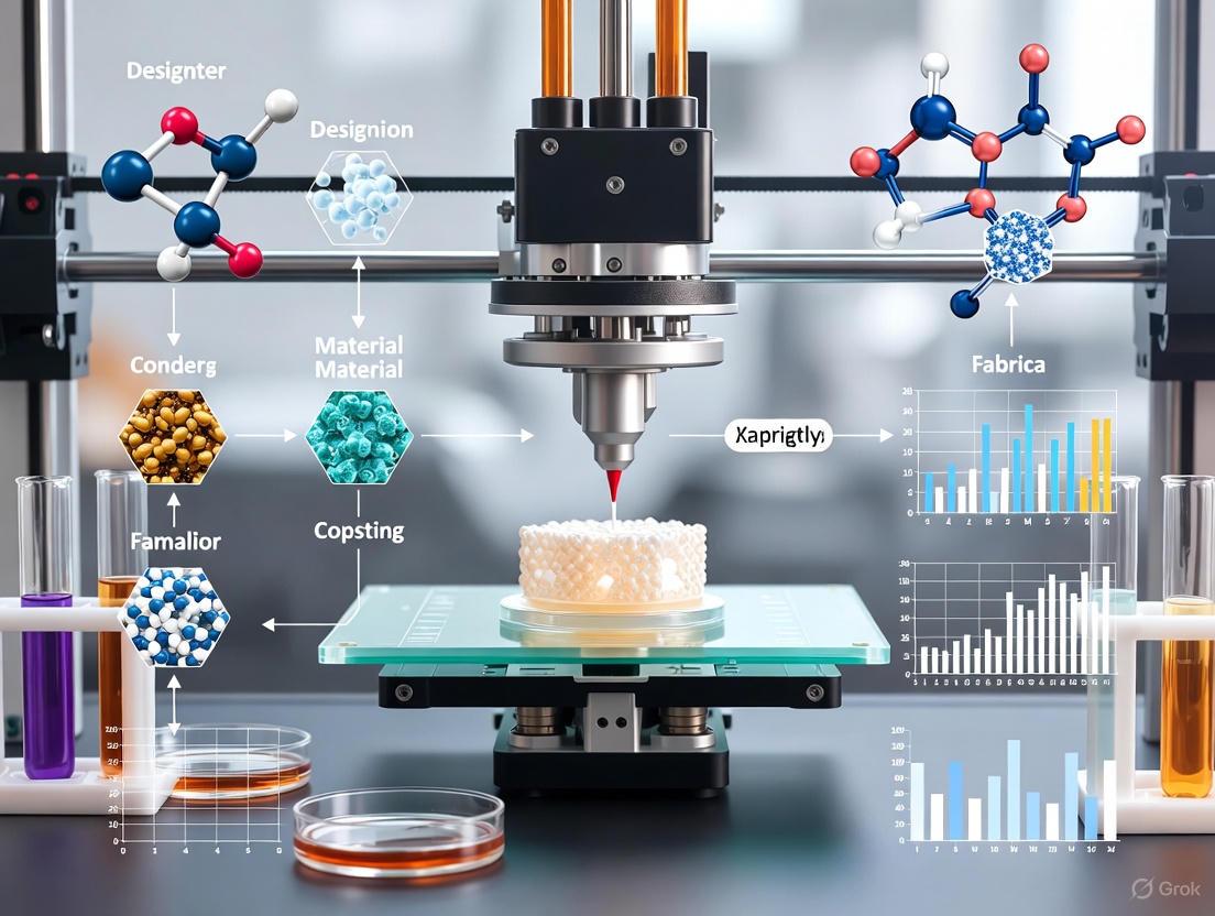
This article explores the transformative role of 3D printing in creating custom material designs, with a focus on applications for researchers and drug development professionals.
Custom Material Design in 3D Printing: From Drug Delivery to High-Strength Composites
Abstract
This article explores the transformative role of 3D printing in creating custom material designs, with a focus on applications for researchers and drug development professionals. It covers the foundational principles of additive manufacturing for composites and pharmaceuticals, detailing methods like fused deposition modeling and stereolithography. The scope extends to advanced applications such as personalized polypills, patient-specific implants, and high-strength composite structures. The content also addresses critical challenges in optimization and quality control, providing a comparative analysis of material properties and performance validation. By integrating the latest research and market trends, this review serves as a comprehensive resource for leveraging 3D printing to develop next-generation customized solutions in medicine and advanced manufacturing.
The Foundations of 3D Printed Material Design: Principles, Materials, and Market Dynamics
Core Principles of Additive Manufacturing for Custom Materials
Additive manufacturing (AM) has evolved from a niche prototyping method into a mainstream production tool capable of creating complex, functional components from custom materials. The core philosophy of Design for Additive Manufacturing (DfAM) blends traditional engineering fundamentals with the unique freedoms and constraints of 3D printing [1]. Unlike traditional manufacturing, which often imposes geometric limitations due to tool access or draft angles, AM rewards organic shapes, internal features, and integrated assemblies that were previously impossible or inefficient to produce [1]. Success in 3D printing custom materials requires anticipating how the printer builds the part, where thermal stresses may form, and how novel materials behave during the printing and cooling processes.
Fundamental DfAM Principles for Custom Materials
The effective application of custom materials hinges on adhering to key DfAM principles during the design phase. These rules ensure printability, structural integrity, and functional performance.
Wall Thickness
Maintaining consistent and appropriate wall thickness is critical for creating strong, reliable parts. Walls that are too thin lack structural support and can lead to cracking or failure, while excessively thick walls can cause internal stress, warping, and unnecessary material consumption [1].
- Principle: Ensure wall thicknesses are consistent throughout the part, especially at corners and transitions.
- Rationale: Prevents deformation during printing and improves overall reliability. The ideal thickness is material-dependent, but often falls within 1 to 3 millimeters for many polymers [1].
Feature Size
The resolution of every AM process imposes a limit on the minimum printable feature size, dictated by the printer's nozzle diameter, laser spot size, or voxel resolution [1].
- Principle: Design features such as holes, thin ribs, and engraved text to be larger than the printer's minimum capable resolution.
- Rationale: Prevents small features from failing to form, closing up, or fusing together during the build process, ensuring both accuracy and functionality [1].
Part Orientation
Part orientation during the build process majorly influences the final component's strength, accuracy, and surface quality [1].
- Principle: Orient the part to align load-bearing features with the plane of the print layers and to minimize the need for support structures.
- Rationale: Layers bond most strongly when forces act along their plane. Strategic orientation also reduces support material, minimizes post-processing, and can improve surface finish by mitigating the "stair-stepping" effect [1].
Internal Channels
The ability to create complex internal channels is a significant advantage of AM, enabling applications in fluidics, cooling, and wiring [1].
- Principle: Design internal passages to be clear of trapped powder or resin and consider long-term manufacturing plans if transitioning to other processes.
- Rationale: Narrow or sharply angled channels can become clogged with unmelted powder or support material, rendering the part non-functional. Designing for clearability ensures consistent manufacturing and performance [1].
Self-Supporting Angles
Gravity significantly impacts each layer as it is deposited. Unsupported overhangs and steep angles are prone to sagging, distortion, or collapse [1].
- Principle: Use gentle transitions, fillets, and angled surfaces that allow each new layer to be adequately supported by the one beneath it.
- Rationale: Self-supporting designs reduce or eliminate the need for dedicated support structures, which shortens print cleanup time, improves surface quality on overhanging faces, and increases build stability [1].
Additive Manufacturing Materials and Their Properties
The capabilities of 3D printing are directly tied to the properties of its feedstocks. Custom materials are broadly categorized as polymers, metals, and emerging composites, each suited for different applications [2].
Table 1: Common Additive Manufacturing Polymer Materials
| Material Category | Example Materials | Key Properties | Typical Applications |
|---|---|---|---|
| Standard Thermoplastics | ABS, PLA, Nylon (PA) | Durability, impact resistance, ease of use | Prototyping, consumer products, gears, bearings [2] |
| High-Performance Thermoplastics | PEEK, PEI, PPSU | Excellent chemical & temperature resistance, high strength | Aerospace, automotive, medical implants [2] |
| Photopolymers (Resins) | Standard, Tough, Flexible, Biocompatible Resins | High resolution, smooth surface finish | Detailed prototypes, visual models, medical devices [2] |
| Elastomers | TPU, Flexible Resins | Soft, rubber-like flexibility | Seals, gaskets, wearables, dampers [2] |
Table 2: Common Metal Powders for Additive Manufacturing
| Metal Category | Example Alloys | Key Properties | Typical Applications |
|---|---|---|---|
| Aluminum Alloys | AlSi10Mg | Lightweight, good strength-to-weight ratio | Aerospace components, heat exchangers [2] |
| Titanium Alloys | Ti-6Al-4V | High strength, low density, corrosion resistance | Patient-specific implants, aerospace structural parts [2] |
| Stainless Steels | 316L, 304 | Corrosion resistance, durability | Medical devices, marine applications, food processing [2] |
| Nickel-based Superalloys | Inconel | High thermal resistance, creep strength | Jet engine components, gas turbines [2] |
Experimental Protocol for High-Throughput Material Discovery
The following protocol outlines a methodology for using AM as a research tool to discover and test new custom materials, leveraging high-throughput techniques [3].
Objective: To rapidly fabricate and characterize a library of material formulations for evaluating printability and mechanical performance.
Research Reagent Solutions
Table 3: Essential Materials for Custom AM Material Research
| Reagent/Material | Function/Description |
|---|---|
| Polymer Resins/Base Powders | Primary material matrix (e.g., PEEK, Nylon, custom photopolymer resins) [2] |
| Functional Fillers | Carbon fibers, glass beads, or ceramic nanoparticles to enhance mechanical, thermal, or electrical properties [2] |
| Photosensitizers | For vat polymerization; compounds that initiate cross-linking upon light exposure [3] |
| AI/ML Software Platform | Artificial intelligence and machine learning tools to guide material design and predict properties [3] |
Step-by-Step Workflow
- Material Formulation: Prepare a library of material variants by blending base polymers with functional fillers at different concentrations.
- Test Coupon Design: Design a standardized test artifact containing features to assess critical parameters: tensile bars, compression pillars, overhang structures, and fine channels.
- High-Throughput Printing: Use a multi-material or automated AM system to print the entire library of test coupons in a single or a minimized number of build jobs.
- Post-Processing: Apply necessary post-processing steps uniformly across the sample set (e.g., UV curing, thermal annealing, support removal).
- Characterization & Data Collection:
- Mechanical Testing: Perform tensile, compression, and flexural tests.
- Dimensional Accuracy: Use coordinate measuring machines (CMM) to verify feature sizes [4].
- Microscopy: Examine layer adhesion, porosity, and filler distribution using SEM.
- Data Analysis & Feedback: Employ AI/ML models to analyze the dataset, identify composition-structure-property relationships, and recommend optimized formulations for the next iteration of discovery [3].
Workflow Visualization
Material Selection and Process Compatibility Framework
Selecting the appropriate material and AM process is a critical decision that depends on the functional requirements of the final part and the capabilities of each manufacturing technology [2].
The successful 3D printing of custom material designs relies on a deep integration of core DfAM principles—governing wall thickness, feature size, orientation, internal channels, and self-supporting angles—with a rigorous understanding of material properties and process compatibility. By adopting structured experimental protocols and high-throughput discovery workflows, researchers can accelerate the development of next-generation materials. This integrated approach, facilitated by AI and machine learning, is key to unlocking new frontiers in additive manufacturing for demanding applications across aerospace, medical, and advanced industrial sectors.
Application Notes & Experimental Protocols
Metals: Powder Bed Fusion of Ti-6Al-4V for Aerospace Components
Application Notes: Ti-6Al-4V is the preferred titanium alloy for aerospace and medical implants due to its high strength-to-weight ratio and biocompatibility [5]. Verification of powder composition is critical, as deviations exceeding 0.5% in key elements can lead to cracking and a 30% reduction in yield strength [5]. Properly verified compositions can achieve tensile strengths of approximately 900 MPa [5].
Experimental Protocol: Powder Verification and DMLS Process
- Step 1: Powder Composition Verification: Analyze metal powder using Inductively Coupled Plasma Mass Spectrometry (ICP-MS) to ensure conformity with ASTM F2924 standards. Acceptable ranges are Al: 5.5-6.75% and V: 3.5-4.5%; oxygen levels must be <0.13% to prevent embrittlement [5].
- Step 2: Powder Bed Fusion (DMLS): Conduct the build process in an argon-gas-purged environment to prevent oxidation of reactive Ti-6Al-4V powder [6]. Use a layer thickness of 20-30 μm [6] and a high-power laser to fully melt the powder.
- Step 3: Stress Relief: Upon build completion, subject the parts, while still on the build platform, to a stress-relief heat treatment cycle [6].
- Step 4: Post-Processing: Separate the parts from the build platform and remove support structures. Apply final finishing via bead blasting or machining to improve surface finish [6].
Thermoplastics: Pellet-Based Printing of Soft Elastomers for Soft Robotics
Application Notes: Pellet-based extrusion printing enables the use of very soft Thermoplastic Elastomers (TPEs), down to Shore Hardness 00-30, for applications like soft robotic actuators [7]. This process allows for the creation of thin (0.2-1.2 mm), airtight membranes capable of inflating to a stretch of 1320% [7].
Experimental Protocol: Printing and Testing of Soft TPE Membranes
- Step 1: Pellet Extrusion Setup: Utilize a pellet-based extruder capable of processing soft TPE pellets. Optimize nozzle temperature and flow rate to prevent clogging and ensure layer adhesion [7].
- Step 2: Print Parameter Calibration: Print test membranes with varying thicknesses (0.2, 0.5, 1.0, 1.2 mm) to establish a relationship between printing parameters and membrane integrity [7].
- Step 3: Tensile and Inflation Testing: Mechanically test printed specimens to determine engineering stress and maximum elongation. Conduct inflation tests on sealed membranes to validate air-tightness and measure maximum stretch capacity [7].
- Step 4: Actuator Functional Testing: Integrate the printed membrane into a soft bending actuator design. Characterize performance by measuring bending angle (e.g., 180°) and blocked force (e.g., 238 times its weight) [7].
Composites: Continuous Fiber Reinforcement for Optomechanical Components
Application Notes: Continuous fiber reinforcement using Markforged technology (Onyx matrix with fiberglass or Kevlar) creates parts that bridge the performance gap between standard plastics and metals [8]. These components are ideal for applications requiring high stiffness-to-weight ratios, vibration resistance, and controlled thermal expansion, such as mirror holders and clamping forks [8].
Experimental Protocol: Fabrication of a Stiffened Mount
- Step 1: Digital Model Preparation: Load the CAD model into the cloud-based Eiger software. Define the print orientation to align continuous fibers with the primary load paths [8].
- Step 2: Parameter Setting: Set a standard layer height of 0.1 mm. Use two perimeter walls and a triangular infill pattern with a density of 37% for the base Onyx material [8].
- Step 3: Continuous Fiber Reinforcement: Specify the number and placement of continuous fiber layers (e.g., two levels of four isotropic layers near the roof and floor). Set nozzle temperatures to 270–280 °C for Onyx and 240–250 °C for the continuous fiber [8].
- Step 4: Post-Processing and Validation: After printing, remove the part and perform mechanical testing. Compare experimental deformation under load with Finite Element Analysis (FEA) simulations to validate the design and printing strategy [8].
Bio-inks: VitroINK for 3D Bioprinting of Tissue Models
Application Notes: VitroINK bioinks represent a new generation of xeno-free, biofunctional materials that maintain high cell viability and support cell-matrix interactions without requiring UV, heat, or chemical crosslinking [9]. They are room-temperature stable, facilitating easier handling and mixing with cells via a dual-syringe system [9].
Experimental Protocol: Bioprinting a Liver Construct for Toxicity Screening
- Step 1: Bioink and Cell Preparation: Thaw VitroINK bioink and primary human hepatocytes. Prepare the bioink-cell mixture either by pre-mixing or loading into a dual-syringe system for mixing immediately before printing [10] [9].
- Step 2: Bioprinting Process: Use a bioprinter (e.g., BIO X) fitted with a temperature-controlled printhead (maintained at 18-22°C). Print the liver construct layer-by-layer into a sterile culture plate [10].
- Step 3: Post-Printing Culture: Transfer the bioprinted construct to an incubator (37°C, 5% CO2) with specialized culture media to support hepatocyte function.
- Step 4: Functional Assay: After a culture period, dose the bioprinted mini-livers with test compounds. Evaluate liver toxicity by measuring standard biomarkers like albumin secretion and urea synthesis, comparing against 2D culture controls [10].
Table 1: Metal Alloy Specifications for 3D Printing
Data sourced from manufacturer specifications and standards (e.g., ASTM) as cited in [5] [6].
| Alloy Type | Key Composition (%) | Ultimate Tensile Strength (MPa) | Yield Stress (MPa) | Elongation at Break (%) | Common Applications |
|---|---|---|---|---|---|
| Ti-6Al-4V | Al: 6, V: 4 [5] | 993 - 1,055 [6] | 855 - 951 [6] | 15 - 18 [6] | Aerospace, Medical Implants |
| AlSi10Mg | Si: 10, Mg: 0.3 [5] | 268 - 345 [6] | 180 - 228 [6] | 8 - 15 [6] | Automotive, Lightweight Structures |
| Stainless Steel 316L | Cr: 17, Ni: 12 [5] | 565 - 586 [6] | 379 - 386 [6] | 75 - 78 [6] | Medical, Chemical Processing |
| Inconel 718 | Ni: 52, Cr: 19 [5] | 958 - 993 (Stress Relieved) [6] | 572 - 676 (Stress Relieved) [6] | 36 - 40 (Stress Relieved) [6] | Aerospace, High-Temp Components |
Table 2: Composite 3D Printing Strategies and Properties
Based on experimental data and characterization from [8].
| Printing Strategy | Reinforcing Material | Key Affected Properties | Anisotropy | Best For |
|---|---|---|---|---|
| FFF with Blends | Particle additives (e.g., bronze, wood) | Aesthetics, Machinability, Flame Resistivity [8] | Low (mainly from layering) | Prototypes, Visual Models |
| FFF with Chopped Fibers | Crushed Carbon, Glass, or Kevlar Fibers [8] | Stiffness, Dimensional Stability, Temp. Resistance [8] | Medium (from layering & fiber orientation) | Stiff, functional housings & fixtures |
| FFF with Continuous Fibers | Continuous Carbon, Glass, or Kevlar strands [8] | Tensile Strength, Stiffness, Impact Resistance [8] | High (controlled, directional reinforcement) | High-performance, load-bearing structures |
Workflow and Pathway Visualizations
Metal Powder Verification and Processing Workflow
Composite 3D Printing with Continuous Fiber
Bio-ink Crosslinking and Culture Workflow
The Scientist's Toolkit: Research Reagent Solutions
Table 3: Essential Materials for 3D Printing Custom Material Designs
| Item | Function | Example Use Case |
|---|---|---|
| Metal Powder (Ti-6Al-4V) | Primary material for DMLS/SLM processes creating high-strength, lightweight metal parts [5]. | Aerospace components, medical implants [5]. |
| PolyTerra PLA | Eco-friendly, biodegradable thermoplastic filament used as the base matrix in sustainable printing strategies [11]. | Sustainable prototypes, non-critical parts in hybrid prints [11]. |
| Onyx (Micro-carbon-filled Nylon) | Base matrix material for Markforged continuous fiber printing; provides a stiff, high-quality surface finish [8]. | Optomechanical mounts, jigs, and fixtures [8]. |
| Continuous Carbon Fiber | Reinforcement material embedded in the Onyx matrix to dramatically increase strength and stiffness [8]. | Primary load-bearing elements in composite prints [8]. |
| VitroINK Bioink | Xeno-free, biofunctional hydrogel for 3D bioprinting that supports cell viability without harsh crosslinking [9]. | Bioprinting liver, skin, and other tissue models for drug screening [10] [9]. |
| TeloCol | A form of collagen bioink, used for dispensing and bioprinting structurally complex biological constructs [10]. | Creating droplet-in-droplet structures, contraction assays [10]. |
Emerging Market Trends and Growth Drivers in Custom Material Printing
The domain of custom material printing is undergoing a profound transformation, evolving from a rapid prototyping tool into a core manufacturing technology capable of producing end-use parts with tailored properties. For researchers and drug development professionals, this shift opens new frontiers in creating highly customized medical devices, implants, and drug delivery systems. The global market for 3D printing materials is projected to grow from USD 3.88 billion in 2025 to USD 10.02 billion by 2030, at a compelling Compound Annual Growth Rate (CAGR) of 20.9% [12]. This growth is primarily driven by the ease of developing customized products and significant reductions in manufacturing costs and process downtime across key industries. Understanding these trends and their underlying drivers provides a critical foundation for strategic research and development investments in this rapidly advancing field.
Table: Global 3D Printing Materials Market Forecast
| Metric | 2025 | 2030 (Projected) | CAGR (2025-2030) |
|---|---|---|---|
| Market Size | USD 3.88 Billion | USD 10.02 Billion | 20.9% [12] |
Market Trends and Quantitative Analysis
The custom material printing landscape is characterized by several convergent trends, each with distinct implications for research and industrial application. The market's momentum is sustained by technological advancements in materials and increasing adoption across high-value industries.
Primary Market Growth Drivers
Quantitative analysis reveals the specific impact of key drivers on the market's growth trajectory [13]:
- Surge in Metal Powder Usage: The adoption of titanium, nickel, and aluminum alloys for serial production in aerospace and medical sectors is a major driver, with an estimated +4.2% impact on the CAGR forecast. This is exemplified by the use of Ti-6Al-4V in flight-critical components and biocompatible implants [13].
- Advances in High-Performance Polymers: Rapid development in materials like Polyetheretherketone (PEEK) and polyetherketoneketone (PEKK) is projected to have a +3.8% impact on CAGR. These polymers are replacing metals in applications requiring high strength-to-weight ratios and thermal resistance [13].
- Demand from Automotive Applications: The automotive sector, particularly electric vehicle (EV) production, is poised for the highest growth rate (24.87% CAGR) [13]. This surge is driven by the use of 3D printing for lightweight coolant manifolds, complex battery mounts, and tooling, reducing assembly tool weight by up to 72% [13].
- Mass-Customization in Healthcare: The trend toward patient-specific implants, prosthetics, and surgical guides continues to gain momentum, with an estimated +2.9% impact on the market CAGR [13].
Market Segmentation and Material Types
The market is diversifying across material types, forms, and end-use industries, each segment exhibiting unique growth dynamics [12] [13].
Table: 3D Printing Materials Market Analysis by Segment
| Segment | Dominant Category | Key Characteristics & Growth |
|---|---|---|
| By Material Type | Plastics (47.25% share in 2024) | Versatile, inexpensive, and broadly available for technologies like FDM, SLA, and SLS. Includes commodity (ABS, PLA) and engineering-grade polymers [13]. |
| Metals (Highest growth: 23.24% CAGR to 2030) | Driven by certified titanium, aluminum, and nickel super-alloys for aerospace and medical applications [13]. | |
| By Form | Filament (68.42% share in 2024) | Dominance driven by hobbyist, education, and engineering adoption via Fused Filament Fabrication (FDM/FFF) printers [13]. |
| Powder (Highest growth rate) | Essential for technologies like SLS and DMLS, enabling enhanced accuracy and complexity for end-use parts [12]. | |
| By End-use Industry | Aerospace & Defense (36.28% share in 2024) | Early adopter of powdered metals for airframe brackets and ducting; lengthy qualification cycles create high barriers [13]. |
| Automotive (Highest growth: 24.87% CAGR) | Fueled by electrification and personalized interiors; used for jigs, fixtures, and low-volume service parts [13]. |
Application-Oriented Experimental Protocols
For researchers entering the field, establishing robust experimental protocols is essential. The following sections provide detailed methodologies for key applications in custom material printing.
Protocol: Fabrication of Patient-Specific Orthopedic Implants
This protocol outlines the workflow for producing a custom titanium cranial plate or spinal cage using Direct Metal Laser Sintering (DMLS) [13].
1. Pre-Production: Design and Preparation
- Medical Imaging and Model Generation: Acquire high-resolution CT scans of the patient's defect site. Use medical imaging software (e.g., Mimics) to convert DICOM data into a 3D surface model (STL file) of the implant.
- Topology Optimization and Lattice Design: Using engineering software (e.g., nTopology), apply topology optimization algorithms to minimize implant weight while maintaining mechanical integrity. Integrate a porous lattice structure (e.g., gyroid or diamond unit cell) into the design to promote bone osseointegration. The pore size should be designed within the range of 300-600 µm.
- Support Structure Generation: Automatically generate support structures using the DMLS machine's build processor software to anchor the part to the build plate and dissipate heat during printing.
2. Production: DMLS Printing
- Material: Ti-6Al-4V ELI (Extra Low Interstitial) grade powder, particle size 15-45 µm.
- Printing Parameters:
- Layer Thickness: 30 µm
- Laser Power: 200 W
- Scan Speed: 1200 mm/s
- Build Chamber Atmosphere: Argon gas, maintaining oxygen level < 0.1%
- Process: The build plate is pre-heated to 200°C. A recoater blade spreads a thin layer of powder. A high-power fiber laser selectively sinters the powder cross-section based on the sliced CAD data. The build plate lowers, and the process repeats for each layer.
3. Post-Processing
- Stress Relief: Heat treat the printed part while still attached to the build plate at 650°C for 2 hours in a vacuum furnace to relieve internal stresses.
- Support Removal: Carefully remove the build plate and separate the implant from the base plate via wire EDM. Manually remove all support structures.
- Hot Isostatic Pressing (HIP): Subject the implant to HIP (920°C, 100 MPa, 2 hours) to eliminate internal porosity and enhance fatigue resistance.
- Surface Finishing: Perform sandblasting with alumina media and electropolishing to achieve a smooth, biocompatible surface finish.
The workflow for this protocol is illustrated in the following diagram:
Diagram 1: Workflow for a Custom Orthopedic Implant
Protocol: High-Performance Polymer Component via FFF
This protocol details the manufacturing of a high-heat, strong component, such as a satellite bracket or automotive fixture, using PEEK or carbon-fiber reinforced filament [14] [13].
1. Material and Printer Preparation
- Material Handling: Seal PEEK or ONYX (carbon fiber filled nylon) filament in a sealed bag with desiccant. Load the spool into a sealed drybox attached to the printer, or pre-dry the filament in an oven at 80°C for 4-6 hours prior to printing.
- Printer Setup: Utilize an industrial FFF printer equipped with an all-metal hot-end capable of reaching at least 400°C and a heated build chamber capable of maintaining 120-150°C. Install a hardened steel nozzle if printing with carbon-fiber reinforced materials.
2. Printing Process
- Build Plate Preparation: Apply a thin, even layer of polyimide (PEI) or adhesive to a clean, heated build plate to ensure part adhesion.
- Critical Printing Parameters:
- Nozzle Temperature: 380-420°C (for PEEK)
- Build Plate Temperature: 120-150°C (for PEEK)
- Chamber Temperature: > 70°C (for PEEK)
- Print Speed: 30-60 mm/s
- Layer Height: 0.1-0.2 mm
- Infill Density: 80-100% for structural parts
- Process Monitoring: Monitor the first layers closely for adhesion. The entire printing process should occur in a draft-free environment to prevent thermal warping.
3. Post-Processing and Validation
- Annealing: To relieve internal stresses and improve crystallinity, anneal the printed PEEK part by heating it in an oven according to a specific thermal profile (e.g., heat to 200°C, hold, then heat to above its glass transition temperature).
- Dimensional Validation: Use Coordinate Measuring Machine (CMM) or laser scanning to verify critical dimensions against the original CAD model.
- Mechanical Testing: Perform tensile, flexural, or impact testing on printed coupons from the same build lot to validate mechanical properties against material datasheet specifications.
The Scientist's Toolkit: Research Reagent Solutions
For experimental work in custom material printing, selecting the appropriate materials and understanding their functions is critical. The following table details key materials used in the featured protocols and related research.
Table: Essential Materials for Custom Material Printing Research
| Material | Type/Form | Primary Function & Key Properties |
|---|---|---|
| Ti-6Al-4V ELI Powder | Metal Powder | Function: Fabrication of load-bearing, biocompatible implants. Properties: High strength-to-weight ratio, excellent corrosion resistance, and biocompatibility per ASTM F136 [13]. |
| PEEK (Polyetheretherketone) | Polymer Filament | Function: Creating high-temperature, chemically resistant components. Properties: High thermal resistance (HDT > 250°C), high strength, sterilizability, and inherent biocompatibility [14] [13]. |
| Onyx (Chopped CF Nylon) | Composite Filament | Function: Base material for strong, stiff, and heat-resistant end-use parts. Properties: Nylon matrix filled with chopped carbon fiber, offering high strength, stiffness, and excellent surface finish [14]. |
| VICTREX AM 200 | High-Performance Polymer | Function: For production-grade parts requiring high dimensional accuracy at elevated temperatures. Properties: PEEK-based material maintaining accuracy at 150°C service temperatures, suitable for hundreds of parts per build [13]. |
| Biocompatible (USP Class VI) Resin | Photopolymer Liquid | Function: Producing surgical guides, dental models, and non-implant medical devices. Properties: Cured resin meets stringent biocompatibility standards (e.g., ISO 10993), allowing for temporary contact with the body [15] [13]. |
| Flame Retardant (FR) Polymer Powder | Polymer Powder | Function: Manufacturing components for electronics, aerospace, and automotive where flame resistance is critical. Properties: Halogen-free, flame-retardant properties (e.g., HP's PA 12 FR), meeting industry safety standards [15] [13]. |
The relationships between these materials, their properties, and their primary research applications are summarized in the following diagram:
Diagram 2: Material-Property-Application Relationships
The emerging trends in custom material printing underscore a paradigm shift from general-purpose prototyping to application-specific, high-performance manufacturing. The robust growth driven by material innovations in metals and high-performance polymers, coupled with escalating demand from the medical, aerospace, and automotive sectors, creates a fertile ground for advanced research. For scientists and drug development professionals, mastering the associated experimental protocols—from designing for additive manufacturing to executing precise post-processing techniques—is no longer optional but essential for leveraging the full potential of this technology. The ability to tailor material properties at the point of manufacture will continue to be the core value proposition, enabling breakthroughs in personalized medicine and complex engineered systems.
The Shift from Prototyping to End-Use Part Production
Additive manufacturing (AM), or 3D printing, has undergone a fundamental transformation, evolving from a technology exclusively for rapid prototyping to a robust method for producing functional, end-use parts [16]. This shift is particularly impactful in fields requiring high levels of customization and complex material designs, such as medical devices and drug development [17] [3]. For researchers and scientists, this transition is not merely about replacing traditional manufacturing but about leveraging the unique capabilities of AM to create parts with customized mechanical properties, intricate internal architectures, and material compositions that were previously impossible to achieve [18]. The core of this evolution lies in the convergence of advanced printing processes, a growing palette of engineering-grade materials, and data-driven design strategies that together ensure the reliability and performance of manufactured components.
Key Drivers and Enabling Technologies
The move toward end-use production is powered by several technological and methodological advances. Advanced Processes and Materials: Vat photopolymerization processes, like Digital Light Processing (DLP), enable the production of highly detailed and complex geometries with precise material structure control [18]. Furthermore, direct-ink writing (DIW) allows for the use of paste-like materials, such as those laden with living cells or functional ceramics, expanding the scope of 3D printing to artificial tissues and large-scale structures [19]. Integrated Quality Control: The adoption of real-time metrology systems, such as in-process monitoring with advanced imaging, allows for the instant detection and correction of defects during printing, a critical development for industries like aerospace and medical devices where part failure is not an option [20]. Data-Driven Design and Production: The integration of numerical modeling and artificial intelligence (AI) is accelerating materials discovery and optimizing print parameters. Validated numerical models allow researchers to evaluate designs under various loading conditions at a fraction of the cost and time of extensive experimental testing [3] [18].
Table 1: Technologies Enabling End-Use 3D Printing
| Technology Category | Specific Example | Function in End-Use Production | Research Application |
|---|---|---|---|
| Advanced Processes | Digital Light Processing (DLP) | Enables high-resolution, precise control over material structure and geometry [18]. | Fabrication of biomedical components with complex internal lattices [18]. |
| Paste Extrusion | Direct-Ink Writing (DIW) | Prints with a wide range of functional, paste-like materials (e.g., cells, concrete, ceramics) [19]. | Creation of artificial tissues and embedded sensors for smart garments [19]. |
| In-Process Monitoring | 3D Metrology Systems | Uses real-time imaging (e.g., X-ray) to detect and correct defects layer-by-layer [20]. | Ensures reliability and perfection of critical parts in aerospace and medical devices [20]. |
| Production Workflow | Time Code (T-Code) | A programming language that synchronizes printhead motion with material switching for continuous fabrication [19]. | Enables smooth material gradients and complex multi-material parts without defects [19]. |
Quantitative Analysis of Printed Part Performance
The mechanical performance of 3D-printed parts is paramount for their qualification in end-use applications. Research demonstrates that mechanical properties can be systematically tuned through geometric design. For instance, a study on DLP-printed PLA resin compared fully solid specimens with those featuring a Voronoi lattice structure, revealing a strategic trade-off between strength and material efficiency [18].
Table 2: Mechanical Performance of Solid vs. Voronoi DLP-Printed PLA Resin Specimens
| Mechanical Property | Solid Specimen Performance | Voronoi Specimen Performance | Implications for Design |
|---|---|---|---|
| Tensile Strength | Higher | Lower | Solid structures are superior for applications requiring high load-bearing capacity under tension [18]. |
| Bending/Flexural Performance | High strength | Better performance per unit mass despite lower absolute load capacity [18]. | Voronoi structures are highly efficient for applications where flexural loading and weight are critical factors [18]. |
| Material Efficiency | Low (fully dense) | High (porous structure) | Voronoi lattices optimize material consumption, reducing weight and waste while maintaining mechanical integrity [18]. |
| Failure Mode | Brittle fracture with striations and bubble-shaped irregularities [18]. | Clean, brittle failure along structural voids; fragmented surfaces [18]. | Design dictates failure mode; solid structures fail in a more monolithic way, while lattices fail in a predictable, controlled manner [18]. |
Experimental Protocols for End-Use Part Evaluation
A rigorous, multi-stage protocol is essential for developing and validating 3D-printed components for end-use applications. The following workflow provides a structured methodology for researchers.
Stage 1: Specimen Design and Preparation
- Objective: Create digital models for mechanical testing.
- Procedure:
- Software: Use a CAD package such as SolidWorks Premium.
- Standard Specimens: Model tensile and bending test specimens according to ISO 527-2 and ISO 178:2019 standards, respectively [18].
- Lattice Design: Use a generative design tool like Grasshopper for Rhino to create a Voronoi lattice structure within the specimen volume. This optimizes material consumption and can impart energy-absorption properties [18].
- Slicing: Export the models as STL files and prepare them for printing using slicing software (e.g., ChiTuBox for DLP printing) [18].
Stage 2: Material Processing and Printing
- Objective: Fabricate test specimens with high precision.
- Procedure:
- Material: Select a commercially available PLA-like photopolymer resin.
- Printer: Use a Digital Light Processing (DLP) 3D printer. The DLP process is chosen for its ability to cure entire layers at once, providing high accuracy and smooth surface finishes [18].
- Post-Processing: Conduct all necessary post-printing procedures as per the resin manufacturer's guidelines, which may include washing in isopropyl alcohol and post-curing under UV light.
Stage 3: Experimental Mechanical Testing
- Objective: Empirically determine key mechanical properties.
- Procedure:
- Tensile Test: Perform uniaxial tensile tests to determine tensile strength, modulus of elasticity, and elongation at break. This reveals the material's behavior under pulling stress [18].
- Three-Point Bending Test: Perform flexural tests to determine flexural strength and modulus. This is crucial for applications where the component will experience bending loads [18].
- Analysis: Compare the results of solid and Voronoi specimens to understand the structure-property relationship.
Stage 4: Numerical Modeling and Validation
- Objective: Develop a predictive numerical model to reduce future development costs.
- Procedure:
- Software: Use a finite element analysis (FEA) package such as Ansys.
- Model Development: Create a numerical model of the test specimens, defining material properties based on initial experimental data.
- Simulation: Run simulations of the tensile and bending tests.
- Validation: Correlate the simulation results with the experimental data from Stage 3. A strong correlation confirms the model's reliability for preliminary design verification of future components [18].
Stage 5: Microstructural Analysis
- Objective: Understand the root causes of failure and identify printing defects.
- Procedure:
- Microscopy: Use microscopic analysis to examine the fracture surfaces of the tested specimens.
- Identification: Look for features such as striations, bubbles, and the nature of the fracture (brittle vs. ductile) [18].
- Feedback: Use these insights to identify potential issues in the printing process or material and inform further design iterations.
Essential Research Reagent Solutions
The following materials and software are critical for executing the experimental protocols for the development of end-use 3D printed parts.
Table 3: Key Research Reagents and Materials for End-Use 3D Printing Research
| Item Name | Function/Description | Application in Research Context |
|---|---|---|
| PLA-like Photopolymer Resin | A biodegradable, liquid resin that cures under UV light to form a rigid, high-resolution part [18]. | Base material for vat photopolymerization (e.g., DLP); used for creating precise mechanical and biomedical components [18]. |
| Voronoi Lattice Algorithm | A generative algorithm that creates organic, cellular structures within a design volume. | Used in CAD to design lightweight, energy-absorbing interior structures that optimize the strength-to-weight ratio of a part [18]. |
| Finite Element Analysis (FEA) Software | Software (e.g., Ansys) that predicts how a product reacts to real-world forces, vibration, and other physical effects [18]. | Enables virtual testing of 3D-printed designs under mechanical load, significantly reducing the need for physical prototypes [18]. |
| Direct-Ink Writing (DIW) Ink | A paste-like, complex fluid material that can contain polymers, living cells, ceramics, or concrete [19]. | Allows printing of functional, multi-material structures for advanced applications in bioprinting, electronics, and construction [19]. |
| In-Process Metrology System | A system that uses real-time imaging (e.g., X-ray) to monitor the printing process layer-by-layer [20]. | Provides closed-loop quality control by detecting defects during the build process, essential for certifying critical end-use parts [20]. |
The maturation of 3D printing from a prototyping tool to a production-ready technology represents a paradigm shift for research and development. This transition is underpinned by a holistic approach that integrates material science, advanced processes, and computational design. The ability to precisely control internal geometry, as demonstrated by the tunable performance of Voronoi structures, allows researchers to engineer material properties to meet specific functional requirements. Furthermore, the framework of experimental validation coupled with predictive numerical modeling creates a robust and efficient pathway for developing reliable end-use parts. As these technologies continue to converge—with enhancements in AI-driven design, high-throughput material discovery, and real-time process control—3D printing is poised to become the cornerstone of manufacturing for highly customized, performance-critical applications in medicine, aerospace, and beyond.
Methodologies and Breakthrough Applications in Pharmaceutical and Composite Printing
Three-dimensional (3D) printing, or additive manufacturing, is revolutionizing the pharmaceutical industry by enabling the fabrication of personalized medicines that offer solutions unattainable by traditional mass production [21]. This technology builds complex structures through successive layering of materials based on a digital design, allowing for unprecedented flexibility and precision in dosage form design [22]. The global 3D printed drug market is predicted to grow at an impressive 12.3% compound annual growth rate between 2025 and 2032, increasing from USD 63.45 million to USD 160.5 million, reflecting strong industry confidence in this technology's potential [21].
A particularly promising application of 3D printing in pharmaceutics is the creation of polypills - single dosage forms containing multiple active pharmaceutical ingredients (APIs) [23]. This approach is especially valuable for conditions requiring complex treatment regimens, such as metabolic syndrome, where patients often need multiple medications targeting different aspects of their condition [24]. Compared to traditional manufacturing processes like tablet compression or capsule filling, 3D printing facilitates the combination of previously incompatible APIs into different compartments within a single pill and allows for advanced customization of release profiles [21]. Although the regulatory landscape for 3D-printed pharmaceuticals is still evolving, the FDA's 2015 approval of SPRITAM (levetiracetam), the first 3D-printed drug, has sparked increased interest in further developing these applications [23] [21].
Key 3D Printing Technologies in Pharmaceutical Manufacturing
Several 3D printing techniques have been utilized in pharmaceutical manufacturing, each with distinct mechanisms and applications. The table below summarizes the primary technologies used in pharmaceutical applications:
Table 1: Key 3D Printing Technologies Used in Pharmaceutical Applications
| Technology | Process Description | Key Applications | Advantages | Limitations |
|---|---|---|---|---|
| Fused Deposition Modeling (FDM) [22] | Uses a heated nozzle to extrude a continuous strand of molten polymer which is layered and cooled to form a 3D object | Oral dosage forms, implants, modified-release tablets, polypills | Simple operation, easy parameter control, versatile | Requires thermal stability of APIs, need for filament preparation |
| Inkjet-Based Printing [22] | Deposits layers of photopolymer resin which are cured using a UV light source to form a 3D object | Tablets, implants, orodispersible films | Suitable for heat-sensitive materials, precise dosing | Limited material options, potential stability issues with UV curing |
| Powder-Based Printing [22] | Binds powder particles together using a binder solution or laser to create a 3D structure | Fast-dissolving tablets, complex medication release profiles, novel-shaped tablets | No heat stress on APIs, versatile material options | Post-processing required, potential powder handling issues |
| Stereolithography (SLA) [22] | Uses a laser to solidify liquid resin layer by layer via photopolymerization | Microneedles, high-precision dosage forms | High resolution and accuracy, smooth surface finish | Limited biocompatible materials, resin toxicity concerns |
Among these techniques, Fused Deposition Modeling (FDM) has emerged as the most widely used technology in pharmaceutical research, particularly for manufacturing polypills, due to its relatively simple operation, ease of parameter control, and versatility in creating complex dosage forms [22] [24]. The FDM process is typically coupled with Hot-Melt Extrusion (HME) to produce drug-loaded filaments suitable for printing [24].
Figure 1: Experimental workflow for developing FDM-printed polypills
Quantitative Data and Research Findings
Performance Metrics of 3D Printing Technologies
Research has demonstrated that various 3D printing technologies can achieve clinically acceptable levels of accuracy for pharmaceutical applications. A comparative study of 12 different 3D printers used in dentistry (which shares precision requirements with pharmaceutical applications) showed that all tested printers could produce reliable, reproducible models with mean errors below clinically relevant thresholds [25]. The most accurate printers in the study (Envision One, Envision D4K, Ackuretta Sol and Asiga Max UV) achieved overall trueness under 35 μm, well within acceptable limits for pharmaceutical dosage forms [25].
Table 2: Accuracy Performance of 3D Printing Technologies in Pharmaceutical Applications
| Performance Metric | Range/Value | Technology/Context | Clinical Relevance |
|---|---|---|---|
| Dimensional Trueness [25] | < 35 μm (best performers) to < 260 μm | Dental models (indicative for pharmaceutical tools) | All within clinically acceptable limits (< 300-500 μm) |
| Drug Loading Efficiency [24] | High loading with desired precision | FDM for potent drugs in small doses | Enables accurate dosing for potent APIs |
| Production Speed [26] | 5-10 times faster than previous generations | Recent desktop FDM printers | Facilitates on-demand manufacturing |
| Material Waste [22] | Significantly reduced compared to conventional methods | Various pharmaceutical 3D printing technologies | Cost-effective for small batches |
Case Study: Polypill for Metabolic Syndrome
A significant demonstration of 3D printing's potential in pharmaceuticals is the development of a polypill for metabolic syndrome, a complex condition characterized by at least three of the following: insulin resistance, hypertension, dyslipidemia, type 2 diabetes, obesity, inflammation, and non-alcoholic fatty liver disease [24]. Researchers successfully manufactured a polypill using FDM 3D printing technology containing three APIs:
- Nifedipine as an antihypertensive drug
- Simvastatin as an antihyperlipidemic drug
- Gliclazide as an antiglycemic drug [24] [21]
This formulation exhibited a dual-release profile, combining faster simvastatin release (within 6 hours) with a 24-hour sustained release for nifedipine and gliclazide, showcasing the potential for personalized treatment of metabolic syndrome [24]. The researchers utilized Hansen solubility parameters (HSPs) as predictors to guide the formation of amorphous solid dispersions between drugs and polymers, ensuring miscibility and enhanced oral bioavailability [24]. The HSPs varied from 18.3 for nifedipine, 24.6 for simvastatin, and 7.0 for gliclazide, while the total solubility parameter for the excipient mixture was 27.3±0.5, enabling the formation of amorphous solid dispersions particularly for simvastatin and gliclazide [24].
Table 3: Formulation Parameters for Metabolic Syndrome Polypill
| Parameter | Nifedipine | Simvastatin | Gliclazide | Polymer System |
|---|---|---|---|---|
| Therapeutic Category | Antihypertensive | Antihyperlipidemic | Antiglycemic | Matrix Former |
| Target Release Profile | 24-hour sustained release | <6 hour release | 24-hour sustained release | Controlled Release |
| Hansen Solubility Parameter [24] | 18.3 | 24.6 | 7.0 | 27.3 ± 0.5 |
| Solid State in Formulation [24] | Partially crystalline | Amorphous dispersion | Amorphous dispersion | Amorphous |
Experimental Protocols and Methodologies
Protocol: Fabrication of FDM 3D-Printed Polypills
This protocol outlines the methodology for developing polypills containing multiple APIs using Fused Deposition Modeling (FDM) 3D printing technology, based on established research for metabolic syndrome treatment [24].
Materials and Equipment
Table 4: Essential Research Reagent Solutions for FDM-Printed Polypills
| Reagent/Material | Function/Purpose | Examples/Specifications |
|---|---|---|
| Active Pharmaceutical Ingredients [24] | Therapeutic agents | Nifedipine, Simvastatin, Gliclazide (or other API combinations) |
| Polymer Matrix [22] [21] | Filament formation, controlled release | HPMC (various viscosity grades), PVA, PLA - pharmaceutical grade |
| Plasticizers [22] | Enhance filament flexibility | Polyethylene glycol, glycerin, triethyl citrate |
| Hot-Melt Extruder [24] | Production of drug-loaded filaments | Single or twin-screw extruder with temperature control |
| FDM 3D Printer [24] | Fabrication of dosage forms | Dual-extrusion capable for multi-material printing |
| Characterization Equipment [24] | Quality assessment | DSC, XRD, dissolution apparatus, HPLC |
Step-by-Step Procedure
Pre-formulation Studies
- Determine Hansen solubility parameters (HSPs) for all APIs and polymers to predict miscibility and compatibility [24].
- Calculate the total solubility parameter (δ) using the equation: δ = √(δd² + δp² + δh²), where δd, δp, and δh represent dispersion, polar, and hydrogen bonding components, respectively.
- Select polymer systems with HSP values close to the APIs to facilitate amorphous solid dispersion formation.
Filament Preparation via Hot-Melt Extrusion (HME)
- Pre-blend APIs with polymer matrix and plasticizers using a mortar and pestle or blender for small batches, or a tumble blender for larger batches.
- Process the physical mixture using a hot-melt extruder with temperature settings appropriate for the polymer system (typically 10-20°C above the polymer's glass transition temperature).
- For temperature-sensitive APIs, consider processing at lower temperatures with suitable plasticizers.
- Collect the extruded filament and ensure diameter consistency (typically 1.75 mm or 2.85 mm for compatibility with FDM printers).
- Store filaments in sealed containers with desiccant to prevent moisture absorption.
3D Printing Process
- Design the polypill structure using CAD software, considering the internal architecture for controlled release.
- For multi-material printing, design separate components for different drug compartments.
- Convert the CAD file to STL format and import into slicing software.
- Set printing parameters: nozzle temperature (based on filament properties), build plate temperature (to ensure adhesion), layer height (typically 100-200 μm), printing speed (30-60 mm/s), and infill density (usually 100% for solid dosage forms).
- Load drug-loaded filaments and initiate the printing process.
- For dual-drug systems, utilize multiple extruders or pause printing for filament switching.
Post-processing and Characterization
- Remove printed polypills from the build plate and carefully detach any support structures if present.
- Conduct quality control tests including dimensional analysis (using digital calipers or microscopy), weight variation, and visual inspection.
- Perform solid-state characterization using XRD and DSC to confirm amorphous dispersion formation.
- Conduct in vitro drug release studies using USP dissolution apparatus with appropriate media.
- Analyze drug content using HPLC or UV-Vis spectroscopy.
Figure 2: Material compatibility decision pathway for FDM formulation
Applications and Future Perspectives
The applications of 3D printing in pharmaceuticals extend far beyond polypills for metabolic syndrome. Research has demonstrated successful 3D printing of various dosage forms including oral controlled release systems, micropills, microchips, implants, microneedles, rapid dissolving tablets, and multiphase release dosage forms [22]. The technology enables unique control over drug release and the shape of dosage forms, potentially making it the key technique for individualized dosage forms [27].
Future advancements in pharmaceutical 3D printing are likely to focus on several key areas:
- Artificial Intelligence Integration: AI-powered 3D printing can enhance efficiency, precision, and user experience through model search and generation, autonomous slicing, intelligent material recognition, and real-time defect detection [26].
- Expanded Material Options: Continued development of pharmaceutical-grade polymers with tailored properties will expand the possibilities for dosage form design [21].
- Regulatory Framework Development: As the technology matures, clearer regulatory pathways will emerge to facilitate broader adoption in pharmaceutical manufacturing [21].
- Point-of-Care Manufacturing: The portability and ease of use of 3D printers make them ideal for usage in hospital wards, in-patient pharmacies, specialist clinics, and community pharmacies for on-demand medication preparation [22].
The integration of 3D printing into pharmaceutical practice represents a paradigm shift from traditional "one-size-fits-all" medicine toward truly personalized treatment approaches. By enabling precise control over dose, release profile, and combination therapies, 3D printing has the potential to significantly improve therapeutic outcomes, particularly for patients with complex medication needs such as those with metabolic syndrome, polypharmacy, or unique physiological requirements.
The evolution of 3D bioprinting has positioned it as a transformative technology in regenerative medicine, enabling the fabrication of complex, patient-specific tissue constructs. This capability is central to advancing beyond the limitations of traditional transplantation methods. The core of this approach lies in the creation of customized scaffolds that act as temporary, three-dimensional extracellular matrices (ECMs). These structures are designed to direct cellular behavior—including adhesion, proliferation, and differentiation—and facilitate the formation of new, functional tissue [28] [29]. The precision of additive manufacturing, particularly through computer-aided design (CAD), allows for unprecedented control over scaffold architecture, permitting the optimization of both biological and mechanical performance for specific clinical needs [28]. This document details the critical design parameters, experimental protocols, and material solutions that underpin the development of advanced bioprinted scaffolds within a broader research context focused on 3D printing of custom material designs.
Core Design Parameters: The Role of Porosity
Among various design parameters, porosity is a critical determinant of scaffold success, as it directly influences both biological response and mechanical integrity [28]. Porosity is not a monolithic property but is defined by several interconnected characteristics that must be carefully balanced.
Table 1: Key Porosity Parameters and Their Impact on Scaffold Performance
| Parameter | Biological Influence | Mechanical Influence | Design Considerations |
|---|---|---|---|
| Pore Size | Affects cell adhesion, infiltration, and tissue-specific differentiation (e.g., osteogenic, chondrogenic) [28]. | Influences local stiffness and stress distribution. | Optimal size is cell and tissue-type dependent; often requires a gradient to mimic native tissue [28]. |
| Pore Geometry | Specific curvatures and shapes can enhance or inhibit focal adhesion formation [28]. | Determines load-bearing capacity and structural stability. | Controlled via CAD and printing path; hexagonal or grid-like structures often provide good stability [28]. |
| Interconnectivity | Governs cell migration, uniform tissue formation, angiogenesis, and nutrient/waste diffusion [28] [29]. | Affects overall structural cohesion and resistance to compression. | A high degree of interconnectivity is essential for biological functionality, even if it slightly reduces stiffness [28]. |
| Distribution | Influences homogeneity of tissue growth and formation of organized structures (e.g., neural networks, bone canaliculi) [28]. | Impacts anisotropic mechanical behavior. | Can be designed as gradients to create zones with different mechanical or biological properties [28]. |
Mastering the interplay of these porosity parameters is fundamental to guiding specific cellular responses and developing scaffolds tailored for tissues ranging from bone and cartilage to neural and vascular networks [28].
Experimental Protocols for Advanced Bioprinting
Protocol 1: Design and Fabrication of a Multi-Material Hydrogel Scaffold
This protocol outlines the synthesis and characterization of a composite bioink based on alginate (Alg), carboxymethyl cellulose (CMC), and gelatin methacrylate (GelMA), balancing printability, stability, and biocompatibility [30].
1. Bioink Formulation and Preparation
- Materials: Alginate powder, Carboxymethyl Cellulose (CMC), GelMA powder, Polyethylene glycol diacrylate (PEGDA), Lithium Phenyl (2,4,6-trimethylbenzoyl) phosphinate (LAP) photoinitiator, Dulbecco's Phosphate Buffered Saline (DPBS).
- Procedure:
- Weigh 0.5 g GelMA (5 wt%), 0.3 g PEGDA (3 wt%), and 0.05 g LAP (0.5 wt%) into a glass vial.
- Add 10 mL DPBS. Cover the vial with aluminum foil to prevent premature crosslinking.
- Heat the mixture to 47°C for 1 hour with stirring to ensure complete dissolution.
- Filter the solution through a 0.22 µm sterile filter and maintain at 37°C until use [30].
- Separately, prepare alginate and CMC solutions to achieve final combined concentrations of 4% Alg and 10% CMC when mixed with the GelMA base [30].
2. Rheological Characterization and Printability Assessment
- Objective: To correlate bioink rheology with printing performance.
- Methods:
- Shear-Thinning Behavior: Perform a flow sweep test on a rotational rheometer (e.g., MCR102) with shear rates from 0.01 to 100 s⁻¹ at the printing temperature (e.g., 26°C). A significant decrease in viscosity with increasing shear rate indicates good extrudability [30] [31].
- Viscoelasticity: Conduct an amplitude sweep at a fixed frequency (e.g., 100 rad/s) to identify the linear viscoelastic (LVE) region and yield stress. A dominant storage modulus (G') within the LVE region indicates good shape fidelity post-printing [30].
- Thixotropy: Perform a step-rate test, alternating between low and high shear rates to mimic the printing and deposition phases. Rapid recovery of viscosity after shear cessation is crucial for layer stacking [30].
Table 2: Target Rheological Properties for Printability
| Property | Target Value/Range | Measurement Technique | Significance |
|---|---|---|---|
| Shear-Thinning Index (n) | n < 1 (Power-law model) | Flow Sweep Test | Ensures easy extrusion under pressure and minimal cell shear stress [30]. |
| Yield Stress (τ₀) | > 50 Pa (application-dependent) | Amplitude Sweep Test | Provides shape retention and self-supporting ability after deposition [30]. |
| Structural Recovery | > 85% G' recovery within 10s | Thixotropic Loop Test | Enables stable multi-layer fabrication without deformation [30]. |
3. Printing and Cross-linking
- Printing: Utilize a extrusion-based bioprinter equipped with a temperature-controlled printhead. Set the printing temperature to 26°C for optimal viscosity [30] [31].
- Dual Cross-linking:
- Ionic Cross-linking: Immediately after printing, mist the scaffold with a 100-200 mM CaCl₂ solution.
- Photo-cross-linking: Follow with UV light exposure (e.g., 365 nm, 5-10 mW/cm²) for 60-120 seconds to cure the GelMA and PEGDA components. This dual approach creates scaffolds with variable stiffness and long-term stability [30].
The following workflow summarizes the key stages of this protocol from material preparation to final validation.
Protocol 2: Embedded Bioprinting for Cellular Alignment
This protocol describes the fabrication of multilayered arterial tissues with controlled cellular alignment using an embedded 3D bioprinting approach, which is also adaptable for skeletal and cardiac muscle tissues [31].
1. Supporting Bath and Bioink Preparation
- Supporting Bath:
- Dissolve 5 g of Pluronic F-127 in 50 mL of 1x PBS overnight at 4°C.
- Heat the solution to 50°C and stir at 600 rpm. Add 3 g of Hydroxypropylmethyl cellulose (H-HPMC) and stir for 15 minutes.
- Cool to room temperature under slow stirring (200 rpm). Transfer to an acrylic printing box, avoiding air bubbles. Sterilize with UV light (≥40 W) for >15 minutes [31].
- Bioink: Prepare a cell-laden GelMA bioink as in Protocol 1, with a recommended cell concentration of 10⁶ cells/mL. Load the bioink into a sterile syringe and maintain at the printing temperature of 26°C in the dark [31].
2. Fluid Property Measurement and Flow Rate Modeling
- Objective: To predict the printing flow rate for consistent filament diameter and cellular alignment.
- Property Measurement:
- Thermal Parameters: Use a thermal conductivity analyzer (e.g., TPS 2500 S) with a Kapton sensor to measure the thermal conductivity and specific heat of the bioink and supporting bath at 20°C.
- Density: Measure using a density meter (e.g., DMA 5000 M) across a temperature range (20°C to 30°C).
- Surface Tension: Determine using a contact angle goniometer via the sessile drop method.
- Rheology: Perform temperature sweep (40°C to 10°C at -2°C/min) and shear rate sweep (0.01 to 100 s⁻¹) tests as described in Protocol 1 [31].
- Flow Rate Prediction Model:
- Model the nozzle immersed in the supporting bath using CAD software (e.g., SOLIDWORKS).
- Import the model into computational fluid dynamics (CFD) software (e.g., ANSYS Workbench).
- Input measured fluid properties as parameters and simulate flow to determine the optimal flow rate that produces the desired filament diameter for cellular alignment [31].
3. Embedded Printing and Post-Processing
- Printing Path Generation: Manually design a 3D printing trajectory that promotes cellular alignment, as commercial slicing software may not support this.
- Printing: Perform printing with the nozzle immersed ~5 mm in the supporting bath, using the flow rate determined from the CFD model.
- Bath Removal: After printing, carefully remove the construct from the supporting bath using a removal bath composed of 2.5 mL PEG400 in 50 mL of 1x PBS [31].
The Scientist's Toolkit: Research Reagent Solutions
The following table catalogues essential materials and their functions for developing and characterizing bioinks and bioprinted scaffolds.
Table 3: Essential Research Reagents and Materials for Bioprinting
| Reagent/Material | Function and Role in Bioprinting | Example Application |
|---|---|---|
| Alginate (Alg) | A natural polymer that forms a gentle ionic gel with divalent cations (e.g., Ca²⁺); provides initial structural integrity and biocompatibility [30]. | Used in composite bioinks for its rapid gelation and synergy with other polymers like GelMA [30]. |
| Gelatin Methacrylate (GelMA) | A photocrosslinkable hydrogel derived from gelatin; contains RGD peptide motifs that promote cell adhesion and proliferation [30]. | Serves as a primary bioink component for creating stable, cell-supportive scaffolds; concentration tunes stiffness [30]. |
| Carboxymethyl Cellulose (CMC) | A cellulose derivative used as a viscosity modifier and rheological agent to enhance the shear-thinning properties of bioinks [30]. | Combined with Alg and GelMA to improve printability and filament formation [30]. |
| Lithium Phenyl (2,4,6-trimethylbenzoyl) phosphinate (LAP) | A cytocompatible photoinitiator that cleaves under UV light to generate radicals, initiating the cross-linking of methacrylated polymers [30]. | Used for UV cross-linking of GelMA and PEGDA components in bioinks [30]. |
| Pluronic F-127 | A thermoreversible block copolymer that acts as a sacrificial support material; it is fluid when cold and solid when warm [31]. | Key component of the supporting bath in embedded bioprinting, providing temporary mechanical support during printing [31]. |
| Hydroxypropylmethyl cellulose (H-HPMC) | A cellulose ether used to modify the viscosity and viscoelastic properties of solutions. | Added to Pluronic F-127 supporting baths to fine-tune its rheological characteristics for improved printing fidelity [31]. |
| Polyethylene glycol diacrylate (PEGDA) | A synthetic, photopolymerizable macromer used to increase the cross-linking density and mechanical strength of hydrogel networks [30]. | Incorporated into GelMA bioinks to enhance the final scaffold's mechanical properties and stability [30]. |
The integration of precise scaffold design, advanced biomaterials, and sophisticated bioprinting protocols, as detailed in these application notes, is pushing the boundaries of regenerative medicine. The critical role of controlled porosity in directing biological function, combined with robust protocols for manufacturing complex tissue constructs, provides a foundational framework for research. Emerging techniques, such as dual-light processing for multi-material structures [32] and the increasing integration of AI and computational modeling [28] [31], promise to further enhance the precision and capabilities of this field. By adhering to systematic design and characterization principles, researchers can continue to develop increasingly biomimetic and functional tissues for therapeutic applications.
The integration of continuous fibers and nanoparticles into polymer matrices represents a frontier in the creation of high-performance composites for additive manufacturing. These materials transcend the capabilities of conventional polymers and short-fiber reinforcements, offering unparalleled specific stiffness and strength, enhanced multifunctionality, and superior thermal properties [33] [34]. The synergy between continuous fibers, which provide macro-scale structural reinforcement, and nanoparticles, which enhance matrix properties at the micro-scale, enables the fabrication of complex, load-bearing components tailored for demanding sectors such as aerospace, automotive, and biomedical engineering [35] [8]. This document, framed within broader research on 3D printing of custom material designs, provides detailed application notes and experimental protocols to guide researchers in the processing, characterization, and application of these advanced composites.
Material Systems and Performance Data
The performance of 3D-printed composites is fundamentally governed by the selection of the reinforcement and matrix materials. Continuous fibers are the primary load-bearing constituent, while the matrix binds the fibers and transfers stress.
Continuous Fiber Reinforcements
Different fiber types offer a range of mechanical, economic, and functional properties, as summarized in Table 1.
Table 1: Performance Characteristics of Common Continuous Fibers for 3D Printing
| Fiber Type | Tensile Strength (MPa) | Tensile Modulus (GPa) | Key Advantages | Primary Applications | Citations |
|---|---|---|---|---|---|
| Carbon | 3500 – 7000 | 230 – 600 | Superior strength-to-weight ratio, high stiffness, corrosion resistance | Aerospace, automotive, high-performance sports | [35] |
| Glass | 2000 – 4000 | 70 – 90 | Cost-effective, higher fracture strain (toughness) | Construction, marine, consumer goods | [35] |
| Aramid | 3000 – 4000 | 70 – 140 | Excellent impact resistance, thermal stability | Ballistic protection, aerospace | [35] |
| Natural (Flax) | 300 – 1000 | 20 – 60 | Sustainable, biodegradable, low density | Eco-friendly products, biomedical | [35] |
Matrix Materials and Nanoparticle Enhancement
The polymer matrix can be enhanced with nanoparticles to improve its intrinsic properties, which in turn boosts the overall composite performance. Key matrix materials include Nylon (PA) and its composites (e.g., Onyx, a micro-carbon-fiber filled nylon), Polylactic Acid (PLA) for its biodegradability, and high-performance thermoplastics like Polyetheretherketone (PEEK) for high-temperature applications [35] [8]. Nanoparticles such as carbon nanotubes (CNTs) or graphene can be incorporated to enhance electrical and thermal conductivity, interlaminar shear strength, and fracture toughness of the polymer matrix, contributing to a more robust fiber-matrix interface [34].
Quantitative Performance of Advanced Composites
The strategic combination of fibers and matrix yields significant mechanical enhancements. Table 2 summarizes quantitative data from recent studies on advanced composite systems.
Table 2: Mechanical Performance of Selected 3D-Printed Advanced Composites
| Composite Material | Fiber Volume Fraction | Tensile Strength (MPa) | Flexural Modulus (GPa) | Key Innovation / Property Enhancement | Citations |
|---|---|---|---|---|---|
| Helical CCF/PLA | Not Specified | Not Specified | 202% increase vs. non-twisted | Spiral fiber architecture for manipulating mechanical and sensing responses. | [33] |
| CCF/PA6 | Not Specified | Significantly improved vs. pure PA6 | Not Specified | Interfacial optimization between fiber and matrix. | [35] |
| CFRP (Onyx + Fiberglass) | Not Specified | Experimentally determined* | Experimentally determined* | Young's modulus determined via tensile testing for FEA simulation. | [8] |
| CF/Nylon (Markforged) | Not Specified | ~800 MPa | Not Specified | High strength suitable for replacing metal optomechanical components. | [8] |
*The Young's modulus for the CFRP (Onyx + Fiberglass) material was determined experimentally for simulation inputs, with values varying based on printing parameters like infill density and pattern [8].
Experimental Protocols
Protocol 1: Fused Filament Fabrication (FFF) of Continuous Fiber Composites
This protocol details the procedure for fabricating continuous fiber-reinforced composites using a dual-nozzle FFF platform, such as the Markforged Mark Two or similar systems [8].
1. Materials and Equipment:
- 3D Printer: Dual-extrusion FFF printer capable of printing composite materials (e.g., Markforged Mark Two, Anisoprint A-series).
- Base Material Filament: Micro-carbon-fiber filled nylon (e.g., Onyx) or pure thermoplastic spool.
- Continuous Fiber Filament: Spool of continuous carbon, glass, or Kevlar fiber.
- Software: Slicing software (e.g., Markforged Eiger, Ultimaker Cura).
- Adhesive: Bed adhesive (e.g., water-soluble school glue).
2. Pre-Printing Procedure:
- CAD Model Preparation: Design the component using CAD software, considering anisotropic properties and optimal fiber orientation for load paths.
- Slicing and Toolpath Generation:
- Import the CAD model (STL file) into the slicing software.
- Select the base material (e.g., Onyx) for the overall structure.
- Define the continuous fiber reinforcement layout. Specify the number of perimeter layers and the infill pattern (e.g., triangular, isotropic) for the fiber.
- Program the fiber path to align with principal stress directions. Software like Anisoprint's allows for varying fiber direction and density ("anisoprinting").
- Set printing parameters:
- Nozzle temperature (base material): 270-280 °C.
- Nozzle temperature (continuous fiber): 240-250 °C.
- Bed temperature: As recommended for the material.
- Layer height: 0.1 mm.
- Infill density: Adjustable (e.g., 37% standard).
- Bed Adhesion: Apply a thin layer of adhesive to the build plate to prevent warping and improve first-layer adhesion.
3. Printing Execution:
- Initiate the print job. The printer will co-extrude the base material and the continuous fiber according to the predefined toolpaths.
- Monitor the initial layers to ensure proper adhesion and fiber deposition.
4. Post-Processing:
- Once printing is complete and the build chamber has cooled, carefully remove the part from the build plate.
- Remove any support structures (if used). For Markforged Onyx, supports are typically breakaway.
- Parts can be sanded, painted, or further machined as required.
Protocol 2: Fabrication of Helical-Gradient Continuous Fiber Composites
This protocol describes an integrated method for creating composites with spirally arranged fibers, which exhibit superior mechanical and sensing properties [33].
1. Materials and Equipment:
- Integrated Platform: Custom fabrication platform combining a resin-coating, fiber-twisting extrusion process with a 3D printer.
- Materials: Continuous carbon fibers (e.g., 1K T300), PLA pellets, acetone, dichloromethane.
2. Experimental Workflow:
- Fiber Desizing: Treat the continuous carbon fibers with acetone to remove sizing agents.
- Resin Coating: Pre-coat the desized fibers with a PLA solution in dichloromethane.
- Helical Twisting-Extrusion:
- Feed the coated continuous fiber through the integrated platform.
- Program the twisting mechanism to impart a helical gradient to the fiber bundle. This can involve independently controlling the twist of single strands and multi-strand bundles.
- Co-extrude the twisted fiber with molten PLA in a single-stream process to form a continuous filament with a controlled helical architecture.
- 3D Printing: Use the produced helical filament in a subsequent 3D printing process to fabricate composite specimens. This process enables support-free printing of complex structures.
The logical workflow for this advanced process is outlined below.
The Scientist's Toolkit: Research Reagent Solutions
Table 3: Essential Materials for High-Performance Composite Research
| Item Name | Function / Role in Research | Typical Examples / Specifications |
|---|---|---|
| Continuous Carbon Fiber | Primary reinforcement; provides high strength and stiffness. | 1K T300 (Toray); supplied on spools for FFF. |
| Matrix Thermoplastic | Binds fibers; transfers load; determines thermal/chemical resistance. | PLA (biodegradable), Nylon (Onyx), PEEK (high-temp). |
| Solvents | Fiber desizing and polymer dissolution for coating. | Acetone (99.5%), Dichloromethane (99.5%). |
| Hardened Nozzle | Extrudes abrasive composite materials without degradation. | Hardened steel or ruby nozzle; diameter ≥ 0.4 mm. |
| Bed Adhesive | Ensures first-layer adhesion and prevents warping. | PVA-based glue (e.g., Elmer's Washable School Glue). |
Functional Integration and Applications
The convergence of continuous fibers and functional matrices leads to composites that are not only structural but also smart and multi-functional.
Sensing Composites
By leveraging the electrical conductivity of carbon fibers, composites can be engineered to sense their own strain and damage. Research has demonstrated spiral fiber architectures that act as embedded sensors [33]. A resistive strain sensor made from helical carbon fiber composites achieved a 562% increase in sensitivity compared to conventional materials. Furthermore, capacitance–resistance hybrid sensors can be printed to simultaneously detect distance, angle, and pressing position, opening avenues for large-area tactile sensing in aerospace and health monitoring [33].
Specific Application Cases
- Aerospace Lightweight Structures: Used for manufacturing brackets, drone arms, and other components where high stiffness-to-weight ratio is critical, replacing aluminum parts [35] [8].
- Automotive Energy-Absorbing Components: Designed for crashworthiness, utilizing the high specific energy absorption of continuous fiber composites [35].
- Optomechanical Components: 3D-printed composite parts like mirror holders and clamping forks successfully replace metal parts, offering lighter weight, easier modification, and adequate vibration resistance and shape accuracy for precision optical systems [8].
The decision-making process for selecting a composite printing strategy based on application requirements is visualized below.
The 3D printing of high-performance composites reinforced with continuous fibers and nanoparticles provides a powerful pathway for creating custom material designs with tailored mechanical and functional properties. The protocols and data outlined in these application notes offer a foundation for researchers to explore this rapidly advancing field. Future progress will be driven by AI-driven process optimization, the development of novel sustainable materials, and improved multi-material integration techniques, further solidifying the role of these composites in next-generation industrial and biomedical applications [35] [36].
Application Notes: Advanced Materials for 3D Printing
The 3D printing of custom material designs represents a frontier in materials science, enabling the creation of structures with previously unattainable properties. This document details two groundbreaking material forms—bendable concrete and dual-phase functional polymers—that are reshaping the capabilities of additive manufacturing within a research context. These materials address critical limitations of traditional formulations, offering new paradigms for constructing resilient infrastructure and complex, multi-part functional devices.
Bendable Concrete (Engineered Cementitious Composite - ECC)
Bendable concrete, or Engineered Cementitious Composite (ECC), is an ultra-ductile cementitious material designed to overcome the inherent brittleness of conventional concrete. Its development is pivotal for 3D printing applications where the incorporation of traditional steel reinforcement is incompatible with automated deposition processes [37].
- Core Innovation and Research Significance: The fundamental breakthrough lies in its ability to sustain significant tensile strain, up to 11.9% higher than standard mixes, without catastrophic failure [37]. This "strain-hardening" behavior under tension is a departure from the brittle fracture typical of ordinary concrete. For 3D printing research, this property is instrumental in enabling the creation of structural components that can withstand seismic events, ground settlement, and other dynamic loads, all without the need for embedded steel rebar that disrupts the printing workflow.
- Material Composition and Mechanism: The ductility is achieved through a meticulously designed fiber-reinforced microstructure. A high volume of short, randomly dispersed polymeric fibers (e.g., Polyvinyl Alcohol (PVA) or Ultra-High Molecular Weight Polyethylene (UHMWPE)) acts as a micro-reinforcement network [37]. When the cementitious matrix is stressed and micro-cracks begin to form, the fibers bridge these cracks. Instead of a crack propagating freely, the fibers yield, transferring stress across the crack and allowing the material to deform plastically while still carrying a load. The specific composition often includes supplementary cementitious materials like fly ash and silica fume to enhance fresh and hardened properties [37].
Table 1: Quantitative Performance Data for 3D-Printed Bendable Concrete Mixes
| Material Component | Function | Typical Proportion by Volume | Key Property Achieved |
|---|---|---|---|
| Cement | Primary binder | Base component | Provides compressive strength |
| Fly Ash / Silica Fume | Supplementary cementitious material | Varies | Enhances workability, durability, and sustainability |
| Fine Aggregate | Filler | Balanced for extrudability | Contributes to buildability and reduces shrinkage |
| Polyvinyl Alcohol (PVA) Fibers | Micro-reinforcement | Critical volume fraction (e.g., 2%) | Enables tensile strain capacity and crack control |
| Superplasticizer | Chemical admixture | Dosage for optimal flow | Ensures extrudability and pumpability |
| Water | Reactant and lubricant | Optimized for rheology | Governs hydration and fresh-state properties |
Dual-Phase Functional Polymers
Functional polymers for 3D printing are being engineered with spatially controlled properties, allowing a single print to contain multiple, distinct material phases. This capability is transformative for manufacturing complex devices with integrated, disposable support structures or regions with tailored mechanical and chemical functions.
- Core Innovation and Research Significance: The innovation centers on a single resin formulation that can be cured into two distinct solid phases based on the wavelength of applied light [38]. This wavelength-selective curing enables the simultaneous printing of both a permanent, robust structure and temporary, dissolvable supports. This eliminates the need for manual, post-print support removal—a labor-intensive and wasteful step in traditional vat photopolymerization—and facilitates the creation of intricate internal channels, interlocking parts, and encapsulated geometries that are otherwise impossible to produce.
- Material Composition and Mechanism: The resin is typically a mixture of two or more monomers and a photo-initiator system. Upon exposure to ultraviolet (UV) light, one set of reactions is triggered, leading to the formation of a highly cross-linked, durable polymer network that is resistant to solvents. When the same resin is exposed to visible light, a different reaction pathway dominates, resulting in a loosely cross-linked or linear polymer structure that remains soluble [38]. The addition of a "bridging" monomer is often required to ensure the mechanical integrity of the UV-cured structure [38]. This allows researchers to "set" the functional parts of a device while "programming" the supports for later dissolution in a food-safe solvent like baby oil, or even back into the original resin monomer for closed-loop recycling [38].
Table 2: Research Reagent Solutions for Dual-Phase Polymer 3D Printing
| Reagent / Material | Function in Formulation | Research Application |
|---|---|---|
| Primary Monomers (e.g., Acrylates) | Building blocks of the polymer network | Forms the backbone of both the permanent and dissolvable phases |
| Bridging Monomer | Enhances cross-linking under specific wavelengths | Strengthens the permanent (UV-cured) structure to prevent failure in solvent [38] |
| UV Photoinitiator | Initiates polymerization upon UV exposure | Selectively solidifies the permanent, non-dissolvable regions of the print |
| Visible Light Photoinitiator | Initiates polymerization upon visible light exposure | Solidifies the temporary, dissolvable support structures |
| Food-Safe Solvent (e.g., Baby Oil) | Dissolution medium | Selectively removes support material without damaging the primary structure [38] |
Experimental Protocols
Protocol for Formulation and Printing of Bendable Concrete
This protocol outlines a systematic, three-step strategy for developing and validating a 3D-printable bendable concrete mix, integrating rheological control and mechanical performance evaluation [39].
Workflow Diagram: Bendable Concrete Development
Title: Bendable Concrete Development Workflow
Step 1: Flow-State Stage – Assessing Extrudability
Objective: To ensure the fresh concrete mix can be pumped and extruded smoothly without segregation or blockage [40].
- Materials: Cement, fly ash, silica fume, fine sand, PVA fibers, superplasticizer (Polycarboxylate Ether-based), water.
- Equipment: High-shear mixer, rheometer, extrudability test rig.
- Procedure:
- Mix Proportioning: Weigh all dry components (cement, supplements, sand). Gradually add water and superplasticizer while mixing in a high-shear mixer for 5-10 minutes.
- Fiber Incorporation: Slowly add the PVA fibers to the slurry to ensure uniform dispersion without balling. Mix for an additional 3-5 minutes.
- Rheological Testing: Characterize the static and dynamic yield stress of the mixture using a rheometer. The mix must demonstrate low enough viscosity for pumping but rapid static yield stress recovery after extrusion.
- Extrudability Test: Load the mixture into a piston-driven extruder. Extrude a continuous filament through a target nozzle (e.g., 15-25 mm diameter) and observe for consistency, surface finish, and absence of tearing or blockage. A successful mix will produce a smooth, continuous filament.
Step 2: Structural Build-Up Stage – Assessing Buildability
Objective: To quantify the ability of the deposited material to support subsequent layers without deformation (i.e., "green strength") [41] [39].
- Materials: Freshly mixed ECC from Step 1.
- Equipment: 3D concrete printer (gantry or robotic arm), penetration needle test apparatus.
- Procedure:
- Printability Test: Program the printer to construct a simple vertical wall (e.g., 200 mm tall, 3 beads wide). The printing process should be continuous.
- In-situ Observation: Monitor the printed structure for signs of deformation, buckling, or lateral collapse during and immediately after printing.
- Green Strength Measurement: Using a penetration needle test, periodically measure the shear strength of the printed material over time. This data helps define the critical structuration rate and the maximum allowable printing speed and layer height.
Step 3: Hardened-State Stage – Mechanical Characterization
Objective: To validate the mechanical performance and "bendable" properties of the hardened, printed concrete [37].
- Materials: Cured ECC specimens printed and cured under controlled conditions (e.g., 7, 28 days).
- Equipment: Universal testing machine (UTM), digital image correlation (DIC) system for strain measurement.
- Procedure:
- Specimen Preparation: Print and cure "dog-bone" shaped specimens for direct tensile tests and prismatic beams for flexural tests.
- Uniaxial Tensile Test: Load the dog-bone specimen in the UTM. Measure stress-strain behavior. A successful ECC will exhibit multiple cracking and strain-hardening behavior, with a tensile strain capacity exceeding 3%.
- Four-Point Bending Test: Test prism beams to failure. Compare the load-deflection curves with those of conventional concrete. The ECC will demonstrate significant deflection and energy absorption capacity.
- Microstructural Analysis: Use scanning electron microscopy (SEM) on fractured surfaces to analyze fiber distribution, fiber pull-out mechanisms, and fiber-matrix bonding.
Protocol for Vat Photopolymerization with Dual-Phase Resins
This protocol describes the method for printing complex, multi-part objects using a single resin that forms both permanent and dissolvable structures based on the wavelength of light [38].
Workflow Diagram: Dual-Phase Polymer Printing
Title: Dual-Phase Polymer Printing and Recycling
Step 1: Resin Preparation and Digital File Preparation
Objective: To prepare the dual-cure resin and the digital model with integrated, dissolvable supports.
- Materials: Primary monomers, bridging monomer, UV photoinitiator, visible light photoinitiator.
- Equipment: Amber glassware, magnetic stirrer, CAD software, slicing software capable of assigning different light sources to model and support structures.
- Procedure:
- Resin Formulation: In amber glassware to prevent premature curing, combine the primary monomers. Add the bridging monomer (e.g., 1-5% by weight). Separately, dissolve the UV and visible light photoinitiators in small amounts of monomer before adding them to the main mixture. Stir until a homogeneous solution is achieved.
- Model and Support Generation: Design the target 3D model (e.g., interlocking gears, a hollow lattice) in CAD software. Use the software's automated support generation feature to create supports for overhangs and internal cavities. Assign the model body to be cured by UV light and the support structures to be cured by visible light in the slicing software.
Step 2: Simultaneous Dual-Wavelength Printing
Objective: To fabricate the object with its integrated support structure in a single, automated print job.
- Materials: Prepared dual-cure resin.
- Equipment: Custom or modified vat photopolymerization 3D printer equipped with both a UV light projector (e.g., 365 nm) and a visible light projector (e.g., 405 nm, 460 nm).
- Procedure:
- Printer Setup: Load the sliced file containing the dual light patterns. Fill the printer vat with the formulated resin.
- Print Execution: Initiate the print. The printer will project patterns of UV light to solidify the permanent parts of the model and simultaneously project patterns of visible light to solidify the support structures. The process continues layer-by-layer until complete.
Step 3: Support Dissolution and Material Recycling
Objective: To remove the support material without manual intervention and recycle the dissolved polymer.
- Materials: Printed part with supports, food-safe solvent (e.g., baby oil).
- Equipment: Ultrication bath, collection containers, filtration setup.
- Procedure:
- Dissolution: Upon print completion, remove the build platform from the printer. Submerge the entire printed structure (part and supports) in a bath of baby oil. Agitation (e.g., via ultrasonic bath) can accelerate the process. The visible-light-cured supports will dissolve, leaving the UV-cured structure intact.
- Part Retrieval: Remove the final, clean functional part from the solvent bath.
- Solvent Recycling: The solvent now contains dissolved support polymer. This solution can be filtered and blended directly with fresh resin monomer for subsequent prints, enabling a closed-loop, low-waste process [38].
Overcoming Design and Fabrication Challenges in Custom 3D Printing
Strategies for Sustainable Printing and Material Reduction
Additive manufacturing (AM) has revolutionized prototyping and production across numerous fields, including biomedical and drug development research. However, its environmental footprint, particularly material consumption and waste generation, presents a significant challenge. For researchers and scientists, implementing sustainable printing practices is crucial not only for reducing environmental impact but also for enhancing cost-efficiency and material performance in experimental workflows. These application notes provide detailed, actionable protocols for integrating material reduction strategies and sustainable material usage into 3D printing research, with a specific focus on applications relevant to scientific and drug development laboratories.
Material Reduction and Design Optimization Strategies
Strategic design and process optimization can drastically reduce material usage without compromising the structural or functional integrity of printed components, which is essential for custom laboratory apparatus, microfluidic devices, and tissue scaffolds.
Topology Optimization and Lightweighting
Principle: Computational design techniques are used to distribute material only where it is mechanically necessary, creating lightweight, efficient structures [42].
Application Workflow:
- Define Design Space: In your CAD software, model the maximum volume the part is allowed to occupy.
- Apply Constraints and Loads: Specify fixed boundary conditions, applied forces, and performance targets (e.g., maximum allowable deformation or natural frequency).
- Run Optimization: Use topology optimization software (often integrated into advanced CAD packages) to generate a material layout that meets the set constraints. The result is an organic, lightweight structure.
- Post-Processing: Smooth and prepare the optimized geometry for 3D printing, ensuring printability.
Lattice Structure Integration
Principle: Replacing solid volumes with internal micro-architectures or lattices can achieve weight reductions of up to 90% while maintaining specific mechanical properties [42].
Protocol for Lattice Generation:
- Select Lattice Type: Choose a unit cell (e.g., gyroid, cubic, tetrahedral) based on the desired mechanical response (isotropic, flexible, rigid).
- Define Cell Parameters: Specify cell size and beam thickness to control density and porosity.
- Apply to Volume: Use dedicated lattice generation software or modules to fill the target volume within the part.
- Validate Structure: Perform a finite element analysis (FEA) simulation to ensure the lattice meets the functional requirements.
Strategic Reinforcement
Principle: For components requiring high mechanical performance, a dual-material approach can be used, whereby a sustainable but weaker base material is strategically reinforced with a stronger material only in critical stress zones [11].
Experimental Protocol:
- Stress Analysis: Perform an FEA simulation on the part to identify high-stress regions [11].
- Model Segmentation: Segment the digital model into two bodies: one for the base material and one for the reinforcement material [11].
- Printing Setup: Use a dual-extrusion 3D printer. Load the eco-friendly filament (e.g., recycled PLA) in one extruder and the high-performance filament (e.g., Tough PLA) in the other [11].
- Printing: Execute the print job, allowing the printer to switch extruders at the designated layers or regions. This method has been shown to recover up to 70% of the strength of a full-strength part while using a fraction of the high-performance material [11].
Table 1: Quantitative Impact of Material Reduction Strategies
| Strategy | Typical Material Savings | Key Software/Tools | Best-Suited Applications |
|---|---|---|---|
| Topology Optimization | 20-70% [42] | nTopology, Ansys, Autodesk Fusion 360 | Structural brackets, custom jigs, robotic end-effectors |
| Lattice Integration | 50-90% [42] | nTopology, Materialise 3-matic | Tissue engineering scaffolds, lightweight insulation, fluidic mixers |
| Strategic Reinforcement | ~80% reduction of high-performance plastic [11] | SustainaPrint (MIT), Simplify3D | Functional prototypes, load-bearing lab equipment |
Sustainable Material Selection and Recycling
Choosing appropriate materials and implementing closed-loop recycling within a lab setting are fundamental to sustainable research practices.
Material Selection Guide
Table 2: Sustainable Material Options for Research Applications
| Material Class | Example Materials | Key Properties | Research Applications |
|---|---|---|---|
| Biodegradable Polymers | PLA, PHA | Biodegradable under specific conditions, low toxicity | Disposable labware, single-use microfluidics, plant-based scaffolds |
| Bio-based & Recycled Polymers | Recycled PET/G (rPET/G), Nylon from castor oil | Reduced carbon footprint, mechanical properties similar to virgin material | Durable jigs and fixtures, sample holders, housing for custom instruments |
| High-Performance Sustainable Composites | Polyolefins (PP, PE) with recycled content, wood-filled PLA | Chemical resistance (PP/PE), aesthetic appeal | Chemical fluidic manifolds, sample containers, presentation models |
In-House Filament Recycling Protocol
Recycling failed prints and support material into new filament closes the waste loop and reduces raw material costs.
Materials:
- Waste Stream: Clean, sorted 3D printing waste (PLA, ABS).
- Equipment: Shredder/granulator, filament extruder, spooler.
- Safety: Personal protective equipment (PPE) including safety glasses and gloves.
Step-by-Step Procedure:
- Waste Collection and Sorting: Collect failed prints and supports. Crucially, sort by material type to avoid cross-contamination that can degrade properties.
- Shredding: Use a granulator to shred the waste into small, uniform flakes (~3-5 mm).
- Drying: Dry the flakes in a vacuum oven or food dehydrator at 65-75°C for at least 4 hours to remove moisture. Moisture causes bubbling and poor extrusion.
- Extrusion: Feed the dried flakes into the hopper of the filament extruder. Set the temperature profile according to the polymer (e.g., 180-210°C for PLA).
- Spooling and Sizing: Monitor the extruded filament diameter using a laser gauge. Adjust the puller speed to maintain a consistent diameter (e.g., 1.75 mm ± 0.05 mm) and spool the filament.
- Quality Control: Test the recycled filament's tensile strength and diameter consistency before use in critical applications.
Experimental Protocols for Sustainable Printing
Protocol: Print Parameter Optimization for Material Efficiency
Objective: To determine the optimal set of printing parameters that minimizes material usage while meeting functional requirements for a given part.
Materials:
- 3D printer (FDM recommended for accessibility)
- CAD model of a standardized test part (e.g., a tensile bar or a small container)
- Virgin or recycled PLA filament
- Slicing software (e.g., Ultimaker Cura, PrusaSlicer)
Method:
- Identify Key Variables: Select parameters known to affect material use and strength: Layer Height, Infill Density, Infill Pattern, Wall Count, and Print Speed.
- Design of Experiment (DOE): Create an experimental matrix (e.g., a Taguchi array or full factorial) to systematically test combinations of these parameters.
- Printing: Slice the standard test part for each parameter combination in the DOE. Record the estimated print time and material usage from the slicer.
- Testing: Print all specimens. For each, record the actual mass and subject it to relevant functional tests (e.g., tensile testing, compression testing, or leak testing for fluidic devices).
- Data Analysis: Use statistical analysis (e.g., Analysis of Variance - ANOVA) to identify which parameters have a significant effect on the performance and material usage. Determine the Pareto-optimal set of parameters that offer the best trade-off.
Protocol: Validation of Strategic Reinforcement
Objective: To empirically validate the mechanical performance of a hybrid-printed part against versions printed entirely in base and reinforcement materials.
Materials:
- Dual-extrusion 3D printer
- Eco-friendly filament (e.g., PolyTerra PLA)
- High-performance filament (e.g., Tough PLA or ABS)
- CAD software with FEA capability
- Universal Testing Machine (e.g., Instron)
Method:
- Design and Analysis: Create a CAD model of a hook or a cantilever beam. Perform FEA to identify the high-stress region [11].
- Model Segmentation: Modify the model to create two components: the main body and a reinforcement patch for the high-stress area [11].
- Printing: Print three versions of the part:
- Version A: Entirely from eco-friendly filament.
- Version B: Entirely from high-performance filament.
- Version C: Hybrid print, with eco-friendly material as the main body and high-performance material as the reinforcement [11].
- Mechanical Testing: Mount each part in a universal testing machine and subject it to a controlled load until failure (e.g., apply a tensile load to the hook or a point load to the beam).
- Analysis: Record the failure load and displacement for each specimen. Calculate the percentage of strength recovered in the hybrid part compared to the full-strength version.
The Researcher's Toolkit
Table 3: Key Research Reagent Solutions for Sustainable 3D Printing
| Item | Function/Description | Example Application in Research |
|---|---|---|
| Polylactic Acid (PLA) | A biodegradable polymer derived from renewable resources like corn starch. | Primary material for prototyping, disposable microfluidic chips, and educational models. |
| Recycled PETG (rPETG) | Filament produced from post-consumer PET plastic. Offers good chemical resistance and toughness. | Printing lab equipment housings, chemical solvent racks, and durable sample containers. |
| Soluble Support Material | Support material (e.g., PVA, BVOH) that dissolves in water, reducing post-processing damage and waste. | Creating complex, intricate channels in microfluidic devices or assemblies with internal voids. |
| Finite Element Analysis (FEA) Software | Computational tool for simulating physical forces and identifying areas of high stress. | Used in topology optimization and for determining regions requiring strategic reinforcement [11]. |
| Filament Recycler/Extruder | A device that shreds plastic waste and extrudes it into new filament spools. | Closing the material loop in the lab by recycling failed prints into new experimental filament. |
Workflow and Pathway Visualizations
Strategic Reinforcement Workflow
Closed-Loop Material Recycling Pathway
Computational Design and Topology Optimization for Printability
Computational design represents a paradigm shift in how engineers approach 3D printing, moving beyond traditional CAD to leverage algorithms that automatically generate, analyze, and optimize geometries. Within pharmaceutical and material science research, these methodologies enable the creation of complex, performance-driven structures that would be impossible to produce conventionally. Topology optimization specifically employs mathematical models to distribute material within a defined design space, satisfying performance constraints while minimizing or maximizing objective functions such as weight, compliance, or fluid permeability [43]. This approach is particularly valuable for developing custom drug delivery systems (DDSs) and lightweight medical implants where control over internal architecture directly influences release kinetics and biocompatibility [44].
The integration of these computational techniques with additive manufacturing (AM) is transformative. It facilitates a closed-loop workflow from digital design to physical part, allowing researchers to fabricate complex, high-value metamaterials. These metamaterials exhibit properties defined by their engineered microarchitecture rather than their base material composition alone, opening new frontiers in personalized medicine [44]. For drug development professionals, this means unprecedented precision in designing dosage forms with tailored release profiles, multi-drug combinations (polypills), and patient-specific medical devices.
Key Software Tools for Optimization
The effectiveness of computational design hinges on specialized software capable of translating performance requirements into printable geometries. The table below summarizes core software tools relevant to pharmaceutical and material design research.
Table 1: Key Software for Topology Optimization and Lattice Generation
| Software Tool | Primary Function | Key Features | Relevance to Pharmaceutical/Material Research |
|---|---|---|---|
| Altair Inspire [43] | Topology Optimization | Physics-based optimization, manufacturing constraints | Ideal for optimizing mechanical load-bearing implants and device components. |
| nTopology [43] | Generative Design | Advanced lattice structures, field-driven design | Creates complex, tunable porous structures for drug elution or tissue scaffolds. |
| Autodesk Netfabb [43] | Build Preparation & Optimization | Topology optimization, support tuning, build prep | Streamlines the entire AM workflow from optimization to print preparation. |
| Materialise 3-matic [43] | Mesh-Based Design | Lightweighting, surface texturing, mesh cleanup | Modifies existing anatomical models (e.g., from scans) for implant design. |
| Materialise Magics [45] | Data and Build Preparation | Lattice creation, support generation, nesting | Essential for preparing and managing complex builds in a GMP-compliant environment. |
| ParaMatters CogniCAD [43] | Structural Optimization | High automation, generative design | Rapidly explores design alternatives for functional components in medical devices. |
These tools enable researchers to replace solid volumes with optimized lattice structures, achieving weight reductions of up to 40% while maintaining structural integrity [46]. In drug delivery, this principle translates to designing high-surface-area matrices that provide precise control over drug release kinetics.
Experimental Protocols for Topology Optimization and Printability
Protocol 1: Topology Optimization of a Load-Bearing Implant Component
This protocol outlines the steps to design a lightweight, mechanically efficient component for a custom medical implant.
1. Problem Definition and Design Space Creation:
- Objective: Minimize component mass while maintaining structural stiffness under defined load conditions.
- Constraints: Identify fixed boundary conditions, load locations, and magnitudes based on biomechanical data.
- Design Space: Model the maximum allowable volume the part can occupy using CAD software (e.g., Autodesk Fusion 360, SolidWorks). This is the "envelope" within which the optimization algorithm will work.
- Preserved Regions: Define non-optimizable areas, such as screw holes or mating surfaces, that must remain in their original geometry.
2. Simulation Setup:
- Apply the boundary conditions and loads within the optimization software (e.g., Altair Inspire).
- Define the material properties of the intended print material (e.g., titanium TI-6AI-4V for metal implants or medical-grade polyamide for polymers).
- Set the optimization target (e.g., 50% mass reduction) and the minimum safety factor.
3. Optimization and Design Interpretation:
- Run the topology optimization solver. The output will be a generative mesh suggesting an optimal material layout.
- Interpret the result and use smoothing tools to create a watertight, manifold geometry suitable for printing. This often involves converting the organic mesh into a B-Rep (Boundary Representation) CAD model.
4. Design for Additive Manufacturing (DfAM):
- Orientation Analysis: Use software (e.g., Magics SG+) to determine the optimal build orientation to minimize support structures and maximize critical surface quality [43].
- Support Generation: Apply automated or custom support structures tailored to the printing technology (SLA for resins, break-away supports for FDM).
- Lattice Integration (Optional): For further weight reduction, replace non-critical solid sections with lattice structures using nTopology or Materialise 3-matic [43].
5. Printing and Validation:
- Machine Setup: Select appropriate printer (SLS for complex nylon parts, SLA for high-detail resins). For metals, use SLM or EBM.
- Print Verification: Perform a build simulation in software like Magics to detect potential issues like recoater interference [45].
- Post-Processing: Conduct stress-relief annealing, remove supports, and surface finish as required.
- Validation: Measure critical dimensions and perform mechanical testing (e.g., compression testing) to validate the optimized part against simulation predictions.
Protocol 2: Generating and Tuning Lattice Structures for Drug Release Control
This protocol details the creation of lattice-based matrices to modulate the release profile of an active pharmaceutical ingredient (API).
1. Lattice Selection and Unit Cell Design:
- Objective: Select a lattice unit cell that influences fluid ingress and diffusion path.
- Cell Type: Choose from standard geometries (e.g., Cubic, Gyroid, Diamond). Gyroids are often preferred for their high surface-area-to-volume ratio and tortuous porosity.
- Software: Utilize nTopology or Materialise 3-matic for advanced lattice generation [43].
- Parameter Definition: Set the unit cell size, beam thickness (strut diameter), and pore size. These parameters directly control the mechanical properties of the metamaterial and the diffusion pathway for drug release [44].
2. Lattice Integration and Model Preparation:
- Import Base Model: Import the solid model of the dosage form (e.g., a tablet or implant scaffold).
- Boolean Operation: Use the lattice volume to replace a defined region of the solid model, creating a porous core or a gradient structure.
- File Export: Export the final model in a high-resolution format such as 3MF to preserve color and metadata, or STL for slicing.
3. Slicing and Print Preparation:
- Slicer Setup: Import the model into a slicer (Ultimaker Cura, PrusaSlicer).
- Material Assignment: For multi-material printers (IDEX, AMS), assign different materials to the solid shell and lattice core if required.
- Print Settings: For SLA/DLP: Configure layer thickness (e.g., 50-100 μm), exposure time, and lift speed. For SLS: Set laser power, scan speed, and bed temperature according to the polymer powder (e.g., PA12).
4. In-Vitro Release Testing:
- Dissolution Setup: Use a USP-compliant dissolution apparatus (e.g., Basket or Paddle method).
- Sampling and Analysis: Withdraw samples at predetermined time intervals and analyze the drug concentration via HPLC or UV-Vis spectroscopy.
- Data Correlation: Correlate the observed release profile (e.g., sustained, pulsatile) with the lattice parameters (pore size, surface area) to refine the computational model for future iterations.
The Scientist's Toolkit: Essential Research Reagents and Materials
Successful experimentation in computational design and 3D printing relies on a carefully selected suite of software, materials, and hardware.
Table 2: Essential Research Reagents and Materials
| Item | Function/Description | Application Example |
|---|---|---|
| Photopolymerizable Resin (SLA) [47] | Liquid resin that cures under UV light to form solid objects. | Fabricating high-resolution dosage forms with complex internal channels. |
| Pharmaceutical-Grade Polymer Filaments (FDM) [47] | Thermoplastic polymers (e.g., PVA, PLA) loaded with API for FDM printing. | Printing tailored solid dosage forms; PVA is used as a soluble support. |
| Medical-Grade Polyamide (PA12/PA11) Powder (SLS) [43] | Fine polymer powder sintered by a laser to create robust, porous parts. | Manufacturing durable medical devices, implants, and porous scaffolds without supports. |
| Metal Alloy Powder (e.g., Ti-6Al-4V) [46] | Spherical metal powder for SLM/EBM printing of implants. | Producing patient-specific, load-bearing implants with optimized lattice structures. |
| Slicer Software (e.g., Ultimaker Cura) [48] | Translates 3D models into printer instructions (G-code), including color/filament changes. | Preparing models for print, adding pause commands for manual filament swaps. |
| Multi-Material System (e.g., AMS, IDEX) [48] | Hardware enabling automatic filament switching during a single print job. | Printing complex models incorporating multiple materials or colors without manual intervention. |
Workflow Visualization
The following diagram illustrates the integrated computational and experimental workflow for developing an optimized 3D-printed drug delivery system.
Integrated Computational and Experimental Workflow
This iterative workflow begins with defining a performance goal, such as a specific drug release profile or mechanical strength. The core computational steps of simulation and optimization generate a design, which is then printed and physically tested. The critical feedback loop ensures the computational model is refined based on empirical data, closing the gap between digital prediction and physical reality [43] [44].
Ensuring Interlayer Adhesion and Minimizing Voids in Composites
In the additive manufacturing (AM) of composite materials, two intertwined challenges critically define the structural integrity and functional performance of the final part: achieving strong interlayer adhesion and minimizing the formation of internal voids. The layer-by-layer nature of AM processes, particularly Material Extrusion (MEX), often results in anisotropic mechanical properties, where the bond between deposited layers (interlayer adhesion) is a primary weakness [49]. Concurrently, voids or porosities formed during printing act as stress concentrators, significantly compromising mechanical, visual, and dimensional properties [50]. For researchers developing custom materials, understanding and controlling these phenomena is paramount. This document outlines application notes and experimental protocols, framed within academic research, to address these critical production challenges.
Understanding the Core Challenges
The Mechanism of Interlayer Adhesion
In polymer-based MEX, interlayer adhesion is a thermally driven diffusion process. When a hot filament is deposited onto a previous layer, the interface heats up, allowing polymer chains to diffuse across the boundary. This process, known as polymer chain diffusion or "healing," forms the basis of the bond [49]. The strength of this bond is therefore directly governed by the thermal conditions at the interface, which are influenced by nozzle temperature, build chamber temperature, and the cooling rate.
The Origin and Impact of Voids
Voids are undesirable internal cavities not filled by the matrix or reinforcement. They can be categorized as:
- Micro voids: Form within the feedstock material (e.g., within composite pellets or filaments) [51].
- Inter-bead voids: Occur between adjacent extruded roads within a single layer [50].
- Interlayer voids: Form between successive layers due to incomplete bonding [51].
The formation of voids is exacerbated in fiber-reinforced composites due to increased melt viscosity and differences in the coefficient of thermal expansion between the fiber and matrix [52]. These voids reduce the load-bearing area, create points of stress concentration, and can facilitate moisture absorption, leading to premature failure under mechanical loading [50] [52] [51].
Quantitative Data on Key Parameters
Table 1: Summary of Key Process Parameters and Their Quantitative Effects on Adhesion and Void Content
| Parameter | Primary Effect | Quantitative Impact on Void Content | Quantitative Impact on Mechanical Strength |
|---|---|---|---|
| Nozzle Temperature | Influences polymer viscosity and chain diffusion. | Void volume fraction decreases with optimal temperature increase [52]. | Increased strength up to a point; excessive heat can degrade polymer [49]. |
| Build Platform Temperature | Controls cooling rate and interlayer re-heating. | Higher temperature reduces interlayer voids by improving weldability [49]. | Direct correlation with improved interlayer adhesion and Z-strength [49]. |
| Layer Height | Affects pressure on underlying layer and extrusion profile. | Smaller layer height can reduce interlayer gaps but requires optimal compensation [49]. | An optimal value exists; too small can cause over-compression, too large weakens adhesion [49]. |
| Print Speed | Influences shear rate and contact time for diffusion. | Higher speeds can increase void formation due to reduced deposition time [49]. | Generally inversely related to bond quality; must be balanced with temperature [49]. |
| Post-Process Consolidation (Pressure & Temperature) | Collapses voids and improves fiber impregnation. | Roller compression reduced void volume fraction in SCF/ABS beads [52]. Hot-press consolidation measurably increased density of GFRP specimens [51]. | Epoxy infiltration of PP lattices increased Energy Absorption (EA) by ~37% [53]. |
Table 2: Post-Processing Techniques for Void Reduction and Adhesion Improvement
| Technique | Methodology | Key Controlling Parameters | Reported Efficacy |
|---|---|---|---|
| Hot-Press Consolidation [51] | Application of pressure at elevated temperature post-print. | Temperature, pressure, duration. | Density increase confirmed void reduction across all tested fiber orientations [51]. |
| Epoxy Infiltration [53] | Low-viscosity epoxy is vacuum- or ultrasonically-assisted into the 3D-printed porous structure. | Epoxy viscosity (150-1070 cP), infiltration method, cure cycle. | Medium viscosity (~500 cP) yielded optimal specific energy absorption (0.84 J/g) [53]. |
| In-situ Roller Compression [52] | A roller compresses the freshly extruded bead immediately after deposition. | Roller gap, roller speed, roller temperature. | Reduced void volume fraction in a single deposited bead compared to non-compressed beads [52]. |
Experimental Protocols
Protocol 1: Optimizing Temperature-Sensitive Parameters for Interlayer Adhesion
Objective: To systematically determine the optimal combination of nozzle and build platform temperatures for maximizing the interlayer adhesion strength of a new composite filament.
Materials and Equipment:
- MEX 3D Printer with an enclosed build chamber (if available)
- Custom composite filament
- Standardized tensile testing dog-bone specimen CAD model (e.g., ASTM D638 Type I or Type IV)
- Tensile Testing Machine
- Digital Calipers
- Scanning Electron Microscope (SEM) (for failure surface analysis)
Procedure:
- Design of Experiment (DoE): Create a parameter matrix varying Nozzle Temperature (e.g., 3-5 levels around the filament manufacturer's recommendation) and Build Platform Temperature (e.g., 3 levels from low to just below the material's glass transition temperature, Tg). Keep other parameters (print speed, layer height, fan speed) constant.
- Specimen Fabrication: For each parameter set in the DoE, print a minimum of five (5) tensile specimens. Crucially, orient the specimens so that the tensile stress during testing is perpendicular to the print layers (Z-axis orientation). This ensures failure occurs at the interlayer bonds.
- Conditioning: Condition all printed specimens in a controlled environment (e.g., 23°C, 50% RH) for at least 24 hours before testing.
- Tensile Testing: Test all specimens at a constant crosshead speed according to ASTM D638. Record the ultimate tensile strength (UTS) and note the location of failure.
- Data Analysis:
- Calculate the mean UTS for each parameter set.
- Use analysis of variance (ANOVA) to identify statistically significant effects of nozzle and bed temperature on UTS.
- Plot response surfaces to visualize the optimal processing window.
- Failure Analysis: Examine the fracture surfaces of tested specimens using SEM to characterize the failure mode (cohesive vs. adhesive). Well-bonded interfaces will show a more cohesive, rough fracture surface.
Protocol 2: Quantifying Void Content via Micro-Computed Tomography (µCT)
Objective: To non-destructively characterize the void volume fraction, size, distribution, and sphericity within a 3D-printed composite sample.
Materials and Equipment:
- 3D-printed composite sample (a small cube or a segment of a tensile bar is sufficient)
- High-resolution 3D Micro-Computed Tomography (µCT) System
- Image processing software (e.g., Avizo, ImageJ with 3D plugins, or manufacturer-specific software)
Procedure:
- Sample Preparation: No specific preparation is needed, which is a key advantage of µCT. Ensure the sample is clean and free of loose debris.
- µCT Scanning:
- Mount the sample securely on the staging platform.
- Set the scanning parameters (voltage, current, exposure time) based on the material's density. For polymer composites, typical voltages range from 60-90 kV.
- Achieve a voxel size (resolution) small enough to detect the voids of interest; this often requires a resolution of a few microns.
- Perform a 360-degree scan, acquiring several hundred to thousands of projections.
- Image Reconstruction: Use the scanner's software to reconstruct the 2D projections into a 3D volume dataset.
- Image Segmentation and Analysis:
- Import the 3D volume into image processing software.
- Apply filters (e.g., non-local means filter) to reduce noise while preserving edges.
- Use global or local thresholding algorithms to segment the voids from the solid material. This step is critical and may require validation.
- Perform a quantitative analysis on the segmented void phase to calculate:
- Total Void Volume Fraction (%)
- Void Size Distribution
- Void Sphericity (a value of 1.0 represents a perfect sphere)
- Spatial Distribution of voids (e.g., are they concentrated at interlayer boundaries?)
- Reporting: Correlate the void metrics with the printing parameters used to fabricate the sample and with mechanical test data if available.
Protocol 3: Post-Process Void Reduction via Viscosity-Controlled Epoxy Infiltration
Objective: To enhance the mechanical properties and reduce internal porosity of a 3D-printed lattice or porous structure through optimized epoxy infiltration.
Materials and Equipment:
- 3D-printed polypropylene or other porous polymer structure
- Two-component epoxy system (e.g., MasterBond EP114 or equivalent)
- Rotational Viscometer
- Ultrasonic Bath
- Vacuum Desiccator (optional, for degassing)
- Oven for curing
Procedure:
- Viscosity Calibration:
- Prepare the epoxy resin and hardener mixture according to the manufacturer's ratio.
- Use a rotational viscometer to measure the mixed epoxy's viscosity at room temperature.
- To achieve lower viscosities for better infiltration, gently warm the epoxy mixture in a water bath or oven, measuring the viscosity at specific temperature intervals (e.g., 25°C, 30°C, 35°C). This creates a viscosity-temperature profile. Note: Monitor pot life carefully at elevated temperatures.
- Sample Infiltration:
- Pre-weigh the dry 3D-printed sample.
- Place the sample in a container and submerge it in the epoxy mixture at the target viscosity.
- Place the container in an ultrasonic bath and sonicate for 10-15 minutes. The ultrasonic energy helps displace trapped air and drives the epoxy into micro-voids.
- For deeper penetration, a vacuum-assisted infiltration can be performed after sonication by placing the submerged sample in a vacuum desiccator for 15-30 minutes.
- Draining and Curing:
- Remove the sample from the epoxy bath and allow excess resin to drain.
- Wipe away large droplets carefully to avoid blocking surface pores.
- Cure the epoxy according to the manufacturer's specifications, which may involve a step-cure cycle (e.g., a low-temperature hold to allow air/volatiles to escape before full cure) to minimize void formation within the epoxy itself [54].
- Evaluation:
- Weigh the sample post-cure to calculate the mass uptake of epoxy.
- Perform mechanical testing (e.g., compression for lattice structures) and compare with as-printed samples.
- Use µCT (Protocol 2) to visually confirm the reduction in internal porosity.
Visual Workflows and Pathways
Diagram Title: Adhesion and Void Reduction Workflows
The Scientist's Toolkit: Research Reagent Solutions
Table 3: Essential Materials and Equipment for Research on Adhesion and Voids
| Item / Reagent | Function / Application | Key Considerations |
|---|---|---|
| High-Resolution µCT System | Non-destructive 3D visualization and quantification of internal voids, including size, shape, and distribution [52]. | Resolution (voxel size) is critical for detecting micro-voids. Image processing expertise is required for accurate segmentation. |
| Two-Part Epoxy Infiltration System (e.g., MasterBond EP114 [54]) | Post-process void filling and matrix enhancement for 3D-printed porous structures. | Ultra-low viscosity (e.g., 500-1500 cP) is ideal for deep penetration. Viscosity is highly temperature-dependent [53]. |
| Rotational Viscometer | Measures and monitors the viscosity of epoxy resins during infiltration process optimization [53]. | Essential for establishing a reliable viscosity-temperature profile for the chosen epoxy. |
| Ultrasonic Bath | Assists infiltration by using cavitation to displace trapped air from deep pores within the printed structure [53]. | Prevents surface bubble formation and ensures deeper resin penetration. |
| Controlled Environment 3D Printer | Printer with an enclosed and heated build chamber to manage thermal history and minimize warping and thermal stress. | Critical for processing high-performance polymers (e.g., PEEK, Nylon) and achieving consistent interlayer temperatures [49]. |
| Digital Image Correlation (DIC) System | Full-field strain mapping during mechanical testing to identify localized deformation and failure initiation at voids or weak interlayers [55]. | Provides visual evidence of stress concentrations correlated with internal defects. |
Quality Control Protocols for Pharmaceutical and Medical Device Printing
Three-dimensional (3D) printing, or additive manufacturing, is a transformative technology in the healthcare sector, enabling the production of personalized pharmaceutical products and patient-specific medical devices. This technology constructs objects layer by layer from digital models, offering unparalleled flexibility in design and manufacturing [56]. For pharmaceuticals, this facilitates the creation of customized dosage forms with tailored drug release profiles, while in medical devices, it allows for the production of implants and prosthetics that precisely match a patient's anatomy [57] [58]. However, the unique nature of 3D printing, particularly its tendency toward small batches and high customization, presents significant challenges for quality control (QC) and regulatory compliance [59]. Establishing robust, science-based quality control protocols is therefore critical to ensuring the safety, efficacy, and performance of 3D-printed healthcare products, and is a fundamental requirement for their successful integration into clinical practice and regulatory approval [57] [60].
This document outlines essential quality control protocols for 3D-printed pharmaceuticals and medical devices, framed within a research context on custom material designs. It provides a structured approach covering critical quality attributes, material controls, process validation, and final product testing.
Quality by Design (QbD) Framework
A systematic Quality by Design (QbD) approach is the recommended foundation for developing and controlling 3D-printed pharmaceuticals and medical devices [57]. QbD is a holistic concept that builds quality into the product from the initial development stages, rather than relying solely on testing the final output.
The following diagram illustrates the core iterative process of the QbD framework as applied to 3D printing, linking critical material attributes and process parameters to the final product's quality.
Figure 1: The QbD Framework for 3D Printing. This iterative process ensures product quality is systematically designed and controlled.
Quality Control for Pharmaceutical Products
The quality control of 3D-printed pharmaceutical products (3DPPs) must extend beyond traditional tests to address attributes intrinsic to the additive manufacturing process [57].
Critical Quality Attributes (CQAs) for 3D-Printed Pharmaceuticals
The table below summarizes the key CQAs for 3D-printed pharmaceuticals, categorizing them and linking them to critical process parameters.
Table 1: Critical Quality Attributes for 3D-Printed Pharmaceutical Products
| Category | Critical Quality Attribute (CQA) | Relevant 3D Printing Technology | Influential Critical Process Parameter (CPP) |
|---|---|---|---|
| Structural & Dimensional | Structural fidelity / Print accuracy [57] | All, especially FDM, SSE | Nozzle diameter, layer height, printing speed, temperature |
| Layer adhesion / Layer bonding strength [57] | FDM, DPE | Nozzle temperature, build plate temperature, printing speed | |
| Surface roughness [58] | FDM, SLS | Layer height, printing temperature, post-processing | |
| Performance-Based | Drug content uniformity [57] | FDM, SSE, DPE | Homogeneity of feedstock, extrusion rate, nozzle pathing |
| Dissolution profile / Drug release kinetics [57] [60] | FDM, SSE | Infill density/pattern, polymer matrix, excipient composition | |
| Degradation products / Stability [57] | FDM | Printing temperature, polymer degradation, storage conditions | |
| API-Specific | Spatial distribution of API [57] | Multi-material printing | Print head alignment, feedstock switching precision |
| Solid-state form of API (e.g., crystalline/amorphous) [60] | FDM (HME) | Hot-melt extrusion temperature, cooling rate |
Experimental Protocol: Quality Control of a Fused Deposition Modeling (FDM) Printed Tablet
This protocol provides a detailed methodology for the fabrication and quality control testing of a simple FDM-printed immediate-release tablet.
1. Objective: To fabricate and characterize a drug-loaded FDM-printed tablet, assessing its critical quality attributes including dosage form accuracy, drug content, and dissolution profile.
2. Materials and Reagents:
- Printer: Fused Deposition Modeling (FDM) 3D printer.
- Filament: Drug-loaded thermoplastic filament (e.g., API dispersed in PVA or PLA).
- Software: Computer-Aided Design (CAD) software and slicing software.
- Analytical Balance: For weighing filaments and finished tablets.
- Dissolution Apparatus: USP-compliant dissolution tester (e.g., Apparatus II, paddle).
- HPLC System: For drug assay and related substance analysis.
- Calipers / Micrometer: For dimensional analysis.
- pH Buffer Solutions: For dissolution media.
3. Methodology:
- Step 1: Digital Design and Slicing
- Design a standard convex tablet geometry (e.g., 10mm diameter, 4mm height) using CAD software.
- Import the design into slicing software. Set critical process parameters (CPPs): Nozzle temperature (°C), Build plate temperature (°C), Layer height (mm), Printing speed (mm/s), and Infill density/pattern (%).
- Generate the G-code file for printing.
Step 2: Printing Process
- Load the drug-loaded filament into the FDM printer.
- Initiate the printing process using the predefined G-code.
- Monitor the process for any inconsistencies like nozzle clogging or warping.
Step 3: Post-processing
- Carefully remove the printed tablet from the build plate.
- If necessary, remove any support structures.
- Visually inspect for gross defects.
Step 4: Quality Control Testing
- Dimensional Accuracy: Measure the diameter, height, and weight of individual tablets (n=10) using calipers and an analytical balance. Compare against the CAD model specifications.
- Drug Content Uniformity: Crush and dissolve individual tablets (n=10) in a suitable solvent. Analyze the drug content using a validated HPLC method. The acceptance value (AV) should comply with pharmacopeial standards (e.g., AV ≤ 15%).
- In Vitro Dissolution Testing: Place tablets (n=6) in the dissolution apparatus containing 900 mL of pH 6.8 phosphate buffer at 37±0.5°C, with paddles rotating at 50 rpm. Withdraw samples at predetermined time points (e.g., 5, 10, 15, 30, 45, 60 minutes). Analyze drug concentration via HPLC and plot the dissolution profile.
4. Data Analysis:
- Report mean ± standard deviation for dimensional and weight measurements.
- Calculate the drug content and content uniformity.
- Model the dissolution data and report parameters like T~80%~ (time for 80% drug release).
Quality Control for Medical Devices
Quality control for 3D-printed medical devices must ensure they meet stringent requirements for mechanical performance, biocompatibility, and sterility, especially for implants [58] [61].
Critical Quality Attributes (CQAs) for 3D-Printed Medical Devices
The table below outlines the key CQAs for 3D-printed medical devices, particularly implants and surgical guides.
Table 2: Critical Quality Attributes for 3D-Printed Medical Devices
| Category | Critical Quality Attribute (CQA) | Relevant 3D Printing Technology | Influential Critical Process Parameter (CPP) |
|---|---|---|---|
| Mechanical Performance | Tensile, compressive, and flexural strength [58] | DMLS, SLS, FDM (PEEK) | Laser power, scan speed, build orientation, infill pattern |
| Fatigue resistance and elongation at break [58] | DMLS, SLS | Porosity, post-processing (heat treatment), surface finish | |
| Elastic modulus matching bone (to prevent stress shielding) [58] | DMLS, SLS (porous structures) | Unit cell design, pore size, porosity percentage | |
| Structural & Morphological | Porosity and pore size distribution [58] | SLS, DMLS | Laser power, particle size of powder, layer thickness |
| Surface topography and roughness (for osseointegration) [58] | SLA, DMLS | Layer height, laser spot size, post-processing (e.g., etching) | |
| Dimensional accuracy vs. digital model [62] | All | Machine calibration, material shrinkage, support strategy | |
| Biological & Safety | Biocompatibility (per ISO 10993 series) [61] | All | Material selection, sterilization method, post-processing residuals |
| Sterility assurance [56] | All | Validation of sterilization method (e.g., gamma, EtO, steam) |
Experimental Protocol: Characterization of a 3D-Printed Orthopedic Bone Scaffold
This protocol details the manufacturing and QC testing for a porous titanium alloy (Ti-6Al-4V) bone scaffold fabricated via Direct Metal Laser Sintering (DMLS).
1. Objective: To fabricate and characterize a porous titanium bone scaffold, assessing its mechanical properties, architectural parameters, and surface characteristics relevant to orthopedic implantation.
2. Materials and Reagents:
- Printer: Direct Metal Laser Sintering (DMLS) or Laser Powder Bed Fusion (LPBF) system.
- Material: Ti-6Al-4V powder, grade 23 (ELI - Extra Low Interstitial).
- Software: CAD software with lattice generation capabilities.
- Micro-CT Scanner: For non-destructive internal structural analysis.
- Universal Mechanical Tester: For compression testing.
- Scanning Electron Microscope (SEM): For surface morphology and pore structure analysis.
- Profilometer: For surface roughness (Ra) measurement.
- Sterilization Equipment: Autoclave or gamma irradiator.
3. Methodology:
- Step 1: Digital Design
- Design a cylindrical scaffold with a defined diamond or gyroid lattice structure to achieve a target porosity of 60-70%.
- Set the nominal pore size to 300-500 μm to facilitate bone ingrowth.
Step 2: Printing and Post-Processing
- Print the scaffolds using optimized DMLS parameters: Laser power (W), Scan speed (mm/s), Hatch spacing (μm), and Layer thickness (μm).
- Subject the printed scaffolds to stress relief and hot isostatic pressing (HIP) to reduce internal stresses and eliminate porosity.
- Clean the scaffolds to remove any residual powder.
Step 3: Quality Control Testing
- Micro-CT Imaging: Scan the scaffolds (n=5) to reconstruct 3D models. Analyze the effective porosity, pore size distribution, and interconnectivity against the designed values.
- Mechanical Compression Testing: Perform uniaxial compression tests on scaffolds (n=5) until failure. Determine the compressive modulus and yield strength.
- Surface Characterization: Use SEM to visualize the surface morphology, including powder particle fusion and micro-scale surface texture. Use a profilometer to quantify the surface roughness (Ra).
- Sterilization: Validate a sterilization cycle (e.g., steam autoclaving at 121°C for 20 minutes) and confirm sterility using biological indicators.
4. Data Analysis:
- Compare the as-printed architectural data (from Micro-CT) with the as-designed model.
- Report the mean compressive modulus and strength, comparing them to the mechanical properties of human trabecular bone.
- Document SEM micrographs and average surface roughness values.
The Scientist's Toolkit: Essential Research Reagents & Materials
The following table lists key materials and reagents essential for research in 3D printing of pharmaceuticals and medical devices.
Table 3: Essential Research Reagents and Materials for 3D Printing
| Item | Function/Application | Examples & Notes |
|---|---|---|
| Pharmaceutical-Grade Polymers | Acts as a carrier or matrix for the Active Pharmaceutical Ingredient (API); controls drug release [57] [60]. | PVA (water-soluble, immediate-release); PLA (brittle, modified release); HPMC (hydrophilic, controlled release); PCL (erodible, long-term release). |
| Metal Alloy Powders | Raw material for printing load-bearing, permanent medical implants via powder bed fusion [58] [61]. | Ti-6Al-4V (high strength, excellent biocompatibility); Co-Cr alloys (wear resistance for joints); 316L Stainless Steel (corrosion resistance). |
| Bioinks / Biomaterials | Used in bioprinting and creating scaffolds for tissue engineering; may contain living cells or bioactive factors [58] [61]. | Alginate, Gelatin Methacryloyl (GelMA), Fibrin. Must be biocompatible and provide a suitable environment for cell growth. |
| Pharmaceutical Solvents | Used in semi-solid extrusion (SSE) or for preparing solutions and suspensions for printing [60]. | Ethanol, Water (Purified), Glycerol. Must be volatile or compatible with the formulation to achieve desired rheology. |
| Quality Control Standards | Reference materials for calibrating analytical instruments and validating test methods [61]. | USP standard for API, reference strain for sterility testing, standard roughness samples for profilometer calibration. |
Integrated Quality Control Workflow
A robust QC strategy for 3D-printed healthcare products involves multiple stages, from raw material inspection to final product release. The following diagram outlines this integrated workflow, highlighting critical control points.
Figure 2: Integrated QC Workflow for 3D-Printed Products. This workflow shows the critical stages where quality must be verified.
The adoption of 3D printing in pharmaceuticals and medical devices holds immense promise for personalized medicine. However, this potential can only be realized through the implementation of rigorous, science-based quality control protocols that address the unique challenges of additive manufacturing. By adopting a QbD framework, defining and monitoring CQAs, CMAs, and CPPs, and employing the detailed experimental protocols and toolkits outlined in this document, researchers and manufacturers can ensure the consistent production of safe, effective, and high-quality 3D-printed healthcare products. As the regulatory landscape evolves [57] [61], these foundational QC principles will be paramount for successful translation from research to clinical application.
Validation, Performance Analysis, and Comparative Material Assessment
The transition of additive manufacturing (AM) from a prototyping technology to a method for producing functional, end-use parts necessitates a rigorous understanding of the mechanical properties of 3D-printed materials [63] [64]. For researchers developing custom material designs, particularly in demanding fields like drug development and biomedical devices, predicting part durability is critical for application success. Unlike traditionally manufactured materials, the mechanical behavior of 3D-printed structures is not solely defined by the base material; it is a complex function of the printing process itself [65]. This application note provides a structured framework for the mechanical characterization of 3D-printed polymers, detailing standardized testing protocols and analyzing key factors influencing performance, from basic tensile strength to long-term durability.
Fundamentals of Mechanical Properties and Testing
Mechanical property testing evaluates a material's behavior under various loading conditions, providing quantitative data essential for design and validation [65]. For 3D-printed parts, these properties are profoundly influenced by the layer-by-layer construction, which introduces anisotropic behavior—meaning properties vary depending on the direction of measurement [65] [66].
Key Mechanical Properties:
- Tensile Strength: The maximum stress a material can withstand while being stretched before failing. Yield strength marks the transition from elastic (recoverable) to plastic (permanent) deformation [65].
- Elastic Modulus (Young's Modulus): A measure of a material's stiffness, calculated as the slope of the stress-strain curve in its elastic region [65].
- Ductility: The ability of a material to undergo plastic deformation before fracture, often measured as percent elongation [65].
- Flexural Strength: The resistance of a material to bending forces [67].
- Toughness: The ability of a material to absorb energy and plastically deform without fracturing, represented by the total area under the stress-strain curve [65].
- Fatigue Strength: The highest stress a material can withstand for a given number of cyclic loading cycles without failing [63] [64].
Standardized Mechanical Testing Protocols
Adherence to standardized testing protocols is vital for generating reliable, comparable, and reproducible data. The following table summarizes key international standards for polymer testing.
Table 1: Standardized Mechanical Testing Protocols for Polymers
| Test Type | Common ASTM Standard | Key Measured Properties | Typical Specimen Geometry |
|---|---|---|---|
| Tensile Test | ASTM D638 [68] | Tensile Strength, Yield Strength, Elongation, Elastic Modulus | Dog-bone (Type I-V) [68] |
| Compression Test | ASTM D695 [68] | Compressive Strength, Modulus | Cylindrical or Cubic [65] |
| Flexural Test | ASTM D790 [68] | Flexural Strength, Flexural Modulus | Bar-shaped specimen [67] |
| Shear Test | ASTM D5379 [68] | Shear Strength, Modulus | Notched beam (Iosipescu) [66] |
| Fatigue Test | (Varies, often adapted from metal standards) [64] | Fatigue life (S-N curves), Endurance limit | Varies, often dog-bone [63] |
Detailed Protocol: Tensile Testing per ASTM D638
Principle: This test determines the mechanical properties of plastics under uniaxial tensile forces. A dumbbell-shaped specimen is gripped at both ends and pulled at a constant speed until failure [68].
Materials and Equipment:
- Universal Testing Machine: Equipped with a suitable load cell and grips.
- Extensometer or Digital Image Correlation (DIC) System: For accurate strain measurement. Research indicates strain gauges may be less suitable for materials like PLA, while DIC and extensometers provide more consistent results [66].
- Specimens: A minimum of five specimens per printing direction is recommended [68].
Procedure:
- Specimen Fabrication: Print specimens according to the required ASTM D638 type (e.g., Type I for general use). Key dimensions for a Type I specimen include [68]:
- Width of narrow section (W): 13 ±0.5 mm
- Length of narrow section (L): 57 ±0.5 mm
- Overall length (LO): 165 mm minimum
- Gage length (G): 50 ±0.25 mm
- Conditioning: Condition specimens at 23 ±2°C and 50 ±5% relative humidity for a specified period before testing [68].
- Test Setup: Measure the cross-sectional area of the specimen's narrow section. Mount the specimen in the grips, ensuring it is aligned axially. Attach the extensometer or set up the DIC system.
- Testing: Set the crosshead speed according to the material being tested (typically from 5 to 500 mm/min, with softer materials tested at higher speeds) [68]. Start the test and record the force and displacement data until specimen failure.
- Data Analysis: Calculate engineering stress (force/original area) and strain (change in length/original length). From the resulting stress-strain curve, determine the elastic modulus, yield strength, ultimate tensile strength, and elongation at break [65].
The Scientist's Toolkit: Research Reagent Solutions
Selecting appropriate materials and parameters is fundamental to research in custom material design. The table below outlines high-performance options and key printing parameters that function as critical "reagents" in the experimental process.
Table 2: Essential Research Materials and Parameters for 3D Printing
| Category | Item | Function & Rationale |
|---|---|---|
| High-Performance Materials | PEEK (Polyether Ether Ketone) | Offers exceptional tensile strength (>100 MPa) and thermal resistance (up to 250°C), ideal for extreme environments in aerospace and automotive applications [69]. |
| ULTEM (PEI) | Provides high strength and inherent flame retardancy (UL94 V-0), suitable for functional prototypes in regulated industries [69]. | |
| PA12 CF (Carbon-Fiber Reinforced Nylon) | Balances good tensile strength (≈56 MPa) with very high stiffness, excellent for lightweight, rigid structural components [69]. | |
| PPS GF (Glass-Filled Polyphenylene Sulfide) | Delivers high tensile strength (≈126 MPa) and outstanding chemical/thermal resistance, perfect for aggressive industrial environments [69]. | |
| Critical Process Parameters | Build Orientation | Defines the part's alignment relative to the build platform; a major driver of anisotropic mechanical properties [65] [67]. |
| Raster Orientation | Controls the angle at of the deposited filament paths within each layer; significantly impacts fatigue life and strength [63] [64]. | |
| Infill Density & Pattern | Determines the internal solid structure of a part, allowing for optimization of the strength-to-weight ratio and material usage [65]. |
Advanced Considerations: Durability and Fatigue
For end-use applications, a part's resistance to long-term, cyclic loading—its fatigue behavior—is often more critical than its static strength. Fatigue failure can occur at stress levels far below the material's ultimate tensile strength [63] [64].
Factors Influencing Fatigue in 3D-Printed Parts
The fatigue life of a 3D-printed polymer is highly sensitive to printing parameters and the resulting microstructure [64]:
- Raster Orientation: Studies consistently show that a ±45° raster orientation provides superior fatigue resistance compared to 0° or 90° alignments. This is because the 45° orientation promotes more uniform stress distribution and mitigates crack propagation along the layer lines [63] [64].
- Build Orientation: The direction in which a part is built affects interlayer adhesion and stress concentrations. For instance, under tensile and flexural loading, the Y build orientation (flat on the build plate) often yields the best fatigue performance [64].
- Infill Density and Pattern: Higher infill densities generally improve fatigue strength. Furthermore, "cross-over" infill patterns like honeycomb or grid can offer better fatigue resistance than simple rectilinear patterns [64].
- Layer Height: While thinner layers can improve surface finish, moderately higher layer heights can sometimes favor fatigue strength by creating stronger bead cross-sections, though this is process- and material-dependent [64].
Diagram 1: Fatigue failure process and key factors in 3D-printed polymers.
Experimental Protocol for Fatigue Testing
Objective: To characterize the fatigue life (S-N curve) of a 3D-printed material under cyclic loading.
Equipment: Servo-hydraulic or electromechanical fatigue testing machine, capable of applying cyclic loads.
Procedure:
- Specimen Preparation: Fabricate standardized specimens (e.g., ASTM D638 Type I) with controlled printing parameters (orientation, infill, layer height).
- Test Setup: Mount the specimen in the testing machine. Define the loading parameters: waveform (typically sinusoidal), frequency (to avoid hysteretic heating in polymers), and stress ratio (R = σmin / σmax).
- Testing: Subject multiple specimens to different levels of cyclic stress amplitude, all below the material's yield strength. Record the number of cycles until failure for each stress level.
- Data Analysis: Plot the applied stress (S) against the number of cycles to failure (N) for each specimen to create an S-N curve. This curve allows for the estimation of the material's endurance limit—the stress level below which fatigue failure is unlikely to occur [63] [64].
Data Presentation and Analysis
Accurate measurement and presentation of data are crucial. The following table compiles mechanical properties for common 3D printing materials, highlighting the impact of material composition.
Table 3: Mechanical Properties of Common 3D Printing Materials
| Material | Technology | Tensile Strength (MPa) | Elastic Modulus (GPa) | Notable Properties |
|---|---|---|---|---|
| PLA | FDM | ~48-58 [69] [66] | ~3.5 [66] | Biodegradable, stiff, brittle |
| ABS | FDM | ~40-45 [64] | ~2.1-2.7 [64] | Good toughness, impact resistance |
| PEEK | FDM | >100 [69] | N/A | High temp, chemical resistance |
| PA12 (Nylon) | MJF/SLS | ~48 [69] | ~1.7 [69] | Balanced strength and ductility |
| PA12-CF | FDM | ~56 [69] | ~8.3 [69] | High stiffness, lightweight |
| PPS GF | FDM | ~126 [69] | ~11 [69] | Highest strength, high temp resistance |
| Tough Resin | SLA/MSLA | 40-45 [69] | N/A | ABS-like, for functional prototypes |
A systematic approach to mechanical property testing is indispensable for advancing the field of custom material 3D printing. Researchers must move beyond single-point tensile strength data and embrace a holistic characterization strategy that includes durability metrics like fatigue. The protocols and data outlined herein provide a foundation for generating robust, comparable data. By rigorously controlling and reporting printing parameters such as build and raster orientation, and by employing standardized testing methods, researchers can establish the complex process-structure-property relationships needed to design and fabricate reliable, high-performance 3D-printed components for critical applications in drug development and beyond. The synergy between material, process parameters, and final performance underscores the need for an integrated experimental framework in this rapidly evolving field.
The integration of 3D printing technologies into pharmaceutical manufacturing, particularly at the point-of-care, represents a paradigm shift towards personalized medicine. This transition necessitates the development of robust quality assurance (QA) frameworks specifically designed for small-batch, on-demand production of dosage forms. Unlike conventional large-scale manufacturing, pharmaceutical 3D printing focuses on tailoring drug products to individual patient needs, creating unique challenges for ensuring dosage accuracy and content uniformity [70]. The critical nature of these attributes is paramount; they are fundamental to ensuring patient safety, achieving the desired therapeutic efficacy, and complying with evolving regulatory standards for personalized medicines [71]. This document outlines application notes and detailed protocols to guide researchers and drug development professionals in establishing rigorous QA processes for pharmaceutical 3D printing, with a focus on extrusion-based techniques.
Regulatory and Quality Framework
The regulatory landscape for 3D-printed pharmaceuticals is dynamically evolving. Agencies like the U.S. Food and Drug Administration (FDA) and the European Medicines Agency (EMA) are adapting policies to accommodate point-of-care manufacturing [70]. The EMA's Quality Innovation Group (QIG) is actively developing guidance, including a forthcoming 'Questions and Answers' document on 3D printing and decentralized manufacturing [70]. A cornerstone of the regulatory approach is the application of a risk-based quality assurance system, aligned with existing principles of Good Manufacturing Practice (GMP). This system must comprehensively cover the entire process: the printable formulation (ink), the 3D printer itself, and the final printed dosage form [60]. The framework should be built upon a foundation of Critical Material Attributes (CMAs), Critical Process Parameters (CPPs), and Critical Quality Attributes (CQAs) to ensure the consistent production of high-quality, personalized dosage forms [71].
Critical Quality Attributes (CQAs) for Printed Dosage Forms
For 3D-printed solid oral dosage forms, specific CQAs must be meticulously monitored and controlled. The following table summarizes the key CQAs related to dosage accuracy and content uniformity, along with their specifications and analytical methods.
Table 1: Critical Quality Attributes for 3D-Printed Dosage Forms
| Critical Quality Attribute (CQA) | Description & Significance | Target Specification | Recommended Analytical Method |
|---|---|---|---|
| Content Uniformity | Uniform distribution of the Active Pharmaceutical Ingredient (API) within a single batch of printed dosage forms. Critical for dose accuracy. | Complies with Ph. Eur. Chapter 2.9.40 or USP <905>; Acceptable Value (AV) ≤ 15 | HPLC or UV-Vis spectroscopy of individual printlets [72] |
| Mass Uniformity | Consistency in the weight of individual printed dosage forms. A proxy for volumetric dosing accuracy in extrusion printing. | Complies with Ph. Eur. Chapter 2.9.5 or USP <2091>; relative standard deviation (RSD) < 5% | High-precision analytical balance [72] |
| Dose Accuracy | Correlation between the designed (theoretical) drug dose and the measured drug content in the printed dosage form. | R² value ≥ 0.99 between designed and measured dose [72] | Drug content analysis (e.g., HPLC) across a range of printed doses |
| Drug Release Profile | The rate and extent of drug release from the printed dosage form, which can be tailored by the printing process. | Consistent with designed release profile (e.g., immediate, modified) | USP dissolution apparatus (I, II, or IV) with HPLC analysis |
| Structural Integrity | The physical stability and mechanical strength of the printed dosage form, affecting handling and storage. | No physical defects (cracking, layer separation); sufficient hardness for intended use | Texture analysis, visual inspection, friability testing |
Research by Johannesson et al. demonstrates the feasibility of achieving these standards, showing a high correlation (R² = 0.99) between the weight of Semi-Solid Extrusion (SSE) printed tablets and their drug content, with compliance to Ph. Eur. requirements for mass and content uniformity [72].
Experimental Protocols for QA Assessment
Protocol: Assessment of Content and Mass Uniformity
1. Objective: To ensure that a batch of 3D-printed dosage forms exhibits consistent drug content and mass, complying with pharmacopeial standards. 2. Materials:
- Calibrated high-precision analytical balance
- HPLC system with validated method for the API
- Appropriate solvents for extraction
- Volumetric flasks, pipettes
- At least 30 individually printed dosage forms from a single batch
3. Methodology:
- Mass Uniformity: Individually weigh 30 printed dosage forms. Calculate the average mass, standard deviation, and relative standard deviation (RSD).
- Content Uniformity:
- Place each of the 30 individual dosage forms into separate volumetric flasks.
- Add a suitable solvent to extract the API, using sonication if necessary.
- Dilute the solutions to volume and filter.
- Analyze the drug content of each solution using the HPLC method.
- Calculate the average drug content, standard deviation, and RSD. Apply Ph. Eur. Chapter 2.9.40 or USP <905> criteria for acceptance. 4. Data Analysis: The batch complies if the AV for content uniformity is ≤ 15 and the RSD for mass is < 5%. Any outlier should be investigated as a potential process failure.
Protocol: Verification of Dose Accuracy Across a Range
1. Objective: To establish a correlation between the designed dose (e.g., via software-controlled volume or infill) and the actual drug content in the printed dosage form. 2. Materials:
- Pharmaceutical 3D printer (e.g., SSE, FDM)
- Printable formulation (ink) with characterized API concentration
- HPLC system
- Analytical balance
3. Methodology:
- Design a set of at least 5 dosage forms with varying target doses (e.g., 5 mg, 10 mg, 15 mg, 20 mg, 25 mg). This is typically achieved by scaling the volume of the printed object in the slicing software.
- Print 10 replicates of each dose level.
- For each dose level, determine the average actual drug content using HPLC analysis (as in Protocol 4.1).
- Plot the measured average drug content against the target dose for all dose levels. 4. Data Analysis: Perform linear regression analysis on the data. A strong linear correlation (e.g., R² ≥ 0.99) indicates high dose accuracy and a predictable, well-controlled printing process [72].
Diagram 1: QA workflow for pharmaceutical 3D printing, tracing CMA, CPP, and CQA.
The Scientist's Toolkit: Research Reagent Solutions
Successful implementation of QA in pharmaceutical printing relies on specific materials and technologies. The following table details essential components for research in this field.
Table 2: Essential Research Reagents and Materials for Pharmaceutical 3D Printing QA
| Category / Item | Function in QA & Research | Examples & Notes |
|---|---|---|
| Printable Polymers | Form the matrix of the dosage form; control drug release and provide structural integrity. | Hydrophilic (HPMC, PVA) for immediate release; Insoluble (PLA, EC) for sustained release. Must have appropriate rheology. |
| Plasticizers | Modify the flexibility and printability of polymer-based inks, preventing clogging. | Glycerol, Polyethylene Glycol (PEG). Critical for FDM filament fabrication and SSE pastes. |
| Lipidic Excipients | Enable formulation of poorly water-soluble drugs; enhance bioavailability. | Capmul MCM EP (mixed glycerides), Captex 355 (triglycerides) [72]. |
| Suspending Agents | Provide rheological properties to prevent API settling in inks, ensuring content uniformity. | Croscarmellose Sodium, Xanthan Gum. Ensure homogeneity of the pre-print formulation. |
| Process Analytical Technology (PAT) | Enables real-time, non-destructive monitoring of CQAs during manufacturing. | Near-Infrared (NIR) spectroscopy for content uniformity; optical coherence tomography for structure [71]. |
As pharmaceutical manufacturing evolves towards personalized, point-of-care production, robust quality assurance for 3D printing is not just beneficial—it is imperative. By establishing rigorous experimental protocols focused on dosage accuracy and content uniformity, and by integrating modern tools like PAT, researchers can build a compelling data-driven foundation for this transformative technology. Adherence to a framework built on CMAs, CPPs, and CQAs, coupled with proactive engagement with evolving regulatory guidance, will be crucial for translating the promise of personalized 3D-printed medicines into safe and effective clinical reality.
Comparative Analysis of Printing Technologies for Different Material Classes
Additive Manufacturing (AM), or 3D printing, has revolutionized prototyping and production by enabling the fabrication of complex, custom-designed parts across various industries. This technology builds objects layer-by-layer from three-dimensional model data, offering unparalleled freedom of design, mass customization, and waste minimization [73]. The choice of 3D printing process is intrinsically linked to the selection of materials, which ultimately determines the mechanical properties, functional characteristics, and aesthetic appearance of the final part [74]. This document provides a comparative analysis of major 3D printing technologies, focusing on their compatibility with different material classes, resultant material properties, and optimal applications. It further details standardized experimental protocols for the characterization of 3D-printed parts, supporting rigorous research within a thesis on custom material designs.
The three most established plastic 3D printing processes are Fused Deposition Modeling (FDM), Stereolithography (SLA), and Selective Laser Sintering (SLS), each utilizing distinct material forms and consolidation mechanisms [74].
- Fused Deposition Modeling (FDM): Also known as Fused Filament Fabrication (FFF), FDM printers melt and extrude thermoplastic filaments, depositing them through a nozzle layer-by-layer [74]. It is renowned for its cost-effectiveness and broad material selection but often exhibits lower resolution and anisotropic mechanical properties due to layer adhesion issues [74] [73].
- Stereolithography (SLA): SLA employs a laser to cure thermosetting liquid resins into hardened plastic in a process called photopolymerization [74]. This technology yields parts with the highest resolution, accuracy, and smoothest surface finish among plastic 3D printing processes. SLA parts are typically isotropic, meaning their strength is consistent regardless of orientation [74].
- Selective Laser Sintering (SLS): This technology uses a high-powered laser to fuse small particles of thermoplastic powder [74]. A key advantage of SLS is that unused powder acts as a built-in support structure, allowing for the creation of complex geometries. Parts produced are strong and have good mechanical properties but can have a grainy surface finish [73].
For metals, Direct Metal Laser Sintering (DMLS) is a prominent powder bed fusion technique. DMLS uses a laser to sinter pure metal powder, producing parts with properties comparable to wrought metals. Notably, DMLS produces parts with material properties that are nearly isotropic [75].
The materials used in these processes fall into two main categories for plastics:
- Thermoplastics: These polymers can undergo multiple melt and solidification cycles (e.g., PLA, ABS, Nylon), making them reversible and, in some cases, recyclable. They are common in FDM and SLS [74].
- Thermosetting Plastics: These materials (e.g., photopolymer resins) undergo an irreversible curing process (e.g., via UV light in SLA), forming a permanent solid state [74].
Table 1: Primary 3D Printing Technologies and Their Material Compatibility.
| Technology | Material Form | Material Class | Key Material Examples |
|---|---|---|---|
| FDM (FFF) | Filament | Thermoplastics & Composites | ABS, PLA, PETG, Nylon, TPU, Composites (carbon fiber, woodfill) [74] [76] |
| SLA | Liquid Resin | Thermosetting Polymers | Standard, Tough, Rigid, Flexible, Castable, Dental & Medical Resins [74] |
| SLS | Powder | Thermoplastics | Nylon (Polyamide) and its composites [74] |
| DMLS | Powder | Metals & Alloys | Stainless Steel (17-4 PH, 316L), Aluminum (AlSi10Mg), Inconel [75] |
Comparative Analysis of Material Properties and Performance
The mechanical properties of 3D-printed parts are not solely dependent on the base material but are significantly influenced by the printing process and parameters. For instance, FDM parts are inherently anisotropic, with strength varying depending on the orientation of the print layers, whereas SLA and DMLS parts exhibit more isotropic behavior [74] [75]. Printing parameters such as infill density, infill pattern, print orientation, and number of outer shells also heavily influence mechanical performance [76].
Table 2: Comparative Mechanical Properties of Common 3D Printing Materials.
| Material | Technology | Tensile Strength (MPa) | Elongation at Break (%) | Hardness | Notable Properties |
|---|---|---|---|---|---|
| PLA | FDM | 30 - 65 [76] | Low (Brittle) | - | Easy to print, rigid, biodegradable [74] |
| ABS | FDM | ~30 [76] | Moderate | - | Tough, durable, heat and impact resistant [74] [76] |
| Nylon | SLS/FDM | High | High | - | Strong, durable, lightweight, flexible [74] |
| Tough Resin | SLA | Similar to ABS | Similar to ABS | - | Functional prototypes; handles compression, stretching, bending [74] |
| Stainless Steel 316L | DMLS | 92 ksi (~634 MPa) [75] | 58% [75] | 94 HRB [75] | Corrosion resistant, good ductility |
| Stainless Steel 17-4 PH | DMLS | 198 ksi (~1365 MPa) [75] | 13% [75] | 42 HRC [75] | High strength, can be heat-treated |
| Aluminum AlSi10Mg | DMLS | 50 ksi (~345 MPa) [75] | - | 59 HRB [75] | Good strength-to-weight ratio, thermal conductivity |
Recent comparative studies highlight the nuanced performance of different technologies, even with the same nominal material. A 2025 study on monolithic zirconia for dental crowns compared milling (subtractive manufacturing) with 3D printing (additive manufacturing). It found that while both methods offered comparable fracture resistance, 3D-printed zirconia demonstrated enhanced reliability and consistency in mechanical properties, as indicated by a higher Weibull modulus. However, the milled zirconia exhibited superior surface finish and microhardness, though glazing significantly improved the surface roughness of 3D-printed versions, narrowing the performance gap [77].
Furthermore, research on FDM parameters shows that mechanical performance is highly load-specific. A systematic study of eleven filaments, including standard polymers and composites, found that a gyroid infill pattern at 75% density and 0° orientation could increase the bending modulus by up to ~35% for common thermoplastics and ~30% for stone-filled polymers compared to other patterns. However, for tensile stiffness, the variation between infill patterns for conventional polymers remained below 5%, underscoring the need for application-driven parameter selection [76].
Application Notes and Protocol for Experimental Characterization
The Scientist's Toolkit: Research Reagent Solutions
Table 3: Essential Materials and Equipment for Experimental Characterization of 3D-Printed Parts.
| Item | Function/Application | Reference |
|---|---|---|
| Universal Testing Machine | Measures tensile, compressive, and flexural strength of printed specimens. | [77] [76] |
| Vickers Microhardness Tester | Indents material surface with a diamond pyramid to measure resistance to plastic deformation. | [77] |
| Contact Profilometer | Precisely measures surface roughness by tracing a stylus across the specimen surface. | [77] |
| Scanning Electron Microscope (SEM) | Provides high-resolution imaging for fractographic analysis and examination of layer adhesion and defects. | [77] |
| Thermocycling Chamber | Simulates aging and environmental stress by subjecting samples to repeated temperature cycles. | [77] |
| Weibull Analysis Software | Statistical tool for analyzing failure data and predicting the reliability and lifetime of materials, especially ceramics and brittle polymers. | [77] [78] |
Experimental Protocol: Mechanical and Surface Characterization
This protocol outlines a standardized method for evaluating the fracture resistance and surface characteristics of 3D-printed specimens, adapted from comparative studies [77] [76].
1. Objective: To determine the fracture resistance, surface roughness, and microhardness of 3D-printed specimens and compare them with equivalent subtractively manufactured counterparts.
2. Materials and Equipment:
- 3D-printed and control (e.g., milled) specimens (e.g., crowns, tensile bars)
- Universal Testing Machine
- Vickers Microhardness Tester
- Contact Profilometer
- Scanning Electron Microscope (SEM)
- Thermocycling Chamber
- Polishing and glazing materials (if applicable)
3. Methodology:
- Step 1: Specimen Preparation and Grouping
- Fabricate a minimum of 12 specimens per test group (e.g., Group I: Milled, Group II: 3D-Printed) to ensure statistical power [77].
- For relevant applications, subject all specimens to a simulated aging process, such as 5,000 thermocycles between 5°C and 55°C [77].
- Subdivide specimens for different post-processing (e.g., polished vs. glazed) and testing (e.g., roughness vs. microhardness).
Step 2: Surface Roughness Assessment
- Measure the surface roughness (Ra, Rz) of both glazed and unglazed specimens using a contact profilometer.
- Perform measurements on a minimum of 12 specimens per group and condition [77].
- Conduct statistical analysis (e.g., two-way ANOVA) to assess the association between the manufacturing method and glazing with surface roughness, with a significance level set at p < 0.05 [77].
Step 3: Microhardness Testing
- Perform Vickers microhardness tests on unglazed specimens.
- Apply a standard load and dwell time, and measure the diagonal of the indentation to calculate the hardness value.
- A minimum of 12 specimens per group should be tested [77].
- Compare groups using an independent samples t-test.
Step 4: Fracture Resistance Testing
- Load specimens in a universal testing machine at a crosshead speed of 1 mm/min until fracture occurs [77].
- Record the maximum load (in Newtons) sustained by each specimen as its fracture resistance.
- Perform fractographic analysis on the fractured surfaces using SEM to identify the origin of failure and the nature of crack propagation [77].
Step 5: Data and Statistical Analysis
- Calculate the mean and standard deviation for all measured properties (fracture resistance, roughness, microhardness).
- Use Weibull analysis to determine the Weibull modulus (m) and characteristic strength of the materials, which indicates reliability and structural homogeneity [77] [78].
- A higher Weibull modulus suggests a narrower strength distribution and a more reliable material.
Diagram 1: Experimental characterization workflow for 3D-printed materials.
Key Challenges and Future Perspectives
Despite significant advancements, several challenges persist in the 3D printing of custom material designs. A primary issue is anisotropic behavior, particularly in FDM, where mechanical strength is direction-dependent due to the layer-by-layer construction and potential for void formation [73]. Furthermore, the limited material libraries for some technologies and a lack of comprehensive, process-specific material data sheets hinder predictive design [75]. Challenges also include high costs for industrial systems, limitations in mass production speed, and the frequent need for post-processing to achieve desired surface finishes [73].
Future research is focused on overcoming these barriers. The development of novel, application-specific materials, including multi-material composites and functionally graded materials, is a key area of innovation [79]. Statistical methods like the Taguchi Methodology and Weibull Analysis, along with Artificial Intelligence (AI) and Machine Learning (ML), are increasingly being employed to optimize printing parameters and predict part quality [78]. Finally, efforts to enable in-space manufacturing demonstrate the push towards adapting 3D printing for extreme environments, which involves overcoming challenges related to vacuum, microgravity, and temperature fluctuations [80].
Regulatory Pathways and Standardization for 3D Printed Medical Products
The integration of 3D printing technology, also known as additive manufacturing (AM), into the medical product lifecycle represents a paradigm shift in the development of personalized medicine. This technology enables the fabrication of patient-specific implants, anatomical models, and complex drug dosage forms that are impossible to produce with traditional manufacturing. However, its rapid advancement has outpaced the development of a harmonized regulatory framework, presenting significant challenges for researchers and product developers [57]. A clear regulatory pathway is essential for translating innovative 3D-printed custom material designs from the research bench to clinical application.
This document provides application notes and experimental protocols to guide researchers and drug development professionals through the current regulatory and standardization landscape. It is framed within a broader thesis on 3D printing of custom material designs, emphasizing a science- and risk-based approach aligned with regulatory expectations. The focus is on critical parameters related to feedstock materials and printing processes that impact the quality, safety, and efficacy of the final product [57].
Current Regulatory Framework
Global Regulatory Landscape
Currently, no dedicated, harmonized regulatory framework exists globally for 3D-printed medical products. Regulatory bodies apply existing guidelines while developing new ones to address the unique challenges of additive manufacturing.
Table 1: Global Regulatory Status for 3D-Printed Medical Products (as of 2025)
| Regulatory Body | Key Documents/Guidances | Status of 3D-Printed Products | Focus Areas |
|---|---|---|---|
| U.S. FDA | - Technical Considerations for Additive Manufactured Medical Devices (2017) [81]- Emerging Technology Program (ETP) [57] | - First approved 3D-printed drug (Spritam, 2015) [57]>100 approved 3D-printed medical devices [82] | Risk assessment, process validation, material controls, quality assurance [82] |
| European Medicines Agency (EMA) | - Medical Device Regulation (MDR) (EU) 2017/745 [57]- Innovative Task Force (ITF) for novel technologies [57] | No specific framework for 3D-printed pharmaceuticals; no authorized 3D-printed product as of 2025 [57] | Risk-based approach, quality, and safety standards per MDR [61] |
| International Coalition | - ICH Q13 (Continuous Manufacturing) [57]- IMDRF Definitions for Personalized Medical Devices [83] | Defines "patient-matched" vs. "custom-made" medical devices [83] | Harmonizing definitions and technical requirements for personalized devices |
The foundational U.S. FDA guidance for devices outlines technical considerations, including device design, printing material controls, post-processing, and testing [81]. For pharmaceuticals, the approval of Spritam (levetiracetam) for epilepsy via the Emerging Technology Program marked a milestone, yet it remains an exception rather than the norm [57]. A significant regulatory gap is the application of traditional ICH Q6 guidelines to 3D-printed pharmaceuticals, which fail to fully address unique Critical Quality Attributes (CQAs) like structural fidelity, layer adhesion strength, and spatial distribution of APIs [57].
The Critical Role of Standardization
International standards are crucial for ensuring quality, safety, and interoperability. They provide a common language and methodology for researchers and manufacturers.
Table 2: Key International Standards for Medical 3D Printing
| Standard | Title/Focus | Relevance to Researchers |
|---|---|---|
| ISO/ASTM 52900:2021 [83] | Additive Manufacturing - General Principles - Terminology | Standardizes terminology for the seven AM process categories (e.g., Material Extrusion, Powder Bed Fusion). |
| ISO/ASTM 52927:2024 [61] | Additive manufacturing — Requirements for part qualification — Principles for testing and validation of parts produced by additive manufacturing | Provides methodological tools for validating and monitoring the printing process to ensure device quality. |
| ISO 13485 [61] | Medical devices — Quality management systems | Specifies requirements for a quality management system where an organization needs to demonstrate its ability to provide medical devices that consistently meet customer and regulatory requirements. |
| ISO/IEC DIS 3532-1 [83] | (In development) Standard for the manufacturing process of 3D-printed implants. | Outlines the phases for manufacturing 3D-printed implants, from image acquisition to post-market surveillance. |
Adherence to these standards is increasingly seen as a prerequisite for regulatory approval and market success. The ISO/ASTM 52900 standard is particularly important for correctly classifying the 3D-printing technology used [61].
Quality by Design (QbD) and Critical Parameter Workflow
A Quality by Design (QbD) framework is a systematic, science-based approach to product development that builds quality into the product from the outset, rather than relying solely on testing the final product (Quality by Test) [57]. For 3D-printed medical products, this is essential due to the complexity and interconnectedness of process parameters and material attributes.
The following workflow diagram illustrates the application of QbD principles to the development of 3D-printed medical products, linking critical inputs to the final product quality.
Defining Critical Elements
- Target Product Profile (TPP): A prospective summary of the quality characteristics of the drug product [57]. For a 3D-printed implant, this could include dosage strength, dissolution profile, and structural dimensions.
- Critical Material Attributes (CMAs): Physical, chemical, or microbiological properties of input materials that should be within an appropriate limit or distribution to ensure the desired product quality [57]. For 3D printing, this includes properties of the printable ink or powder, such as viscosity, particle size distribution, and polymer molecular weight.
- Critical Process Parameters (CPPs): Parameters of the manufacturing process whose variability has a direct impact on a Critical Quality Attribute (CQA) and therefore should be monitored or controlled to ensure the process produces the desired quality [57]. In 3D printing, this includes layer thickness, printing speed, nozzle temperature, and laser power.
- Critical Quality Attributes (CQAs): Physical, chemical, biological, or microbiological properties or characteristics that should be within an appropriate limit, range, or distribution to ensure the desired product quality [57]. For 3D-printed products, CQAs extend beyond traditional attributes (e.g., assay, purity) to include structural fidelity, porosity, layer adhesion strength, and surface roughness.
Experimental Protocols for Critical Parameter Analysis
This section provides detailed methodologies for characterizing CMAs and CPPs to control CQAs effectively.
Protocol 1: Characterization of Feedstock Material Attributes (CMAs)
1.0 Objective: To determine the critical material attributes of a polymer-based filament (e.g., PLA, PCL) for material extrusion 3D printing that influence printability and final product performance.
2.0 Materials & Equipment:
- Filament spool (1.75 mm diameter)
- Thermogravimetric Analyzer (TGA)
- Differential Scanning Calorimeter (DSC)
- Rheometer (preferably with a slit die capillary)
- Tensile testing machine (for raw filament)
- Scanning Electron Microscope (SEM)
3.0 Methodology:
- 3.1 Thermal Properties (DSC):
- Cut a 5-10 mg sample from the filament.
- Run a heat-cool-heat cycle from -50°C to 250°C at a rate of 10°C/min under a nitrogen purge.
- Analyze the thermogram to determine the glass transition temperature (Tg), melting temperature (Tm), and crystallinity percentage. These values inform the optimal nozzle and build plate temperatures [83].
- 3.2 Rheological Properties:
- Perform a dynamic frequency sweep test at the printing temperature.
- Measure the complex viscosity, storage modulus (G'), and loss modulus (G") as a function of frequency.
- The melt flow behavior is critical for predicting extrusion uniformity and layer adhesion. A shear-thinning behavior is typically desirable.
- 3.3 Mechanical Properties of Filament:
- Following ASTM D638, test the raw filament in tension to determine its ultimate tensile strength and elongation at break.
- This serves as a baseline for comparing the strength of the printed object and assessing material degradation.
- 3.4 Morphological Analysis (SEM):
- Image the cross-section of the filament. Analyze for particle size and distribution (for composite filaments), presence of voids, and uniformity of diameter.
4.0 Data Analysis: Establish specification limits for key CMAs like Tg, Tm, and complex viscosity at a reference frequency. Filaments falling outside these limits are likely to cause printing failures or compromise final product CQAs.
Protocol 2: Optimization of Printing Process Parameters (CPPs)
1.0 Objective: To identify the optimal critical process parameters for a material extrusion 3D printer to achieve target CQAs (e.g., dimensional accuracy, tensile strength).
2.0 Materials & Equipment:
- Qualified filament from Protocol 1.
- Fused Deposition Modeling (FDM) 3D printer.
- CAD model of a standardized test specimen (e.g., a tensile bar).
- Coordinate Measuring Machine (CMM) or digital calipers.
- Tensile testing machine (for printed specimens).
3.0 Methodology (Design of Experiments - DoE):
- 3.1 Factor Selection: Identify key CPPs: Nozzle Temperature, Print Speed, Layer Height, and Infill Density.
- 3.2 DoE Matrix: Create a DoE matrix (e.g., a 2^4 full factorial design) to systematically vary the CPPs.
- 3.3 Printing: Print multiple replicates of the standardized test specimen for each set of conditions in the DoE matrix.
- 3.4 Response Measurement: For each set of printed specimens, measure the following CQAs:
- Dimensional Accuracy: Using CMM, measure critical dimensions and compare to the CAD model.
- Tensile Strength: Test specimens according to ASTM D638.
- Surface Roughness: Measure using a profilometer.
- Visual Inspection: Check for warping, stringing, and layer adhesion defects.
4.0 Data Analysis: Perform statistical analysis (e.g., ANOVA, response surface modeling) on the data to build a model that predicts the CQAs based on the CPPs. Use this model to define the design space—the combination of CPPs where product CQAs are consistently met.
Regulatory Pathways and Point-of-Care Manufacturing
A significant trend is the migration of 3D printing to point-of-care (PoC) settings, such as hospital labs, for manufacturing patient-specific anatomical models, surgical guides, and custom implants [61] [84]. This shift introduces unique regulatory challenges concerning quality control and oversight.
Pathways for PoC Manufactured Devices
Regulators distinguish between custom-made and patient-matched devices, which have different regulatory pathways [83]. A custom-made device is produced per a healthcare professional's specific request for an individual patient, typically under the professional's responsibility. A patient-matched device is based on a patient's anatomy but is designed and produced by a manufacturer operating within a predefined, regulated design envelope [83]. Researchers developing PoC solutions must define their regulatory strategy early, based on this distinction.
Protocol 3: Quality Management for Point-of-Care 3D Printing
1.0 Objective: To establish a quality management system (QMS) framework for the PoC manufacturing of a patient-specific anatomical model for surgical planning.
2.0 Materials & Equipment:
- Hospital Information System (HIS)/PACS (for DICOM images)
- Segmentation and printing software (e.g., Materialise Mimics)
- 3D printer (e.g., Stereolithography - SLA)
- Biocompatible, sterilizable resin
- Document control system
3.0 Methodology:
- 3.1 Process Workflow Documentation:
- Map and document every step, from physician order and DICOM data acquisition to segmentation, printing, post-processing, and delivery to the surgical team.
- The diagram below outlines a standardized workflow integrating QMS steps to ensure compliance.
- 3.2 Software Validation:
- Validate the segmentation and printing preparation software to ensure it reliably produces accurate models. This includes testing with standardized digital phantoms.
- 3.3 Process Validation:
- Establish and validate the printing process for each material and printer combination. This includes demonstrating that the printer can consistently produce parts with the required dimensional accuracy and structural fidelity.
- 3.4 Material and Biocompatibility Controls:
- Use only materials with established biocompatibility certifications (e.g., USP Class VI, ISO 10993) for models that contact skin or are used in sterile fields.
- 3.5 Personnel Training and Document Control:
- Maintain records of training for all personnel involved in the process.
- Implement a document control system for standard operating procedures (SOPs), design history files, and device history records for each model produced.
The Scientist's Toolkit: Research Reagent Solutions
Table 3: Essential Materials and Reagents for 3D Printing Medical Product Research
| Item/Category | Function/Description | Research Considerations |
|---|---|---|
| Photopolymer Resins (for SLA/DLP) | Liquid resins that solidify upon exposure to specific light wavelengths. | Biocompatibility (e.g., Class I / IIa), mechanical properties (e.g., flexibility, strength), and post-curing requirements must be characterized for intended use [61]. |
| Metal Alloy Powders (for LPBF) | Fine powders (e.g., Ti-6Al-4V, CoCr) fused by high-energy lasers. | Particle size distribution and flow characteristics are CMAs. Ti alloys offer biocompatibility and modulus similar to bone [61] [83]. New bioresorbable magnesium alloys are emerging [61]. |
| Polymer Filaments (for FDM) | Thermoplastic filaments (e.g., PLA, PCL, ABS) extruded through a heated nozzle. | Glass transition (Tg) and melting temperatures (Tm) are key CMAs. Biodegradable polymers like PCL/β-TCP composites are used for bio-absorbable implants [83]. |
| Bio-inks (for Bioprinting) | Hydrogels containing living cells and biomaterials (e.g., collagen, gelatin methacrylate). | Must provide structural support and a permissive microenvironment for cell growth and function. Key CMAs include viscosity, gelation kinetics, and cell viability post-printing [85]. |
| Support Materials | Sacrificial materials used to support overhanging structures during printing. | Must be easily removable without damaging the part or leaving residues. Compatibility with the primary material is critical. |
The regulatory pathway for 3D-printed medical products is evolving, with a clear emphasis on Quality by Design, risk management, and adherence to emerging international standards. For researchers in custom material designs, success depends on a deep understanding of the interplay between CMAs, CPPs, and CQAs, and on rigorously documenting this relationship through structured experimental protocols. As regulatory bodies work towards a more harmonized framework, adopting these practices will not only facilitate regulatory approval but also ensure that innovative, safe, and effective 3D-printed medical products can successfully reach patients. The future will see greater integration of AI for process optimization and real-time quality control, further embedding 3D printing as a cornerstone of personalized medicine [61] [84].
Conclusion
The integration of 3D printing into custom material design marks a paradigm shift in manufacturing, particularly for biomedical and advanced industrial applications. The synthesis of insights across the four intents reveals a clear trajectory: foundational material science enables sophisticated methodologies in drug personalization and composite fabrication, while advanced optimization and rigorous validation ensure safety, performance, and reliability. For biomedical research, the future lies in harnessing AI-driven design, expanding the library of biocompatible and functional materials, and establishing robust decentralized manufacturing frameworks for point-of-care therapies. The convergence of these technologies promises to accelerate the development of truly patient-specific medical solutions, from genetically tailored pharmaceuticals to functional tissue constructs, ultimately redefining the boundaries of personalized medicine and customized material science.
References
- 1. Design for Additive Manufacturing: 5 Rules Every Engineer ... [https://avidpd.com/additive-manufacturing/design-for-additive-manufacturing-5-rules-every-engineer-should-know/]
- 2. Additive Manufacturing Materials: The Future of 3D Printing [https://www.labmanager.com/additive-manufacturing-materials-the-future-of-3d-printing-34341]
- 3. Using 3D printing as a research tool for materials discovery [https://www.sciencedirect.com/science/article/pii/S2666998623000145]
- 4. Custom Parts Manufacturing Guide 2025 [https://www.hippsc.com/blogs/blog/custom-parts-manufacturing-guide-2025?srsltid=AfmBOopPUDJDL-A-In89ycBY-5DivkHjRAxOHIkKvZ_aUIMgV2joFk85]
- 5. How to Verify Metal Powder for 3D Printing Composition [https://blog.met3dp.com/blog/how-to-verify-metal-powder-for-3d-printing-composition-everything-you-need-to-know-in-2025/]
- 6. Metal 3D Printing Materials Guide [https://www.protolabs.com/resources/guides-and-trend-reports/metal-3d-printing-materials-guide/]
- 7. Pellet-based 3D printing of soft thermoplastic elastomeric ... [https://www.sciencedirect.com/science/article/pii/S0264127525010093]
- 8. The Use of Composite 3D Printing in the Design ... [https://www.mdpi.com/2673-3161/6/4/81]
- 9. Bioinks for 3D Bioprinting - VitroINK [https://www.thewellbio.com/bioinks/]
- 10. Application notes - 3D Bioprinting [https://www.cellink.com/application-notes/]
- 11. A greener way to 3D print stronger stuff [https://news.mit.edu/2025/greener-way-3d-print-stronger-stuff-0904]
- 12. 3D Printing Materials Market Size, Industry Share Growth ... [https://www.marketsandmarkets.com/Market-Reports/3d-printing-materials-market-1295.html]
- 13. 3D Printing Materials Market Size & Share Analysis 2025 [https://www.mordorintelligence.com/industry-reports/3d-printing-materials-market]
- 14. 3D Printing Materials Guide for Engineers [https://markforged.com/resources/learn/3d-printing-basics/how-do-3d-printers-work/3d-printing-materials]
- 15. Guide to 3D Printing Materials: Types, Applications, and ... [https://formlabs.com/blog/3d-printing-materials/?srsltid=AfmBOoq5mxP60OiksAdZtER2-UdfmnUGj8LyNwuCrBX6pwdawUQeJT-a]
- 16. Prototyping Versus Production—Still Think They're Different? [https://www.engineering.com/prototyping-versus-production-still-think-theyre-different/]
- 17. Polymer 3D Printing Review: Materials, Process, and Design Strategies for Medical Applications | MDPI [https://www.mdpi.com/2073-4360/13/9/1499]
- 18. Experimental and Numerical Research of 3D DLP-Printed ... [https://pmc.ncbi.nlm.nih.gov/articles/PMC12073731/]
- 19. University of Hawaiʻi Researchers Map Physics Behind ... [https://3dprintingindustry.com/news/university-of-hawai%CA%BBi-researchers-map-physics-behind-paste-like-3d-printing-246799/]
- 20. Top 11 3D Printing Innovations from Around the World in 2025 [https://triangleip.com/3d-printing-innovations/]
- 21. 3D printing in the pharma industry [https://pharma.iff.com/blogs/breaking-the-mold-discover-how-3d-printed-solid-oral-dosage-forms-could-reshape-the-pharmaceutical-industry]
- 22. Practical Application of 3D Printing for Pharmaceuticals in ... [https://pmc.ncbi.nlm.nih.gov/articles/PMC10385973/]
- 23. Fabrication of Polypill Pharmaceutical Dosage Forms ... [https://pubmed.ncbi.nlm.nih.gov/39458614/]
- 24. Engineering of 3D printed personalized polypills for the ... [https://www.sciencedirect.com/science/article/pii/S0378517323006142]
- 25. A comparison of trueness and precision of 12 3D printers ... [https://www.nature.com/articles/s41405-022-00108-6]
- 26. 3D Printing Market highlights August 2025 - daydream.eu [https://www.daydream.eu/3d-printing-market-highlights-august-2025/]
- 27. 3D printing of pharmaceutical dosage forms [https://www.sciencedirect.com/science/article/pii/S0169409X24003260]
- 28. Recent Advances in 3D Bioprinting of Porous Scaffolds for ... [https://pmc.ncbi.nlm.nih.gov/articles/PMC12470888/]
- 29. Bioprinted Scaffolds for Biomimetic Applications: A State-of ... [https://www.mdpi.com/2313-7673/10/9/595]
- 30. A comprehensive protocol for hydrogel-based bioink design [https://pubs.rsc.org/en/content/articlehtml/2025/tb/d5tb00737b]
- 31. Protocol for embedded 3D bioprinting of arterial models ... [https://pmc.ncbi.nlm.nih.gov/articles/PMC12148410/]
- 32. UT Austin 3D Prints Soft and Hard Materials in One Process [https://ifun3d.com/news/dual-light-3d-printing-soft-hard-materials]
- 33. Novel 3D Printed Continuous Fiber-Reinforced Composites [https://www.sciencedirect.com/science/article/abs/pii/S2214860425003392]
- 34. A critical review of the past, present, and future of 3D ... [https://www.sciencedirect.com/science/article/abs/pii/S0263822325009845]
- 35. 3D Printing Continuous Fiber Reinforced Polymers [https://www.mdpi.com/2073-4360/17/12/1601]
- 36. 3D Printing Trends for 2025: Executive Survey of Leading ... [https://3dprintingindustry.com/news/3d-printing-trends-for-2025-executive-survey-of-leading-additive-manufacturing-companies-236247/]
- 37. UNM researchers build the future of 3D printing with bendable ... [https://engineering.unm.edu/news/2025/01/unm-researchers-build-the-future-of-3d-printing-with-bendable-concrete.html]
- 38. New 3D printing method enables complex designs and ... [https://news.mit.edu/2025/new-3d-printing-method-enables-complex-designs-creates-less-waste-0603]
- 39. A three-step development strategy for 3D printable ... [https://www.sciencedirect.com/science/article/pii/S2214509525003389]
- 40. 3D Concrete Printing: Revolutionizing Modern Construction [https://www.giatecscientific.com/education/3d-concrete-printing-in-modern-construction/]
- 41. How 3D-Printed Concrete Is Becoming Structural in 2025 [https://www.aectechnicalsg.com/beyond-the-curve-how-3d-printed-concrete-is-becoming-structural-in-2025/]
- 42. Enhancing Sustainability And Waste Reduction In 3D Printing [https://jlc3dp.com/blog/enhancing-sustainability-and-waste-reduction-in-3d-printing]
- 43. 3D optimization software for smarter, lightweight 3D printing [https://sinterit.com/3d-printing-guide/software-for-3d-printing/3d-optimization-software/]
- 44. 3D printed metamaterials: properties, fabrication, and drug ... [https://www.sciencedirect.com/science/article/pii/S0169409X25001218]
- 45. Materialise Magics | 3D Printing Data and Build ... [https://www.materialise.com/en/industrial/software/magics-data-build-preparation]
- 46. 3D Printing Manufacturing Trends 2025: Complete Guide [https://www.womp.com/blogs/the-future-of-3d-printing-and-manufacturing-transforming-product-development-in-2025]
- 47. 3D Printing Technologies in Personalized Medicine ... [https://pmc.ncbi.nlm.nih.gov/articles/PMC9967161/]
- 48. How to Add Different Color Text to a 3D Print: A Design and ... [https://jlc3dp.com/blog/how-to-add-different-color-text-to-a-3d-print]
- 49. Influence of Temperature on Interlayer Adhesion and ... [https://www.mdpi.com/2504-4494/9/6/196]
- 50. A review of void reduction strategies in material extrusion- ... [https://www.sciencedirect.com/science/article/pii/S2214860423000763]
- 51. Void Content Reduction in 3D Printed Glass Fiber ... [https://www.mdpi.com/2504-477X/6/5/128]
- 52. Effect of Process Parameters on Void Distribution, Volume ... [https://pmc.ncbi.nlm.nih.gov/articles/PMC9740531/]
- 53. Infiltration-Assisted Mechanical Strengthening of 3D ... [https://pmc.ncbi.nlm.nih.gov/articles/PMC12526441/]
- 54. Void-Free Bond Lines | MasterBond.com [https://www.masterbond.com/techtips/void-free-bond-lines]
- 55. Strength in Adhesion: A Multi-Mechanics Review Covering ... [https://www.mdpi.com/2073-4360/17/19/2600]
- 56. What To Know About 3D Printing in Healthcare? [https://arterexmedical.com/3d-printing/]
- 57. Toward a harmonized regulatory framework for 3D-printed ... [https://pmc.ncbi.nlm.nih.gov/articles/PMC12619793/]
- 58. Innovative 3D printing technologies and advanced ... [https://pmc.ncbi.nlm.nih.gov/articles/PMC11850356/]
- 59. 3D Printing in Medical Device Manufacturing: A High-Level ... [https://www.oliverhcp.com/news-and-resources/packtalk/3d-printing-in-medical-device-manufacturing-a-high-level-overview]
- 60. Expert opinion on extrusion-based pharmaceutical 3D ... [https://www.sciencedirect.com/science/article/pii/S0378517325011585]
- 61. Game-changing 3D printing manufacturing trends for 2025 [https://medical-technology.nridigital.com/medical_technology_jan25/3d_printing_manufacturing_trends_for_202]
- 62. Three-dimensional printing of medical devices [https://www.ibanet.org/three-dimensional-printing-medical-devices]
- 63. A review of the fatigue behavior of 3D printed polymers [https://www.sciencedirect.com/science/article/abs/pii/S2214860418308662]
- 64. Effect of 3D Printing Parameters on the Fatigue Properties ... [https://www.mdpi.com/2076-3417/13/2/904]
- 65. Mechanical property testing | Additive Manufacturing and ... [https://fiveable.me/additive-manufacturing-and-3d-printing/unit-10/mechanical-property-testing/study-guide/wFwDctczHrW7lL6m]
- 66. Experimental determination of mechanical properties of 3D ... [https://www.sciencedirect.com/science/article/pii/S0142941825001746]
- 67. Influence of Printing Orientation on the Mechanical ... [https://www.mdpi.com/2073-4360/17/3/265]
- 68. Mechanical Testing Protocol [https://www.pollen.am/mechanical_testing_protocol_general_introduction/]
- 69. Guide to durable materials for 3D printing [https://www.weerg.com/guides/strongest-material-for-3d-printing]
- 70. A framework for conducting clinical trials involving 3D printing ... [https://pmc.ncbi.nlm.nih.gov/articles/PMC12350472/]
- 71. Pharmaceutical Quality Control: 2025 Playbook [https://www.pharmanow.live/pharma-manufacturing/quality-control-in-pharmaceutical-manufacturing]
- 72. Quality attributes for printable emulsion gels and 3D ... [https://www.pharmaexcipients.com/news/quality-attributes-printable-gels/]
- 73. Additive manufacturing (3D printing): A review of materials ... [https://www.sciencedirect.com/science/article/pii/S1359836817342944]
- 74. Guide to 3D Printing Materials: Types, Applications, and ... [https://formlabs.com/blog/3d-printing-materials/?srsltid=AfmBOoqIt7wiZiiSZWZdYQAJFvMpLIJBvWzRUDWIhEe3PqL1WeNeve1U]
- 75. 3D Printing Materials Guide [https://www.protolabs.com/resources/guides-and-trend-reports/selecting-the-right-material-for-3d-printing/]
- 76. Comparative Analysis of the Mechanical Properties ... [https://www.mdpi.com/2673-3161/6/3/70]
- 77. a comparative analysis of 3D printing and milling techniques [https://pubmed.ncbi.nlm.nih.gov/40707985/]
- 78. Statistical methods for design and testing of 3D-printed polymers [https://pmc.ncbi.nlm.nih.gov/articles/PMC9976681/]
- 79. Multi-material additive manufacturing: A systematic review ... [https://www.sciencedirect.com/science/article/pii/S026412752300076X]
- 80. 3D SOFT MATERIAL PRINTER FOR IN-SPACE ... [https://hammer.purdue.edu/articles/thesis/3D_SOFT_MATERIAL_PRINTER_FOR_IN-SPACE_MANUFACTURING_EXPERIMENT/22644112]
- 81. 3D Printing of Medical Devices [https://www.fda.gov/medical-devices/products-and-medical-procedures/3d-printing-medical-devices]
- 82. FDA seeks to pave regulatory pathway that keeps pace ... [https://www.plasticstoday.com/medical/fda-seeks-to-pave-regulatory-pathway-that-keeps-pace-with-advances-in-3d-printed-medical-devices]
- 83. Medical Applications of 3D Printing and Standardization ... [https://pmc.ncbi.nlm.nih.gov/articles/PMC10409621/]
- 84. 3D Planning and Printing at Point of Care: 2025 Trends [https://www.materialise.com/en/inspiration/articles/trends-3d-planning-printing-point-of-care-2025]
- 85. Inside TIME's 2025 Inventions List: 3D Printing's Standouts ... [https://3dprint.com/321315/inside-times-2025-inventions-list-3d-printings-standouts-and-surprises-and-the-ones-they-missed/]