CRISPR-Cas9: A Step-by-Step Guide to Mechanism, Applications, and Optimization for Research and Therapeutics
This article provides a comprehensive guide to CRISPR-Cas9 gene editing, detailing its foundational mechanism derived from bacterial immunity and its step-by-step workflow from design to analysis.
CRISPR-Cas9: A Step-by-Step Guide to Mechanism, Applications, and Optimization for Research and Therapeutics
Abstract
This article provides a comprehensive guide to CRISPR-Cas9 gene editing, detailing its foundational mechanism derived from bacterial immunity and its step-by-step workflow from design to analysis. Tailored for researchers, scientists, and drug development professionals, it explores diverse methodological applications in both basic research and clinical trials, addresses critical troubleshooting for challenges like off-target effects and delivery, and validates techniques through comparative analysis with next-generation editors like base and prime editing. The content synthesizes the latest 2025 clinical updates and technological advancements to serve as a strategic resource for therapeutic development and experimental design.
The CRISPR-Cas9 Blueprint: From Bacterial Immunity to Programmable Gene Editing
Clustered Regularly Interspaced Short Palindromic Repeats (CRISPR) and their CRISPR-associated (Cas) proteins constitute a sophisticated adaptive immune system found in prokaryotes, providing sequence-specific protection against mobile genetic elements (MGEs) such as viruses and plasmids [1] [2]. This system allows bacteria and archaea to acquire immunological memory of previous infections, enabling them to mount a targeted defense upon subsequent encounters with the same genetic elements [3]. The CRISPR-Cas system stores fragments of foreign DNA as "spacers" within the host genome, transcribes these sequences into RNA guides, and uses them to direct Cas nucleases to cleave complementary invading nucleic acids [1]. The unprecedented precision of this mechanism has been harnessed for revolutionary genome editing technologies, but its natural biological context represents a remarkable evolutionary adaptation for prokaryotic defense [4] [3].
Historical Discovery and Key Milestones
The discovery of CRISPR unfolded through several decades of incremental research, beginning with initial observations of unusual genetic structures and culminating in the mechanistic understanding we have today. The timeline below summarizes the key historical milestones in CRISPR research, from its initial discovery to its application as a gene-editing tool [2] [4] [3]:
Francisco Mojica's crucial observation that spacer sequences matched viral and plasmid DNA suggested CRISPR's function in adaptive immunity [3]. This hypothesis was experimentally validated in 2007 by Barrangou et al., who demonstrated that Streptococcus thermophilus could acquire new spacers from infecting phages and thereby gain resistance [3]. The subsequent discovery that Cas proteins are DNA-cutting endonucleases paved the way for repurposing CRISPR-Cas9 as a programmable gene-editing tool by Emmanuelle Charpentier and Jennifer Doudna, who later received the Nobel Prize in Chemistry in 2020 for this groundbreaking work [2] [3].
Molecular Architecture of CRISPR-Cas Systems
Core Genomic Components
The CRISPR-Cas locus exhibits a conserved architectural organization across prokaryotic species, consisting of several essential genetic elements [1] [2]:
CRISPR Array: Composed of short (28-37 bp) palindromic repeats separated by unique spacers (typically 32-38 bp) derived from previously encountered MGEs [3]. The array is usually preceded by an AT-rich leader sequence that contains promoters for transcription [2].
cas Genes: Located adjacent to the CRISPR array, these genes encode the Cas proteins that execute all stages of the immune response, including adaptation, expression, and interference [2].
Table: Core Components of a Typical CRISPR Locus
| Component | Size Range | Function | Conservation |
|---|---|---|---|
| Repeats | 28-37 base pairs | Form palindromic structures; separate spacers | Highly conserved within array |
| Spacers | 32-38 base pairs | Store genetic memory of past infections | Unique to each immunization event |
| Leader Sequence | ~500 base pairs | Contains promoter elements; initiation of transcription | AT-rich; conserved within species |
| cas Genes | Variable | Encode proteins for immune function | Varies by CRISPR type and subtype |
Classification of CRISPR-Cas Systems
CRISPR-Cas systems exhibit remarkable diversity, which researchers have categorized into distinct classes, types, and subtypes based on their genetic architecture and mechanistic principles [1] [2] [4]:
Class 1 Systems (Types I, III, IV): Utilize multi-subunit effector complexes for interference [1]. For example, Type I systems employ the Cascade complex for crRNA processing and target recognition, coupled with the Cas3 protein for degradation [1].
Class 2 Systems (Types II, V, VI): Employ single, large effector proteins for interference [1] [4]. This class includes the well-characterized Cas9 (Type II), Cas12 (Type V), and Cas13 (Type VI) proteins [3].
Table: Major CRISPR-Cas System Classification and Features
| Class | Type | Signature Protein | Effector Complex | Target |
|---|---|---|---|---|
| Class 1 | I | Cas3 | Multi-subunit (Cascade) | DNA |
| Class 1 | III | Cas10 | Multi-subunit | DNA/RNA |
| Class 1 | IV | Unknown | Multi-subunit | DNA |
| Class 2 | II | Cas9 | Single protein | DNA |
| Class 2 | V | Cas12 | Single protein | DNA |
| Class 2 | VI | Cas13 | Single protein | RNA |
The CRISPR-Cas Immune Mechanism: A Three-Stage Process
The functional execution of CRISPR-Cas immunity occurs through three distinct stages: adaptation, expression, and interference. The following diagram illustrates the complete stepwise process, from initial infection to target destruction:
Stage 1: Adaptation - Acquiring Immunological Memory
The adaptation phase represents the immunization step where the CRISPR system captures molecular memories of invading genetic elements [1] [2]. This process involves:
Protospacer Selection: The Cas1-Cas2 complex recognizes and acquires short fragments (~30-40 bp) of foreign DNA called protospacers, often adjacent to a short Protospacer Adjacent Motif (PAM) sequence that distinguishes self from non-self DNA [1] [4].
Spacer Integration: The Cas1-Cas2 complex integrates the selected protospacer as a new spacer into the CRISPR array, typically at the leader-proximal end, creating a chronological record of infections [1]. This integration involves duplication of the repeat sequence, resulting in an expanded array that serves as the genetic memory of past infections [1].
Stage 2: Expression and Processing - Generating Guide RNAs
Upon subsequent infection, the CRISPR array is transcribed and processed to generate functional guide RNAs [1] [4]:
pre-crRNA Transcription: The entire CRISPR array is transcribed as a long precursor CRISPR RNA (pre-crRNA) from the leader sequence promoter [1].
crRNA Maturation: The pre-crRNA is processed into short, mature CRISPR RNAs (crRNAs) by Cas proteins (Class 1) or with the involvement of tracrRNA and RNase III (Class 2) [1] [4]. Each mature crRNA contains a single spacer sequence that serves as the guide for target recognition.
Stage 3: Interference - Target Degradation
The final interference stage represents the execution of immunological function [1] [4]:
Complex Assembly: The mature crRNAs assemble with Cas proteins to form effector complexes (Class 1) or guide single effector proteins (Class 2) to surveil the cell for matching nucleic acid sequences.
Target Recognition and Cleavage: Upon encountering complementary sequences in invading DNA/RNA, the Cas nucleases are activated to create double-strand breaks or targeted degradation, effectively neutralizing the threat [1]. Critical to this process is the PAM requirement, which prevents autoimmunity by ensuring that the CRISPR array itself (which lacks PAM sequences) is not targeted [2].
Evolutionary Origins and Natural Function
Evolutionary Trajectory from Mobile Genetic Elements
Comparative genomic analyses reveal that CRISPR-Cas systems originated from MGEs, creating an evolutionary arms race between defense systems and parasitic elements [1]. Key evolutionary insights include:
The adaptation module (Cas1-Cas2) originated from casposons, a distinct type of transposon that uses a Cas1 homolog as its transposase [1]. This ancestral relationship explains the integrase activity central to spacer acquisition.
Class 2 effector modules derive from nucleases encoded by various MGEs [1]. For instance, Cas9 appears to have evolved from RNA-guided nucleases present in transposable elements.
The origin of Class 1 effector complexes remains less clear, though recent discoveries suggest they may have evolved from signal transduction systems involved in stress-induced programmed cell death [1].
Quantitative Analysis of CRISPR System Efficacy
Recent research has quantitatively compared the efficacy of different CRISPR systems in eliminating antibiotic resistance genes. The table below summarizes findings from a study evaluating the eradication efficiency of carbapenem resistance genes KPC-2 and IMP-4 using three distinct CRISPR systems [5]:
Table: Comparison of CRISPR Systems in Eliminating Antibiotic Resistance Genes
| CRISPR System | Signature Nuclease | Target Gene | Eradication Efficiency | Key Advantages |
|---|---|---|---|---|
| CRISPR-Cas9 | Cas9 | KPC-2 & IMP-4 | 100% elimination | Well-characterized, reliable DSBs |
| CRISPR-Cas12f1 | Cas12f1 | KPC-2 & IMP-4 | 100% elimination | Compact size (half of Cas9) |
| CRISPR-Cas3 | Cas3 | KPC-2 & IMP-4 | 100% elimination (highest copy number reduction) | Processive degradation creating large deletions |
This comparative study demonstrated that while all three systems successfully eliminated the resistance genes and restored antibiotic sensitivity, the CRISPR-Cas3 system showed superior eradication efficiency based on qPCR analysis of resistant plasmid copy numbers [5]. All systems also effectively blocked horizontal transfer of resistant plasmids with efficiency up to 99% [5].
Experimental Protocols for CRISPR Research
Core Methodology: Eliminating Antibiotic Resistance Genes
The following protocol outlines the methodology used to assess CRISPR efficacy against antibiotic resistance genes, as demonstrated in the comparative study of Cas9, Cas12f1, and Cas3 systems [5]:
Target Design and Plasmid Construction
Target Selection: Design spacer sequences complementary to target regions within resistance genes (e.g., positions 542-576 bp of KPC-2 and 213-248 bp of IMP-4) [5].
PAM Consideration: Ensure appropriate protospacer adjacent motif recognition:
Plasmid Assembly: Clone spacer sequences into appropriate CRISPR plasmids using BsaI restriction sites and ligation. Transform into competent E. coli cells carrying resistance plasmids [5].
Efficiency Assessment Protocol
Transformation: Introduce CRISPR plasmids into model drug-resistant bacteria (E. coli DH5α carrying pKPC-2 or pIMP-4) using high-efficiency transformation protocols [5].
Elimination Verification: Screen transformants via colony PCR to confirm eradication of resistance genes [5].
Phenotypic Confirmation: Perform antibiotic sensitivity testing to verify resensitization to appropriate antibiotics (e.g., ampicillin) [5].
Quantitative Analysis: Utilize qPCR to compare copy numbers of resistance plasmids before and after CRISPR treatment, normalizing to chromosomal control genes [5].
Essential Research Reagents and Materials
Table: Key Reagents for CRISPR-Cas Experimental Research
| Reagent/Material | Specification | Experimental Function | Example Application |
|---|---|---|---|
| Cas Nuclease Expression Plasmid | pCas9, pCas12f1, or pCas3 vectors | Provides nuclease component | Source of Cas protein for targeted cleavage [5] |
| Guide RNA Cloning Vector | Contains BsaI restriction sites | Scaffold for spacer insertion | Customization of target specificity [5] |
| Spacer Oligonucleotides | 20-34 nt target-specific sequences | Defines targeting specificity | Guides Cas complex to specific genomic loci [5] |
| Drug-Resistant Model Plasmid | e.g., pKPC-2 or pIMP-4 in pSEVA551 backbone | Serves as experimental target | Evaluation of resistance gene elimination [5] |
| Competent Cells | E. coli DH5α or other suitable strains | Host for plasmid propagation | Transformation and amplification of CRISPR constructs [5] |
| Selection Antibiotics | Tetracycline, chloramphenicol, kanamycin | Maintains plasmid selection | Selective pressure for transformants [5] |
| qPCR Reagents | Primers, probes, master mix | Quantitative assessment | Measures eradication efficiency [5] |
The CRISPR-Cas system represents a remarkable natural innovation in prokaryotic biology—an adaptive immune system that maintains a genetic record of past infections and directs sequence-specific elimination of pathogens. Its molecular mechanisms, involving coordinated stages of adaptation, expression, and interference, showcase the sophistication of bacterial defense strategies. The evolutionary origins of these systems from the very mobile genetic elements they now combat illustrate the dynamic arms race driving microbial evolution. As research continues to unravel the complexities of diverse CRISPR-Cas systems, their fundamental biology continues to inspire transformative applications across medicine, biotechnology, and synthetic biology.
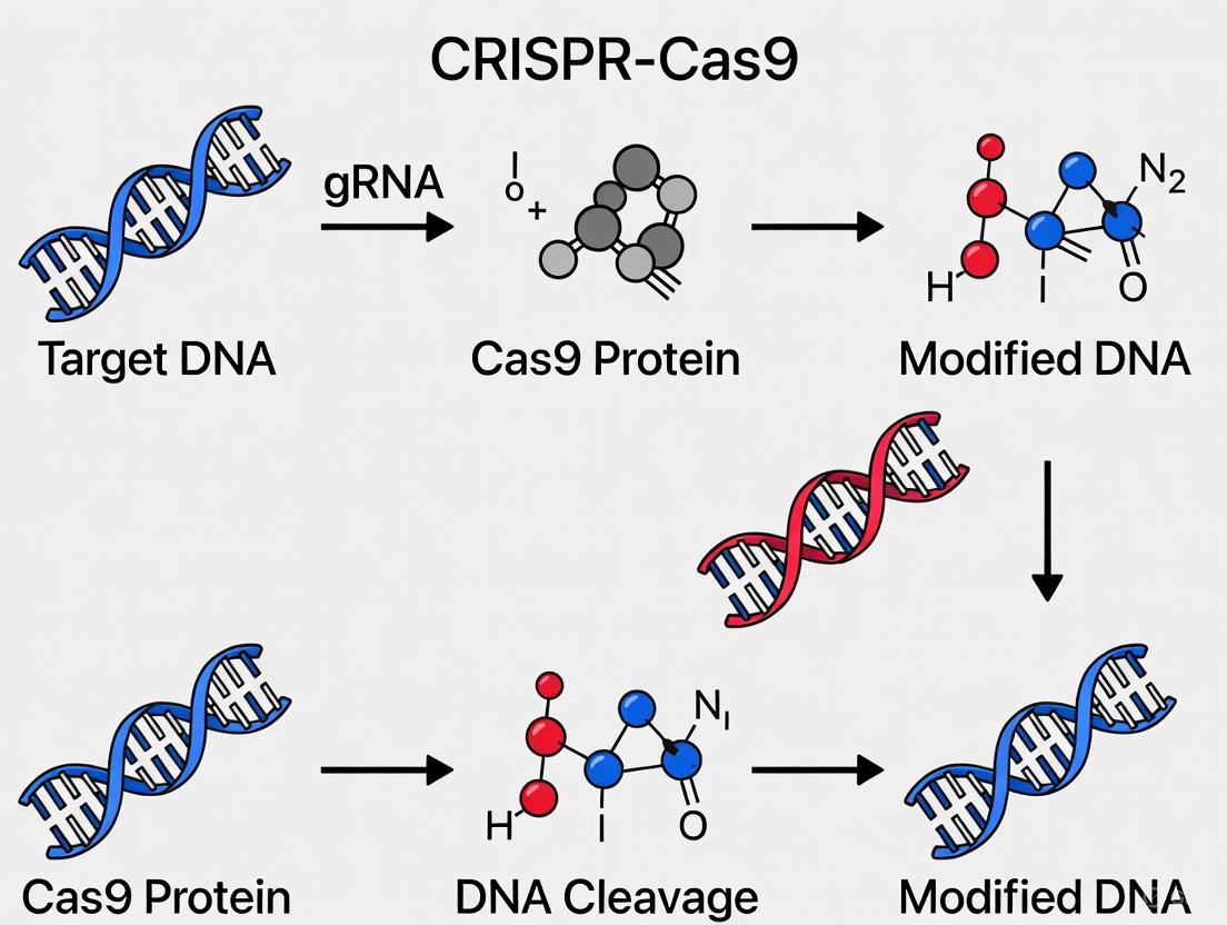
The CRISPR-Cas9 system represents a revolutionary genome-editing technology derived from an adaptive immune mechanism in bacteria and archaea [6] [7]. At its core, this powerful tool consists of two fundamental molecular components: the Cas9 enzyme, which acts as a programmable DNA-cutting endonuclease, and a guide RNA (gRNA), which provides targeting specificity to direct Cas9 to precise genomic locations [4] [8]. The elegant simplicity of this two-component system—where protein function is directed by RNA-based programming—has democratized genetic engineering, enabling researchers to manipulate genes with unprecedented precision and efficiency across diverse biological systems [6] [7].
This technical guide examines the structural and functional characteristics of both Cas9 and gRNA, explores their mechanistic interplay in genome editing, details experimental methodologies for their implementation, and highlights recent advances in CRISPR technology relevant to therapeutic development. Understanding these core components at a deep level is essential for researchers aiming to harness CRISPR-Cas9 for advanced applications in basic research and drug development.
The Cas9 Enzyme: Structure and Function
Architectural Organization and Functional Domains
The Cas9 nuclease exhibits a bilobed architecture composed of a recognition lobe (REC) and a nuclease lobe (NUC), which together facilitate RNA-guided DNA targeting and cleavage [9]. The REC lobe, primarily responsible for guide RNA binding and recognition, contains several key domains including the bridge helix and REC1, REC2, and REC3 domains that stabilize the gRNA-Cas9 complex and facilitate binding between the guide RNA and target DNA [9]. The NUC lobe houses the catalytic centers for DNA cleavage, containing the HNH and RuvC nuclease domains, along with the PAM-interacting (PI) domain that serves as an initial checkpoint for target recognition [9].
Table 1: Primary Functional Domains of the Cas9 Enzyme
| Domain/Lobe | Structural Features | Functional Role |
|---|---|---|
| REC Lobe | Alpha-helical structure containing REC1, REC2, REC3, and bridge helix domains | Facilitates gRNA binding and recognition; stabilizes gRNA-Cas9 complex; enables target DNA binding |
| NUC Lobe | Contains HNH and RuvC nuclease domains and PAM-interacting domain | Catalyzes DNA cleavage; recognizes PAM sequence |
| HNH Domain | ββα-metal fold structure | Cleaves the DNA strand complementary to the gRNA (target strand) |
| RuvC Domain | RNase H-like fold structure | Cleaves the non-complementary DNA strand (non-target strand) |
| PAM-Interacting Domain | Positively charged binding channel | Recognizes protospacer adjacent motif (PAM); initiates DNA binding |
The Cas9 enzyme requires the presence of a specific protospacer adjacent motif (PAM) sequence adjacent to its target site—a short, guanine-rich sequence (5'-NGG-3' for SpCas9) that serves as a binding signal and prevents the enzyme from targeting the bacterium's own CRISPR array [4] [7]. This structural organization enables Cas9 to perform its function as a programmable DNA endonuclease, with the REC and NUC lobes cooperating to ensure specific targeting and efficient cleavage of DNA sequences.
Cas9 Variants and Engineering Advances
The native Cas9 enzyme from Streptococcus pyogenes (SpCas9) has been extensively engineered to overcome limitations such as off-target effects, PAM restrictions, and delivery constraints. These engineered variants significantly expand the therapeutic potential of CRISPR technology. Key advances include:
High-Fidelity Cas9 Variants: Engineered versions such as SpCas9-HF1, eSpCas9(1.1), and HypaCas9 incorporate mutations in the REC or NUC lobes that reduce tolerance for mismatches between the gRNA and target DNA, substantially minimizing off-target editing while maintaining robust on-target activity [9].
Catalytically Inactivated Cas9 (dCas9): Created through point mutations in both HNH and RuvC nuclease domains, dCas9 retains DNA binding capability but lacks cleavage activity [10] [9]. This variant serves as a programmable DNA-binding platform for CRISPR interference (CRISPRi), epigenetic modification, and transcriptional regulation when fused to effector domains [10].
Cas9 Nickases (nCas9): These variants contain a mutation in either the HNH or RuvC domain, enabling single-strand DNA breaks rather than double-strand breaks [10]. When used with paired gRNAs targeting opposite strands, nCas9 creates staggered double-strand breaks with enhanced specificity and reduced off-target effects [9].
Compact Cas9 Orthologs: Recently characterized smaller Cas9 variants, such as the Type II-D Cas9 from a Nitrospirae bacterium (NsCas9d) comprising only 762 amino acids, offer advantages for viral vector delivery, particularly in therapeutic contexts where packaging constraints limit payload size [11]. This compact enzyme recognizes a 5'-NRG-3' PAM and generates 3-nt 5' overhangs that facilitate predictable DNA repair processes [11].
AI-Designed Cas9 Proteins: Breakthroughs in machine learning and protein language models have enabled the computational design of novel Cas9-like effectors with optimal properties. The OpenCRISPR-1 protein, designed using models trained on 1 million CRISPR operons, exhibits comparable or improved activity and specificity relative to SpCas9 while being 400 mutations away in sequence space [12].
Guide RNA (gRNA): Design and Function
Molecular Composition and Targeting Mechanism
The guide RNA serves as the programmable targeting component of the CRISPR-Cas9 system, dictating specificity through complementary base pairing with target DNA sequences. In native bacterial systems, the guide RNA exists as a dual-RNA structure consisting of CRISPR RNA (crRNA) and trans-activating crRNA (tracrRNA) [8]. The crRNA contains a customizable 17-20 nucleotide sequence complementary to the target DNA, while the tracrRNA forms a scaffold that stabilizes the crRNA and facilitates Cas9 binding [8] [7].
For most research and therapeutic applications, these two components are combined into a single-guide RNA (sgRNA) molecule—a chimeric RNA transcript that simplifies experimental design and implementation [8]. The sgRNA maintains the critical functional regions of both native RNAs: the customizable spacer sequence that determines DNA targeting specificity, and the scaffold region that enables Cas9 binding and activation [8].
The targeting mechanism begins with the sgRNA directing Cas9 to genomic locations complementary to its spacer sequence. Cas9 first identifies appropriate PAM sequences, then unwinds the adjacent DNA to allow hybridization between the target DNA and the sgRNA spacer region [7]. Perfect complementarity between the sgRNA spacer and target DNA, particularly in the "seed sequence" proximal to the PAM, triggers conformational changes in Cas9 that activate its nuclease domains [9].
Figure 1: gRNA Structure and DNA Targeting Mechanism
Strategic gRNA Design Considerations
Effective gRNA design is paramount for successful CRISPR experiments, directly influencing both on-target efficiency and off-target effects. Multiple factors must be considered during the design process:
PAM Availability and Positioning: The required PAM sequence must be present adjacent to the target site, with Cas9 typically cleaving 3-4 nucleotides upstream of the PAM [8] [7]. Different Cas orthologs and variants recognize distinct PAM sequences, expanding the targetable genomic space [8].
Sequence Specificity and Off-Target Potential: The sgRNA sequence should be unique within the genome to minimize off-target effects. Bioinformatics tools evaluate potential off-target sites with similar sequences, particularly those with mismatches in the distal region from the PAM [8] [9].
GC Content and Thermodynamic Properties: Optimal GC content (typically 40-60%) promotes stable sgRNA-DNA binding without excessive stability that might reduce specificity [9]. Extreme GC content (>80% or <20%) can compromise editing efficiency [8].
Genomic Accessibility: The target site should reside in chromatin regions accessible to the Cas9-sgRNA complex, as epigenetic modifications and chromatin condensation can significantly reduce editing efficiency [9].
Table 2: gRNA Design Parameters and Optimization Strategies
| Design Parameter | Optimal Range | Impact on Editing | Optimization Strategy |
|---|---|---|---|
| Spacer Length | 17-23 nucleotides | Shorter spacans increase specificity but may reduce on-target efficiency; longer spacans have opposite effects | Adjust based on application: 20nt standard, 17-18nt for enhanced specificity |
| GC Content | 40-60% | Moderate GC content ensures stable binding without excessive rigidity that reduces specificity | Avoid extremes (<20% or >80%) |
| Seed Sequence | 8-12 bases proximal to PAM | Critical for recognition and cleavage; requires perfect complementarity | Ensure perfect match to target in seed region |
| Off-Target Score | Minimize potential off-targets | Predicts and reduces unintended editing at similar genomic sites | Use multiple bioinformatics tools (CHOPCHOP, Synthego) |
Experimental Protocols: From Component Preparation to Analysis
CRISPR-Cas9 Delivery Methods and Workflows
Successful genome editing requires efficient delivery of both Cas9 and gRNA into target cells. The choice of delivery format and method significantly impacts editing efficiency, specificity, and potential applications. Common delivery approaches include:
Ribonucleoprotein (RNP) Complexes: Pre-assembled complexes of purified Cas9 protein and synthetic sgRNA offer rapid action, reduced off-target effects (due to transient activity), and no risk of genomic integration [9]. RNP delivery is particularly suitable for therapeutic applications where precise temporal control is essential [9].
mRNA/sgRNA Co-delivery: In vitro transcribed or synthetic Cas9 mRNA and sgRNA provide transient expression with reduced immune responses compared to plasmid DNA [9]. This approach enables efficient editing in sensitive cell types while minimizing persistent Cas9 expression.
Plasmid DNA Vectors: DNA plasmids encoding both Cas9 and sgRNA sequences allow for sustained expression but increase the risk of off-target effects and potential genomic integration [8] [9]. Plasmid-based approaches benefit from simpler preparation but may trigger stronger immune responses.
Viral Vectors: Adenoviral (AV) and adeno-associated viral (AAV) vectors enable efficient in vivo delivery but face limitations including constrained packaging capacity (particularly for SpCas9) and potential immunogenicity [6]. Lentiviral vectors allow stable integration but raise safety concerns for therapeutic applications [9].
Figure 2: CRISPR-Cas9 Experimental Workflow
The Scientist's Toolkit: Essential Research Reagents
Table 3: Key Research Reagents for CRISPR-Cas9 Experiments
| Reagent Category | Specific Examples | Function & Application |
|---|---|---|
| Cas9 Expression Systems | SpCas9 plasmid, Hi-Fi Cas9 mRNA, recombinant Cas9 protein | Provides the nuclease component in various formats suitable for different delivery methods and specificity requirements |
| gRNA Synthesis Platforms | Synthetic sgRNA, IVT sgRNA, plasmid-encoded sgRNA | Generates the targeting component with varying quality, cost, and preparation time considerations |
| Delivery Reagents | Lipid nanoparticles, electroporation systems, viral packaging systems | Enables intracellular delivery of CRISPR components through chemical, physical, or biological methods |
| Design Bioinformatics | CHOPCHOP, Synthego Design Tool, Cas-OFFinder | Facilitates gRNA design with off-target prediction and efficiency scoring algorithms |
| Editing Detection Tools | T7E1 assay, TIDE analysis, NGS validation kits | Confirms on-target editing and identifies potential off-target effects through molecular analysis |
| Cell Culture Components | Appropriate cell lines, growth media, selection antibiotics | Provides the biological context for editing and subsequent expansion of modified cells |
The continued evolution of Cas9 enzymes and guide RNA designs is expanding the therapeutic potential of CRISPR technology. Recent developments in structural biology have revealed novel compact Cas9 variants with unique properties, while AI-driven protein design has generated entirely new editors with optimized characteristics [11] [12]. These advances, coupled with improved delivery strategies and enhanced specificity systems, are addressing key challenges in clinical translation.
For research and drug development professionals, understanding the intricate relationship between Cas9 and gRNA—from fundamental molecular mechanisms to practical experimental considerations—provides the foundation for innovative applications. As CRISPR technology progresses toward broader therapeutic implementation, this core knowledge enables researchers to select appropriate editing platforms, design effective targeting strategies, and interpret experimental outcomes within the complex landscape of genomic manipulation. The future of CRISPR-based therapeutics will undoubtedly build upon these fundamental components, leveraging their programmable nature to address increasingly sophisticated challenges in genetic medicine.
The Protospacer Adjacent Motif (PAM) is a short, specific DNA sequence that serves as the essential molecular address for CRISPR-Cas systems, enabling precise DNA targeting and cleavage. This technical guide explores the fundamental role of the PAM in facilitating self versus non-self discrimination in bacterial adaptive immunity and its critical function in modern genome engineering applications. We examine the structural mechanisms of PAM recognition, detail the varying PAM requirements across diverse Cas nucleases, and provide comprehensive experimental protocols for accounting for PAM constraints in CRISPR experiment design. Within the broader context of how CRISPR-Cas9 functions step-by-step, understanding PAM requirements is paramount for developing effective research strategies and therapeutic applications, from basic gene knockouts to advanced clinical trials.
The CRISPR-Cas system functions as an adaptive immune system in prokaryotes, protecting bacteria and archaea from foreign genetic material such as bacteriophages and plasmids [13] [14]. This system maintains a genetic memory of previous infections through CRISPR arrays - short stretches of DNA composed of alternating conserved repeats and target-specific spacers derived from foreign genetic elements [14]. When transcribed and processed into CRISPR RNAs (crRNAs), these sequences guide Cas effector proteins to recognize and cleave complementary invading DNA sequences [14].
The PAM serves as the critical first step in target recognition, typically appearing as a short DNA sequence (usually 2-6 base pairs) immediately adjacent to the target DNA region (protospacer) [13] [14]. This motif functions as a fundamental recognition signal that enables the CRISPR system to distinguish between self and non-self DNA [13] [14]. Without the presence of the correct PAM sequence, Cas effector proteins cannot effectively bind to or cleave target DNA, regardless of the degree of complementarity with the guide RNA [13].
The structural basis of PAM recognition involves direct protein-DNA interactions between the Cas nuclease and the PAM sequence [14]. These interactions destabilize the adjacent DNA duplex, facilitating interrogation of the downstream sequence by the crRNA and enabling RNA-DNA pairing when a matching target is present [15]. This mechanism ensures that only DNA sequences flanked by the appropriate PAM are recognized as legitimate targets, thereby preventing autoimmune reactions against the bacterium's own CRISPR arrays, which lack PAM sequences [13].
The Molecular Mechanism of PAM Recognition
Structural Basis of PAM-Dependent Target Recognition
The molecular mechanism of PAM recognition involves precise protein-DNA interactions that initiate the process of target DNA identification. Structural studies have revealed that Cas effector proteins contain specific PAM-interaction domains that directly contact the DNA major groove to read the PAM sequence [14]. For the commonly used Streptococcus pyogenes Cas9 (SpCas9), this recognition occurs through a arginine-rich motif within the C-terminal domain of the protein that makes specific contacts with the minor groove of the PAM duplex [14].
Upon encountering potential target DNA, the Cas nuclease first scans the DNA for the presence of its cognate PAM sequence through three-dimensional diffusion [14]. When the correct PAM is identified, the protein undergoes a conformational change that promotes local DNA melting, enabling the formation of an R-loop structure where the target strand displaces from its complement and pairs with the crRNA [14]. This process effectively positions the DNA scissile bonds within the Cas nuclease catalytic sites for cleavage [14].
The requirement for PAM recognition serves two critical biological functions. First, it provides a mechanism for self versus non-self discrimination, ensuring that the Cas nuclease does not target the bacterial genome where the spacer sequences are stored in CRISPR arrays without adjacent PAM sequences [13] [14]. Second, it increases the specificity and efficiency of target location by providing an initial anchor point that dramatically reduces the search space for potential targets within the vast genomic landscape [14].
PAM Locations Across Different CRISPR Systems
The location of the PAM relative to the target sequence varies significantly between different types of CRISPR-Cas systems, which has important implications for guide RNA design and targeting capabilities:
Figure 1: PAM locations vary by CRISPR system type. Type I and V systems typically have 5' PAMs, while Type II systems have 3' PAMs. This orientation affects guide RNA design and targeting strategies.
PAM Sequences Across Cas Nuclease Variants
Natural PAM Diversity
Different Cas nucleases isolated from various bacterial species recognize distinct PAM sequences, providing researchers with a diverse toolkit for genome engineering applications. The PAM requirement represents one of the primary differentiators between Cas protein variants and significantly influences targeting range and specificity [13] [14].
Table 1: PAM Sequences for Various CRISPR Nucleases
| CRISPR Nuclease | Organism Isolated From | PAM Sequence (5' to 3') | Targeting Considerations |
|---|---|---|---|
| SpCas9 | Streptococcus pyogenes | NGG | Most commonly used nuclease; broad targeting capability [13] |
| SaCas9 | Staphylococcus aureus | NNGRR(T) or NNGRR(N) | Smaller size beneficial for viral packaging [13] |
| NmeCas9 | Neisseria meningitidis | NNNNGATT | Longer PAM increases specificity but reduces targeting range [13] |
| CjCas9 | Campylobacter jejuni | NNNNRYAC | Intermediate PAM length balances specificity and targeting [13] |
| Cas12a (Cpf1) | Lachnospiraceae bacterium | TTTV | T-rich PAM; creates staggered cuts [13] |
| hfCas12Max | Engineered from Cas12i | TN and/or TNN | Engineered variant with relaxed PAM requirements [13] |
| Cas12b | Alicyclobacillus acidiphilus | TTN | Thermostable variant useful for specific applications [13] |
| Cas3 | In silico analysis of various prokaryotic genomes | No PAM requirement | Unique helicase-nuclease activity [13] |
This natural diversity of PAM specificities enables researchers to select the most appropriate nuclease for their specific experimental needs, particularly when targeting genomic regions that may lack common PAM sequences like the canonical NGG motif recognized by SpCas9 [13].
Engineered Cas Variants with Altered PAM Specificities
Protein engineering approaches have significantly expanded the PAM recognition capabilities beyond naturally occurring Cas variants. Directed evolution and structure-guided engineering have produced Cas9 variants with altered PAM specificities, substantially increasing the targetable genomic space [13] [14].
Notable engineered variants include:
- xCas9: Recognizes a broad range of PAM sequences including NG, GAA, and GAT [14]
- SpCas9-NG: Engineered to recognize NG PAMs instead of the canonical NGG [14]
- SpRY: A nearly PAM-less Cas9 variant capable of recognizing NRN and to a lesser extent NYN PAMs [14]
These engineered variants demonstrate the flexibility of PAM recognition and provide researchers with tools to target previously inaccessible genomic loci. However, it's important to note that these engineered proteins often exhibit variable editing efficiencies across different target sites and may require additional optimization for specific applications [14].
PAM Considerations in Experimental Design
Guide RNA Design Relative to PAM
The design of guide RNAs is fundamentally constrained by the PAM requirement of the selected Cas nuclease. The targeting portion of the guide RNA must be complementary to the DNA sequence immediately adjacent to the PAM [13] [16]. For most applications, the PAM sequence itself is excluded from the guide RNA design to prevent self-targeting of the CRISPR constructs [13].
The optimal positioning of the cut site varies depending on the specific genetic manipulation being performed:
- Knockout experiments: Target constitutively expressed exons, preferably 5' exons, to increase the likelihood of generating frameshift mutations that completely disrupt gene function [16]
- HDR-mediated editing: Select cut sites as close as possible to the desired edit (ideally less than 10 bp away) to maximize recombination efficiency [16]
- Base editing: Position the target nucleotide within the specific editing window of the base editor, which is typically 3-10 nucleotides upstream of the PAM [16]
- Prime editing: Design pegRNAs with the edit located downstream (3') of the nick site [16]
- CRISPRi/a: Target promoter regions or transcription start sites for optimal transcriptional repression or activation [16]
When no suitable PAM is available near the desired target site, researchers can consider alternative strategies including selecting a different Cas nuclease with compatible PAM requirements, using engineered Cas variants with altered PAM specificities, or targeting the opposite DNA strand [13] [16].
Experimental Workflow for PAM-Centric CRISPR Experiments
A standardized experimental approach that accounts for PAM constraints ensures successful CRISPR genome engineering outcomes:
Figure 2: Comprehensive CRISPR experimental workflow highlighting critical steps where PAM considerations influence experimental design and execution decisions.
The Scientist's Toolkit: Essential Research Reagents
Table 2: Essential Research Reagents for CRISPR Experiments
| Reagent Category | Specific Examples | Function in CRISPR Experiments |
|---|---|---|
| Cas Nucleases | SpCas9, SaCas9, Cas12a, Base Editors | Effector proteins that cleave or modify target DNA [16] |
| Guide RNA Vectors | U6-driven gRNA expression plasmids | Express the target-specific guide RNA component [16] |
| Delivery Tools | Lipofectamine, Electroporation systems, Lentiviral particles | Introduce CRISPR components into cells [16] [17] |
| Validation Primers | Target-specific PCR primers, Sequencing primers | Amplify and sequence target loci to confirm edits [18] |
| Analysis Software | ICE, MAGeCK, CRISPRanalyzeR | Quantify editing efficiency and analyze screen data [19] [18] |
| Cell Culture Reagents | Selection antibiotics, Culture media | Maintain and select successfully transfected cells [16] |
Advanced Applications and Clinical Relevance
PAM Considerations in Therapeutic Development
The translation of CRISPR technology from basic research to clinical applications has highlighted the critical importance of PAM selection in therapeutic development. Recent clinical advances demonstrate how PAM requirements influence therapeutic strategy:
- Casgevy (exa-cel): The first FDA-approved CRISPR-based medicine for sickle cell disease and transfusion-dependent beta thalassemia utilizes ex vivo editing of hematopoietic stem cells, where PAM availability at the BCL11A enhancer was a key consideration in target selection [20]
- In vivo CRISPR therapies: Intellia Therapeutics' phase I trial for hereditary transthyretin amyloidosis (hATTR) uses LNP-delivered CRISPR-Cas9 targeting the TTR gene in the liver, requiring careful PAM selection for efficient editing [20]
- Personalized CRISPR treatments: The landmark case of an infant with CPS1 deficiency treated with bespoke in vivo CRISPR therapy demonstrated the need for rapid PAM analysis and guide RNA design for ultra-rare genetic conditions [20]
The choice of Cas nuclease and corresponding PAM requirements directly impacts the therapeutic targeting range, with efforts focused on developing engineered Cas variants with relaxed PAM specificities to increase the number of targetable disease-causing mutations [14] [20].
Emerging Technologies and Future Directions
Recent technological advances continue to expand our ability to manipulate and overcome PAM limitations:
- PAM-less Cas variants: Engineered Cas proteins with significantly reduced PAM requirements, such as SpRY, enable targeting of previously inaccessible genomic regions [14]
- Phage-delivered CRISPR: Bacteriophages engineered to deliver CRISPR components specifically to bacterial pathogens, leveraging natural PAM recognition for precision antimicrobial activity [20]
- Multiplexed editing systems: Technologies enabling simultaneous targeting of multiple genomic loci with different PAM requirements, expanding complex genome engineering capabilities [19]
- Single-cell CRISPR screening: Advanced screening methods that combine CRISPR perturbations with single-cell RNA sequencing, requiring careful PAM consideration in library design [19]
These emerging technologies demonstrate the ongoing evolution of CRISPR tools and the central role that PAM understanding plays in enabling new applications across basic research, biotechnology, and therapeutic development.
The PAM sequence serves as the essential molecular address that directs CRISPR-Cas systems to their precise DNA targets. Its fundamental role in self versus non-self discrimination, target recognition, and cleavage activation makes it a critical consideration in all CRISPR experimental designs. The diversity of natural PAM specificities across different Cas nucleases, combined with engineered variants with altered PAM recognition, provides researchers with an expanding toolkit for genome engineering applications. As CRISPR technology advances toward broader therapeutic implementation, understanding and innovating around PAM constraints will continue to drive progress in precision genome editing. The systematic integration of PAM considerations into experimental design, from basic research to clinical applications, ensures the continued responsible development and application of these powerful genome engineering technologies.
The CRISPR-Cas9 system represents a transformative technology in the field of genome engineering, derived from an adaptive immune mechanism in prokaryotes that protects against viral infections [4]. This system functions as a precise, programmable tool for making targeted modifications to DNA sequences in a wide range of organisms, with profound implications for therapeutic development, agricultural biotechnology, and basic research [21] [4]. The technology has rapidly become the preferred method for genome editing due to its simplicity, efficiency, and precision compared to previous technologies like zinc finger nucleases (ZFNs) and transcription activator-like effector nucleases (TALENs) [4] [22].
At its core, the CRISPR-Cas9 system consists of two fundamental components: the Cas9 nuclease, an enzyme that acts as a "molecular scissor" to cut DNA, and a guide RNA (gRNA), which directs Cas9 to a specific target sequence in the genome [21] [4]. The system operates through a coordinated process of DNA recognition, cleavage, and subsequent repair by cellular mechanisms, enabling researchers to disrupt, insert, or modify genes with unprecedented control [4] [22]. This technical guide provides an in-depth examination of the molecular mechanism of CRISPR-Cas9, with particular focus on the structural basis of DNA recognition, the cleavage process, and the cellular repair pathways that ultimately generate the desired genetic modifications.
Molecular Components of CRISPR-Cas9
Cas9 Nuclease Structure and Function
The Cas9 protein is a multi-domain DNA endonuclease that serves as the catalytic engine of the CRISPR-Cas9 system. The most commonly used variant, derived from Streptococcus pyogenes (SpCas9), consists of 1368 amino acids and contains several functionally distinct domains [4]. Structurally, Cas9 is organized into two primary lobes: the recognition (REC) lobe and the nuclease (NUC) lobe [4].
The REC lobe, comprised of REC1 and REC2 domains, is primarily responsible for binding to the guide RNA [4]. The NUC lobe contains three key domains: the RuvC domain, which cleaves the non-target DNA strand; the HNH domain, which cleaves the target DNA strand complementary to the guide RNA; and the PAM-interacting domain, which recognizes the protospacer adjacent motif essential for target recognition [4] [23]. Upon binding to the guide RNA, Cas9 undergoes a conformational change that shifts it into an active, DNA-binding configuration, priming the system for target recognition and cleavage [22].
Guide RNA Design and Function
The guide RNA is a synthetic RNA molecule that combines two naturally occurring RNA components: the CRISPR RNA (crRNA), which contains the ~20 nucleotide spacer sequence complementary to the target DNA, and the trans-activating crRNA (tracrRNA), which serves as a binding scaffold for the Cas9 protein [4] [17]. In engineered CRISPR systems, these are often combined into a single-guide RNA (sgRNA) for simplicity [4].
The specificity of CRISPR-Cas9 is determined by the guide RNA sequence, which can be designed to target virtually any genomic locus followed by an appropriate PAM sequence [22]. The 5' end of the sgRNA contains the target-specific sequence, while the 3' end forms a hairpin structure that interacts with Cas9 [22]. Proper sgRNA design is critical for experimental success, as it must be highly specific to the target site to minimize off-target effects while maintaining efficiency in guiding Cas9 to the intended genomic location [21] [17].
PAM Sequence Requirements
The protospacer adjacent motif (PAM) is a short, specific DNA sequence (typically 2-6 base pairs) adjacent to the target DNA region that is essential for Cas9 recognition and cleavage [13]. For SpCas9, the PAM sequence is 5'-NGG-3', where "N" can be any nucleotide base [4] [13]. The PAM is located directly downstream (3') of the DNA region targeted for cleavage [13].
The PAM serves a critical function in self versus non-self discrimination in bacterial immunity, ensuring that the Cas9 nuclease does not target the bacterial genome itself [13]. In genome engineering applications, the PAM requirement constrains the targetable sites within a genome, though this limitation can be partially addressed by using Cas9 orthologs from different bacterial species or engineered Cas9 variants with altered PAM specificities [22] [13].
Table 1: Common Cas Nucleases and Their PAM Sequences
| CRISPR Nuclease | Organism Source | PAM Sequence (5' to 3') |
|---|---|---|
| SpCas9 | Streptococcus pyogenes | NGG |
| SaCas9 | Staphylococcus aureus | NNGRRT or NNGRRN |
| NmeCas9 | Neisseria meningitidis | NNNNGATT |
| Cas12a (Cpf1) | Lachnospiraceae bacterium | TTTV |
| hfCas12Max | Engineered from Cas12i | TN and/or TNN |
| AacCas12b | Alicyclobacillus acidiphilus | TTN |
DNA Recognition Mechanism
Target Site Identification
The DNA recognition process begins when the Cas9-sgRNA complex scans the genome for potential target sequences [22]. This process involves two distinct recognition steps: first, identification of the correct PAM sequence, and second, verification of complementarity between the sgRNA spacer and the target DNA [13].
The PAM-interacting domain of Cas9 initially surveys DNA for the presence of the appropriate PAM sequence [4] [13]. When Cas9 identifies a potential PAM, it triggers local DNA melting, unwinding the double helix to allow the sgRNA to interrogate the adjacent sequence for complementarity [4] [22]. This two-step recognition mechanism ensures that Cas9 only cleaves DNA at sites containing both the correct PAM and sufficient complementarity to the sgRNA spacer sequence [13].
Conformational Activation
Upon PAM recognition, Cas9 undergoes significant conformational changes that activate its DNA-binding capability [23] [22]. Structural studies using cryo-electron microscopy have revealed that guide RNA binding induces a shift in Cas9 from an inactive to an active configuration, with surface-exposed, positively-charged grooves becoming available for DNA interaction [23] [22].
The HNH and RuvC nuclease domains remain in inactive conformations until successful target binding occurs [23]. Recent structural evidence has identified multiple conformational states of the HNH domain, with the active cleavage state positioning the catalytic residues immediately adjacent to the scissile phosphate bonds in the target DNA [23]. This conformational proofreading mechanism contributes to the specificity of CRISPR-Cas9 by ensuring that nuclease activity only occurs after correct target identification.
Complementarity Verification
Following PAM recognition and DNA unwinding, the seed sequence (8-10 nucleotides at the 3' end of the sgRNA targeting region) begins annealing to the target DNA [22]. If perfect complementarity exists in the seed region, annealing continues in a 3' to 5' direction along the entire spacer sequence [22].
The location and number of mismatches between the sgRNA and target DNA significantly impact cleavage efficiency [22]. Mismatches in the seed region near the PAM typically abolish cleavage, while mismatches in the distal 5' region are more tolerated [22]. This verification process ensures that Cas9 cleavage only occurs at sites with sufficient complementarity to the sgRNA, providing an important layer of specificity to prevent off-target effects.
Diagram 1: DNA Recognition Process. This flowchart illustrates the sequential steps in CRISPR-Cas9 target recognition, from initial PAM search to conformational activation.
DNA Cleavage Activation
Double-Strand Break Formation
Once the Cas9-sgRNA complex successfully binds to a target sequence with sufficient complementarity, the nuclease domains undergo final activation to generate a double-strand break (DSB) in the DNA [4] [23]. The cleavage event is catalyzed by two distinct nuclease domains: the HNH domain cleaves the DNA strand complementary to the sgRNA (target strand), while the RuvC domain cleaves the opposite, non-complementary strand (non-target strand) [4] [22].
These coordinated cleavage events occur approximately 3-4 nucleotides upstream of the PAM sequence and typically result in blunt-ended DNA breaks, though the precise cleavage pattern can vary slightly depending on the specific Cas nuclease used [4] [22]. Structural studies have revealed that in the active cleavage state, the HNH domain rotates approximately 170° from its position in the inactive complex, bringing its active site into proximity with the target DNA strand [23]. This dramatic conformational change positions the catalytic residues for in-line attack on the scissile phosphodiester bonds.
Structural Transitions in Cleavage Activation
Advanced structural biology techniques, including cryo-electron microscopy, have captured Cas9 in multiple conformational states during the cleavage process [23]. The HNH domain exists in at least three distinct states: an inactive state (State 1) where the active site is positioned more than 32 Å from the cleavage site; an intermediate state (State 2) with the active site approximately 19 Å from the cleavage site; and an active cleavage state (State 3) where the catalytic residues are properly positioned for DNA cleavage [23].
The transition between these states involves significant domain movements, including a helix-to-loop conformational change in the L2 linker region (residues 906-923) that enables proper positioning of the HNH domain [23]. In the active cleavage state, the HNH domain contacts the REC1 and PI domains primarily through segments of residues 861-864, 872-876, and 903-906, stabilizing the complex for efficient DNA cleavage [23].
Table 2: Key Cas9 Nuclease Domains and Their Functions
| Domain | Location in Cas9 | Primary Function | Catalytic Residues |
|---|---|---|---|
| HNH | Residues 775-906 | Cleaves target DNA strand complementary to sgRNA | H840 |
| RuvC | Residues 1-180, 810-900 | Cleaves non-target DNA strand | D10, E762, H983, D986 |
| REC Lobe | Residues 1-307, 480-713 | Binds guide RNA and facilitates target recognition | N/A |
| PAM-Interacting | Residues 1100-1368 | Recognizes PAM sequence and initiates DNA binding | R1333, R1335 |
Cleavage Kinetics and Efficiency
The efficiency of DNA cleavage by CRISPR-Cas9 depends on multiple factors, including sgRNA design, chromatin accessibility, and cellular context [21] [22]. Optimal sgRNAs typically have target sequences of 18-20 nucleotides with minimal potential for off-target binding [21] [17]. The GC content of the target sequence also influences cleavage efficiency, with moderate GC content (40-60%) generally providing optimal results [17].
Recent studies comparing different Cas enzymes have revealed that Cas9 produces more unintended large-scale repair events than Cas12a, an important consideration for therapeutic applications where precision is critical [24]. Engineered high-fidelity Cas9 variants (such as eSpCas9, SpCas9-HF1, and HypaCas9) incorporate mutations that reduce off-target effects while maintaining on-target activity, providing improved specificity for applications requiring high precision [22].
Diagram 2: DNA Cleavage Activation. This diagram illustrates the process from target recognition to double-strand break formation, highlighting key requirements and catalytic residues.
Cellular Repair Pathways
Non-Homologous End Joining (NHEJ)
Following the generation of a double-strand break by Cas9, cellular DNA repair mechanisms are activated to restore genomic integrity [4]. The predominant and most efficient repair pathway is non-homologous end joining (NHEJ), which is active throughout the cell cycle and functions by directly ligating the broken DNA ends without requiring a template [4] [22].
NHEJ is an error-prone process that frequently results in small insertions or deletions (indels) at the cleavage site [4] [22]. When these indels occur within the coding sequence of a gene, they can cause frameshift mutations that introduce premature stop codons, leading to gene knockout [22]. The efficiency of NHEJ makes it particularly useful for applications aimed at gene disruption, though the stochastic nature of the resulting mutations means that outcomes can vary between cells [22].
Homology-Directed Repair (HDR)
The homology-directed repair (HDR) pathway provides a more precise mechanism for DNA repair that utilizes a homologous DNA template to accurately restore the missing sequence [4] [22]. This pathway is primarily active in the late S and G2 phases of the cell cycle when sister chromatids are available as templates [4].
In CRISPR genome engineering applications, HDR can be harnessed to introduce specific genetic modifications by providing an exogenous donor DNA template containing the desired sequence flanked by homology arms complementary to the regions surrounding the cleavage site [4] [22]. While HDR enables precise gene editing, including gene corrections, insertions, and point mutations, its efficiency is typically lower than NHEJ and varies significantly based on cell type, cell cycle stage, and the design of the donor template [4].
Repair Pathway Regulation and Manipulation
The balance between NHEJ and HDR pathways is tightly regulated within cells and can be influenced experimentally to enhance desired editing outcomes [4]. Researchers have developed various strategies to promote HDR over NHEJ, including chemical inhibition of key NHEJ factors, cell cycle synchronization, and the use of modified Cas9 variants that favor HDR [4].
Recent advances include the identification of specific DNA repair factors that influence editing outcomes, such as mismatch repair proteins that drive specific base edit outcomes and the E3 ubiquitin ligase RFWD3 that mediates certain transversion mutations [24]. Understanding and manipulating these repair pathways is crucial for achieving predictable editing outcomes in both research and therapeutic contexts.
Table 3: Comparison of Cellular DNA Repair Pathways in CRISPR-Cas9 Editing
| Repair Pathway | Template Requirement | Efficiency | Fidelity | Primary Applications | Key Regulatory Factors |
|---|---|---|---|---|---|
| Non-Homologous End Joining (NHEJ) | None | High | Error-prone (indels) | Gene knockout, gene disruption | DNA-PK, Ku70/80, XRCC4, Ligase IV |
| Homology-Directed Repair (HDR) | Homologous DNA template | Low to moderate | High (precise) | Gene correction, precise insertion, point mutations | BRCA1, BRCA2, Rad51, RPA |
| Microhomology-Mediated End Joining (MMEJ) | Microhomology regions | Moderate | Error-prone (deletions) | Targeted deletions | PARP1, MRE11, CtIP |
Experimental Protocols and Methodologies
Guide RNA Design and Validation
The first critical step in any CRISPR experiment is the design and validation of target-specific guide RNAs [21] [17]. A standardized protocol for sgRNA design includes:
Target Selection: Identify the specific genomic region to be modified. For gene knockouts, target exons near the 5' end of the coding sequence to maximize the probability of generating frameshift mutations [17] [22].
PAM Identification: Locate available PAM sequences (5'-NGG-3' for SpCas9) adjacent to the target site [13]. If no suitable PAM is available, consider alternative Cas nucleases with different PAM requirements [22] [13].
Specificity Verification: Use computational tools (such as those available from CRISPR design platforms) to assess potential off-target sites across the genome [21] [22]. Select sgRNAs with minimal sequence similarity to other genomic regions, especially in the seed sequence near the PAM [22].
Synthesis and Cloning: Synthesize oligonucleotides corresponding to the selected target sequence and clone them into appropriate expression vectors [17]. Most CRISPR systems use U6 polymerase III promoters for sgRNA expression [22].
Validation: Verify sgRNA functionality before large-scale experiments using surrogate reporter systems or T7E1 mismatch assays on a small scale [17].
Delivery Methods for CRISPR Components
Effective delivery of CRISPR components into target cells is essential for successful genome editing [21] [17]. The optimal delivery method depends on the cell type and application:
Lipid Nanoparticles (LNPs): Particularly effective for in vivo delivery, with natural tropism for liver cells [20]. LNPs can encapsulate Cas9 mRNA or protein along with sgRNA and have been used in clinical trials for hereditary transthyretin amyloidosis (hATTR) and hereditary angioedema (HAE) [20].
Viral Vectors: Adenovirus (AV), adeno-associated virus (AAV), and lentivirus (LV) vectors provide efficient delivery, though with size limitations (especially for AAV) and potential immunogenicity concerns [17]. Viral vectors are suitable for both in vitro and in vivo applications [17].
Electroporation: Effective for ex vivo applications using primary cells and cell lines [17]. Creates temporary pores in cell membranes through electrical current, allowing entry of CRISPR components [17].
Microinjection: Used for precise delivery into zygotes or individual cells, commonly employed in the generation of genetically modified organisms [17].
Recent clinical advances have demonstrated the potential for redosing when using LNP delivery, as LNPs do not trigger the same immune responses as viral vectors [20].
Analysis and Validation of Editing Outcomes
Comprehensive analysis of CRISPR editing outcomes is essential to confirm desired modifications and detect potential off-target effects [21]:
Initial Validation: Confirm editing efficiency 48-72 hours post-transfection using T7E1 assay, tracking of indels by decomposition (TIDE), or next-generation sequencing [21] [17].
Clonal Isolation: For precise edits, isolate single-cell clones by limiting dilution or fluorescence-activated cell sorting (FACS) [17].
Genotypic Analysis: Perform genomic DNA extraction, PCR amplification of the target region, and sequencing to characterize modifications at the molecular level [17]. Sanger sequencing coupled with decomposition analysis or next-generation sequencing provides comprehensive assessment of editing outcomes [21].
Phenotypic Validation: Confirm functional consequences of genetic modifications through Western blotting, qRT-PCR, or functional assays specific to the target gene [17].
Off-Target Assessment: Evaluate potential off-target sites predicted by in silico tools using targeted sequencing or more comprehensive methods like GUIDE-seq or CIRCLE-seq [22].
Diagram 3: CRISPR Experimental Workflow. This diagram outlines the key phases and steps in a typical CRISPR-Cas9 experiment, from initial design to final validation.
Research Reagent Solutions
Successful implementation of CRISPR-Cas9 technology requires careful selection of appropriate reagents and tools. The following table outlines essential materials and their functions in CRISPR experiments:
Table 4: Essential Research Reagents for CRISPR-Cas9 Experiments
| Reagent Category | Specific Examples | Function | Considerations |
|---|---|---|---|
| Cas9 Expression Systems | SpCas9, SaCas9, HiFi Cas9 variants | Catalyzes DNA cleavage at target sites | Choose based on PAM requirements, size constraints, and fidelity needs |
| Guide RNA Vectors | U6-driven sgRNA plasmids, multiplex gRNA arrays | Directs Cas9 to specific genomic targets | Consider cloning strategy and potential for multiplexing |
| Delivery Tools | Lipid nanoparticles (LNPs), AAV vectors, Electroporation systems | Introduces CRISPR components into cells | Selection depends on cell type, efficiency requirements, and application (in vivo vs. in vitro) |
| Repair Templates | Single-stranded oligodeoxynucleotides (ssODNs), double-stranded DNA donors | Provides template for HDR-mediated precise editing | Design homology arms (typically 800-1000 bp for dsDNA, 30-60 nt for ssODNs) |
| Validation Assays | T7E1 kits, NGS platforms, Antibodies for phenotypic confirmation | Confirms editing efficiency and characterizes outcomes | Implement multiple validation methods for robust results |
| Cell Culture Resources | Appropriate media, selection antibiotics, cloning reagents | Supports growth and isolation of edited cells | Include controls for experimental variability |
Current Applications and Clinical Translation
Therapeutic Developments
CRISPR-Cas9 technology has rapidly advanced toward clinical applications, with several notable successes in recent years [20]. The first CRISPR-based therapeutic, Casgevy (exagamglogene autotemcel), received regulatory approval for the treatment of sickle cell disease and transfusion-dependent beta thalassemia [20]. This ex vivo therapy involves editing patient-derived hematopoietic stem cells to reactivate fetal hemoglobin production, providing a potentially curative approach for these inherited blood disorders [20].
In vivo CRISPR therapies have also demonstrated promising results in clinical trials. Intellia Therapeutics' phase I trial for hereditary transthyretin amyloidosis (hATTR) represents the first systemically administered CRISPR-Cas9 therapy, using lipid nanoparticles to deliver Cas9 mRNA and sgRNA targeting the TTR gene in liver cells [20]. Results showed sustained reduction of TTR protein levels by approximately 90%, with clinically meaningful improvements in neuropathy scores [20]. Similar approaches are being investigated for hereditary angioedema (HAE), with phase I/II trials demonstrating 86% reduction in kallikrein levels and significant reduction in inflammation attacks [20].
Technological Innovations
Recent advances in CRISPR technology have expanded its capabilities beyond standard gene editing [22] [24]. Base editing systems enable direct conversion of one nucleotide to another without creating double-strand breaks, offering greater precision and potentially improved safety profiles [24]. Prime editing represents a further refinement, allowing for all possible base-to-base conversions as well as small insertions and deletions without requiring donor templates or double-strand breaks [24].
Novel delivery approaches continue to enhance the applicability of CRISPR therapies. The development of lipid nanoparticles (LNPs) with improved tissue specificity and the engineering of viral vectors with enhanced tropism and capacity are addressing previous limitations in delivery efficiency [20] [25]. Additionally, the demonstration that LNP-delivered CRISPR therapies can be redosed represents a significant advancement for treating genetic disorders that may require multiple treatments [20].
Research and Diagnostic Applications
Beyond therapeutic applications, CRISPR-Cas9 has become an indispensable tool in basic research, enabling high-throughput genetic screening, disease modeling, and functional genomics [22]. The technology's programmability and scalability make it ideal for genome-wide loss-of-function screens to identify genes involved in specific biological processes or disease states [22].
CRISPR-based diagnostics have also emerged as powerful tools for detecting pathogens and genetic variants [24]. These systems typically leverage Cas enzymes (such as Cas12, Cas13, or Cas14) that exhibit collateral cleavage activity upon target recognition, enabling amplification of detection signals [24]. Recent developments include one-pot assays combining isothermal amplification with CRISPR detection for rapid, point-of-care diagnosis of infectious diseases like monkeypox and antibiotic-resistant bacteria [24].
The step-by-step mechanism of CRISPR-Cas9—from DNA recognition through cleavage to cellular repair—represents a remarkable convergence of bacterial immunity and programmable genome engineering [4]. The precise molecular interactions between the Cas9 nuclease, guide RNA, and target DNA enable researchers to make targeted modifications to virtually any genomic locus, provided the appropriate PAM sequence is present [23] [13]. The cellular repair pathways that process Cas9-induced breaks ultimately determine the editing outcomes, with NHEJ typically generating gene disruptions and HDR enabling precise modifications when a donor template is provided [4] [22].
As CRISPR technology continues to evolve, ongoing refinements to Cas nucleases, delivery methods, and repair pathway manipulation are expanding its applications and improving its precision [22] [24]. The successful translation of CRISPR-based therapies from bench to bedside represents a milestone in the field of genetic medicine, offering promising treatments for previously untreatable genetic disorders [20]. However, challenges remain in ensuring specific targeting, efficient delivery to relevant tissues, and achieving predictable editing outcomes across diverse cell types and organisms [4] [20].
The rapid progress in CRISPR technology, coupled with its extensive adoption across biological research and therapeutic development, ensures that it will remain at the forefront of genetic engineering for the foreseeable future [21] [22]. Continued investigation into the fundamental mechanisms of CRISPR systems will undoubtedly yield further innovations, enhancing both our understanding of biological systems and our ability to intervene therapeutically in human disease.
The CRISPR-Cas9 system has revolutionized biomedical research by providing an unprecedented tool for precise genome modification. This revolutionary gene-editing technology can be used to modify or correct precise regions of our DNA to treat serious diseases [26]. At its core, the CRISPR-Cas9 system consists of two key components: the Cas9 enzyme, which acts as "molecular scissors" to cut DNA, and a guide RNA (gRNA) that specifies the location at which Cas9 will cut [26]. However, the CRISPR-Cas9 machinery itself does not perform the genetic modification—it only creates the initial double-strand break (DSB). The actual genetic editing occurs through the cell's endogenous DNA damage repair (DDR) mechanisms, primarily Non-Homologous End Joining (NHEJ) and Homology-Directed Repair (HDR) [27] [28].
When DNA damage occurs, a series of DNA Damage Repair pathways are activated to sense and fix the disrupted sequences. These pathways are essential for maintaining genomic integrity across all organisms [27]. While multiple repair mechanisms exist—including Base Excision Repair (BER), Nucleotide Excision Repair (NER), and Mismatch Repair (MMR)—HDR and NHEJ represent the two key pathways responsible for repairing the double-strand breaks created by CRISPR-Cas9 [27]. Researchers strategically leverage these endogenous DNA repair pathways to generate genetically edited organisms, furthering the study of human disease and the development of new therapeutics [27] [28].
This technical guide explores the mechanistic basis of NHEJ and HDR, their distinct roles in CRISPR-based genome editing, and practical methodologies for harnessing these pathways to achieve specific genetic outcomes. Within the broader context of how CRISPR-Cas9 works step by step, understanding and controlling these cellular repair processes represents the crucial final stage that determines the success and precision of genome editing experiments.
Fundamental Mechanisms of NHEJ and HDR
Non-Homologous End Joining (NHEJ): The Rapid Response Pathway
Non-Homologous End Joining (NHEJ) is the cell's primary and most efficient mechanism for repairing double-strand breaks. This pathway operates by quickly rejoining broken DNA ends without requiring a homologous template [27]. The term "non-homologous" refers to the fact that the two broken ends of the DNA are indiscriminately rejoined (ligated) back together with minimal reference to DNA sequence [27]. While this makes NHEJ fast and active throughout all phases of the cell cycle, this speed comes at the cost of precision—NHEJ often leads to small insertions or deletions (INDELs) at the repair site [27].
The error-prone nature of NHEJ stems from its repair mechanism. A commonly observed phenomenon accompanying DSBs is the creation of very small single-stranded overhangs. These single nucleotide overhangs can create regions of "microhomology" that can help guide DNA repair machinery, sometimes allowing for the perfect repair of the DNA. Unfortunately, this does not occur a majority of the time [27]. Imprecise repair frequently results in the loss or gain of a small number of nucleotides (typically 1-10 base pairs), effectively knocking out the gene of interest due to INDEL formation resulting in loss of function, frameshift mutations, or creation of a premature stop codon [27]. These characteristics make NHEJ particularly useful for gene knockout studies where the goal is to disrupt gene function.
Homology-Directed Repair (HDR): The Precision Pathway
Homology-Directed Repair (HDR) represents a more precise DNA repair mechanism that utilizes homologous sequences as a template for accurate DSB repair. Unlike NHEJ, which identifies any two broken ends of DNA and "sticks" them back together, the HDR pathway proteins recognize homologous sequences of DNA (from a sister chromatid, a donor homology plasmid, single stranded ODN, etc.) near the region of the DSB and uses those homologous regions as a template for precise damage correction [27].
In CRISPR-Cas9 gene editing, researchers can leverage HDR by designing a donor template that includes the DNA sequence they want to insert, flanked by regions of homology that match the ends of the cut DNA [27]. This allows for precise edits, making HDR ideal for applications such as gene knockins, precise point mutations, or creating transgenic models with specific genetic modifications [27]. However, HDR has significantly lower efficiency compared to NHEJ, as it only occurs during certain phases of the cell cycle (S and G2), where homologous DNA is naturally available [27]. Another important consideration when designing a gene edit with HDR is to ensure the homology arms are as close to the DSB as possible to maximize efficiency [27].
Table 1: Key Characteristics of NHEJ and HDR Repair Pathways
| Characteristic | Non-Homologous End Joining (NHEJ) | Homology-Directed Repair (HDR) |
|---|---|---|
| Template Requirement | No template required | Requires homologous donor template |
| Cell Cycle Activity | Active throughout all phases | Restricted to S and G2 phases |
| Repair Precision | Error-prone (often creates INDELs) | High precision |
| Repair Speed | Fast response | Slower process |
| Primary Applications | Gene knockouts, gene disruption | Gene correction, precise insertions, knockins |
| Efficiency in Mammalian Cells | High (dominant pathway) | Low (typically <10% of repaired breaks) |
| Key Proteins Involved | Ku70/80, DNA-PKcs, XRCC4, DNA Ligase IV | BRCA1, BRCA2, RAD51, CtIP |
Visualizing the Core Mechanisms of NHEJ and HDR
The following diagram illustrates the fundamental cellular decision process between NHEJ and HDR pathways following a CRISPR-Cas9 induced double-strand break:
Strategic Implementation for Specific Edit Types
Harnessing NHEJ for Gene Knockouts
The error-prone nature of NHEJ makes it ideally suited for creating gene knockouts. When the goal is to disrupt gene function rather than create a precise edit, NHEJ's efficiency and tendency to create INDELs become advantageous rather than problematic. To generate knockouts using NHEJ, researchers need three essential components: Cas9 nuclease (delivered as protein or plasmid), single guide RNAs (sgRNA) complexed with Cas9, and PCR primers for validation via sequencing [27].
The process works by designing sgRNAs that target critical exonic regions of the gene of interest. When Cas9 creates a DSB at this site, NHEJ repairs the break but typically introduces small insertions or deletions. These INDELs can disrupt the reading frame of the gene, leading to premature stop codons and subsequent degradation of the transcript via nonsense-mediated decay (NMD) [29]. The high efficiency of NHEJ means that a significant proportion of treated cells will contain disruptive mutations, making it possible to generate knockout cell lines or organisms with high success rates.
NHEJ can also be used for more extensive deletions by employing multiple guide RNAs. By using two guide RNAs that target separate sites, researchers can delete entire segments of DNA between the cleavage sites—after cleavage, the two separate ends are joined together while the intervening sequence is removed [26]. This approach was successfully demonstrated in chicken primordial germ cells (PGCs), where researchers used paired gRNAs to delete an entire 4.2 kb provirus (EAV-HP) responsible for blue eggshell color [30].
Leveraging HDR for Precise Genetic Modifications
HDR is the pathway of choice when precise genetic modifications are required, such as introducing specific point mutations, inserting epitope tags, or creating conditional alleles. The key to successful HDR-based editing lies in the design and delivery of the donor template, which contains the desired modification flanked by homology arms that match the sequences surrounding the cut site [27].
Several donor template formats are available, each with specific advantages:
- Double-stranded DNA (dsDNA) plasmids: These typically have long homology arms (500-1000 bp) and are suitable for inserting larger constructs, such as fluorescent protein genes or selection cassettes.
- Single-stranded oligodeoxynucleotides (ssODNs): These short single-stranded DNA templates (typically 100-200 nt) are ideal for introducing single nucleotide changes or small tags and show higher HDR efficiency in some systems.
- Double-stranded DNA PCR fragments: These linear dsDNA fragments with shorter homology arms (50-100 bp) can be rapidly generated and work well for various knock-in applications.
The ORANGE (Open Resource for the Application of Neuronal Genome Editing) toolkit provides an excellent example of HDR implementation for endogenous protein tagging in neurons. This system uses a CRISPR/Cas9 knock-in template vector containing a U6-driven gRNA expression cassette, the donor sequence containing the fluorescent tag, and a Cas9 expression cassette driven by a universal β-actin promoter [31]. The donor sequence is generated by standard PCR with primers introducing a short linker and Cas9 target sequences flanking the donor, creating a flexible system that can be adapted to tag virtually any protein of interest [31].
Quantitative Comparison of Editing Outcomes
Understanding the expected efficiency and outcomes of NHEJ versus HDR editing is crucial for experimental planning. The following table summarizes quantitative data from multiple studies demonstrating the performance characteristics of each pathway:
Table 2: Quantitative Comparison of NHEJ and HDR Editing Outcomes
| Parameter | NHEJ-Mediated Editing | HDR-Mediated Editing | Experimental Context |
|---|---|---|---|
| Typical Efficiency | 29-69% indel formation [30] | Typically <10% of alleles [32] | Chicken PGC provirus deletion [30] |
| Optimal Cell Cycle Phase | All phases [27] | S and G2 phases [27] | Mammalian cells [27] |
| Template Design | Not applicable | 50-800 bp homology arms recommended [33] | General guideline [33] |
| Deletion Size Capability | Up to 4.2 kb demonstrated [30] | Limited by template design | Chicken PGCs [30] |
| Precision Rate | Low (high INDEL frequency) | High (when successful) | General observation [27] |
| Multiplexing Capability | High (multiple gRNAs) | Challenging | Chicken PGCs [30] |
Advanced Methodologies and Protocols
Experimental Workflow for Pathway-Specific Editing
The following diagram outlines a comprehensive experimental workflow for designing and executing a CRISPR-Cas9 gene editing experiment tailored to specifically harness either NHEJ or HDR pathways:
Enhancing HDR Efficiency: Methodological Approaches
The low efficiency of HDR relative to NHEJ represents a significant technical challenge in precision genome editing. Several well-established methodologies can be employed to enhance HDR rates:
1. Cell Cycle Synchronization: Since HDR is primarily active in the S and G2 phases of the cell cycle, synchronizing cells to these phases can significantly improve HDR efficiency. This can be achieved through chemical treatments such as nocodazole (G2/M arrest) or mimosine (G1/S arrest), followed by release into the cell cycle [27].
2. NHEJ Pathway Inhibition: Suppressing key proteins in the NHEJ pathway can push the cell to favor HDR. This can be accomplished using siRNA against NHEJ components (e.g., Ku70, Ku80, DNA ligase IV) or chemical inhibitors such as Scr7 (DNA ligase IV inhibitor) or NU7026 (DNA-PKcs inhibitor) [27].
3. Optimized Donor Template Design: The design of the donor template significantly impacts HDR efficiency. Key considerations include:
- Using single-stranded oligodeoxynucleotides (ssODNs) as templates, which improve HDR efficiency by offering a quick, efficient repair guide [27].
- Ensuring close proximity of the edit to the Cas9 cut site (within 10 bp or less).
- Incorporating modified bases to protect the donor template from degradation.
- Optimizing homology arm length (typically 30-50 nt for ssODNs, 500-1000 bp for plasmid donors).
4. Cas9 Variants and Delivery Optimization: The use of high-fidelity Cas9 variants can improve editing specificity, while RNP (ribonucleoprotein) complex delivery often yields higher HDR efficiency compared to plasmid-based delivery [30] [33]. Timing of donor template delivery relative to CRISPR components may also be optimized—some protocols recommend delivering the donor template 4-24 hours after CRISPR components.
Quantitative Evaluation of Editing Efficiency: qEva-CRISPR Method
Accurate quantification of editing outcomes is essential for evaluating experimental success. The qEva-CRISPR method represents a significant advancement in quantitative evaluation of CRISPR/Cas9-mediated modifications. This ligation-based dosage-sensitive method allows for parallel analysis of target and selected off-target sites, overcoming limitations of earlier detection methods [29].
The qEva-CRISPR protocol involves:
- Designing specific probes for both wild-type and edited sequences
- Multiplex ligation-dependent probe amplification to simultaneously quantify different alleles
- Capillary electrophoresis separation and fluorescence detection for precise quantification
This method detects all types of mutations, including point mutations and large deletions, with sensitivity independent of mutation type. Unlike earlier methods like T7 endonuclease I (T7E1) or Surveyor nuclease assays, qEva-CRISPR can successfully analyze targets located in 'difficult' genomic regions and can distinguish sequences generated by NHEJ versus HDR [29].
For researchers requiring absolute quantification of editing efficiencies, digital PCR (dPCR) provides an alternative methodology. As demonstrated in chicken PGC editing experiments, dPCR enabled absolute quantification of provirus deletion efficiencies, revealing 29% efficiency with wildtype Cas9 and 69% efficiency when a high-fidelity Cas9 variant was employed [30].
Table 3: Essential Research Reagents for NHEJ and HDR Genome Editing
| Reagent Category | Specific Examples | Function in Experiment | Considerations |
|---|---|---|---|
| CRISPR Nucleases | Wildtype SpCas9, High-fidelity Cas9 variants (e.g., SpCas9-HF1) | Induces targeted double-strand breaks | High-fidelity variants reduce off-target effects [30] |
| Delivery Systems | Electroporation, Lipofection, Viral vectors (lentivirus, AAV) | Introduces editing components into cells | RNP delivery reduces off-target effects; viral vectors allow stable expression [33] |
| Donor Templates | ssODNs, dsDNA plasmids with homology arms, PCR fragments | Provides repair template for HDR | ssODNs ideal for point mutations; plasmid donors for larger insertions [27] |
| Efficiency Enhancers | NHEJ inhibitors (Scr7, NU7026), Cell cycle synchronizing agents | Increases HDR: NHEJ ratio | Timing critical for cell cycle synchronization [27] |
| Validation Tools | T7E1 assay, Surveyor assay, TIDE analysis, Digital PCR, qEva-CRISPR | Quantifies editing efficiency and specificity | Digital PCR provides absolute quantification; qEva-CRISPR allows multiplex analysis [29] [30] |
| Specialized Systems | ORANGE toolkit, HITI (Homology-Independent Targeted Integration) | Enables editing in challenging systems (e.g., neurons) | HITI uses NHEJ for precise integration in postmitotic cells [31] |
Applications and Future Perspectives
Therapeutic Applications and Clinical Translation
The strategic application of NHEJ and HDR pathways has enabled remarkable advances in therapeutic genome editing. Clinical trials have demonstrated the potential of both approaches for treating genetic disorders:
NHEJ-Based Therapies: The first FDA-approved CRISPR-based therapy, Casgevy, utilizes NHEJ to disrupt the BCL11A gene in hematopoietic stem cells to treat sickle cell disease and transfusion-dependent beta thalassemia [20]. This approach effectively knocks out a gene whose suppression promotes fetal hemoglobin production, ameliorating disease symptoms.
HDR-Based Therapies: While more challenging therapeutically, HDR-based approaches are advancing toward clinical application. Recent breakthroughs include a personalized in vivo CRISPR treatment for an infant with CPS1 deficiency, developed and delivered in just six months [20]. This landmark case demonstrates the potential for rapid development of bespoke HDR-based therapies for rare genetic disorders.
In Vivo Editing Advances: Recent clinical trials have demonstrated the feasibility of in vivo genome editing using lipid nanoparticle (LNP) delivery. Intellia Therapeutics' phase I trial for hereditary transthyretin amyloidosis (hATTR) represents the first clinical trial for a CRISPR-Cas9 therapy delivered by LNP, achieving approximately 90% reduction in disease-related protein levels [20]. Notably, the LNP delivery system enables redosing—a significant advantage over viral vector delivery systems [20].
Emerging Technologies and Future Directions
The field of CRISPR-based genome editing continues to evolve rapidly, with several emerging technologies poised to enhance our ability to harness cellular repair pathways:
HDR Efficiency Optimization: Ongoing research focuses on developing more robust methods for enhancing HDR efficiency, including engineered Cas9 variants with altered kinetics, small molecule screening to identify novel HDR enhancers, and fusion proteins that recruit HDR factors to target sites [32].
Novel Delivery Strategies: Advances in delivery technology, particularly organ-specific LNPs and novel viral vectors, will expand the therapeutic potential of both NHEJ and HDR-based editing approaches [20].
Alternative Precise Editing Platforms: While HDR remains the gold standard for precision editing, newer technologies such as base editing and prime editing offer alternative pathways to precise genome modification without requiring double-strand breaks or donor templates, potentially bypassing some limitations of HDR [32].
Multiplexed Editing Applications: Tools like the ORANGE toolkit enable multiplexed labeling of endogenous proteins in neurons, allowing simultaneous investigation of multiple protein species within single cells [31]. Similar approaches could be expanded to other cell types and applications.
As these technologies mature, researchers and therapeutic developers will possess an increasingly sophisticated toolkit for harnessing cellular repair pathways to achieve desired genetic outcomes with greater precision and efficiency. The strategic selection between NHEJ and HDR pathways, coupled with appropriate methodological enhancements, will continue to drive advances in both basic research and clinical applications of CRISPR genome editing.
CRISPR in Action: Experimental Workflows and Translational Applications in Biomedicine
The CRISPR-Cas9 system has revolutionized biological research and therapeutic development by providing an unprecedented ability to precisely modify genomes. This bacterial adaptive immune system has been harnessed as a programmable genome engineering tool that allows researchers to edit DNA with exceptional precision and efficiency [21] [6]. The technology's core principle involves a two-component system: a Cas nuclease that cuts DNA and a guide RNA (gRNA) that directs the nuclease to a specific genomic location [21]. This review delineates the CRISPR-Cas9 workflow within the context of a broader thesis on its step-by-step functionality, presenting a systematic trilogy of design, edit, and analyze phases that form the foundation of effective genome engineering. Understanding this workflow is crucial for researchers and drug development professionals seeking to leverage this powerful technology for target validation, disease modeling, and therapeutic development [34] [35].
Phase One: Design - Strategic Planning for Precision
The design phase constitutes the critical foundational stage where strategic decisions determine the success of the entire CRISPR experiment. This stage requires meticulous planning of the targeting strategy and selection of appropriate molecular tools.
Guide RNA Design Principles
The guide RNA (gRNA) serves as the targeting component of the CRISPR system, dictating specificity and efficiency. The gRNA is typically a 20-nucleotide sequence complementary to the target DNA site [33]. Effective gRNA design must maximize on-target efficiency while minimizing potential off-target effects, which occur when the CRISPR system binds and cuts at unintended genomic locations with similar sequences [21]. The specificity of the guide RNA profoundly influences experimental success, as unintentional binding to random sites can have detrimental cellular effects [21]. Proprietary algorithms and design tools are available to assess potential off-target effects and predict on-target efficiency, helping researchers select optimal gRNA sequences [33]. Single guide RNA (sgRNA) formats that combine crRNA and tracrRNA into a single molecule have simplified design and delivery processes [33].
CRISPR Nuclease Selection
The selection of the appropriate Cas nuclease represents another crucial design decision. While the canonical Cas9 from Streptococcus pyogenes remains widely used, various alternative nucleases with distinct properties are now available [21]. The choice of nuclease must align with experimental requirements and target sequence constraints. Each nuclease has specific Protospacer Adjacent Motif (PAM) requirements—short DNA sequences adjacent to the target site that are essential for recognition and cleavage [36] [6]. For example, SpCas9 recognizes a 5'-NGG-3' PAM sequence [33]. Cas12a (formerly Cpf1) offers an alternative with different PAM requirements, potentially enabling targeting of genomic regions inaccessible to Cas9 [33]. Beyond standard nucleases, engineered variants like dead Cas9 (dCas9) serve as programmable DNA-binding platforms for precision transcriptional regulation without DNA cleavage [6].
Table 1: Overview of Common CRISPR Nucleases and Their Properties
| Nuclease | PAM Sequence | Key Features | Best Applications |
|---|---|---|---|
| SpCas9 | 5'-NGG-3' | High efficiency, well-characterized | General knockout and knock-in studies |
| Cas12a | 5'-TTTN-3' | Creates staggered ends, simpler gRNA | Multiplexed editing, AT-rich regions |
| dCas9 | 5'-NGG-3' | Catalytically inactive | Transcriptional regulation, epigenome editing |
| Base Editors | Varies by Cas domain | Direct nucleotide conversion without DSBs | Point mutation correction |
Figure 1: CRISPR Design Phase Core Components. The design phase requires simultaneous consideration of guide RNA (gRNA) design and nuclease selection, with particular attention to PAM requirements and off-target effect minimization.
Phase Two: Edit - Executing Genomic Modification
The editing phase encompasses the delivery of CRISPR components into cells and the subsequent cellular processes that accomplish genomic modification.
Delivery Methods for CRISPR Components
Effective delivery of CRISPR components into target cells is pivotal for successful genome editing. The chosen method must balance efficiency with cellular viability while considering the experimental context. Common approaches include:
- Ribonucleoprotein (RNP) Complexes: Preassembled complexes of Cas9 protein and gRNA provide immediate activity with rapid degradation, resulting in higher efficiency and reduced off-target effects due to transient presence [33].
- Electroporation: Application of electrical fields to facilitate cellular entry of CRISPR components, offering high transfection efficiency particularly valuable for difficult-to-transfect cell types like primary cells [33].
- Lipofection: Lipid-based nanoparticles that encapsulate and deliver CRISPR plasmids or RNA, providing a simple and efficient method suitable for various cell types [33].
- Viral Vectors: Engineered viruses, particularly lentivirus and adeno-associated virus (AAV), enable efficient delivery, though AAV has limited loading capacity that may constrain CRISPR component size [6].
The selection of an appropriate delivery method depends on factors including target cell type, desired editing permanence, and specific CRISPR components employed [33].
DNA Repair Pathways and Editing Outcomes
Once delivered to cells, the Cas nuclease creates double-strand breaks (DSBs) at target DNA sites [36]. These breaks activate endogenous cellular repair mechanisms that determine the editing outcome:
- Non-Homologous End Joining (NHEJ): The predominant repair pathway in mammalian cells, NHEJ directly ligates broken DNA ends without a template. This error-prone process often results in small insertions or deletions (indels) that can disrupt gene function, making it ideal for gene knockout experiments [36] [33].
- Homology-Directed Repair (HDR): A more precise mechanism that uses homologous DNA templates to repair breaks. When researchers provide an exogenous donor DNA template, HDR can facilitate specific nucleotide changes, insertions, or gene corrections, enabling precision genome editing [36] [33].
HDR occurs less frequently than NHEJ and is primarily active in dividing cells, presenting challenges for precision editing in non-dividing cells like neurons [31]. Strategies to enhance HDR efficiency include optimizing donor template design and synchronizing cell cycles [33].
Table 2: Comparison of DNA Repair Pathways in CRISPR Editing
| Parameter | Non-Homologous End Joining (NHEJ) | Homology-Directed Repair (HDR) |
|---|---|---|
| Repair Template | None required | Donor DNA template required |
| Efficiency | High | Low to moderate |
| Cell Cycle Preference | Active throughout cell cycle | Preferred in S/G2 phases |
| Editing Outcome | Random insertions/deletions (indels) | Precise nucleotide changes |
| Primary Application | Gene knockouts, gene disruption | Gene correction, precise insertions |
| Key Limitations | Error-prone, heterogeneous outcomes | Low efficiency, requires donor design |
Figure 2: CRISPR Edit Phase Workflow. The edit phase begins with delivery of CRISPR components, leading to double-strand breaks (DSBs) that are resolved through cellular repair pathways, primarily NHEJ or HDR.
Phase Three: Analyze - Validation and Assessment
The analysis phase constitutes the critical validation step where editing efficiency and specificity are quantified, confirming successful genomic modification.
Methodologies for Analyzing CRISPR Edits
Multiple methodologies exist for assessing CRISPR editing outcomes, each with distinct advantages, limitations, and appropriate applications:
- Next-Generation Sequencing (NGS): Considered the gold standard for CRISPR analysis, targeted NGS provides comprehensive assessment of editing outcomes with high sensitivity and accuracy [37]. This approach enables detailed characterization of indel spectra and frequencies but requires substantial resources, time, and bioinformatics expertise [37].
- Inference of CRISPR Edits (ICE): A user-friendly computational tool that uses Sanger sequencing data to determine editing efficiency and characterize indel patterns [37]. ICE provides NGS-comparable accuracy (R² = 0.96) at lower cost, making it accessible for laboratories without specialized sequencing infrastructure [37].
- Tracking of Indels by Decomposition (TIDE): Another Sanger sequencing-based analysis method that decomposes sequencing chromatograms to quantify editing efficiency [37]. While cost-effective, TIDE has limitations in detecting complex editing outcomes compared to ICE [37].
- T7 Endonuclease 1 (T7E1) Assay: A gel-based method that detects mismatches in heteroduplex DNA formed by annealing wild-type and edited sequences [37]. This approach provides rapid, low-cost assessment of editing but lacks quantitative accuracy and detailed sequence information [37].
Selection of an appropriate analysis method depends on experimental requirements, available resources, and the necessary level of resolution. For therapeutic applications where comprehensive characterization is essential, NGS remains preferable, while research applications may be adequately served by ICE or TIDE analyses [37].
Advanced Analysis for Complex Models
Recent technological advances address analytical challenges in complex biological contexts. CRISPR-StAR (Stochastic Activation by Recombination) introduces an internal control mechanism that overcomes limitations of conventional screening in heterogeneous models like organoids or in vivo tumors [38]. This method uses Cre-inducible sgRNA expression and single-cell barcoding to generate paired experimental and control populations within each clone, effectively controlling for intrinsic and extrinsic heterogeneity [38]. Such innovations enable higher-resolution genetic screening in physiologically relevant models, accelerating therapeutic target identification.
Table 3: Comparison of CRISPR Analysis Methods
| Method | Principle | Sensitivity | Cost | Time | Information Obtained |
|---|---|---|---|---|---|
| Next-Generation Sequencing | High-throughput DNA sequencing | Very High | High | Days to weeks | Complete sequence characterization, indel spectrum |
| ICE Analysis | Computational analysis of Sanger data | High | Medium | 1-2 days | Editing efficiency, major indel products |
| TIDE Analysis | Decomposition of Sanger chromatograms | Medium | Medium | 1-2 days | Editing efficiency, simple indels |
| T7E1 Assay | Mismatch cleavage detection | Low | Low | Hours | Presence of editing (non-quantitative) |
The Scientist's Toolkit: Essential Research Reagents
Successful execution of CRISPR experiments requires specific molecular tools and reagents. The following essential components constitute the core CRISPR toolkit:
- Guide RNA Expression Constructs: DNA templates for sgRNA transcription, typically driven by U6 promoters [31]. Modified gRNAs with Alt-R modifications can enhance stability and editing efficiency while reducing cellular toxicity [33].
- Cas Nuclease Expression Systems: Plasmid, mRNA, or protein formats for Cas nuclease delivery. Ribonucleoprotein (RNP) complexes offer immediate activity with reduced off-target effects [33].
- Donor DNA Templates: Single-stranded or double-stranded DNA molecules containing homologous sequences for HDR-mediated precise editing [33]. Optimal design includes homology arms flanking the desired modification [33].
- Delivery Reagents: Transfection reagents (lipids, polymers) or electroporation systems appropriate for the target cell type [33].
- Validation Primers: PCR primers flanking the target site for amplification of edited regions prior to analysis [37] [39].
- Positive Control gRNAs: Validated gRNAs targeting known essential genes or easily detectable loci for system validation [21].
Therapeutic Applications and Future Perspectives
The standardized CRISPR workflow has accelerated biomedical research and therapeutic development. In drug discovery, CRISPR screening enables genome-wide target identification and validation, particularly for complex diseases [34] [35]. Pooled CRISPR screens with comprehensive sgRNA libraries facilitate systematic investigation of gene-drug interactions across the genome [35]. Integration with organoid models and emerging technologies like artificial intelligence further enhances the scale and precision of target discovery [35].
Therapeutic applications continue to advance, with ongoing clinical trials demonstrating the potential of CRISPR-based approaches for treating genetic disorders, cancers, and infectious diseases [6]. Current research focuses on improving spatiotemporal control through chemical, genetic, and physical regulation to enhance specificity and safety [36]. Innovations in delivery systems, particularly non-viral vectors like lipid nanoparticles, address critical challenges in therapeutic implementation [6].
Despite remarkable progress, challenges remain in minimizing off-target effects, managing data complexity, and addressing ethical considerations [35]. Ongoing technological refinements and methodological advancements continue to expand CRISPR capabilities, promising to further transform biomedical research and therapeutic development.
sgRNA Design Principles for Optimal Targeting and Efficiency
The CRISPR-Cas9 system has revolutionized genome editing by providing researchers with a precise and programmable method for manipulating DNA sequences. This bacterial adaptive immune system has been repurposed as a powerful biotechnology tool that functions like a genetic scalpel, enabling targeted modifications in the genomes of virtually any organism. The system's operation hinges on the coordinated activity of two fundamental components: the Cas9 nuclease, which acts as the molecular scissors that cut DNA, and the single guide RNA (sgRNA), which serves as the GPS that directs these scissors to a specific genomic location [4].
The sgRNA is a synthetically engineered RNA molecule that combines two natural RNA elements: the CRISPR RNA (crRNA), which contains the ~20-nucleotide sequence complementary to the target DNA, and the trans-activating CRISPR RNA (tracrRNA), which serves as a binding scaffold for the Cas9 protein [8]. This fusion creates a single-molecule guide that simplifies experimental design and implementation. The critical importance of sgRNA design cannot be overstated, as the sequence of the sgRNA directly determines both the efficiency of editing at the intended target site (on-target efficiency) and the potential for unintended editing at similar sites elsewhere in the genome (off-target effects) [8] [40]. Consequently, optimal sgRNA design represents the most crucial step in planning successful CRISPR experiments, forming the foundation for all subsequent genome engineering applications in basic research and therapeutic development.
The Molecular Mechanism of CRISPR-Cas9 Genome Editing
Step-by-Step Breakdown of the CRISPR-Cas9 System
The CRISPR-Cas9 genome editing mechanism can be systematically divided into three sequential biological steps: recognition, cleavage, and repair [4]. Each step is essential for achieving precise genetic modifications and is governed by specific molecular interactions.
Step 1: Recognition and Complex Formation - The process initiates when the Cas9 nuclease, in complex with the sgRNA, scans the genome in search of a protospacer adjacent motif (PAM) sequence. For the most commonly used Cas9 from Streptococcus pyogenes (SpCas9), the PAM sequence is 5'-NGG-3', where "N" can be any nucleotide base [4] [41]. Once Cas9 identifies a PAM site, it partially unwinds the adjacent DNA duplex and enables the sgRNA to attempt pairing with the target DNA sequence through Watson-Crick base complementarity. If the sgRNA sequence demonstrates sufficient complementarity to the target DNA, a stable Cas9-sgRNA-DNA complex forms, positioning the nuclease domains for activation.
Step 2: DNA Cleavage - Following successful target recognition, the Cas9 nuclease undergoes a conformational change that activates its two distinct nuclease domains: the HNH domain cleaves the DNA strand complementary to the sgRNA (target strand), while the RuvC domain cleaves the non-complementary strand (non-target strand) [4]. This coordinated cleavage activity typically occurs 3 base pairs upstream of the PAM sequence and results in a precise double-strand break (DSB) in the DNA backbone, creating predominantly blunt-ended DNA fragments [4].
Step 3: DNA Repair and Editing Outcomes - The cellular DNA repair machinery detects the DSB and initiates one of two primary repair pathways. The first, non-homologous end joining (NHEJ), is an error-prone mechanism that directly ligates the broken DNA ends, often resulting in small insertions or deletions (indels) at the cleavage site [4]. When designed to disrupt a protein-coding sequence, these indels can produce frameshift mutations that effectively knockout gene function. The second pathway, homology-directed repair (HDR), operates with higher fidelity when a donor DNA template with homology to the target region is present. This pathway enables precise gene insertion or replacement, allowing researchers to introduce specific genetic modifications [4].
Figure 1: The CRISPR-Cas9 Genome Editing Mechanism. This diagram illustrates the three fundamental steps of CRISPR-Cas9-mediated genome editing: target recognition, DNA cleavage, and cellular repair pathways that lead to different editing outcomes.
PAM Sequence Requirements and Cas Protein Variants
The PAM sequence represents an absolute requirement for Cas9 activity and serves as a critical recognition element that prevents the nuclease from targeting the bacterial CRISPR locus itself. Different Cas nucleases isolated from various bacterial species recognize distinct PAM sequences, which directly influences their potential target range within a genome [8]. For example, while SpCas9 requires a 5'-NGG-3' PAM, Staphylococcus aureus Cas9 (SaCas9) recognizes 5'-NNGRR(N)-3', and the high-fidelity Cas12 variant hfCas12Max utilizes 5'-TN-3' and/or 5'-(T)TNN-3' PAM sequences [8]. This PAM diversity enables researchers to select Cas proteins that best suit their specific targeting needs, particularly when targeting genomic regions with limited PAM availability for SpCas9.
Fundamental Principles of sgRNA Design
Core Design Parameters for Optimal sgRNA Activity
Designing highly effective sgRNAs requires careful consideration of multiple sequence-based parameters that collectively influence on-target efficiency and specificity. The following principles represent the current consensus from large-scale empirical studies evaluating thousands of sgRNAs [8] [40] [41].
Target Sequence Length - For SpCas9, the optimal target sequence length is typically 20 nucleotides immediately upstream of the PAM sequence, though functional sgRNAs can range from 17-23 nucleotides [8]. Shorter sequences may reduce off-target effects but can compromise specificity if too short, while longer sequences maintain specificity but may exhibit reduced activity due to increased structural constraints.
GC Content - The GC content of the sgRNA significantly impacts its stability and hybridization energy to the target DNA. Optimal sgRNAs should possess GC content between 40-80%, with ideal performance typically observed in the 40-60% range [8] [41]. sgRNAs with excessively high GC content (>80%) may form stable secondary structures that interfere with Cas9 binding, while those with low GC content (<20%) may demonstrate reduced binding stability and editing efficiency.
Sequence Specificity and Uniqueness - The target sequence must be unique within the genome to minimize off-target effects. Bioinformatics tools should be employed to ensure the selected 20-nucleotide sequence (plus PAM) occurs only once in the target genome, with particular attention to genomic sites that differ by only a few nucleotides, as these represent potential off-target sites [41].
Positioning Within the Target Gene - For gene knockout applications, sgRNAs targeting exonic regions closer to the 5' end of the coding sequence (CDS) are generally preferred, as indels introduced early in the protein-coding sequence are more likely to produce frameshift mutations and premature stop codons that effectively disrupt gene function [41].
Advanced Design Considerations
Beyond these core parameters, several advanced considerations can further optimize sgRNA performance:
Nucleotide Composition - Empirical evidence indicates that sgRNAs with specific nucleotide preferences at particular positions demonstrate enhanced activity. For example, guanine nucleotides at position 20 (adjacent to the PAM) and cytosine nucleotides at position 19 are associated with higher editing efficiency, while thymine nucleotides at position 16 should generally be avoided when possible [41].
Secondary Structure - The sgRNA itself should be evaluated for potential internal secondary structures that might occlude the Cas9 binding site or the seed sequence (positions 1-12 adjacent to the PAM) that is critical for target recognition. Computational tools can predict and score potential hairpin formations that might impair sgRNA function.
Epigenetic Context - The chromatin accessibility of the target region can significantly influence editing efficiency. Targets in open chromatin regions (euchromatin) typically exhibit higher editing efficiency compared to those in closed chromatin regions (heterochromatin) due to differential Cas9 accessibility [42].
Quantitative Assessment of sgRNA Efficiency and Specificity
On-Target Efficiency Prediction Algorithms
Several sophisticated algorithms have been developed to predict sgRNA on-target efficiency based on large-scale empirical data. These scoring systems evaluate specific sequence features to generate efficiency scores that correlate with observed editing outcomes [41].
Table 1: Key sgRNA On-Target Efficiency Prediction Algorithms
| Algorithm Name | Development Basis | Key Features | Applications/Tools |
|---|---|---|---|
| Rule Set 1 | Data from 1,841 sgRNAs [41] | Position-specific nucleotide preferences | CHOPCHOP |
| Rule Set 2 | Data from 4,390 sgRNAs [40] [41] | Gradient-boosted regression trees | CHOPCHOP, CRISPOR |
| Rule Set 3 | Data from 47,000 sgRNAs across 7 datasets [41] | Incorporates tracrRNA sequence variations | GenScript, CRISPick |
| CRISPRscan | 1,280 gRNAs validated in zebrafish [41] | Nucleotide frequency and position weights | CHOPCHOP, CRISPOR |
| Lindel | ~1.16 million mutation events [41] | Predicts indel patterns and frameshift ratio | CRISPOR |
These algorithms have evolved substantially over time, with Rule Set 3 representing the current state-of-the-art by incorporating tracrRNA sequence variations that were overlooked in earlier versions [41]. The continued refinement of these predictive models highlights the importance of large-scale empirical data in understanding the complex relationship between sgRNA sequence and editing efficiency.
Off-Target Effect Prediction and Minimization
Minimizing off-target effects is equally crucial as maximizing on-target efficiency, particularly for therapeutic applications. Several computational approaches have been developed to assess and quantify off-target potential [41] [43].
Table 2: Primary sgRNA Off-Target Assessment Methods
| Method | Basis | Scoring Approach | Applications |
|---|---|---|---|
| Homology Analysis | Genome-wide sequence similarity | Counts sequences with ≤3 mismatches | Multiple tools |
| MIT (Hsu) Score | Indel data from 700+ gRNA variants [41] | Position-weighted mismatch scoring | Original MIT tool |
| Cutting Frequency Determination (CFD) | Activity data from 28,000 gRNAs [41] | Position-specific mismatch matrix | CRISPick, GenScript |
| CRISOT | Molecular dynamics simulations of RNA-DNA hybrids [43] | Machine learning on interaction fingerprints | CRISOT tool suite |
The CRISOT framework represents a significant advancement in off-target prediction by incorporating molecular dynamics simulations to characterize RNA-DNA interaction fingerprints at atomic resolution [43]. This approach has demonstrated superior performance compared to hypothesis-driven and traditional learning-based methods, highlighting the value of mechanistic understanding in predictive modeling.
Experimental Workflow for sgRNA Design and Validation
Comprehensive sgRNA Design Pipeline
Figure 2: sgRNA Design and Validation Workflow. This flowchart outlines the comprehensive process for designing and validating highly efficient and specific sgRNAs, incorporating both computational prediction and experimental assessment.
sgRNA Format and Synthesis Methods
Once designed, sgRNAs can be produced in several formats, each with distinct advantages and limitations [8]:
Plasmid-Expressed sgRNA - The sgRNA sequence is cloned into a plasmid vector and introduced into cells, where cellular RNA polymerase transcribes the sgRNA. While cost-effective for long-term experiments, this approach can lead to prolonged sgRNA expression, potentially increasing off-target effects, and requires 1-2 weeks for cloning before experiments can begin [8].
In Vitro-Transcribed (IVT) sgRNA - sgRNA is transcribed from a DNA template outside the cell using RNA polymerase (e.g., T7 RNA polymerase), then purified and delivered directly to cells. This method typically requires 1-3 days for synthesis and purification but can yield lower-quality sgRNA if not carefully optimized [8].
Synthetic sgRNA - sgRNA is chemically synthesized using solid-phase synthesis where individual ribonucleotides are sequentially added. This approach produces highly pure, reproducible sgRNA without the need for cloning or transcription, making it ideal for standardized experiments and therapeutic applications [8].
Validation Methodologies for Assessing Editing Efficiency
Comparative Analysis of Editing Efficiency Assessment Methods
Accurately measuring editing efficiency is crucial for evaluating sgRNA performance. Multiple methods are available, each with distinct strengths, limitations, and appropriate use cases [42].
Table 3: Methods for Assessing CRISPR-Cas9 Editing Efficiency
| Method | Principle | Key Advantages | Key Limitations | Best Applications |
|---|---|---|---|---|
| T7 Endonuclease I (T7EI) | Cleaves mismatched heteroduplex DNA | Simple, low-cost, quick results | Semi-quantitative, low sensitivity | Initial screening |
| TIDE/ICE | Decomposition of Sanger sequencing traces | Quantitative, provides indel spectrum | Accuracy depends on sequencing quality | Routine validation |
| Droplet Digital PCR (ddPCR) | Quantitative PCR with fluorescent probes | Highly precise, absolute quantification | Requires specific probe design | Therapeutic development |
| Next-Generation Sequencing (NGS) | High-throughput sequencing of target site | Most comprehensive, captures all edits | Higher cost, complex data analysis | Publication-quality data |
| Fluorescent Reporter Systems | Live-cell fluorescence upon editing | Enables enrichment of edited cells | Only applicable to engineered cells | Screening applications |
Recent comparative studies have demonstrated that methods like TIDE/ICE and ddPCR provide more accurate quantification of editing efficiency compared to traditional T7EI assays, with ddPCR offering particularly precise measurements for therapeutic applications where quantitative accuracy is paramount [42].
Off-Target Validation Techniques
While computational prediction helps identify potential off-target sites, experimental validation remains essential for comprehensive sgRNA characterization. Several methods are commonly employed:
Guideseq - A genome-wide method that captures double-strand breaks by incorporating sequencing adapters, providing unbiased identification of off-target sites without prior prediction [43].
Circleseq - An in vitro approach that uses circularized genomic DNA to detect cleavage events across the entire genome with high sensitivity [43].
Targeted Sequencing - Amplification and deep sequencing of computationally predicted off-target sites provides a cost-effective method for validating potential off-target effects at specific loci.
Advanced Applications and Future Directions
Specialized sgRNA Design Considerations
As CRISPR technology advances, specialized sgRNA design principles have emerged for specific applications:
CRISPRa/i (Activation/Interference) - For gene regulation applications, sgRNAs must target specific positions relative to the transcription start site (TSS), typically within 200 nucleotides upstream for activation and closer to the TSS for interference [44].
Base Editing - sgRNAs for base editing applications should position the target nucleotide within the editing window of the base editor (typically positions 4-8 for cytosine base editors and positions 4-7 for adenine base editors) while considering sequence context that may influence editing efficiency [45].
Prime Editing - Prime editing guide RNAs (pegRNAs) require both a target-specific sequence and a reverse transcription template containing the desired edit, necessitating specialized design tools that optimize both components simultaneously [42].
High-Throughput Screening with Optimized Libraries
Genome-wide CRISPR screens represent one of the most powerful applications of CRISPR technology, and specialized sgRNA libraries have been developed for this purpose. Recent research demonstrates that smaller, more optimized libraries can perform as well as or better than larger libraries when guides are selected using principled criteria [46]. For example, the Vienna library, which selects guides using VBC scores, achieved strong performance with only 3-6 guides per gene, reducing screening costs while maintaining sensitivity [46]. Dual-targeting strategies, where two sgRNAs target the same gene simultaneously, can further enhance knockout efficiency, though they may trigger a heightened DNA damage response in some contexts [46].
Table 4: Essential Research Reagents for CRISPR sgRNA Experiments
| Reagent/Resource | Function | Key Considerations |
|---|---|---|
| Cas9 Nuclease | Creates double-strand breaks at target DNA | Choose between wild-type, high-fidelity, or nickase variants |
| sgRNA | Guides Cas9 to specific genomic locations | Select optimal format: synthetic, IVT, or plasmid-expressed |
| Delivery Vector | Introduces CRISPR components into cells | Lentiviral, adenoviral, or plasmid-based systems |
| Validation Primers | Amplify target region for efficiency analysis | Design primers ~200-300bp flanking the target site |
| HDR Template | Provides repair template for precise editing | Single-stranded or double-stranded DNA with homology arms |
| Cell Line | Provides cellular context for editing | Consider transfection efficiency and repair pathway activity |
Optimal sgRNA design represents the cornerstone of successful CRISPR genome editing experiments. By carefully considering the fundamental principles of target selection, leveraging sophisticated prediction algorithms for both on-target efficiency and off-target effects, and implementing rigorous experimental validation, researchers can significantly enhance the success of their genome editing applications. The continued development of more accurate predictive models, particularly those incorporating molecular mechanisms like the CRISOT framework, promises to further improve sgRNA design capabilities. As CRISPR technology continues to evolve toward therapeutic applications, the principles outlined in this guide will remain essential for achieving precise, efficient, and specific genome modifications that advance both basic research and clinical development.
The Clustered Regularly Interspaced Short Palindromic Repeats and associated Cas9 protein (CRISPR-Cas9) system has emerged as the most effective, efficient, and accurate genome editing tool in living cells [4]. This technology, adapted from a natural bacterial immune system, functions like a precise genetic scissor, capable of removing, adding, or altering sections of DNA [7]. Its operation relies on two core components: a guide RNA (gRNA) that specifies the target DNA sequence, and the Cas9 nuclease that cuts the DNA at the designated location [4] [7].
The therapeutic potential of CRISPR-Cas9 is vast, with applications being investigated across medicine, agriculture, and biotechnology. In medicine, it offers promise for treating cancers, HIV, and genetic disorders such as sickle cell disease, cystic fibrosis, and Duchenne muscular dystrophy [4]. However, this potential cannot be realized without effective methods to deliver the CRISPR machinery into the nucleus of target cells. The genetic material (DNA or RNA) is highly vulnerable in its naked form and can be degraded by biological fluids or trigger unwanted immune responses [47]. Therefore, the development of safe and efficient delivery vectors—the microscopic "delivery trucks" that transport genetic cargo—is a fundamental challenge in the field [48] [49]. As of late 2025, the delivery landscape is rapidly evolving, marked by both breakthroughs in clinical applications and significant ongoing challenges [20].
Core CRISPR-Cas9 Mechanism: A Primer
Before delving into delivery methods, it is essential to understand the basic step-by-step mechanism of the CRISPR-Cas9 system.
Step 1: Recognition and Complex Formation. The process begins with the formation of a complex between the Cas9 enzyme and a synthetically designed single-guide RNA (sgRNA). This sgRNA is a combined molecule derived from the natural crRNA and tracrRNA components [4] [7]. The Cas9/sgRNA complex then scans the cell's DNA, searching for a specific short sequence known as the Protospacer Adjacent Motif (PAM). For the commonly used Cas9 from Streptococcus pyogenes, the PAM sequence is 5'-NGG-3' [7].
Step 2: DNA Cleavage. Once the complex locates a PAM site, the sgRNA unwinds the adjacent DNA and checks for complementarity with its own sequence. If a match is found, the Cas9 enzyme is activated and creates a double-stranded break (DSB) in the DNA, precisely 3 base pairs upstream of the PAM sequence [4]. This cut is achieved through two distinct nuclease domains within Cas9: the HNH domain cleaves the DNA strand complementary to the sgRNA, while the RuvC domain cleaves the non-complementary strand [4].
Step 3: DNA Repair and Genetic Outcome. The cell perceives the DSB as damage and activates one of two primary endogenous repair pathways to fix the break [4] [49]:
- Non-Homologous End Joining (NHEJ): This is an error-prone pathway that directly ligates the broken DNA ends. It often results in small random insertions or deletions (indels) at the cleavage site, which can disrupt or "knock out" a gene [4] [7]. This is useful for disabling dominant disease-causing genes.
- Homology-Directed Repair (HDR): This is a more precise pathway that requires a donor DNA template. The cell uses this template to repair the break, allowing for the insertion of a correct gene or specific genetic alteration. While HDR is ideal for gene correction, it is less efficient than NHEJ and primarily occurs in dividing cells [4] [7].
The following diagram illustrates this core mechanism.
Delivery System Technologies
The CRISPR-Cas9 components can be delivered in three primary formats: DNA (plasmid), mRNA (for Cas9 translation) with a separate gRNA, or as a pre-assembled Ribonucleoprotein (RNP) complex of Cas9 protein and gRNA [49]. The choice of delivery vector is critical and is broadly divided into two categories: viral and non-viral.
Viral Vectors
Viral vectors are engineered viruses that have been stripped of their disease-causing genes but retain their natural ability to efficiently enter cells and deliver genetic material [48] [47]. They are among the most common delivery vehicles in FDA-approved gene therapies and clinical trials [48].
The general mechanism for viral vector-based gene delivery is outlined below.
The most widely used viral vectors include:
Adeno-Associated Viruses (AAVs): AAVs are small, non-enveloped viruses with a single-stranded DNA genome. They are prized for their low immunogenicity and long-term transgene expression without integrating into the host genome [47]. However, their main limitation is a small packaging capacity (~4.7 kb), which is too small for the standard SpCas9 but can accommodate smaller Cas9 variants or base editors [49] [47]. AAVs are frequently used in in vivo therapies, as evidenced by recent preclinical successes in correcting vascular diseases [50].
Adenoviruses (AdVs): Adenoviruses have a larger double-stranded DNA genome and a much higher packaging capacity (up to 30 kb for "gutless" third-generation vectors) [47]. They can efficiently infect both dividing and non-dividing cells. Their major drawback is that they can trigger severe immune responses, which can limit re-dosing and pose safety concerns [47].
Lentiviruses (LVs): Lentiviruses are a subclass of retroviruses that can integrate their genetic material into the host genome, leading to stable, long-term expression [47]. This makes them ideal for ex vivo applications, such as engineering chimeric antigen receptor (CAR) T-cells for cancer therapy [51]. A key safety consideration is the risk of insertional mutagenesis, where integration disrupts an important host gene [47].
Non-Viral Vectors
Non-viral vectors rely on physical or chemical methods to deliver CRISPR components. They generally offer a better safety profile with lower immunogenicity and no risk of insertional mutagenesis [49] [47]. A major advancement in this category is the use of Lipid Nanoparticles (LNPs).
The delivery pathway for LNP-based CRISPR systems is distinct from viral methods.
LNPs are tiny, spherical vesicles composed of lipids that can encapsulate CRISPR components. They have risen to prominence due to their success in mRNA COVID-19 vaccines and have since been validated in CRISPR clinical trials [20]. A key operational advantage of LNPs is their suitability for redosing. Unlike viral vectors, which often trigger strong immune responses that prevent repeated administration, LNPs do not have this limitation. In 2025, Intellia Therapeutics reported the first-ever redosing of participants in an in vivo CRISPR trial for hATTR, and a personalized therapy for an infant with CPS1 deficiency successfully employed three separate LNP doses [20].
Other notable non-viral methods include:
Electroporation: This physical method uses an electric field to create temporary pores in the cell membrane, allowing CRISPR components (often as RNP) to enter directly into the cytoplasm. It is highly efficient for ex vivo applications (e.g., editing hematopoietic stem cells for sickle cell disease) but can cause significant cell death and stress [49].
Other Physical Methods: Microinjection delivers CRISPR components directly into a single cell (e.g., a zygote) using a fine needle. It is highly precise but low-throughput and requires skilled personnel [49].
Other Chemical Methods: These include polymer-based nanoparticles and cell-penetrating peptides (CPPs), which form complexes with CRISPR cargo to facilitate cellular uptake. They are generally easy to prepare and have low cytotoxicity, but often suffer from relatively low delivery efficiency compared to viral vectors or LNPs [49] [47].
Comparative Analysis: Viral vs. Non-Viral Delivery
The choice between viral and non-viral delivery systems involves a careful trade-off between efficiency, safety, cargo capacity, and clinical applicability. The following tables provide a structured, quantitative comparison of these technologies.
Table 1: Quantitative Comparison of Key Delivery Vector Properties
| Property | Viral Vectors (AAV, LV, AdV) | Non-Viral Vectors (LNP, Electroporation) |
|---|---|---|
| Cargo Capacity | AAV: ~4.7 kb [47]Lentivirus: ~8 kb [47]Adenovirus: up to 30 kb [47] | Effectively unlimited for ex vivo (e.g., Electroporation) [49]. Limited by LNP size for in vivo. |
| Immunogenicity | Moderate to High (Risk of immune reaction, especially with AdV) [47] | Low to Moderate (LNPs have lower immunogenicity than viruses) [49] |
| Integration into Genome | Lentivirus: Yes (risk of insertional mutagenesis) [47]AAV: Mostly non-integrating [47] | No (Eliminates risk of insertional mutagenesis) [47] |
| Manufacturing & Cost | Complex production, High cost [47] | Easier to prepare, Low cost [49] |
| Redosing Potential | Low (Pre-existing and triggered immunity block reuse) [20] | High (LNPs enable multiple doses, as demonstrated clinically) [20] |
| Typical Application | AAV/LV: In vivo gene therapy [20]LV: Ex vivo cell engineering (e.g., CAR-T) [51] | LNP: In vivo systemic delivery (e.g., liver targets) [20]Electroporation: Ex vivo cell editing [49] |
Table 2: Summary of Advantages and Disadvantages
| Vector Type | Advantages | Disadvantages and Clinical Challenges |
|---|---|---|
| Viral Vectors | • High delivery efficiency [47]• Sustained long-term gene expression [47]• Well-established clinical use [48] | • Limited cloning capacity (especially AAV) [47]• Significant immunogenicity [4] [47]• Risk of insertional mutagenesis (LV) [47]• Complex and costly manufacturing [47] |
| Non-Viral Vectors | • Favorable safety profile (low cytotoxicity, no genomic integration) [49] [47]• Large cargo capacity [47]• Redosing is feasible (LNPs) [20]• Easier to scale up and lower cost [49] | • Variable and often lower delivery efficiency than viruses [49] [47]• Vulnerability to extracellular and intracellular barriers [47]• Potential toxicity of some materials (e.g., inorganic nanoparticles) [7] |
Experimental Protocols and Clinical Workflows
Ex Vivo Cell Therapy Workflow (e.g., for Sickle Cell Disease)
This protocol is the basis for the first approved CRISPR therapy, Casgevy.
- Cell Harvest: Collect CD34+ hematopoietic stem and progenitor cells (HSPCs) from the patient's bone marrow or mobilized peripheral blood.
- Cell Activation: Activate the harvested cells in culture to make them more receptive to gene editing.
- CRISPR Delivery via Electroporation: Deliver the CRISPR-Cas9 components (typically as RNP complexes) into the activated cells using electroporation. The target is the BCL11A gene to reactivate fetal hemoglobin [4] [51].
- Quality Control and Expansion: Culture the edited cells and perform rigorous quality control checks, including checks for editing efficiency and viability.
- Patient Conditioning: Administer myeloablative conditioning (e.g., busulfan) to the patient to clear the bone marrow and make space for the new cells.
- Reinfusion: Transplant the CRISPR-edited HSPCs back into the patient via intravenous infusion.
In Vivo Systemic Therapy Workflow (e.g., for hATTR)
This protocol, used in Intellia Therapeutics' trials, demonstrates the use of LNPs for in vivo delivery.
- Formulation: Encapsulate CRISPR-Cas9 mRNA and the target-specific sgRNA (designed to knock out the TTR gene) into lipid nanoparticles (LNPs) [20].
- Administration: Administer a single dose of the LNP formulation to the patient via intravenous infusion [20].
- Delivery and Uptake: The LNPs circulate systemically and naturally accumulate in liver cells, the primary site of TTR protein production.
- Intracellular Processing: The LNPs fuse with the endosomal membrane within the hepatocytes, releasing their cargo into the cytoplasm. The mRNA is translated into functional Cas9 protein, which complexes with the sgRNA.
- Gene Editing: The RNP complex enters the nucleus, creates a DSB in the TTR gene, and the error-prone NHEJ repair knocks out the gene.
- Efficacy Monitoring: Monitor treatment efficacy through serial blood tests to measure the reduction in TTR protein levels, which shows a rapid and sustained decrease (e.g., ~90% reduction) [20].
The Scientist's Toolkit: Essential Research Reagents
Table 3: Key Research Reagent Solutions for CRISPR Delivery Studies
| Research Reagent | Function and Application in CRISPR Delivery |
|---|---|
| Cas9 Nuclease Variants (e.g., SpCas9, HiFi Cas9) | The core "scissor" enzyme. High-fidelity (HiFi) variants are used to minimize off-target effects [7]. |
| Guide RNA (sgRNA) | The "GPS" that directs Cas9 to the specific DNA target sequence. Must be designed for high specificity and efficiency [4]. |
| Lipid Nanoparticles (LNPs) | The primary non-viral delivery system for in vivo applications. Commercially available LNP kits can be used to encapsulate CRISPR RNPs or mRNA [20] [49]. |
| Viral Vectors (AAV, Lentivirus) | Used for stable and efficient gene delivery. AAV is standard for in vivo delivery of smaller editors, while Lentivirus is common for ex vivo cell engineering and in vitro studies [47]. |
| Electroporation Systems (e.g., Nucleofector) | Essential physical delivery equipment for hard-to-transfect primary cells (e.g., T-cells, HSPCs) in ex vivo workflows [49]. |
| Donor DNA Template | A single-stranded or double-stranded DNA oligonucleotide containing the desired corrective sequence for precise HDR-mediated gene correction or insertion [4] [7]. |
The development of delivery systems is as crucial as the refinement of the CRISPR-Cas9 editing machinery itself. Viral vectors, particularly AAVs and Lentiviruses, offer high efficiency and durable expression, making them powerful tools for both research and approved therapies. However, the emergence of non-viral methods, especially LNPs, represents a paradigm shift. LNPs address critical limitations of viral vectors, including immunogenicity, cargo constraints, and the inability to re-dose, thereby expanding the therapeutic horizon.
The future of CRISPR delivery lies in the continued optimization of both viral and non-viral platforms. Key directions include engineering novel capsids and LNPs with tropism for organs beyond the liver, developing smaller and more precise CRISPR effectors to fit within viral vectors, and creating sophisticated hybrid systems. As the field advances, the choice of delivery vector will remain a foundational decision, dictating the safety, efficacy, and ultimate clinical success of next-generation CRISPR-based medicines.
The Clustered Regularly Interspaced Short Palindromic Repeats (CRISPR) and their associated protein (Cas-9) system represents the most effective, efficient, and accurate genome editing technology in living cells [4]. Originally discovered as an adaptive immune system in prokaryotes to defend against viruses, CRISPR-Cas9 has been repurposed as a programmable genetic scissor, revolutionizing molecular biology and therapeutic development [4]. This adaptive immune system functions by incorporating short fragments of viral DNA (spacers) into a genomic region called the CRISPR array, creating a genetic memory of previous infections that protects bacteria from repeated viral attacks [4].
The CRISPR-Cas9 system has superseded earlier gene-editing technologies like Zinc Finger Nucleases (ZFN) and Transcription Activator-Like Effector Nucleases (TALENs) due to its superior efficiency, simpler design, and lower cost [4]. While ZFN and TALENs require complex protein engineering for each new DNA target, CRISPR-Cas9 can be redirected to new genomic loci simply by redesigning the guide RNA sequence [4]. The technology's core components include two essential elements: guide RNA (gRNA) and the CRISPR-associated (Cas-9) protein [4]. The most commonly used nuclease, Cas-9 protein from Streptococcus pyogenes (SpCas-9), is a large multi-domain DNA endonuclease that functions as a genetic scissor [4].
Molecular Mechanism of CRISPR-Cas9
The mechanism of CRISPR-Cas9 genome editing comprises three fundamental steps: recognition, cleavage, and repair [4].
Components and Recognition
The CRISPR-Cas9 system requires two key components: the Cas9 nuclease and a synthetic guide RNA (sgRNA) [4]. The Cas9 protein consists of two primary lobes: the recognition (REC) lobe, containing REC1 and REC2 domains responsible for binding guide RNA, and the nuclease (NUC) lobe, composed of RuvC, HNH, and Protospacer Adjacent Motif (PAM) interacting domains [4]. The sgRNA is a chimeric non-coding RNA created by fusing CRISPR RNA (crRNA) with trans-activating CRISPR RNA (tracrRNA) [4]. The crRNA component (18-20 base pairs in length) specifies the target DNA through complementary base pairing, while the tracrRNA serves as a binding scaffold for the Cas9 nuclease [4].
Target recognition begins when the Cas9-sgRNA complex scans DNA for specific short sequences known as Protospacer Adjacent Motifs (PAMs) [4]. For the standard SpCas9, the PAM sequence is 5'-NGG-3' (where N can be any nucleotide base) [4]. Once Cas9 identifies a PAM site, it triggers local DNA melting, enabling the sgRNA to form an RNA-DNA hybrid with the target DNA strand [4]. This PAM recognition is a critical prerequisite for DNA recognition and subsequent cleavage, and its restriction to 5'-NGG-3' imposes a major constraint on the breadth of Cas9-mediated genome editing [52].
DNA Cleavage and Repair Mechanisms
Following successful target recognition, the Cas9 nuclease induces a double-stranded break (DSB) in the DNA at a position 3 base pairs upstream of the PAM sequence [4]. Cleavage is executed by two distinct nuclease domains: the HNH domain cleaves the complementary strand, while the RuvC domain cleaves the non-complementary strand, resulting in predominantly blunt-ended DSBs [4].
After cleavage, the DSB is repaired by the cell's endogenous DNA repair machinery through one of two primary pathways [4]:
- Non-Homologous End Joining (NHEJ): This pathway facilitates the repair of DSBs by directly joining DNA fragments without a homologous template. NHEJ is active throughout the cell cycle and represents the predominant cellular repair mechanism. However, it is error-prone and often results in small random insertions or deletions (indels) at the cleavage site, which can generate frameshift mutations or premature stop codons [4].
- Homology-Directed Repair (HDR): This highly precise mechanism requires a homologous DNA template and is most active in the late S and G2 phases of the cell cycle. In CRISPR gene editing, HDR enables precise gene insertion or replacement by using an exogenous donor DNA template with sequence homology to the predicted DSB site [4].
Figure 1: CRISPR-Cas9 Mechanism: From DNA Recognition to Repair
Therapeutic Delivery Approaches: Ex Vivo vs. In Vivo Editing
Therapeutic genome editing strategies are fundamentally categorized into two distinct delivery approaches: ex vivo and in vivo editing. These approaches differ in their technical execution, target tissues, and clinical applications [53].
Ex Vivo Gene Therapy
Ex vivo gene therapy involves extracting cells from a patient, genetically modifying them in a controlled laboratory environment, and then reintroducing the edited cells back into the patient's body [53]. This approach is particularly advantageous for targeting accessible tissues such as blood and skin, and is the current standard for treating hematological disorders [53].
The laboratory-based modification process enables researchers to confirm that the newly introduced genetic material functions as intended before reinfusion [54]. Additionally, ex vivo editing allows for precise quality control measures, including genomic sequencing to verify the desired genetic change and screen for potential off-target effects [53]. The edited cells – typically stem cells in the case of blood disorders – are then expanded in culture and transplanted back into the patient, where they begin replacing disease-causing cells [53].
In Vivo Gene Therapy
In vivo gene therapy involves directly introducing therapeutic genetic material or genome-editing components into the patient's body [53]. This approach is essential for targeting organs that cannot be easily accessed or removed, such as the brain, liver, or eyes [53].
In vivo delivery requires protective vehicles (vectors) to transport genetic material to target cells while evading degradation [53]. Viral vectors, particularly modified viruses stripped of their disease-causing abilities, are commonly employed for this purpose [54]. Non-viral delivery methods, including lipid nanoparticles (LNPs), are also gaining prominence, especially for CRISPR-Cas9 therapies [20]. These vectors are typically administered via infusion or direct injection into the target organ [54].
Table 1: Comparative Analysis of Ex Vivo vs. In Vivo Gene Editing Approaches
| Parameter | Ex Vivo Editing | In Vivo Editing |
|---|---|---|
| Definition | Cells are removed from the patient, edited externally, and reintroduced [53] | Genetic changes are made directly to cells inside the patient's body [53] |
| Target Tissues | Accessible tissues (blood, skin); commonly used for blood disorders [53] [54] | Internal organs that cannot be easily removed (brain, liver, eyes) [53] |
| Delivery Method | Electroporation or viral transduction in laboratory setting [55] | Viral vectors (AAV) or non-viral vectors (LNPs) delivered via infusion/injection [53] [54] |
| Clinical Examples | Casgevy for sickle cell disease and β-thalassemia; CAR-T for blood cancers [53] [20] | Zolgensma for spinal muscular atrophy; Luxturna for Leber congenital amaurosis [53] |
| Advantages | Precise quality control; ability to verify edits pre-delivery; lower immunogenicity risk [53] [54] | Less invasive; suitable for inaccessible organs; potentially more scalable [53] |
| Limitations | Complex logistics; requires specialized facilities; high cost; limited to certain cell types [53] | Potential immune responses to vectors; off-target concerns; lower editing efficiency in some tissues [4] [20] |
| Scalability | Lower scalability due to patient-specific manufacturing and complex logistics [53] | Higher scalability as treatments can be manufactured in bulk and administered like traditional drugs [53] |
Strategic Considerations for Therapeutic Development
Technical and Manufacturing Considerations
The choice between ex vivo and in vivo approaches has profound implications for therapeutic development. Ex vivo therapies require sophisticated infrastructure for cell collection, transport, processing, and reinfusion [53]. The complex logistics of handling living cells under carefully controlled conditions, often over long distances, contributes significantly to their high cost and limited scalability [53]. Each patient's treatment constitutes a separate manufacturing batch, making standardization challenging [53].
In vivo therapies, while potentially more scalable, face different challenges related to delivery efficiency and biosafety [56]. Viral vectors, particularly adeno-associated viruses (AAVs), remain the most common delivery vehicles but can trigger immune responses and have limited packaging capacity [54]. Lipid nanoparticles (LNPs) have emerged as a promising non-viral alternative, especially for liver-targeted therapies, with the added advantage of enabling redosing – as demonstrated in recent clinical trials where participants safely received multiple doses of LNP-delivered CRISPR treatments [20].
Clinical and Regulatory Considerations
From a clinical perspective, ex vivo editing offers greater control over the editing process and allows comprehensive pre-implantation validation through quality control checks including genomic sequencing to confirm on-target editing and detect off-target effects [53]. However, patients typically require conditioning regimens (such as chemotherapy) to clear endogenous cells and make space for the edited cells, adding to the treatment burden and risk profile [20].
In vivo editing faces greater uncertainty regarding delivery efficiency and distribution throughout target tissues, making precise dosing more challenging [56]. However, the minimally invasive nature of intravenous or localized injections offers practical advantages for patients [54]. Recent breakthroughs in in vivo editing include the first personalized CRISPR treatment for an infant with CPS1 deficiency, which was developed and delivered in just six months – demonstrating the potential for rapid customization to address rare genetic conditions [20].
Advanced Engineering and Experimental Approaches
Expanding PAM Compatibility
A significant limitation of conventional CRISPR-Cas9 systems is their restriction to genomic loci adjacent to 5'-NGG-3' PAM sequences [4] [52]. To overcome this constraint, researchers have engineered Cas9 variants with altered PAM specificities through directed evolution [52]. Notable variants include:
- VQR variant (D1135V, R1335Q, T1337R): Recognizes 5'-NGA-3' PAM [52]
- VRER variant (D1135V, G1218R, R1335E, T1337R): Recognizes 5'-NGCG-3' PAM [52]
- EQR variant (D1135E, R1335Q, T1337R): Recognizes 5'-NGAG-3' PAM [52]
Advanced computational analyses, including molecular dynamics simulations and graph-theory approaches, reveal that efficient PAM recognition involves not only direct contacts between PAM-interacting residues and DNA but also a distal network that stabilizes the PAM-binding domain and preserves long-range communication with the REC3 domain [52]. This insight highlights that engineering Cas9 variants with expanded PAM compatibility requires consideration of both local stabilization and global allosteric networks [52].
Enhanced Imaging and Validation Techniques
Advanced imaging technologies are critical for validating CRISPR editing efficiency and specificity. Conventional CRISPR imaging tools based on dCas9-fused fluorescent proteins suffer from high background fluorescence and nonspecific nucleolar accumulation [57]. Recent developments address these limitations through fluorogenic CRISPR (fCRISPR) systems that utilize engineered sgRNAs coupled with degron-tagged fluorescent proteins [57].
These fluorogenic proteins remain unstable and non-fluorescent unless bound to specific RNA hairpins (Pepper aptamers) incorporated into the sgRNA scaffold [57]. The resulting ternary complexes (dCas9:sgRNA:fluorogenic protein) enable high-contrast genomic DNA imaging with significantly improved signal-to-noise ratios (up to 116 SNR, representing a 26-fold improvement over conventional dCas9-GFP systems) [57]. This technology facilitates real-time tracking of chromosome dynamics, DNA double-strand breaks, and repair processes – providing powerful validation tools for both ex vivo and in vivo editing applications [57].
Figure 2: Decision Framework for Selecting Therapeutic Editing Approaches
Research Reagent Solutions
Table 2: Essential Research Reagents for CRISPR-Based Therapeutic Development
| Reagent Category | Specific Examples | Research Function | Therapeutic Application |
|---|---|---|---|
| Cas9 Variants | SpCas9 (wild-type), VQR, VRER, EQR variants [52] | Engineered PAM specificity to expand targetable genomic loci [52] | Broadening the range of treatable genetic mutations |
| Delivery Vehicles | Lipid Nanoparticles (LNPs), AAV vectors, Electroporation systems [55] [20] | Efficient delivery of editing components to target cells or tissues [56] | Clinical administration of therapeutics; LNP enables redosing [20] |
| Editing Validation | fCRISPR systems, NGS-based sequencing assays [57] | Confirm on-target editing and detect off-target effects [57] | Quality control and safety assessment for clinical applications |
| Stem Cell Media | Cell culture media formulations for hematopoietic stem cells [55] | Support cell viability and proliferation during ex vivo manipulation [53] | Manufacturing of cell-based therapies like Casgevy |
| Donor Templates | Single-stranded DNA, AAV donor vectors [4] | Enable precise HDR-mediated gene correction or insertion [4] | Therapeutic gene correction for monogenic disorders |
The strategic selection between ex vivo and in vivo editing approaches represents a fundamental consideration in CRISPR-based therapeutic development. Ex vivo editing offers greater control and validation capabilities, making it particularly suitable for accessible cells like hematological stem cells, as demonstrated by approved therapies for sickle cell disease and beta thalassemia [53] [20]. In vivo editing provides a less invasive alternative for targeting internal organs and offers greater potential for scalability, with emerging clinical successes in liver-directed therapies [20].
Future advancements will likely focus on overcoming the current limitations of both approaches. For ex vivo editing, this includes streamlining manufacturing processes to reduce costs and complexity [53]. For in vivo editing, priority areas include developing novel delivery vectors with enhanced tissue specificity and reduced immunogenicity [56] [20]. The ongoing refinement of Cas9 variants with expanded PAM compatibility and improved specificity will further broaden the therapeutic landscape [52]. As the field progresses, the optimal therapeutic strategy may increasingly involve synergistic application of both approaches, tailored to specific disease pathologies and patient needs.
Clustered Regularly Interspaced Short Palindromic Repeats and CRISPR-associated protein 9 (CRISPR-Cas9) represents a transformative genome-editing technology derived from a bacterial adaptive immune system [58]. This system has evolved from a fundamental biological discovery to a powerful therapeutic tool, enabling precise modification of DNA sequences in living cells. The technology's arrival in the clinical arena marks a paradigm shift in how we approach the treatment of genetic disorders, moving beyond symptom management toward potential curative interventions. This technical guide examines the current clinical landscape of CRISPR-Cas9, focusing on two pioneering applications: sickle cell disease and hereditary transthyretin (hATTR) amyloidosis. We will explore the underlying mechanisms, delivery strategies, experimental protocols, and clinical outcomes that define the forefront of genomic medicine.
Fundamental Mechanisms of CRISPR-Cas9
The CRISPR-Cas9 system functions as a programmable DNA endonuclease capable of creating targeted double-strand breaks (DSBs) in genomic DNA [58]. This process involves two key molecular components: the Cas9 protein, which executes the DNA cleavage, and a guide RNA (gRNA), which directs Cas9 to a specific DNA sequence through complementary base-pairing [58] [59]. The system's activity unfolds in three principal stages: adaptation, expression, and interference [59].
Upon binding to the target DNA sequence, the Cas9 protein induces DSBs at a site adjacent to a protospacer adjacent motif (PAM), a short DNA sequence essential for target recognition [59]. The cellular repair machinery then addresses these breaks primarily through two pathways: non-homologous end joining (NHEJ), which often results in small insertions or deletions (indels) that disrupt gene function, or homology-directed repair (HDR), which enables precise genetic modifications using a donor DNA template [58] [59]. The following diagram illustrates this core mechanism:
CRISPR-Cas9 in Sickle Cell Disease
Disease Background and Therapeutic Strategy
Sickle cell disease (SCD) is a monogenic, autosomal recessive blood disorder caused by a specific point mutation in the β-globin gene (HBB) [60]. This mutation results in the production of abnormal hemoglobin S (HbS), which polymerizes under deoxygenated conditions, distorting red blood cells into a characteristic sickle shape [60] [61]. These sickled cells cause vaso-occlusive crises, hemolytic anemia, organ damage, and reduced life expectancy [61].
The therapeutic strategy for CRISPR-Cas9 in SCD does not directly correct the HBB mutation but instead targets the BCL11A gene, a transcriptional repressor of fetal hemoglobin (HbF) [62]. Naturally, HbF production declines after birth as adult hemoglobin synthesis increases. By disrupting the BCL11A gene, CRISPR-Cas9 reactivates HbF production, which does not sickle and can effectively compensate for the defective adult hemoglobin [60] [62]. This approach was validated through foundational research demonstrating that natural mutations in BCL11A confer resistance to SCD symptoms [62].
Clinical Protocol and Methodology
The approved therapy, Casgevy, utilizes an ex vivo editing approach [61]. The detailed protocol involves:
- Hematopoietic Stem Cell (HSC) Collection: Peripheral blood is collected from the patient, and CD34+ hematopoietic stem and progenitor cells are isolated through apheresis [60] [61].
- Ex Vivo Editing: The collected HSCs are transfected with the CRISPR-Cas9 system targeting the BCL11A gene. This typically involves electroporation to deliver the ribonucleoprotein complex (Cas9 protein and sgRNA) into the cells [60] [59].
- Myeloablative Conditioning: Patients undergo chemotherapy (commonly busulfan) to create marrow space for the edited cells [61].
- Reinfusion: The CRISPR-edited CD34+ cells are infused back into the patient, where they engraft in the bone marrow and begin producing red blood cells with elevated HbF levels [61].
The following diagram illustrates this therapeutic strategy and workflow:
Clinical Trial Data and Outcomes
The safety and efficacy of Casgevy were evaluated in an ongoing single-arm, multi-center trial involving adult and adolescent patients with SCD and a history of severe vaso-occlusive crises [61]. The primary efficacy outcome was freedom from severe VOC episodes for at least 12 consecutive months during the 24-month follow-up period.
Table 1: Clinical Outcomes from Casgevy Trial for Sickle Cell Disease
| Parameter | Result | Follow-up Period |
|---|---|---|
| Patients with sufficient follow-up | 31 of 44 treated | 24 months |
| Achieved freedom from severe VOCs | 29 patients (93.5%) | At least 12 consecutive months |
| Successful engraftment rate | 100% | Post-infusion |
| Graft failure or rejection | 0 patients | Throughout trial |
The most common side effects included low levels of platelets and white blood cells, mouth sores, nausea, musculoskeletal pain, abdominal pain, vomiting, febrile neutropenia, headache, and itching [61]. All treated patients achieved successful engraftment with no instances of graft failure or rejection [61].
CRISPR-Cas9 in hATTR Amyloidosis
Disease Background and Therapeutic Strategy
Hereditary transthyretin (TTR) amyloidosis is a progressive, autosomal dominant disorder caused by mutations in the TTR gene that lead to the production of misfolded transthyretin protein [63] [64]. These misfolded proteins aggregate into amyloid fibrils that accumulate in various tissues, including nerves and the heart, causing polyneuropathy and cardiomyopathy [63].
The therapeutic strategy for hATTR involves an in vivo CRISPR-Cas9 approach that directly targets and inactivates the TTR gene in hepatocytes, the primary site of TTR production [20] [63]. This strategy utilizes lipid nanoparticles (LNPs) as delivery vehicles to transport the CRISPR-Cas9 components systemically [20]. Unlike ex vivo approaches, the editing occurs directly inside the patient's body.
Clinical Protocol and Methodology
The investigational therapy, nexiguran ziclumeran (nex-z), employs the following methodology:
- Formulation: The CRISPR-Cas9 system (most likely encoded in mRNA format for Cas9 and gRNA) is encapsulated within lipid nanoparticles (LNPs) [20].
- Administration: The LNP formulation is administered to patients via a single intravenous infusion [63].
- Hepatic Delivery: Following systemic administration, the LNPs naturally accumulate in the liver due to their physicochemical properties and tropism for hepatocytes [20].
- In Vivo Editing: The LNPs fuse with cell membranes and release their cargo into hepatocytes. The Cas9 protein is expressed, complexes with the gRNA, and enters the nucleus to create a DSB in the TTR gene. Repair via NHEJ results in gene knockout [63].
- Outcome: Successful editing reduces the production of both mutant and wild-type TTR protein from the liver, preventing further amyloid deposition and potentially halting disease progression [63].
The following diagram illustrates this in vivo delivery and editing strategy:
Clinical Trial Data and Outcomes
In a phase I, open-label trial involving 36 patients with hereditary amyloidosis and polyneuropathy, nex-z demonstrated potent and durable reduction of serum TTR levels [63]. The primary objectives were to evaluate safety, tolerability, and pharmacodynamics.
Table 2: Clinical Outcomes from Phase I Trial of nex-z for hATTR Amyloidosis
| Parameter | Result | Follow-up Period |
|---|---|---|
| Number of patients | 36 | Median 25.5 months |
| Reduction in serum TTR | 90% reduction | 28 days post-treatment |
| Durability of TTR reduction | 92% reduction | 24 months post-treatment |
| Disease progression | Stable in 29 patients, improved in 2, worsened in 2 | 24 months (FAP stage) |
| Common adverse events | Infusion-related reactions (21 patients), decreased thyroxine (8), headache (4) | Post-infusion |
Treatment was generally well-tolerated with a favorable safety profile, though phase III trials were temporarily paused due to a serious adverse event (a patient death from sudden cardiac death at 9 months, which investigators did not attribute to the treatment, and a separate case of severe liver toxicity) [64]. Researchers remain optimistic about the therapy's potential, noting the durable biochemical and functional benefits observed [63] [64].
Comparative Analysis of Delivery Methods
The two clinical applications highlight fundamentally different delivery paradigms for CRISPR-Cas9. The following table compares these critical approaches:
Table 3: Comparison of CRISPR-Cas9 Delivery Methods in Clinical Applications
| Feature | Ex Vivo (SCD - Casgevy) | In Vivo (hATTR - nex-z) |
|---|---|---|
| Editing Location | Outside the body (cells edited in culture) | Inside the body (direct systemic administration) |
| Delivery Vehicle | Electroporation (for RNP delivery) | Lipid Nanoparticles (LNP) |
| Target Tissue/Cells | Hematopoietic Stem Cells (CD34+) | Hepatocytes |
| Key Advantage | High control over editing efficiency; easier safety monitoring | Less invasive; potential for broader application |
| Key Challenge | Complex logistics; requires myeloablative conditioning | Potential immune reactions; lower editing efficiency in some tissues |
| Dosing Potential | Typically single administration | Potential for redosing (as LNPs don't trigger strong immune memory like viral vectors) [20] |
The Scientist's Toolkit: Essential Research Reagents
The translation of CRISPR-Cas9 from bench to bedside relies on a suite of specialized reagents and tools. The following table details key solutions used in the development of these therapies.
Table 4: Essential Research Reagents for CRISPR-Cas9 Therapeutics
| Reagent / Solution | Function | Application in Featured Therapies |
|---|---|---|
| Guide RNA (gRNA) | Directs Cas9 to specific genomic locus via complementary base pairing. | BCL11A-targeting gRNA (Casgevy); TTR-targeting gRNA (nex-z) [60] [63]. |
| Cas9 Nuclease | Executes double-strand DNA break at the target site. | Wild-type Streptococcus pyogenes Cas9 is commonly used [58]. |
| Lipid Nanoparticles (LNPs) | Nano-scale carriers for in vivo delivery of CRISPR components. | Delivery vehicle for systemic administration of nex-z [20] [63]. |
| Electroporation Systems | Physical method using electrical pulses to create transient pores in cell membranes for reagent delivery. | Standard for ex vivo delivery of RNP complexes into HSCs for Casgevy [60] [59]. |
| Hematopoietic Stem Cell Media | Specialized culture media supporting the survival and proliferation of CD34+ HSCs. | Essential for maintaining cell viability during the ex vivo editing process for Casgevy [60]. |
| Anti-CRISPR Proteins | Naturally occurring inhibitors of Cas9 activity; used to enhance specificity. | Emerging tool to reduce off-target effects by rapidly inactivating Cas9 after editing is complete [65]. |
The clinical approval of Casgevy for sickle cell disease and the advanced development of nex-z for hATTR amyloidosis represent watershed moments for CRISPR-Cas9 technology and genomic medicine as a whole. These applications demonstrate the versatility of genome editing, showcasing both ex vivo and in vivo delivery strategies tailored to specific disease pathologies. The robust clinical outcomes, characterized by high response rates and durable effects, validate the therapeutic potential of this technology. However, challenges remain, including optimizing delivery efficiency, managing potential immune responses, ensuring long-term safety, and improving accessibility. As research continues, further refinements such as base editing, prime editing, and the integration of AI for gRNA design promise to enhance the precision and expand the scope of CRISPR-Cas9 therapies [58] [65]. These pioneering applications in hematology and metabolic disease pave the way for a new generation of treatments for a broad spectrum of genetic disorders.
The Clustered Regularly Interspaced Short Palindromic Repeats (CRISPR) and CRISPR-associated protein 9 (Cas9) system represents a transformative genome-editing technology that has revolutionized biomedical research and therapeutic development. Originally discovered as an adaptive immune system in prokaryotes that defends against viruses or bacteriophages, CRISPR-Cas9 has been repurposed as a highly efficient, precise, and programmable tool for modifying DNA sequences in living cells [4]. This revolutionary technology, for which Dr. Emmanuelle Charpentier and Dr. Jennifer Doudna received the Nobel Prize, enables researchers to modify or correct precise regions of DNA to treat serious diseases [26]. Unlike previous gene-editing tools such as zinc finger nucleases (ZFN) and Transcription Activator-Like Effector Nucleases (TALENs), which were challenging to engineer, expensive, and time-consuming, CRISPR-Cas9 provides a more accessible and efficient platform for genetic manipulation [4].
The CRISPR-Cas9 system functions as a ribonucleoprotein complex with two fundamental components: the Cas9 endonuclease enzyme, which acts as "molecular scissors" to cut DNA, and a guide RNA (gRNA), which directs Cas9 to a specific genomic location through complementary base pairing [4] [26]. The mechanism of CRISPR-Cas9 genome editing involves three sequential steps: recognition, cleavage, and repair. Initially, the designed gRNA recognizes and binds to the target DNA sequence through Watson-Crick base pairing. The Cas9 nuclease then creates double-stranded breaks (DSBs) at a site 3 base pairs upstream of a Protospacer Adjacent Motif (PAM) sequence, which for the commonly used Streptococcus pyogenes Cas9 is 5'-NGG-3' [4]. Finally, the cellular DNA repair machinery is activated to repair the DSB through one of two primary pathways: error-prone non-homologous end joining (NHEJ) or high-fidelity homology-directed repair (HDR) [4].
While the NHEJ pathway frequently results in small insertions or deletions (indels) that can disrupt gene function—creating knockouts—this review focuses on the more precise strategies for gene correction and insertion that leverage the HDR pathway and other advanced approaches. These sophisticated applications hold tremendous promise for developing therapeutic interventions for genetic disorders by not merely disrupting problematic genes but rather correcting them or inserting beneficial sequences [4] [26].
Fundamental Mechanisms of CRISPR-Cas9 Action
Molecular Components and Their Functions
The CRISPR-Cas9 system requires two essential components to function: the Cas9 protein and a guide RNA. The Cas9 protein is a multi-domain DNA endonuclease responsible for cleaving the target DNA to form a double-stranded break. Structurally, Cas9 consists of two primary lobes: the recognition (REC) lobe and the nuclease (NUC) lobe. The REC lobe, composed of REC1 and REC2 domains, is responsible for binding the guide RNA. The NUC lobe contains three critical domains: the RuvC domain, which cleaves the non-complementary strand of target DNA; the HNH domain, which cleaves the complementary strand; and the PAM-interacting domain, which confers PAM specificity and initiates binding to target DNA [4].
The guide RNA is a synthetic fusion of two natural RNA components: the CRISPR RNA (crRNA), which contains the 18-20 nucleotide targeting sequence that specifies the DNA target through complementary base pairing, and the trans-activating CRISPR RNA (tracrRNA), which serves as a binding scaffold for the Cas9 nuclease. In most experimental applications, these are combined into a single guide RNA (sgRNA) molecule to simplify delivery [4]. The programmability of the CRISPR-Cas9 system stems from the ability to easily design sgRNAs with different targeting sequences, enabling researchers to direct the Cas9 nuclease to virtually any genomic locus that is adjacent to a PAM sequence.
The DNA Cleavage and Repair Process
The process of CRISPR-Cas9-mediated genome editing begins with the formation of the ribonucleoprotein complex, where the sgRNA binds to the Cas9 protein. This complex then scans the genome searching for complementary DNA sequences adjacent to appropriate PAM sequences. Once a potential target is identified, the Cas9 enzyme triggers local DNA melting, allowing the formation of an RNA-DNA hybrid between the sgRNA and the target DNA [4]. If the complementarity is sufficient, particularly in the seed sequence region adjacent to the PAM, the Cas9 protein undergoes a conformational change that activates its nuclease domains.
The activated Cas9 enzyme creates a blunt-ended, double-stranded break approximately 3 base pairs upstream of the PAM sequence through the coordinated activity of its HNH and RuvC nuclease domains. The HNH domain cleaves the DNA strand complementary to the sgRNA guide sequence, while the RuvC domain cleaves the opposite strand [4]. This DSB then triggers the cellular DNA damage response, initiating one of two primary repair pathways that determine the editing outcome.
The following diagram illustrates the fundamental step-by-step mechanism of CRISPR-Cas9 action:
Key Strategies for Gene Correction and Insertion
Homology-Directed Repair for Precise Editing
Homology-directed repair (HDR) is the primary cellular mechanism for achieving precise gene correction and insertion with CRISPR-Cas9. Unlike the error-prone NHEJ pathway, HDR is a high-fidelity repair process that requires a homologous DNA template to accurately repair double-stranded breaks. This pathway is most active during the late S and G2 phases of the cell cycle when sister chromatids are available as natural templates [4]. In CRISPR genome editing applications, researchers can harness this mechanism by providing an exogenous donor DNA template containing the desired genetic modification along with the CRISPR-Cas9 components.
The donor template for HDR must contain sequences homologous to the regions flanking the Cas9-induced break site (typically 500-1000 base pairs on each side) and must include the desired genetic change—whether a specific point mutation correction, a small insertion, or a complete gene sequence. When a DSB occurs, the cellular repair machinery uses this donor template as a reference, copying the sequence information to incorporate the precise edit at the target locus [26]. This approach enables a variety of precise genetic modifications, including correction of disease-causing point mutations, insertion of therapeutic transgenes, or introduction of specific tags for protein visualization and purification.
The efficiency of HDR-mediated editing is generally lower than NHEJ due to several biological constraints. HDR competes with the more dominant NHEJ pathway, requires specific cell cycle phases, and depends on efficient delivery of the donor template to the nucleus. Consequently, researchers have developed various strategies to enhance HDR efficiency, including cell cycle synchronization, using chemical inhibitors of NHEJ pathway components, and optimizing the design and delivery of donor templates [4].
Advanced Strategies for Large Deletions and Insertions
Beyond precise point mutations, CRISPR-Cas9 can facilitate larger genomic alterations through sophisticated multi-guide RNA approaches. For large deletions, two sgRNAs are designed to target sequences flanking the region to be removed. When co-delivered with Cas9, these sgRNAs create concurrent double-stranded breaks at both target sites, resulting in the excision of the intervening sequence. The cellular repair machinery then joins the two distant breaks, effectively deleting the entire segment between them [26].
This strategy was successfully demonstrated in a study aiming to delete a 4.2 kb provirus (EAV-HP) inserted in the SLCO1B3 gene of Araucana chickens, which is responsible for blue eggshell color. Researchers designed pairs of gRNAs targeting the entire provirus region and achieved deletion efficiencies of 29% with wildtype Cas9 and 69% when using a high-fidelity Cas9 variant [30]. Digital PCR assays confirmed complete provirus removal in selected cell clones, demonstrating the power of this approach for large sequence deletions.
For more substantial insertions, such as therapeutic transgenes or reporter constructs, researchers typically employ HDR with specially designed donor templates. These templates contain the insertion cassette flanked by homology arms that correspond to the sequences adjacent to the cut site. Recent advances have improved the efficiency of large insertions through the development of optimized delivery methods, novel Cas variants with enhanced activity, and the use of single-stranded DNA templates that better mimic natural recombination intermediates.
The following experimental workflow illustrates the key steps in implementing CRISPR-Cas9 for gene correction and insertion:
Quantitative Analysis of Editing Efficiencies
The efficacy of CRISPR-Cas9-mediated gene correction and insertion strategies varies significantly depending on the specific approach, cell type, and experimental conditions. The table below summarizes quantitative data on editing efficiencies from key studies, providing researchers with realistic expectations for different applications:
Table 1: Editing Efficiencies of CRISPR-Cas9 Strategies for Gene Correction and Insertion
| Editing Strategy | Target | Cell Type | Efficiency | Validation Method | Reference |
|---|---|---|---|---|---|
| HDR-mediated correction | Point mutations | Various mammalian cells | 1-20% | Sequencing, functional assays | [4] |
| Large deletion (2 gRNAs) | 4.2 kb provirus | Chicken PGCs | 29% (wtCas9), 69% (HiFi Cas9) | Digital PCR, sequencing | [30] |
| Gene insertion | Transgene | Various mammalian cells | 0.5-10% | Flow cytometry, sequencing | [26] |
| Gene disruption (NHEJ) | Various genes | Mammalian cells | 40-80% | T7EI assay, sequencing | [4] |
The substantial variation in HDR efficiency highlights the technical challenges of precise gene editing compared to simpler gene knockout approaches. Factors influencing HDR efficiency include cell cycle stage, donor template design and delivery, Cas9 version, and target locus accessibility. The notably higher efficiency for large deletions using the high-fidelity Cas9 variant demonstrates how protein engineering can enhance specific applications [30].
Recent advances in CRISPR technology have led to the development of more precise editing systems with improved efficiency. Base editing and prime editing technologies, which do not rely on double-stranded breaks or donor templates, have shown promising results for certain applications with reduced off-target effects. Additionally, the optimization of delivery methods, particularly for in vivo applications, has significantly improved the therapeutic potential of these approaches [20].
Research Reagent Solutions for Gene Editing Experiments
Successful implementation of CRISPR-Cas9 gene correction and insertion strategies requires careful selection of appropriate reagents and tools. The following table outlines essential research reagents and their specific functions in gene editing experiments:
Table 2: Essential Research Reagents for CRISPR-Cas9 Gene Correction and Insertion Experiments
| Reagent Category | Specific Examples | Function in Experiment | Considerations for Selection |
|---|---|---|---|
| Cas9 Nuclease Variants | Wildtype SpCas9, HiFi Cas9, eSpCas9 | Creates double-stranded breaks at target sites | High-fidelity variants reduce off-target effects; consider PAM requirements |
| Guide RNA Vectors | U6-driven sgRNA plasmids, chemically modified sgRNAs | Targets Cas9 to specific genomic loci | Optimize for delivery method; consider chemical modifications for stability |
| Donor Template Formats | ssODN, dsDNA with homology arms, AAV vectors | Provides template for HDR-mediated precise editing | ssODNs for small edits; dsDNA for larger insertions; optimize homology arm length |
| Delivery Systems | Lipid nanoparticles (LNPs), Viral vectors (AAV, lentivirus), Electroporation | Introduces editing components into cells | LNPs suitable for in vivo delivery; electroporation effective for ex vivo applications |
| Efficiency Enhancers | NHEJ inhibitors (e.g., Scr7), Cell cycle synchronizing agents | Increases HDR efficiency relative to NHEJ | Can improve precise editing but may have cellular toxicity |
| Validation Tools | T7 endonuclease I assay, digital PCR, Sanger sequencing, NGS | Confirms editing efficiency and specificity | Digital PCR provides absolute quantification; NGS identifies off-target effects |
The selection of appropriate Cas9 variants deserves particular attention. While wildtype Cas9 from Streptococcus pyogenes (SpCas9) remains widely used, high-fidelity variants such as HiFi Cas9 have demonstrated significantly improved specificity with reduced off-target effects while maintaining robust on-target activity [30]. For the donor template, single-stranded oligodeoxynucleotides (ssODNs) are typically used for small edits (up to 100 bp), while double-stranded DNA templates with homology arms are preferred for larger insertions. Viral vectors, particularly adeno-associated viruses (AAVs), can serve as efficient donor template delivery systems due to their high transduction efficiency and capacity for large DNA fragments.
Emerging delivery technologies, particularly lipid nanoparticles (LNPs), have shown remarkable success in clinical applications. LNPs offer advantages over viral delivery methods, including reduced immunogenicity and the potential for redosing, as demonstrated in recent clinical trials for hereditary transthyretin amyloidosis (hATTR) where participants safely received multiple doses [20].
Experimental Protocols for Gene Correction
HDR-Mediated Gene Correction Protocol
The following detailed protocol outlines the key steps for implementing HDR-mediated gene correction in mammalian cells:
Target Selection and gRNA Design:
- Identify the target sequence within your gene of interest, ensuring it contains a PAM sequence (5'-NGG-3' for SpCas9) adjacent to the desired edit site.
- Design sgRNAs with the target-specific 20-nucleotide sequence using established bioinformatics tools. Prioritize guides with high on-target efficiency scores and minimal predicted off-target effects.
- For gene correction (rather than insertion), design sgRNAs that create a DSB as close as possible to the mutation site to maximize HDR efficiency.
Donor Template Design and Preparation:
- For point mutations, design single-stranded oligodeoxynucleotides (ssODNs) of 100-200 nucleotides with the corrected sequence positioned centrally.
- Include homologous arms of 40-90 nucleotides on each side of the edit. Consider incorporating silent mutations in the PAM sequence or seed region to prevent re-cleavage of corrected sequences.
- For larger insertions, create double-stranded DNA donors with 500-1000 bp homology arms flanking the insertion cassette.
Delivery of CRISPR Components:
- For mammalian cells, use lipofection or electroporation to co-deliver Cas9 protein (or mRNA) with sgRNA and donor template.
- Optimal ratios typically use 100-200 pmol Cas9 protein, 60-120 pmol sgRNA, and 100-200 pmol ssODN donor per million cells.
- For difficult-to-transfect cells, consider using all-in-one plasmid systems or ribonucleoprotein (RNP) complexes.
Screening and Validation:
- Allow 48-72 hours for editing to occur, then extract genomic DNA using standard protocols.
- Initial screening can be performed using mismatch detection assays (e.g., T7 Endonuclease I) to estimate editing efficiency.
- For precise quantification of HDR efficiency, use digital PCR or next-generation sequencing.
- Isolate single-cell clones by limiting dilution and expand for comprehensive analysis of correctly edited clones.
This protocol was successfully implemented in a study achieving precise provirus deletion in chicken primordial germ cells (PGCs), where researchers used digital PCR for absolute quantification of editing efficiencies, revealing 69% deletion efficiency with high-fidelity Cas9 [30].
Large DNA Fragment Insertion Protocol
For inserting larger DNA fragments (e.g., reporter genes, therapeutic transgenes), the following protocol modifications are recommended:
Dual gRNA Design: Design two sgRNAs that flank the insertion site to create a double-stranded break with overhangs that can enhance HDR efficiency for larger inserts.
Donor Template Construction:
- Clone the insertion cassette into a plasmid backbone flanked by homology arms (800-1000 bp each) corresponding to the target locus.
- Consider using adeno-associated virus (AAV) vectors as donor templates, as they naturally promote homologous recombination.
Enhanced HDR Conditions:
- Synchronize cells in S/G2 phase by serum starvation or chemical treatment before editing.
- Add NHEJ inhibitors such as Scr7 (2-5 μM) or NU7026 (10 μM) during the first 24-48 hours post-transfection to favor HDR.
- Use Cas9 fusion proteins with HDR-enhancing domains (e.g., Cas9-Rad52) to improve recombination efficiency.
Selection and Expansion:
- Include a selection marker (e.g., puromycin resistance, fluorescent protein) in the donor template to enrich for successfully edited cells.
- Perform antibiotic selection 48-72 hours post-transfection for 5-7 days.
- Isolve and expand single-cell clones for validation using junction PCR, Southern blotting, and functional assays.
Recent clinical advances have demonstrated the therapeutic potential of these approaches. In a landmark case, researchers developed a personalized in vivo CRISPR therapy for an infant with CPS1 deficiency, delivering the treatment via lipid nanoparticles (LNPs) and achieving significant clinical improvement with no serious side effects [20]. This case establishes a proof of concept for rapid development of bespoke CRISPR therapies for genetic disorders.
Emerging Technologies and Future Directions
The field of CRISPR-based gene correction and insertion continues to evolve rapidly, with several emerging technologies showing promise for enhancing precision and efficiency. Artificial intelligence tools, such as CRISPR-GPT developed at Stanford Medicine, are now accelerating gene-editing experimental design and troubleshooting. This AI tool uses years of published data to hone experimental designs, predict off-target effects, and suggest optimization strategies, potentially reducing the development timeline for new therapies from years to months [66].
Novel CRISPR systems beyond Cas9 are also expanding the toolbox for gene correction. The comparison of CRISPR-Cas9 with Cas12f1 and Cas3 systems for eradicating antibiotic resistance genes revealed that CRISPR-Cas3 showed higher eradication efficiency than both Cas9 and Cas12f1 systems [5]. While Cas12f1 is notably smaller—approximately half the size of Cas9—making it advantageous for delivery constraints, Cas3 demonstrates unique processive degradation of target DNA that may be particularly useful for certain applications.
The therapeutic application of CRISPR technologies has reached significant milestones recently, with the first approval of CRISPR-based medicine—Casgevy for sickle cell disease (SCD) and transfusion-dependent beta thalassemia (TBT)—and the successful implementation of personalized in vivo CRISPR therapy for rare genetic disorders [20]. These advances highlight the transition of CRISPR from a research tool to a therapeutic platform, though challenges remain in delivery, efficiency, and safety across different tissue types.
Future directions in the field include the development of more sophisticated delivery systems capable of targeting specific tissues and organs, the engineering of novel Cas variants with expanded PAM preferences and reduced off-target effects, and the integration of CRISPR with other therapeutic modalities to address complex genetic disorders. As these technologies mature, they hold the promise of enabling precise correction of disease-causing mutations across a wide range of genetic conditions, ultimately fulfilling the therapeutic potential of genome editing.
Navigating CRISPR Challenges: A Guide to Troubleshooting and Enhancing Precision
{#document-context .context} This guide examines the critical challenge of off-target effects in CRISPR-Cas9 genome editing, framed within the broader thesis of understanding the step-by-step mechanism of CRISPR-Cas9. It is designed to equip researchers, scientists, and drug development professionals with advanced strategies and practical methodologies to quantify, analyze, and minimize off-target activity, thereby enhancing the fidelity and safety of therapeutic applications.
{#introduction .section}
The CRISPR-Cas9 system functions through a sequence of key steps: recognition, where the guide RNA (gRNA) directs the Cas9 nuclease to a target DNA sequence; cleavage, where Cas9 creates a double-strand break (DSB) 3 base pairs upstream of a Protospacer Adjacent Motif (PAM); and repair, where cellular mechanisms like Non-Homologous End Joining (NHEJ) or Homology-Directed Repair (HDR) resolve the break [4]. While revolutionary, this process is inherently imperfect. A significant challenge is the "off-target effect," where the Cas nuclease exhibits non-specific activity, causing DSBs at sites other than the intended target due to tolerable mismatches between the gRNA and DNA [67]. These unintended edits can confound experimental results and, critically, pose substantial safety risks in clinical settings, including the potential for oncogenic mutations [68] [67]. Ensuring fidelity is therefore paramount for the responsible development and clinical translation of CRISPR-based therapies.
{#prediction-methods .section}
Prediction and In Silico Guide RNA Design
The first and most crucial strategy for minimizing off-target effects begins in silico with careful gRNA design and selection.
Computational Prediction and Selection
A primary defense against off-target effects is the use of sophisticated bioinformatics tools during gRNA design. Software such as CRISPOR employs specialized algorithms to score and rank all possible gRNAs for a target site based on their predicted on-target efficiency and off-target potential [67]. Guides with high similarity to other genomic sites are flagged. Researchers should select gRNAs from the top of these rankings, which typically represent guides with high on-target activity and a lower risk of off-target editing [67].
Guide RNA Optimization
The gRNA sequence itself can be engineered to enhance specificity:
- GC Content: gRNAs with higher GC content in their sequence form more stable DNA:RNA duplexes upon binding to the target, which can increase on-target editing and reduce off-target binding [67].
- Guide Length: Truncating the gRNA to 17-20 nucleotides (from the standard 20-22) can reduce its tolerance for mismatches and lower the risk of off-target activity [67].
- Chemical Modifications: Incorporating specific chemical modifications, such as 2'-O-methyl analogs (2'-O-Me) and 3' phosphorothioate bonds (PS), into synthetically produced gRNAs can increase editing efficiency at the target site while simultaneously reducing off-target edits [67].
{#detection-methods .section}
Experimental Detection and Analysis of Off-Target Edits
After performing CRISPR editing, it is essential to experimentally detect and quantify any off-target events. The table below summarizes the key methodologies.
Table 1: Methods for Detecting and Analyzing CRISPR Off-Target Effects
| Method Category | Specific Examples | Key Principle | Best Use Case |
|---|---|---|---|
| Candidate Site Sequencing | — | Sanger or NGS sequencing of specific genomic loci predicted by design tools [67]. | Initial validation when off-target risk is predicted to be low. |
| Targeted Sequencing | GUIDE-seq, CIRCLE-seq, DISCOVER-seq, TEG-seq, CAST-seq | Enrichment and sequencing of sites bound by Cas protein or sites undergoing NHEJ repair; CAST-seq specifically quantifies chromosomal rearrangements [67] [69]. | Comprehensive, genome-wide profiling of off-target cleavage and structural variations. |
| Whole Genome Sequencing (WGS) | — | Provides a full, unbiased analysis of the entire genome for any edits [67]. | Gold standard for comprehensive safety profiling, including chromosomal aberrations; costly. |
| Sanger-based Analysis Tool | ICE (Inference of CRISPR Edits) | Uses Sanger sequencing data and an algorithm to model editing efficiency and indel profiles from a mixed population of cells [18]. | Rapid, low-cost initial analysis of editing efficiency and major indel contributions. |
The following workflow diagram outlines the strategic decision-making process for selecting and applying these detection methods.
Figure 1: A strategic workflow for detecting and analyzing CRISPR off-target effects after genome editing.
{#minimization-strategies .section}
Strategic Minimization of Off-Target Effects
Several strategic approaches can be employed to minimize the occurrence of off-target effects, focusing on the core components of the CRISPR system and its delivery.
Selection and Engineering of High-Fidelity Cas Nucleases
The choice of nuclease is a primary determinant of editing fidelity. While the wild-type Cas9 from Strepterevisiae pyogenes (SpCas9) is widely used, it has a known tolerance for mismatches [67]. Several advanced alternatives have been developed:
Table 2: Comparison of CRISPR Nucleases and Their Fidelity Profiles
| Nuclease System | Key Feature | Reported Fidelity | Considerations |
|---|---|---|---|
| Wild-Type SpCas9 | Standard nuclease; requires 5'-NGG PAM [4]. | Baseline fidelity; can tolerate 3-5 bp mismatches [67]. | Benchmark for comparison; off-target risk is well-documented. |
| High-Fidelity SpCas9 Variants | Engineered mutants (e.g., eSpCas9, SpCas9-HF1) [67]. | Reduced off-target cleavage compared to wild-type [67]. | May have reduced on-target editing efficiency [67]. |
| Cas12a (Cpf1) | Requires 5'-TTTN PAM; creates staggered cuts [5]. | Different off-target profile than SpCas9 [67]. | Alternative PAM preference can expand targetable sites. |
| Cas12f1 | Ultra-small size (~half of SpCas9) [5]. | Eradicates resistance genes with high efficacy [5]. | Advantageous for delivery; efficacy compared in Table 3. |
| Cas3 | Creates large, processive deletions in target DNA [5]. | Highest eradication efficiency in a comparative study [5]. | Not suitable for precise edits; ideal for complete gene disruption. |
| Base & Prime Editors | Uses catalytically impaired Cas (dCas9 or nCas9) fused to enzyme; does not create DSBs [67]. | Dramatically reduced off-target effects due to absence of DSBs [67]. | Limited to specific nucleotide conversions; smaller editing window. |
Quantitative data from a comparative study on eradicating antibiotic resistance genes highlights the performance differences between these systems.
Table 3: Quantitative Eradication Efficiency of Different CRISPR Systems against Carbapenem Resistance Genes
| CRISPR System | Target Gene | Eradication Efficiency | Resulting Phenotype |
|---|---|---|---|
| CRISPR-Cas9 | KPC-2 / IMP-4 | 100% elimination of target gene from plasmid [5]. | Resensitization to ampicillin [5]. |
| CRISPR-Cas12f1 | KPC-2 / IMP-4 | 100% elimination of target gene from plasmid [5]. | Resensitization to ampicillin [5]. |
| CRISPR-Cas3 | KPC-2 / IMP-4 | 100% elimination; higher eradication efficiency than Cas9/Cas12f1 per qPCR [5]. | Resensitization to ampicillin [5]. |
Optimization of Delivery Methods and Cargo
The format and vehicle used to deliver CRISPR components into cells significantly influence the duration of nuclease activity, which directly impacts off-target rates.
- CRISPR Cargo: Using DNA plasmids for delivery leads to prolonged expression of Cas9 and gRNA, as the sequences must be transcribed and translated, increasing the window for off-target activity [67]. Superior alternatives include delivering pre-complexed ribonucleoproteins (RNPs), which consist of the purified Cas9 protein and gRNA. RNP delivery leads to rapid editing and rapid degradation of the components, sharply reducing the persistence of nuclease activity and, consequently, off-target effects [67].
- Delivery Vehicle: The choice of vector (e.g., viral vs. non-viral) affects the kinetics and concentration of CRISPR components in the cell. Short-term, high-efficiency delivery is generally desirable to minimize off-target risk [67].
The following diagram synthesizes these minimization strategies into a cohesive overview.
Figure 2: Core strategies for minimizing CRISPR off-target effects, focusing on nuclease choice, gRNA design, and delivery.
{#research-toolkit .section}
The Scientist's Toolkit: Key Reagents and Materials
Successful and faithful CRISPR experimentation relies on a suite of essential reagents and tools. The following table details key solutions for conducting off-target assessments.
Table 4: Research Reagent Solutions for Off-Target Assessment
| Reagent / Tool | Function | Example / Note |
|---|---|---|
| gRNA Design Software | Predicts potential off-target sites and scores gRNAs for specificity. | CRISPOR is a widely used example [67]. |
| High-Fidelity Cas9 Expression Plasmid | Provides a source of engineered Cas nuclease with reduced off-target activity. | Plasmids for eSpCas9 or SpCas9-HF1 are available from various repositories [67]. |
| Ribonucleoprotein (RNP) Complex | The pre-complexed Cas protein and gRNA for direct delivery, reducing off-target windows. | Can be formed in vitro with purified recombinant Cas9 protein and synthetic gRNA [67]. |
| Synthetic Chemically-Modified gRNA | Enhances stability and specificity; reduces off-target editing. | Available from commercial suppliers (e.g., Synthego) with 2'-O-Me and PS modifications [67]. |
| Off-Target Detection Kit | A commercial kit for a specific detection method (e.g., GUIDE-seq). | Streamlines library prep and sequencing for validated workflows. |
| ICE Analysis Tool | A free online software for analyzing CRISPR editing efficiency and indel profiles from Sanger data. | Inference of CRISPR Edits (ICE) by Synthego [18]. |
{#conclusion .section}
Addressing off-target effects is not a single-step exercise but an integral part of the entire CRISPR workflow, from initial computational design to final validation. A multi-pronged strategy—combining careful gRNA selection, the use of high-fidelity or alternative nucleases, optimized delivery methods for transient activity, and rigorous detection protocols—is essential to improve the fidelity of CRISPR-Cas9 genome editing. As the technology progresses toward broader clinical application, these strategies form the bedrock of developing safe and effective genetic therapies. Continuous innovation in nuclease engineering, predictive algorithms, and sensitive detection assays will further solidify the foundation of precise and trustworthy genome editing.
The Clustered Regularly Interspaced Short Palindromic Repeats (CRISPR) and CRISPR-associated protein 9 (Cas9) system has revolutionized genetic engineering, providing researchers with an unprecedented ability to precisely edit genomes across diverse organisms. This bacterial adaptive immune system has been repurposed as a programmable genome editing tool that enables targeted modifications with relative ease compared to previous technologies like Zinc Finger Nucleases (ZFNs) and Transcription Activator-Like Effector Nucleases (TALENs) [4]. The CRISPR-Cas9 system functions through two fundamental components: the Cas9 nuclease, which acts as a molecular scissor to create double-stranded breaks in DNA, and a guide RNA (gRNA) that directs Cas9 to specific genomic locations through complementary base pairing [4] [22]. The target recognition requires both base pairing to the gRNA sequence and the presence of a Protospacer Adjacent Motif (PAM)—a short, conserved DNA sequence adjacent to the target site [4] [70].
The design of the gRNA represents perhaps the most critical determinant of CRISPR experiment success. An ideal gRNA must balance two essential properties: high on-target activity (efficient cleavage at the intended site) and minimal off-target effects (cleavage at unintended sites) [71] [70]. While the fundamental mechanism of CRISPR-Cas9 is well-established, the practical challenge lies in selecting optimal gRNA sequences from among thousands of possible candidates—a task that has become increasingly dependent on sophisticated computational approaches [71] [70]. This technical guide explores the computational framework for gRNA optimization, providing researchers with methodologies to enhance the specificity and efficiency of their genome editing experiments.
Computational Approaches for gRNA Efficiency Prediction
Feature-Based Predictors of gRNA Activity
Computational tools for gRNA design have evolved significantly, progressing from simple alignment-based algorithms to sophisticated machine learning models. These tools analyze multiple sequence-based features that correlate with cleavage efficiency, allowing researchers to prioritize gRNAs with the highest predicted activity [70]. The following table summarizes the key nucleotide features that influence gRNA efficiency:
Table 1: Nucleotide Features Correlated with gRNA Efficiency
| Feature Category | Features Associated with HIGH Efficiency | Features Associated with LOW Efficiency |
|---|---|---|
| Overall Nucleotide Usage | High adenine (A) count; Adenine in middle positions; AG, CA, AC, TA dinucleotides [70] | High uracil (U) and guanine (G) count; GG, GGG motifs; UU, GC dinucleotides [70] |
| Position-Specific Nucleotides | Guanine or adenine in position 19; Cytosine in positions 16 & 18; Guanine in position 20 [70] | Cytosine or uracil in position 20; Uracil in positions 17-20; Thymine in PAM (TGG) [70] |
| Sequence Motifs | TT, GCC at the 3' end; CGG PAM (especially CGGH) [70] | Poly-nucleotide repeats (especially GGGG) [70] |
| Structural Features | GC content between 40-60% [70] | GC content >80% or <20% [70] |
These features are integrated into predictive algorithms through various computational approaches. Early hypothesis-driven tools applied empirically derived rules based on experimental observations, while contemporary learning-based systems utilize machine learning models trained on large-scale CRISPR screening data [70]. The evolution of these computational methods has progressively improved prediction accuracy, with deep learning approaches now demonstrating particular promise due to their capacity for automated feature extraction from raw sequence data [70].
Classification of Computational Design Tools
The landscape of gRNA design tools can be categorized into three distinct computational approaches, each with characteristic strengths and limitations:
Table 2: Classification of Computational gRNA Design Tools
| Tool Category | Methodology | Representative Tools | Advantages | Limitations |
|---|---|---|---|---|
| Alignment-Based (Candidate-Retrieval) | Identifies potential gRNAs by scanning genome for PAM sites and retrieving adjacent sequences [70] | Basic genome browsers, early design tools | Simple implementation; Comprehensive candidate identification | Does not predict efficiency; No off-target assessment [70] |
| Hypothesis-Driven (Rule-Based) | Applies empirically derived rules based on known efficiency correlates (GC content, position-specific nucleotides) [70] | Initial versions of popular design tools | Interpretable rules; Fast computation | Limited to known features; May miss complex patterns [70] |
| Learning-Based (Machine/Deep Learning) | Utilizes models trained on large CRISPR datasets to predict efficiency based on multiple features [70] | DeepCRISPR, CRISPRscan, newer versions of established tools | Higher predictive accuracy; Automated feature discovery; Handles complex interactions [70] | Requires large training datasets; "Black box" limitations; Computational intensity [70] |
The progression from alignment-based to learning-based tools represents a significant advancement in predictive capability. Contemporary evaluations suggest that learning-based tools, particularly those employing deep learning architectures, generally outperform other approaches by integrating multiple predictive features through sophisticated algorithmic frameworks [70]. However, optimal tool selection often depends on the specific experimental context, as performance can vary across different cell types and organisms [70].
Figure 1: Computational gRNA Design Workflow. This diagram illustrates the multi-step bioinformatics pipeline for selecting optimal gRNA sequences, incorporating various computational tool types for efficiency prediction.
Strategies for Minimizing Off-Target Effects
Understanding the Molecular Basis of Off-Target Cleavage
Off-target effects represent a significant challenge in CRISPR applications, particularly for therapeutic development where precise editing is critical. These unintended cleavages occur when the gRNA binds to and activates Cas9 at genomic loci with significant sequence similarity to the intended target [71]. The molecular basis for off-target activity stems from the Cas9 enzyme's tolerance for minor mismatches between the gRNA and target DNA, especially when these mismatches occur in positions distal to the PAM sequence [22]. Specifically, mismatches in the seed sequence—the 8-10 nucleotides immediately adjacent to the PAM—are more disruptive to Cas9 binding than mismatches in the distal region [22]. This understanding has informed the development of computational prediction algorithms that weight mismatch positions differently when assessing potential off-target sites.
The Cutting Frequency Determination (CFD) score has emerged as one of the most widely used metrics for quantifying off-target potential. This scoring system accounts for both the position and identity of mismatches, giving greater weight to mismatches in the seed region that more significantly reduce off-target activity [71]. Other computational approaches include comprehensive genome-wide alignment to identify all possible off-target sites with up to three or four mismatches, followed by weighted scoring based on experimental data of how different mismatch patterns affect cleavage efficiency [71] [70].
Computational and Experimental Approaches to Enhance Specificity
Multiple strategies have been developed to minimize off-target effects, combining computational screening with engineered system components:
Extended gRNA Specificity Checks: Modern bioinformatics tools perform comprehensive genome-wide alignments to identify sequences with significant homology to candidate gRNAs. Tools like those offered by ATUM and E-CRISP incorporate mismatch tolerance profiles to flag gRNAs with potential off-target sites, even with up to 3-4 nucleotide mismatches [71].
Truncated gRNAs: Using gRNAs with shorter complementary regions (17-18 nucleotides instead of 20) has been shown to increase specificity by requiring more perfect matches for efficient cleavage, though this may come at the cost of reduced on-target activity [22].
High-Fidelity Cas9 Variants: Protein engineering has produced enhanced specificity Cas9 variants such as eSpCas9(1.1), SpCas9-HF1, and HypaCas9, which contain mutations that reduce off-target editing while maintaining on-target efficiency [22]. These variants typically work by weakening non-specific interactions with the DNA backbone or enhancing proofreading capabilities.
Dual Nickase Systems: Utilizing two Cas9 nickase molecules (Cas9n) with paired gRNAs that target opposite DNA strands requires simultaneous binding at adjacent sites to create a double-strand break, dramatically increasing specificity as the probability of off-target pairs is exponentially lower [22].
Figure 2: Integrated Strategies for Minimizing Off-Target Effects. This diagram illustrates the complementary computational and experimental approaches for enhancing CRISPR-Cas9 specificity.
Experimental Validation and Workflow Integration
Prototype Validation Systems
While computational prediction provides valuable guidance, experimental validation remains essential for confirming gRNA efficiency and specificity. Various rapid assessment platforms have been developed that enable preliminary testing without proceeding directly to full organism editing. Mesophyll protoplast systems have emerged as particularly valuable for plant research, allowing rapid evaluation of gRNA activity prior to undertaking lengthy stable transformation experiments [72].
For instance, an optimized CRISPR/Cas9 system using maize mesophyll protoplasts achieved high yields of viable protoplasts and transfection efficiency of approximately 50% [72]. This system enabled rapid assessment of nine gRNAs targeting three key floral repressors, with editing efficiencies ranging from 0.4% to 23.7% across different maize genotypes [72]. The maintained protoplast viability for up to seven days post-transfection allows for extended observation of editing outcomes and provides a resource-efficient approach for gRNA validation [72]. Similar rapid validation approaches have been developed for other systems, including human pluripotent stem cells [73] and yeast [74], each with optimized protocols specific to the organism.
Comprehensive Experimental Protocol for gRNA Validation
The following detailed protocol outlines a standardized approach for gRNA validation, adaptable to various experimental systems:
In Silico Design Phase:
- Input the target genomic sequence into multiple computational design tools (e.g., CHOPCHOP, CRISPR Design Tool, ATUM) [73].
- Select 3-5 candidate gRNAs based on consensus high scores for both on-target efficiency and specificity across different algorithms.
- Analyze potential off-target sites using genome-wide alignment with tools that implement CFD scoring or similar weighted mismatch evaluation [71].
- Finalize gRNA designs with incorporation of appropriate cloning overhangs for the selected expression system.
Molecular Cloning:
- Clone candidate gRNA sequences into appropriate expression vectors containing the Cas9 nuclease. Systems allowing co-expression of fluorescent markers (e.g., GFP) or selectable markers (e.g., puromycin resistance) enable enrichment of transfected cells [73].
- Verify constructs by Sanger sequencing to ensure correct gRNA sequence and orientation.
Delivery and Transfection:
- Deliver CRISPR constructs to the chosen validation system (protoplasts, cell culture, etc.) using optimized transformation methods. For protoplast systems, polyethylene glycol (PEG)-mediated transfection has proven effective [72].
- Include appropriate controls: empty vector, non-targeting gRNA, and previously validated positive control gRNA.
Efficiency Assessment:
- Harvest cells 48-72 hours post-transfection and extract genomic DNA using standardized protocols [73].
- Amplify the target region by PCR using primers flanking the anticipated cut site.
- Utilize T7 Endonuclease I or Surveyor assays to detect insertion/deletion (indel) mutations, or employ barcoded deep sequencing for more quantitative assessment [73].
- Calculate editing efficiency as the percentage of sequenced reads containing indels at the target site.
Specificity Validation:
- For top-performing gRNAs, assess potential off-target sites identified during computational design by amplifying and sequencing these regions.
- Alternatively, employ genome-wide methods such as CIRCLE-seq or GUIDE-seq for unbiased off-target detection in particularly sensitive applications.
This comprehensive validation workflow ensures that only the most effective and specific gRNAs proceed to full-scale experiments, conserving resources and increasing the likelihood of successful genome editing outcomes.
Essential Research Reagents and Tools
Successful gRNA optimization requires both computational resources and experimental reagents. The following table catalogues essential components of the gRNA optimization toolkit:
Table 3: Essential Research Reagents and Computational Tools for gRNA Optimization
| Category | Specific Tool/Reagent | Function/Purpose | Examples/Notes |
|---|---|---|---|
| Computational Tools | gRNA Design Platforms | Identify potential gRNAs with high predicted efficiency and specificity [71] [73] | CHOPCHOP, ATUM, E-CRISP, CRISPR Design Tool |
| Off-Target Prediction Algorithms | Quantify potential off-target effects through genome-wide alignment [71] [70] | CFD scoring, Cutting Frequency Determination | |
| Machine Learning Predictors | Predict gRNA efficiency using models trained on experimental data [70] | DeepCRISPR, CRISPRscan | |
| CRISPR Components | Cas9 Expression Systems | Provide nuclease function for DNA cleavage [74] [73] | Wild-type SpCas9, High-fidelity variants (eSpCas9, SpCas9-HF1) |
| gRNA Expression Vectors | Enable delivery and expression of guide RNAs [74] [73] | U6-promoter driven vectors, tRNA-sgRNA systems for enhanced expression [74] | |
| Validation Reagents | Protoplast/Cell Systems | Provide rapid assessment platform for gRNA activity [72] | Maize mesophyll protoplasts, human pluripotent stem cells [72] [73] |
| Detection Assays | Quantify editing efficiency and specificity [73] | T7E1 assay, Surveyor assay, barcoded deep sequencing | |
| Selection Markers | Enrich for transfected cells [73] | Fluorescent proteins (GFP), antibiotic resistance (puromycin) |
The integration of these computational and experimental resources creates a comprehensive framework for gRNA optimization. Particularly noteworthy is the advancement in machine learning approaches, which leverage growing datasets from genome-wide CRISPR screens to continuously improve prediction accuracy [70]. The tRNA-sgRNA expression architecture has demonstrated particularly high efficiency (92.5% gene disruption in Yarrowia lipolytica) [74], while optimized delivery systems like PEG-mediated transfection of protoplasts provide accessible validation platforms [72].
The optimization of gRNA design represents a cornerstone of successful CRISPR-based research, bridging computational prediction and experimental validation. As the field advances, several emerging trends promise to further enhance our ability to design highly specific and efficient gRNAs. The integration of deep learning approaches is expected to accelerate, with models becoming increasingly sophisticated in their ability to predict gRNA activity across diverse cell types and organisms [70]. The development of expanded PAM compatibility through engineered Cas variants like xCas9 and SpCas9-NG will provide greater targeting flexibility, while multiplexed systems enabling simultaneous targeting of multiple genomic loci continue to improve in efficiency and usability [22].
For researchers embarking on CRISPR experiments, a balanced approach that leverages the strengths of both computational prediction and empirical validation remains essential. Beginning with multiple design algorithms to select candidate gRNAs, followed by rigorous experimental testing in appropriate model systems, provides the most reliable path to successful genome editing. As computational tools continue to evolve and incorporate larger training datasets from diverse biological contexts, the prediction accuracy will further improve, potentially reducing the need for extensive empirical testing. However, the fundamental requirement for experimental validation will remain, ensuring that the theoretical predictions of computational tools translate to efficient and precise genome editing in practice. Through the strategic integration of these computational and experimental approaches, researchers can maximize both the efficiency and specificity of their CRISPR applications, accelerating progress across basic research, agricultural improvement, and therapeutic development.
The Clustered Regularly Interspaced Short Palindromic Repeats and associated Cas9 nuclease (CRISPR-Cas9) system has revolutionized biomedical research by providing an adaptable immune system from bacteria and archaea that can be harnessed for precise genome editing. This technology offers unprecedented potential for treating genetic diseases by directly modifying faulty genes. However, the transformative potential of CRISPR-based therapeutics is constrained by a fundamental challenge: the efficient, safe, and targeted delivery of CRISPR components to relevant cells and tissues in vivo [75] [76]. The CRISPR machinery—typically comprising the Cas nuclease and a guide RNA (gRNA)—cannot passively enter cells and requires specialized transport vehicles [77].
The ideal delivery vector must fulfill multiple critical functions: protect its nucleic acid or protein cargo from degradation, enhance cellular internalization, facilitate endosomal escape to avoid lysosomal degradation, and target specific cell types to minimize off-target effects [75]. Currently, no universal delivery system exists that optimally meets all these requirements across different tissues and applications. This technical guide examines the current landscape of advanced delivery vectors, with particular focus on lipid nanoparticles (LNPs) and other innovative systems, providing researchers with a comprehensive overview of strategies to overcome the persistent delivery hurdle in CRISPR-based therapeutics.
Cargo Formats and Their Delivery Implications
CRISPR-Cas9 can be delivered in three primary forms, each with distinct advantages and challenges that influence vector selection [75]:
- DNA Forms: plasmid DNA encoding Cas9 and gRNA offers stable structure and sustained, long-term expression, facilitating enhanced editing activity. However, prolonged expression increases off-target risks, and DNA carries the risk of unintended integration into the host genome [75].
- mRNA Forms: mRNA molecules encoding the Cas9 protein eliminate genome integration risks and have a short half-life, reducing off-target effects. However, mRNA is susceptible to degradation by nucleases, can trigger immune responses, and requires efficient translation to reach therapeutic thresholds [75].
- RNP Forms: preassembled Ribonucleoprotein complexes of Cas9 protein and gRNA offer rapid editing action, superior editing efficiency, and the lowest off-target effects. Challenges include difficult manufacturing, stability concerns, and a current lack of efficient in vivo delivery vectors [75].
Table 1: Comparison of CRISPR-Cas9 Cargo Formats
| Cargo Format | Advantages | Disadvantages | Ideal Delivery Method |
|---|---|---|---|
| DNA (plasmid) | Stable structure; sustained long-term expression; high editing activity [75] | Risk of host genome integration; prolonged expression increases off-target effects [75] | Viral vectors (AAV, Lentivirus) [75] |
| mRNA | No genome integration risk; short half-life reduces off-target effects; instantaneous translation [75] | Susceptible to nuclease degradation; can trigger immune responses; requires efficient translation [75] | Lipid Nanoparticles (LNPs) [75] [20] |
| Ribonucleoprotein (RNP) | Lowest off-target effects; rapid editing action; superior editing efficiency [75] | Difficult and expensive manufacturing; lack of efficient in vivo delivery vectors [75] | Virus-Like Particles (VLPs); Electroporation (ex vivo) [75] [78] |
Advanced Delivery Vector Systems
Viral Vector Systems
Viral vectors leverage the natural infectious mechanisms of viruses to achieve high delivery efficiency.
- Adeno-Associated Virus (AAV) Vectors: AAV-based vectors are the most widely utilized and FDA-approved for gene therapy. They offer minimal pathogenicity, targeted tissue tropism through various serotypes, and long-lasting gene expression. Critical limitations include a constrained packaging capacity (~4.7 kb) that may not accommodate larger CRISPR systems, persistence in vivo leading to potential continuous expression and increased off-target effects, and the risk of genomic integration events [75]. AAV vectors primarily deliver DNA cargo [75].
- Lentivirus (LV) Vectors: LV vectors are enveloped viruses that use single-stranded RNA as cargo. They efficiently infect dividing and non-dividing cells. However, their integration into the host genome raises significant safety concerns for CRISPR applications, as this can lead to more severe off-target effects and insertional mutagenesis [75].
- Engineered Virus-Like Particles (eVLPs): eVLPs represent an innovative hybrid approach, designed to deliver preassembled Cas9 RNP complexes. They mimic viral structures but lack viral genetic material, reducing immunogenicity and genotoxic risks. Recent advances show promising results, with one study achieving up to 99% editing efficiency in vitro and 16.7% average efficiency in mouse retinal pigment epithelium following subretinal injection [79].
Non-Viral Vector Systems
Non-viral systems address key limitations of viral vectors, particularly immunogenicity and packaging constraints.
- Lipid Nanoparticles (LNPs): LNPs have emerged as a leading platform for in vivo delivery of CRISPR mRNA and RNP complexes. They offer low immunogenicity, ease of assembly, stable complex formation with nucleic acids, and scalability for industrial commercialization [75]. A significant advantage is their suitability for repeat dosing, unlike viral vectors which often trigger immune responses preventing re-administration [20]. A landmark 2025 trial successfully delivered the world's first personalized CRISPR therapy via LNP to an infant with a severe metabolic disease (CPS1 deficiency), demonstrating rapid development timelines and safe administration of multiple doses [20]. LNPs naturally accumulate in the liver, making them ideal for hepatic targets, but research is actively focusing on modifying their surface chemistry and structure to target other tissues [20].
- Lipid Nanoparticle Spherical Nucleic Acids (LNP-SNAs): This novel nanostructure, developed at Northwestern University, represents a significant architectural advancement. It features an LNP core carrying CRISPR cargo (Cas9 mRNA, gRNA, and a DNA repair template) wrapped in a dense, protective shell of DNA [77]. This DNA coating serves a dual purpose: it shields the cargo and facilitates cellular uptake through interactions with cell surface receptors. In laboratory tests, LNP-SNAs entered cells up to three times more effectively than standard LNPs, reduced toxicity, and boosted gene-editing efficiency threefold while improving the success rate of precise DNA repairs by over 60% [77].
- Receptor-Targeted Non-Viral Delivery: Emerging strategies involve appending specific targeting molecules (e.g., peptides, antibodies) directly to the Cas9 protein or its carrier. These molecules act as "keys" that bind to specific "lock" receptors on target cell types, promoting selective cellular internalization. This approach aims to achieve higher specificity than viral tropism allows, creating a potentially plug-and-play platform adaptable to different cell types [78].
Table 2: Performance Comparison of Advanced Delivery Systems
| Delivery System | Cargo Compatibility | Targeting Efficiency | Immunogenicity | Key Advantage | Reported Editing Efficiency |
|---|---|---|---|---|---|
| AAV Vectors | DNA (size-limited) [75] | High (serotype-dependent) [75] | Moderate to High [75] [78] | Long-lasting expression [75] | Varies by target; bystander edits reported [75] |
| Standard LNPs | mRNA, RNP [75] [20] | Primarily liver [20] | Low [75] | Repeat dosing potential; scalable [20] | ~90% protein reduction in hATTR trial [20] |
| LNP-SNAs | mRNA, RNP, DNA template [77] | Tunable [77] | Low (expected) | Enhanced cellular uptake & precise repair [77] | 3x boost in efficiency vs. standard LNPs [77] |
| Engineered VLPs | RNP [79] | Moderate [79] | Low [79] | Delivers preassembed RNP [75] | Up to 99% in vitro, 16.7% in vivo (mouse retina) [79] |
Experimental Protocols for Key Delivery Systems
Protocol: LNP-SNA Synthesis and Testing for CRISPR Delivery
This protocol, adapted from Mirkin et al., details the creation and validation of the novel LNP-SNA system [77].
1. Synthesis of LNP-SNA Core:
- Prepare an LNP core using a microfluidic device by mixing ionizable lipids, phospholipids, cholesterol, and PEG-lipid in a ethanol phase with an aqueous phase containing the CRISPR cargo (e.g., Cas9 mRNA, sgRNA, and DNA HDR template).
- Formulate the LNP at a precise ratio to encapsulate the large CRISPR components efficiently.
2. Surface Functionalization:
- Conjugate short, dense strands of DNA (20-30 nt) to the surface of the pre-formed LNPs via thiol or maleimide chemistry.
- Purify the resulting LNP-SNAs using tangential flow filtration or size exclusion chromatography to remove unconjugated DNA and free LNPs.
3. In Vitro Transfection and Editing Analysis:
- Culture relevant cell types (e.g., HEK293T, primary human T-cells, iPSCs).
- Add LNP-SNAs to the cell culture medium at varying concentrations (e.g., 0.1-1.0 mg/mL total lipid).
- Incubate for 24-72 hours and then analyze:
- Cellular Uptake: Quantify internalization using flow cytometry (e.g., via fluorescently labelled LNP-SNAs).
- Cytotoxicity: Measure cell viability using assays like MTT or CellTiter-Glo.
- Editing Efficiency: Harvest genomic DNA and perform next-generation sequencing (NGS) of the target locus to quantify indel frequency or precise HDR rates.
Protocol: Targeted RNP Delivery via Conjugation
This protocol outlines a strategy for delivering RNP complexes to specific cell types by conjugating them to targeting ligands [78].
1. RNP Complex Formation:
- Purify recombinant Cas9 protein and synthesize sgRNA with high fidelity.
- Pre-complex the Cas9 protein with the sgRNA at a molar ratio of 1:1.2 (protein:RNA) in a suitable buffer. Incubate at 25°C for 10-15 minutes to form the RNP.
2. Site-Specific Conjugation:
- Engineer the Cas9 protein to contain a unique surface-accessible cysteine residue or a recognized tag (e.g., SNAP-tag, HALO-tag).
- React the modified RNP complex with a targeting ligand (e.g., antibody fragment, peptide, small molecule) that is functionalized with a complementary reactive group (e.g., maleimide for cysteine, substrate for the tag).
- Purify the conjugated RNP using fast protein liquid chromatography (FPLC) or affinity chromatography to remove unconjugated ligands and free RNP.
3. Validation of Targeted Delivery:
- Binding Specificity: Use flow cytometry or live-cell imaging to confirm the binding of conjugated RNPs to cells expressing the target receptor versus receptor-negative control cells.
- Functional Delivery: Treat target and non-target cells with conjugated RNPs and measure genome editing at the intended locus via T7E1 assay or NGS. Compare editing efficiency to non-conjugated RNP controls.
The Scientist's Toolkit: Essential Reagents for Delivery Research
Table 3: Key Research Reagent Solutions for CRISPR Delivery Studies
| Reagent / Material | Function in Delivery Research | Example Application |
|---|---|---|
| Ionizable Cationic Lipids | Core component of LNPs; encapsulates and protects nucleic acid cargo; facilitates endosomal escape [75] [20] | Formulating LNPs for mRNA delivery [20] |
| AAV Serotypes (e.g., AAV2, AAV9) | Provides specific tissue tropism for viral vector delivery [75] | Delivering CRISPR-DNA to the liver (AAV8/9) or retina (AAV2) [75] |
| Recombinant Cas9 Protein | Essential for forming RNP complexes; can be engineered for conjugation [75] [78] | Production of RNPs for electroporation or targeted delivery via conjugation [78] |
| Chemically Modified gRNA | Enhances stability and reduces immunogenicity of gRNA; improves editing efficiency [75] | Used in both RNP and mRNA/LNP delivery formats to boost performance |
| Alt-R HDR Enhancer Protein | Recombinant molecule that increases homology-directed repair (HDR) efficiency [79] | Improving precise gene insertion when co-delivered with CRISPR machinery [79] |
| PEG-Lipid Conjugates | Component of LNPs that reduces opsonization and extends circulation half-life in vivo [75] | Surface functionalization of LNPs for improved pharmacokinetics |
| Targeting Ligands (e.g., Antibodies, Peptides) | Directs the delivery vehicle to specific cell surface receptors for targeted delivery [78] | Conjugating to Cas9 RNP or LNP surface to achieve cell-type-specific editing |
Visualization of Delivery Strategies and Workflows
The following diagrams illustrate the core concepts and experimental workflows for advanced CRISPR delivery systems.
Diagram 1: LNP-SNA Delivery Mechanism. The LNP-SNA, comprising an LNP core with a protective DNA coating, binds to cell surface receptors, is internalized, escapes the endosome, and releases its CRISPR cargo to enable gene editing in the nucleus [77].
Diagram 2: Decision Workflow for Delivery Strategy. A strategic workflow for selecting an appropriate CRISPR delivery system based on the therapeutic goal, cargo format, and target cell accessibility [75] [78].
The field of CRISPR delivery is evolving rapidly, moving beyond conventional viral vectors and first-generation LNPs. Innovations such as LNP-SNAs, eVLPs, and targeted RNP delivery systems are demonstrating remarkable improvements in efficiency, specificity, and safety in preclinical models [77] [79]. The successful clinical application of LNP-based CRISPR therapy for hereditary transthyretin amyloidosis (hATTR) and the landmark personalized therapy for CPS1 deficiency underscore the translational potential of these advanced vectors [20].
Future progress hinges on the continued development of modular and programmable delivery platforms that can be tailored to diverse tissue targets beyond the liver. The convergence of structural nanomedicine, biomaterials science, and synthetic biology will be crucial in designing next-generation vectors that fully unlock the therapeutic promise of CRISPR-Cas9, ultimately enabling the treatment of a broad spectrum of genetic diseases.
The CRISPR-Cas9 system has revolutionized genetic engineering by providing an unprecedented ability to modify DNA sequences in living cells. This bacterial adaptive immune system has been repurposed as a programmable genome editing tool that consists of two fundamental components: a guide RNA (gRNA) that specifies the target DNA sequence through complementary base pairing, and the Cas9 nuclease that creates a double-stranded break (DSB) at the targeted site [4]. The cellular machinery then repairs this break primarily through one of two pathways: the error-prone non-homologous end joining (NHEJ) that often results in insertions or deletions (indels) disrupting gene function, or the more precise homology-directed repair (HDR) that can incorporate specific genetic changes using a donor DNA template [4] [80].
While the therapeutic potential of this technology is immense, concerns about off-target effects—unintended edits at genomic sites with sequence similarity to the target—have prompted the development of novel Cas enzymes with enhanced specificity [4] [81] [80]. This technical guide explores two strategic approaches to improving CRISPR safety: high-fidelity Cas9 variants (with a focus on HiFi Cas9) and compact Cas enzymes that offer both practical delivery advantages and potentially enhanced specificity.
Core Mechanism: How CRISPR-Cas9 Functions Step-by-Step
Understanding the improvements offered by novel Cas enzymes requires a foundational knowledge of the standard CRISPR-Cas9 mechanism. The process can be divided into three essential phases:
- Recognition and Binding: The Cas9 nuclease remains inactive until complexed with a guide RNA. This ribonucleoprotein complex then scans the genome, searching for a protospacer adjacent motif (PAM) sequence—5'-NGG-3' for Streptococcus pyogenes Cas9 (SpCas9). Once a PAM is located, the gRNA unwinds the adjacent DNA and checks for complementarity with its spacer sequence [4].
- Cleavage and DSB Formation: Upon successful recognition of a target sequence complementary to the gRNA, the Cas9 protein undergoes a conformational change that activates its nuclease domains. The HNH domain cleaves the DNA strand complementary to the gRNA, while the RuvC domain cleaves the non-complementary strand, resulting in a blunt-ended double-strand break typically 3 base pairs upstream of the PAM sequence [4] [81].
- DNA Repair: The cell detects the DSB and initiates repair. The dominant NHEJ pathway ligates the broken ends, often introducing small insertions or deletions (indels) that can disrupt gene function. If a donor DNA template is provided, the less frequent HDR pathway can be harnessed to introduce precise nucleotide changes or insert new genetic sequences [4].
The following diagram illustrates this core mechanism and the subsequent repair pathways.
HiFi Cas9: Enhanced Specificity Without Compromising Efficiency
Development and Mechanism of HiFi Cas9
The challenge with early high-fidelity Cas9 variants like eSpCas9(1.1) and SpCas9-HF1 was their significant reduction in on-target editing efficiency, particularly when delivered as ribonucleoprotein (RNP) complexes—the preferred method for therapeutic applications due to its transient presence and reduced off-target effects [82]. To address this limitation, Vakulskas et al. developed HiFi Cas9 using an unbiased bacterial screening system that selected for mutants capable of cleaving on-target sites while sparing off-target sites, even under the demanding conditions of RNP delivery [82].
HiFi Cas9 contains a single point mutation (R691A) that appears to fine-tune the balance between DNA binding affinity and cleavage activity. This mutation is located in the REC3 domain of Cas9, a region critical for guide RNA and DNA interaction. The R691A substitution likely reduces non-specific DNA binding interactions while preserving the energy necessary for on-target cleavage, thereby maintaining high on-target activity while discriminating more effectively against mismatched off-target sites [82].
Table 1: Comparison of High-Fidelity Cas9 Variants
| Cas9 Variant | Key Mutations | On-Target Efficiency (vs. WT) | Off-Target Reduction | Optimal Delivery Format |
|---|---|---|---|---|
| Wild-Type (WT) SpCas9 | None | 100% (Reference) | Reference level | Plasmid, mRNA, RNP |
| HiFi Cas9 | R691A | ~70-100% of WT in RNP format [82] | Up to 20-fold reduction [82] | RNP (shows best profile) |
| eSpCas9(1.1) | K848A, K1003A, R1060A | ~23% of WT in RNP format [82] | Moderate reduction | Plasmid |
| SpCas9-HF1 | N497A, R661A, Q695A, Q926A | ~4% of WT in RNP format [82] | Significant reduction | Plasmid |
| HyperDriveCas9 | Combines fidelity (e.g., from HypaCas9) and hyperactivity mutations [83] | Higher than parent fidelity variant [83] | Maintains low off-target profile [83] | RNP |
Experimental Protocol: Assessing HiFi Cas9 Editing
To evaluate the performance of novel Cas enzymes like HiFi Cas9, researchers typically conduct parallel assessments of on-target and off-target activity. Below is a generalized protocol for a comparative analysis.
Objective: To compare the editing efficiency and specificity of HiFi Cas9 against Wild-Type Cas9 at multiple genomic loci.
Materials:
- Wild-Type Cas9 protein (commercially available)
- HiFi Cas9 protein (commercially available)
- Synthetic guide RNAs targeting loci of interest (e.g., HBB, CCR5)
- Cultured human cells (e.g., HEK293T, primary CD34+ HSPCs)
- Transfection reagent (e.g., Lipofectamine CRISPRMAX) for RNP delivery
- Lysis buffer and PCR reagents
- Next-generation sequencing (NGS) platform and resources for data analysis
Methodology:
- RNP Complex Formation: For each target locus, pre-complex Wild-Type or HiFi Cas9 protein with the corresponding sgRNA at a molar ratio of 1:2 (e.g., 10 µg Cas9: 5 µg sgRNA) in a suitable buffer. Incubate at room temperature for 10-20 minutes.
- Cell Transfection/Electroporation: Deliver the formed RNP complexes into the cells. For immortalized cell lines like HEK293T, lipid-based transfection can be used. For sensitive primary cells like CD34+ HSPCs, use a specialized electroporation system (e.g., Lonza 4D-Nucleofector).
- On-Target Analysis (Amplicon-Seq):
- Harvest Cells: Collect cells 72-96 hours post-editing.
- Genomic DNA Extraction: Isolate genomic DNA using a commercial kit.
- PCR Amplification: Design primers flanking the on-target site (~250-350 bp amplicon). Add Illumina adapter sequences via a second round of PCR.
- NGS and Data Analysis: Sequence the amplicons on a MiSeq or similar platform. Use CRISPR-specific analysis tools (e.g., CRISPResso2) to quantify the percentage of indels at the target site.
- Off-Target Analysis:
- In Silico Prediction: Use tools like Cas-OFFinder to generate a list of potential off-target sites with up to 5 mismatches.
- Targeted Deep Sequencing: Design amplicons for the top ~20-50 predicted off-target sites. Follow steps 3c and 3d to quantify editing at these loci.
- Validation: Confirm any detected off-target edits by Sanger sequencing or TIDE decomposition.
Expected Outcome: HiFi Cas9 should demonstrate comparable on-target editing efficiency to Wild-Type Cas9 (e.g., >60% indel frequency in CD34+ cells at the HBB locus) while showing a significant reduction or complete absence of edits at the predicted off-target sites [82].
The workflow for this key experiment, from design to analysis, is summarized below.
Smaller Cas Enzymes: Compact Alternatives for Enhanced Delivery and Safety
Beyond fidelity improvements, the size of the Cas nuclease presents a significant bottleneck for therapeutic delivery, especially when using the adeno-associated virus (AAV) vector, which has a limited packaging capacity of ~4.7 kb. The standard SpCas9 (∼4.2 kb) alone nearly fills this capacity, leaving little room for regulatory elements and gRNA expression cassettes. This has driven the exploration of naturally smaller or engineered compact Cas variants [84].
Table 2: Comparison of Smaller Cas Enzyme Variants
| Enzyme | Size (aa / kb) | PAM Sequence | Key Features | Therapeutic Utility |
|---|---|---|---|---|
| SpCas9 (WT) | ~1368 aa / ~4.2 kb | 5'-NGG-3' [4] | Reference nuclease; well-characterized | Limited for AAV delivery due to size |
| Cas12a (Cpf1) | ~1300 aa / ~3.9 kb | 5'-TTTV-3' [81] | Creates staggered cuts; requires only crRNA | More suitable for AAV than SpCas9 |
| CasΦ (Cas12j) | ~700-800 aa / ~2.2 kb | 5'-TBN-3' [84] | Ultra-compact; found in huge phages | Enables complex AAV delivery cargo |
| Cas14 | ~400-700 aa / ~1.8-2.5 kb | ssDNA target [84] | Very small; targets single-stranded DNA | Primarily used in diagnostics |
| IscB (enDelIscB) | ~400-500 aa / ~1.5-2.0 kb | 5'-NAC-3' (flexible) [85] | Ancestor of Cas9; engineered for high activity (48.9-fold increase) [85] | Promising for AAV delivery; base editor fusions available |
Notable Examples and Companies:
- Mammoth Biosciences is leveraging the ultra-small CasΦ (Cas12j) and Cas14 systems. Their compact size facilitates AAV packaging for in vivo delivery to tissues beyond the liver and enables the development of multiplexed therapies [84].
- Engineered IscB (enDelIscB): A recent breakthrough involved the systematic engineering of the IscB protein, a putative ancestor of Cas9, to create "enDelIscB" with a 48.9-fold increase in activity. This variant recognizes a flexible NAC protospacer adjacent motif (PAM). Fusion of enDelIscB with effector domains has created efficient miniature genome-editing tools (e.g., enDelIscB-T5E, ICBE, and IABE) that have been successfully used to generate mouse models, highlighting their in vivo utility [85].
These smaller variants not only solve the delivery problem but often exhibit novel biochemical properties—such as different PAM requirements and cleavage patterns—that can inherently alter and sometimes improve their specificity profile compared to SpCas9.
The Scientist's Toolkit: Essential Reagents and Methods
Table 3: Key Research Reagent Solutions for Novel Cas Enzyme Studies
| Reagent / Method | Function | Example Use Case |
|---|---|---|
| Recombinant HiFi Cas9 Protein | Core nuclease for RNP formation; R691A point mutation reduces off-targets [82]. | Ex vivo editing of hematopoietic stem cells (HSCs) for sickle cell disease therapy. |
| enDelIscB Plasmid or mRNA | Engineered, compact nuclease for delivery via AAV or other size-limited vectors [85]. | In vivo editing where AAV packaging capacity is a constraint. |
| Lipid Nanoparticles (LNPs) | In vivo delivery vehicle for Cas9 RNPs or mRNA/sgRNA; tropism for liver cells [20]. | Systemic administration for liver-targeted therapies (e.g., hATTR, HAE). |
| Electroporation Systems | Physical method for delivering RNP complexes ex vivo into hard-to-transfect cells. | Gene editing in primary T-cells or CD34+ HSPCs for immunotherapies. |
| GUIDE-seq / CIRCLE-seq | Unbiased, genome-wide methods for identifying off-target cleavage sites [81]. | Comprehensive safety profiling of a novel guide RNA or Cas variant. |
| Prime Editors / Base Editors | Alternative CRISPR systems that do not create DSBs, offering potentially safer editing profiles [58]. | Correction of point mutations without inducing indels (e.g., β-thalassemia). |
The continued evolution of CRISPR-Cas technology is fundamentally addressing the critical challenges of safety and delivery that are paramount for therapeutic translation. The development of HiFi Cas9 represents a significant advancement by providing a nuclease that maintains high on-target activity while dramatically reducing off-target effects in therapeutically relevant RNP delivery formats [82]. Concurrently, the exploration and engineering of smaller Cas enzymes, such as CasΦ and enDelIscB, are breaking the delivery barriers imposed by AAV packaging limits, while also offering novel biochemical properties [85] [84].
These two strategic paths—enhancing fidelity and minimizing size—are not mutually exclusive and are increasingly converging. The future of CRISPR therapeutics lies in a diverse toolbox of engineered enzymes, allowing researchers to select the optimal nuclease for each specific application, whether it requires the proven high efficiency and fidelity of HiFi Cas9 for ex vivo cell engineering or the compact size of CasΦ for complex in vivo gene therapy regimens. As these tools mature, they will undoubtedly accelerate the development of safe and effective CRISPR-based treatments for a wide spectrum of genetic diseases.
The CRISPR-Cas9 system has revolutionized genetic engineering by enabling precise, targeted genome editing. At the heart of this technology lies the guide RNA (gRNA), a molecular component that directs the Cas9 nuclease to specific DNA sequences. However, a significant challenge impeding the transition from research to clinical applications is the inherent instability of native gRNA molecules. Unmodified gRNAs are highly susceptible to degradation by ubiquitous cellular nucleases and can trigger unwanted immune responses in primary human cells, leading to poor editing efficiencies and cell death [86].
The groundbreaking recognition in 2015 that synthetic gRNA could be chemically modified to overcome these limitations marked a critical advancement in CRISPR therapeutics [86]. Chemical modifications serve as protective armor for gRNAs, substantially increasing their stability, reducing immunogenicity, and enhancing overall editing efficiency—particularly in therapeutically relevant primary cells such as T cells and hematopoietic stem cells where early CRISPR experiments showed disappointing results [86]. This technical guide examines the strategic application of chemical modifications to gRNAs, providing researchers with methodologies to enhance CRISPR-based experiments and therapeutic development.
Fundamental Concepts of gRNA Architecture
Structural Anatomy of gRNA
To understand chemical modification strategies, one must first appreciate the structural components of a gRNA. The single-guide RNA (sgRNA) molecule, approximately 100 nucleotides long, is a chimera of two distinct functional elements [86]:
- crRNA region: Comprises the 5' end with a 17-20 nucleotide sequence complementary to the target DNA site.
- tracrRNA region: Forms the 3' end (65-85 nucleotides) that serves as a binding handle for the Cas9 nuclease.
The seed region (8-10 bases at the 3' end of the crRNA sequence) plays a particularly crucial role in target binding and is therefore typically excluded from modification strategies to prevent impairing hybridization efficiency [86].
At the molecular level, the gRNA backbone consists of alternating phosphate groups and ribose sugars connected by phosphodiester bonds. The ribose rings are 5-carbon sugars with hydroxyl groups at each carbon position, providing primary sites for chemical modification [86].
Table: Key Structural Elements of gRNA and Their Functions
| Structural Element | Location | Function | Modification Considerations |
|---|---|---|---|
| crRNA | 5' end (17-20 nt) | Target recognition via complementarity | Avoid modifications in seed region (last 8-10 nt) |
| tracrRNA | 3' end (65-85 nt) | Cas9 nuclease binding | Tolerates extensive modifications |
| Phosphodiester Backbone | Throughout molecule | Structural integrity | Phosphorothioate modifications increase nuclease resistance |
| Ribose Sugar | Throughout molecule | Structural moiety | 2'-position modifications enhance stability |
gRNA Structure and Modification Sites
Chemical Modification Strategies: Mechanisms and Applications
Backbone Modifications
Phosphorothioate (PS) Bonds represent one of the most widely utilized backbone modifications. This approach substitutes a non-bridging oxygen atom in the phosphate group with sulfur, creating nuclease-resistant linkages that dramatically improve gRNA stability against exonuclease degradation [86]. PS modifications are particularly valuable at the vulnerable 5' and 3' termini where exonuclease activity is most prevalent. Studies demonstrate that incorporating PS bonds at both terminal significantly extends gRNA half-life in cellular environments without substantially impairing guide function [86].
Ribose Sugar Modifications
The 2' position of the ribose sugar provides a key modification site for enhancing gRNA stability:
- 2'-O-Methyl (2'-O-Me): Addition of a methyl group (-CH₃) to the 2' hydroxyl of the ribose sugar represents the most common naturally occurring RNA modification [86]. This modification not only protects against nuclease degradation but also reduces immune activation, making it particularly valuable for in vivo applications.
- 2'-Fluoro (2'-F): Substitution of the 2' hydroxyl group with fluorine provides even greater stabilization against nucleases than 2'-O-Me modifications [86]. However, the feasibility of this modification depends on the gRNA synthesis method.
These sugar modifications are frequently combined with PS bonds in what are termed MS modifications (2'-O-methyl 3' phosphorothioate), which provide synergistic stabilization exceeding either modification alone [86]. More advanced combinations such as 2'-O-methyl-3'-phosphonoacetate (MP) have demonstrated additional benefits including reduced off-target editing while maintaining on-target efficiency [86].
Strategic Placement of Modifications
The location of chemical modifications critically influences their effectiveness and compatibility with different CRISPR systems:
- Terminal Modifications: The 5' and 3' ends are primary targets for modification as they are particularly vulnerable to exonuclease degradation. For SpCas9, modifications at both ends are well-tolerated [86].
- Nuclease-Specific Considerations: Different Cas nucleases exhibit varying tolerance for modifications. While SpCas9 functions well with 5' and 3' modifications, Cas12a cannot tolerate 5' modifications [86]. High-fidelity variants like hfCas12Max may require specific modification patterns distinct from SpCas9.
- Seed Region Avoidance: The seed region at the 3' end of the crRNA sequence must remain unmodified to ensure proper hybridization with the target DNA [86].
Table: Quantitative Effects of Chemical Modifications on gRNA Performance
| Modification Type | Stability Improvement | Editing Efficiency | Immune Response Reduction | Best Applications |
|---|---|---|---|---|
| Phosphorothioate (PS) | High (exonuclease protection) | Moderate increase | Minimal direct effect | All gRNA formats, especially termini |
| 2'-O-Methyl (2'-O-Me) | Moderate to high | Maintains or slightly improves | Significant reduction | In vivo applications, immunogenic cell types |
| 2'-Fluoro (2'-F) | Very high | Maintains | Significant reduction | Challenging environments, high nuclease activity |
| MS (2'-O-Me + PS) | Very high | Significant improvement | Moderate reduction | Primary cells, therapeutic development |
| MP (2'-O-Me-3'-PACE) | High | Maintains with reduced off-targets | Moderate reduction | Applications requiring high specificity |
Experimental Protocols for Evaluating Modified gRNAs
Protocol: Assessing gRNA Stability in Cellular Environments
Purpose: To quantitatively compare the stability and functional half-life of chemically modified gRNAs versus unmodified controls in relevant cell cultures.
Materials:
- Synthetic modified and unmodified gRNAs (HPLC-purified)
- Appropriate Cas9 protein or expression vector
- Target cell line (e.g., HEK293, primary T cells, or other relevant models)
- Transfection reagent (e.g., Lipofectamine CRISPRMAX) or electroporation system
- RNA extraction kit
- qRT-PCR reagents with specific gRNA detection probes
- Amplicon sequencing library preparation kit
Methodology:
- Transfection: Introduce constant amounts of Cas9 with modified or unmodified gRNAs into cells using appropriate delivery methods. Include multiple replicates and controls.
- Time-Course Sampling: Harvest cells at predetermined time points (e.g., 0, 2, 4, 8, 12, 24 hours post-transfection).
- gRNA Quantification: Extract total RNA and quantify intact gRNA using specific stem-loop qRT-PCR assays.
- Functional Assessment: At 48-72 hours post-transfection, harvest cells for genomic DNA extraction and amplicon sequencing of target loci to determine editing efficiency.
- Data Analysis: Calculate gRNA half-life from degradation curves and correlate with editing efficiency measurements.
Expected Outcomes: Modified gRNAs typically demonstrate extended half-lives (2-5 fold increase) and corresponding improvements in editing efficiency, particularly at later time points [86].
Protocol: Evaluating Immune Activation
Purpose: To measure innate immune responses to modified versus unmodified gRNAs in immune-competent cells.
Materials:
- Primary human peripheral blood mononuclear cells (PBMCs) or specialized reporter cell lines
- Modified and unmodified gRNAs
- ELISA kits for IFN-α, IFN-β, and other cytokines
- qPCR reagents for immune gene expression analysis
- Cell culture equipment and reagents
Methodology:
- Cell Stimulation: Treat PBMCs with modified or unmodified gRNAs using appropriate transfection reagents.
- Cytokine Measurement: Collect supernatants at 6-24 hours post-treatment and quantify cytokine levels by ELISA.
- Gene Expression Analysis: Extract RNA at 6-8 hours and analyze expression of interferon-stimulated genes (ISGs) by qPCR.
- Viability Assessment: Measure cell viability using MTT or similar assays at 24 hours.
Interpretation: Effective modification strategies typically show significantly reduced cytokine production and ISG expression while maintaining higher cell viability [86].
gRNA Modification Experimental Workflow
Clinical Applications and Therapeutic Developments
The implementation of chemically modified gRNAs has enabled numerous advances in CRISPR-based therapeutics. In ex vivo cell therapies, modified gRNAs have proven essential for achieving efficient editing in challenging primary cell types such as T cells and hematopoietic stem cells [86]. For in vivo applications, stabilization against nucleases is particularly critical as unmodified gRNAs are rapidly degraded in biological fluids.
Recent clinical successes highlight the importance of these modification strategies. The first personalized in vivo CRISPR therapy for an infant with CPS1 deficiency utilized lipid nanoparticles to deliver CRISPR components, with the patient safely receiving multiple doses [20]. The ability to redose without significant immune reaction suggests careful engineering of both delivery vehicles and nucleic acid components. Similarly, Intellia Therapeutics' phase I trial for hereditary transthyretin amyloidosis (hATTR) demonstrated sustained protein reduction over two years following a single systemic dose of LNP-delivered CRISPR therapy [20].
Ongoing clinical trials continue to leverage chemical modification strategies. Intellia's treatment for hereditary angioedema (HAE) using CRISPR-Cas9 to reduce kallikrein production has shown promising results, with participants receiving higher doses experiencing an 86% reduction in target protein and most becoming attack-free [20]. These clinical achievements underscore how chemical modifications of gRNAs have evolved from research tools to essential components of therapeutic development.
The Scientist's Toolkit: Research Reagent Solutions
Table: Essential Reagents for gRNA Chemical Modification Research
| Reagent/Service | Provider Examples | Function & Application | Key Considerations |
|---|---|---|---|
| Chemically Modified sgRNAs | Synthego, Integrated DNA Technologies (IDT) | Ready-to-use modified gRNAs with various modification patterns | Available in different purity grades (HPLC); customizable modification patterns |
| Custom Alt-R CRISPR gRNAs | IDT | Chemically synthesized gRNAs with option for 2'-Fluoro, 2'-O-Methyl modifications | High purity ideal for translational applications; modifiable via custom gRNA tool |
| UNCOVERseq Services | IDT | Off-target nomination using enhanced GUIDE-seq methodology | Identifies potential off-target sites for safety assessment |
| rhAmpSeq CRISPR Analysis | IDT | Targeted sequencing for off-target confirmation | Provides deep understanding of editing risks; accelerates path to clinic |
| HDR Enhancer Protein | IDT (manufactured by Aldevron) | Improves HDR efficiency in difficult-to-edit cells | Designed for therapeutic applications; maintains safety and cell health |
| Cas9 mRNA | IDT, Aldevron | High-quality nuclease component for CRISPR editing | Supports early discovery to clinical stages; rigorous quality standards |
Chemical modification of gRNAs has evolved from a specialized optimization to a fundamental requirement for robust CRISPR experiments, particularly in therapeutically relevant primary cells and in vivo applications. The strategic incorporation of modifications such as phosphorothioate bonds, 2'-O-methyl, and 2'-fluoro groups has demonstrated significant improvements in gRNA stability, editing efficiency, and safety profiles.
As CRISPR technology progresses toward broader clinical application, further innovation in modification chemistry continues to emerge. Advanced architectures such as spherical nucleic acids (SNAs) that combine gRNAs with dense DNA shells show promising enhancements in cellular uptake and gene-editing efficiency [87] [88]. The ongoing development of novel lipid nanoparticles and targeting moieties promises to further improve the delivery and specificity of modified gRNAs [25] [89].
For researchers embarking on CRISPR experiments, particularly in challenging cell types or with therapeutic goals, incorporating chemically modified gRNAs represents a critical best practice. The continued refinement of modification patterns and their integration with advanced delivery systems will undoubtedly unlock new possibilities for genome editing across basic research and clinical applications.
Bench to Bedside: Validating CRISPR Experiments and Comparing Editing Technologies
Analytical Methods for Verifying Editing Efficiency and Specificity
Within the broader thesis on the step-by-step functionality of CRISPR-Cas9 research, the critical validation step that follows the delivery of the ribonucleoprotein (RNP) complex into cells is the analytical verification of editing outcomes. After the Cas9 nuclease creates a double-strand break (DSB) and cellular repair mechanisms via non-homologous end joining (NHEJ) or homology-directed repair (HDR) introduce modifications, researchers must employ robust methods to confirm both the efficiency and specificity of these edits [90]. The choice of analytical method is crucial, as it directly impacts the interpretation of experimental results and the success of downstream applications in drug development and functional genomics. This guide provides an in-depth examination of the current methodologies, their protocols, and their appropriate applications for researchers and scientists engaged in CRISPR-Cas9 research.
Multiple methods have been developed to assess CRISPR-Cas9 editing outcomes, each with distinct strengths, limitations, and optimal use cases. These techniques range from simple, rapid enzymatic assays to comprehensive sequencing-based approaches, allowing researchers to select the appropriate level of analysis based on their specific needs for quantitative precision, detail of editing outcomes, and resource constraints.
The table below summarizes the primary methods used for verifying CRISPR editing efficiency:
Table 1: Comparison of Key CRISPR Analytical Methods
| Method | Principle | Information Output | Throughput | Relative Cost | Key Applications |
|---|---|---|---|---|---|
| T7 Endonuclease I (T7EI) Assay [42] [37] | Cleavage of heteroduplex DNA at mismatch sites | Semi-quantitative indel percentage | Medium | Low | Initial screening, gRNA optimization |
| Tracking of Indels by Decomposition (TIDE) [42] [37] | Decomposition of Sanger sequencing chromatograms | Indel efficiency, specific indels and their frequencies | Medium | Low-Medium | Rapid efficiency analysis, small-scale studies |
| Inference of CRISPR Edits (ICE) [42] [37] [18] | Advanced decomposition of Sanger sequencing data | Editing efficiency, knockout score, detailed indel spectrum | High | Low-Medium | Detailed characterization without NGS |
| Droplet Digital PCR (ddPCR) [42] | Absolute quantification using partitioned reactions | Precise frequency of specific edits | Medium | Medium-High | Validation of specific known edits |
| Next-Generation Sequencing (NGS) [91] [37] | High-throughput sequencing of target loci | Comprehensive indel spectrum, precise quantification | High | High | Gold-standard validation, detailed profiling |
Detailed Methodologies and Protocols
T7 Endonuclease I (T7EI) Assay
The T7EI assay is a mismatch cleavage method that provides a cost-effective, rapid initial assessment of editing efficiency without requiring sequencing.
Experimental Protocol [42]:
- PCR Amplification: Amplify a 300-800 bp region surrounding the CRISPR target site from genomic DNA using high-fidelity PCR.
- DNA Denaturation and Renaturation: Purify the PCR product and subject it to a heteroduplex formation process by denaturing at 95°C for 5-10 minutes, then slowly cooling to room temperature (ramp rate of -0.3°C/sec to -0.1°C/sec).
- T7EI Digestion: Incubate 200-500 ng of the reannealed PCR product with 1 μL of T7 Endonuclease I enzyme in an appropriate reaction buffer at 37°C for 30-90 minutes.
- Analysis: Separate the digestion products by agarose gel electrophoresis (1-2% gel). Cleaved fragments indicate the presence of indels.
- Efficiency Calculation: Use densitometric analysis of gel bands to estimate editing efficiency with the formula: % indel = [1 - √(1 - (b + c)/(a + b + c))] × 100, where a is the integrated intensity of the undigested PCR product, and b and c are the integrated intensities of the cleavage products.
Inference of CRISPR Edits (ICE) Analysis
ICE utilizes Sanger sequencing data to provide NGS-like quantification of editing outcomes at a significantly reduced cost, making it suitable for high-throughput applications.
Experimental Protocol [18]:
- Sample Preparation: Extract genomic DNA from edited and control (unedited) cells. Amplify the target region via PCR using primers flanking the edit site.
- Sanger Sequencing: Submit the purified PCR products for Sanger sequencing in both forward and reverse directions.
- Data Upload: Upload the sequencing chromatogram files (.ab1) for both control and edited samples to the ICE webtool (available at ice.synthego.com).
- Parameter Input: Input the gRNA target sequence (excluding the PAM sequence) and select the appropriate nuclease (e.g., SpCas9, Cas12a) used in the experiment. For knock-in analysis, also provide the donor sequence.
- Analysis and Interpretation: ICE outputs several key metrics:
- Indel Percentage: The overall editing efficiency.
- Knockout Score (KO Score): The proportion of cells with frameshift or large (21+ bp) indels likely to result in functional gene knockout.
- Knock-in Score (KI Score): The proportion of sequences with the desired knock-in edit.
- Model Fit (R²): Indicates confidence in the analysis.
- Indel Spectrum: A detailed breakdown of the specific insertion and deletion events and their relative abundances.
Next-Generation Sequencing (NGS) for Comprehensive Analysis
NGS represents the gold standard for CRISPR analysis, providing the most comprehensive view of editing outcomes, including complex modifications that may be missed by other methods.
Experimental Protocol [91] [37]:
- Library Preparation: Design primers with appropriate adapters to amplify a region of 200-400 bp surrounding the target site. Perform a limited-cycle PCR to create the sequencing library.
- Sequencing: Use a targeted amplicon sequencing approach on an NGS platform (e.g., Illumina MiSeq) with sufficient read depth (typically >10,000x coverage) to detect low-frequency events.
- Bioinformatic Analysis:
- Demultiplexing: Assign reads to respective samples based on barcodes.
- Quality Control: Filter reads based on quality scores and remove adapter sequences.
- Alignment: Map reads to the reference genome sequence.
- Variant Calling: Use specialized tools (e.g., CRISPResso2, CRISPResso) to identify and quantify insertion and deletion events relative to the cut site.
- Data Interpretation: Analyze the distribution and frequency of all indels. Calculate the overall editing efficiency and the proportion of frameshift mutations. Assess potential unexpected events like large deletions or complex rearrangements.
Diagram 1: CRISPR Analysis Workflow. This workflow outlines the key decision points and processes for the major analytical methods following genomic DNA extraction and PCR amplification.
The Scientist's Toolkit: Essential Research Reagents
Successful validation of CRISPR editing requires specific reagents and tools at each stage of the process. The following table details key solutions and their functions in analytical workflows.
Table 2: Essential Research Reagents for CRISPR Validation
| Reagent/Tool | Function | Application Notes |
|---|---|---|
| High-Fidelity DNA Polymerase | Accurate amplification of the target genomic locus for downstream analysis. | Critical for minimizing PCR-introduced errors that could be mistaken for real edits. |
| T7 Endonuclease I | Recognizes and cleaves mismatched base pairs in heteroduplex DNA. | Used in the T7EI assay; sensitive to incubation time and temperature. |
| Sanger Sequencing Services | Provides chromatogram data (.ab1 files) for sequence confirmation. | Required for TIDE and ICE analysis; cost-effective for medium-throughput studies. |
| ICE (Inference of CRISPR Edits) Software | Web-based tool for deconvoluting complex Sanger sequencing data from edited samples. | Provides NGS-like data from Sanger sequencing; outputs efficiency, KO score, and indel spectrum [18]. |
| TIDE (Tracking of Indels by Decomposition) Software | Algorithmic decomposition of sequencing chromatograms to quantify indel frequencies. | An earlier tool for Sanger analysis; less capable with complex edits compared to ICE [37]. |
| NGS Platform & Bioinformatics Pipeline | Enables deep sequencing of target amplicons and computational analysis of editing outcomes. | The most comprehensive method; requires specialized expertise and resources [91]. |
| Droplet Digital PCR (ddPCR) System | Absolute quantification of specific edit types using water-oil emulsion droplet technology. | Excellent for validating the frequency of a known, specific edit (e.g., a particular HDR event) [42]. |
Advanced Considerations and Emerging Technologies
Assessing Specificity and Off-Target Effects
While assessing on-target efficiency is crucial, a complete analytical workflow must also evaluate editing specificity to identify potential off-target effects. Methods for assessing specificity include:
- Computational Prediction and In Silico Analysis: Tools that predict potential off-target sites based on sequence similarity to the gRNA.
- Genome-Wide Sequencing: Whole-genome sequencing (WGS) provides the most comprehensive assessment but is costly and requires sophisticated bioinformatic analysis.
- Circularization for In Vitro Reporting of Cleavage Effects (CIRCLE-Seq): An in vitro method that uses purified genomic DNA and Cas9 to identify potential off-target sites with high sensitivity.
RNA-Seq for Transcriptional Characterization
DNA-level analysis alone may not capture the full functional impact of CRISPR edits. RNA sequencing (RNA-seq) can identify unintended transcriptional consequences, such as aberrant splicing, interchromosomal translocations, and the activation of neighboring genes, providing a more holistic validation of editing outcomes [91]. The use of de novo transcriptome assembly tools like Trinity has been shown to identify unexpected changes not detectable by DNA-focused methods.
The Role of Artificial Intelligence
Artificial intelligence is beginning to play a transformative role in CRISPR analytics. AI-driven tools are improving the accuracy of off-target prediction, optimizing gRNA design for higher efficiency, and enhancing the analysis of complex editing outcomes [35]. Machine learning models, particularly RNN-GRU and deep neural networks, are being leveraged to create more accurate prediction frameworks, thereby streamlining the transfer learning process in CRISPR experimental design.
The selection of an appropriate analytical method is a critical determinant of success in CRISPR-Cas9 research. The choice involves balancing factors such as required detail, throughput, resources, and project goals. While the T7EI assay offers a quick initial check, and Sanger-based methods like ICE provide a cost-effective balance of detail and throughput, NGS remains the gold standard for comprehensive characterization. As CRISPR technology continues to evolve, integrating multiple validation methods—including RNA-seq for transcriptional assessment and AI-enhanced prediction tools—will provide the most robust verification of both editing efficiency and specificity, thereby ensuring the reliability of research outcomes and the safety of therapeutic applications.
The advent of Clustered Regularly Interspaced Short Palindromic Repeats (CRISPR) and its associated protein Cas9 has ushered in a transformative era in molecular biology and therapeutic development [4]. This technology, often described as "genetic scissors," enables precise modification of target genes with unprecedented accuracy and efficiency [92]. For researchers and drug development professionals, interpreting clinical trial data for CRISPR-based therapies requires a specialized understanding of both the fundamental molecular mechanisms and the unique efficacy and safety endpoints relevant to gene editing applications.
CRISPR-Cas9 functions as a sophisticated genome editing tool derived from a natural adaptive immune system in prokaryotes that defends against viruses or bacteriophages [4] [6]. The system's therapeutic potential lies in its ability to induce targeted double-stranded breaks (DSBs) in DNA, prompting the cell to repair these breaks through endogenous DNA repair pathways [93]. The transition of CRISPR-Cas9 from basic research to clinical applications represents a paradigm shift in how we approach genetic diseases, cancers, and other complex disorders [93] [6].
This guide provides a technical framework for interpreting efficacy and safety endpoints in CRISPR-Cas9 clinical trials, with a specific focus on the mechanistic basis of the technology and its implications for trial design and data analysis. We will examine quantitative data from recent trials, detail essential experimental protocols, and visualize key molecular pathways to equip researchers with the analytical tools necessary for critical evaluation of this emerging therapeutic class.
Core Mechanism of CRISPR-Cas9 Gene Editing
Molecular Components
The CRISPR-Cas9 system requires two fundamental molecular components to function [4] [93]:
Cas9 Nuclease: A large (1368 amino acids) multi-domain DNA endonuclease, often derived from Streptococcus pyogenes (SpCas9), that acts as the catalytic engine cleaving target DNA. The protein consists of two primary lobes: the recognition (REC) lobe responsible for binding guide RNA, and the nuclease (NUC) lobe composed of RuvC, HNH, and PAM-interacting domains [4].
Guide RNA (gRNA): A synthetic single-guide RNA (sgRNA) that combines the functions of two natural RNA components: CRISPR RNA (crRNA), which specifies the target DNA sequence through complementary base pairing, and trans-activating CRISPR RNA (tracrRNA), which serves as a binding scaffold for the Cas9 nuclease [4] [93]. The gRNA is typically 18-20 base pairs in length and can be designed to target almost any gene sequence in the genome [4].
Step-by-Step Molecular Mechanism
The mechanism of CRISPR-Cas9 genome editing proceeds through three distinct phases: recognition, cleavage, and repair [4].
Step 1: Recognition and Binding The sgRNA directs Cas9 to the target sequence in the gene of interest through complementary base pairing. The Cas9 protein remains inactive in the absence of sgRNA [4]. Critical to this recognition process is the Protospacer Adjacent Motif (PAM), a short (2-5 base-pair) conserved DNA sequence downstream of the cut site [4] [6]. For the most commonly used SpCas9 nuclease, the PAM sequence is 5'-NGG-3' (where N can be any nucleotide base) [4] [6]. Once Cas9 identifies the appropriate PAM sequence, it triggers local DNA melting followed by formation of an RNA-DNA hybrid [4] [93].
Step 2: DNA Cleavage After successful target recognition, the Cas9 protein undergoes conformational activation for DNA cleavage [4]. The HNH domain cleaves the complementary strand, while the RuvC domain cleaves the non-complementary strand of the target DNA [4] [93]. This coordinated action produces predominantly blunt-ended double-stranded breaks (DSBs) at a precise site 3 base pairs upstream of the PAM sequence [4].
Step 3: DNA Repair The induced DSB activates the cell's endogenous DNA repair machinery, which proceeds through one of two primary pathways [4] [93]:
Non-Homologous End Joining (NHEJ): An error-prone repair mechanism that directly ligates broken DNA ends without a template, often resulting in small random insertions or deletions (indels) at the cleavage site. These indels can disrupt gene function, leading to gene knockout. NHEJ is the predominant repair pathway in somatic cells and operates throughout the cell cycle [4] [93].
Homology-Directed Repair (HDR): A precise repair mechanism that uses a homologous DNA template to accurately repair the break. This pathway allows for specific gene insertions or nucleotide substitutions but is inherently less efficient than NHEJ and is restricted primarily to the late S and G2 phases of the cell cycle [4] [93].
Figure 1: CRISPR-Cas9 Mechanism Overview. This diagram illustrates the step-by-step process of CRISPR-Cas9 genome editing, from complex formation through DNA cleavage and repair pathway activation.
Efficacy Endpoints in CRISPR Clinical Trials
Molecular and Biochemical Efficacy Metrics
In CRISPR-based therapeutic trials, efficacy endpoints extend beyond conventional clinical measures to include molecular and biochemical metrics that directly reflect gene editing activity [94] [95] [96]. The recent Phase 1 trial of CTX310, a CRISPR-Cas9 therapy targeting angiopoietin-like protein 3 (ANGPTL3) for lipid management, demonstrates this multi-layered efficacy assessment approach [94] [95] [96].
ANGPTL3 Editing Efficiency CTX310 demonstrated robust, dose-dependent reductions in circulating ANGPTL3 protein, with maximal effects observed at higher dose levels [94] [96]:
Table 1: ANGPTL3 Reduction Across Dose Levels
| Dose Level (mg/kg) | Mean Reduction in ANGPTL3 | Time Point |
|---|---|---|
| 0.1 | 10% | Day 30 |
| 0.3 | 9% | Day 30 |
| 0.6 | -33% | Day 30 |
| 0.7 | -80% | Day 30 |
| 0.8 | -73% | Day 30 |
Lipid Parameter Improvements The functional consequences of ANGPTL3 editing were assessed through clinically relevant lipid parameters [94] [95]:
Table 2: Lipid Changes in CTX310 Trial
| Parameter | Dose Level | Mean Reduction | Maximum Reduction | Time Point |
|---|---|---|---|---|
| Triglycerides | 0.8 mg/kg | -55% | -84% | Day 60 |
| LDL Cholesterol | 0.8 mg/kg | -49% | -87% | Day 60 |
| Triglycerides* | >0.6 mg/kg | -60% | Not reported | Day 60 |
In participants with elevated baseline TG (>150 mg/dL)
Disease-Specific Efficacy Endpoints
Beyond biochemical markers, CRISPR trials incorporate disease-specific clinical efficacy endpoints. For example, in a phase I clinical trial of CRISPR-Cas9 PD-1-edited T cells in patients with refractory non-small-cell lung cancer, researchers assessed standard oncology endpoints including median progression-free survival (7.7 weeks) and median overall survival (42.6 weeks) [97]. The simultaneous measurement of molecular editing efficiency and clinical outcomes provides a comprehensive efficacy profile that establishes both biologic activity and therapeutic benefit.
Safety Endpoints and Risk Assessment
Primary Safety Monitoring Parameters
Safety assessment in CRISPR clinical trials requires careful monitoring of both general therapeutic risks and gene-editing-specific toxicities [94] [96] [97]. The safety profile of CTX310 from the Phase 1 trial demonstrates this comprehensive approach [94] [96]:
Table 3: Safety Profile of CTX310 in Phase 1 Trial
| Safety Parameter | Incidence | Severity | Outcome |
|---|---|---|---|
| Treatment-related serious adverse events | 0% | None | No dose-limiting toxicities |
| Infusion-related reactions | 20% (3/15 participants) | Grade 2 | All resolved; participants completed infusions |
| Liver transaminase elevations | 7% (1/15 participants) | Grade 2 (3-5x baseline) | Peaked at Day 4, resolved by Day 14 |
| Allergic reaction | 1 participant | Not specified | Resolved next day with supportive care |
In the PD-1-edited T cell trial for non-small-cell lung cancer, all treatment-related adverse events were grade 1/2, demonstrating a favorable safety profile for ex vivo CRISPR-edited cell therapies [97].
Gene Editing-Specific Safety Concerns
CRISPR-based therapies introduce unique safety considerations that require specialized monitoring endpoints [4] [93] [6]:
Off-Target Effects: Unintended editing at genomic sites with sequence similarity to the target site. Assessment requires comprehensive genomic analysis such as next-generation sequencing. In the PD-1-edited T cell trial, the median mutation frequency of off-target events was 0.05% at 18 candidate sites [97].
Immunogenicity: Immune reactions against bacterial-derived Cas9 protein. Monitoring includes assessment of anti-Cas9 antibodies and associated inflammatory responses [4] [6].
On-Target, Off-Tumor Effects: Editing in non-target tissues expressing the target gene, particularly concerning for systemically delivered therapies [93] [6].
Long-Term Persistence: For in vivo gene editing, monitoring the duration of editing activity and potential for delayed adverse events is essential [93].
Experimental Protocols and Methodologies
Clinical Trial Design Considerations
The Phase 1 trial of CTX310 employed an open-label, dose-escalation design to assess safety and preliminary efficacy [94] [96]. Key methodological elements included:
Patient Population: Adults with uncontrolled hypercholesterolemia, hypertriglyceridemia, or mixed dyslipidemia despite maximally tolerated lipid-lowering therapy. Specific enrollment criteria included homozygous familial hypercholesterolemia (HoFH), severe hypertriglyceridemia (sHTG), heterozygous familial hypercholesterolemia (HeFH), or mixed dyslipidemias with elevated TG and LDL [94].
Dosing Regimen: Single intravenous doses of CTX310 ranging from 0.1 to 0.8 mg/kg (lean body weight) across sequential cohorts [94] [96].
Endpoint Assessment: Primary endpoints focused on safety and tolerability, with secondary endpoints including changes in circulating ANGPTL3 protein, TG, and LDL levels [94] [96].
Molecular Validation Methods
Robust assessment of gene editing outcomes requires multiple complementary analytical approaches:
Next-Generation Sequencing (NGS): Comprehensive analysis of editing efficiency at on-target sites and screening for potential off-target events [97].
Protein Quantification: ELISA-based measurement of target protein reduction (e.g., ANGPTL3) to confirm functional consequences of gene editing [94] [96].
Cell-Based Assays: For ex vivo edited therapies, flow cytometry and functional assays validate both editing efficiency and cellular function [97].
Figure 2: Clinical Trial Workflow for CRISPR Therapeutics. This diagram outlines the key stages in CRISPR clinical trial execution, from patient screening through endpoint assessment.
The Scientist's Toolkit: Essential Research Reagents
Successful development and evaluation of CRISPR-Cas9 therapies requires specialized reagents and methodologies [4] [93] [6]:
Table 4: Essential Reagents for CRISPR-Cas9 Research
| Reagent Category | Specific Examples | Research Function | Considerations |
|---|---|---|---|
| Cas9 Variants | SpCas9, SaCas9, CjCas9 | DNA cleavage engine | PAM specificity, size constraints for delivery |
| Guide RNA Design | sgRNA, crRNA:tracrRNA duplex | Target specificity | On-target efficiency, off-target potential |
| Delivery Systems | LNP, AAV, Electroporation | Cellular delivery of editing components | Packaging capacity, tropism, immunogenicity |
| Editing Templates | ssODN, dsDNA donor | HDR-mediated precise editing | Size, homology arm design, modification type |
| Detection Assays | T7E1, TIDE, NGS | Editing efficiency quantification | Sensitivity, quantitative accuracy, off-target detection |
| Cell Culture Models | Primary cells, iPSCs, cell lines | In vitro efficacy and safety testing | Physiological relevance, editing efficiency |
The interpretation of clinical trial data for CRISPR-Cas9 therapies demands a sophisticated understanding of both molecular mechanisms and clinical trial methodology. As demonstrated by recent clinical trials, comprehensive assessment requires integration of quantitative molecular endpoints with traditional safety and efficacy measures. The unique aspects of gene editing, including potential for long-lasting effects after single administration, necessitate specialized monitoring for off-target effects, immunogenicity, and long-term persistence.
For researchers and drug development professionals, critical evaluation of CRISPR clinical data should focus on the strength of molecular evidence supporting target engagement, the rigor of safety monitoring for gene-editing-specific risks, and the clinical relevance of efficacy endpoints. As the field advances with newer technologies like base editing and prime editing, the framework for interpreting clinical trial data will continue to evolve, requiring ongoing attention to both the technical details and ethical considerations of genome editing in human therapeutics.
The Clustered Regularly Interspaced Short Palindromic Repeats (CRISPR) and CRISPR-associated protein 9 (Cas9) system represents a transformative genome engineering technology that has evolved from a prokaryotic adaptive immune defense into a precise gene-editing tool [98]. The core CRISPR-Cas9 system functions through two fundamental components: a Cas nuclease that creates double-strand breaks in DNA and a guide RNA (gRNA) that directs the nuclease to a specific genomic locus via Watson-Crick base pairing [21] [99]. This technology has revolutionized biomedical research and therapeutic development by enabling precise manipulation of cellular DNA sequences with unprecedented ease and accuracy compared to previous technologies like zinc-finger nucleases (ZFNs) and transcription activator-like effector nucleases (TALENs) [99].
The therapeutic application of CRISPR-Cas9 typically follows a structured workflow encompassing three critical phases: design (selecting optimal guide RNA and components), edit (introducing CRISPR components into cells), and analysis (verifying editing efficiency and outcomes) [21]. CRISPR-based therapies can be administered through two primary approaches: ex vivo editing, where cells are modified outside the body before transplantation, and in vivo editing, where therapeutic components are delivered directly to target tissues within the patient [100]. While ex vivo approaches have yielded the first approved CRISPR therapies (e.g., Casgevy for sickle cell disease and beta thalassemia), in vivo delivery represents the next frontier for treating a broader range of genetic disorders [20] [101].
This case study examines a landmark 2025 clinical achievement that exemplifies the convergence of several advanced CRISPR technologies: a fully personalized, in vivo CRISPR therapy developed for an infant with a rare, life-threatening genetic disorder. This case serves as a proof-of-concept for rapid development of bespoke gene-editing therapies and illustrates the technical and regulatory pathway for future personalized genomic medicines.
Case Background: CPS1 Deficiency
In early 2025, a multi-institutional team achieved a historic milestone by developing and administering the first personalized in vivo CRISPR therapy to an infant with carbamoyl phosphate synthetase 1 (CPS1) deficiency [20]. CPS1 deficiency is a rare autosomal recessive urea cycle disorder that results in the inability to detoxify ammonia, leading to life-threatening hyperammonemia, neurological damage, and high mortality in infancy if untreated [20].
- Patient Profile: The recipient was an infant identified as "KJ" with confirmed CPS1 deficiency
- Clinical Urgency: Without intervention, the condition carries poor prognosis due to metabolic crises
- Therapeutic Challenge: Conventional management involves strict protein restriction and ammonia-scavenging medications, which are insufficient for severe cases
- Innovation Timeline: The complete therapeutic development pathway—from conception to FDA approval and administration—was accomplished in just six months, establishing a new paradigm for rapid development of personalized genetic medicines [20]
Technical and Methodological Analysis
Therapeutic Design and Mechanism
The therapeutic strategy employed a knockout approach through non-homologous end joining (NHEJ) repair rather than gene correction. The intervention targeted the CPS1 gene to disrupt its function, though the exact molecular mechanism (whether through direct mutation correction or an alternative pathway) was not fully detailed in available reports [20]. This approach aligns with established CRISPR methodologies where the system creates a double-strand break at the target site, triggering the cell's endogenous DNA repair mechanisms [99].
Table 1: Core CRISPR Components and Their Functions in the Therapeutic Context
| Component | Type/Form | Function in Therapy |
|---|---|---|
| Cas Nuclease | Likely Cas9 mRNA | Creates double-strand breaks at target DNA sequence |
| Guide RNA | Custom-designed sgRNA | Directs Cas nuclease to specific CPS1 gene locus |
| Delivery Vehicle | Lipid Nanoparticles (LNPs) | Encapsulates and delivers CRISPR components to hepatocytes |
| Target Cells | Hepatocytes | Liver cells where CPS1 enzyme is primarily expressed |
Delivery System and Administration
The therapy utilized lipid nanoparticles (LNPs) for in vivo delivery, representing a significant advancement over viral vector-based approaches [20]. LNPs are synthetic nanoparticles composed of ionizable lipids that self-assemble into vesicles capable of encapsulating nucleic acids and facilitating cellular entry through endocytosis [101] [99].
- Administration Route: Intravenous infusion, enabling systemic delivery
- Targeting Specificity: Natural tropism of LNPs for hepatocytes, the relevant cell type for CPS1 expression
- Dosing Regimen: Multiple doses administered safely—a distinctive advantage of LNP delivery over viral vectors, which typically trigger immune responses preventing re-dosing [20]
The LNP delivery platform has demonstrated particular effectiveness for liver-targeted therapies because systemically administered LNPs naturally accumulate in hepatic tissue due to anatomical and physiological characteristics of the liver vasculature [20] [101].
Development Workflow and Protocol
The accelerated six-month development timeline required a highly coordinated, parallel-process approach across multiple specialized institutions. The workflow integrated target identification, guide RNA design, LNP formulation, safety testing, and regulatory approval in a compressed timeframe.
Diagram 1: Therapeutic development workflow from diagnosis to outcome assessment, completed within a six-month timeline.
Quantitative Outcomes and Efficacy Measures
The therapeutic intervention demonstrated compelling clinical results, with quantitative metrics indicating successful editing and physiological improvement.
Table 2: Quantitative Efficacy Outcomes from the Case Study
| Parameter | Pre-Treatment Status | Post-Treatment Assessment | Clinical Significance |
|---|---|---|---|
| Ammonia Detoxification | Impaired, requiring medication | Improved, reduced medication dependence | Reduced risk of metabolic crisis |
| Growth Parameters | Compromised | Progressive improvement observed | Indicator of overall health improvement |
| Editing Efficiency | N/A | Confirmed via molecular analysis | Verification of target engagement |
| Dosing Schedule | N/A | Three doses safely administered | Demonstration of LNP re-dosing capability |
| Adverse Events | N/A | No serious side effects reported | Favorable safety profile established |
Notably, the ability to administer multiple doses represented a significant advantage of the LNP delivery platform, as this approach would be contraindicated with viral vectors due to immune recognition and potential inflammatory responses [20]. Each successive dose contributed to increased editing percentages in target cells, demonstrating a cumulative therapeutic effect without evidence of dose-limiting toxicity.
Technical Challenges and Solutions
Delivery Optimization
The successful implementation addressed several historical challenges in CRISPR therapeutics:
- Immunogenicity: Pre-existing immunity to bacterial Cas proteins represents a significant hurdle for CRISPR therapies. The LNP delivery system, combined with the use of mRNA encoding Cas9 rather than the protein itself, may have mitigated immune recognition compared to alternative approaches [99].
- Off-Target Editing: The guide RNA was carefully designed and validated to maximize on-target activity while minimizing potential off-target effects, a critical safety consideration for clinical applications [21] [100].
- Manufacturing Timeline: The compressed development schedule required parallel processing of component production, formulation optimization, and safety testing, establishing a new benchmark for rapid therapeutic development.
Safety Considerations
Non-clinical safety assessment addressed multiple risk factors inherent to genome editing technologies, including:
- Verification of target engagement specificity
- Assessment of potential immunostimulatory responses
- Evaluation of editing persistence and distribution
- Analysis of potential impact on non-target tissues [100]
The absence of serious adverse events following administration and the ability to safely administer multiple doses provided preliminary validation of the favorable risk-benefit profile for this therapeutic approach.
Research Reagents and Technical Toolkit
Table 3: Essential Research Reagents and Materials for In Vivo CRISPR Therapeutics
| Reagent/Material | Category | Function in Research & Development |
|---|---|---|
| Cas9 mRNA | Nucleic Acid Payload | Encodes the Cas nuclease protein; translated upon cellular entry |
| Guide RNA | Nucleic Acid Payload | Provides targeting specificity through complementary base pairing |
| Ionizable Lipids | LNP Component | Forms biodegradable nanoparticle structure; enables endosomal escape |
| Helper Lipids | LNP Component | Stabilizes nanoparticle structure and enhances delivery efficiency |
| PEG-Lipids | LNP Component | Provides stealth properties and modulates pharmacokinetics |
| In Vitro Transcribed RNA | Research Tool | Enables screening of guide RNA activity and specificity |
| Next-Generation Sequencing | Analytical Tool | Assesses on-target editing efficiency and detects potential off-target events |
| Cell Culture Models | Research System | Provides preliminary assessment of editing efficiency and toxicity |
Implications and Future Directions
This case study demonstrates the feasibility of developing personalized in vivo CRISPR therapies within a clinically relevant timeframe, establishing a regulatory and manufacturing precedent for bespoke genetic medicines. The successful implementation has several broader implications:
- Regulatory Pathway: Establishes a potential framework for accelerated approval of platform-based genetic medicines for rare diseases [20]
- Manufacturing Paradigm: Demonstrates that personalized therapies can be produced within timelines compatible with urgent medical needs
- Delivery Advancements: Validates LNP-based delivery as a viable platform for in vivo genome editing, with particular relevance for liver-targeted applications
- Dosing Strategy: Confirms the feasibility of multiple administration, enabling titration to therapeutic effect
The primary challenge moving forward, as noted by IGI's Fyodor Urnov, is scaling this approach "from CRISPR for one to CRISPR for all" – transforming a bespoke demonstration into a broadly accessible therapeutic platform [20].
This landmark case coincides with broader progress in the CRISPR clinical landscape, which as of 2025 includes approximately 250 gene-editing clinical trials across multiple therapeutic areas including blood disorders, cancers, infectious diseases, and cardiovascular conditions [102]. The field continues to evolve with advances in novel editing systems (base editors, prime editors), delivery technologies, and targeting strategies that promise to expand the therapeutic scope beyond hepatic disorders to neurological, muscular, and other systemic conditions.
The 2025 personalized in vivo CRISPR therapy for CPS1 deficiency represents a watershed moment in genomic medicine, demonstrating the convergence of multiple technological advances to address previously untreatable genetic disorders. The case establishes precedent for rapid development of patient-specific therapies, validates LNP-based delivery for in vivo genome editing, and demonstrates a favorable safety profile enabling dose titration.
As the field progresses, the principles and methodologies exemplified in this case will likely inform development of CRISPR-based interventions for an expanding spectrum of genetic disorders, potentially transforming therapeutic approaches for both rare and common diseases. The successful integration of target identification, delivery engineering, manufacturing, and regulatory strategy provides a template for the next generation of precision genetic medicines.
The advent of CRISPR-Cas technology has revolutionized genetic engineering, providing researchers with unprecedented tools for precise genome manipulation. While the foundational CRISPR-Cas9 system has become synonymous with gene editing, recent advancements have yielded more precise technologies, notably base editing, that address some of Cas9's limitations. Both systems originate from bacterial defense mechanisms but have been engineered for distinct applications in biomedical research and therapeutic development [4] [6].
Understanding the operational mechanisms, precision profiles, and application landscapes of these technologies is crucial for selecting the appropriate tool for specific research or therapeutic objectives. This technical guide provides a comprehensive comparison of CRISPR-Cas9 and base editing technologies, focusing on their molecular mechanisms, editing outcomes, and practical applications within drug development and research contexts.
Molecular Mechanisms: A Step-by-Step Technical Analysis
CRISPR-Cas9: DNA Cleavage and Cellular Repair
The CRISPR-Cas9 system functions through a multi-step process that results in double-stranded DNA breaks (DSBs) followed by cellular repair:
Recognition and Binding: The Cas9-sgRNA complex randomly interrogates cellular DNA, first identifying a short protospacer adjacent motif (PAM) sequence adjacent to the target site. For the most commonly used Streptococcus pyogenes Cas9 (SpCas9), the PAM sequence is 5'-NGG-3' (where N is any nucleotide) [4] [103].
DNA Melting and Hybridization: Upon PAM recognition, the Cas9 protein unwinds the DNA duplex, allowing the sgRNA to hybridize with the complementary DNA strand (the protospacer) through Watson-Crick base pairing [4].
Cleavage: Successful hybridization activates the Cas9 nuclease domains. The HNH domain cleaves the DNA strand complementary to the sgRNA, while the RuvC domain cleaves the non-complementary strand, generating a predominantly blunt-ended double-stranded break 3 base pairs upstream of the PAM sequence [4] [34].
Repair: The cellular machinery repairs the DSB through one of two primary pathways:
- Non-Homologous End Joining (NHEJ): An error-prone process active throughout the cell cycle that directly ligates broken ends, often resulting in small insertions or deletions (indels) that can disrupt gene function [4] [34].
- Homology-Directed Repair (HDR): A precise repair pathway that uses a homologous DNA template to faithfully restore sequence information, potentially incorporating designed changes if an exogenous donor template is provided [4] [103].
Base Editing: Chemical Conversion Without Double-Strand Breaks
Base editing represents a paradigm shift from cutting to direct chemical conversion, enabling single-nucleotide changes without DSBs. The system comprises three essential components:
Catalytically Impaired Cas Protein: Typically a Cas9 nickase (nCas9) that cuts only the non-edited DNA strand or dead Cas9 (dCas9) with no cleavage activity [104] [105].
Deaminase Enzyme: A nucleobase deaminase that chemically modifies target nucleotides in single-stranded DNA. Cytosine base editors (CBEs) use cytidine deaminases, while adenine base editors (ABEs) use engineered adenosine deaminases [105] [106].
Guide RNA: Directs the base editor to the specific target genomic locus [104].
The base editing mechanism proceeds through these steps:
Target Binding and Strand Separation: The base editor complex binds to DNA through sgRNA complementarity, displacing a short stretch of non-target DNA strand to form an R-loop structure [105].
Deamination: The deaminase enzyme acts on exposed nucleotides within a defined "editing window" (typically nucleotides 4-8, counting from the PAM-distal end). CBEs deaminate cytosine to uracil, while ABEs deaminate adenine to inosine [104] [105].
Cellular Processing and Repair:
- In CBEs: Uracil is recognized as thymine during DNA replication or repair. The inclusion of uracil glycosylase inhibitor (UGI) prevents excision of uracil by base excision repair pathways, enhancing editing efficiency [105] [106].
- In ABEs: Inosine is interpreted as guanine by cellular machinery [105] [106].
- Nicking the non-edited strand by nCas9 directs the mismatch repair machinery to preferentially replace the non-edited nucleotide, further enhancing editing efficiency [105].
Quantitative Comparison: Performance and Applications
Table 1: Key Characteristics of CRISPR-Cas9 and Base Editing Technologies
| Parameter | CRISPR-Cas9 | Cytosine Base Editors (CBEs) | Adenine Base Editors (ABEs) |
|---|---|---|---|
| Primary Editing Action | Double-stranded DNA break | Chemical conversion of C to T | Chemical conversion of A to G |
| Repair Mechanism | NHEJ (predominant) or HDR | Direct chemical conversion with cellular processing | Direct chemical conversion with cellular processing |
| Editing Window | Cleavage 3 bp upstream of PAM | ~5 nucleotide window (positions 4-8) | ~5 nucleotide window (positions 4-8) |
| Editing Outcomes | Indels (NHEJ) or precise edits (HDR with donor) | C•G to T•A transition | A•T to G•C transition |
| Typical Efficiency | High for gene disruption (NHEJ); low for precise edits (HDR) | Moderate to high (37% average in initial BE3) [105] | High (up to 50% in ABE7.10) [105] |
| Indel Formation | High (primary outcome of NHEJ) | Greatly reduced (~1.1% with BE3) [105] | Greatly reduced (<1% in many cases) |
| Cell Cycle Dependence | HDR requires S/G2 phases; NHEJ active throughout | Active in both dividing and non-dividing cells | Active in both dividing and non-dividing cells |
| Therapeutic Applicability | Gene disruption; requires HDR for precise correction | Corrects ~14% of pathogenic SNVs (C•G to T•A) | Corrects ~48% of pathogenic SNVs (A•T to G•C) |
Table 2: Applications in Research and Therapeutic Contexts
| Application Domain | CRISPR-Cas9 | Base Editing |
|---|---|---|
| Gene Disruption/Knockout | Excellent - high efficiency indel formation | Limited - primarily for point mutations |
| Point Mutation Correction | Limited - requires HDR with donor template | Excellent - direct correction without DSBs |
| Large Sequence Insertion/Deletion | Good - with HDR and donor template | Not possible |
| High-Throughput Screening | Excellent - for loss-of-function studies [34] | Emerging - for functional consequence of point mutations |
| In Vivo Therapeutic Editing | Limited by DSB-associated risks | Promising - reduced safety concerns |
| Agricultural Biotechnology | Established - for trait improvement | Emerging - for precise trait modification |
Experimental Protocols: Key Methodologies
Protocol for CRISPR-Cas9 Gene Editing
Objective: Targeted gene disruption via NHEJ-mediated indel formation.
Materials:
- SpCas9 nuclease (as plasmid, mRNA, or ribonucleoprotein)
- Target-specific sgRNA
- Delivery system (electroporation, lipofection, or viral vector)
- Target cells
Methodology:
- sgRNA Design: Design 20-nt spacer sequence complementary to target site with 5'-NGG PAM adjacent. Verify specificity using algorithms like BLAST to minimize off-target effects [103].
- Complex Formation: Complex SpCas9 with sgRNA at molar ratio of 1:2 for 10-20 minutes at room temperature to form ribonucleoprotein (RNP) complexes.
- Delivery: Introduce RNP complexes into target cells using appropriate method. For hard-to-transfect cells, electroporation typically achieves highest efficiency.
- Analysis: Harvest cells 48-72 hours post-editing. Assess editing efficiency by T7E1 assay, TIDE analysis, or next-generation sequencing.
Technical Notes: RNP delivery minimizes off-target effects due to transient activity. Include negative control sgRNA to identify non-specific effects [103].
Protocol for Base Editing
Objective: Targeted point mutation installation without double-strand breaks.
Materials:
- Base editor (BE3, BE4, or ABE7.10) as plasmid, mRNA, or protein
- Target-specific sgRNA designed for base editing window
- Delivery system
- Target cells
Methodology:
- sgRNA Design: Identify target base position within editing window (typically positions 4-8 of protospacer). Verify PAM availability (5'-NGG for SpCas9-derived editors).
- Editor Delivery: Co-deliver base editor and sgRNA using appropriate method. For therapeutic applications, mRNA delivery often provides optimal balance of efficiency and safety.
- Optimization: For CBEs, consider including uracil glycosylase inhibitor (UGI) to prevent uracil excision and improve efficiency [105].
- Analysis: Harvest cells 72-96 hours post-editing. Analyze by Sanger sequencing or next-generation sequencing. Specifically assess bystander edits within the activity window.
Technical Notes: Base editors exhibit varying efficiencies depending on sequence context. Perform preliminary testing with multiple sgRNAs to identify optimal conditions [105] [106].
The Scientist's Toolkit: Essential Research Reagents
Table 3: Essential Reagents for Genome Editing Research
| Reagent Category | Specific Examples | Function & Application |
|---|---|---|
| CRISPR Nucleases | SpCas9, SaCas9, CjCas9 | DNA cleavage with varying PAM requirements and sizes [103] |
| Base Editors | BE3, BE4, ABE7.10, Target-AID | Programmable point mutation installation [105] [106] |
| Guide RNA Components | Synthetic sgRNA, crRNA+tracrRNA | Target specification and Cas protein recruitment |
| Delivery Systems | AAV vectors, lipid nanoparticles, electroporation | Intracellular delivery of editing components [103] [6] |
| Validation Tools | T7E1 assay, next-generation sequencing, digital PCR | Assessment of editing efficiency and specificity |
| Cell Culture Models | HEK293T, iPSCs, primary cells | Experimental systems for editing evaluation [34] |
Therapeutic Applications and Clinical Translation
The therapeutic landscape for genome editing has expanded dramatically, with both technologies showing promise for addressing genetic disorders:
CRISPR-Cas9 Clinical Applications
CRISPR-Cas9 has demonstrated significant therapeutic potential, particularly for diseases where gene disruption provides therapeutic benefit:
Sickle Cell Disease and β-Thalassemia: Ex vivo editing of hematopoietic stem cells to disrupt the BCL11A gene, a repressor of fetal hemoglobin, has shown clinical success with increased fetal hemoglobin expression and reduced transfusion requirements [4] [107].
Cancer Immunotherapies: Engineering chimeric antigen receptor (CAR)-T cells through targeted integration of CAR genes into specific genomic loci to enhance antitumor activity [6].
Base Editing Clinical Applications
Base editing offers advantages for diseases caused by specific point mutations:
Familial Hypercholesterolemia: Verve Therapeutics has initiated clinical trials using ABEs to disrupt the PCSK9 gene in hepatocytes via intravenously delivered lipid nanoparticles, reducing LDL cholesterol levels in preclinical models [104].
Sickle Cell Disease: BEAM-101, an investigational therapy from Beam Therapeutics, uses base editing to recreate natural protective mutations in the β-globin gene to increase fetal hemoglobin production [107].
HIV Resistance: Simultaneous disruption of CCR5 and CXCR4 receptors in CD4+ T cells using base editing creates cells resistant to HIV infection, potentially offering a functional cure [104].
The choice between CRISPR-Cas9 and base editing technologies depends fundamentally on the desired genetic outcome and therapeutic context. CRISPR-Cas9 remains the superior tool for gene disruption, large insertions, and deletions where double-strand breaks are acceptable or desirable. Base editing offers a more precise, safer alternative for point mutation correction with reduced genotoxic risk, particularly valuable for therapeutic applications in non-dividing cells.
Ongoing refinements in both technologies—including enhanced specificity, expanded targeting scope, and improved delivery systems—continue to broaden their applications in both basic research and clinical therapeutics. As the field advances, the strategic combination of these complementary technologies promises to unlock new possibilities for genetic medicine and precision therapeutics.
The advent of clustered regularly interspaced short palindromic repeats (CRISPR) and their associated protein (Cas-9) represents a transformative milestone in molecular biology, offering an unprecedented ability to modify genetic material with high efficiency and accuracy [4]. This revolutionary genome-editing tool has rapidly become indispensable across diverse disciplines, from functional genomics to therapeutic development. CRISPR-Cas9 functions as a programmable genetic scissor, utilizing a guide RNA (gRNA) sequence to direct the Cas9 nuclease to specific genomic locations where it introduces double-stranded breaks (DSBs) in DNA [108]. The cellular repair of these breaks then facilitates the introduction of targeted genetic modifications.
However, the very mechanism that makes CRISPR-Cas9 so powerful—the creation of DSBs—also constitutes a significant limitation. DSB repair can lead to unintended mutations, including insertions and deletions (indels), and poses challenges for therapeutic applications where precision is paramount [109]. To address these limitations, prime editing has emerged as a more versatile and precise method for genome modification that operates without requiring DSBs or donor DNA templates [110]. This "search-and-replace" genome editing technology significantly expands the capabilities of CRISPR systems while reducing the risks of unwanted mutations [109].
This technical assessment examines the relative versatility of CRISPR-Cas9 and prime editing for addressing complex mutations, providing researchers and drug development professionals with a comparative analysis of their mechanisms, efficiencies, and practical applications in both basic research and therapeutic contexts.
Fundamental Mechanisms: A Technical Comparison
The CRISPR-Cas9 Architecture and Workflow
The CRISPR-Cas9 system comprises two essential components: the Cas9 nuclease and a guide RNA (gRNA) [4]. The Cas9 protein, most commonly derived from Streptococcus pyogenes (SpCas9), is a large multi-domain DNA endonuclease that cleaves target DNA to create double-stranded breaks. Structurally, Cas9 consists of two primary lobes: the recognition (REC) lobe, containing REC1 and REC2 domains responsible for binding guide RNA, and the nuclease (NUC) lobe, composed of RuvC, HNH, and protospacer adjacent motif (PAM) interacting domains [4].
The mechanism of CRISPR-Cas9 genome editing unfolds through three sequential steps: recognition, cleavage, and repair [4]. The designed gRNA directs Cas9 to recognize the target sequence in the gene of interest through complementary base pairing. The Cas9 nuclease then creates DSBs at a site 3 base pairs upstream of the PAM sequence, a short (2-5 base pair) conserved DNA sequence downstream of the cut site that varies depending on the bacterial species [4]. For SpCas9, the PAM sequence is 5'-NGG-3', where N can be any nucleotide base [4]. Finally, the DSB is repaired by the host cellular machinery through one of two primary pathways:
- Non-homologous end joining (NHEJ): This pathway facilitates the repair of DSBs by joining DNA fragments through an enzymatic process without requiring exogenous homologous DNA. NHEJ is active in all phases of the cell cycle and represents the predominant cellular repair mechanism [4]. However, it is error-prone and often results in small random insertions or deletions (indels) at the cleavage site, which can generate frameshift mutations or premature stop codons [4].
- Homology-directed repair (HDR): This highly precise mechanism requires a homologous DNA template and is most active in the late S and G2 phases of the cell cycle [4]. In CRISPR gene editing, HDR enables precise gene insertion or replacement by adding a donor DNA template with sequence homology at the predicted DSB site [4].
CRISPR-Cas9 Genome Editing Mechanism
The Prime Editing Architecture and Workflow
Prime editing represents a significant evolution beyond the CRISPR-Cas9 system, engineered to overcome key limitations associated with DSBs. This technology combines the DNA-targeting capabilities of CRISPR with a reverse transcriptase enzyme to enable precise genetic modifications without DSBs [111]. The prime editing system consists of three fundamental components:
- Cas9 nickase (nCas9): A modified version of Cas9 containing mutations in one of the two main amino acid residues responsible for DNA cleavage activity. This nCas9 can still pair with a gRNA and locate the DNA sequence complementary to the gRNA spacer but can only nick one strand of the DNA rather than creating a DSB [111].
- Reverse transcriptase (RT): An enzyme derived from the Moloney murine leukemia virus (MMLV-RT) that can read an RNA sequence and transcribe it into a single-stranded DNA sequence. This RT is fused to nCas9 through a peptide linker [111].
- Prime-editing guide RNA (pegRNA): A complex molecular guide that serves both to direct the editor to a specific DNA sequence and to encode the genetic modification to be introduced. A pegRNA consists of four essential parts: (1) a spacer sequence that targets the PE complex to a specific DNA sequence; (2) a scaffold that binds to the nCas9 protein; (3) a primer-binding site (PBS) that anneals to the target DNA and acts as a primer for the RT; and (4) an RT template sequence that serves as a template for the RT to synthesize the DNA sequence containing the desired edit [111].
The prime editing process operates through a sophisticated multi-step mechanism that enables precise "search-and-replace" functionality without DSBs [111] [109]:
- Target recognition and binding: The prime editor complex, comprising nCas9-reverse transcriptase and pegRNA, binds to the target DNA sequence.
- Strand nicking: The nCas9 component nicks the non-target strand of DNA, exposing a 3'-hydroxyl group.
- Reverse transcription: The exposed 3' end acts as a primer for the reverse transcriptase to extend the DNA using the RT template provided by the pegRNA, synthesizing a new DNA strand containing the desired edit.
- Flap resolution and integration: Cellular mechanisms remove the unedited 5' flap and ligate the edited 3' flap to the complementary DNA strand, incorporating the edit into the genome.
- Complementary strand editing (in PE3/PE3b systems): An additional sgRNA directs nicking of the non-edited strand to encourage the cellular repair machinery to use the newly synthesized edited strand as a template, thereby increasing editing efficiency [111].
Prime Editing Search-and-Replace Mechanism
Comparative Technical Analysis
Editing Scope and Precision
The fundamental distinction between CRISPR-Cas9 and prime editing lies in their editing scope and precision. While both systems enable genetic modifications, their capabilities and potential applications differ significantly.
CRISPR-Cas9 excels at gene disruption through indel mutations introduced via NHEJ repair, making it particularly suitable for functional gene knockout studies [4]. When combined with donor DNA templates, it can facilitate larger insertions or replacements through HDR. However, HDR efficiency is typically low compared to NHEJ, and the requirement for DSBs presents significant safety concerns for therapeutic applications, including potential chromosomal rearrangements and activation of p53-mediated stress responses [109].
Prime editing offers a substantially broader editing scope, capable of performing all 12 possible base-to-base conversions, as well as targeted small insertions and deletions, without requiring DSBs or donor DNA templates [111] [109]. This versatility makes it particularly well-suited for correcting point mutations—which account for approximately 58% of known human genetic diseases—as well as for introducing precise small insertions or deletions. The technology's ability to perform these edits without creating DSBs significantly reduces the risk of unintended mutations and chromosomal abnormalities, addressing a key limitation of CRISPR-Cas9 [109].
Table 1: Editing Capabilities Comparison
| Editing Feature | CRISPR-Cas9 | Prime Editing |
|---|---|---|
| DSB Formation | Yes, double-stranded breaks | No, only single-strand nicks |
| Base Substitutions | Limited (requires HDR) | All 12 possible conversions |
| Small Insertions | Possible with HDR | Yes (up to 100+ bp) |
| Small Deletions | Yes (via NHEJ) | Yes (precise deletion) |
| Donor DNA Requirement | Required for precise edits | Not required |
| Primary Repair Pathway | NHEJ (error-prone) | Flap resolution |
| Theoretical Off-Target Effects | Higher (DSB-dependent) | Lower (nick-based) |
Efficiency and Fidelity Metrics
Recent advances in both CRISPR-Cas9 and prime editing have yielded progressive improvements in editing efficiency and fidelity, though each technology presents distinct performance characteristics.
CRISPR-Cas9 typically demonstrates high editing efficiency for gene disruption via NHEJ, with rates often exceeding 80% in many cell types [4]. However, HDR-mediated precise editing occurs at substantially lower frequencies, generally ranging from 1% to 20% depending on cell type, target locus, and experimental conditions [4]. The technology's fidelity has been improved through the development of high-fidelity Cas9 variants (such as SpCas9-HF1, evoCas9, and HiFi Cas9) and engineered Cas9 nickase systems that reduce off-target effects [112].
Prime editing has undergone rapid evolution since its initial development in 2019, with successive generations demonstrating progressively improved efficiency. The initial PE1 system achieved editing efficiencies of approximately 10-20% in HEK293T cells [109]. PE2, which incorporated an engineered reverse transcriptase, improved editing efficiency to 20-40%, while PE3 and PE3b, which utilize an additional sgRNA to nick the non-edited strand, further increased efficiency to 30-50% [109]. The most recent advancements include PE4 and PE5 systems that incorporate dominant-negative MLH1 (MLH1dn) to inhibit mismatch repair, achieving efficiencies of 50-70% and 60-80%, respectively [109]. Most notably, a 2025 study reported the development of a next-generation prime editor (vPE) that achieves comparable efficiency to previous editors but with up to 60-fold lower indel errors, enabling edit:indel ratios as high as 543:1 [113].
Table 2: Efficiency and Fidelity Metrics
| Performance Metric | CRISPR-Cas9 | Prime Editing |
|---|---|---|
| Typical Editing Efficiency | 1-20% (HDR) >80% (NHEJ) | 30-80% (latest systems) |
| Indel Formation | High (NHEJ pathway) | Very low (vPE: 543:1 edit:indel ratio) |
| Key Limitations | PAM requirement Off-target effects DSB-associated risks | pegRNA design complexity Delivery challenges MMR antagonism |
| Recent Improvements | High-fidelity variants Base editors Expanded PAM variants | PE4/PE5 with MLH1dn vPE with reduced errors Engineered pegRNAs |
Practical Implementation Considerations
The successful implementation of either editing technology requires careful consideration of multiple practical factors, including design complexity, delivery challenges, and experimental workflows.
CRISPR-Cas9 benefits from relatively straightforward experimental design, with numerous established protocols and computational tools available for gRNA design and validation. The primary design consideration involves identifying a target site with an appropriate PAM sequence and ensuring gRNA specificity to minimize off-target effects [112]. Delivery approaches include viral vectors (such as AAV, lentivirus), lipid nanoparticles (LNPs), and physical methods (such as electroporation) [112]. However, the size of SpCas9 (∼4.2 kb) presents challenges for packaging into certain viral vectors with limited capacity, particularly AAV (∼4.7 kb capacity) [112].
Prime editing implementation involves greater complexity, particularly in pegRNA design, which requires optimization of both the primer binding site (PBS) and reverse transcription template (RTT) sequences [111] [114]. The extended length of pegRNAs (typically 120-145 nucleotides) compared to standard gRNAs (∼100 nucleotides) presents synthetic challenges and potential stability issues [114]. Additionally, the larger size of the prime editor construct (nCas9-RT fusion) further complicates delivery, particularly for viral vector approaches. Recent advancements addressing these challenges include engineered pegRNAs (epegRNAs) with modified 3' structures that enhance stability, and optimized PBS and RTT length designs that improve editing efficiency [111] [113].
Experimental Protocols for Complex Mutations
CRISPR-Cas9 Protocol for Gene Correction via HDR
The following protocol outlines a standardized approach for correcting point mutations using CRISPR-Cas9-mediated HDR in mammalian cells:
gRNA Design and Validation:
- Design gRNAs targeting 20 bp sequences adjacent to NGG PAM sites within 10-50 bp of the target mutation.
- Validate gRNA specificity using tools such as CRISPOR or ChopChop to minimize off-target effects.
- Select 2-3 high-scoring gRNAs for empirical testing.
Donor Template Design:
- Design single-stranded oligodeoxynucleotides (ssODNs) or double-stranded DNA donors containing the desired correction flanked by homologous arms (50-100 bp each side).
- Incorporate silent mutations in the PAM sequence or protospacer when possible to prevent re-cleavage after editing.
- Include restriction enzyme sites or other detectable markers to facilitate screening.
Delivery and Transfection:
- Deliver CRISPR components (Cas9 + gRNA) and donor template using appropriate methods:
- Electroporation: Use 1-5 µg of Cas9 expression plasmid or ribonucleoprotein (RNP) complex with 0.5-2 µg donor template per 10^6 cells.
- Lipid nanoparticles: Formulate Cas9 mRNA/sgRNA or RNP complexes with optimized lipid mixtures.
- For viral delivery, package components in lentiviral or AAV vectors (consider size constraints).
- Deliver CRISPR components (Cas9 + gRNA) and donor template using appropriate methods:
Analysis and Validation:
- Harvest cells 48-72 hours post-transfection for initial efficiency assessment.
- Extract genomic DNA and amplify target region by PCR.
- Analyze editing efficiency using T7E1 assay, TIDE analysis, or next-generation sequencing.
- Clone edited cells and validate corrections by Sanger sequencing of individual clones.
- Perform off-target analysis at predicted sites and functional validation of corrected phenotype.
Prime Editing Protocol for Complex Mutations
This protocol details the implementation of prime editing for correcting diverse mutation types, incorporating recent advancements from PE3 and PE5 systems:
pegRNA Design:
- Design pegRNA spacer sequence (20 nt) targeting the genomic locus of interest with nCas9 PAM (NGG) positioned appropriately.
- Optimize primer binding site (PBS) length: Test 8-15 nt sequences with melting temperature of ~30°C.
- Design reverse transcription template (RTT) containing desired edit(s) with 10-15 nt homologous sequence downstream of edit.
- For PE3/PE3b systems, design additional nicking sgRNA targeting non-edited strand, 40-100 bp from the pegRNA nick site.
Vector Assembly:
- Clone prime editor construct (nCas9-RT fusion) and pegRNA into appropriate expression vectors.
- For PE5 systems, include MLH1dn expression cassette to suppress mismatch repair.
- Consider all-in-one vectors vs. separate configurations based on delivery constraints.
Delivery and Transfection:
- Transfect cells using optimized methods:
- HEK293T cells: Use PEI or lipofectamine-based transfection with 1 µg prime editor + 0.5 µg pegRNA plasmid per 24-well.
- Hard-to-transfect cells: Use nucleofection with RNP complexes containing purified prime editor protein and in vitro transcribed pegRNA.
- For therapeutic applications, employ lipid nanoparticle (LNP) formulations optimized for pegRNA stability.
- Transfect cells using optimized methods:
Optimization and Analysis:
- Test multiple pegRNA designs in parallel to identify optimal configurations.
- Harvest cells 72-96 hours post-transfection to allow for editing completion.
- Analyze editing efficiency using targeted sequencing (amplicon sequencing recommended).
- Calculate precise editing rates and indel frequencies from sequencing data.
- For clonal analysis, isolate single cells and expand for comprehensive genomic and functional characterization.
The Scientist's Toolkit: Essential Research Reagents
Successful implementation of genome editing technologies requires access to specialized reagents and tools. The following table outlines essential components for both CRISPR-Cas9 and prime editing workflows:
Table 3: Essential Research Reagents for Genome Editing
| Reagent Category | Specific Examples | Function | Considerations |
|---|---|---|---|
| Editor Proteins | SpCas9, HiFi Cas9, Base editors, Prime editors (PE2, PE3, PE5) | Catalytic components that execute DNA modification | PAM requirements, size constraints, fidelity profiles |
| Guide RNAs | sgRNAs, pegRNAs, nicking sgRNAs | Target recognition and editing specification | Specificity, stability, design complexity |
| Delivery Systems | AAV vectors, Lentivirus, Lipid nanoparticles, Electroporation systems | Intracellular delivery of editing components | Packaging capacity, cell type compatibility, toxicity |
| Template DNA | ssODNs, dsDNA donors, Plasmid templates | Homology-directed repair templates | Length, modification, nuclear accessibility |
| Validation Tools | T7E1 enzyme, Restriction enzymes, NGS assays, Antibiotic selection | Detection and quantification of editing outcomes | Sensitivity, specificity, throughput capacity |
| Cell Culture | HDR enhancers (e.g., RS-1), MMR inhibitors, Cell lines, Culture media | Cellular environment optimization | Toxicity, cost, experimental variability |
Therapeutic Applications and Clinical Outlook
Current Clinical Landscape
The therapeutic potential of genome editing technologies is rapidly being realized in clinical settings, with both CRISPR-Cas9 and prime editing showing promising applications for treating genetic disorders.
CRISPR-Cas9 has already achieved regulatory milestones, with the first CRISPR-based medicine, Casgevy, receiving approval for treating sickle cell disease (SCD) and transfusion-dependent beta thalassemia (TBT) [20]. This ex vivo therapy involves editing patient-derived hematopoietic stem cells to reactivate fetal hemoglobin production, thereby compensating for the defective adult hemoglobin. Additional clinical trials are investigating CRISPR-Cas9 for hereditary transthyretin amyloidosis (hATTR) using lipid nanoparticle (LNP) delivery to target the TTR gene in hepatocytes, with studies showing approximately 90% reduction in disease-related protein levels sustained over two years [20].
Prime editing is currently in earlier stages of therapeutic development but shows considerable promise for addressing a broader range of genetic mutations. Preclinical studies have demonstrated successful in vivo prime editing in mouse models of genetic disorders, including Leber's congenital amaurosis (retina), hereditary tyrosinemia (liver), and phenylketonuria (liver) [111]. The technology's ability to correct point mutations without DSBs makes it particularly attractive for treating neurological disorders, with recent reports of successful prime editing in the mouse brain, liver, and heart [111]. The enhanced specificity profiles of next-generation prime editors (such as vPE with edit:indel ratios of 543:1) position this technology for rapid translation into clinical applications [113].
Addressing Complex Mutation Scenarios
The versatility of editing technologies becomes particularly evident when addressing different categories of genetic mutations:
For large gene deletions or insertions, CRISPR-Cas9 currently offers more established approaches, particularly when combined with viral vector delivery of donor templates or when utilizing the efficiency of NHEJ-mediated integration strategies.
For point mutations and small indels, prime editing demonstrates clear advantages in precision and safety, particularly for therapeutic applications where minimizing DSB-induced genotoxicity is paramount. The technology's ability to perform all 12 possible base-to-base conversions without DSBs enables correction of the majority of known pathogenic single-nucleotide polymorphisms.
For multiplexed editing scenarios requiring simultaneous modification of multiple genomic loci, both technologies face challenges. CRISPR-Cas9 can induce significant genomic instability when creating multiple concurrent DSBs, while prime editing encounters delivery limitations for multiple large pegRNA constructs. Recent advances in RNA polymerase III expression systems for pegRNAs show promise for addressing these multiplexing challenges.
The comparative analysis of CRISPR-Cas9 and prime editing reveals a nuanced landscape where each technology offers distinct advantages depending on the specific research or therapeutic context. CRISPR-Cas9 remains the preferred option for applications requiring efficient gene disruption, large insertions, or when working with mutation types that benefit from NHEJ-mediated repair. Its well-established protocols, broader user base, and increasingly refined toolkit make it ideal for many research applications and specific therapeutic approaches where DSB risks are manageable.
Prime editing represents a significant advancement in precision editing capabilities, particularly suited for correcting point mutations and small indels with minimal genotoxic risk. The technology's ability to perform diverse editing operations without DSBs or donor templates, combined with recent improvements in efficiency and fidelity, positions it as the emerging standard for therapeutic applications requiring precise genetic correction. However, the current complexities of pegRNA design and delivery challenges mean that implementation requires greater optimization and expertise.
For researchers and drug development professionals, the strategic selection between these technologies should be guided by the specific mutation type being targeted, the required precision, delivery constraints, and the therapeutic context. As both technologies continue to evolve—with CRISPR-Cas9 developing enhanced specificity and prime editing addressing efficiency and delivery limitations—their complementary strengths will likely establish them as synergistic tools in the genome editing arsenal, each fulfilling distinct roles in the progression from basic research to clinical applications for complex genetic diseases.
The Clustered Regularly Interspaced Short Palindromic Repeats (CRISPR) and CRISPR-associated protein 9 (Cas9) system has revolutionized genetic engineering, transitioning from a prokaryotic immune mechanism to the most versatile and precise genome-editing tool available today [4] [6]. This RNA-guided system enables researchers to make targeted modifications to DNA sequences in living cells with unprecedented efficiency and accuracy [108]. The clinical application of this technology has progressed rapidly, with the first CRISPR-based medicine, Casgevy (exagamglogene autotemcel), receiving regulatory approval in late 2023 for sickle cell disease (SCD) and transfusion-dependent beta thalassemia (TBT) [20] [102]. As of 2025, the clinical landscape encompasses approximately 250 gene-editing therapeutic candidates in development, with over 150 trials currently active [102]. This whitepaper examines the current state of late-stage CRISPR-Cas9 clinical trials, framing these advances within the fundamental step-by-step mechanics of how the technology functions at the molecular level.
The Step-by-Step Mechanism of CRISPR-Cas9 Genome Editing
Molecular Components of the System
The CRISPR-Cas9 system requires two fundamental components to function: the Cas9 nuclease and a guide RNA (gRNA) [4] [112].
- Cas9 Nuclease: This enzyme, derived from Streptococcus pyogenes (SpCas9), acts as "molecular scissors" [108]. It is a large, multi-domain DNA endonuclease (1368 amino acids) consisting of two primary lobes: the recognition lobe (REC1 and REC2 domains) that binds the guide RNA, and the nuclease lobe (NUC) containing RuvC, HNH, and PAM-interacting domains responsible for DNA cleavage [4] [6].
- Guide RNA (gRNA): This synthetic RNA molecule is a chimeric fusion of two naturally occurring RNA components: CRISPR RNA (crRNA) and trans-activating CRISPR RNA (tracrRNA) [4] [93]. The 5' end of the gRNA contains an 18-20 nucleotide sequence designed to be complementary to the target DNA site, while the 3' end forms a hairpin structure that serves as a binding scaffold for the Cas9 protein [4] [112].
The Three-Step Genome Editing Process
The mechanism of CRISPR-Cas9-mediated genome editing can be divided into three distinct stages: recognition, cleavage, and repair [4].
Step 1: Recognition and Binding
The Cas9-gRNA complex scans the genome searching for a specific short DNA sequence adjacent to the target site called the Protospacer Adjacent Motif (PAM) [6] [112]. For the most commonly used SpCas9, the PAM sequence is 5'-NGG-3' (where N is any nucleotide) [4]. Once Cas9 identifies a PAM sequence, it partially unwinds the adjacent DNA duplex, allowing the gRNA to form complementary base pairs with the target DNA strand [93] [6]. If the gRNA sequence demonstrates sufficient complementarity to the target DNA, the complex becomes fully activated for DNA cleavage [93].
Step 2: DNA Cleavage
Upon successful binding to the target site, the Cas9 enzyme induces a precise double-strand break (DSB) in the DNA backbone approximately 3 base pairs upstream of the PAM sequence [4]. Cleavage is accomplished through the coordinated activity of two distinct nuclease domains within Cas9: the HNH domain cleaves the DNA strand complementary to the gRNA, while the RuvC domain cleaves the opposite, non-complementary strand [4] [93]. This results in a predominantly blunt-ended double-strand break [4].
Step 3: DNA Repair and Genetic Modification
The cellular DNA repair machinery addresses the induced double-strand break through one of two primary pathways, which determines the final genetic outcome [4] [112]:
- Non-Homologous End Joining (NHEJ): This error-prone repair pathway directly ligates the broken DNA ends without a template, often resulting in small random insertions or deletions (indels) at the cleavage site [4]. These indels can disrupt the open reading frame of a gene, leading to gene knockout—a valuable approach for disrupting disease-causing genes [112].
- Homology-Directed Repair (HDR): This precise repair pathway uses a homologous DNA template (typically supplied exogenously) to repair the break [4]. HDR allows for specific gene insertions, corrections, or replacements and is the preferred mechanism for therapeutic correction of pathogenic mutations [93]. However, HDR is inherently less efficient than NHEJ and is restricted to specific cell cycle phases (late S and G2) [93].
The following diagram illustrates the complete CRISPR-Cas9 mechanism:
Advanced CRISPR Editing Technologies
Beyond standard CRISPR-Cas9, several enhanced editing platforms have been developed to address limitations of the original system:
- Base Editing: This technology uses catalytically impaired Cas9 proteins (nuclease-deficient) fused to single-stranded DNA-modifying enzymes such as cytidine deaminase or adenine deaminase [93]. Base editors can directly convert one DNA base to another (C→T or A→G) without creating double-strand breaks, enabling precise single-nucleotide changes with reduced indel formation [93].
- Prime Editing: This more recent innovation uses a Cas9 nickase fused to a reverse transcriptase enzyme and employs a specialized prime editing guide RNA (pegRNA) [93]. Prime editors can perform all 12 possible base-to-base conversions as well as small insertions and deletions without requiring double-strand breaks or donor DNA templates [93].
Late-Stage Clinical Trials in 2025
As of February 2025, the CRISPR clinical trial landscape has expanded significantly across multiple therapeutic areas [102]. The following table summarizes key late-stage (Phase 2/3 and Phase 3) trials based on current registrations:
Table 1: Selected Late-Stage CRISPR Clinical Trials in 2025
| Therapeutic Area | Condition | Editing Approach | Delivery Method | Target | Trial Phase | Sponsor |
|---|---|---|---|---|---|---|
| Hemoglobinopathies | Sickle Cell Disease (SCD) | CRISPR-Cas9 Knockout | Ex Vivo (CD34+ HSPCs) | BCL11A | Phase 3 [102] | CRISPR Therapeutics/Vertex [20] |
| Hemoglobinopathies | Transfusion-Dependent Beta Thalassemia (TDT) | CRISPR-Cas9 Knockout | Ex Vivo (CD34+ HSPCs) | BCL11A | Phase 3 [102] | CRISPR Therapeutics/Vertex [20] |
| Hereditary Amyloidosis | hATTR (Cardiomyopathy) | CRISPR-Cas9 Knockout | In Vivo (LNP) | TTR | Phase 3 [20] | Intellia Therapeutics [20] |
| Hereditary Amyloidosis | hATTR (Neuropathy) | CRISPR-Cas9 Knockout | In Vivo (LNP) | TTR | Phase 3 [20] | Intellia Therapeutics [20] |
| Cardiovascular Disease | Heterozygous Familial Hypercholesterolemia | CRISPR-Cas9 Knockout | In Vivo (LNP) | ANGPTL3 | Phase 1 (Late-Breaking) [115] | CRISPR Therapeutics [115] |
| Autoimmune Diseases | Refractory Autoimmune Disease | CRISPR-Cas9 | Not Specified | Not Disclosed | Phase 1/2 [102] | CRISPR Therapeutics [102] |
| Immunodeficiencies | Various Immunodeficiencies | Not Specified | Not Specified | Not Specified | Phase 3 [102] | Multiple [102] |
Analysis of Clinical Trial Trends
The current clinical landscape demonstrates several important trends:
- Therapeutic Area Expansion: While hematological conditions initially dominated the field, trials now encompass cardiovascular diseases, autoimmune conditions, hereditary amyloidosis, metabolic disorders, and various cancers [102].
- Delivery Method Evolution: Early approvals utilized ex vivo approaches where cells are edited outside the body before reinfusion [20]. Current trials increasingly investigate in vivo delivery using lipid nanoparticles (LNPs) that target specific organs, particularly the liver [20] [115].
- Editing Strategy Refinement: Later-stage trials continue to employ gene disruption strategies (knockout) rather than more complex precise correction approaches, highlighting the technical challenges of achieving efficient HDR in therapeutic contexts [20].
Experimental Protocols for Key Clinical Applications
Ex Vivo Gene Editing for Hemoglobinopathies
The protocol for CASGEVY (exa-cel) exemplifies the ex vivo editing approach for sickle cell disease and beta thalassemia [20]:
- Hematopoietic Stem Cell Collection: CD34+ hematopoietic stem and progenitor cells (HSPCs) are collected from the patient via apheresis after mobilization from bone marrow [20].
- CRISPR-Cas9 Electroporation: Cells are transfected with CRISPR-Cas9 components targeting the BCL11A gene enhancer region using electroporation [20]. The BCL11A gene encodes a transcriptional repressor of fetal hemoglobin [20].
- Quality Control and Expansion: Edited cells undergo rigorous quality control assessment, including on-target editing efficiency quantification, off-target activity screening, and viability testing [20].
- Patient Conditioning: Patients receive myeloablative busulfan conditioning to clear bone marrow niches for the engraftment of edited cells [20].
- Reinfusion: The CRISPR-edited CD34+ cells are infused back into the patient where they engraft in the bone marrow and reconstitute the hematopoietic system with red blood cells that produce fetal hemoglobin instead of defective adult hemoglobin [20].
The following workflow diagram illustrates this process:
In Vivo Gene Editing for Hereditary Amyloidosis
Intellia Therapeutics' Phase 3 trials for hereditary transthyretin amyloidosis (hATTR) demonstrate the in vivo approach [20]:
- LNP Formulation: CRISPR-Cas9 components (typically mRNA encoding Cas9 and sgRNA targeting the TTR gene) are encapsulated in liver-tropic lipid nanoparticles (LNPs) [20].
- Systemic Administration: Patients receive a single intravenous infusion of the LNP formulation [20].
- Hepatocyte Transfection: LNPs accumulate preferentially in the liver due to their natural tropism, where they are internalized by hepatocytes and release their CRISPR payload [20].
- Gene Editing and Protein Reduction: CRISPR-Cas9 components enter the nucleus and disrupt the TTR gene, reducing production of the disease-causing transthyretin protein by approximately 90% as measured in serum [20].
- Therapeutic Effect: Reduced TTR protein levels correlate with stabilization or improvement of disease-related symptoms in both cardiomyopathy and neuropathy forms of hATTR [20].
The Scientist's Toolkit: Essential Research Reagents
Table 2: Key Research Reagents for CRISPR-Cas9 Experiments
| Reagent/Solution | Function | Technical Considerations |
|---|---|---|
| Cas9 Expression System | Provides the nuclease component for DNA cleavage | Available as plasmid DNA, mRNA, or recombinant protein (RNP); protein delivery reduces off-target effects and immune responses [93] [6] |
| Guide RNA (gRNA) | Targets Cas9 to specific genomic loci | Designed with 18-20 nt complementarity to target; specificity must be verified computationally; chemical modifications enhance stability [4] [112] |
| Delivery Vectors | Introduces CRISPR components into cells | Viral vectors (AAV, lentivirus) for persistent expression; lipid nanoparticles (LNPs) for in vivo delivery; electroporation for ex vivo applications [93] [6] |
| HDR Donor Template | Provides homology for precise edits | Single-stranded oligodeoxynucleotides (ssODNs) for small edits; double-stranded DNA templates for larger insertions; design includes homologous arms flanking desired change [4] [112] |
| PAM Variants | Alternative Cas proteins with different PAM requirements | Expands targetable genomic space; examples include Cas12a (TTTV PAM), SaCas9 (NNGRRT PAM), and engineered SpCas9 variants with altered PAM specificities [6] [112] |
| Repair Pathway Modulators | Influences DNA repair outcome | Small molecule inhibitors (e.g., AZD7648 for DNA-PKcs) can enhance HDR efficiency but may cause unintended genomic damage; requires careful validation [116] |
Technical Challenges and Safety Considerations
Despite promising clinical advances, several technical challenges remain:
- Off-Target Effects: The potential for CRISPR-Cas9 to cleave unintended genomic sites with sequence similarity to the target remains a concern [4] [6]. Strategies to mitigate this include using high-fidelity Cas9 variants, optimizing gRNA design, and employing dual nickase systems that require two adjacent binding events for double-strand break formation [112].
- Delivery Efficiency: Achieving effective in vivo delivery to target tissues while avoiding non-specific distribution continues to challenge the field [6]. Different tissues and cell types require specialized delivery approaches, and further development of cell-type-specific vectors is needed [93].
- Unintended Structural Variations: Recent research has revealed that CRISPR editing can sometimes cause large-scale structural rearrangements, including kilobase- and even megabase-scale deletions that extend from the cut site [116]. These potentially oncogenic alterations may be missed by standard short-read sequencing methods and require specialized long-read sequencing or ddPCR approaches for detection [116].
- HDR Efficiency Limitations: The low efficiency of homology-directed repair compared to error-prone NHEJ remains a barrier to precise therapeutic gene correction [93]. While small molecule enhancers like AZD7648 can increase HDR rates, they may also exacerbate large-scale structural damage, highlighting the need for safer alternatives [116].
The clinical landscape for CRISPR-Cas9 therapeutics in 2025 reflects both the remarkable progress and ongoing challenges in the field. With multiple late-stage trials advancing across diverse disease areas and the first regulatory approvals achieved, CRISPR-based medicines are establishing themselves as a transformative therapeutic modality. The step-by-step mechanism of CRISPR-Cas9—from guide RNA-target DNA recognition through Cas9-mediated cleavage to cellular repair—provides the fundamental framework understanding these clinical applications. Continued refinement of editing precision, delivery technologies, and safety assessment methods will be essential to fully realize the potential of this powerful technology and expand its applications to address a broader range of human diseases.
Conclusion
CRISPR-Cas9 has fundamentally transformed biomedical research and entered an era of clinical validation, with approved therapies for conditions like sickle cell disease and promising late-stage trials for hATTR amyloidosis and hereditary angioedema. Mastering its step-by-step mechanism, from gRNA design to leveraging cellular repair pathways, is fundamental. However, successful application requires carefully navigating persistent challenges in off-target effects and delivery efficiency. The future of gene editing is expanding beyond standard CRISPR-Cas9 with the rise of more precise, dual-strand-break-free technologies like base editing and prime editing. For researchers and drug developers, the path forward involves integrating these advanced tools, developing smarter delivery solutions like organotropic LNPs, and establishing robust regulatory pathways for personalized therapies, ultimately paving the way for a new class of transformative genetic medicines.
References
- 1. and evolution of Origins -Cas CRISPR - PMC systems [https://pmc.ncbi.nlm.nih.gov/articles/PMC6452270/]
- 2. Frontiers | Harnessing bacterial : immunity -Cas CRISPR as... system [https://www.frontiersin.org/journals/cellular-and-infection-microbiology/articles/10.3389/fcimb.2025.1588446/full]
- 3. - Wikipedia CRISPR [https://en.wikipedia.org/wiki/CRISPR]
- 4. Mechanism and Applications of CRISPR/Cas-9-Mediated ... [https://pmc.ncbi.nlm.nih.gov/articles/PMC8388126/]
- 5. Comparison of CRISPR-Cas9, CRISPR-Cas12f1, and ... [https://pmc.ncbi.nlm.nih.gov/articles/PMC12131857/]
- 6. CRISPR/Cas9 therapeutics: progress and prospects [https://www.nature.com/articles/s41392-023-01309-7]
- 7. What is CRISPR-Cas9 and how does it work? | INTEGRA [https://www.integra-biosciences.com/united-states/en/blog/article/what-crispr-cas9-and-how-does-it-work]
- 8. The Complete Guide to Understanding CRISPR sgRNA [https://www.synthego.com/guide/how-to-use-crispr/sgrna/]
- 9. Cas9 Nuclease: Structure, Mechanism & Genome Editing | Danaher Life Sciences [https://lifesciences.danaher.com/us/en/library/cas9-nuclease.html]
- 10. Cas9 - Wikipedia [https://en.wikipedia.org/wiki/Cas9]
- 11. Compact Cas9d Enzyme Revealed as Promising Genome-Editing Tool----Institute of Biophysics of Chinese Academy of Sciences [https://english.ibp.cas.cn/research_23463/Research_progress/202509/t20250909_1054297.html]
- 12. Design of highly functional genome editors by modelling ... [https://www.nature.com/articles/s41586-025-09298-z]
- 13. Importance of the PAM Sequence in CRISPR Experiments [https://www.synthego.com/guide/how-to-use-crispr/pam-sequence/]
- 14. Deciphering, communicating, and engineering the CRISPR ... [https://pmc.ncbi.nlm.nih.gov/articles/PMC5235977/]
- 15. What Is the PAM Sequence for CRISPR and Where Is It? | IDT [https://www.idtdna.com/pages/support/faqs/what-is-a-pam-sequence-and-where-is-it-located]
- 16. Plan Your CRISPR Experiment [https://www.addgene.org/guides/crispr/plan-your-experiment/]
- 17. Explaining CRISPR-CAS9 System- Step by Step [https://geneticeducation.co.in/explaining-crispr-cas9-system-step-by-step/]
- 18. How To Use ICE: A Detailed Guide for Analyzing CRISPR ... [https://www.synthego.com/guide/how-to-use-crispr/ice-analysis-guide/]
- 19. Bioinformatics approaches to analyzing CRISPR screen data [https://pmc.ncbi.nlm.nih.gov/articles/PMC10019185/]
- 20. CRISPR Clinical Trials: A 2025 Update [https://innovativegenomics.org/news/crispr-clinical-trials-2025/]
- 21. How To Use CRISPR: Your Guide to Successful Genome ... [https://www.synthego.com/guide/how-to-use-crispr/]
- 22. CRISPR Guide [https://www.addgene.org/guides/crispr/]
- 23. Structural insights into DNA cleavage activation of CRISPR ... [https://www.nature.com/articles/s41467-017-01496-2]
- 24. CMN Weekly (23 May 2025) - Your ... [https://crisprmedicinenews.com/news/cmn-weekly-23-may-2025-your-weekly-crispr-medicine-news/]
- 25. Principles of CRISPR-Cas9 technology: Advancements in ... [https://www.sciencedirect.com/science/article/pii/S1773224724000066]
- 26. Gene Editing [https://crisprtx.com/gene-editing]
- 27. HDR vs NHEJ: DNA Repair Pathway Comparison [https://invivobiosystems.com/crispr/hdr-vs-nhej/]
- 28. HDR vs. NHEJ: CRISPR Gene Editing Techniques [https://www.zeclinics.com/blog/hdr-vs-nhej-in-crispr-gene]
- 29. qEva-CRISPR: a method for quantitative evaluation of ... [https://pmc.ncbi.nlm.nih.gov/articles/PMC6158505/]
- 30. Quantitative analysis of CRISPR/Cas9-mediated provirus ... [https://www.nature.com/articles/s41598-022-19861-7]
- 31. ORANGE: A CRISPR/Cas9-based genome editing toolbox ... [https://pmc.ncbi.nlm.nih.gov/articles/PMC7176289/]
- 32. CRISPR-Cas9-mediated homology-directed repair for ... [https://www.sciencedirect.com/science/article/pii/S2162253124002312]
- 33. CRISPR Gene Editing Workflow: A Step-by-Step Guide [https://www.cd-genomics.com/blog/crispr-gene-editing-workflow-a-step-by-step-guide/]
- 34. Cornerstones of CRISPR-Cas in drug development and therapy [https://pmc.ncbi.nlm.nih.gov/articles/PMC5459481/]
- 35. CRISPR screening redefines therapeutic target ... [https://www.sciencedirect.com/science/article/pii/S2095177925001741]
- 36. Recent Updates of the CRISPR/Cas9 Genome Editing ... [https://pmc.ncbi.nlm.nih.gov/articles/PMC11164216/]
- 37. How to Pick the Best CRISPR Data Analysis Method for ... [https://www.synthego.com/guide/how-to-use-crispr/analysis-methods/]
- 38. CRISPR-StAR enables high-resolution genetic screening ... [https://www.nature.com/articles/s41587-024-02512-9]
- 39. Analyze CRISPR Editing [https://www.geneious.com/tutorials/analyze-crispr-editing]
- 40. Optimized sgRNA design to maximize activity and minimize ... [https://pmc.ncbi.nlm.nih.gov/articles/PMC4744125/]
- 41. A Guide to Efficient CRISPR gRNA Design: Principles and ... [https://www.genscript.com/a-guide-to-efficient-crispr-grna-design-principles-and-design-tools.html]
- 42. Comparative Analysis of Methods for Assessing On-Target ... [https://pmc.ncbi.nlm.nih.gov/articles/PMC11932317/]
- 43. Genome-wide CRISPR off-target prediction and ... [https://www.nature.com/articles/s41467-023-42695-4]
- 44. Choosing the right tool for designing guide RNAs [https://www.takarabio.com/learning-centers/gene-function/gene-editing/gene-editing-tools-and-information/sgrna-design-tools?srsltid=AfmBOoowdsERWhr9cpEhNjmUmrgOsSAHTsbeqNPA4BY5tSNiv4AFzUVG]
- 45. CRISPR Design Tools [https://www.synthego.com/crispr-design-tools/]
- 46. A benchmark comparison of CRISPRn guide-RNA design ... [https://bmcgenomics.biomedcentral.com/articles/10.1186/s12864-025-11386-3]
- 47. Appraisal for the Potential of Viral and Nonviral Vectors in ... [https://pmc.ncbi.nlm.nih.gov/articles/PMC9407213/]
- 48. Viral and Non-viral Vectors | Lentiviral, Adenoviral & AAV [https://www.thegenehome.com/how-does-gene-therapy-work/vectors]
- 49. Advances in CRISPR Delivery Methods - PubMed Central - NIH [https://pmc.ncbi.nlm.nih.gov/articles/PMC9835311/]
- 50. CMN Weekly (12 September 2025) [https://crisprmedicinenews.com/news/cmn-weekly-12-september-2025-your-weekly-crispr-medicine-news/]
- 51. Therapeutic Approach [https://crisprtx.com/gene-editing/therapeutic-approach]
- 52. Design Rules for Expanding PAM Compatibility in CRISPR ... [https://pmc.ncbi.nlm.nih.gov/articles/PMC12424760/]
- 53. In vivo and ex vivo gene therapies explained [https://www.genomicseducation.hee.nhs.uk/blog/in-vivo-and-ex-vivo-gene-therapies-explained/]
- 54. In Vivo vs. Ex Vivo Gene Therapy - QPS [https://www.qps.com/2023/08/30/in-vivo-vs-ex-vivo-gene-therapy/]
- 55. CRISPR Market: Size, Trends, & Forecasts (2025-2034) [https://www.forinsightsconsultancy.com/reports/crispr-market]
- 56. The Future of Gene Therapy: A Review of In Vivo and Ex ... [https://pubmed.ncbi.nlm.nih.gov/38363528/]
- 57. Fluorogenic CRISPR for genomic DNA imaging [https://www.nature.com/articles/s41467-024-45163-9]
- 58. Revolution of Biotechnology with CRISPR [https://www.nature.com/articles/s12276-025-01452-x]
- 59. Challenges and Opportunities in the Application of CRISPR ... [https://pmc.ncbi.nlm.nih.gov/articles/PMC12503735/]
- 60. Sickle Cell Gene Therapy Using CRISPR [https://www.synthego.com/crispr-sickle-cell-disease/]
- 61. FDA Approves First Gene Therapies to Treat Patients with ... [https://www.fda.gov/news-events/press-announcements/fda-approves-first-gene-therapies-treat-patients-sickle-cell-disease]
- 62. Creating the World's First CRISPR Medicine, for Sickle Cell ... [https://hms.harvard.edu/news/creating-worlds-first-crispr-medicine-sickle-cell-disease]
- 63. CRISPR therapy shows promise in amyloidosis ... [https://www.healio.com/news/cardiology/20251009/crispr-geneediting-therapy-may-benefit-patients-with-amyloidosis-polyneuropathy]
- 64. ATTR Gene Therapy Shows Durable Benefit Despite ... [https://patientworthy.com/2025/11/21/attr-gene-therapy-shows-durable-benefit-despite-safety-concerns/]
- 65. A boost for the precision of genome editing [https://news.mit.edu/2025/boost-precision-genome-editing-0820]
- 66. AI-powered CRISPR could lead to faster gene therapies ... [https://med.stanford.edu/news/all-news/2025/09/ai-crispr-gene-therapy.html]
- 67. CRISPR Off-Target Editing: Prediction, Analysis, and More [https://www.synthego.com/blog/crispr-off-target-editing/]
- 68. Off-target effects in CRISPR-Cas genome editing for ... [https://www.sciencedirect.com/science/article/pii/S2162253125001908]
- 69. Methods for detecting off-target effects of CRISPR/Cas9 [https://pubmed.ncbi.nlm.nih.gov/41213415/]
- 70. CRISPR–Cas9 gRNA efficiency prediction: an overview of ... [https://pmc.ncbi.nlm.nih.gov/articles/PMC9023298/]
- 71. The Bioinformatics of CRISPR gRNA Design [https://ardigen.com/the-bioinformatics-of-crispr-grna-design/]
- 72. Rapid assessment of CRISPR gRNAs with optimized ... [https://pubmed.ncbi.nlm.nih.gov/40938388/]
- 73. Comprehensive protocols for CRISPR/Cas9-based gene ... [https://pmc.ncbi.nlm.nih.gov/articles/PMC4988528/]
- 74. Optimizing the CRISPR/Cas9 system for gene editing in ... [https://www.sciencedirect.com/science/article/pii/S2667370325000050]
- 75. Unlocking mRNA-driven CRISPR-Cas9 gene therapy via ... [https://pmc.ncbi.nlm.nih.gov/articles/PMC12594903/]
- 76. Overcoming the Delivery Challenges in CRISPR/Cas9 Gene ... [https://pmc.ncbi.nlm.nih.gov/articles/PMC12434829/]
- 77. CRISPR's Efficiency Triples With Spherical Nucleic Acid ... [https://news.feinberg.northwestern.edu/2025/09/05/crisprs-efficiency-triples-with-spherical-nucleic-acid-delivery-system/]
- 78. Overcoming CRISPR Delivery Hurdles for Therapeutic ... [https://www.synthego.com/blog/crispr-delivery-therapeutics/]
- 79. CMN Weekly (10 October 2025) [https://crisprmedicinenews.com/news/cmn-weekly-10-october-2025-your-weekly-crispr-medicine-news/]
- 80. CRISPR Gene Therapy: Applications, Limitations, and ... [https://pmc.ncbi.nlm.nih.gov/articles/PMC7427626/]
- 81. Assessing and advancing the safety of CRISPR-Cas tools [https://www.nature.com/articles/s41467-023-35886-6]
- 82. A high-fidelity Cas9 mutant delivered as ... [https://pmc.ncbi.nlm.nih.gov/articles/PMC6107069/]
- 83. Mutational rescue of the activity of high-fidelity Cas9 enzymes [https://www.sciencedirect.com/science/article/pii/S2667237524000870?dgcid=rss_sd_all]
- 84. Ten CRISPR companies to watch in 2025 [https://www.labiotech.eu/best-biotech/crispr-companies/]
- 85. CMN Weekly (28 November 2025) - Your Weekly CRISPR ... [https://crisprmedicinenews.com/news/cmn-weekly-28-november-2025-your-weekly-crispr-medicine-news/]
- 86. Demystifying CRISPR gRNA Chemical Modifications [https://www.synthego.com/blog/chemically-modified-grna/]
- 87. CMN Weekly (5 September 2025) [https://crisprmedicinenews.com/news/cmn-weekly-5-september-2025-your-weekly-crispr-medicine-news/]
- 88. New Nanostructure Enhances CRISPR Delivery [https://www.regmednet.com/new-nanostructure-enhances-crispr-delivery/]
- 89. Nanotechnology-Based Delivery of CRISPR/Cas9 for ... [https://pmc.ncbi.nlm.nih.gov/articles/PMC12346122/]
- 90. Comparing Gene Editing Platforms: CRISPR vs. Traditional ... [https://blog.crownbio.com/comparing-gene-editing-platforms-crispr-vs-traditional-methods]
- 91. Techniques for Validating CRISPR Changes Using RNA- ... [https://pmc.ncbi.nlm.nih.gov/articles/PMC12027141/]
- 92. CRISPR–Cas9: A History of Its Discovery and Ethical ... [https://pmc.ncbi.nlm.nih.gov/articles/PMC9377665/]
- 93. Advancing CRISPR genome editing into gene therapy clinical ... [https://pmc.ncbi.nlm.nih.gov/articles/PMC12094669/]
- 94. Press Release [https://ir.crisprtx.com/news-releases/news-release-details/crispr-therapeutics-announces-positive-phase-1-clinical-data]
- 95. First-in-Human Trial Suggests CRISPR-Cas9 Therapy ... [https://www.acc.org/Latest-in-Cardiology/Articles/2025/11/03/16/19/sat-956am-crispr-aha-2025]
- 96. Phase 1 Trial of CRISPR-Cas9 Gene Editing Targeting ... [https://pubmed.ncbi.nlm.nih.gov/41211945/]
- 97. Safety and feasibility of CRISPR-edited T cells in patients ... [https://pubmed.ncbi.nlm.nih.gov/32341578/]
- 98. Visualization Analysis of CRISPR Gene-editing Knowledge ... [https://pmc.ncbi.nlm.nih.gov/articles/PMC8682952/]
- 99. In vivo delivery of CRISPR-Cas9 therapeutics - PubMed Central [https://pmc.ncbi.nlm.nih.gov/articles/PMC8424283/]
- 100. Non-clinical safety considerations on genome editing using ... [https://www.sciencedirect.com/science/article/pii/S2352304225002740]
- 101. In Vivo [https://crisprtx.com/focus-areas/in-vivo]
- 102. Overview CRISPR Clinical Trials 2025 - Learn | Innovate [https://crisprmedicinenews.com/clinical-trials/]
- 103. CRISPR-Cas9 DNA Base-Editing and Prime-Editing - PMC [https://pmc.ncbi.nlm.nih.gov/articles/PMC7503568/]
- 104. Explainer: What Are Base Editors and How Do They Work? [https://crisprmedicinenews.com/news/explainer-what-are-base-editors-and-how-do-they-work/]
- 105. Base editing: precision chemistry on the genome and ... [https://pmc.ncbi.nlm.nih.gov/articles/PMC6535181/]
- 106. CRISPR Base Editing Explained | ABE & CBE in Gene ... [https://www.synthego.com/crispr-base-editing-guide/]
- 107. Base Editing vs. CRISPR: Precision in Gene Therapy [https://kactusbio.com/blogs/gene-editing/base-editing-vs-crispr-navigating-precision-gene-therapy?srsltid=AfmBOorud4jm71LOB9Wq134EI7aFjzTGVM6pk9cC8lQEsaHJYzM0T9kc]
- 108. What is CRISPR ? Unraveling the Secrets of Gene Editing... | Medium [https://gafowler.medium.com/what-is-crispr-unraveling-the-secrets-of-gene-editing-technology-0f257150f956]
- 109. Emerging trends in prime editing for precision genome ... [https://pmc.ncbi.nlm.nih.gov/articles/PMC12322275/]
- 110. Review Prime editing: advances and therapeutic applications [https://www.sciencedirect.com/science/article/pii/S0167779923000859]
- 111. Explainer: What Is Prime Editing and What Is It Used For? [https://crisprmedicinenews.com/news/explainer-what-is-prime-editing-and-what-is-it-used-for/]
- 112. What is CRISPR - Cas and how does it work? | INTEGRA 9 [https://www.integra-biosciences.com/china/en/blog/article/what-crispr-cas9-and-how-does-it-work]
- 113. Engineered prime editors with minimal genomic errors [https://www.nature.com/articles/s41586-025-09537-3]
- 114. Prime Editing as a Precision Gene Editing Tool [https://www.synthego.com/guide/crispr-methods/prime-editing/]
- 115. Press Release [https://ir.crisprtx.com/news-releases/news-release-details/crispr-therapeutics-present-late-breaking-data-american-heart]
- 116. CRISPR Enhancer Increases Precision – and Destroys DNA [https://crisprmedicinenews.com/news/crispr-enhancer-increases-precision-and-destroys-dna/]