CRISPR Screening in Mammalian Cells: A Comprehensive Guide from Basics to Clinical Translation
This article provides a comprehensive overview of CRISPR screening technologies in mammalian systems, tailored for researchers, scientists, and drug development professionals.
CRISPR Screening in Mammalian Cells: A Comprehensive Guide from Basics to Clinical Translation
Abstract
This article provides a comprehensive overview of CRISPR screening technologies in mammalian systems, tailored for researchers, scientists, and drug development professionals. It covers foundational principles of CRISPR-Cas9 systems and screening modalities (CRISPRko, CRISPRi, CRISPRa), explores methodological approaches including pooled versus arrayed screens and functional assays, addresses critical troubleshooting and optimization strategies for challenging cell models, and validates findings through comparative analysis with orthogonal technologies and advanced bioinformatics. The content synthesizes current advancements to equip researchers with practical knowledge for designing robust screening campaigns that accelerate therapeutic discovery.
Understanding CRISPR Screening: Core Principles and System Selection
The Clustered Regularly Interspaced Short Palindromic Repeats (CRISPR) and CRISPR-associated (Cas) system represents a transformative technology derived from a prokaryotic adaptive immune system. In bacteria, this mechanism detects and eliminates foreign genetic elements by integrating short sequences from invading viruses into the host genome, which then guide Cas nucleases to cleave homologous viral DNA upon re-infection [1]. Scientists have repurposed this two-component system—consisting of a guide RNA (gRNA) and a Cas nuclease—into a versatile genome engineering tool that has rapidly surpassed earlier technologies like zinc finger nucleases (ZFNs) and transcription activator-like effector nucleases (TALENs) due to its simplicity, scalability, and adaptability [1]. This application note details the core mechanisms of CRISPR-Cas9 and provides detailed protocols for its application in mammalian genome editing, framed within the context of CRISPR screening for functional genomics.
Core Mechanisms and System Components
Molecular Machinery of CRISPR-Cas9
The engineered CRISPR system requires two fundamental components: a CRISPR-associated endonuclease (most commonly Cas9 from Streptococcus pyogenes) and a guide RNA (gRNA or sgRNA). The gRNA is a short synthetic RNA composed of a constant scaffold sequence necessary for Cas-binding and a user-defined ~20-nucleotide spacer sequence that specifies the genomic target through complementary base-pairing [1]. The ability to redirect the Cas nuclease to any genomic locus simply by modifying the targeting sequence within the gRNA constitutes the foundational simplicity and power of the technology [1].
The genomic target for Cas9 can be any ~20 nucleotide DNA sequence, provided it meets two essential conditions: 1) the sequence is unique compared to the rest of the genome to minimize off-target effects, and 2) the target is present immediately adjacent to a Protospacer Adjacent Motif (PAM) [1]. For the commonly used SpCas9, the PAM sequence is 5'-NGG-3', where 'N' is any nucleotide. This requirement ensures the nuclease distinguishes between self and non-self DNA in its native bacterial context [1] [2].
The mechanism proceeds through several critical steps. First, the gRNA and Cas9 protein form a ribonucleoprotein (RNP) complex. gRNA binding induces a conformational change in Cas9, shifting it into an active DNA-binding configuration [1]. As Cas9 scans the genome, the spacer sequence of the gRNA attempts to anneal to potential DNA targets. The seed sequence (8–10 bases at the 3' end of the gRNA targeting sequence) initiates annealing to the target DNA; if perfect complementarity exists in this region, annealing continues in a 3' to 5' direction [1]. Upon successful recognition of a target sequence followed by the correct PAM, Cas9 undergoes a second conformational change that activates its nuclease domains (RuvC and HNH), which cleave opposite strands of the target DNA. This results in a double-strand break (DSB) located ~3–4 nucleotides upstream of the PAM sequence [1].
DNA Repair Pathways and Editing Outcomes
Cellular repair of the CRISPR-induced DSB occurs primarily through two endogenous pathways, each yielding distinct genetic outcomes:
- Non-Homologous End Joining (NHEJ): An efficient but error-prone repair pathway that directly ligates break ends without a homologous template. NHEJ frequently introduces small insertion or deletion mutations (indels) at the DSB site. When targeted to open reading frames, these indels often cause frameshift mutations leading to premature stop codons, effectively disrupting gene function and generating knockouts [1] [2].
- Homology-Directed Repair (HDR): A less efficient but high-fidelity pathway that uses a homologous DNA template to accurately repair the break. By co-delivering a designed donor DNA template with sequence homology to the target region, researchers can harness HDR to introduce precise genetic modifications, including specific nucleotide changes or insertion of reporter genes [2].
Figure 1: Core CRISPR-Cas9 Mechanism. The Cas9 enzyme and guide RNA form a ribonucleoprotein complex that creates a double-strand break at DNA targets adjacent to a PAM sequence. Cellular repair pathways then generate either knockout or precise editing outcomes.
Advanced CRISPR Tool Development
Engineered Cas Variants for Enhanced Functionality
The native SpCas9 system has been extensively engineered to overcome limitations in specificity, targeting range, and functionality, yielding specialized variants for advanced applications:
High-Fidelity Cas9s were developed to reduce off-target effects while maintaining robust on-target activity. These include eSpCas9(1.1) (weakened interactions with non-target DNA strand), SpCas9-HF1 (disrupted interactions with DNA phosphate backbone), HypaCas9 (increased proofreading), evoCas9, and Sniper-Cas9 [1].
PAM-Flexible Cas9s address the constraint imposed by the NGG PAM requirement. Variants like xCas9 (recognizes NG, GAA, and GAT PAMs), SpCas9-NG (NG PAM), SpG (NGN PAM), and SpRY (NRN/NYN PAM) significantly expand the targetable genomic landscape [1].
Specialized Function Cas Enzymes enable diverse applications beyond simple DSB generation. Catalytically inactive "dead" Cas9 (dCas9), created through D10A and H840A mutations, binds DNA without cleavage and serves as a programmable DNA-binding platform for fusing effector domains including transcriptional activators, repressors, epigenetic modifiers, and base editors [1]. Cas9 nickase (Cas9n), a D10A mutant with one active nuclease domain, generates single-strand breaks rather than DSBs and can be used in paired configurations to enhance specificity [1].
Table 1: Engineered Cas9 Variants and Their Applications
| Cas Variant | Key Properties | Primary Applications | PAM Sequence |
|---|---|---|---|
| Wild-type SpCas9 | Standard nuclease activity, NGG PAM | Gene knockout, screening | NGG |
| High-Fidelity (e.g., SpCas9-HF1) | Reduced off-target effects | Therapeutic applications, sensitive genetic screens | NGG |
| PAM-Flexible (e.g., xCas9) | Expanded targeting range | Targeting gene deserts, precise editing | NG, GAA, GAT |
| Cas9 Nickase (nCas9) | Single-strand DNA nicking | Paired nicking for enhanced specificity | NGG |
| dCas9 | Catalytically inactive | Transcriptional regulation, epigenome editing | NGG |
| Cas12a (Cpf1) | Different nuclease architecture, T-rich PAM | Multiplexed editing, AT-rich targets | TTTN |
CRISPR Screening Platforms
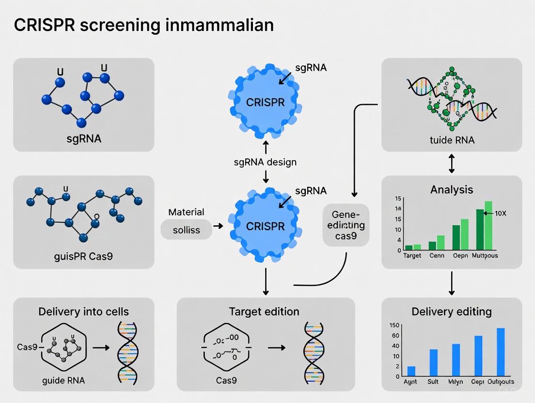
The scalability of gRNA design has enabled genome-wide CRISPR screening, an powerful approach for unbiased functional genomics. In pooled screening formats, libraries containing thousands to hundreds of thousands of gRNAs are delivered to populations of cells, followed by selection pressures and high-throughput sequencing to identify gRNAs that become enriched or depleted under specific conditions [3] [4]. Recent advances have extended this technology from in vitro models to in vivo contexts, enabling genetic dissection in physiologically relevant environments [3].
Key considerations for genome-wide screening include achieving sufficient library coverage (traditionally 250× coverage per sgRNA), ensuring efficient delivery to target cells, and implementing appropriate phenotypic selections [3]. For in vivo screens, delivery remains a significant challenge, with lentiviral vectors, adeno-associated viruses (AAVs), and non-viral methods like transposon systems each offering distinct advantages and limitations for different tissue types [3].
Figure 2: Genome-wide CRISPR Screening Workflow. The process involves designing a comprehensive sgRNA library, delivering it to target cells, applying phenotypic selection, and identifying hits through next-generation sequencing.
Application Notes: Genome-Wide Screening in Primary Human NK Cells
Experimental Platform and Workflow
The PreCiSE platform enables genome-wide CRISPR screening in primary human natural killer (NK) cells, overcoming previous technical challenges in editing these difficult-to-transfect immune cells [5]. The optimized protocol involves:
Primary Human NK Cell Isolation and Expansion: NK cells are isolated from cord blood and expanded with engineered universal antigen-expressing feeder cells (uAPCs) plus IL-2 (200 IU/ml) [5].
Retroviral Library Delivery: A genome-wide sgRNA library (77,736 sgRNAs targeting 19,281 genes with 500 non-targeting controls) is delivered via retroviral transduction at low multiplicity of infection (MOI) to ensure single-copy integration [5].
Cas9 Protein Electroporation: Cells are electroporated with Cas9 ribonucleoprotein complexes to ensure efficient editing while minimizing persistent Cas9 expression that could increase off-target effects [5].
Functional Validation: Successful editing is confirmed through targeted ablation of the NK cell surface marker PTPRC (CD45), achieving 90.1% ± 0.1% loss of CD45 expression versus 0% in Cas9 mock-electroporated controls [5].
Phenotypic Selection: Edited NK cells undergo three sequential challenges with pancreatic cancer cells (Capan-1) at an effector-to-target ratio of 1:1 to model tumor-induced dysfunction, with phenotypic assessment via CD107a (LAMP1) degranulation marker expression and spectral flow cytometry [5].
Key Findings and Therapeutic Implications
This screening approach identified several critical regulators of NK cell antitumor activity. Transcription factor screening targeting 1,632 TFs with 11,364 unique guides revealed PRDM1 (Blimp-1) and RUNX3 as key transcriptional regulators that suppress NK cell proliferation and antitumor response [5]. Genome-wide screening further identified MED12, ARIH2, and CCNC as critical checkpoints whose ablation significantly improved NK cell antitumor activity against multiple treatment-refractory human cancers both in vitro and in vivo [5].
Mechanistically, ablation of these targets enhanced both innate and CAR-mediated NK cell function through improved metabolic fitness, increased proinflammatory cytokine secretion, and expansion of cytotoxic NK cell subsets [5]. Single-cell RNA sequencing analysis of patient samples confirmed elevated PRDM1 and RUNX3 expression in tumor-infiltrating NK cells compared to healthy donors, validating their clinical relevance as potential therapeutic targets [5].
Essential Protocols for Mammalian Genome Editing
sgRNA Design and Validation
Successful genome editing begins with careful sgRNA design, which largely determines both on-target efficiency and off-target specificity [2]. The following protocol outlines best practices:
Target Site Selection: Identify 20-nucleotide target sequences immediately 5' of an NGG PAM sequence using established bioinformatic tools (e.g., CHOPCHOP, CRISPR Design Tool). Prioritize targets with high predicted on-target activity scores and minimal off-target potential based on seed sequence uniqueness [2].
Specificity Considerations: Select target sequences with minimal homology to other genomic regions, especially in the seed region (8-12 bases proximal to PAM). Mismatches in this region most effectively reduce off-target cleavage [1].
Experimental Validation: For critical applications, validate sgRNA efficiency using in vitro cleavage assays prior to cellular experiments. Clone sgRNA into expression vectors, transcribe in vitro, incubate with Cas9 protein and target DNA plasmid, and assess cleavage efficiency by gel electrophoresis [2].
Vector Selection: For knockout screens, clone validated sgRNAs into lentiviral vectors enabling co-expression with Cas9 and selection markers. For therapeutic applications, consider high-fidelity Cas9 variants to minimize off-target effects [1] [2].
CRISPR Delivery Methods for Mammalian Cells
Table 2: Comparison of CRISPR Delivery Methods
| Delivery Method | Targetable Cell Types | Advantages | Disadvantages |
|---|---|---|---|
| Lentiviral Vectors | Dividing cells, some primary cells (hepatocytes, CNS with injection) | Stable sgRNA integration, large packaging capacity (8-10 kb) | Difficult for most extrahepatic sites, insertional mutagenesis risk |
| AAV Vectors | Liver, CNS, T cells, muscle, broader tropism | Low immunogenicity, clinical experience | Small packaging capacity (5-6 kb), episomal (transient without transposon) |
| Electroporation (RNP) | Primary immune cells, stem cells, difficult-to-transfect cells | Rapid editing, reduced off-targets, no vector DNA | Limited to ex vivo applications, optimization required |
| Hydrodynamic Injection + Transposon | Hepatocytes in vivo | Efficient liver delivery, stable expression | Limited to hepatic delivery, requires specialized equipment |
Protocol: Pooled CRISPR Screening in Primary Human Cells
This protocol adapts the PreCiSE platform for genome-wide screening in primary human immune cells [5]:
Day 1: Cell Preparation
- Isolate primary NK cells from cord blood or peripheral blood.
- Begin expansion using irradiated uAPCs at 1:1 ratio in complete media supplemented with IL-2 (200 IU/ml).
- Incubate at 37°C, 5% CO₂.
Day 5: Library Transduction
- Harvest expanded NK cells, count, and assess viability (>90% required).
- Transduce cells with retroviral sgRNA library at MOI <0.3 to ensure single integration events.
- Centrifuge at 1000 × g for 90 minutes at 32°C (spinoculation).
- Resuspend in fresh media with IL-2 and return to incubator.
Day 6: Cas9 Electroporation
- Harvest transduced cells and electroporate with Cas9 RNP complex using optimized pulse codes.
- Include non-electroporated controls to assess transduction efficiency.
- Recover cells in pre-warmed media with IL-2.
Day 7: Selection and Expansion
- Begin puromycin selection (concentration optimized for cell type) to eliminate non-transduced cells.
- Continue expansion with uAPCs and IL-2.
- Monitor viability and cell counts daily.
Days 14-28: Phenotypic Selection
- Subject edited cells to relevant biological challenge (e.g., tumor cell co-culture, cytokine deprivation, drug treatment).
- For exhaustion models, perform sequential tumor challenges at 1:1 effector:target ratio.
- Harvest cells for genomic DNA extraction and flow cytometry analysis at multiple time points.
Sample Processing and Sequencing
- Extract genomic DNA using silica column-based methods.
- Amplify integrated sgRNA sequences with barcoded primers for multiplexing.
- Perform next-generation sequencing (Illumina platform recommended).
- Analyze sequencing data using established pipelines (MAGeCK, PinAPL-Py) to identify significantly enriched or depleted sgRNAs.
Table 3: Essential Research Reagents for CRISPR Screening
| Reagent Category | Specific Examples | Function & Application Notes |
|---|---|---|
| Cas9 Expression Systems | SpCas9, HiFi Cas9, dCas9-VP64, dCas9-KRAB | Core nuclease function; high-fidelity variants reduce off-targets; dCas9 fusions for transcriptional regulation |
| Delivery Vehicles | VSVG-pseudotyped lentivirus, AAV6/8/9, electroporation equipment | Introduce CRISPR components into target cells; choice depends on cell type and application |
| sgRNA Libraries | Genome-wide (Brunello, GeCKO), focused (kinase, TF), custom libraries | Collections of sgRNAs for specific screening applications; include non-targeting controls |
| Cell Culture Reagents | IL-2 for NK cells, recombinant cytokines, uAPCs, serum-free media | Support growth and function of primary cells during screening |
| Selection Agents | Puromycin, blasticidin, fluorescent markers (GFP) | Enumerate for successfully transduced cells |
| Analysis Tools | NGS platforms, flow cytometers, bioinformatics software (MAGeCK) | Assess editing efficiency and screen outcomes |
Safety Considerations and Technical Challenges
Despite its transformative potential, CRISPR-Cas9 technology presents important safety considerations that must be addressed, particularly for clinical applications. Beyond well-documented off-target effects at sites with sequence similarity to the intended target, recent studies reveal more substantial concerns regarding on-target structural variations [6]. These include large kilobase- to megabase-scale deletions, chromosomal translocations, and other complex rearrangements that may escape detection by standard short-read sequencing methods [6].
Particular concern arises from strategies to enhance homology-directed repair efficiency through inhibition of DNA-PKcs, a key NHEJ pathway component. The DNA-PKcs inhibitor AZD7648, while promoting HDR, has been shown to dramatically increase frequencies of megabase-scale deletions and chromosomal translocations—in some cases by thousand-fold [6]. These findings highlight the complex trade-offs between editing efficiency and genomic integrity, emphasizing the need for comprehensive genotoxicity assessment in therapeutic development.
Figure 3: CRISPR Safety Considerations. Beyond intended edits, CRISPR can induce various unintended structural variations. Some enhancement strategies exacerbate these risks, necessitating appropriate mitigation approaches.
Risk mitigation strategies include using high-fidelity Cas9 variants (e.g., HiFi Cas9), paired nickase systems, and implementing comprehensive genomic integrity assessment methods capable of detecting large structural variations (e.g., CAST-Seq, LAM-HTGTS) [6]. For therapeutic applications, careful consideration should be given to whether HDR enhancement is truly necessary, as selective advantages of corrected cells or moderate editing efficiencies may suffice for clinical benefit without introducing unnecessary genotoxic risk [6].
The CRISPR-Cas9 system has evolved from a bacterial immune mechanism into an extraordinarily versatile platform for mammalian genome engineering. Its application in large-scale screening approaches continues to reveal novel biological insights and therapeutic targets across diverse cellular contexts and disease states. The protocols and application notes detailed here provide a framework for implementing these technologies while acknowledging both their transformative potential and important technical limitations. As the field advances, continued refinement of editing specificity, delivery efficiency, and safety assessment will further expand the research and therapeutic applications of this powerful technology.
Class 2 CRISPR systems, characterized by their single effector protein, have revolutionized genetic engineering and functional genomics. Within this class, Type II (Cas9) and Type V (Cas12) systems have become indispensable tools for unraveling gene function in mammalian cells [7]. Their programmability and precision have made them particularly valuable for pooled genetic screens, enabling the systematic identification of genes involved in health, disease, and therapeutic response [8]. The core innovation lies in the RNA-guided nature of these effectors; a customizable guide RNA (gRNA) directs the Cas protein to a specific genomic locus or nucleic acid sequence for cleavage or binding. This review details the molecular architectures, comparative capabilities, and practical application of these systems within the context of modern CRISPR screening, providing a foundational resource for researchers embarking on functional genetic studies.
Molecular Mechanisms and System Comparisons
Core Mechanisms of Cas9 and Cas12
The Type II CRISPR system, centered on the Cas9 protein, functions as a bilobed structure composed of a recognition (REC) lobe and a nuclease (NUC) lobe [7]. Its activity requires two nuclease domains: the HNH domain, which cleaves the DNA strand complementary to the guide RNA, and the RuvC domain, which cleaves the non-complementary strand [7]. This results in a blunt-ended double-strand break (DSB) three nucleotides upstream of the Protospacer Adjacent Motif (PAM), which for the commonly used Streptococcus pyogenes Cas9 is a 5'-NGG-3' sequence [7]. The system relies on a single guide RNA (sgRNA) that fuses the functions of the endogenous crRNA and tracrRNA.
In contrast, Type V effectors like Cas12a (also known as Cpf1) possess a single RuvC-like nuclease domain responsible for cleaving both DNA strands [9]. This results in a staggered double-strand break with a 5' overhang [9]. A key differentiator of Cas12a is its PAM requirement; it typically recognizes a 5'-TTTN-3' PAM, which is richer in adenine and thymine compared to the GC-rich Cas9 PAM [9] [10]. Furthermore, upon recognizing and cleaving its target double-stranded DNA (cis-cleavage), Cas12a exhibits transient collateral activity, non-specifically cleaving surrounding single-stranded DNA (ssDNA) [9]. This trans-cleavage activity is the foundation for many diagnostic applications.
Comparative Analysis of Class 2 CRISPR Systems
The following table summarizes the key characteristics of Cas9 and Cas12, highlighting their distinctions and respective advantages.
Table 1: Comparative Analysis of Type II (Cas9) and Type V (Cas12) CRISPR Systems
| Feature | Type II (Cas9) | Type V (Cas12a) |
|---|---|---|
| Effector Protein | Cas9 | Cas12a (Cpf1), Cas12f1 |
| PAM Sequence | 5'-NGG-3' (GC-rich) [7] | 5'-TTTN-3' (AT-rich) [9] [10] |
| Guide RNA | Single guide RNA (sgRNA) [7] | CRISPR RNA (crRNA) [9] |
| Cleavage Mechanism | Dual HNH & RuvC domains [7] | Single RuvC domain [9] |
| Cleavage Pattern | Blunt ends [7] | Staggered ends with 5' overhang [9] |
| Collateral Activity | No | Yes (ssDNA trans-cleavage) [9] |
| Primary Screening Applications | Gene knockout (CRISPRn), Interference (CRISPRi), Activation (CRISPRa) [8] | Gene knockout, diagnostics (e.g., DETECTR) [9] [10] |
Recent studies have further expanded the CRISPR toolbox. For instance, the much smaller size of Cas12f1 (half the size of Cas9) facilitates easier delivery, while CRISPR-Cas3 has been shown to achieve high eradication efficiency of antibiotic resistance genes in bacterial models, though its application in mammalian cells is less common [10].
Application Notes for CRISPR Screening in Mammalian Cells
CRISPR Knockout (CRISPRn), Interference (CRISPRi), and Activation (CRISPRa)
Beyond simple knockout screens, modified CRISPR systems allow for reversible and tunable control of gene expression, which is invaluable for dissecting gene function.
- CRISPRn (Nuclease): The wild-type Cas9 introduces DSBs, which are repaired by error-prone non-homologous end joining (NHEJ), often resulting in frameshift mutations and gene knockout [8]. This is ideal for identifying essential genes and loss-of-function phenotypes.
- CRISPRi (Interference): A catalytically dead Cas9 (dCas9) is fused to repressive domains like the KRAB domain. This complex binds to the promoter or coding region of a target gene without cutting the DNA, recruiting chromatin-modifying proteins to silence transcription [8]. CRISPRi is highly specific and reversible, making it suitable for studying essential genes.
- CRISPRa (Activation): The same dCas9 is fused to transcriptional activators (e.g., VP64, p65, Rta) to recruit RNA polymerase and co-activators, leading to targeted gene overexpression [8]. Advanced systems like the SunTag or SAM use scaffold strategies to recruit multiple activator molecules, significantly potentiating gene expression [8].
Pooled CRISPR Screening Workflow
Pooled CRISPR screens are a powerful, unbiased method for discovering genes involved in a biological process of interest across the entire genome.
Table 2: Key Reagents and Materials for Pooled CRISPR Screening
| Reagent/Material | Function/Description | Example/Note |
|---|---|---|
| sgRNA Library | Pooled collection of lentiviral transfer plasmids, each encoding a unique sgRNA. | Genome-wide libraries typically contain 4-6 sgRNAs per gene and require high coverage (e.g., 250x per sgRNA) [3]. |
| Lentiviral Vectors | Delivery vehicle for stably integrating the sgRNA and Cas transgene into the host cell genome. | Often pseudotyped with VSVG glycoprotein; crucial for achieving high infection efficiency [8] [3]. |
| Cas9-Expressing Cells | Mammalian cell line engineered to constitutively or inducibly express the Cas9 nuclease. | Using stable cell lines simplifies screening by reducing the number of genetic elements to deliver [3]. |
| Selection Antibiotics | To select for successfully transduced cells. | e.g., Puromycin, Blasticidin. |
| Next-Generation Sequencing (NGS) Platform | To quantify sgRNA abundance before and after selection. | Determines which sgRNAs are enriched or depleted under selective pressure [8]. |
The generalized workflow for a pooled negative selection (drop-out) screen is as follows:
Step 1: Library Design and Cloning. A pooled sgRNA library is designed, typically targeting each gene with multiple sgRNAs to ensure robustness. The library is cloned into a lentiviral transfer plasmid [8] [4]. Step 2: Lentivirus Production. The plasmid library is used to produce lentiviral particles in a packaging cell line (e.g., HEK293T). Step 3: Cell Transduction. A culture of Cas9-expressing mammalian cells is transduced with the lentiviral library at a low Multiplicity of Infection (MOI ~0.3) to ensure most cells receive only one sgRNA. Cells are then selected with antibiotics to generate a representationally stable screening pool [8]. Step 4: Phenotypic Selection. The pooled cell population is divided and subjected to a biological challenge (e.g., drug treatment, viral infection) over multiple generations. A reference sample is harvested at the start (T0), and the experimental group is harvested after the challenge (T-final) [8] [4]. Step 5: Sequencing and Quantification. Genomic DNA is extracted from both T0 and T-final populations. The integrated sgRNA sequences are PCR-amplified and quantified via NGS to determine the relative abundance of each sgRNA [8]. Step 6: Bioinformatic Analysis. Depletion or enrichment of specific sgRNAs in the T-final sample relative to T0 is calculated using specialized tools (e.g., MAGeCK). Statistically significant hits indicate genes that confer sensitivity or resistance to the applied challenge [4].
Detailed Protocol: A Pooled CRISPR-KO Screen for Drug Resistance Genes
This protocol outlines the steps to identify genetic modifiers of sensitivity to a drug-like compound in a human cell line, a common application in chemical biology and drug target discovery [8].
Pre-Screen Preparation
- Cell Line: Obtain a human cancer cell line (e.g., K562, A375) expressing Cas9 under a constitutive promoter.
- sgRNA Library: Select a genome-wide CRISPR knockout library (e.g., Brunello or Brie library).
- Reagents: Prepare DMEM or RPMI-1640 culture media, fetal bovine serum (FBS), penicillin-streptomycin, polybrene (8 µg/mL), puromycin, and the drug compound of interest.
Screen Execution
- Library Transduction:
- Day 0: Seed 200 million Cas9-expressing cells in a large flask.
- Day 1: Replace medium with fresh medium containing 8 µg/mL polybrene. Add the lentiviral sgRNA library at an MOI of 0.3, ensuring >500x coverage of the library (e.g., for a 100,000 sgRNA library, transduce at least 50 million cells). Mix gently and culture for 24 hours.
- Selection of Transduced Cells:
- Day 3: Replace the medium with fresh medium containing the appropriate selection antibiotic (e.g., 2 µg/mL puromycin). Continue selection for 5-7 days until >90% of non-transduced control cells are dead.
- Phenotypic Challenge:
- Day 10: Harvest the pooled, selected cells. Count the cells and split into two arms:
- Control Arm: Culture cells in standard medium. Harvest 50 million cells as the T0 reference point. Continue culturing the rest, maintaining a minimum of 500x library coverage at all times.
- Treatment Arm: Culture cells in medium containing the IC50-IC80 concentration of the drug compound. Culture both arms for 3-4 weeks, passaging cells every 2-3 days to maintain log-phase growth.
- Day 10: Harvest the pooled, selected cells. Count the cells and split into two arms:
- Sample Harvest and Sequencing:
- After ~14 population doublings, harvest 50 million cells from both the Control and Treatment arms. Extract genomic DNA using a maxi-prep kit. Perform a two-step PCR to amplify the integrated sgRNA cassettes from the genomic DNA and attach Illumina sequencing adapters and barcodes. Pool the PCR products and perform NGS on an Illumina platform to a depth of 5-10 million reads per sample.
Post-Screen Analysis
- sgRNA Count Quantification: Use a tool like
MAGeCK countto align the NGS reads to the library reference and generate a count table for each sgRNA in the T0, final Control, and final Treatment samples. - Hit Identification: Run
MAGeCK testto compare sgRNA abundances between the Treatment and Control arms. Genes enriched in the Treatment arm (with multiple sgRNAs showing significant positive scores) are resistance hits, suggesting their knockout confers survival advantage. Genes depleted in the Treatment arm are sensitivity hits, suggesting they are essential for survival under drug pressure [8] [4]. - Validation: Top candidate genes must be validated using individual sgRNAs in a low-throughput format and orthogonal assays (e.g., Western blot, RT-qPCR) to confirm the phenotype.
The strategic application of Class 2 CRISPR systems, particularly Cas9 and Cas12, has fundamentally advanced our capacity for functional genomic screening in mammalian cells. The choice between systems depends on the experimental goal: Cas9-based knockout, interference, and activation screens remain the gold standard for probing gene function, while Cas12 variants offer advantages in diagnostics and specific targeting contexts. As the field progresses, the integration of artificial intelligence for guide RNA design and outcome prediction, alongside improvements in delivery technologies like lipid nanoparticles (LNPs), is poised to further enhance the efficiency, precision, and safety of these powerful tools [11] [12]. By following the detailed protocols and understanding the comparative strengths outlined in this article, researchers can effectively leverage these systems to uncover novel biological mechanisms and therapeutic targets.
CRISPR-based screening technologies have revolutionized functional genomics in mammalian cells, providing researchers with a powerful toolkit to systematically interrogate gene function. While the foundational CRISPR-Cas9 system enables permanent gene knockout, advanced derivatives known as CRISPR interference (CRISPRi) and CRISPR activation (CRISPRa) offer sophisticated temporal control over gene expression. CRISPRi achieves targeted gene repression without altering the DNA sequence, whereas CRISPRa enables precise transcriptional activation. These three modalities—Knockout (ko), Interference (i), and Activation (a)—each possess distinct mechanisms and applications that make them uniquely suited for different biological questions in basic research and drug discovery. This article delineates the operational principles, experimental protocols, and practical considerations for implementing these technologies in genetic screens, providing a comprehensive resource for scientists engaged in mammalian cell research.
Molecular Mechanisms and Comparative Profiles
The functional diversity of CRISPR screening modalities stems from engineered variations of the core CRISPR-Cas9 system, primarily through modifications to the Cas9 nuclease and its associated effector domains.
CRISPR Knockout (CRISPRko) utilizes the wild-type Cas9 nuclease, which creates double-strand breaks (DSBs) in the DNA at sites specified by the guide RNA (gRNA). These breaks are primarily repaired by the error-prone non-homologous end joining (NHEJ) pathway, often resulting in insertions or deletions (indels) that disrupt the open reading frame and lead to permanent gene knockout [1].
CRISPR Interference (CRISPRi) employs a catalytically dead Cas9 (dCas9), which lacks nuclease activity but retains DNA-binding capability. When targeted to a gene's promoter region, dCas9 alone can cause steric hindrance, blocking transcription. For more potent repression in mammalian cells, dCas9 is typically fused to a transcriptional repressor domain, such as the Krüppel associated box (KRAB), which recruits additional proteins to silence gene expression [13].
CRISPR Activation (CRISPRa) also uses dCas9 but is fused to transcriptional activator domains, such as VP64, p65, or the SunTag system. When guided to a gene's promoter or enhancer region, this complex recruits the cellular transcription machinery to drive gene expression, often achieving supra-physiological levels [13].
The table below summarizes the core characteristics of these three modalities.
Table 1: Core Characteristics of CRISPR Screening Modalities
| Feature | CRISPR Knockout (ko) | CRISPR Interference (i) | CRISPR Activation (a) |
|---|---|---|---|
| Cas9 Type | Wild-type (active nuclease) | dCas9 (catalytically dead) | dCas9 (catalytically dead) |
| Core Mechanism | DNA cleavage → NHEJ repair → Indels | Steric hindrance + recruitment of repressors | Recruitment of transcriptional activators |
| Genetic Outcome | Permanent gene knockout | Reversible gene knockdown | Tunable gene overexpression |
| Key Effector Domain | N/A | KRAB | VP64, p65, SunTag |
| Effect on Essential Genes | Lethal, precluding study | Enables study of hypomorphic phenotypes | Can reveal oncogenic potential |
The following diagram illustrates the fundamental mechanisms of action for each modality.
Experimental Protocols for Screening
Executing a successful CRISPR screen requires meticulous planning, from library design to data analysis. The following protocol outlines the key steps for a pooled negative selection screen to identify genes essential for cell growth, a common application for CRISPRko.
Workflow for a Pooled CRISPRko Screen
- Library Selection and Design: Choose a genome-wide or sub-library of sgRNAs targeting your genes of interest. A typical library uses 4-10 sgRNAs per gene, plus non-targeting control sgRNAs [14] [15]. The library is cloned into a lentiviral backbone.
- Lentiviral Production: Produce lentivirus containing the pooled sgRNA library in HEK293T cells. Determine the viral titer.
- Cell Transduction and Selection: Transduce your target mammalian cells (e.g., K562, HeLa) at a low Multiplicity of Infection (MOI ~0.3-0.5) to ensure most cells receive only one sgRNA. Select transduced cells with antibiotics (e.g., puromycin) for 5-7 days. This is the "Day 0" or "T0" timepoint.
- Screen Propagation and Harvest: Split the cell population into replicates and continue culturing for 2-4 weeks (e.g., Day 21, "T21") to allow depletion of cells carrying sgRNAs targeting essential genes.
- Genomic DNA (gDNA) Extraction and Sequencing: Harvest cells at T0 and T21. Extract gDNA and perform PCR amplification of the integrated sgRNA sequences using primers compatible with Illumina sequencing.
- Data Analysis: Sequence the PCR products and count the reads for each sgRNA. Use specialized algorithms (e.g., MAGeCK, casTLE) to compare sgRNA abundance between T0 and T21, identifying sgRNAs (and their target genes) that are significantly depleted or enriched [14] [16].
The workflow is visualized in the diagram below.
Critical Parameters for CRISPRi/a Screens
While the overall workflow for CRISPRi and CRISPRa screens is similar to CRISPRko, key differences must be considered:
- Cell Line Engineering: Stable cell lines expressing dCas9-KRAB (for CRISPRi) or dCas9-activator (for CRISPRa) must be generated and validated prior to the screen [16] [13].
- gRNA Design: sgRNAs for CRISPRi/a must be designed to bind to the promoter or transcriptional start site of the target gene, rather than the coding exon. This requires accurate annotation of promoter regions [13].
- Controls: Include non-targeting gRNAs and gRNAs targeting known essential and non-essential genes as negative and positive controls, respectively.
Performance and Application Data
The choice between CRISPRko, CRISPRi, and CRISPRa is dictated by the biological question. A systematic comparison revealed that CRISPRko and RNAi (a knockdown technology conceptually similar to CRISPRi) can identify distinct sets of essential genes, suggesting they reveal different aspects of biology [14]. Combining data from both knockout and knockdown/activation screens provides a more robust and comprehensive understanding of gene function.
Table 2: Quantitative Performance and Primary Applications
| Screening Modality | Typical Efficiency/Effect | Key Strengths | Ideal Screening Contexts |
|---|---|---|---|
| CRISPRko | High knockout efficiency; Indels via NHEJ [1] | Permanent loss-of-function; Identifies fitness genes; Well-established analysis | Genome-wide loss-of-function; Identification of essential genes [14] |
| CRISPRi | Strong repression (up to 80-99%) with dCas9-KRAB [13] | Reversible; Tunable knockdown; Fewer off-targets vs. RNAi; Studies essential genes | Functional dissection of essential genes; Drug target validation [13] |
| CRISPRa | Strong activation with multi-domain systems (e.g., SunTag) [13] | Endogenous gene overexpression; Gain-of-function studies | Identifying drivers of drug resistance; Oncogene screening [15] [13] |
Case Study: Dual CRISPRko/CRISPRa Screen for Drug Resistance
A powerful application of these technologies is elucidating mechanisms of drug resistance. A 2020 study employed dual genome-wide CRISPR knockout and CRISPR activation screens to identify genes that regulate cellular resistance to ATR inhibitors (ATRi) in cancer cells [15].
- The CRISPRko screen identified genes whose loss confers resistance (e.g., MED12, KNTC1), potentially by disrupting pathways that promote cell death in response to ATR inhibition.
- The parallel CRISPRa screen identified genes whose overexpression drives resistance, pointing to potential mechanisms like enhanced DNA repair or suppression of apoptosis.
This dual approach provided a comprehensive landscape of genetic determinants of ATRi resistance, highlighting how KO and A screens can reveal complementary biological insights within the same study.
The Scientist's Toolkit: Essential Reagents and Materials
Successful implementation of CRISPR screens relies on a core set of high-quality reagents. The following table lists essential components and their functions.
Table 3: Essential Reagents for CRISPR Screening
| Reagent / Material | Function and Critical Notes |
|---|---|
| sgRNA Library | Pooled collection of guide RNAs; Critical for specificity. Arrayed synthetic libraries are recommended for high editing efficiency and low off-targets [17]. |
| Cas9 Enzyme | Wild-type for KO; Catalytically dead (dCas9) for i/a. High-fidelity variants (e.g., eSpCas9, SpCas9-HF1) reduce off-target effects [1]. |
| Lentiviral Packaging System | For efficient delivery of CRISPR components into mammalian cells (e.g., HEK293T cells, psPAX2, pMD2.G plasmids). |
| Cell Culture Reagents | Optimized media and supplements for the target mammalian cell line; Puromycin for selection post-transduction. |
| Next-Generation Sequencing (NGS) Platform | For quantifying sgRNA abundance from genomic DNA of screen samples. Essential for deconvoluting results [18]. |
| Bioinformatics Software | Tools like MAGeCK [16] and casTLE [14] for statistical analysis of sgRNA enrichment/depletion. |
Troubleshooting and Technical Considerations
Even well-designed screens can face challenges. Below are common issues and recommended solutions.
Table 4: Common Technical Challenges and Solutions
| Challenge | Potential Cause | Recommended Solution |
|---|---|---|
| Poor Screen Performance / Low Signal | Low viral titer; Ineffective delivery; Insensitive cell line. | Re-titer virus; Optimize delivery method (e.g., electroporation); Use a cell line with robust fitness phenotypes. |
| High False Positive/Negative Rate | Ineffective sgRNAs; Poor library coverage; Off-target effects. | Use validated library designs with multiple sgRNAs/gene; Maintain >500x library coverage during screening; Use bioinformatic tools to filter out common off-targets [1]. |
| Inconsistent Results Between Modalities | Fundamental biological differences (knockout vs. knockdown); Technology-specific artifacts. | Combine data using statistical frameworks like casTLE [14]; Validate key hits with orthogonal assays (e.g., cDNA rescue). |
| Low Editing/Efficiency (CRISPRi/a) | Poor dCas9 expression; Inaccessible chromatin at target; Suboptimal gRNA design. | Validate dCas9 and effector expression; Use ATAC-seq data to inform target site selection; Design gRNAs to target nucleosome-free regions [13]. |
A significant consideration is the inherent heterogeneity in screening technologies. For example, a CRISPRko screen can generate a mixture of true knockouts, heterozygotes, and wild-type cells due to variable editing outcomes, which can influence the observed phenotype [14]. Acknowledging and accounting for this variability during data interpretation is crucial.
CRISPR-Cas9 has revolutionized functional genomics, enabling systematic perturbation of genes to uncover their functions in health and disease. For researchers and drug development professionals applying CRISPR screening in mammalian cells, a thorough understanding of three foundational components is critical: the design of guide RNAs (gRNAs), the constraints imposed by Protospacer Adjacent Motif (PAM) requirements, and the decisive role of cellular DNA repair pathways. The PAM, a short DNA sequence adjacent to the target site, is an absolute requirement for Cas nuclease activity and serves as a recognition signal that licenses the Cas complex to cleave DNA [19] [20]. Following cleavage, cellular repair pathways—primarily non-homologous end joining (NHEJ) or homology-directed repair (HDR)—determine the mutational outcome, leading to either gene knockouts or precise edits [21]. This application note details the essential protocols and design principles for successful CRISPR screen design and analysis, framed within the context of mammalian cell research for drug target discovery.
Core Principles: PAM Sequences and Cellular Repair Pathways
The Critical Role of the Protospacer Adjacent Motif (PAM)
The PAM is a short, specific DNA sequence (typically 2-6 base pairs) located directly adjacent to the DNA region targeted for cleavage by the CRISPR-Cas system. Its primary function is to allow the Cas nuclease to distinguish between self and non-self DNA, preventing the bacterial CRISPR system from attacking its own genome [19]. From a practical standpoint, the PAM is indispensable for CRISPR experiments because Cas nucleases will only interrogate a potential target site if the correct PAM is present. The Cas nuclease destabilizes the adjacent DNA sequence, allowing the guide RNA to pair with the matching target DNA [20]. The specific PAM sequence requirement depends entirely on the Cas nuclease used. For the commonly used Streptococcus pyogenes Cas9 (SpCas9), the PAM sequence is 5'-NGG-3', where "N" can be any nucleotide base [19] [20]. This requirement means that every potential target site in the genome must be followed by a GG dinucleotide.
DNA Repair Pathways Determine Editing Outcomes
Once the Cas nuclease creates a double-strand break (DSB), the cell's innate DNA repair machinery takes over. The outcome of a CRISPR experiment is therefore dictated by which repair pathway is activated [21]. The two primary pathways are:
- Non-Homologous End Joining (NHEJ): This is the cell's dominant and error-prone repair pathway. NHEJ directly ligates the broken DNA ends, often resulting in small insertions or deletions (indels). When these indels occur within a protein-coding exon, they can cause frameshift mutations that lead to premature stop codons and effectively knock out the gene [22] [21]. NHEJ is the basis for most CRISPR knockout (CRISPRko) screens.
- Homology-Directed Repair (HDR): This is a more precise, but less frequent, pathway that functions in the S and G2 phases of the cell cycle. HDR uses a DNA template—either a sister chromatid or an exogenously supplied donor DNA template—to repair the break accurately. In CRISPR experiments, researchers can co-deliver a designed donor template to direct specific edits, such as inserting a therapeutic gene or introducing a specific point mutation, in a process known as knock-in [21].
Table 1: Overview of DNA Repair Pathways in CRISPR Genome Editing
| Pathway | Mechanism | Templates Required | Primary Outcome | Common CRISPR Applications |
|---|---|---|---|---|
| Non-Homologous End Joining (NHEJ) | Direct ligation of broken ends | None | Error-prone, creates indels | Gene knockout (CRISPRko) |
| Homology-Directed Repair (HDR) | Uses homologous sequence as a template | Donor DNA template (endogenous or exogenous) | Precise edits | Gene knock-in, specific point mutations |
The following diagram illustrates the critical decision point after a Cas-induced double-strand break and the two primary repair pathways that determine the experimental outcome.
Experimental Protocols for CRISPR Screening
Protocol: A Workflow for Pooled CRISPR Knockout Screening
This protocol outlines the key steps for performing a pooled CRISPR knockout screen in mammalian cells to identify genes essential for cell viability or drug resistance, a common application in drug target discovery [22] [21].
gRNA Library Design and Cloning:
- Design: Select a genome-wide or focused gRNA library. Each gene is typically targeted by 4-6 gRNAs to ensure statistical robustness. The gRNA sequence (usually 20 nt) is designed to target an early exon of the gene and must be immediately 5' to a compatible PAM sequence (e.g., NGG for SpCas9) [19] [22].
- Cloning: The pooled oligonucleotide library is synthesized and cloned into a lentiviral gRNA expression vector. The vector also contains a selection marker (e.g., puromycin resistance).
Lentivirus Production and Cell Line Preparation:
- Virus Production: The lentiviral plasmid library is transfected into a packaging cell line (e.g., HEK293T) to produce lentiviral particles containing the gRNA library.
- Cell Line: A mammalian cell line (e.g., HeLa, HL-60) that stably expresses the Cas9 nuclease is generated or obtained. The Cas9 can be constitutively expressed or induced.
Library Transduction and Selection:
- Transduction: The target cells are transduced with the lentiviral library at a low Multiplicity of Infection (MOI ~0.3) to ensure most cells receive only one gRNA. This achieves a coverage of 500-1000 cells per gRNA to maintain library representation.
- Selection: Cells are selected with an antibiotic (e.g., puromycin) for 3-5 days to eliminate untransduced cells, creating a stable pool of mutant cells.
Phenotypic Selection and Analysis:
- Experimental Arms: The selected cell pool is split into two groups: a control arm and a treatment arm (e.g., exposed to a drug candidate).
- Time Point: Cells are passaged for 2-3 weeks to allow for phenotypic enrichment (depletion of essential genes or enrichment of resistance genes).
- Genomic DNA (gDNA) Extraction: gDNA is extracted from both control and treatment populations at the end point.
Sequencing and Bioinformatic Analysis:
- Amplification and Sequencing: The integrated gRNA sequences are amplified from the gDNA by PCR and prepared for next-generation sequencing (NGS).
- Analysis: NGS reads are aligned to the library reference. gRNA abundance is quantified and compared between treatment and control groups using specialized algorithms (e.g., MAGeCK) to identify significantly depleted or enriched gRNAs and their target genes [22].
The overall workflow for a typical pooled CRISPR screen, from library design to hit identification, is summarized below.
Protocol: Assessing Editing Outcomes with an eGFP-to-BFP Conversion Assay
This protocol provides a method to rapidly quantify and distinguish between NHEJ and HDR outcomes in a cell population, which is crucial for optimizing knock-in efficiency [23].
Generation of eGFP-Positive Reporter Cell Line:
- A mammalian cell line (e.g., HEK293) is stably transduced with a lentiviral construct expressing enhanced Green Fluorescent Protein (eGFP). A clonal population with high, uniform eGFP expression is selected.
Design and Transfection of CRISPR Reagents:
- gRNA and Donor Template: Design a gRNA to target a region within the eGFP gene. Co-design a single-stranded oligodeoxynucleotide (ssODN) donor template encoding the mutations to convert eGFP to Blue Fluorescent Protein (BFP). The donor must contain homologous arms flanking the Cas9 cut site.
- Transfection: Transfect the eGFP-positive cells with the ribonucleoprotein (RNP) complex (Cas9 protein + gRNA) along with the ssODN donor template using a method suitable for the cell line (e.g., electroporation).
Cell Handling and Fluorescence Measurement:
- Incubation: Culture the transfected cells for 5-7 days to allow for expression of the edited protein.
- Flow Cytometry (FACS): Analyze the cells using a flow cytometer. Measure fluorescence in the FITC (green) and Pacific Blue (blue) channels.
Data Analysis and Interpretation:
- HDR Efficiency: The percentage of BFP-positive cells (successful HDR) is calculated from the total live cell population.
- NHEJ Efficiency: The percentage of eGFP-negative (non-fluorescent) cells indicates a successful knockout via NHEJ, provided the edits disrupt the eGFP reading frame.
- Editing Success: The remaining eGFP-positive, BFP-negative cells represent unedited cells or cells with in-frame NHEJ repairs that did not disrupt fluorescence.
The Scientist's Toolkit: Reagents and Computational Tools
Successful execution of CRISPR screens relies on a suite of specialized reagents and computational tools for design, analysis, and validation.
Table 2: Research Reagent Solutions for CRISPR Screening
| Reagent / Tool | Function | Example Products / Variants |
|---|---|---|
| Cas Nuclease | Engineered endonuclease that creates DSB at target site. | SpCas9 (NGG PAM), SaCas9 (NNGRRT PAM), Cas12a/Cpf1 (TTTV PAM), High-fidelity variants (e.g., Alt-R S.p. HiFi Cas9) [19] [20] |
| Guide RNA (gRNA) | Synthetic RNA that directs Cas nuclease to specific genomic locus. | Chemically modified synthetic sgRNA (increases stability and efficiency), crRNA:tracrRNA complexes [20] |
| Lentiviral Vector | Efficient delivery system for gRNA library into mammalian cells. | Plasmids with U6 promoter for gRNA expression, antibiotic resistance markers (e.g., puromycin) |
| Donor Template | Provides homologous sequence for HDR-mediated precise editing. | Single-stranded ODNs (ssODNs), double-stranded DNA (dsDNA) donors with homology arms |
| Analysis Software | Computational tool to analyze NGS or Sanger data from editing experiments. | ICE (Inference of CRISPR Edits) for Sanger data, MAGeCK for NGS screen data [24] [22] |
Table 3: Key Bioinformatics Tools for CRISPR Screen Data Analysis
| Tool | Primary Function | Key Methodology | Application Context |
|---|---|---|---|
| MAGeCK | Identifies enriched/depleted genes from CRISPR screens | Robust Rank Aggregation (RRA) on sgRNA counts [22] | Genome-wide dropout/sorting screens |
| BAGEL | Identifies essential genes using a Bayesian framework | Compares sgRNA abundance to a reference set of essential genes [22] | CRISPRko viability screens |
| ICE | Quantifies editing efficiency and indel profiles from Sanger data | Compares sequencing traces from edited vs. control samples [24] | Validation of editing in individual clones |
| CRISPhieRmix | Integrates multiple screens to improve hit identification | Hierarchical mixture model [22] | Meta-analysis of screening data |
| scMAGeCK | Links gene perturbations to transcriptomic phenotypes in single cells | RRA or linear regression on single-cell RNA-seq data [22] | Single-cell CRISPR screens (Perturb-seq) |
Advanced Applications: Expanding the Scope of CRISPR Screens
The core CRISPR-Cas9 system has been engineered to enable a diverse range of screening modalities beyond simple gene knockouts, greatly enhancing its utility in functional genomics and drug discovery [22] [21].
CRISPR Interference (CRISPRi): This system uses a catalytically "dead" Cas9 (dCas9) fused to a transcriptional repressor domain like KRAB. dCas9 binds to the DNA without cutting it, and KRAB silences the target gene. CRISPRi is ideal for studying essential genes, as it is reversible and creates hypomorphic rather than null alleles. It is also the preferred method for targeting non-coding elements like enhancers and long non-coding RNAs (lncRNAs) [22] [21].
CRISPR Activation (CRISPRa): This gain-of-function approach uses dCas9 fused to strong transcriptional activators (e.g., VP64, p65, SAM complex). By targeting the dCas9-activator to gene promoters, researchers can overexpress genes of interest. CRISPRa screens are powerful for identifying genes that confer drug resistance or that can rescue a disease phenotype [22] [21].
Single-Cell CRISPR Screens: Technologies like Perturb-seq and CROP-seq combine pooled CRISPR screening with single-cell RNA sequencing (scRNA-seq). This allows researchers to not only identify which genes are essential for a phenotype but also to observe the full transcriptomic consequences of each perturbation in individual cells, uncovering underlying regulatory networks [22].
Table 4: Overview of Advanced CRISPR Screening Modalities
| Screening Type | Core Machinery | Primary Genetic Effect | Key Application in Drug Discovery |
|---|---|---|---|
| CRISPR Knockout (CRISPRko) | Wild-type Cas9 | Permanent gene disruption via indels | Identify essential genes and drug targets [22] |
| CRISPR Interference (CRISPRi) | dCas9-KRAB fusion | Reversible gene knockdown | Study essential genes and regulatory elements [22] [21] |
| CRISPR Activation (CRISPRa) | dCas9-activator fusion | Gene overexpression | Identify genes conferring resistance or therapeutic benefit [22] [21] |
| Single-Cell CRISPR Screens | Cas9 + scRNA-seq | Gene knockout with transcriptomic readout | Uncover gene networks and cellular heterogeneity in response to perturbation [22] |
Mastering the principles of guide RNA design, PAM requirements, and cellular repair pathways is fundamental to designing and executing successful CRISPR screens in mammalian cells. The continued diversification of Cas nucleases with novel PAM specificities expands the targetable genome, while sophisticated protocols and analytical tools enable precise dissection of gene function at scale. For researchers in drug development, these methodologies provide a powerful pipeline for the systematic identification and validation of novel therapeutic targets, accelerating the journey from gene discovery to clinical application. As CRISPR screening technologies evolve towards higher resolution in single cells and more physiologically relevant model systems, their impact on functional genomics and target discovery will only continue to grow.
The field of functional genomics has undergone a revolutionary transformation over the past decade, marked by a fundamental transition from RNA interference (RNAi)-based technologies to CRISPR-Cas9-based systems. This evolution has redefined our approach to understanding gene function, enabling unprecedented precision and scalability in genetic screening. Functional genomics aims to bridge the critical gap between genetic information and biological function by systematically perturbing genes and analyzing resulting phenotypic changes [25]. While RNAi technology provided the first scalable method for loss-of-function studies in mammalian cells, its limitations prompted the development of more robust approaches. The emergence of CRISPR-Cas9 as a programmable genome-editing tool has addressed many of these challenges, offering permanent gene disruption, higher specificity, and greater versatility in experimental design [26] [14]. This shift has been particularly transformative for drug discovery and therapeutic target identification, allowing researchers to systematically identify essential genes and drug-gene interactions with enhanced confidence and reproducibility [27]. The historical progression from RNAi to CRISPR represents not merely a technological improvement but a fundamental paradigm shift in how we interrogate gene function at scale.
Historical Background: From RNAi to CRISPR
The Era of RNA Interference (RNAi)
RNA interference emerged in the early 2000s as the first high-throughput technology for gene silencing in mammalian cells. The technology leverages a conserved endogenous pathway where introduced double-stranded RNAs trigger sequence-specific degradation of complementary mRNA molecules [26]. Several RNAi reagents were developed, including short interfering RNAs (siRNAs) and short hairpin RNAs (shRNAs), which could be delivered via viral transduction to achieve stable gene knockdown [26]. This approach enabled genome-scale loss-of-function screens that identified genes involved in various biological processes and disease states. The major advantages of RNAi included its ability to achieve partial knockdowns (potentially revealing phenotypes for essential genes) and its applicability to large-scale screening. However, significant limitations emerged: incomplete gene knockdown due to variable reagent efficiency, extensive off-target effects through miRNA-like deregulation, and high rates of false positives and false negatives that complicated data interpretation [26] [14]. These constraints highlighted the need for more precise and reliable perturbation technologies.
The CRISPR Revolution
The discovery of the CRISPR-Cas9 system and its development into a genome-editing tool marked the beginning of a new era in functional genomics. The natural CRISPR system, first characterized by Francisco Mojica in the 1990s [28], functions as an adaptive immune system in bacteria. Key discoveries followed, including Alexander Bolotin's identification of Cas9 and the protospacer adjacent motif (PAM) in 2005 [28], and the seminal 2012 publications by Charpentier, Doudna, and Siksnys demonstrating that Cas9 could be programmed with a single guide RNA to create targeted double-strand breaks in DNA [28]. The transformative potential for eukaryotic cells was realized in January 2013, when Feng Zhang and George Church's laboratories simultaneously reported successful genome editing in human and mouse cells [28]. Unlike RNAi, which reduces gene expression at the mRNA level, CRISPR-Cas9 introduces permanent changes to the DNA sequence itself, typically resulting in complete gene knockout through frameshift mutations [26]. This fundamental difference in mechanism underlies CRISPR's advantages in consistency, potency, and specificity for functional genomic applications.
Comparative Performance: RNAi versus CRISPR
Table 1: Systematic comparison of RNAi and CRISPR-Cas9 screening technologies
| Feature | RNAi | CRISPR-Cas9 |
|---|---|---|
| Mechanism of Action | mRNA degradation/translational inhibition | DNA double-strand breaks → indels → frameshifts |
| Molecular Effect | Gene knockdown (partial reduction) | Gene knockout (complete disruption) |
| Target Specificity | Moderate (off-targets via seed matches) | High (requires perfect complementarity) |
| Phenotype Strength | Variable, dose-dependent | Typically strong, binary |
| Screening Reproducibility | Moderate | High |
| Technical Validation Rate | ~30-60% | ~70-90% |
| Essential Gene Detection | Identifies ~60% at 1% FPR | Identifies ~60% at 1% FPR |
| Additional Hits | ~3,100 genes at 10% FPR | ~4,500 genes at 10% FPR |
| Overlap Between Technologies | ~1,200 genes identified in both | ~1,200 genes identified in both |
| Primary Applications | Loss-of-function studies, drug target ID | Knockout, activation, inhibition, base editing |
Direct comparative studies reveal both complementary and distinct characteristics of each technology. In parallel screens conducted in the K562 chronic myelogenous leukemia cell line, both RNAi and CRISPR showed similar precision in detecting essential genes (AUC >0.90), with each identifying approximately 60% of gold standard essential genes at a 1% false positive rate [14]. However, each technology also identified thousands of unique hits not detected by the other, suggesting they may capture different biological aspects [14]. Notably, CRISPR screens identified electron transport chain genes as essential, while RNAi screens uniquely detected chaperonin-containing T-complex subunits [14]. This differential enrichment of biological processes highlights how each technology can reveal distinct genetic vulnerabilities. Statistical frameworks like casTLE (Cas9 high-Throughput maximum Likelihood Estimator) that combine data from both technologies have demonstrated improved performance in essential gene identification, achieving AUC of 0.98 and recovering >85% of gold standard essential genes at ~1% FPR [14].
CRISPR-Based Screening Approaches: Technical Advances
Diverse CRISPR Screening Modalities
The core CRISPR-Cas9 system has been extensively engineered to enable diverse screening applications beyond simple gene knockouts. The catalytically dead Cas9 (dCas9) variant, generated by mutating the RuvC and HNH nuclease domains, retains DNA-binding capability without creating double-strand breaks [21]. This foundational modification has enabled the development of multiple screening modalities:
CRISPR Knockout (CRISPRko): Uses wild-type Cas9 to create double-strand breaks, resulting in frameshift mutations and gene knockout through non-homologous end joining (NHEJ) repair [22]. This approach is preferred for complete gene inactivation and essential gene identification.
CRISPR Interference (CRISPRi): Employes dCas9 fused to transcriptional repressors like KRAB (Krüppel-associated box) to block transcription initiation or elongation, achieving reversible gene silencing without DNA damage [22] [21]. This method is valuable for studying essential genes and non-coding elements.
CRISPR Activation (CRISPRa): Utilizes dCas9 fused to transcriptional activators such as VP64, VP64-p65-Rta (VPR), or synergistic activation mediator (SAM) to enhance gene expression [22] [21]. This enables gain-of-function screens that complement loss-of-function approaches.
More recently, base editors and prime editors have further expanded the CRISPR screening toolbox. Base editors enable precise single-nucleotide changes without double-strand breaks, while prime editors allow targeted insertions, deletions, and all possible base-to-base conversions [25]. These technologies have enabled variant-focused screens that functionally annotate single-nucleotide variants of unknown significance, as demonstrated by Kim and colleagues, who used prime-editor tiling arrays to identify EGFR variants conferring drug resistance [21].
Advanced Screening Readouts and Applications
Early CRISPR screens primarily relied on cell viability or fluorescence-activated cell sorting (FACS) as readouts, but recent advances have dramatically diversified the phenotypic measurements possible. The integration of CRISPR with single-cell RNA sequencing (scRNA-seq) technologies like Perturb-seq, CRISP-seq, and CROP-seq enables high-resolution assessment of transcriptomic changes resulting from genetic perturbations [22] [21]. This approach allows simultaneous analysis of hundreds of individual genetic perturbations and their effects on global gene expression patterns in complex cell populations.
Perturbomics represents a systematic functional genomics approach that annotates genes based on phenotypic changes induced by CRISPR-mediated perturbations [21]. This method has been successfully applied to identify novel therapeutic targets for cancer, cardiovascular diseases, and neurodegenerative disorders. For example, CRISPR screens have identified genes whose modulation sensitizes cancer cells to targeted therapies, reveals mechanisms of drug resistance, and uncovers vulnerabilities in specific genetic backgrounds [21] [27].
Table 2: Bioinformatics tools for CRISPR screen data analysis
| Tool Name | Year | Statistical Method | Primary Application | Key Features |
|---|---|---|---|---|
| MAGeCK | 2014 | Negative binomial distribution + Robust Rank Aggregation | CRISPRko screens | First specialized workflow, widely adopted |
| MAGeCK-VISPR | 2015 | Negative binomial + Maximum likelihood estimation | CRISPR chemogenetic screens | Integrated workflow with QC visualization |
| BAGEL | 2016 | Reference gene set distribution + Bayes factor | Essential gene identification | Bayesian framework for essentiality |
| CRISPhieRmix | 2018 | Hierarchical mixture model | CRISPRi/a screens | Expectation-maximization algorithm |
| JACKS | 2019 | Bayesian hierarchical modeling | Multiplexed screens | Improved quantification of guide activity |
| DrugZ | 2019 | Normal distribution + Sum z-score | CRISPR drug-gene interactions | Identifies drug resistance/sensitivity genes |
| scMAGeCK | 2020 | RRA/Linear regression | Single-cell CRISPR screens | Links perturbations to transcriptomes |
| SCEPTRE | 2020 | Negative binomial regression | Single-cell perturbation screens | Handers low multiplicity of infection |
Experimental Protocols: From RNAi to CRISPR Screening
Protocol: Genome-scale RNAi Screening
Principle: This protocol uses lentiviral-delivered shRNA libraries to achieve stable gene knockdown in mammalian cells, followed by phenotypic selection and sequencing-based quantification of shRNA abundance.
Materials:
- shRNA Library: Arrayed or pooled shRNA library (e.g., 5-10 shRNAs per gene)
- Cell Line: Appropriate mammalian cell line for biological question
- Lentiviral Packaging System: For library delivery
- Selection Antibiotics: For stable cell line selection
- Next-Generation Sequencing Platform: For shRNA quantification
Procedure:
- Library Amplification and Validation: Amplify shRNA library plasmid DNA and sequence validate to ensure representation.
- Lentivirus Production: Package shRNA library into lentiviral particles using HEK293T cells and concentration determination.
- Cell Infection: Infect target cells at low MOI (0.3-0.5) to ensure single integration events.
- Selection: Apply puromycin selection (or other appropriate antibiotic) for 3-7 days to establish stable knockdown pools.
- Phenotypic Application: Split cells into treatment and control arms (e.g., drug treatment vs. DMSO control).
- Harvesting: Collect cells at multiple time points (typically day 0, day 7, day 14).
- Genomic DNA Extraction: Isolve high-quality genomic DNA from all samples.
- shRNA Amplification and Sequencing: PCR amplify shRNA inserts from genomic DNA and sequence using NGS.
- Data Analysis: Process sequencing data to quantify shRNA abundance changes between conditions.
Critical Considerations:
- Maintain ≥500 cells per shRNA throughout selection to prevent library bottlenecking
- Include biological replicates to ensure reproducibility
- Use non-targeting shRNA controls for normalization
- Account for seed-based off-target effects in data interpretation
Protocol: Pooled CRISPR Knockout Screening
Principle: This protocol uses lentiviral-delivered sgRNA libraries with Cas9-expressing cells to generate gene knockouts, followed by phenotypic selection and NGS-based quantification of sgRNA abundance.
Materials:
- sgRNA Library: Pooled sgRNA library (e.g., 4-10 sgRNAs per gene)
- Cas9-Expressing Cell Line: Stable Cas9-expressing cells or wild-type cells with Cas9 delivery
- Lentiviral Packaging System: For sgRNA library delivery
- Selection Antibiotics: For stable cell selection
- Next-Generation Sequencing Platform: For sgRNA quantification
- MAGeCK Software: For computational analysis
Procedure:
- Library Amplification: Transform and amplify sgRNA library plasmid DNA in electrocompetent E. coli to maintain diversity.
- Lentivirus Production: Package sgRNA library into lentiviral particles using HEK293T cells, then titer and concentrate.
- Cell Infection: Infect Cas9-expressing cells at MOI of 0.3-0.5 to ensure single sgRNA integration.
- Selection: Apply puromycin selection for 3-7 days to establish stable sgRNA expression.
- Reference Sample Collection: Harvest 50-100 million cells as Day 0 reference time point.
- Phenotypic Application: Split remaining cells into experimental arms and apply selection pressure for 14-21 days.
- Endpoint Harvesting: Collect surviving cells at experimental endpoint.
- Genomic DNA Extraction: Isolate genomic DNA from all samples (Day 0 and endpoint).
- sgRNA Amplification and Sequencing: PCR amplify sgRNA cassettes from genomic DNA with barcoded primers for multiplexed NGS.
- Data Analysis: Process FASTQ files through MAGeCK pipeline to identify significantly enriched/depleted sgRNAs and genes.
Critical Considerations:
- Maintain ≥500 cells per sgRNA throughout screen (≥1000x library coverage)
- Include non-targeting control sgRNAs for normalization
- Validate Cas9 activity and editing efficiency before screening
- Perform Western blot or T7E1 assay to confirm knockout efficiency for hit genes
Protocol: CRISPR Screen Data Analysis with MAGeCK
Principle: This computational protocol uses the MAGeCK workflow to identify significantly enriched or depleted genes from CRISPR screen sequencing data.
Materials:
- Computing Resources: Linux/server environment with sufficient memory and storage
- FASTQ Files: Raw sequencing files from screen samples
- sgRNA Library Annotation File: Tab-delimited file mapping sgRNAs to genes
- Sample Metadata: File describing experimental conditions and replicates
Procedure:
- Quality Control and Read Counting:
Quality Assessment:
Differential Analysis:
Visualization and Downstream Analysis:
Critical Considerations:
- Use negative control sgRNAs for normalization when available
- Check read count distribution and sgRNA dropout rates
- Validate top hits using orthogonal approaches (e.g., individual knockouts)
- Perform pathway enrichment analysis on significant hits
The Scientist's Toolkit: Essential Research Reagents
Table 3: Essential research reagents for CRISPR-based functional genomics
| Reagent Category | Specific Examples | Function and Application |
|---|---|---|
| CRISPR Nucleases | Wild-type SpCas9, SaCas9, Cpf1 | Induce double-strand breaks for gene knockout |
| CRISPR Effector Domains | dCas9-KRAB, dCas9-VPR, dCas9-p300 | Transcriptional repression/activation or epigenetic modification |
| sgRNA Libraries | Genome-wide knockout (Brunello), CRISPRi v2, SAM | Target specific gene sets at scale with optimized sgRNAs |
| Delivery Systems | Lentiviral, AAV, lipid nanoparticles | Introduce CRISPR components into target cells |
| Cell Lines | Cas9-expressing lines (e.g., HEK293T-Cas9), iPSCs | Provide cellular context for screening with stable Cas9 expression |
| Selection Markers | Puromycin, blasticidin, fluorescent proteins | Enrich for successfully transduced cells |
| Sequencing Reagents | Illumina sequencing primers, barcoded adapters | Amplify and sequence sgRNA representations |
| Analysis Software | MAGeCK, BAGEL, PinAPL-Py | Identify significantly enriched/depleted genes from screen data |
| Validation Reagents | siRNA, antibodies, qPCR assays | Confirm screen hits using orthogonal methods |
The evolution from RNAi to CRISPR-based functional genomics represents one of the most significant technological transitions in modern biology. While RNAi established the foundation for systematic loss-of-function screening in mammalian cells, CRISPR technologies have addressed many of its limitations through more complete gene disruption, higher specificity, and greater versatility [14] [27]. The development of diverse CRISPR modalities—including knockout, interference, activation, base editing, and prime editing—has created an expansive toolkit for functional genomics that enables both loss-of-function and gain-of-function studies at unprecedented scale [25] [21].
Current challenges in CRISPR screening include minimizing off-target effects, improving in vivo delivery, and handling the computational complexity of large-scale screening data [29] [27]. Future directions point toward more physiologically relevant screening systems, including organoid-based models, single-cell multi-omics readouts, and in vivo screening approaches [21] [27]. The integration of artificial intelligence and machine learning with CRISPR screening data holds promise for predicting gene function and genetic interactions [27]. As these technologies continue to mature, CRISPR-based functional genomics will play an increasingly central role in therapeutic target identification, drug discovery, and precision medicine, ultimately accelerating our understanding of gene function in health and disease.
Executing Successful Screens: From Library Design to Phenotypic Readouts
In the field of functional genomics, CRISPR screening has emerged as a powerful methodology for unbiased identification of genes involved in biological processes and disease pathologies. The foundation of any successful CRISPR screen lies in the careful design of the guide RNA (gRNA) library, which determines the scope and precision of genetic perturbations. Library design strategies have evolved to encompass three primary categories: genome-wide libraries for comprehensive discovery, focused libraries for investigating specific pathways, and custom libraries for tailored experimental needs. The strategic selection and design of these libraries are critical for researchers aiming to elucidate gene function and identify novel therapeutic targets in mammalian cells [30] [21].
The versatility of CRISPR-Cas systems has enabled the development of diverse perturbation modalities beyond simple gene knockout, including transcriptional activation (CRISPRa), interference (CRISPRi), and epigenetic silencing (CRISPRoff). Each modality requires specialized library design considerations to maximize perturbation efficacy while minimizing off-target effects. This application note outlines key principles, design parameters, and practical protocols for implementing these library strategies within the context of mammalian cell research and drug discovery pipelines [31] [21].
Library Types and Applications
Genome-Wide Libraries
Genome-wide CRISPR libraries are designed to target every known gene in an organism, enabling unbiased discovery of genes involved in biological processes without prior assumptions about gene function. These libraries provide the most comprehensive approach for functional genetic screening but require significant experimental resources and computational infrastructure.
Table 1: Key Parameters for Genome-Wide Library Design
| Parameter | Typical Specification | Considerations |
|---|---|---|
| Genomic Coverage | All protein-coding genes (~19,000-20,000 human genes) | Non-coding RNAs and regulatory elements can be included |
| gRNAs per Gene | 4-10 | Increased numbers improve statistical confidence and knockout efficiency |
| Library Size | ~75,000-100,000 gRNAs | Varies with gRNA density and additional non-targeting controls |
| Control gRNAs | 100-1,000 non-targeting gRNAs | Essential for benchmarking background noise and normalization |
| Coverage Requirement | 250-500x per gRNA | Minimum 250x coverage recommended for robust statistical power |
Recent advances in genome-wide library design have addressed previous limitations through innovative approaches such as quadruple-guide RNA (qgRNA) vectors, where four distinct gRNAs targeting the same gene are combined in a single construct. This multi-guide approach significantly enhances perturbation efficacy, with reported ablation efficiencies of 75-99% for gene knockout and 76-92% for epigenetic silencing experiments [31]. The ALPA (automated liquid-phase assembly) cloning method enables high-throughput construction of these arrayed qgRNA libraries, facilitating the systematic perturbation of 19,000-22,000 human protein-coding genes with minimal recombination artifacts [31].
For in vivo applications in mammalian models, genome-wide screening presents additional challenges related to delivery efficiency and cellular coverage. Achieving sufficient library representation in animal models requires careful consideration of delivery mechanisms and may necessitate pooling samples from multiple animals to maintain adequate gRNA coverage [3].
Focused Libraries
Focused or sub-library strategies target a predefined subset of genes, typically belonging to specific functional categories, pathways, or disease-associated genomic regions. These libraries offer several practical advantages for hypothesis-driven research, including reduced cost, higher throughput, and simplified data analysis.
Table 2: Applications of Focused CRISPR Libraries
| Application Domain | Target Genes | Typical Library Size | Perturbation Mode |
|---|---|---|---|
| Drug Target Validation | Kinases, phosphatases, druggable genome | 5,000-10,000 gRNAs | Knockout, CRISPRi |
| Pathway Analysis | Genes in specific signaling pathways | 1,000-5,000 gRNAs | Knockout, Activation |
| Disease Mechanism | Genes from GWAS studies | 2,000-8,000 gRNAs | Knockout, Base editing |
| Synthetic Lethality | DNA repair pathways, paralogs | 500-2,000 gRNAs | Knockout |
Focused libraries are particularly valuable for follow-up studies to validate hits from genome-wide screens or to investigate specific biological mechanisms in depth. The reduced size of focused libraries enables higher coverage with fewer cells, making them suitable for challenging experimental systems such as primary cells, co-cultures, and in vivo models where cell numbers are limited [3]. Additionally, focused libraries allow for increased gRNA density per gene (6-10 gRNAs), enhancing the reliability of phenotype-genotype associations and providing better resolution of essential domains within protein-coding sequences.
Custom Library Design
Custom CRISPR libraries offer maximum flexibility to address specialized research questions that fall outside standard library designs. Researchers can design custom libraries to target specific genomic regions, non-coding elements, multiple isoforms, or combinations of genes with unique experimental requirements.
Key considerations for custom library design include:
- Variant-specific targeting: Designing gRNAs that account for single-nucleotide polymorphisms (SNPs) or mutations present in specific cell lines or patient-derived models [31] [32].
- Isoform-specific perturbation: Creating gRNAs that selectively target specific transcript variants to dissect isoform-specific functions [32].
- Multi-species applications: Developing libraries for non-model organisms or comparative genomics studies.
- Specialized screening modalities: Optimizing gRNA design for base editing, prime editing, or epigenetic modulation screens [21].
Custom libraries require careful bioinformatic design and rigorous quality control, but provide the precise tools needed for advanced mechanistic studies and personalized therapeutic target discovery.
gRNA Design Principles and Workflow
Fundamental Design Parameters
Effective gRNA design balances multiple parameters to maximize on-target efficiency while minimizing off-target effects. The following factors represent critical considerations in guide RNA design:
On-target activity predictions utilize algorithms such as those developed by Doench et al. to score gRNAs based on sequence composition, position-specific nucleotide preferences, and thermodynamic properties. These algorithms generate scores typically ranging from 0-1, with higher scores indicating greater predicted cutting efficiency. Most design tools apply a threshold (e.g., ≥0.4) to select gRNAs with high predicted on-target activity [32] [33].
Off-target potential assessment involves scanning the reference genome to identify regions with sequence similarity to the intended target. Mismatch tolerance varies depending on position within the gRNA, with PAM-distal mismatches generally being more tolerated than PAM-proximal mismatches. Off-target scores (range 0-1) indicate the inverse probability of off-target cutting, with higher scores denoting lower off-target potential [32].
Target position within the gene significantly impacts the likelihood of generating a functional knockout. For protein-coding genes, targeting exons near the 5' end of the coding sequence increases the probability that frameshift mutations will disrupt critical functional domains. The relative target position is scored from 0-1, with lower values indicating proximity to the N-terminus [32] [33].
SNP probability accounts for common genetic variations that might impair gRNA binding. Guides overlapping with known SNPs, particularly those with high allele frequencies, should be avoided to ensure consistent performance across different genetic backgrounds [32].
Isoform coverage is crucial for genes with multiple transcript variants. Guides should ideally target exons common to all relevant isoforms unless isoform-specific perturbation is desired. The fraction of transcripts covered represents the proportion of isoforms that contain the target sequence [32].
Design Workflow
The gRNA design process follows a systematic workflow to identify optimal guides for each target gene. The diagram below illustrates this process:
Design Considerations by Perturbation Modality
The optimal gRNA design strategy varies significantly depending on the intended CRISPR application:
For gene knockout (CRISPRko), gRNAs should target early coding exons (typically within the first 50% of the coding sequence) to maximize the probability of generating frameshift mutations that disrupt functional protein domains. The target site should be common to all relevant transcript isoforms unless isoform-specific knockout is desired [33].
For CRISPR activation (CRISPRa) and interference (CRISPRi), gRNAs must target promoter regions rather than coding sequences, typically within 200 base pairs upstream of the transcription start site. The narrow targeting window for these modalities often limits the number of possible gRNAs, requiring a balance between optimal location and sequence quality [33].
For base editing and prime editing, the target site must position the desired edit within the specific editing window of the enzyme being used (typically nucleotides 4-8 for cytosine base editors and 3-9 for adenine base editors). This locational constraint often takes precedence over sequence quality metrics [21].
For epigenetic editing (CRISPRoff), gRNAs should target gene promoters with consideration of local chromatin environment and DNA methylation patterns. The quadruple-sgRNA approach has demonstrated particularly high efficacy for epigenetic silencing, achieving 76-92% reduction in gene expression [31].
Experimental Protocol for CRISPR Screening
Library Selection and Preparation
Materials:
- CRISPR library (commercial or custom-designed)
- Lentiviral packaging plasmids (psPAX2, pMD2.G)
- HEK293T cells for virus production
- Appropriate culture media and reagents
- Target cells expressing Cas9 (or compatible CRISPR effector)
Procedure:
Library Selection: Based on your research question, select an appropriate library type (genome-wide, focused, or custom). For genome-wide screens, the T.spiezzo (deletion) and T.gonfio (activation/silencing) libraries provide comprehensive coverage with quadruple-guide designs [31].
Library Amplification: Transform the library plasmid pool into electrocompetent E. coli cells using high-efficiency transformation protocols to maintain library diversity. Plate on large-format antibiotic selection plates to ensure adequate representation of all gRNAs.
Plasmid Preparation: Harvest bacterial biomass from all plates and perform maxiprep DNA purification. Quantify DNA concentration and verify library complexity by next-generation sequencing of the gRNA cassette region.
Lentivirus Production:
- Seed HEK293T cells in advanced DMEM supplemented with 10% FBS at 70-80% confluency in tissue culture plates.
- Co-transfect the library plasmid with packaging plasmids (psPAX2 and pMD2.G) using PEI transfection reagent at a 3:1 PEI:DNA ratio.
- Replace media 6-8 hours post-transfection with fresh complete media.
- Collect viral supernatant at 48 and 72 hours post-transfection, filter through 0.45μm membranes, and concentrate using PEG-it virus precipitation solution or ultracentrifugation.
- Aliquot and store at -80°C until use.
Virus Titer Determination:
- Serially dilute lentiviral particles and transduce target cells in the presence of polybrene (8μg/mL).
- After 48-72 hours, select with appropriate antibiotics (e.g., puromycin) for 5-7 days.
- Calculate titer based on percentage of surviving cells and dilution factors.
Cell Transduction and Screening
Materials:
- Target cells (capable of expressing Cas9 nuclease)
- Lentiviral library particles
- Polybrene or other transduction enhancers
- Selection antibiotics
- Phenotype-specific reagents (drugs, FACS markers, etc.)
Procedure:
Cell Preparation: Culture target cells expressing Cas9 nuclease or appropriate CRISPR effector in optimal growth medium. For dCas9-VPR or dCas9-KRAB screens, use cells stably expressing these effectors [31].
Pilot Transduction: Perform test transductions with a range of MOIs (0.1-0.5) to determine the optimal multiplicity of infection that achieves 30-50% transduction efficiency without multiple integrations per cell.
Library Transduction:
- Scale up transduction to cover the entire library with at least 250-500x coverage per gRNA. For a library with 75,000 gRNAs, this requires a minimum of 18.75 million cells.
- Incubate cells with viral particles and polybrene (8μg/mL) for 24 hours, then replace with fresh complete medium.
- Begin antibiotic selection 48 hours post-transfection and maintain for 5-7 days to eliminate untransduced cells.
Phenotypic Selection:
- For positive selection screens (e.g., drug resistance), apply selective pressure (drug treatment, nutrient deprivation, etc.) for 7-14 days.
- For negative selection screens (e.g., essential genes), monitor cell proliferation over time, typically 14-21 days, with periodic cell passaging to maintain logarithmic growth.
- For FACS-based screens, stain cells with appropriate antibodies or fluorescent markers and sort populations based on the phenotype of interest.
Sample Collection:
- Harvest at least 20 million cells from both the experimental and reference (initial plasmid library or unselected population) groups to maintain library representation.
- Extract genomic DNA using maxiprep protocols suitable for next-generation sequencing.
Sequencing and Data Analysis
Procedure:
gRNA Amplification and Sequencing:
- Amplify gRNA sequences from genomic DNA using two-step PCR with barcoded primers compatible with your sequencing platform.
- Purify PCR products using size-selection beads and quantify using fluorometric methods.
- Pool equimolar amounts of each sample and sequence on an Illumina platform to achieve at least 50-100 reads per gRNA.
Bioinformatic Analysis:
- Demultiplex sequencing data and align reads to the library reference.
- Count gRNA reads in each sample and normalize for sequencing depth.
- Using specialized tools (MAGeCK, CERES, etc.), compare gRNA abundance between experimental conditions to identify significantly enriched or depleted gRNAs.
- Perform gene-level analysis by aggregating data from multiple gRNAs targeting the same gene.
- Conduct pathway enrichment analysis to identify biological processes and mechanisms associated with the screening phenotype.
Hit Validation:
- Select top candidate genes for validation using individual gRNAs or alternative perturbation methods.
- Confirm phenotype using orthogonal assays distinct from the primary screen readout.
- Perform mechanistic studies to establish the biological role of validated hits in the phenotype of interest.
Research Reagent Solutions
Table 3: Essential Reagents for CRISPR Screening
| Reagent Category | Specific Examples | Function and Application |
|---|---|---|
| CRISPR Effectors | SpCas9, dCas9-VPR, dCas9-KRAB, Cas12a | Core editing proteins for different perturbation modalities |
| Library Vectors | lentiCRISPRv2, pYJA5 (for ALPA cloning) | Backbone plasmids for gRNA expression and delivery |
| Delivery Tools | Lentivirus (VSVG-pseudotyped), AAV vectors, Transposon systems | Efficient library delivery to target cells |
| Design Algorithms | Synthego CRISPR Design Tool, Benchling, Crisflash | Computational tools for optimal gRNA selection and off-target prediction |
| Cell Line Engineering | Cas9-expressing cell lines, Inducible dCas9 systems | Engineered cells ready for CRISPR screening |
| Selection Markers | Puromycin, Blasticidin, Fluorescent proteins (TagBFP, eGFP) | Selection and tracking of successfully transduced cells |
| Screening Readouts | FACS markers, Cell viability assays, scRNA-seq | Phenotypic assessment following genetic perturbation |
Strategic design of CRISPR libraries is fundamental to successful functional genomics screening in mammalian cells. The selection between genome-wide, focused, and custom libraries should be guided by the research objectives, available resources, and experimental constraints. Implementation of robust gRNA design principles—considering on-target efficiency, off-target potential, and modality-specific requirements—ensures high-quality perturbations that yield biologically meaningful results. As CRISPR screening technologies continue to evolve, incorporating advances such as quadruple-guide designs, single-cell readouts, and improved delivery systems will further enhance the resolution and scope of target identification studies in basic research and drug discovery.
CRISPR-based genetic screening has become an indispensable tool in functional genomics, enabling the unbiased discovery of genes involved in biological processes and disease mechanisms. For researchers working with mammalian cells, one of the most fundamental decisions is choosing between pooled and arrayed screening formats. This choice profoundly impacts experimental design, resource requirements, and the biological questions that can be addressed. Pooled screens combine all genetic perturbations in a single culture, making them cost-effective for genome-scale investigations, while arrayed screens maintain perturbations in separate wells, enabling complex phenotypic readouts and direct target identification. Understanding the strengths, limitations, and appropriate applications of each format is essential for designing effective screening strategies in mammalian cell research and drug development.
Core Concepts and Comparative Analysis
Fundamental Mechanistic Differences
The core distinction between pooled and arrayed screening formats lies in how genetic perturbations are delivered and tracked:
Pooled Screening: A library of single guide RNAs (sgRNAs) is introduced into a heterogeneous population of cells via lentiviral transduction at low multiplicities of infection, ensuring most cells receive a single genetic perturbation. Cells are cultured together under selective pressure, after which sgRNAs that confer sensitivity or resistance are identified through next-generation sequencing [34] [4].
Arrayed Screening: Individual sgRNAs or sets targeting single genes are arranged in multiwell plates (typically 96-, 384-, or 1536-well format). Each well receives a distinct genetic perturbation, allowing for direct association between phenotype and target gene without requiring sequencing-based deconvolution [35] [36].
Strategic Comparison of Screening Formats
Table 1: Comparative analysis of pooled versus arrayed CRISPR screening formats
| Parameter | Pooled Screening | Arrayed Screening |
|---|---|---|
| Throughput | Ideal for genome-scale screens (thousands of genes) | Better suited for focused screens (hundreds of genes) [36] |
| Cost Considerations | Lower cost per target for large libraries | Higher cost per target but lower total cost for focused screens [36] |
| Phenotypic Readouts | Primarily limited to viability, proliferation, or selectable markers | Compatible with diverse readouts: high-content imaging, morphology, electrophysiology, secretion [36] |
| Experimental Complexity | Requires sequencing and bioinformatics infrastructure | Requires liquid handling automation and plate readers [35] |
| Hit Identification | Indirect, through sequencing sgRNA abundance | Direct, as each well corresponds to a known target [36] |
| Safety Considerations | Typically uses lentiviral delivery with integration concerns | Uses non-integrating RNPs, enhancing safety [36] |
| Primary Applications | Discovery-phase, genome-wide identification of hits [34] [37] | Validation, secondary screening, and focused mechanistic studies [35] [36] |
Advantages and Technical Considerations of Arrayed Screening
Arrayed CRISPR screens provide several distinct advantages that make them particularly valuable for specific research scenarios:
Detection of Subtle Effects: Genes with minor phenotypic effects may be missed in pooled formats where only a fraction of cells is perturbed, but can be detected in arrayed screens where most cells in a well share the same perturbation [36].
Minimized Bystander Effects: In pooled screens, cells undergoing stress or death from one perturbation can affect neighboring cells with different perturbations through secreted factors or contact-dependent mechanisms. Arrayed screens confine such effects to individual wells, maintaining clearer genotype-phenotype relationships [36].
Complex Phenotypic Assessment: The well-based format enables detailed characterization using high-content imaging, analysis of extracellular metabolites, and measurement of multiple parameters simultaneously [36] [38].
Experimental Protocols and Methodologies
Protocol 1: Pooled CRISPR-knockout Screening with Hit Validation Using CelFi Assay
This integrated protocol describes a complete workflow from genome-scale screening to hit validation, combining established pooled screening methods with the recently developed Cellular Fitness (CelFi) validation assay [34].
Stage 1: Genome-wide Pooled Screening
sgRNA Library Design and Construction:
- Select a genome-wide sgRNA library (e.g., Brunello, GeCKO v2) with 4-6 sgRNAs per gene and non-targeting control sgRNAs.
- Clone sgRNA sequences into lentiviral backbone vectors using golden gate assembly or similar methods.
Lentiviral Production:
- Transfect HEK-293T cells with sgRNA library plasmid and packaging plasmids (psPAX2, pMD2.G) using lipid-based transfection.
- Collect viral supernatant at 48h and 72h post-transfection, concentrate using ultracentrifugation or precipitation methods.
Cell Transduction and Selection:
- Transduce target cells at low MOI (0.3-0.5) to ensure single integration events.
- Add puromycin (1-5 µg/mL, concentration depends on cell line) 24h post-transduction for 48-72h to select successfully transduced cells.
Phenotypic Selection:
- Apply selective pressure relevant to biological question (e.g., drug treatment, nutrient stress, time passage).
- Maintain cells for 14-21 population doublings to allow phenotypic manifestation.
- Harvest cell pellets at multiple timepoints for genomic DNA extraction.
sgRNA Amplification and Sequencing:
- Extract genomic DNA using salt precipitation or column-based methods.
- Amplify integrated sgRNA cassettes via two-step PCR with barcoded primers for multiplexing.
- Sequence on Illumina platform (minimum 50x coverage per sgRNA).
Bioinformatic Analysis:
- Process sequencing data with MAGeCK or similar tools to identify significantly enriched/depleted sgRNAs [22].
- Generate gene-level scores using robust rank aggregation (RRA) algorithm.
- Select candidate hits based on statistical significance (FDR < 0.05) and effect size.
Stage 2: Hit Validation Using CelFi Assay
sgRNA Design and RNP Complex Formation:
- Design 2-3 sgRNAs per candidate gene from primary screen.
- Chemically synthesize sgRNAs or use in vitro transcription.
- Complex 10-50 µM sgRNA with 10-50 µM S. pyogenes Cas9 protein (IDT, Sigma) to form ribonucleoproteins (RNPs) by incubating at room temperature for 10-20 minutes [34].
Cell Transfection:
- Use electroporation (Neon, Amaxa) or lipid-based methods to deliver RNPs to target cells.
- For suspension cells (e.g., Nalm6): Use 1-2×10⁶ cells, 100-200 pmol RNP complex.
- For adherent cells (e.g., HCT116, DLD1): Trypsinize, resuspend in electroporation buffer, transfer 1-2×10⁶ cells.
Time-course Monitoring:
- Passage cells maintaining at least 500,000 cells per time point to prevent bottleneck effects.
- Harvest cell aliquots at days 3, 7, 14, and 21 post-transfection for genomic DNA extraction.
Indel Analysis by Targeted Sequencing:
- Amplify target regions by PCR using barcoded primers.
- Sequence amplicons using Illumina MiSeq or similar platform (minimum 5000x coverage).
- Analyze sequencing data with CRIS.py or similar tools to categorize indels as in-frame, out-of-frame (OoF), or 0-bp [34].
Fitness Ratio Calculation:
- Calculate fitness ratio as (OoF indels at day 21)/(OoF indels at day 3).
- Interpret results: Ratio < 1 indicates negative selection (fitness defect); Ratio ≈ 1 indicates neutral effect; Ratio > 1 indicates positive selection (fitness advantage) [34].
Protocol 2: Arrayed CRISPR Screening Using Digital Microfluidics Platform
This protocol describes a miniaturized, automated arrayed screening approach ideal for precious primary cells or low-input applications, utilizing digital microfluidics (DMF) technology [35].
Stage 1: Platform Setup and sgRNA Library Preparation
DMF System Configuration:
- Initialize DMF electroporation platform with 48 independently programmable reaction sites.
- Integrate with liquid handling robotics for automated reagent dispensing.
- Load SBS-format cartridges onto platform.
Arrayed sgRNA Library Design:
- Select 3-5 sgRNAs per target gene with optimized on-target efficiency scores.
- Include non-targeting control sgRNAs and essential gene positive controls.
- Arrange in 384-well format with controls distributed across plates.
RNP Complex Preparation:
- For each sgRNA, prepare RNP complexes by mixing:
- 2 µL of 60 µM sgRNA (synthesized crRNA:tracrRNA duplex or sgRNA)
- 2 µL of 60 µM S. pyogenes Cas9 protein
- Incubate 10 minutes at room temperature.
- Transfer 1 µL RNP complexes to destination wells of DMF cartridge.
- For each sgRNA, prepare RNP complexes by mixing:
Stage 2: Low-Input Cell Transfection
Primary Cell Preparation:
DMF Electroporation:
- Dispense cell suspension droplets alongside RNP-containing droplets.
- Apply optimized electrical parameters (typical: 200-400 V, 10-30 ms pulse duration).
- Execute parallel electroporation across 48 reaction sites.
Post-electroporation Recovery:
- Combine electroporated cells with recovery medium.
- Transfer to 384-well cell culture plates pre-filled with growth medium.
- Centrifuge plates at 300 × g for 1 minute to sediment cells.
Stage 3: Phenotypic Assessment and Analysis
Multiparameter Phenotyping:
- For immune cells: Analyze surface markers (e.g., LAG-3, PD-1) by flow cytometry 72-96h post-editing [35].
- For intracellular phenotypes: Fix and stain for specific targets at relevant timepoints.
- For secreted factors: Collect supernatant for cytokine analysis (IFNγ, TNFα) by ELISA or multiplex assays.
High-content Image Acquisition and Analysis:
- For adherent cells: Image using high-content screening systems (e.g., PerkinElmer Opera, ImageXpress).
- Acquire 4-20 fields per well to ensure adequate cell numbers.
- Extract morphological features, intensity measurements, and spatial relationships.
Data Normalization and Hit Calling:
- Apply B-score or LOESS normalization to correct for spatial biases [38].
- For each target, compare to non-targeting control wells using linear mixed effects models to account for plate-to-plate variation [38].
- Establish significance thresholds based on false discovery rate (FDR < 0.1) and effect size (fold-change > 2).
The Scientist's Toolkit: Essential Research Reagents and Platforms
Table 2: Key research reagents and platforms for CRISPR screening
| Category | Specific Examples | Function and Application |
|---|---|---|
| CRISPR Enzymes | S. pyogenes Cas9 protein, dCas9-KRAB (for CRISPRi), dCas9-SAM (for CRISPRa) [22] | Core editing machinery; catalytically dead variants for modulation without cleavage |
| sgRNA Formats | Chemically synthesized sgRNA, crRNA:tracrRNA duplexes [36] | Target specification; two-part system often provides higher efficiency |
| Delivery Systems | Lentiviral vectors (pooled), Digital Microfluidics (arrayed), Lonza Nucleofector [34] [35] [36] | Introduction of editing components; choice depends on format and cell type |
| Cell Models | Immortalized lines (HCT116, DLD1), primary cells (T cells, myoblasts), Cas9-expressing variants [34] [35] | Screening context; stable Cas9 lines improve efficiency and reproducibility |
| Analysis Tools | MAGeCK, BAGEL, CRIS.py, custom R packages for arrayed data [34] [22] [38] | Data processing, normalization, and hit identification |
| Selection Agents | Puromycin, Blasticidin, Geneticin (G418) | Selection of successfully transduced cells in pooled screens |
| Validation Assays | CelFi assay, T7E1 mismatch detection, flow cytometry, western blot [34] [23] | Confirmation of screening hits and mechanistic follow-up |
Integration with Research Objectives: A Decision Framework
The choice between pooled and arrayed screening formats should be guided by specific research goals, available resources, and downstream applications:
When to Choose Pooled Screening
- Genome-scale Discovery: Initial unbiased identification of genes involved in biological processes (e.g., cancer vulnerabilities, viral host factors) [34] [37].
- Viability-based Phenotypes: Questions focused on cell fitness, proliferation, or survival under selective pressure [34] [4].
- Budget Constraints: When screening cost per gene is a primary consideration for large-scale efforts [36].
- Adequate Cell Numbers: When >1 million cells are available for screening to ensure proper library representation.
When to Choose Arrayed Screening
- Validation Studies: Confirmation of hits from primary pooled screens [36].
- Complex Phenotypes: Investigations requiring high-content imaging, morphology assessment, or secretory profiles [36].
- Limited Cell Input: Working with precious primary cells or patient samples where cell numbers are limited [35].
- Chemical-Genetic Interactions: Screens combining genetic perturbations with compound treatments in factorial designs.
- Safety-sensitive Environments: Situations where lentiviral use is restricted and non-integrating RNP delivery is preferred [36].
Integrated Approaches
Increasingly, the most powerful screening strategies combine both formats: using pooled screens for primary discovery followed by arrayed screens for validation and mechanistic investigation. The development of validation assays like CelFi [34] and miniaturized platforms [35] has strengthened this integrated approach, enabling researchers to move efficiently from genome-scale discovery to validated hits with clear phenotypic consequences.
The decision between pooled and arrayed CRISPR screening formats represents a fundamental strategic choice in functional genomics research. Pooled screens offer unparalleled scale for discovery-phase research, while arrayed screens provide precision and phenotypic depth for focused investigations. As technologies advance—including improved RNP delivery, miniaturized platforms, and sophisticated analytical methods—the integration of both approaches continues to accelerate biological discovery. By carefully matching screening strategies to specific research questions and leveraging the complementary strengths of each format, researchers can maximize insights into gene function and identify promising therapeutic targets with greater efficiency and confidence.
The efficacy of CRISPR-Cas9 genome editing is fundamentally constrained by the need to deliver large molecular complexes—including the Cas nuclease and guide RNA (gRNA), and often a donor DNA template—into the nucleus of target cells. The choice of delivery method directly impacts editing efficiency, specificity, cell viability, and practical applicability in high-throughput screening. This application note details three principal delivery platforms—lentiviral transduction, lipid nanoparticles (LNPs), and electroporation—within the context of CRISPR screening in mammalian cells. We provide a quantitative comparison, detailed protocols for implementation, and a curated toolkit of essential reagents to guide researchers and drug development professionals in selecting and optimizing the appropriate delivery strategy for their experimental and therapeutic goals.
Quantitative Comparison of Delivery Methods
The table below summarizes the key performance characteristics of lentiviral transduction, lipid nanoparticles (LNPs), and electroporation for CRISPR-Cas9 delivery, based on current research data.
Table 1: Performance Comparison of CRISPR-Cas9 Delivery Methods
| Feature | Lentiviral Transduction | Lipid Nanoparticles (LNPs) | Electroporation |
|---|---|---|---|
| Primary Cargo Delivered | DNA (for Cas9/gRNA expression) [39] | mRNA (for Cas9 translation) and gRNA; or RNP [39] [40] | RNP complexes, mRNA, or DNA [39] [35] |
| Typical Editing Efficiency | Varies with design and titer; suitable for long-term expression [39] | Up to 90% knockout in primary T cells; >90% viability [40] | >90% transfection efficiency in primary human T cells and myoblasts [35] |
| Advantages | Stable, long-term expression; high transduction efficiency; infects dividing and non-dividing cells [39] | High efficiency; low cytotoxicity; scalable; minimal immunogenicity vs. viral methods [39] [40] | High efficiency for a wide range of cargos (RNP, mRNA, DNA); direct delivery to cytoplasm/nucleus [35] |
| Disadvantages/Challenges | Random integration into host genome; cargo size limitation; risk of insertional mutagenesis; production challenges with retro-transduction [39] [41] | Must escape endosomes to avoid degradation; requires optimization of lipid composition and cell culture media [39] [40] | High cell mortality if not optimized; requires specialized equipment; can be low-throughput in conventional formats [35] |
| Ideal Use Cases | Stable cell line generation; long-term knockdown/activation studies; in vivo delivery [39] [42] | Therapeutic in vivo delivery (e.g., COVID-19 vaccines); ex vivo editing of primary cells (T cells, HSCs) [39] [43] [40] | High-efficiency editing in hard-to-transfect cells (e.g., primary cells, stem cells); RNP delivery for minimal off-target effects [39] [35] |
Detailed Methodologies and Protocols
Protocol 1: High-Efficiency CRISPR Editing in Primary T Cells Using Lipid Nanoparticles (LNPs)
This protocol is adapted from recent work demonstrating high knockout efficiency and viability in primary human T cells and CD34+ hematopoietic stem and progenitor cells (HSCs) using a novel LNP platform [40].
1. Reagent Preparation:
- Ionizable LNPs: Formulate LNPs using a NanoAssemblr NxGen mixing platform. LNPs should encapsulate Cas9 mRNA and sgRNA targeting genes of interest (e.g., TCR/CD52 for T cells).
- Cells: Isolate and activate human primary T cells using standard methods (e.g., CD3/CD28 activation).
- Media: Optimize culture media; the specific media and supplements used significantly impact editing efficiency and require systematic testing [40].
2. Transfection Procedure:
- Add the formulated LNPs directly to the activated T cells in culture.
- Incubate the cells under optimized conditions. The kinetics of treatment (transfection duration) is a critical parameter that must be optimized [40].
3. Post-Transfection Analysis:
- Assess editing efficiency 48-72 hours post-transfection. For knockout experiments, use flow cytometry to measure the population of TCR-/CD52- double knockout cells, which can reach up to 90% [40].
- Measure cell viability, which can be maintained at >90% with optimized LNP formulations and culture conditions [40].
- For functional assays, evaluate proliferation capacity and, in the case of HSCs, perform colony-forming unit assays to confirm multilineage differentiation potential is preserved [40].
Figure 1: LNP-mediated CRISPR delivery workflow for primary T cells. Optimizing media and transfection kinetics is crucial for high efficiency and viability [40].
Protocol 2: Low-Input, High-Throughput CRISPR Delivery via Digital Microfluidics Electroporation
This protocol utilizes a next-generation digital microfluidics (DMF) electroporation platform, enabling high-efficiency genome engineering with as few as 3,000 primary human cells per condition, making it ideal for screening with rare cell populations [35].
1. Platform and Cartridge Setup:
- Utilize a DMF electroporation platform with a planar electrode array capable of running 48 independently programmable reactions.
- Load a disposable cartridge into the system. The system should be compatible with automated liquid handlers for high-throughput workflows [35].
2. Sample and Payload Loading:
- Using an integrated liquid handler, deposit the payload (e.g., CRISPR-Cas9 RNP complexes or mRNA) onto the bottom plate substrate of the cartridge.
- Load the cell suspension (as low as 3,000 cells per edit) and any additional payloads onto the cartridge. The system manipulates discrete nanoliter- to microliter-scale droplets [35].
3. Electroporation Execution:
- Run the platform with user-defined electrical parameters. The "Tri-Drop" configuration, where a central cell suspension droplet is flanked by two conductive buffer droplets, creates a transient electroporation zone that minimizes Joule heating and maintains high cell viability [35].
4. Post-Electroporation Recovery and Analysis:
- Offload the cells from the cartridge for recovery and culture.
- Determine transfection efficiency and editing outcomes using flow cytometry, next-generation sequencing, or high-content imaging. This system has demonstrated up to 76.5% GFP expression in primary human myoblasts and over 90% in primary T cells using EGFP mRNA [35].
Figure 2: Digital microfluidics electroporation workflow. This method enables high-efficiency, low-input CRISPR editing, ideal for high-throughput screening [35].
Notes on Lentiviral Vector Production and Challenges
While a full protocol for lentiviral production is beyond the scope of this note, a critical challenge in manufacturing is retro-transduction. This occurs when producer cells (often HEK293T) are transduced by the lentiviruses they are producing, leading to a significant loss of harvestable infectious vector—estimated between 60% and 97% [41]. This drastically increases production costs and can affect vector quality.
Strategy to Mitigate Retro-transduction:
- LDLR Knockout: The vesicular stomatitis virus glycoprotein G (VSV-G) used for pseudotyping enters cells via the Low-Density Lipoprotein Receptor (LDLR). Knocking out the LDLR gene in producer cell lines is a strategy to reduce retro-transduction. However, findings on its effectiveness are controversial, with some groups reporting increased yield and others observing impaired cellular functions [41]. Researchers must carefully validate the impact of this modification on their specific production system.
The Scientist's Toolkit: Essential Research Reagents
The following table lists key reagents and their functions for implementing the described CRISPR delivery methods.
Table 2: Essential Reagents for CRISPR Delivery Workflows
| Reagent/Material | Function in CRISPR Delivery |
|---|---|
| Ionizable Lipids & LNP Formulations | Core components of synthetic LNPs that self-assemble to encapsulate and protect CRISPR nucleic acids (mRNA, sgRNA) and facilitate cellular entry [39] [40]. |
| CRISPR Ribonucleoprotein (RNP) Complexes | Pre-assembled complexes of Cas9 protein and guide RNA. Enable immediate activity upon delivery, reducing off-target effects and allowing delivery via electroporation and some LNPs [39] [35]. |
| Stable Lentiviral Producer Cell Lines | Cell lines (e.g., based on GPRG/GPRTG) engineered to inducibly produce lentiviral vectors upon stimulation, facilitating scalable and consistent virus production compared to transient transfection [41]. |
| Digital Microfluidics (DMF) Electroporation Cartridge | A disposable device with a planar electrode array that manipulates discrete droplets containing cells and reagents, enabling automated, high-throughput, low-volume electroporation [35]. |
| Optimized Cell Culture Media & Supplements | Specially formulated media and additives are critical for maintaining high cell viability and achieving high editing efficiency post-delivery, particularly for sensitive primary cells like T cells and HSCs [35] [40]. |
Within the framework of functional genomics, CRISPR screening has emerged as a powerful methodology for systematically elucidating gene function in mammalian cells. The power of a CRISPR screen is ultimately realized through the functional assays applied to measure the phenotypic consequences of genetic perturbations. These assays, which range from simple viability measurements to complex multiparametric analyses, form the critical link between genotype and phenotype, enabling researchers to identify genes involved in specific biological processes and disease states. This application note provides a detailed overview of three cornerstone assay methodologies—viability screens, fluorescence-activated cell sorting (FACS)-based assays, and multiparametric phenotypic analysis—within the context of CRISPR screening. It offers structured protocols, quantitative data comparisons, and essential resources to guide researchers in the design and implementation of robust functional genomic studies.
Viability Screens
Principle and Application
Viability screens represent the most straightforward approach for identifying genes essential for cellular survival and proliferation (fitness genes). In a typical positive selection viability screen, cells carrying sgRNAs that confer a survival advantage under a selective pressure (e.g., drug treatment, nutrient deprivation) become enriched in the population. Conversely, in a negative selection screen, sgRNAs targeting genes essential for cell survival are depleted from the population over time [21] [8]. These screens are fundamental for identifying cancer-specific essential genes, which represent potential therapeutic targets [8].
Experimental Protocol
Protocol: Pooled CRISPR Viability Screen
Step 1: Library Transduction
- Transduce a population of Cas9-expressing cells with a pooled lentiviral sgRNA library at a low Multiplicity of Infection (MOI ~0.3-0.5) to ensure most cells receive a single sgRNA.
- Include a selection step (e.g., puromycin) for 2-3 days to eliminate non-transduced cells. This is your initial timepoint (T0).
Step 2: Cell Passaging and Harvest
- Split the transduced cell population into replicate cultures.
- Maintain the cells in exponential growth for a period sufficient for phenotypic manifestation (typically 2-3 weeks), passaging them as needed to prevent over-confluence.
- Harvest a representative sample of cells at the end point (T-final). For a drug screen, one set of replicates would be treated with the compound of interest, while the control set would receive vehicle.
Step 3: Genomic DNA (gDNA) Extraction and Sequencing
- Extract gDNA from a minimum of 1,000 cells per sgRNA in the library from both T0 and T-final samples. For a genome-wide library with 100,000 sgRNAs, this requires ~100 million cells.
- Amplify the integrated sgRNA sequences from the gDNA by PCR using primers that add Illumina sequencing adapters.
- Purify the PCR amplicons and quantify them by next-generation sequencing (NGS).
Step 4: Data Analysis
- Map the sequenced reads to the reference sgRNA library to determine the count for each sgRNA in T0 and T-final samples.
- Normalize read counts and use specialized algorithms (e.g., MAGeCK, CERES) to identify sgRNAs and genes that are significantly enriched or depleted in the final population compared to T0 [21].
Table 1: Key Reagents for Viability Screens
| Reagent / Material | Function | Example / Note |
|---|---|---|
| Cas9-Expressing Cell Line | Provides the nuclease for CRISPR-mediated gene knockout. | Can be generated by stable transduction or use of commercially available lines. |
| Pooled sgRNA Library | Delivers genetic perturbations to the cell population. | Genome-wide (e.g., Brunello) or focused libraries available from various vendors. |
| Lentiviral Packaging System | Produces viral particles to deliver sgRNA library into cells. | Essential for high-efficiency, stable integration of sgRNAs. |
| Selection Antibiotic | Enriches for cells that have successfully integrated the sgRNA vector. | Puromycin is commonly used. |
| NGS Library Prep Kit | Amplifies and prepares sgRNA sequences for sequencing. | Critical for accurate quantification of sgRNA abundance. |
Pathway and Workflow
The diagram below illustrates the logical workflow of a typical pooled CRISPR viability screen.
Figure 1: Workflow of a pooled CRISPR viability screen. Cells are transduced, selected, and passaged before sequencing identifies enriched or depleted sgRNAs.
FACS-Based Sorting Assays
Principle and Application
FACS-based assays enable the enrichment of cells based on specific markers, such as surface proteins, intracellular signaling molecules, or fluorescent reporters. This approach allows for the screening of a wider range of phenotypes beyond viability, including cell differentiation, activation states, and signaling pathway activity [21]. For instance, CRISPR screens have used FACS to isolate T cells based on activation markers (CD25/CD69) or to sort cells based on the nuclear translocation of transcription factors like RelA [44].
Experimental Protocol
Protocol: FACS-Based CRISPR Screen for Nuclear Translocation
This protocol outlines a screen to identify genes regulating the LPS-induced nuclear translocation of the RelA (p65) transcription factor [44].
Step 1: Cell Preparation and Stimulation
- Generate a pooled knockout cell library by transducing Cas9-expressing THP-1 cells (a monocytic cell line) with your sgRNA library of interest and selecting for stable integration.
- Differentiate THP-1 cells into macrophage-like cells using PMA.
- Stimulate the pooled cell library with LPS (e.g., 100 ng/mL for 30-60 mins) to activate the TLR4 pathway and induce RelA nuclear translocation. Include an unstimulated control.
Step 2: Immunostaining
- Fix and permeabilize the cells.
- Stain the cells with a primary antibody against RelA, followed by a fluorescently-labeled secondary antibody (e.g., Alexa Fluor 488).
- Optional counterstain: Use DAPI or Hoechst to label nuclear DNA.
Step 3: FACS Analysis and Sorting
- Analyze the cells using a flow cytometer or cell sorter. For nuclear translocation, a high-content readout is needed.
- Gate and sort two distinct populations based on the fluorescence intensity and subcellular pattern: i) Cells with high nuclear-to-cytoplasmic RelA ratio (translocated), and ii) Cells with low nuclear-to-cytoplasmic RelA ratio (non-translocated).
- Collect a sufficient number of cells from each population (e.g., 10-20 million) for downstream analysis.
Step 4: Downstream Processing and Analysis
- Extract gDNA from the sorted populations and a pre-sort reference sample.
- Perform PCR amplification and NGS of the sgRNA cassettes as described in the viability screen protocol.
- Identify sgRNAs that are enriched in the "non-translocated" population (indicating that the knocked-out gene is required for RelA translocation) or the "translocated" population (indicating the gene is a negative regulator).
Table 2: Key Reagents for FACS-Based Sorting Assays
| Reagent / Material | Function | Example / Note |
|---|---|---|
| Pooled Knockout Cell Library | Provides a heterogeneous population of genetically perturbed cells for sorting. | Can be custom-made or pre-designed libraries targeting specific pathways (e.g., kinases). |
| Stimulus / Inducer | Induces the phenotypic change of interest. | LPS for immune activation, growth factors, drugs. |
| Fixation/Permeabilization Buffer | Preserves cellular architecture and allows antibody entry. | Paraformaldehyde for fixation; saponin or Triton X-100 for permeabilization. |
| Fluorescently-Conjugated Antibodies | Labels specific proteins or modifications for detection by FACS. | Anti-RelA, anti-CD25, anti-CD69, etc. |
| High-Speed Cell Sorter | Physically separates cell populations based on fluorescence parameters. | Enables isolation of rare cell populations. |
Multiparametric Phenotypic Analysis
Principle and Application
Multiparametric assays represent the cutting edge of functional phenotyping, measuring multiple parameters simultaneously to capture complex cellular states, such as morphological changes, protein localization, and organellar dynamics [44] [45] [46]. These high-content analyses are particularly powerful for studying phenotypes where suitable biomarkers for FACS are unavailable, especially when combined with label-free techniques and machine learning [44]. Applications include classifying mitochondrial morphology in T cells, autophagosome dynamics, and systematically identifying regulators of organelle biology, such as primary cilia [44] [46].
Experimental Protocol
Protocol: Microscopy-Based Pooled CRISPR Screening
This protocol is adapted from platforms that enable pooled CRISPR screening with microscopy-based readouts [44] [46].
Step 1: Generate and Prepare the Cell Library
- Create a pooled knockout cell library as described in previous protocols.
- Seed the library onto multiwell imaging plates (e.g., 96-well or 384-well glass-bottom plates) at an appropriate density for imaging.
Step 2: Phenotypic Induction and Staining
- Apply any necessary treatments to induce the phenotype of interest (e.g., chloroquine to induce autophagosome accumulation [44]).
- For fixed-cell imaging, stain the cells with fluorescent antibodies or dyes targeting the structures of interest (e.g., MitoTracker for mitochondria, anti-LC3 for autophagosomes, antibodies for ciliary markers [46]).
- For live-cell imaging, use cells expressing fluorescent protein tags (e.g., LC3-GFP).
Step 3: High-Content Imaging and Image Analysis
- Image the entire well or multiple fields per well using an automated high-content microscope.
- Use image analysis software (e.g., CellProfiler) to extract hundreds of morphological features for each cell (e.g., size, shape, texture, intensity, and object counts).
- Train a machine learning classifier (e.g., Support Vector Machine - SVM) on a training set of cells with known phenotypes to automatically classify the high-content phenotypes of all cells in the library [44].
Step 4: Cell Sorting and Hit Deconvolution
- Based on the classification scores, use a high-content cell sorter (e.g., based on ghost cytometry or imaging flow cytometry) to physically isolate cells exhibiting the target phenotype [44].
- Extract gDNA from the sorted populations, sequence the integrated sgRNAs via NGS, and analyze the data to identify genes whose knockout induces the phenotype.
Advanced Workflow for High-Content Analysis
The following diagram outlines the integrated workflow of a multiparametric screen using machine learning and high-content cell sorting.
Figure 2: Workflow for a microscopy-based multiparametric CRISPR screen. Cells are imaged, classified by machine learning, sorted based on complex phenotypes, and finally sequenced.
Table 3: Key Reagents for Multiparametric Phenotypic Analysis
| Reagent / Material | Function | Example / Note |
|---|---|---|
| Glass-Bottom Imaging Plates | Provides a high-quality optical surface for microscopy. | Essential for high-resolution, automated imaging. |
| Fluorescent Probes / Antibodies | Labels multiple cellular compartments and proteins. | MitoTracker, Phalloidin (actin), immunofluorescence antibodies. |
| Automated High-Content Microscope | Acquires thousands of high-resolution images automatically. | Captures complex morphological data. |
| Image Analysis Software | Extracts quantitative features from cellular images. | CellProfiler, ImageJ, commercial solutions. |
| Machine Learning Classifier | Automates the identification and scoring of complex phenotypes. | Support Vector Machine (SVM) as used in Ghost Cytometry [44]. |
| High-Content Cell Sorter | Sorts cells based on high-content, image-based phenotypes. | Ghost cytometry-based sorters or imaging flow cytometers [44]. |
The Scientist's Toolkit: Research Reagent Solutions
The table below consolidates the essential materials and reagents required for executing the functional assays described in this note.
Table 4: Essential Research Reagent Solutions for CRISPR Functional Assays
| Category | Item | Critical Function |
|---|---|---|
| Core Screening Components | Cas9-Expressing Cell Line | Provides the genome editing machinery. |
| Pooled sgRNA Lentiviral Library | Delivers specific genetic perturbations at high throughput. | |
| Lentiviral Packaging System | Generates infectious viral particles for library delivery. | |
| General Molecular Biology | Selection Antibiotic (e.g., Puromycin) | Enriches for successfully transduced cells. |
| gDNA Extraction Kit | Isols genomic DNA for sgRNA amplification. | |
| NGS Library Preparation Kit | Prepares sgRNA amplicons for deep sequencing. | |
| FACS-Based Assays | Fluorescently-Conjugated Antibodies | Labels specific proteins for detection and sorting. |
| Fixation/Permeabilization Buffers | Preserves and prepares cells for intracellular staining. | |
| High-Speed Cell Sorter | Physically isolates cells based on fluorescence. | |
| Multiparametric Analysis | Glass-Bottom Imaging Plates | Ensures optimal optical clarity for microscopy. |
| Multiplex Fluorescent Probes/Dyes | Simultaneously labels multiple cellular structures. | |
| Automated Microscope & Analysis Software | Captures and quantifies complex cellular phenotypes. | |
| High-Content Cell Sorter | Sorts cells based on morphological features [44]. |
CRISPR screening has redefined the landscape of modern drug discovery by providing a precise and scalable platform for functional genomics. This technology leverages the CRISPR-Cas9 system to perform unbiased, genome-wide interrogation of gene function, enabling the systematic identification of genes involved in disease pathways and therapeutic responses [27]. The development of extensive single-guide RNA (sgRNA) libraries allows for high-throughput screening that systematically investigates gene-drug interactions across the entire genome, accelerating therapeutic target identification and drug discovery with unprecedented precision [27]. Clustered regularly interspaced short palindromic repeats (CRISPR)-Cas9 screening technology has found broad applications in identifying drug targets for various diseases, including cancer, infectious diseases, metabolic disorders, and neurodegenerative conditions, playing a crucial role in elucidating drug mechanisms and facilitating drug screening [27].
The fundamental principle underlying CRISPR screening in drug discovery is the directed perturbation of gene expression followed by phenotypic analysis. The CRISPR system consists of a programmable guide RNA (gRNA) and a Cas9 nuclease that together form a ribonucleoprotein complex. The Cas9 nuclease creates double-strand breaks in DNA, triggering innate repair mechanisms that can be harnessed for gene editing [45]. Beyond simple knockout, adapted CRISPR systems enable various functional perturbations including gene activation (CRISPRa) using an activator protein recruited to the gene locus, and gene inhibition (CRISPRi) which employs the Kruppel-associated box (KRAB) bound to inactive Cas9 (dCas9) to temporarily block transcription without DNA damage [47]. The combined use of CRISPRko, CRISPRi, and CRISPRa provides a comprehensive genome-wide screen that can identify loss-of-function, gain-of-function, and altered-function phenotypes in a way that is specific in its targeting and operates independently of available host cell machinery [47].
Key Applications in Drug Discovery
Target Identification and Validation
Target identification represents the crucial first step in the drug discovery pipeline, aiming to identify genes associated with a disease of interest. CRISPR-mediated loss-of-function screens systematically perturb large sets of genes to discover therapeutic targets in an unbiased fashion [45]. Gene disruptions in healthy cells that recapitulate a disease phenotype implicate the gene's association with the disease, while genetic disruptions in diseased cells that restore a normal phenotype can mimic the therapeutic effect of a drug [45].
A powerful application of CRISPR screening for target identification is the CRISPRres (CRISPR-induced resistance) approach, which exploits the local genetic variation created by CRISPR-Cas-induced non-homologous end-joining repair to generate a wide variety of functional in-frame mutations in essential genes [48]. Using large sgRNA tiling libraries, this method rapidly derives and identifies drug resistance mutations, enabling direct identification of a drug's molecular target. The approach has been validated using known drug-target pairs and successfully applied to identify nicotinamide phosphoribosyltransferase (NAMPT) as the main target of the anticancer agent KPT-9274 [48].
Mechanism of Action Studies
Understanding the mechanism of action of small molecules remains a major challenge in drug development. CRISPR-based chemical-genetic methods address this challenge by systematically profiling the effects of genetic perturbations on drug sensitivity [49]. The central tenet is that sensitivity to a small molecule is influenced by the expression level of its molecular target, an principle established through earlier work in yeast models [49].
For drugs with unknown mechanisms of action, target hypotheses emerge from identifying genes whose expression levels modulate sensitivity. CRISPR screening enables the creation of genetic dependency profiles for each drug, which cluster with profiles of drugs sharing similar mechanisms of action [49]. This pattern-matching approach allows for the classification of poorly characterized drugs by similarity of their genetic dependencies to reference compounds with known mechanisms [49].
Combination Therapy Development
Combination therapies represent a promising strategy to overcome drug resistance in cancer treatment, but identifying effective combinations from thousands of possibilities presents a substantial challenge [50]. CRISPR screening provides a genetically clean approach to significantly speed up this discovery process [51]. By knocking out specific druggable genes and then treating cells with known chemotherapeutics, researchers can identify synergistic effects that inform rational drug combinations [51].
This approach was successfully applied to neuroblastoma, where researchers used CRISPR screening to identify that combining doxorubicin with inhibition of the DNA repair protein PRKDC (DNA-dependent protein kinase catalytic subunit) produced a strong synergistic effect greater than simply adding the effects of the two drugs alone [51]. The mechanism involves doxorubicin creating DNA damage while PRKDC inhibition prevents repair of that damage, causing enhanced cancer cell death [51].
Table 1: CRISPR Screening Applications in Drug Discovery
| Application Area | Key Objective | CRISPR Approach | Output |
|---|---|---|---|
| Target Identification | Discover genes associated with disease phenotypes | Genome-wide knockout or activation screens | List of putative therapeutic targets |
| Target Validation | Confirm causal relationship between gene and phenotype | Secondary screens with different gRNAs or orthogonal methods | Validated targets with high confidence |
| Mechanism of Action | Elucidate how drugs work at molecular level | Chemical-genetic screens profiling drug sensitivity | Drug-target identification and pathway mapping |
| Combination Therapy | Identify synergistic drug pairs | Knockout of druggable genes + drug treatment | Effective drug combinations with mechanistic insight |
| Resistance Mechanisms | Understand how cells evade drug effects | Positive selection screens with drug treatment | Genes whose perturbation confers resistance |
Experimental Protocols
Pooled CRISPR Screening Workflow
Pooled CRISPR screens involve introducing a library of sgRNAs into a single population of cells, enabling the parallel analysis of thousands of genetic perturbations in a single experiment [45]. The standard workflow consists of the following key steps:
Library Design and Selection: Careful design of the sgRNA library is critical for screen success. Libraries should target early exons of protein-coding genes and be evaluated in silico to minimize off-target effects. Most libraries include multiple sgRNAs per gene (typically 3-10) to account for variable efficiency and provide statistical confidence [45]. The selection of an appropriate library depends on the screening goal - genome-wide libraries for unbiased discovery versus focused libraries for specific pathways.
Library Delivery and Cell Transduction: The sgRNA library is cloned into lentiviral vectors and packaged into viral particles. Host cells, which may express Cas9 constitutively or be transduced with Cas9 separately, are transduced with the lentiviral library at a low multiplicity of infection (MOI ~0.3) to ensure most cells receive only one sgRNA [47]. Cells are then selected with antibiotics to generate a stable expression population.
Selection and Phenotypic Assay: The transduced cell population is divided into experimental and control groups. Experimental groups are exposed to the selective pressure of interest, which may include drug treatment, viral infection, or other biological challenges [4]. The specific assay depends on the research question but typically involves either negative selection (identifying essential genes) or positive selection (identifying resistance genes).
Genomic DNA Extraction and Sequencing: After selection, genomic DNA is extracted from both control and experimental cell populations. The integrated sgRNA sequences are amplified by PCR and prepared for high-throughput sequencing to quantify the abundance of each sgRNA [47].
Bioinformatic Analysis: Sequencing data is processed using specialized algorithms such as MAGeCK, STARS, or PinAPL-Py to identify sgRNAs that are significantly enriched or depleted in the experimental condition compared to controls [47]. These sgRNAs correspond to genes involved in the phenotypic response.
In Vivo Screening Using CrAAVe-Seq
Recent advances have enabled CRISPR screening in mammalian tissues in vivo, providing physiological context that cannot be recapitulated in cell culture. The CrAAVe-seq (CRISPR screening by AAV episome-sequencing) platform represents a scalable method for cell-type-specific screening in mouse brain [52]. The protocol involves:
Vector Design: The pAP215 AAV vector contains an mU6-driven sgRNA sequence followed by a Lox71/Lox66-flanked "handle" cassette that undergoes unidirectional inversion in cells expressing Cre recombinase. The construct also expresses a nuclear-localized blue fluorescent protein for visualization [52].
Viral Packaging and Delivery: The sgRNA library is packaged into AAV particles using the PHP.eB capsid, which enables widespread transduction of brain cells. The viral preparation is co-injected with a Cre recombinase vector into neonatal mice, targeting specific cell types through Cre expression under cell-type-specific promoters [52].
Episome Recovery and Sequencing: Unlike traditional approaches that require genomic DNA extraction, CrAAVe-seq exploits the amplification of sgRNA sequences directly from AAV episomes. Nucleic acids are precipitated from brain homogenates, and the Cre-inverted handle sequence is specifically amplified by PCR for sequencing [52].
Data Analysis: Sequencing data is analyzed to quantify sgRNA abundance in specific cell types, identifying genes essential for cellular survival or function in their native physiological environment.
Table 2: Key Parameters for Successful Pooled CRISPR Screens
| Parameter | Optimal Condition | Considerations |
|---|---|---|
| Library Coverage | 500-1000x cells per sgRNA | Ensures statistical power and representation |
| MOI (Multiplicity of Infection) | 0.3-0.5 | Minimizes multiple infections per cell |
| Cell Population | >50 million cells for genome-wide screens | Maintains library complexity |
| Selection Duration | 5-14 population doublings | Allows phenotypic manifestation |
| sgRNAs per Gene | 3-10 | Accounts for variable guide efficiency |
| Replicates | 3+ biological replicates | Ensures reproducibility |
CRISPRres for Target Identification
The CRISPRres protocol enables rapid identification of drug targets through directed mutagenesis of essential genes [48]:
sgRNA Tiling Library Design: Design sgRNAs tiling across coding sequences of potential drug targets, focusing on functional domains and known resistance hotspots. Include multiple sgRNAs per target region to maximize mutation diversity.
Transient CRISPR-Cas9 Expression: Introduce the sgRNA library into target cells through lentiviral transduction, with simultaneous transient expression of SpCas9 nuclease to induce double-strand breaks.
Drug Selection: Treat cells with the compound of interest at multiple concentrations (typically IC50-IC90). Include untreated controls for comparison. Resistant colonies typically appear within 1-3 weeks.
Mutation Identification: Sequence the targeted loci from resistant pools or individual clones. Mutations are typically enriched within 17 bp upstream of the SpCas9 cleavage site and consist of insertions, deletions, and missense mutations.
Functional Validation: Reintroduce identified mutations into native loci using homology-directed repair to confirm they confer resistance without altering protein function.
The Scientist's Toolkit
Table 3: Essential Research Reagents for CRISPR Screening
| Reagent / Tool | Function | Examples & Specifications |
|---|---|---|
| sgRNA Libraries | Target specific genomic loci | GeCKO, Brunello; 3-10 sgRNAs/gene |
| Cas9 Variants | DNA cleavage or binding | SpCas9 (WT for knockout, dCas9 for inhibition/activation) |
| Delivery Systems | Introduce constructs into cells | Lentivirus (in vitro), AAV (in vivo), electroporation |
| Cell Models | Screening context | Immortalized lines, primary cells, organoids, in vivo models |
| Selection Agents | Enrich for desired phenotypes | Antibiotics (puromycin), chemicals, FACS sorting |
| Analysis Software | Identify significant hits | MAGeCK, STARS, PinAPL-Py |
Case Study: Neuroblastoma Combination Therapy
A recent application of CRISPR screening addressed the challenge of identifying effective combination therapies for neuroblastoma, a pediatric solid tumor with limited treatment options [51]. Researchers implemented a scalable screening strategy that bypassed the need to test hundreds of thousands of unique drug-drug combinations.
Instead of conventional drug combination screening, the team used CRISPR to knock out genes corresponding to druggable targets, then treated these engineered cells with standard chemotherapeutics to identify synergistic effects [51]. This approach significantly reduced the experimental burden while providing genetically clean insights into mechanism of action.
The screen identified that combining doxorubicin with knockout of PRKDC (encoding the DNA-dependent protein kinase catalytic subunit) produced a strong synergistic effect. Validation experiments confirmed that the combination of doxorubicin with a PRKDC inhibitor controlled tumor growth more effectively than either agent alone in mouse models [51]. The mechanistic basis involves doxorubicin-induced DNA damage with simultaneous inhibition of the non-homologous end-joining repair pathway through PRKDC inhibition.
To address toxicity concerns, the screening design incorporated non-neuroblastoma cell lines as an "outgroup" to identify combinations selectively toxic to cancer cells while sparing healthy tissues [51]. This comprehensive approach enabled the discovery of novel combination therapies with potential for clinical translation.
CRISPR screening has emerged as an indispensable technology throughout the drug discovery pipeline, from initial target identification to mechanism of action studies and combination therapy development. The versatility of CRISPR systems—enabling gene knockout, inhibition, and activation—provides unprecedented capability to systematically dissect gene function in physiological and disease contexts.
While challenges remain, including off-target effects, data complexity, and the translation of in vitro findings to in vivo systems, ongoing advancements in CRISPR technology and bioinformatics are steadily overcoming these limitations [27]. The integration of CRISPR screening with organoid models, artificial intelligence, and big data technologies promises to further expand the scale, intelligence, and automation of drug discovery [27].
As these technologies mature, CRISPR screening will continue to transform drug discovery by enabling unbiased identification of therapeutic targets, elucidating drug mechanisms of action, and rational design of effective combination therapies across a broad spectrum of human diseases.
Optimizing Screening Performance: Overcoming Technical Challenges
Within the framework of mammalian cell CRISPR screening for drug development, achieving high editing efficiency is a fundamental prerequisite for generating reliable and interpretable data. A failed or inefficient screen can set a research project back by months. Low editing efficiency often manifests as poor knockout performance or low homology-directed repair (HDR) rates, ultimately leading to an inability to discern true phenotypic effects from technical noise. This application note addresses the two most critical levers for maximizing editing efficiency: the strategic design and formulation of guide RNAs (gRNAs) and the meticulous optimization of transfection parameters. By providing a synthesized overview of current best practices and quantitative data, we aim to equip researchers with a systematic protocol to overcome the common challenge of low efficiency in CRISPR experiments.
Guide RNA Optimization
The guide RNA is the targeting component of the CRISPR system, and its design and delivery format are paramount for efficient editing.
Guide RNA Design and Selection
The selection of a highly efficient gRNA sequence is the first and most critical step. It is recommended to test multiple gRNA sequences (typically three to four) for any given genetic target, as their efficiency is difficult to predict a priori [53]. Leveraging publicly available, pre-validated genome-wide libraries can streamline this process. Benchmark studies have demonstrated that libraries designed using modern scoring algorithms, such as the Vienna Bioactivity CRISPR (VBC) score, can achieve superior performance even with fewer guides per gene [54]. For instance, a minimal library comprising the top three VBC-scored guides per gene performed as well as or better than larger, established libraries in essentiality screens [54]. The table below summarizes key design considerations.
Table 1: Key Considerations for Guide RNA Design and Selection
| Factor | Description | Recommendation |
|---|---|---|
| On-target Efficiency | Predicted ability to induce a double-strand break at the intended locus. | Use algorithms like VBC scores or Rule Set 3 to select guides with high predicted on-target activity [54]. |
| Number of Guides | The quantity of different gRNA sequences tested per target gene. | Test a minimum of 3-4 gRNAs per target to increase the probability of identifying a highly effective guide [53]. |
| Dual-targeting Strategy | Using two gRNAs against the same gene to delete the intervening sequence. | Can create more effective knockouts but may induce a stronger DNA damage response; use with caution [54]. |
Guide RNA Format and Stoichiometry
The physical format of the gRNA and its ratio to the Cas9 nuclease are crucial for forming the functional ribonucleoprotein (RNP) complex.
- RNP Complex Stoichiometry: The optimal formation of the RNP complex is foundational for efficient editing. Using nano differential scanning fluorimetry, researchers have demonstrated that an equimolar ratio of Cas9 to gRNA is optimal for efficient RNP complex formation [55]. Deviating from this ratio, particularly using an excess of gRNA, can be detrimental. Excess gRNA has been shown to decrease knock-in efficiency and drastically increase the occurrence of on-target large deletions [55].
- Delivery Cargo Format: The CRISPR-Cas9 system can be delivered in several formats, each with distinct advantages and drawbacks for editing efficiency and specificity [56].
- Plasmid DNA (pDNA): Cost-effective but can lead to prolonged Cas9 expression, increasing off-target risks [57] [56].
- mRNA: Offers faster editing and reduced off-target potential compared to pDNA but suffers from lower stability [57] [56].
- Ribonucleoprotein (RNP): The pre-assembled complex of Cas9 protein and gRNA. This format leads to rapid editing, high specificity, minimized off-target effects, and is considered the gold standard for many applications, especially in sensitive primary cells [57] [58] [56].
The following diagram illustrates the logical workflow for the guide RNA optimization process.
Transfection Parameter Optimization
The method and conditions used to deliver CRISPR components into cells are equally critical as the reagents themselves.
Delivery Methods
Choosing the right delivery method is contingent on the cell type, cargo format, and experimental goal.
- Physical Methods: Electroporation is a widely used, efficient method for RNP delivery, as exemplified by its use in the approved therapy CASGEVY [56]. However, it can cause significant cell death [57]. Innovative microfluidic platforms like the droplet cell pincher (DCP) have recently been shown to outperform electroporation, achieving ~6.5-fold higher single knockout and ~3.8-fold higher knock-in efficiencies by using constriction-based mechanoporation to deliver cargo directly to the nucleus [57].
- Non-Viral Chemical Methods: Lipid-mediated transfection is a common and user-friendly approach. Reagents like Lipofectamine CRISPRMAX are specifically optimized for delivering RNP complexes and are a good starting point for many adherent cell lines [59].
- Viral & Hybrid Systems: For sustained expression, as required for prime editing, lentiviral delivery of pegRNAs combined with stable genomic integration of the prime editor via the piggyBac transposon system has been used to achieve >50% editing efficiency in challenging human pluripotent stem cells [60].
Critical Transfection Parameters
Systematic optimization of transfection conditions is non-negotiable for achieving high efficiency. The following table consolidates optimal parameters identified for RNP-based editing in different contexts.
Table 2: Experimentally Determined Optimal Transfection Parameters for RNP Delivery
| Parameter | Optimal Condition | Experimental Context | Impact / Note |
|---|---|---|---|
| Cas9 : gRNA Ratio | Equimolar (1:1) [55] or 1:2.5 (gRNA:Cas9) [58] | Knock-in in hiPS cells; HDR in BEL-A cells | Excess gRNA reduces KI efficiency and increases large deletions [55]. |
| Cas9 Concentration | 3 µg [58] | HDR in BEL-A cells | Specific to the nucleofection system and cell number. |
| ssODN Donor Concentration | 2 µM [55] or 100 pmol [58] | Knock-in in hiPS and rat embryos; HDR in BEL-A cells | A higher donor concentration can improve HDR efficiency. |
| Small Molecule Enhancers | 0.25 µM Nedisertib (DNA-PKcs inhibitor) [58] | HDR in BEL-A cells | Increased precise genome editing (PGE) efficiency by 24% while maintaining 74% viability [58]. |
The Optimization Protocol
A recommended step-by-step protocol for a systematic optimization of RNP transfection is as follows.
- Select a Positive Control: Begin optimization with a pre-validated positive control gRNA (e.g., targeting a safe harbor locus like AAVS1 or an essential gene like PLK1). This provides a known benchmark to distinguish between reagent/ delivery failure and target-specific issues [53] [61].
- Choose Delivery Method: Select the delivery method (e.g., electroporation, lipofection) most suitable for your cell type and RNP format [59].
- Titrate RNP Complex: While an equimolar ratio is a good start, titrate the absolute amount of the RNP complex. For example, test 1, 2, and 3 µg of Cas9 protein with the corresponding gRNA amount [59] [58].
- Optimize Cell Density: Transfect cells at the recommended confluence (e.g., 70-90% for electroporation, 30-70% for lipid-based methods) [59].
- Evaluate HDR Enhancers: For precise editing, test small molecule inhibitors of the non-homologous end joining (NHEJ) pathway, such as Nedisertib, which was shown to boost HDR efficiency by over 20% [58].
- Assess Editing and Viability: 48-72 hours post-transfection, harvest cells to simultaneously quantify editing efficiency (e.g., via NGS or T7E1 assay) and cell viability. The goal is to find the condition that maximizes editing while maintaining acceptable viability [53] [58].
The following workflow diagram provides a visual summary of this optimization protocol.
The Scientist's Toolkit: Essential Reagents and Materials
The following table lists key reagents and materials crucial for executing a successful CRISPR optimization workflow.
Table 3: Essential Research Reagent Solutions for CRISPR Optimization
| Reagent / Material | Function | Example Use Case |
|---|---|---|
| TrueCut Cas9 Protein v2 | High-purity, ready-to-use Cas9 nuclease for RNP complex assembly. | The standard Cas9 source for RNP-based transfections to ensure consistency and high efficiency [59]. |
| Synthetic gRNA (sgRNA or crRNA:tracrRNA) | Chemically synthesized guide RNA for high purity and reduced immune activation. | Used with Cas9 protein to form RNP complexes; preferred over plasmid-based gRNAs for better specificity [59]. |
| Lipofectamine CRISPRMAX | A lipid-based transfection reagent specifically formulated for RNP delivery. | An effective option for transfecting a variety of mammalian cell lines with CRISPR RNPs [59]. |
| Neon Transfection System | An electroporation system designed for high-efficiency transfection of difficult cell types. | Recommended for achieving maximum editing efficiency in sensitive cells like iPSCs and primary cells [59]. |
| AAVS1 Control gRNA | A positive control targeting a genetically "safe harbor" locus in the human genome. | Serves as a benchmark for editing efficiency and a negative control for phenotypic effects [61]. |
| PLK1 Lethal Control gRNA | A positive control targeting an essential gene, causing rapid cell death upon successful knockout. | Provides a clear, visual readout (cell death) for successful transfection and editing within 72 hours [61]. |
| Nedisertib (DNA-PKcs Inhibitor) | A small molecule inhibitor of the NHEJ DNA repair pathway. | Used as an HDR enhancer to boost the efficiency of precise genome editing by favoring the HDR pathway over NHEJ [58]. |
| Nocodazole | A chemical that inhibits microtubule polymerization, used for cell cycle synchronization. | Can be used to enrich for cells in G2/M phase, where the HDR repair pathway is more active, though it can impact viability [58]. |
Within mammalian cell research, the selection of an appropriate cellular model is a foundational decision that profoundly influences the outcomes and translational relevance of CRISPR screening experiments. Immortalized cell lines, characterized by their capacity for unlimited proliferation, have been a long-standing staple in biological research due to their ease of use and robustness [62] [63]. In contrast, primary cells, isolated directly from living tissue and maintaining their original biological identity without genetic modification, offer superior physiological relevance but present significant technical challenges for genome editing [62] [63]. Stem cells, particularly human pluripotent stem cells (hPSCs) including both embryonic stem cells (ESCs) and induced pluripotent stem cells (iPSCs), represent a third powerful model, combining self-renewal capacity with the potential to differentiate into any cell type, thereby enabling disease modeling and developmental studies [2] [64].
This application note examines the comparative challenges of employing these distinct cell models in CRISPR-based screens, providing detailed protocols and analytical frameworks to guide researchers in navigating the technical complexities associated with each system. The content is structured to support the broader thesis that understanding cell model-specific limitations is crucial for advancing functional genomics and therapeutic discovery.
Comparative Analysis of Cell Model Properties
The selection of a cell model involves critical trade-offs between physiological relevance, experimental practicality, and technical feasibility. The table below summarizes the fundamental characteristics of each system:
Table 1: Fundamental Characteristics of Mammalian Cell Models
| Characteristic | Immortalized Cell Lines | Primary Cells | Stem Cells (hPSCs) |
|---|---|---|---|
| Proliferation Capacity | Unlimited (immortal) [62] | Finite (limited divisions) [62] | Unlimited self-renewal in culture [2] |
| Physiological Relevance | Low; accumulated mutations alter original properties [62] [63] | High; maintain natural state and function [62] [63] | High; can model development and disease when differentiated [2] |
| Genetic Stability | Low; prone to genetic drift and contamination [63] | High; retain genomic and phenotypic stability [63] | High with careful culture; karyotype instability can occur [2] |
| Key Applications | Proof-of-concept studies, high-throughput screens [62] [65] | Preclinical studies, disease modeling, immunotherapies [62] | Disease modeling, developmental biology, cell therapy [2] [64] |
| Common Examples | HEK293, HeLa, Jurkat [65] | T cells, fibroblasts, epithelial cells [62] [65] | Human ES cells, induced pluripotent stem (iPS) cells [2] |
Cell Type-Specific Editing Challenges and Solutions
Immortalized Cell Lines
Challenges: While immortalized lines are notoriously robust and easy to culture, they suffer from significant limitations. Continuous passage leads to genetic and proteomic changes that distance them from their original tissue biology, potentially resulting in misleading experimental outcomes [62] [63]. They are also vulnerable to cross-contamination and misidentification, with studies indicating that a substantial percentage of lines in use are not authentic [63].
Solutions: The primary advantage of cell lines is their high transfection efficiency and divisional rate, which provides ample opportunity for CRISPR components to access the nucleus during cell division [65]. Standard techniques like lipofection and electroporation are highly effective. To mitigate genetic drift, researchers should regularly authenticate cell lines using STR profiling and maintain low-passage frozen stocks [63].
Primary Cells
Challenges: Primary cells are highly sensitive to in vitro conditions and have limited lifespans, making long-term culture and expansion post-editing difficult [62]. They are notoriously difficult to transfect, often resulting in low viability and editing efficiency. Furthermore, immune cells like T cells have innate mechanisms to degrade foreign genetic material, which they may perceive as an infection [62].
Solutions: The ribonucleoprotein (RNP) complex delivery method has proven highly successful for primary cells. This involves pre-complexing the Cas9 protein with the guide RNA before delivery via electroporation or nucleofection [62]. The RNP format is less toxic, has a short cellular half-life (reducing off-target effects), and acts quickly because it bypasses the need for transcription and translation [62] [65]. Optimizing cell culture conditions and using high-activity synthetic sgRNAs are also critical for success [62].
Stem Cells (hPSCs)
Challenges: A major obstacle in hPSC editing is the activation of the p53-mediated DNA damage response upon Cas9-induced double-strand breaks, which can trigger apoptosis and select for p53-deficient mutants with abnormal characteristics [64]. Furthermore, maintaining pluripotency throughout the editing process requires meticulous culture conditions.
Solutions: Using CRISPR interference (CRISPRi) with a nuclease-deactivated Cas9 (dCas9) fused to a KRAB repressor domain avoids creating double-strand breaks, thereby circumventing p53 activation [64]. For knockout experiments, delivering CRISPR components as RNP complexes via nucleofection can also increase efficiency and reduce the time of nuclease activity [2]. Careful clonal expansion and validation are required post-editing to ensure genomic integrity and pluripotency are maintained.
Table 2: Optimized Transfection Methods for Different Cell Models
| Transfection Method | Principle | Best For | Advantages | Limitations |
|---|---|---|---|---|
| Lipofection [65] | Lipid complexes fuse with cell membrane | Immortalized cell lines | Cost-effective, high throughput | Less efficient for sensitive cells |
| Electroporation [65] | Electric pulse forms pores in membrane | Robust cell lines, some primary cells | Fast, high efficiency, easy | Requires optimization, can reduce viability |
| Nucleofection [65] | Electroporation optimized for nuclear delivery | Primary cells, stem cells, difficult-to-transfect cells | High efficiency, direct nuclear delivery | Requires specialized reagents/equipment |
| Microinjection [65] | Microneedle injects components directly | Zygotes, oocytes, single cells | High precision and efficiency | Technically demanding, very low throughput |
| Viral Transduction [65] | Lentivirus/AAV delivers genetic material | Hard-to-transfect cells, stable line generation | High efficiency for many cell types | Time-consuming, safety concerns, costly |
Detailed Experimental Protocols
Protocol 1: CRISPR Knockout in Primary Human T Cells Using RNP Electroporation
This protocol is optimized for high-efficiency editing while maintaining cell viability, critical for applications like CAR-T cell therapy [62].
Reagents and Materials:
- Isolated primary human T cells
- Synthetic sgRNA (e.g., with 2'-O-methyl 3' phosphorothioate modifications for stability) [62]
- Recombinant Cas9 protein
- Electroporation buffer system (e.g., Lonza P3 solution)
- Pre-warmed T cell culture medium (e.g., RPMI-1640 with IL-2)
Procedure:
- sgRNA Design: Design sgRNA with high predicted on-target activity using tools like CHOPCHOP. For T cell targets like CXCR4, ensure specificity to minimize off-target effects [62] [2].
- RNP Complex Formation: Resuspend synthetic sgRNA and Cas9 protein in nuclease-free buffer. Combine at a molar ratio of ~1:1.2 (Cas9:sgRNA). Incubate at room temperature for 10-20 minutes to form the RNP complex.
- Cell Preparation: Isolate T cells from peripheral blood mononuclear cells (PBMCs) using Ficoll density gradient centrifugation. Activate T cells with CD3/CD28 beads for 24-48 hours in medium containing IL-2.
- Electroporation: Wash cells to remove serum and resuspend in electroporation buffer. Mix 1x10^6 cells with the pre-formed RNP complex (e.g., 5 µg of Cas9 protein) in a certified cuvette. Electroporate using a pre-optimized program (e.g., Lonza 4D-Nucleofector, program EO-115). Immediately add pre-warmed medium post-pulse.
- Post-Transfection Culture: Transfer cells to a culture plate with complete medium. Assess viability at 24 hours using trypan blue exclusion. Analyze editing efficiency at 72-96 hours post-electroporation via T7E1 assay or next-generation sequencing of the target locus.
Troubleshooting:
- Low Viability: Reduce RNP complex amount or optimize electroporation voltage/pulse.
- Low Editing Efficiency: Ensure sgRNA activity via in vitro cleavage assay pre-transfection; verify RNP complex formation.
Protocol 2: CRISPRi Screening in Human Induced Pluripotent Stem Cells (hiPSCs)
This protocol enables high-throughput functional genomics in hiPSCs without inducing DNA double-strand breaks, thus avoiding p53-mediated toxicity [64].
Reagents and Materials:
- hiPSC line with doxycycline-inducible KRAB-dCas9 stably integrated at the AAVS1 safe harbor locus [64]
- Lentiviral sgRNA library (e.g., targeting 262 genes with 3,000 sgRNAs including non-targeting controls) [64]
- Essential stem cell culture reagents (e.g., mTeSR medium, Matrigel)
- Doxycycline
- Polybrene
Procedure:
- sgRNA Library Design and Cloning: Design sgRNAs targeting promoter regions of genes of interest (typically -50 to +300 bp from transcription start site). Clone pooled oligonucleotides into a lentiviral expression vector via golden gate assembly [64].
- Lentivirus Production: Generate lentivirus by transfecting HEK293T cells with the sgRNA library plasmid and packaging plasmids using polyethylenimine (PEI). Concentrate virus via ultracentrifugation.
- Cell Preparation and Transduction: Culture hiPSCs to 70% confluency. Transduce with the lentiviral sgRNA library at a low MOI (MOI ~0.3) to ensure single sgRNA integration per cell, in the presence of 8 µg/mL polybrene.
- Selection and Induction: Begin puromycin selection (e.g., 0.5 µg/mL) 48 hours post-transduction to select for successfully transduced cells. Add doxycycline (e.g., 1 µg/mL) to induce KRAB-dCas9 expression and initiate gene repression.
- Phenotypic Selection and Sequencing: Maintain cells under doxycycline treatment for ~10 population doublings. Harvest genomic DNA from ~1x10^7 cells at multiple time points. Amplify integrated sgRNA sequences via PCR and sequence on an Illumina platform to a depth of ~100x library size [66] [64].
- Hit Identification: Process sequencing reads to generate sgRNA count tables. Use specialized algorithms (e.g., MAGeCK) to identify significantly enriched or depleted sgRNAs by comparing abundances to the initial plasmid library or a non-induced control [66].
Essential Quality Controls and Validation
Robust controls are non-negotiable for reliable and interpretable CRISPR screening outcomes across all cell models [61].
Table 3: Essential CRISPR Controls for Functional Genomics
| Control Type | Purpose | Example | Application in Data Analysis |
|---|---|---|---|
| Positive Control [61] | Benchmark editing efficiency and transfection success | Pre-validated sgRNA with high efficiency (e.g., targeting AAVS1 safe harbor) | Establishes baseline for maximum expected editing efficiency in the experiment. |
| Negative/Non-Targeting Control [61] | Distinguish specific gene effects from background noise | sgRNA with no known target in the genome | Provides null distribution for identifying statistically significant hits in screens. |
| Lethal Control [61] | Confirm system is capable of inducing a strong phenotype | sgRNA targeting an essential gene (e.g., PLK1) | Validates that the screening setup can detect strong depletion signals. |
| AAVS1 Safe Harbor Control [61] | Dual-purpose: editing positive & phenotypic negative | sgRNA targeting the AAVS1 locus | Serves as a neutral reference point for phenotypic comparisons in functional assays. |
Visualization of Experimental Workflows
Primary T Cell Editing Workflow
CRISPRi Screening in hiPSCs
The Scientist's Toolkit: Essential Research Reagents
Table 4: Key Reagent Solutions for CRISPR Screening in Mammalian Cells
| Reagent / Tool | Function | Key Considerations |
|---|---|---|
| Synthetic sgRNA [62] | Guides Cas9 to specific genomic target | Chemically modified (2'-O-methyl) versions improve stability and reduce immune response in primary cells. |
| Cas9 Protein [62] | CRISPR nuclease that creates double-strand breaks | High-purity, recombinant protein for RNP formation; includes nuclear localization signals. |
| Cas9 mRNA [65] | Encodes Cas9 nuclease for in vivo translation | Requires careful handling to avoid degradation; may trigger innate immune responses. |
| Electroporation/Nucleofection Systems [62] [65] | Physical delivery method for difficult cells | Cell-type specific programs and solutions are critical for viability and efficiency. |
| Lentiviral Vectors [64] | Efficient delivery for stable expression | Essential for CRISPRi/a screens; allows integration and long-term expression. |
| AAVS1 Targeting Vector [61] [64] | Safe harbor locus for transgene integration | Ensures consistent expression of CRISPR machinery without disrupting endogenous genes. |
| Bioinformatic Analysis Tools (MAGeCK, PinAPL-Py) [66] | Statistical analysis of screening data | Identifies significantly enriched/depleted sgRNAs from NGS data. |
The strategic selection of cell models—whether immortalized lines, primary cells, or stem cells—establishes the foundational parameters for successful CRISPR screening in mammalian systems. Each model presents a unique profile of advantages and constraints, requiring tailored experimental approaches for transfection, culture, and validation. By implementing the detailed protocols, controls, and reagent solutions outlined in this application note, researchers can systematically navigate these challenges to generate robust, physiologically relevant data. As CRISPR technologies continue to evolve, these refined methodologies will prove increasingly vital for advancing functional genomics, target discovery, and the development of novel therapeutic interventions.
The application of CRISPR-Cas9 systems has revolutionized functional genomic screening in mammalian cells, enabling unbiased interrogation of gene function across diverse research areas including cancer biology, immunology, and infectious diseases [4] [67]. Unlike previous technologies such as RNA interference (RNAi), CRISPR-Cas9 drives gene deletion to homozygosity at high frequency, maximizing phenotypic impact and providing more consistent perturbation effects [67]. However, a significant limitation of the CRISPR-Cas9 system lies in its potential for off-target effects—unintended cleavage at genomic sites with sequence similarity to the intended target [68] [69]. These off-target events can arise from tolerance of mismatches and DNA/RNA bulges between the single guide RNA (sgRNA) and target DNA, potentially leading to erroneous experimental conclusions and safety concerns in therapeutic contexts [68] [69]. This application note examines current methodologies for predicting, quantifying, and mitigating off-target effects, providing researchers with practical protocols for enhancing the specificity and reliability of CRISPR screens in mammalian systems.
Computational Prediction Tools for Off-Target Identification
Computational prediction tools provide valuable prior knowledge during sgRNA design by identifying sequences with high potential for off-target effects. These tools can be categorized into four major classes based on their underlying algorithms [69].
Table 1: Categories of Computational Off-Target Prediction Tools
| Category | Underlying Principle | Representative Tools | Key Features |
|---|---|---|---|
| Alignment-Based | Genome-wide scanning for sequences with high similarity to sgRNA | Cas-OFFinder [68], CasOT [68], CHOPCHOP [69], GT-Scan [69] | Adjustable parameters for PAM sequence, mismatch number, and bulge tolerance; exhaustive searching |
| Formula-Based | Position-weighted scoring of mismatches | MIT [68] [69], CCTop [68] [69], CROP-IT [68] | Different weights for PAM-distal and PAM-proximal mismatches; aggregated mismatch contribution |
| Energy-Based | Binding energy modeling of Cas9-gRNA-DNA complex | CRISPRoff [69] | Thermodynamic approach to predict binding affinity |
| Learning-Based | Deep learning algorithms trained on large datasets | CCLMoff [69], DeepCRISPR [68] [69], CRISPR-Net [69] | Automatic feature extraction from sequence data; superior performance on diverse datasets |
Recent advances in deep learning have significantly improved prediction accuracy. The CCLMoff framework incorporates a pre-trained RNA language model trained on 23 million RNA sequences from RNAcentral, enabling it to capture mutual sequence information between sgRNAs and potential target sites [69]. This approach demonstrates strong generalization across diverse next-generation sequencing (NGS)-based detection datasets and successfully identifies biologically relevant features such as the seed region importance [69].
Figure 1: Computational workflow for predicting CRISPR off-target effects during sgRNA design.
Practical Implementation: Using CCLMoff for Off-Target Prediction
Protocol: Computational Off-Target Assessment with CCLMoff
Purpose: To identify potential off-target sites for candidate sgRNAs during experimental design phase.
Input Requirements:
- sgRNA sequence (20nt spacer + PAM sequence)
- Reference genome (e.g., GRCh38 for human studies)
- [Optional] Epigenetic context data (CTCF binding, H3K4me3, chromatin accessibility, DNA methylation)
Procedure:
- Data Preparation: Format sgRNA sequence and convert target DNA sequences to pseudo-RNA by substituting thymine (T) with uracil (U) for compatibility with the RNA language model [69].
- Model Configuration: Access CCLMoff through GitHub repository (github.com/duwa2/CCLMoff) and initialize with pre-trained weights [69].
- Sequence Tokenization: Input embeddings for sgRNA and potential target sites are tokenized at nucleotide level, with a [SEP] delimiter to indicate sequence discontinuity [69].
- Feature Extraction: Process tokenized sequences through 12 transformer blocks with multi-head attention to extract contextual features between sgRNA and target sites [69].
- Classification: Utilize the final hidden layer state of the [CLS] token as input to a multilayer perceptron (MLP) to generate off-target probability scores [69].
- Result Interpretation: Review predicted off-target sites ranked by probability scores, with particular attention to sites in coding regions or known functional elements.
Validation: For high-precision applications, experimentally validate top predicted off-target sites using targeted sequencing [68].
High-Fidelity Cas Variants and Experimental Detection Methods
While computational prediction provides valuable prior assessment, experimental validation remains essential for comprehensive off-target profiling. Multiple methods have been developed to detect Cas9-induced double-strand breaks (DSBs) and their repair products genome-wide [68] [69].
Table 2: Experimental Methods for Detecting CRISPR Off-Target Effects
| Method | Category | Principle | Sensitivity | Advantages | Limitations |
|---|---|---|---|---|---|
| GUIDE-seq [68] [69] | Repair product detection | Integration of dsODNs into DSBs | High | Highly sensitive, cost-effective, low false positive rate | Limited by transfection efficiency |
| CIRCLE-seq [68] [69] | In vitro DSB detection | Circularization of sheared genomic DNA + Cas9 cleavage | High (in vitro) | Ultra-sensitive in vitro profiling | Does not account for cellular context |
| DISCOVER-seq [69] | In vivo DSB detection | Utilizes DNA repair protein MRE11 for ChIP-seq | High in cells | In vivo detection, accounts for cellular context | Requires specific antibodies |
| Digenome-seq [68] [69] | In vitro DSB detection | Cas9 digestion of purified genomic DNA + WGS | High | No cellular context limitations | Expensive, requires high sequencing depth |
| BLISS [68] | In situ DSB detection | Captures DSBs in situ by dsODNs with T7 promoter | Moderate | Direct in situ capture, low-input needed | Only identifies DSBs at detection time |
Protocol: Experimental Validation of Off-Target Effects Using eGFP Disruption Assay
Purpose: To rapidly screen and quantify CRISPR-Cas9 gene editing outcomes and off-target effects in cell populations.
Background: This protocol employs a phenotypic readout through mutation of enhanced green fluorescent protein (eGFP) to blue fluorescent protein (BFP) or non-fluorescent phenotypes, enabling differentiation between non-homologous end joining (NHEJ)-induced gene knockout and homology-directed repair (HDR)-induced mutation [23].
Materials:
- eGFP-positive mammalian cell line (generated via lentiviral transduction)
- CRISPR-Cas9 components (Cas9 nuclease, sgRNAs)
- Flow cytometry equipment with sorting capability
- Tissue culture reagents and equipment
Procedure:
- Cell Line Preparation:
- Generate eGFP-positive cells via lentiviral transduction following standard protocols [23].
- Validate eGFP expression and fluorescence intensity using flow cytometry.
- Maintain cells under standard culture conditions appropriate for the cell type.
Transfection:
- Transfect eGFP-positive cells with CRISPR-Cas9 gene editing reagents targeting the eGFP sequence [23].
- Include appropriate controls: non-targeting sgRNA and mock transfection.
- Optimize transfection parameters for maximum efficiency while maintaining cell viability.
Post-Transfection Analysis:
- Culture transfected cells for 48-72 hours to allow expression of editing outcomes.
- Harvest cells and analyze fluorescence patterns using flow cytometry.
- Measure cell fluorescence in eGFP and BFP channels to distinguish editing outcomes [23].
Data Interpretation:
- Successful HDR: Shift from eGFP to BFP fluorescence
- NHEJ-induced knockout: Loss of fluorescence (non-fluorescent phenotype)
- Off-target assessment: Compare fluorescence patterns between targeted and non-targeting controls
Applications: This protocol is suitable for high-throughput assessment of gene editing techniques and can be adapted for comparing different Cas variants or delivery methods [23].
Integrated Workflow for Off-Target Mitigation
Figure 2: Integrated workflow combining computational prediction and experimental validation for comprehensive off-target assessment.
The Scientist's Toolkit: Essential Research Reagents
Table 3: Key Research Reagents for Off-Target Assessment
| Reagent/Category | Function | Example Applications |
|---|---|---|
| High-Fidelity Cas Variants | Reduced mismatch tolerance; enhanced specificity | eSpCas9, SpCas9-HF1 [68] |
| CCLMoff Software | Deep learning-based off-target prediction | sgRNA design optimization [69] |
| eGFP-BFP Reporter System | Phenotypic readout of editing efficiency | Rapid screening of editing outcomes [23] |
| GUIDE-seq Oligos | Double-stranded oligodeoxynucleotides for DSB tagging | Genome-wide off-target mapping [68] [69] |
| Next-Generation Sequencing Kits | Library preparation and sequencing | GUIDE-seq, CIRCLE-seq, Digenome-seq [68] [69] |
| Flow Cytometry Reagents | Cell staining and analysis | Fluorescence-based assessment of editing efficiency [23] |
Effective mitigation of CRISPR off-target effects requires an integrated approach combining computational prediction with experimental validation. The evolving landscape of deep learning tools like CCLMoff provides increasingly accurate pre-screening capabilities, while high-throughput experimental methods enable comprehensive validation of editing specificity. By implementing the protocols and frameworks outlined in this application note, researchers can significantly enhance the reliability and interpretability of CRISPR screens in mammalian cells, advancing both basic research and therapeutic development.
Within the broader thesis of advancing mammalian cell CRISPR screening, the systematic optimization of transfection conditions represents a critical, yet often bottleneck, stage. The efficacy of CRISPR-based functional genomics and therapeutic development is fundamentally constrained by the efficiency with which CRISPR components can be delivered into target cells. This challenge is particularly acute for difficult-to-transfect cell types, such as primary cells, stem cells, and certain immune cell lines, which are essential for physiologically relevant disease modeling and drug discovery. A survey of CRISPR researchers revealed that 31% identify optimization as the most challenging step in their experimental workflow, underscoring the need for a more rigorous and systematic approach [53].
Traditional optimization methods, which typically test an average of seven conditions, are often insufficient to navigate the complex parameter landscape that influences transfection success [53]. This application note details Synthego's 200-point CRISPR optimization framework, a high-throughput, data-driven strategy designed to overcome the limitations of conventional protocols. By automating the parallel testing of hundreds of transfection conditions and directly measuring editing efficiency via genotyping, this framework enables researchers to identify optimal parameters for challenging cell lines, thereby accelerating the entire CRISPR screening pipeline from target identification to validation.
The 200-Point Optimization Framework
Core Principles and Workflow
The 200-point framework is predicated on the systematic interrogation of a vast experimental space to identify optimal transfection conditions that would likely be missed with narrower, traditional approaches. Unlike methods that rely on surrogate markers like transfection efficiency, this framework directly measures the ultimate parameter of success: genomic editing efficiency [53]. The process involves several key stages, visualized in the workflow below.
The entire process is facilitated by an automated platform, allowing for the rapid testing of up to 200 distinct conditions in parallel. Each condition represents a unique combination of critical parameters such as voltage, pulse length, cell density, and RNP concentration [53]. This high-throughput capability is crucial for mapping the complex, non-linear relationships between these parameters and their combined effect on cell viability and editing efficiency.
Key Optimization Parameters
The framework simultaneously varies multiple physicochemical and biological parameters to find the ideal balance for a given cell line. The table below summarizes the core parameters tested and their standard ranges within the 200-point screen.
Table 1: Key Parameters Tested in the 200-Point Optimization Framework
| Parameter Category | Specific Parameters Tested | Impact on Transfection Outcome |
|---|---|---|
| Electrical Settings | Voltage, Pulse Length, Pulse Number, Interval | Determines pore formation in cell membrane; directly impacts delivery efficiency and cell viability. |
| Cell Preparation | Cell Density, Health Status, Growth Phase | Influences cellular recovery post-electroporation and susceptibility to editing. |
| CRISPR RNP Complex | Concentration, Molar Ratio (sgRNA:Cas9), Complexing Time | Affects the number of functional editors entering the cell and the probability of target cleavage. |
| Post-Transfection Culture | Recovery Medium, Supplements, Handling | Critical for maintaining viability of sensitive cells and allowing time for genomic editing to manifest. |
Experimental Protocol & Application
Step-by-Step Optimization Protocol
The following protocol provides a detailed methodology for implementing a high-throughput optimization screen for a challenging cell line, adapted from Synthego's established workflow [53].
Step 1: Guide RNA and RNP Complex Preparation
- Design and synthesize at least three to four sgRNA sequences for your target locus to account for variable performance [53].
- For the positive control, use a validated sgRNA targeting a housekeeping gene (e.g., from Synthego's Human Controls Kit) to distinguish between optimization failure and sgRNA failure [53].
- Complex purified Cas9 protein with each sgRNA at a predetermined molar ratio (e.g., 1:2) in a suitable buffer. Incubate at room temperature for 10-20 minutes to form ribonucleoprotein (RNP) complexes.
Step 2: Cell Preparation and Plating
- Culture the target cell line (e.g., THP-1, primary T cells) under standard conditions to ensure optimal health and log-phase growth.
- Harvest cells, wash with PBS, and resuspend in an electroporation-compatible buffer at a high cell density (e.g., 1 x 10^5 to 1 x 10^6 cells per condition).
- Dispense cell aliquots into the wells of a 96-well or 384-well electroporation plate.
Step 3: High-Throughput Electroporation
- Using an automated electroporation system, add the pre-formed RNP complexes to each cell aliquot.
- Program the instrument to deliver a matrix of 200 different electroporation conditions, systematically varying voltage, pulse length, and number of pulses as outlined in Table 1.
- Execute the electroporation run. Post-pulse, immediately add pre-warmed recovery medium to each well.
Step 4: Post-Transfection Culture and Analysis
- Transfer cells to a culture plate and incubate under standard conditions for 48-72 hours to allow for genomic editing and repair.
- Harvest cells and extract genomic DNA.
- Perform genotyping of the target locus using a high-throughput method like next-generation sequencing (NGS) or T7E1 assay to quantify the indel formation efficiency for each of the 200 conditions [53].
- Analyze the data to identify the specific electroporation condition that yields the highest editing efficiency while maintaining acceptable cell viability (>70% is often a target).
Illustrative Data and Comparative Performance
The power of this framework is demonstrated by its application to difficult-to-transfect models. The table below compares the outcomes from the 200-point optimization against a standard protocol for a representative challenging cell line.
Table 2: Performance Comparison of Standard vs. 200-Point Optimization in THP-1 Cells
| Optimization Method | Number of Conditions Tested | Editing Efficiency | Cell Viability | Key Advantage |
|---|---|---|---|---|
| Standard Online Protocol | 1 | 7% | >80% | Readily available, minimal effort |
| 200-Point Framework | 200 | >80% | >70% | Discovers non-intuitive, high-performance parameters that dramatically increase efficiency |
In this real-world example, the framework identified a non-obvious set of electroporation parameters that boosted editing efficiency in THP-1 cells by more than ten-fold compared to the standard protocol [53]. This highlights a key advantage: the discovery of high-performance conditions that are not predicted by conventional wisdom or generalized protocols.
Integration with CRISPR Screening Workflows
The 200-point optimization framework is not an isolated exercise but a foundational step that enhances the reliability and scale of downstream CRISPR screening applications. Genome-wide CRISPR screens are powerful tools for unbiased target identification in drug discovery, but their success is contingent on efficient and uniform editing across a large population of cells [45].
Optimized transfection protocols directly address the technical challenges of screening in biologically relevant but difficult-to-transfect models, such as primary human natural killer (NK) cells. A recent genome-wide CRISPR screen in primary NK cells required extensive optimization of electroporation parameters for Cas9 RNP delivery to ensure high editing efficiency and cell viability at the massive scale required for screening [5]. The PreCiSE platform developed for that screen echoes the principles of the 200-point framework, underscoring that thorough optimization is a prerequisite for successful functional genomics in primary cells [5].
Furthermore, the choice between pooled and arrayed screening formats has significant implications for optimization. The diagram below illustrates how optimized delivery is integrated into these workflows.
As shown, pooled screens introduce a library of guide RNAs into a single cell population, requiring a highly efficient and uniform delivery method to ensure each cell receives one guide, which is critical for subsequent NGS and data deconvolution [45]. Arrayed screens, where each guide is delivered to cells in separate wells, also benefit from optimized protocols to ensure consistent editing across thousands of individual experiments, enabling complex phenotypic assays [45]. The 200-point framework provides the optimized delivery foundation that makes both these sophisticated screening modalities possible in challenging, therapeutically relevant cell types.
The Scientist's Toolkit: Essential Reagents and Solutions
The successful implementation of this optimization strategy and subsequent CRISPR screens relies on a suite of specialized reagents and solutions.
Table 3: Key Research Reagent Solutions for CRISPR Optimization and Screening
| Reagent/Solution | Function | Application Notes |
|---|---|---|
| Synthego INDe gRNAs | High-quality guide RNAs for preclinical studies. | Designed for GLP-regulated studies with IND-compliant documentation, facilitating the transition from research to clinic [70]. |
| Positive Control Kits | Species-specific controls to validate editing. | Critical for troubleshooting; confirms the editing machinery works independently of the test gRNA [53]. |
| Validated Cas9 Protein | High-purity, nuclease-free Cas9 for RNP formation. | Essential for achieving high editing efficiency with RNP delivery, especially in sensitive primary cells. |
| Electroporation Buffer Systems | Cell-specific solutions for electroporation. | Formulated to maintain cell viability during and after electrical pulse; performance is cell-type dependent. |
| CRISPR Screening Libraries | Arrayed or pooled collections of sgRNAs. | Genome-wide or focused libraries enable systematic gene perturbation studies [45] [5]. |
Synthego's 200-point optimization framework represents a paradigm shift in the preparation for CRISPR screening in mammalian cells. By moving beyond limited, ad-hoc optimization to a comprehensive and automated system, it directly addresses one of the most significant pain points in functional genomics. This approach enables researchers to systematically unlock challenging but biologically critical cell models, from primary immune cells to stem cells, thereby expanding the frontiers of disease modeling and therapeutic target discovery. Integrating this rigorous optimization step at the outset of a CRISPR screening project de-risks the experimental pipeline and ensures that the resulting data is built upon a foundation of maximal and reproducible editing efficiency.
Within the framework of CRISPR screening in mammalian cells, the reliability of functional genomics data is paramount. The journey from identifying a candidate gene to validating a therapeutic target is fraught with technical challenges, where the integrity of each experimental step dictates the final outcome. This application note provides a detailed protocol for implementing robust quality control (QC) metrics—specifically library coverage, editing efficiency, and phenotypic penetrance—which form the critical triad for ensuring the validity and reproducibility of CRISPR screening data. We focus on practical, quantitative assessments that researchers can implement to minimize false discoveries and enhance the translational potential of their findings in drug development.
Assessing Library Coverage
Library coverage ensures that the complexity of a pooled CRISPR library is sufficiently maintained throughout a screen, from plasmid amplification to final cell population, to prevent stochastic loss of guides and false positives/negatives.
Quantitative Metrics and Standards
- Definition: Coverage (or representation) is the average number of cells representing each single guide RNA (sgRNA) in a population at the start of a screen.
- Calculation:
Coverage = (Total Transduced Cells × Transduction Efficiency) / Number of Unique sgRNAs in the Library. - Minimum Requirement: A minimum coverage of 200-500 cells per sgRNA is recommended for genome-wide screens to ensure each guide is adequately represented [71] [72]. For focused libraries, this should be adjusted based on the total number of guides.
- Empirical Measurement: After transducing cells with the library and applying selection (e.g., puromycin), genomic DNA is harvested from a representative sample of cells (~1,000x library coverage). The sgRNA abundance is quantified by next-generation sequencing (NGS) and compared to the original plasmid library. A strong correlation (Pearson R > 0.9) indicates good representation.
Table 1: Key Metrics for Assessing Library Coverage
| Metric | Target Value | Measurement Method | Implication of Deviation |
|---|---|---|---|
| Cells per sgRNA | 200 - 500 [72] | Cell counting & NGS census | Low coverage increases noise and false positives. |
| Fold-Coverage of Library | > 500x [71] | NGS of plasmid lib vs. post-transduction cells | Insufficient complexity, bottleneck effects. |
| Correlation (Post-transduction vs. Plasmid Library) | Pearson R > 0.9 | NGS and correlation analysis | Biased representation of specific sgRNAs. |
Protocol: Amplification and Titering of a Lentiviral sgRNA Library
This protocol is adapted from a pooled CRISPR screening methodology [71].
Materials:
- Plasmid Library: e.g., Toronto Knock-Out (TKO) library (Addgene #90294) [71].
- Electrocompetent Cells: Endura (Lucigen, 60242-1) or equivalent [73].
- Recovery Medium: SOC or equivalent.
- LB Agar & Liquid Medium with appropriate antibiotic (e.g., Ampicillin) [73].
Procedure:
- Electroporation:
- Thaw electrocompetent cells on ice. For each electroporation, add 2 µL of 50 ng/µL plasmid library to 25 µL of cells in a pre-chilled cuvette (1.0 mm gap).
- Electroporate using manufacturer-recommended settings (e.g., 1.8 kV). Immediately add 975 µL of recovery medium to the cuvette.
- Transfer the cells to a culture tube and incubate at 37°C for 1 hour with shaking (250 rpm).
- Titering and Plating:
- Pool all recovered cells (e.g., from 4 electroporations). Perform a serial dilution (e.g., 10 µL into 990 µL medium for an 800x dilution).
- Plate 20 µL of the dilution onto an LB-agar plate with antibiotic. Also, plate a larger volume (e.g., 150 µL) of the undiluted culture to ensure countable colonies.
- Incubate plates overnight at 37°C.
- Calculation:
- Count the colonies on the plate with the appropriate dilution. Calculate the total number of Colony Forming Units (CFUs) in the original pool.
- CFU Calculation:
CFU/mL = (Number of colonies / Volume plated in mL) × Dilution Factor. - The total CFU count should be at least 200-500 times the number of sgRNAs in the library to ensure sufficient coverage.
Measuring Editing Efficiency
Editing efficiency directly measures the success of CRISPR-Cas9 in creating the intended genetic perturbation, which is a prerequisite for a phenotypic effect.
Quantitative Metrics and Standards
- Definition: The percentage of alleles in a cell population that contain insertions or deletions (indels) at the target site, confirming successful Cas9 cutting and repair by non-homologous end joining (NHEJ).
- Acceptable Thresholds: For robust screening, aim for an average editing efficiency of >70-80% across a sample of targeted loci. Efficiencies below this can lead to high background noise and weak phenotypic penetrance.
- Measurement Tools:
- TIDE (Tracking of Indels by DEcomposition) & ICE (Inference of CRISPR Edits): Web tools that use Sanger sequencing data to deconvolute a mixture of indels and provide an overall editing efficiency score [74].
- CRISPResso2: A more precise tool that uses NGS data to quantify editing efficiency and characterize the spectrum of indel mutations [74].
- Knockout Score: Synthego's ICE tool provides a score that reflects the fraction of alleles that are effectively knocked out (i.e., frameshift indels) [74].
Table 2: Methods for Quantifying Editing Efficiency
| Method | Throughput | Key Output | Advantage |
|---|---|---|---|
| TIDE/ICE Analysis | Medium | Editing Efficiency %, Knockout Score [74] | Fast, cost-effective; uses Sanger sequencing. |
| NGS (e.g., CRISPResso2) | High | Precise editing efficiency, full spectrum of indels, allelic distribution [74] | High accuracy, reveals specific mutation patterns. |
| Digital PCR (dPCR) | High | Copy number variation, on/off-target integration [75] | Absolute quantification without standards, high sensitivity. |
Protocol: Determining Editing Efficiency via TIDE/ICE Analysis
This protocol is based on high-penetrance gRNA screening work [74].
Materials:
- Genomic DNA: From sampled cells, extracted via alkaline lysis or kit.
- PCR Reagents: High-fidelity polymerase (e.g., Q5 from NEB), primers flanking the target site.
- Sanger Sequencing Services.
Procedure:
- Amplify Target Locus:
- Design primers to amplify a ~500-800 bp region surrounding the sgRNA target site. Perform PCR on genomic DNA from edited and wild-type control cells.
- Purify the PCR products (e.g., using Zymo DNA Clean & Concentrator kit).
- Sanger Sequencing:
- Submit the purified PCR product for Sanger sequencing using one of the PCR primers.
- Analysis:
- TIDE: Upload the wild-type and edited sample sequencing chromatogram files to the TIDE website (https://tide.nki.nl). Set the target site sequence and the cut site location. The tool will return an editing efficiency percentage and a breakdown of the predominant indels.
- ICE (Synthego): Upload the same data to the ICE tool. It provides an ICE Score (correlation to the inferred editing mixture) and a Knockout Score, which estimates the percentage of alleles containing frameshift mutations.
Evaluating Phenotypic Penetrance
Phenotypic penetrance measures the consistency and strength of the observable phenotype (e.g., fitness defect) resulting from a genetic perturbation. It is the ultimate validator of a successful screen.
Quantitative Metrics and Standards
- Definition: The proportion of cells within a population, all carrying the same genetic perturbation, that exhibit the expected phenotype.
- Challenges in Complex Models: In vivo screens or those using organoids are plagued by bottlenecks and heterogeneity, where a few clones can dominate the population, masking true gene effects [72]. The signal-to-noise ratio is often poor.
- Advanced Solution: CRISPR-StAR: The CRISPR-StAR (Stochastic Activation by Recombination) method introduces an internal control by activating sgRNAs in only half the progeny of each cell clone after a bottleneck [72]. This controls for clonal heterogeneity and genetic drift, dramatically improving the accuracy of phenotypic penetrance measurement. It has been shown to maintain high reproducibility (Pearson R > 0.68) even at very low cell coverage per sgRNA [72].
Protocol: Implementing Internal Controls with CRISPR-StAR for In Vivo Screening
This protocol summarizes the approach detailed by Schmid et al. [72].
Materials:
- CRISPR-StAR Vector: Contains a Cre-inducible sgRNA construct with a floxed stop cassette and incompatible lox sites (loxP and lox5171) to generate active or inactive sgRNA states.
- Cell Line: Expressing Cas9 and Cre::ERT2.
- Tamoxifen: To induce Cre recombination.
Procedure:
- Library Transduction and Bottleneck:
- Transduce the cell population with the CRISPR-StAR library at high coverage (>500x). Select and then pass the cells through a stringent bottleneck (e.g., engraft into mice or limiting dilution) to mimic a heterogeneous in vivo environment.
- Clonal Expansion and Induction:
- Allow the cells that survive the bottleneck to re-expand into single-cell-derived clones. Each clone is marked by a Unique Molecular Identifier (UMI).
- Administer 4-OH tamoxifen to induce Cre::ERT2. This stochastically generates two populations within each UMI-marked clone: one with the sgRNA in the active state and one with it in the inactive state, serving as an isogenic internal control.
- Phenotypic Readout and Analysis:
- After a phenotypic selection period (e.g., tumor growth), harvest cells and perform NGS to quantify the abundance of active vs. inactive sgRNAs within each UMI clone.
- Analysis: The phenotypic penetrance is calculated by comparing the depletion or enrichment of cells with active sgRNAs relative to their paired inactive controls within the same clonal population. This internal normalization cancels out noise from clonal expansion variability.
The Scientist's Toolkit: Essential Research Reagents
Table 3: Key Reagent Solutions for CRISPR Screening QC
| Reagent / Solution | Function | Example Product / Source |
|---|---|---|
| lentiGuide-Puro Backbone | Lentiviral vector for sgRNA expression and puromycin selection. | Addgene, #52963 [73] |
| High-Fidelity DNA Polymerase | PCR amplification for library construction and amplicon sequencing. | Q5 High-Fidelity (NEB, M0491S) [73] |
| Electrocompetent E. coli | High-efficiency transformation for plasmid library amplification. | Endura Electrocompetent (Lucigen, 60242-1) [73] |
| Esp3I (BsmBI) Restriction Enzyme | Type IIS enzyme for golden gate assembly of sgRNA libraries. | FastDigest Esp3I (Thermo Fisher, FD0454) [73] |
| Cas9 Nuclease | Creates double-strand breaks at DNA target sites. | Cas9-NLS protein (UC Berkeley QB3 Macrolab) [74] |
| Next-Generation Sequencing | Quantifying sgRNA abundance and editing efficiency. | Illumina platforms [71] [72] [74] |
Workflow and Pathway Visualizations
CRISPR Screening QC Workflow
CRISPR Screening QC Workflow
CRISPR-StAR Internal Control Logic
CRISPR-StAR Internal Control Logic
Validating Screening Hits: From Bioinformatics to Clinical Translation
Clustered Regularly Interspaced Short Palindromic Repeats (CRISPR) screening has emerged as a powerful technology in functional genomics, enabling systematic identification of genes associated with specific phenotypes in mammalian cells. The adaptation of CRISPR-Cas9 to pooled library screens represents a major technological advancement over previous RNA interference (RNAi) methods, offering improved specificity and more complete gene knockout [76] [77]. In typical CRISPR knockout (CRISPRko) screens, single-guide RNAs (sgRNAs) direct the Cas9 nuclease to induce targeted double-strand breaks in DNA, resulting in gene knockouts through error-prone non-homologous end joining (NHEJ) repair [22] [78]. The recent expansion of CRISPR工具箱 includes CRISPR interference (CRISPRi) for gene repression and CRISPR activation (CRISPRa) for gene upregulation, further broadening its applications in functional genomics [22].
The critical challenge in genome-scale CRISPR screens lies in the accurate computational analysis of the resulting data to distinguish true biological signals from noise. Bioinformatics pipelines must handle large-scale sequencing data while addressing variable sgRNA efficiency and off-target effects [22]. The primary goal is to identify "hit" genes whose perturbation significantly impacts the phenotype of interest, such as cell viability, drug resistance, or specific cellular functions. Among the numerous computational methods developed, MAGeCK, BAGEL, and CRISPy have emerged as widely-used tools, each employing distinct statistical approaches for robust hit identification [77]. This application note provides detailed protocols and comparisons for these three prominent pipelines, framed within the context of mammalian cell research and therapeutic target discovery.
Comparative Analysis of Bioinformatics Pipelines
MAGeCK (Model-based Analysis of Genome-wide CRISPR/Cas9 Knockout) was one of the first comprehensive workflows specifically designed for CRISPR screen analysis [22] [78]. It utilizes a negative binomial model to account for over-dispersion in sgRNA read counts and implements a robust rank aggregation (RRA) algorithm to identify significantly enriched or depleted genes from multiple sgRNAs targeting the same gene [78] [79]. The MAGeCK-VISPR extension incorporates quality control metrics and a maximum-likelihood estimation (MLE) approach for analyzing complex multi-condition experiments [80] [79].
BAGEL (Bayesian Analysis of Gene EssentiaLity) employs a supervised learning framework that leverages reference sets of core essential and non-essential genes as gold standards [76]. It calculates Bayes Factors to quantify the evidence for gene essentiality by comparing the likelihood of observed sgRNA fold changes under essential versus non-essential models [76] [81]. The updated BAGEL2 version features an improved model with greater dynamic range, multi-target correction to reduce off-target effects, and significantly enhanced computational performance [82] [81].
CRISPRanalyzeR, though less extensively documented in the provided literature, is noted as a web-based platform that integrates multiple analysis approaches, including MAGeCK, sgRSEA, edgeR, and BAGEL, providing a user-friendly interface for researchers without advanced computational expertise [22].
Table 1: Comparative Overview of CRISPR Screen Analysis Tools
| Tool | Primary Statistical Approach | Key Features | Latest Version | Language |
|---|---|---|---|---|
| MAGeCK | Negative binomial distribution + Robust Rank Aggregation (RRA) | Identifies positively and negatively selected genes simultaneously; pathway analysis; quality control | MAGeCK-VISPR (2020) [22] | Python, R |
| BAGEL | Bayesian learning with reference gene sets | Uses gold-standard essential/non-essential genes; calculates Bayes Factors; improved in BAGEL2 | BAGEL2 (2021) [81] | Python |
| CRISPRanalyzeR | Integrates multiple methods (DESeq2, MAGeCK, etc.) | Web-based interface; no installation required; multiple analysis approaches | 1.50 (2018) [22] | Web platform |
Performance Characteristics and Applications
Each algorithm demonstrates distinct strengths in specific screening contexts. MAGeCK shows particular robustness across different experimental conditions, sequencing depths, and sgRNA numbers per gene [78]. Its ability to perform both positive and negative selection screens simultaneously makes it valuable for comprehensive functional genomics studies [78]. In comparative assessments, MAGeCK demonstrated better control of false discovery rates (FDR) and higher sensitivity compared to earlier methods like RIGER and RSA [78].
BAGEL typically achieves higher sensitivity in essential gene identification, often identifying approximately 2,000 fitness genes at 5% FDR in human cell lines [76]. The Bayesian framework allows it to effectively handle screens with multiple timepoints by summing Bayes Factors across timepoints [76]. BAGEL2 further extends this capability by enabling detection of tumor suppressor genes whose knockout increases cellular fitness, addressing a previous limitation [81].
For researchers prioritizing ease of use, CRISPRanalyzeR offers a convenient web-based solution that integrates multiple analysis methods without requiring programming expertise [22]. This makes it particularly valuable for wet-lab researchers conducting CRISPR screens without dedicated bioinformatics support.
Table 2: Recommended Applications for Each Analysis Tool
| Screening Context | Recommended Tool | Rationale |
|---|---|---|
| Essentiality screens | BAGEL2 | Superior sensitivity for detecting fitness genes; uses reference gene sets [76] [81] |
| Multi-condition experiments | MAGeCK-VISPR (MLE) | Specifically designed for complex experimental designs with multiple conditions [80] [79] |
| Combined positive/negative selection | MAGeCK (RRA) | Simultaneously identifies both types of hits; robust performance [78] |
| Limited computational expertise | CRISPRanalyzeR | Web-based platform with multiple integrated methods [22] |
| Drug-gene interactions | DrugZ (with MAGeCKFlute) | Specifically designed for chemogenetic screens [79] [77] |
Experimental Protocols
MAGeCK Workflow Protocol
The MAGeCK pipeline provides a comprehensive analysis workflow from raw sequencing data to hit identification:
Step 1: Data Preprocessing and Quality Control
Begin with raw FASTQ files from next-generation sequencing. Use mageck count to align reads to the sgRNA library reference file, typically allowing no mismatches for optimal accuracy [80] [79]. MAGeCK-VISPR provides comprehensive QC metrics at multiple levels:
- Sequence level: Assess GC content distribution and base quality scores (median >25 recommended)
- Read count level: Evaluate mapping statistics, percentage of zero-count sgRNAs, and Gini index for count distribution evenness
- Sample level: Check correlations between replicates using PCA and correlation analysis
- Gene level: Verify negative selection in ribosomal genes as positive control [80]
Step 2: Read Count Normalization Normalize sgRNA read counts using median normalization to adjust for library size and distribution differences [78] [79]. This step accounts for the over-dispersion characteristic of sgRNA count data, similar to RNA-Seq experiments.
Step 3: Essential Gene Identification
For two-condition comparisons, use mageck test with the RRA algorithm to identify significantly selected genes [78] [79]. The algorithm:
- Models sgRNA read counts with a negative binomial distribution
- Ranks sgRNAs based on p-values from the model
- Applies the α-RRA method to identify genes with sgRNAs skewed toward top or bottom of rankings
- Calculates false discovery rates through permutation tests [78]
Step 4: Advanced Analysis with MAGeCK-MLE
For multi-condition experiments, employ mageck mle which uses maximum likelihood estimation to model complex experimental designs [80] [79]. This approach:
- Explicitly incorporates sgRNA knockout efficiency
- Uses an expectation-maximization (EM) algorithm to iteratively estimate sgRNA efficiency and gene essentiality
- Handles multiple conditions through a design matrix specifying experimental conditions [80]
Step 5: Downstream Analysis Utilize MAGeCKFlute for pathway enrichment analysis, copy number bias correction, and batch effect removal [79]. The pipeline generates publication-quality visualizations including rank plots, volcano plots, and pathway enrichment diagrams.
BAGEL/BAGEL2 Analysis Protocol
The BAGEL pipeline employs a Bayesian framework for gene essentiality classification:
Step 1: Data Preparation and Fold Change Calculation Begin with a tab-separated file of sgRNA read counts. Calculate log fold changes between initial and final timepoints. For copy number correction, use CRISPRcleanR as a preprocessing step [81]:
- Map sgRNA positions and targeted exons using genome annotation (e.g., Gencode v28 for GRCh37)
- Run CRISPRcleanR separately for each replicate to generate copy-number-corrected fold changes
- Combine corrected fold changes into a single file [81]
Step 2: Bayes Factor Calculation
Run BAGEL2 bf function to calculate Bayes Factors using 10-fold cross-validation (recommended for improved performance) [81]. The algorithm:
- Resamples genes into training and test sets using cross-validation
- Estimates fold change distributions for essential and non-essential genes using kernel density estimation
- Calculates guide-level log Bayes Factors as the log-ratio of essential and non-essential distributions
- Applies a linear regression model to extrapolate log ratios in sparse data regions
- Sums sgRNA-level BFs to gene-level BFs [81]
Step 3: Multi-Target Correction Apply BAGEL2's multi-targeting correction to reduce false positives from off-target effects:
- The algorithm estimates and removes "incremental BF" induced by off-target DNA cleavage sites
- Considers off-target sites with up to one mismatch
- Excludes confounding effects from off-target gene knockout [81]
Step 4: Performance Evaluation
Use BAGEL2 pr function to generate precision-recall curves using reference gene sets:
- Core Essential Genes (CEGv2) as positives
- Non-Essential Genes (NEG) as negatives
- Calculate precision (1 - FDR) and recall to evaluate screen quality [76] [81]
Step 5: Hit Calling Select hit genes based on Bayes Factor thresholds. BAGEL2's improved dynamic range enables identification of both essential genes (positive BF) and tumor suppressor genes (negative BF) [81].
The Scientist's Toolkit: Research Reagent Solutions
Table 3: Essential Research Reagents for CRISPR Screening
| Reagent/Library | Function | Application Notes |
|---|---|---|
| Brunello CRISPRko Library | Genome-wide sgRNA library | Improved on-target efficiency; reduced off-target effects [54] |
| GeCKO v2 Library | Genome-scale CRISPR Knock-Out | Early widely-adopted library; used in foundational screens [22] [78] |
| Toronto KnockOut (TKO) Library | Focused CRISPR knockout library | Used in validation studies for BAGEL performance [76] |
| CRISPR-SAM Library | CRISPR activation (CRISPRa) | For gain-of-function screens [22] [80] |
| Core Essential Genes (CEGv2) | Reference gene set | Gold-standard essential genes for BAGEL training [81] |
| Non-Essential Genes (NEG) | Reference gene set | Gold-standard non-essential genes for BAGEL training [81] |
| CRISPRcleanR | Computational correction tool | Corrects copy number biases in CRISPR screens [81] |
| Chronos Algorithm | Gene fitness estimation | Models screen data as time series; alternative analysis method [54] |
Advanced Applications and Future Directions
Single-Cell CRISPR Screening Analysis
Emerging technologies like Perturb-seq, CRISP-seq, and CROP-seq combine CRISPR screening with single-cell RNA sequencing, enabling high-resolution analysis of transcriptional responses to genetic perturbations [22]. Specialized computational methods have been developed for these applications:
- MIMOSCA: Used with Perturb-seq data, applies linear models to identify gene expression changes [22]
- scMAGeCK: Adapted from MAGeCK for single-cell CRISPR screens, utilizes RRA or linear regression approaches [22]
- SCEPTRE: Emplements negative binomial regression with skew-t distribution for single-cell perturbation screens [22]
These methods expand CRISPR screening beyond fitness-based readouts to transcriptomic phenotypes, enabling more comprehensive functional genomics.
In Vivo Screening and Advanced Models
CRISPR screening in complex models like organoids and in vivo systems presents additional challenges including bottleneck effects and biological heterogeneity [72]. CRISPR-StAR (Stochastic Activation by Recombination) has been developed to address these limitations by generating internal controls through Cre-inducible sgRNA expression [72]. This method:
- Overcomes stochastic sgRNA loss during engraftment by initiating screens after bottleneck
- Tracks single-cell-derived clones with unique molecular identifiers (UMIs)
- Generates internal control populations within each clone
- Significantly improves reproducibility in low-coverage scenarios [72]
Such advancements enable more physiologically relevant screening in complex microenvironments, bridging the gap between traditional cell culture and in vivo biology.
Chemogenetic Screens and Drug-Gene Interactions
CRISPR screens combined with drug treatments (chemogenetic screens) identify genes that modulate drug response [22] [77]. DrugZ is a specialized algorithm for this application that:
- Uses normal distribution-based models
- Implements a summed z-score approach
- Identifies synergistic and suppressor drug-gene interactions [77]
Optimal library selection is crucial for these applications, with recent evidence suggesting that smaller, well-designed libraries (e.g., Vienna library with top VBC-scored guides) can outperform larger libraries in both essentiality and drug-gene interaction screens [54].
The selection of an appropriate bioinformatics pipeline is crucial for successful interpretation of CRISPR screening data in mammalian cells. MAGeCK offers a robust, versatile solution for most screening scenarios, particularly for complex multi-condition experiments. BAGEL2 provides superior sensitivity for essential gene identification and now enables detection of tumor suppressor genes. CRISPRanalyzeR delivers accessibility for researchers without computational expertise through its integrated web-based platform.
As CRISPR screening technologies continue to evolve toward more complex models including single-cell readouts and in vivo applications, computational methods must similarly advance to address new challenges in data analysis. The integration of these tools into comprehensive pipelines like MAGeCKFlute demonstrates the growing sophistication of CRISPR screen analysis, enabling researchers to extract meaningful biological insights with increasing precision and reliability. These developments strengthen the foundation for identifying novel therapeutic targets and understanding gene function in mammalian systems.
In functional genomics, establishing confidence in gene-phenotype relationships requires rigorous validation to rule out false positives and technology-specific artifacts. Orthogonal validation—the synergistic use of different methodological approaches to address the same biological question—has become a cornerstone of robust experimental design in mammalian cell research. By combining CRISPR-based technologies with RNA interference (RNAi) and pharmacological inhibition, researchers can distinguish true gene function from off-target effects, ultimately strengthening target identification and validation in drug discovery pipelines.
This application note provides a detailed framework for implementing orthogonal validation strategies within CRISPR screening workflows, featuring standardized protocols, analytical methods, and practical solutions for research and drug development professionals.
Conceptual Framework and Comparative Analysis
Mechanisms of Action Across Technologies
The power of orthogonal validation stems from the distinct mechanisms through which each technology achieves gene perturbation:
- CRISPR Knockout (CRISPRko) utilizes the Cas9 nuclease to create double-strand breaks in DNA, leading to insertions or deletions (indels) that disrupt gene function via non-homologous end joining (NHEJ) repair. This results in permanent, complete gene disruption at the DNA level [83] [17].
- RNA Interference (RNAi) operates through the introduction of double-stranded RNA that engages the endogenous RNA-induced silencing complex (RISC), leading to mRNA degradation or translational inhibition in the cytoplasm. This achieves transient, partial gene knockdown at the transcript level [83] [17].
- Pharmacological Inhibition employs small molecules to directly bind and modulate protein activity, providing acute, dose-dependent, and often reversible perturbation at the functional level.
Technology Comparison for Experimental Design
Table 1: Comparative analysis of gene perturbation technologies for orthogonal validation
| Feature | CRISPRko | CRISPRi | RNAi | Pharmacological Inhibition |
|---|---|---|---|---|
| Mode of Action | DNA cleavage → indels → frameshifts | dCas9-repressor blocks transcription | mRNA degradation/translational blockade | Target protein binding & inhibition |
| Effect Duration | Permanent, heritable | Transient to long-term (epigenetic) | Transient (2-7 days siRNA; longer with shRNA) | Acute, typically reversible |
| Efficiency | Variable (10-95% per allele); clonal selection possible | ~60-90% knockdown | ~75-95% knockdown | Dose-dependent (IC50 determined) |
| Effect Level | DNA (genomic) | Transcription (epigenetic) | mRNA (cytoplasmic) | Protein (functional) |
| Off-Target Effects | Guide RNA-dependent genomic edits | Guide RNA-dependent transcriptional effects | miRNA-like off-targeting; immune activation | Target selectivity issues; polypharmacology |
| Key Applications | Essential gene identification; loss-of-function studies | Tunable knockdown; essential gene studies; functional genomics | Acute knockdown; dose-response studies; therapeutic target validation | Chemical probe validation; druggability assessment |
Experimental Protocols and Workflows
Primary CRISPRko Screening Protocol
Objective: Genome-scale identification of genes essential for cell viability or drug response.
Materials:
- Cas9-expressing mammalian cell line (e.g., TP53/APC DKO gastric organoids) [84]
- Pooled lentiviral sgRNA library (e.g., 12,461 sgRNAs targeting 1,093 genes + 750 non-targeting controls) [84]
- Puromycin selection antibiotic
- Cell culture reagents for 3D organoid maintenance (if using organoid models)
Workflow:
- Library Transduction: Transduce Cas9-expressing cells at low MOI (0.3-0.5) to ensure single sgRNA integration with >1000x cellular coverage per sgRNA.
- Selection: Apply puromycin (1-2 μg/mL) for 5-7 days post-transduction to select successfully transduced cells.
- Timepoint Harvesting: Collect reference sample (T0) 2 days post-selection, then culture remaining cells for phenotype development (typically 14-28 days for negative selection).
- Genomic DNA Extraction: Harvest final timepoint (T1) and extract gDNA using column-based or magnetic bead purification.
- sgRNA Amplification & Sequencing: Amplify sgRNA regions with barcoded primers and perform next-generation sequencing (Illumina platform).
- Bioinformatic Analysis: Process sequencing data through MAGeCK-VISPR workflow for quality control and hit identification [80].
Quality Control Metrics:
- Library representation: >99% sgRNAs detected at T0
- Gini index: <0.2 in plasmid and T0 samples
- Ribosomal gene enrichment: p < 0.001 for negative selection screens
- Pearson correlation between replicates: >0.9
RNAi Validation Protocol
Objective: Confirm CRISPR screening hits using mechanistically distinct knockdown approach.
Materials:
- Target cell line (wild-type or expressing rtTA for inducible systems)
- siRNA pools (3-4 individual siRNAs per target) or lentiviral shRNA constructs
- Lipid-based transfection reagent (for siRNA) or polybrene (for lentiviral transduction)
- RNA extraction kit and qRT-PCR reagents for knockdown validation
Workflow:
- Reagent Design: Select 3-4 pre-validated siRNA sequences per target gene OR design shRNAs using algorithm-based tools.
- Cell Transfection/Transduction:
- For siRNA: Reverse transfect cells in 96-well format (10-50 nM final siRNA concentration)
- For shRNA: Transduce cells with lentiviral particles at MOI 0.5-3, followed by puromycin selection
- Knockdown Validation: At 48-72 hours post-transfection, harvest cells for:
- mRNA extraction and qRT-PCR to assess transcript reduction
- Western blotting to confirm protein reduction (if antibodies available)
- Phenotype Assessment: Subject validated knockdown cells to same phenotypic assay used in primary screen (e.g., viability, drug sensitivity).
- Data Analysis: Compare phenotype effect sizes between CRISPRko and RNAi perturbations.
Validation Criteria: Successful orthogonal validation requires:
- Significant correlation between CRISPRko and RNAi phenotype scores (Pearson r > 0.6)
- Same direction of effect for overlapping hits
- Statistical significance (FDR < 0.1) in both datasets
Pharmacological Inhibition Protocol
Objective: Further validate targets using small molecule inhibitors.
Materials:
- Selective small molecule inhibitors for target proteins
- DMSO vehicle control
- Cell viability assay reagents (e.g., CellTiter-Glo)
- IC50 determination software
Workflow:
- Compound Selection: Identify selective inhibitors with published target engagement data.
- Dose-Response Treatment: Treat cells with 8-point serial dilutions of inhibitor (typically 0.1 nM - 100 μM range) in 384-well format.
- Phenotype Assessment: Incubate for appropriate duration (72-144 hours) and measure endpoint using relevant assay.
- Data Analysis:
- Calculate IC50 values using nonlinear regression
- Compare sensitivity trends with genetic perturbation data
- Assess correlation between genetic dependency and drug sensitivity
Integrated Data Analysis and Interpretation
Bioinformatics Approaches
Effective orthogonal validation requires specialized analytical methods:
CRISPR Screen Analysis:
- MAGeCK-VISPR: Comprehensive workflow for quality control and maximum-likelihood estimation of gene essentiality [80]
- MAGeCK-RRA: Robust rank aggregation for two-condition comparisons [22]
- MAGeCK-MLE: Enables analysis of complex multi-condition experimental designs [80]
Cross-Platform Integration:
- Calculate concordance scores between CRISPR and RNAi phenotypes
- Perform gene set enrichment analysis on overlapping hits
- Construct protein-protein interaction networks for validated targets
Table 2: Key bioinformatics tools for orthogonal validation analysis
| Tool | Primary Function | Key Features | Applicability |
|---|---|---|---|
| MAGeCK-VISPR [80] | CRISPR screen QC & analysis | Quality control metrics, visualization, MLE algorithm | All CRISPR screen types |
| MAGeCK-RRA [22] | Gene ranking in CRISPR screens | Robust rank aggregation, negative binomial model | Two-condition comparisons |
| CRISPhieRmix [22] | Hit calling in CRISPR screens | Hierarchical mixture model, EM algorithm | Screens with high replicate variability |
| DrugZ [22] | Chemogenetic interaction discovery | Normal distribution-based gene ranking | CRISPR + drug combination screens |
| scMAGeCK [22] | Single-cell CRISPR screen analysis | RRA/LR models for single-cell perturbations | CROP-seq, Perturb-seq data |
Interpretation Framework
Strong Validation Evidence:
- Consistent phenotype direction and magnitude across ≥2 perturbation methods
- Dose-response relationship in pharmacological inhibition
- Molecular mechanism consistent with observed phenotype
Potential Artifact Indicators:
- Strong phenotype in one method only
- Inconsistent direction of effects between methods
- Poor correlation between genetic and chemical perturbation
Research Reagent Solutions
Table 3: Essential research reagents for orthogonal validation studies
| Reagent Category | Specific Examples | Function & Application |
|---|---|---|
| CRISPR Systems | SpCas9, dCas9-KRAB (CRISPRi), dCas9-VPR (CRISPRa) [84] | Gene knockout, interference, and activation respectively |
| Delivery Tools | Lentiviral vectors, lipid nanoparticles (LNPs) [11], synthetic sgRNA | Efficient intracellular delivery of editing components |
| RNAi Reagents | siRNA pools, lentiviral shRNAs, miRNA scaffolds [85] | Transcript-level knockdown with varying duration |
| Small Molecule Enhancers | Enoxacin (RNAi enhancer) [85] | Promotes miRNA processing and loading to enhance RNAi efficiency |
| Screening Libraries | Genome-scale sgRNA libraries, arrayed CRISPR libraries [17] | High-throughput functional screening across gene sets |
| Analytical Tools | MAGeCK, CRISPRCloud2, CRISPRAnalyzeR [22] | Bioinformatics analysis of screening data |
Advanced Applications and Case Studies
Gene-Drug Interaction Mapping
CRISPR screening in 3D gastric organoids has demonstrated particular utility for identifying gene-drug interactions. In a recent study [84], researchers combined CRISPRko with cisplatin treatment in primary human gastric organoids to identify genes modulating chemosensitivity. The workflow included:
- Conducting genome-scale CRISPRko screens in TP53/APC DKO gastric organoids
- Challenging edited organoids with IC20-IC80 doses of cisplatin
- Identifying synthetic lethal interactions and resistance mechanisms
- Validating hits using individual sgRNAs and pharmacological inhibitors
This approach uncovered novel regulators of cisplatin response, including an unexpected connection between fucosylation pathways and drug sensitivity.
Specificity Enhancement Through RNAi-CRISPR Hybrid Systems
Emerging approaches leverage RNAi to control CRISPR activity itself, addressing key challenges in gene editing. A recent methodology [85] demonstrates:
- amiRNA-Controlled CRISPR: Artificial miRNAs (amiRNAs) targeting sgRNA spacer sequences can quantitatively inhibit CRISPR activity when combined with the RNAi enhancer enoxacin.
- Specificity Improvement: amiRNA/enoxacin combination tunes sgRNA targeting specificity by suppressing off-target editing.
- Efficiency Enhancement: Blocking endogenous miRNA effects on sgRNAs through sponge constructs increases CRISPR efficiency.
This hybrid approach enables precise spatiotemporal control of CRISPR functions and addresses both off-target effects and variable editing efficiency.
Experimental Workflow Visualization
Integrated Orthogonal Validation Workflow: This diagram illustrates the sequential approach to validating CRISPR screening hits through RNAi and pharmacological methods, with feedback loops for discordant results.
Multi-Level Perturbation Convergence: This diagram shows how consistent phenotypes across DNA, RNA, and protein perturbation levels yield high-confidence hits, while inconsistent results suggest methodological artifacts.
Orthogonal validation combining CRISPR, RNAi, and pharmacological inhibition provides a powerful framework for distinguishing true gene-function relationships from technological artifacts in mammalian cell research. The standardized protocols, analytical frameworks, and reagent solutions outlined in this application note enable researchers to implement robust validation strategies that enhance target confidence in drug discovery pipelines. As CRISPR screening technologies continue to evolve—particularly in physiologically relevant models like 3D organoids—systematic orthogonal validation will remain essential for translating genetic discoveries into therapeutic opportunities.
The systematic interrogation of gene function is a cornerstone of modern biological research and therapeutic development. Two powerful technologies, RNA interference (RNAi) and clustered regularly interspaced short palindromic repeats (CRISPR), have emerged as predominant methods for loss-of-function studies in mammalian cells. This application note provides a comparative analysis of these technologies, detailing their mechanisms, experimental workflows, strengths, and limitations. Framed within the context of CRISPR screening in mammalian cell research, we present structured quantitative comparisons, detailed protocols for implementation, and key reagent solutions to guide researchers in selecting the optimal approach for their specific experimental requirements.
Understanding the fundamental mechanisms of RNAi and CRISPR is essential for selecting the appropriate gene silencing method.
RNA Interference (RNAi): Transcriptional-Level Knockdown
RNAi is an evolutionarily conserved biological process that mediates gene silencing at the mRNA level. The process begins with the introduction of double-stranded RNA (dsRNA) molecules, which are processed by the endonuclease Dicer into small interfering RNAs (siRNAs) or microRNAs (miRNAs) approximately 21 nucleotides in length [17]. These small RNAs associate with the RNA-induced silencing complex (RISC), where the antisense strand guides the complex to complementary mRNA sequences. Upon binding, the Argonaute protein within RISC cleaves the target mRNA, preventing translation of the functional protein [17]. In experimental applications, researchers introduce synthetic siRNAs or express short hairpin RNAs (shRNAs) via viral vectors to harness this natural pathway for targeted gene knockdown.
CRISPR-Cas9: DNA-Level Knockout
The CRISPR-Cas9 system functions as an adaptive immune system in prokaryotes that has been repurposed for precise genome editing in eukaryotic cells. The technology utilizes two key components: a Cas nuclease (most commonly Cas9 from Streptococcus pyogenes) and a guide RNA (gRNA) [17] [86]. The gRNA, analogous to a GPS system, directs the Cas nuclease to a specific DNA sequence complementary to its 20-nucleotide targeting region. Upon binding, the Cas nuclease creates a double-strand break (DSB) in the target DNA [17]. The cell repairs this break primarily through the error-prone non-homologous end joining (NHEJ) pathway, often resulting in insertions or deletions (indels) that disrupt the gene coding sequence and generate premature stop codons, effectively creating a permanent gene knockout [17].
Comparative Analysis: Performance Metrics
Technology Performance Comparison
Table 1: Key Characteristics of RNAi and CRISPR Screening Technologies
| Parameter | RNAi | CRISPR | Experimental Implications |
|---|---|---|---|
| Mechanism of Action | mRNA degradation/translational inhibition [17] | DNA double-strand break with NHEJ repair [17] | RNAi enables partial knockdown; CRISPR creates complete knockout |
| Genetic Effect | Knockdown (transient, reversible) [86] | Knockout (permanent) [86] | CRISPR suitable for complete gene ablation; RNAi for dose-response studies |
| Efficiency Range | Variable (19-47% of constructs achieve >70% knockdown) [87] | High with optimized guides (e.g., RNP format) [17] | RNAi efficiency highly cell-type dependent; CRISPR more consistent |
| Off-Target Effects | Higher (sequence-dependent and independent) [17] | Lower with optimized design tools [17] | CRISPR screens show improved specificity in comparative studies |
| Essential Gene Identification | Superior for identifying highly essential genes [88] | May miss some essential genes [88] | RNAi top hits show better correlation with human essentiality data (gnomAD) |
| Screening Concordance | ~1,200 genes overlap with CRISPR hits [14] | ~4,500 genes identified in Cas9 screens [14] | Technologies identify distinct biological processes; combination improves hit validation |
| Therapeutic Relevance | Better recapitulates partial inhibition by drugs [86] | Models complete gene ablation [86] | RNAi may be more relevant for drug target identification |
Biological Process Enrichment
Comparative screens reveal that RNAi and CRISPR technologies frequently identify distinct essential biological processes, suggesting complementary rather than redundant information [14].
Table 2: Representative Biological Processes Identified by Comparative Screens in K562 Cells
| Technology | Enriched Biological Processes | Representative Complexes |
|---|---|---|
| RNAi | Protein folding, transcriptional regulation | Chaperonin-containing T-complex [14] |
| CRISPR-Cas9 | Cellular respiration, electron transport | Mitochondrial electron transport chain [14] |
| Combined Approach | Comprehensive coverage of essential processes | Both T-complex and electron transport chain [14] |
Experimental Protocols
RNAi Screening Workflow
Library Design and Reagent Selection
Multiple shRNA libraries are commercially available with differing coverages and designs. The three major libraries include the Hannon & Elledge (H&E) library (targeting ~18,000 genes), The RNAi Consortium (TRC) library (targeting ~15,000 genes), and the NKI library (targeting ~8,000 genes) [87]. The TRC library provides the highest redundancy with an average of five constructs per gene, which helps control for off-target effects [87]. For arrayed screens, select 3-5 shRNAs per gene to account for variability in knockdown efficiency. For pooled screens, incorporate molecular barcodes (half-hairpin tags recommended over full-length hairpins) to enable accurate quantification by microarray or sequencing [87].
Delivery and Infection
Materials:
- Lentiviral shRNA vectors (pLKO.1 for TRC library)
- Packaging plasmids (psPAX2, pMD2.G)
- Target cells (validated for viral infectability)
- Polybrene (4-8 μg/mL)
- Puromycin or other selection antibiotics
Procedure:
- Virus Production: Plate HEK293T cells in 10-cm dishes at 60-70% confluency. Co-transfect with shRNA vector and packaging plasmids using preferred transfection method. Harvest virus-containing supernatant at 48 and 72 hours post-transfection.
- Virus Titer Determination: Serially dilute viral supernatant and transduce target cells in the presence of Polybrene. Assess transduction efficiency after 48-72 hours to determine the volume needed for optimal infection.
- Library Transduction: Infect target cells at a low MOI (0.3-0.5) to ensure most cells receive a single shRNA construct. Include sufficient cell numbers to maintain 500-1000x representation of each shRNA construct.
- Selection: Begin antibiotic selection (e.g., 1-2 μg/mL puromycin) 24-48 hours post-infection. Maintain selection for 5-7 days until non-transduced control cells are completely dead.
Phenotypic Analysis and Hit Validation
For viability/proliferation screens, passage cells maintaining library representation for 14-21 days. Harvest genomic DNA at multiple timepoints (T0, Tfinal) for barcode amplification and sequencing. For molecular barcode deconvolution, amplify half-hairpin sequences using PCR with fluorescently-labeled primers and hybridize to complementary microarray probes, or use sequencing-based approaches [87]. Validate top hits using individual shRNAs and measure knockdown efficiency by qRT-PCR and/or immunoblotting.
CRISPR Screening Workflow
Guide RNA Design and Library Selection
Materials:
- Cas9-expressing cell line or Cas9/gRNA delivery system
- sgRNA library (Brunello, Yusa v3, or minimal Vienna libraries)
- Lentiviral packaging system
Procedure:
- Library Selection: Multiple genome-wide libraries are available with varying efficiencies. The Vienna library (3 guides/gene selected by VBC scores) demonstrates performance comparable to larger libraries (e.g., Yusa v3 with 6 guides/gene) while reducing library size by 50% [54]. For applications with limited cells (e.g., in vivo, organoids), consider minimal libraries (2 guides/gene) such as MinLib-Cas9 [54].
- Dual-Targeting Considerations: Dual sgRNA libraries targeting the same gene can enhance knockout efficiency through deletion of intervening sequences, but may induce a stronger DNA damage response [54]. Assess potential fitness costs in pilot studies.
- Delivery Format Selection: Ribonucleoprotein (RNP) complexes comprising synthetic sgRNA and recombinant Cas9 protein provide highest editing efficiency and reduced off-target effects [17]. For large-scale screens, lentiviral delivery remains the most practical approach.
Library Delivery and Screening
Procedure:
- Virus Production and Titering: Follow similar protocol as for RNAi, using sgRNA library plasmids and high-quality lentiviral packaging system.
- Cell Preparation: For lentiviral delivery, use cells stably expressing Cas9 or deliver Cas9 and sgRNA simultaneously. Optimize infection conditions to achieve 30-50% infection efficiency with MOI ~0.3-0.4.
- Transduction and Selection: Transduce cells at sufficient scale to maintain 500-1000x coverage of each sgRNA. Apply selection (puromycin, blasticidin, etc.) 24 hours post-transduction for 5-7 days.
- Phenotype Development: Passage cells for 14-21 population doublings to allow phenotypic manifestation. For negative selection screens (essential gene identification), harvest genomic DNA at T0 and Tfinal timepoints. For positive selection screens (e.g., drug resistance), apply selective pressure and harvest surviving populations.
Sequencing and Analysis
Extract genomic DNA using scaled protocols (e.g., Qiagen Maxi Prep). Amplify sgRNA sequences with indexing primers for multiplexed sequencing. Sequence on Illumina platform to achieve >50x coverage per sgRNA. Analyze using specialized algorithms (MAGeCK, Chronos) to identify significantly enriched or depleted sgRNAs [54] [14].
The Scientist's Toolkit: Research Reagent Solutions
Table 3: Essential Reagents for RNAi and CRISPR Screening
| Reagent Category | Specific Products/Systems | Function | Considerations |
|---|---|---|---|
| RNAi Libraries | TRC shRNA library (Sigma-Aldrich) [87] | Genome-wide gene knockdown | 80,000 constructs targeting 15,000 genes; 5 shRNAs/gene |
| CRISPR Libraries | Vienna library (3 guides/gene) [54] | Genome-wide gene knockout | Minimal library with optimized VBC scores; 50% smaller |
| CRISPR Dual-Targeting | Vienna-dual library [54] | Enhanced knockout efficiency | Two sgRNAs per gene; may increase DNA damage response |
| Delivery Systems | Lentiviral vectors (VSVG-pseudotyped) [3] | Stable nucleic acid delivery | Broad tropism; integrates into genome for persistent expression |
| Alternative Delivery | AAV vectors with transposon [3] | In vivo sgRNA delivery | Broader tropism; transposon enables genomic integration |
| CRISPR Effectors | Cas9 transgenic mice [3] | In vivo screening | Enables tissue-specific screening; simplifies delivery |
| Analysis Algorithms | MAGeCK, Chronos [54] [14] | Screen hit identification | Statistical analysis of sgRNA enrichment/depletion |
| Validation Tools | siPOOLs [88] | RNAi off-target mitigation | Pooled siRNAs to minimize seed-based off-target effects |
Application Guidelines and Decision Framework
Technology Selection Criteria
Choose RNAi screening when:
- Studying essential gene function where complete knockout would be lethal [86]
- Investigating dose-responsive phenotypes or partial loss-of-function [17]
- Modeling therapeutic effects that partially inhibit rather than completely ablate protein function [86]
- Working with validated shRNA libraries in well-characterized systems
- Seeking to identify highly essential genes (RNAi top hits show better correlation with human essentiality data) [88]
Choose CRISPR screening when:
- Complete gene ablation is required to observe a phenotype [17]
- Off-target effects must be minimized [17]
- Studying biological processes less accessible to RNAi (e.g., electron transport chain) [14]
- Conducting in vivo screens with efficient delivery systems [3]
- Performing genome-wide screens with limited cell numbers (using minimal libraries) [54]
Emerging Approaches and Future Directions
Combined screening approaches using both RNAi and CRISPR technologies provide more robust identification of essential genes and biological pathways [14]. Statistical frameworks such as casTLE (Cas9 high-Throughput maximum Likelihood Estimator) can integrate data from both technologies, significantly improving performance in essential gene identification (AUC 0.98 vs. 0.90 for individual technologies) [14].
For in vivo applications, ongoing innovations in delivery systems including engineered lentiviral envelopes with enhanced tropism and AAV vectors with transposon systems for stable sgRNA expression are expanding the range of tissues amenable to genome-wide screening [3]. The development of minimal genome-wide libraries (2-3 guides per gene) with optimized on-target efficiency enables more cost-effective screens and facilitates applications in complex models such as organoids and in vivo systems [54].
Both RNAi and CRISPR technologies offer powerful, complementary approaches for functional genomic screening in mammalian cells. RNAi provides superior capability for studying essential genes through partial knockdown and better models the partial inhibition achieved by many therapeutic compounds. CRISPR enables complete, permanent gene knockout with reduced off-target effects and identifies distinct biological processes. The optimal choice depends on specific experimental goals, with combined approaches providing the most comprehensive functional insights. As both technologies continue to evolve—with improvements in guide RNA design, delivery systems, and analysis algorithms—their synergistic application will further accelerate the systematic functional annotation of mammalian genomes and the identification of novel therapeutic targets.
{# The Application Note}
{#title} From Hit to Target: Secondary Screens in Biologically Relevant Disease Models {#title}
In the functional genomics pipeline, primary genome-wide CRISPR screens generate extensive lists of candidate gene "hits" that influence a phenotype of interest. The transition from these initial hits to a validated, biologically relevant therapeutic target requires rigorous secondary screening. This Application Note details a framework for designing and executing secondary screens that move beyond discovery in standard cell lines to validation in sophisticated, disease-relevant models. By employing focused libraries, multi-omic readouts, and physiologically accurate systems, researchers can effectively triage hits and identify high-priority targets with greater confidence in their therapeutic potential. The protocols herein are framed within the broader thesis that the predictive power of CRISPR screening is maximized when genetic perturbations are studied in contexts that mirror the complex biology of disease.
Summarized Quantitative Data from Key Studies
The table below consolidates key quantitative findings from recent, high-impact studies that exemplify the transition from primary hit identification to secondary validation in disease-relevant models.
Table 1: Summary of Secondary CRISPR Screening Outcomes in Disease Models
| Study Focus | Screening Model & Context | Primary Hit(s) | Secondary Validation Findings | Key Quantitative Data / Phenotypic Outcome |
|---|---|---|---|---|
| Enhancing NK Cell Therapy [5] | Primary human NK cells; genome-wide screen under tumor rechallenge pressure [5]. | Transcription Factors (TFs): PRDM1 (Blimp-1), RUNX3 [5]. | Ablation of PRDM1 or RUNX3 enhances NK cell proliferation and resistance to tumor-induced dysfunction [5]. | • 90.1% ± 0.1% knockout efficiency (CD45 control).• Elevated PRDM1/RUNX3 expression in tumor-infiltrating NK cells from pancreatic cancer patients (scRNA-seq) [5]. |
| Targeting AR in Prostate Cancer [89] | Prostate cancer (C42B) cells with an endogenous AR fluorescent reporter; genome-scale CRISPRi screen [89]. | PTGES3 (novel regulator) [89]. | PTGES3 knockdown reduces AR protein, causes cell-cycle arrest and death in AR-driven models [89]. | • PTGES3 repression decreases AR protein without altering AR mRNA.• Associated with therapy resistance in clinical PCa data [89]. |
| PARP7 Inhibitor Resistance [90] | Lung cancer cell lines (SKMES1, HCC44, H838); whole-genome CRISPR screen with PARP7 inhibitor (RBN2397) [90]. | Resistance hits: AHR, ARNT, MAPK14 (p38α) [90]. | Identified SOCS3 as a key synthetic lethal interaction; SOCS3-null cells show increased sensitivity to PARP7i [90]. | • 7 shared resistance hits across 3 cell lines.• Low nanomolar IC50 for RBN2397 (e.g., ~10-50 nM) [90]. |
Experimental Protocols for Key Assays
Protocol: Rapid Fluorescence-Based Editing Outcome Screen
This protocol uses a phenotypic shift from enhanced Green Fluorescent Protein (eGFP) to Blue Fluorescent Protein (BFP) or a non-fluorescent phenotype to rapidly quantify CRISPR-Cas9 editing outcomes, distinguishing between non-homologous end joining (NHEJ) and homology-directed repair (HDR) [23].
- Step 1: Generation of eGFP-Positive Cell Lines
- Utilize lentiviral transduction to establish a stable cell line constitutively expressing eGFP.
- Confirm uniform and high-level eGFP expression via flow cytometry before proceeding.
- Step 2: Transfection of Gene Editing Reagents
- Design and deliver CRISPR-Cas9 reagents (e.g., Cas9 + gRNA) targeting the eGFP gene. For HDR, co-deliver a donor template encoding BFP.
- Optimization of transfection conditions (e.g., nucleofection parameters) is critical for high efficiency.
- Step 3: Post-Transfection Cell Handling and Fluorescence Measurement
- Culture cells for a sufficient period (e.g., 5-7 days) to allow for protein turnover and the manifestation of the new fluorescent phenotype.
- Harvest cells and prepare them for analysis by Flow Cytometry (FACS).
- Step 4: FACS Data Processing and Analysis
- Use FACS to quantify the percentages of cells within the eGFP-positive, BFP-positive, and non-fluorescent populations.
- The loss of eGFP signal indicates NHEJ-mediated knockout. The acquisition of BFP signal indicates successful HDR.
Protocol: High-Content Single-Cell CRISPR Screening in Immune Cells
This protocol integrates single-cell RNA sequencing (scRNA-seq) with CRISPR screening (CROP-seq/CITE-seq) to delineate the dynamics of transcriptional regulation in complex primary cell populations, such as macrophages [91].
- Step 1: Pooled CRISPR Library Transduction
- Generate a pooled lentiviral library focusing on a targeted gene set (e.g., transcription factors, epigenetic regulators).
- Transduce the library into primary murine macrophages at a low Multiplicity of Infection (MOI) to ensure most cells receive a single guide RNA.
- Step 2: Immune Stimulation and Time-Series Analysis
- Expose the transduced macrophage population to various immune stimuli (e.g., pathogens, cytokines).
- Collect cells at multiple time points to capture the dynamics of the transcriptional response.
- Step 3: Single-Cell Multi-Omic Library Preparation
- For each time point, prepare libraries for CITE-seq (Cellular Indexing of Transcriptomes and Epitopes by Sequencing) and CROP-seq (CRISPR-droplet sequencing).
- This allows for simultaneous capture of: 1) the full transcriptome (RNA), 2) surface protein levels (antibody-derived tags), and 3) the sgRNA identity from each single cell.
- Step 4: Integrated Computational Analysis
- Use bioinformatic pipelines to demultiplex the data, linking each cell's transcriptional and proteomic state to its specific genetic perturbation.
- Apply machine learning to establish functional similarity graphs of regulators and delineate gene regulatory networks.
Visualizing Experimental Workflows and Signaling Pathways
High-Content Single-Cell CRISPR Screening Workflow
{#fig1} Single-cell CRISPR screening workflow.
AHR-PARP7 Signaling Crosstalk Pathway
{#fig2} AHR-PARP7 signaling crosstalk.
The Scientist's Toolkit: Essential Research Reagents
Table 2: Key Reagent Solutions for Advanced CRISPR Screening
| Research Reagent / Solution | Function in Secondary Screening | Specific Examples from Literature |
|---|---|---|
| Endogenous Fluorescent Reporters | Enables quantitative, live-cell tracking of protein abundance or localization in high-throughput formats. | mNG2 split-fluorescent protein tag knocked into the Androgen Receptor (AR) gene to screen for protein level modulators [89]. |
| Specialized Lentiviral Libraries | Deliver sgRNAs for focused genetic perturbation. Includes genome-wide, targeted (e.g., TF-specific), and custom libraries. | A transcription factor (TF) library (1,632 TFs) and a genome-wide library (77,736 sgRNAs) used in primary human NK cell screens [5]. |
| Single-Cell Multi-Omic Platforms | Simultaneously captures the sgRNA identity, full transcriptome, and surface proteome from single cells, revealing heterogeneous responses. | Combined CROP-seq (for sgRNA identity) and CITE-seq (for surface protein) to dissect macrophage immune regulation [91]. |
| Primary Cell Engineering Systems | Facilitates efficient genetic perturbation in hard-to-transfect, physiologically relevant primary cells. | PreCiSE platform: Retroviral sgRNA delivery combined with Cas9 protein electroporation in primary human NK cells [5]. |
| Computational Models for Data Integration | Aids in integrating heterogeneous screening data and predicting perturbation outcomes in silico. | Large Perturbation Model (LPM): A deep-learning model that integrates data from diverse genetic and chemical perturbation experiments [92]. |
The integration of Clustered Regularly Interspaced Short Palindromic Repeats (CRISPR) screening into the drug discovery pipeline is redefining the landscape of therapeutic development by providing a precise and scalable platform for functional genomics [27]. The development of extensive single-guide RNA (sgRNA) libraries enables high-throughput screening (HTS) that systematically investigates gene-drug interactions across the genome [84] [27]. This powerful approach has found broad applications in identifying drug targets for various diseases, including cancer, infectious diseases, and metabolic disorders [27]. This Application Note details the experimental frameworks and recent case studies where CRISPR screening has directly informed the development of therapies now in clinical trials, providing researchers with validated protocols and analytical workflows.
CRISPR Screening Platforms Informing Clinical Development
CRISPR screening platforms have evolved beyond simple knockout (CRISPRko) approaches to include CRISPR interference (CRISPRi), CRISPR activation (CRISPRa), and single-cell modalities, each offering distinct advantages for target identification [84] [22]. The choice of system depends on the biological question, with CRISPRko preferred for clear loss-of-function signals, while CRISPRi/a allows for precise, adjustable gene expression without genomic indels [84] [22].
Table 1: CRISPR Screening Modalities for Therapeutic Target Identification
| Screening Type | Mechanism | Key Components | Primary Applications in Therapy Development |
|---|---|---|---|
| CRISPR Knockout (CRISPRko) | Cas9-induced double-strand breaks lead to frameshift mutations and gene knockout [1]. | Nuclease-active Cas9, sgRNA library [1]. | Identification of essential genes and synthetic lethal interactions [27] [22]. |
| CRISPR Interference (CRISPRi) | dCas9-KRAB fusion protein blocks transcription [84] [22]. | dCas9-KRAB, sgRNAs targeting promoters [84]. | Fine-tuning gene expression; studying essential genes with reduced toxicity [84]. |
| CRISPR Activation (CRISPRa) | dCas9-VPR fusion protein activates transcription [84] [22]. | dCas9-VPR, sgRNAs targeting promoters [84]. | Gain-of-function screens; identifying genes conferring drug resistance [84]. |
| Single-Cell CRISPR Screens | Combines genetic perturbations with single-cell RNA-seq [84] [22]. | Perturb-seq, CRISP-seq, or CROP-seq methodologies [22]. | Uncovering gene regulatory networks and heterogeneous drug responses [84]. |
Case Studies of Clinical Translation
Case Study 1: Cisplatin Response Modulators in Gastric Cancer
A large-scale CRISPR screening campaign in primary human 3D gastric organoids identified novel genes that modulate response to the chemotherapeutic agent cisplatin [84]. This physiologically relevant model preserved the genomic alterations and pathology of primary tissues, enabling a comprehensive dissection of gene-drug interactions [84].
Key Findings:
- TAF6L: Identified as a key gene involved in cell proliferation during the recovery phase following cisplatin-induced DNA damage [84].
- Fucosylation Pathway: An unexpected functional connection between protein fucosylation and cisplatin sensitivity was uncovered [84].
- DNA Repair Convergence: Single-cell CRISPR screens revealed DNA repair pathway-specific transcriptomic convergence in cisplatin-treated organoids [84].
Diagram: CRISPR screening workflow in gastric organoids for cisplatin response.
Case Study 2: In Vivo CRISPR Therapy for Hereditary Transthyretin Amyloidosis (hATTR)
Intellia Therapeutics' phase I trial for hATTR amyloidosis represents the first clinical application of a CRISPR-Cas9 therapy delivered systemically via lipid nanoparticles (LNPs) [11]. This approach emerged from foundational CRISPR screens that validated the knockdown of the TTR gene as a therapeutic strategy.
Clinical Trial Outcomes:
- Protein Reduction: Participants experienced an average of ~90% reduction in levels of the disease-related TTR protein, sustained throughout the trial [11].
- Disease Progression: Functional and quality-of-life assessments showed stability or improvement of disease-related symptoms [11].
- Safety Profile: Mild or moderate infusion-related events were common, with no evidence of serious side effects related to the gene editing mechanism [11].
- Dosing Flexibility: As the treatment uses LNP delivery rather than a viral vector, participants have successfully received multiple doses, a first for in vivo CRISPR therapy [11].
Table 2: Clinical Progress of CRISPR Screening-Informed Therapies
| Therapy / Target | Disease | Delivery System | Development Stage | Key Efficacy Metrics |
|---|---|---|---|---|
| Cisplatin Combination | Gastric Cancer | N/A (Target Identification) | Preclinical [84] | Identification of TAF6L and fucosylation pathway modulators [84]. |
| NTLA-2001 (TTR) | hATTR Amyloidosis | LNP (Systemic) | Phase III [11] | ~90% sustained TTR reduction; disease stabilization/improvement [11]. |
| NTLA-2002 (Kallikrein) | Hereditary Angioedema (HAE) | LNP (Systemic) | Phase I/II [11] | 86% kallikrein reduction; 8/11 patients attack-free (16 weeks) [11]. |
| Personalized CPS1 Editing | CPS1 Deficiency | LNP (Systemic) | First-in-Human [93] [11] | Increased dietary protein tolerance; recovery from illness without hyperammonemia [93]. |
Experimental Protocols
Protocol: Pooled CRISPRko Screening in 3D Organoids
This protocol is adapted from large-scale screening in primary human gastric organoids to identify genes modulating drug response [84].
Day 1: Cell Preparation
- Harvest Cas9-expressing TP53/APC double knockout (DKO) gastric organoids and dissociate into single cells [84].
- Count cells and plate at a density of 1-2x10^6 cells per well in a 6-well plate with complete organoid growth medium.
Day 2: Lentiviral Transduction
- Transduce cells with a pooled lentiviral sgRNA library (e.g., 12,461 sgRNAs targeting membrane proteins) at an MOI of 0.3-0.5 to ensure most cells receive only one sgRNA [84].
- Add polybrene (8 μg/mL) to enhance transduction efficiency.
- Centrifuge plates at 800 x g for 30-60 minutes at 32°C (spinoculation).
- Incubate for 16-24 hours at 37°C, 5% CO₂.
Day 4: Selection and Expansion
- Begin puromycin selection (dose determined by kill curve) to eliminate non-transduced cells.
- Maintain selection for 5-7 days, monitoring for control cell death.
- Harvest a subset of cells as the T0 reference time point (>1000 cells per sgRNA), extracting genomic DNA for NGS library prep [84].
- Split remaining organoids and continue culture, maintaining >1000x coverage.
Day 14-28: Drug Treatment and Harvest
- Add cisplatin (or vehicle control) at predetermined IC₅₀ concentration to culture medium [84].
- Refresh drug/media every 3-4 days.
- Harvest cells at endpoint (T1) after 14-28 days of drug selection, extracting genomic DNA for NGS.
Protocol: Bioinformatics Analysis of Screening Data
The analysis workflow for CRISPR screen data involves multiple steps to identify significantly enriched or depleted genes [22].
Sequence Quality Control and Alignment
- Use FastQC to assess sequencing quality.
- Align reads to the sgRNA library reference using Bowtie 2 or BWA.
Read Count Normalization and sgRNA Enrichment Analysis
- Normalize read counts using median ratio normalization or DESeq2's median of ratios method to adjust for library size and distribution [22].
- Model sgRNA abundance with a negative binomial distribution (as in MAGeCK) to account for over-dispersion common in count data [22].
- Calculate fold-changes and p-values for each sgRNA between T1 and T0 samples.
Gene-Level Analysis and Hit Calling
- Aggregate sgRNA-level statistics to gene-level scores using Robust Rank Aggregation (RRA) in MAGeCK, which identifies genes whose targeting sgRNAs are non-randomly distributed at the top or bottom of the ranked list [22].
- Perform permutation testing to calculate False Discovery Rates (FDR) for significant hits.
- Define hits as genes with FDR < 0.05 and absolute log2 fold-change > 1 compared to non-targeting control sgRNAs.
Diagram: Bioinformatics pipeline for CRISPR screen analysis.
The Scientist's Toolkit: Research Reagent Solutions
Table 3: Essential Research Reagents for CRISPR Screening
| Reagent / Resource | Function | Example/Source |
|---|---|---|
| sgRNA Library | Targets genes genome-wide for systematic perturbation. | Human CRISPR knockout libraries (e.g., membrane protein library [84]); available via Addgene [94]. |
| Cas9 Enzymes | Executes DNA cleavage or transcriptional modulation. | SpCas9 (wild-type), high-fidelity variants (e.g., eSpCas9), dCas9-KRAB (CRISPRi), dCas9-VPR (CRISPRa) [84] [1]. |
| 3D Organoid Models | Provides physiologically relevant screening context. | Primary human gastric organoids (TP53/APC DKO model) [84]. |
| Lipid Nanoparticles (LNPs) | Enables in vivo delivery of CRISPR components. | Used in clinical trials for hATTR (NTLA-2001) and CPS1 deficiency [93] [11]. |
| Bioinformatics Tools | Analyzes NGS data to identify significant hits. | MAGeCK, MAGeCK-VISPR, BAGEL, CRISPhieRmix [22]. |
| Data Repository | Archives and shares published screen datasets. | BioGRID Open Repository of CRISPR Screens (ORCS) [94]. |
CRISPR screening has evolved from a basic research tool to a powerful engine for therapeutic discovery, as demonstrated by its direct role in advancing treatments for genetic diseases and cancers into clinical development. The integration of sophisticated screening platforms—including CRISPRko, CRISPRi/a, and single-cell methods—with physiologically relevant models like 3D organoids provides an unprecedented ability to dissect complex gene-drug interactions. The ongoing clinical trials for hATTR amyloidosis and hereditary angioedema, alongside the groundbreaking personalized therapy for CPS1 deficiency, validate this approach and chart a course for the future of precision medicine. As delivery technologies, particularly LNPs, continue to improve and bioinformatic methods become more sophisticated, the pipeline from CRISPR screening to clinical therapy is expected to accelerate dramatically.
Conclusion
CRISPR screening has revolutionized functional genomics in mammalian cells, providing unprecedented precision for mapping gene-function relationships and identifying therapeutic targets. The integration of robust screening methodologies, advanced bioinformatics, and careful optimization is essential for generating reliable, clinically actionable data. Future directions will focus on enhancing in vivo screening capabilities, developing next-generation editing tools like base editors and CAST systems for larger DNA insertions, and overcoming delivery challenges in complex tissues. As CRISPR-based therapies continue to enter clinical trials, the insights derived from well-executed screens will play an increasingly pivotal role in validating drug targets and personalizing therapeutic strategies for complex diseases, ultimately accelerating the translation of basic research into transformative medicines.
References
- 1. CRISPR Guide [https://www.addgene.org/guides/crispr/]
- 2. Comprehensive protocols for CRISPR/Cas9-based gene ... [https://pmc.ncbi.nlm.nih.gov/articles/PMC4988528/]
- 3. A roadmap toward genome-wide CRISPR screening ... [https://pmc.ncbi.nlm.nih.gov/articles/PMC11960495/]
- 4. High-content CRISPR screening [https://www.nature.com/articles/s43586-021-00093-4]
- 5. Genome-wide CRISPR screens identify critical targets to ... [https://pmc.ncbi.nlm.nih.gov/articles/PMC12396527/]
- 6. The hidden risks of CRISPR/Cas: structural variations and ... [https://www.nature.com/articles/s41467-025-62606-z]
- 7. Class 2 CRISPR/Cas: an expanding biotechnology toolbox for ... [https://cellandbioscience.biomedcentral.com/articles/10.1186/s13578-018-0255-x]
- 8. CRISPRi and CRISPRa screens in mammalian cells for ... [https://pmc.ncbi.nlm.nih.gov/articles/PMC5886776/]
- 9. CRISPR‐driven diagnostics: Molecular mechanisms, clinical ... [https://pmc.ncbi.nlm.nih.gov/articles/PMC12464570/]
- 10. Comparison of CRISPR-Cas9, CRISPR-Cas12f1, and ... [https://pmc.ncbi.nlm.nih.gov/articles/PMC12131857/]
- 11. CRISPR Clinical Trials: A 2025 Update [https://innovativegenomics.org/news/crispr-clinical-trials-2025/]
- 12. Revolutionizing CRISPR technology with artificial intelligence [https://www.nature.com/articles/s12276-025-01462-9]
- 13. CRISPRa and CRISPRi [https://www.synthego.com/guide/crispr-methods/crispri-crispra/]
- 14. Systematic comparison of CRISPR-Cas9 and RNAi ... [https://pmc.ncbi.nlm.nih.gov/articles/PMC4900911/]
- 15. Dual genome-wide CRISPR knockout and ... [https://pmc.ncbi.nlm.nih.gov/articles/PMC7660927/]
- 16. Landscape of enhancer disruption and functional screen in ... [https://pmc.ncbi.nlm.nih.gov/articles/PMC10614365/]
- 17. RNAi vs. CRISPR: Guide to Selecting the Best Gene ... [https://www.synthego.com/blog/rnai-vs-crispr-guide/]
- 18. How to Pick the Best CRISPR Data Analysis Method for ... [https://www.synthego.com/guide/how-to-use-crispr/analysis-methods/]
- 19. What is the PAM Sequence in CRISPR Experiments, and Why is it... [https://www.synthego.com/guide/how-to-use-crispr/pam-sequence/]
- 20. Unlock CRISPR Gene Editing Potential: PAM sequences | IDT [https://www.idtdna.com/page/support-and-education/decoded-plus/crispr-cas-nucleases-understanding-pam-requirements-and-cas-diversification-strategies/]
- 21. Perturbomics: CRISPR–Cas screening-based functional ... [https://www.nature.com/articles/s12276-025-01487-0]
- 22. Bioinformatics approaches to analyzing CRISPR screen data [https://pmc.ncbi.nlm.nih.gov/articles/PMC10019185/]
- 23. Protocol to rapidly screen CRISPR-Cas9 gene editing ... [https://www.sciencedirect.com/science/article/pii/S2666166725003569]
- 24. How To Use ICE: A Detailed Guide for Analyzing CRISPR ... [https://www.synthego.com/guide/how-to-use-crispr/ice-analysis-guide/]
- 25. CRISPR-based functional genomics tools in vertebrate ... [https://www.nature.com/articles/s12276-025-01514-0]
- 26. High-throughput functional genomics using CRISPR-Cas9 [https://pmc.ncbi.nlm.nih.gov/articles/PMC4503232/]
- 27. CRISPR screening redefines therapeutic target ... [https://www.sciencedirect.com/science/article/pii/S2095177925001741]
- 28. CRISPR Timeline [https://www.broadinstitute.org/what-broad/areas-focus/project-spotlight/crispr-timeline]
- 29. CRISPR-Cas9: Transforming Functional Genomics ... [https://pubmed.ncbi.nlm.nih.gov/40129147/]
- 30. CRISPR libraries: A powerful screening tool for function ... [https://www.sciencedirect.com/science/article/pii/S3050538025000389]
- 31. Arrayed CRISPR libraries for the genome-wide activation ... [https://www.nature.com/articles/s41551-024-01278-4]
- 32. CRISPR Guide Design Tools and Algorithm [https://www.stemcell.com/crispr-guide-design-algorithm]
- 33. How To Design Guide RNA for CRISPR [https://www.synthego.com/guide/how-to-use-crispr/design-grna-crispr/]
- 34. Rapid and robust validation of pooled CRISPR knockout ... [https://www.nature.com/articles/s41598-025-96095-3]
- 35. Miniaturized scalable arrayed CRISPR screening in ... [https://www.nature.com/articles/s41598-025-13532-z]
- 36. Pooled vs. Arrayed CRISPR Screen | IDT [https://www.idtdna.com/page/support-and-education/decoded-plus/applications-and-benefits-of-crispr-functional-genomics-screens-using-arrayed-guide-rna-libraries/]
- 37. The application of CRISPR/Cas9–based genome-wide ... [https://www.sciencedirect.com/science/article/pii/S0890850824000562]
- 38. A statistical simulation model to guide the choices of ... [https://pmc.ncbi.nlm.nih.gov/articles/PMC11335118/]
- 39. CRISPR Delivery Methods: Cargo, Vehicles, and Challenges [https://www.synthego.com/blog/delivery-crispr-cas9/]
- 40. Versatile Lipid Nanoparticle Platform for Efficient CRISPR ... [https://www.sciencedirect.com/science/article/pii/S1465324925001409]
- 41. Challenges in lentiviral vector production: retro- ... [https://pmc.ncbi.nlm.nih.gov/articles/PMC12159608/]
- 42. A roadmap toward genome-wide CRISPR screening ... [https://www.sciencedirect.com/science/article/pii/S2666979X25000333]
- 43. CRISPR's Efficiency Triples With Spherical Nucleic Acid ... [https://news.feinberg.northwestern.edu/2025/09/05/crisprs-efficiency-triples-with-spherical-nucleic-acid-delivery-system/]
- 44. Pooled CRISPR screening of high-content cellular ... [https://pmc.ncbi.nlm.nih.gov/articles/PMC10985231/]
- 45. CRISPR Screens: Approaches, Strategies, and Workflow [https://www.synthego.com/guide/crispr-methods/crispr-screen/]
- 46. A microscopy-based CRISPR screening platform enables ... [https://www.sciencedirect.com/science/article/pii/S1534580725006665]
- 47. Use of CRISPR-based screens to identify mechanisms ... [https://pmc.ncbi.nlm.nih.gov/articles/PMC10722205/]
- 48. Target identification of small molecules using large-scale ... [https://www.nature.com/articles/s41467-017-02349-8]
- 49. CRISPR approaches to small molecule target identification [https://pmc.ncbi.nlm.nih.gov/articles/PMC5834945/]
- 50. CRISPR-Cas System Is an Effective Tool for Identifying ... [https://pmc.ncbi.nlm.nih.gov/articles/PMC10670858/]
- 51. CRISPR kickstarts combination drug discovery for ... [https://www.stjude.org/research/progress/2024/crispr-kickstarts-combination-drug-discovery-for-neuroblastoma.html]
- 52. CRISPR screening by AAV episome-sequencing (CrAAVe- ... [https://www.nature.com/articles/s41593-025-02043-9]
- 53. Optimize Your CRISPR Experiments Like a Pro [https://www.synthego.com/blog/crispr-optimization/]
- 54. A benchmark comparison of CRISPRn guide-RNA design ... [https://bmcgenomics.biomedcentral.com/articles/10.1186/s12864-025-11386-3]
- 55. Excess of guide RNA reduces knockin efficiency and ... [https://www.sciencedirect.com/science/article/pii/S2589004223004765]
- 56. CRISPR/Cas9 Delivery Systems to Enhance Gene Editing ... [https://www.mdpi.com/1422-0067/26/9/4420]
- 57. Highly efficient CRISPR-mediated genome editing through ... [https://www.nature.com/articles/s41467-024-52493-1]
- 58. Optimizing CRISPR methodology for precise gene editing ... [https://pmc.ncbi.nlm.nih.gov/articles/PMC12580712/]
- 59. General CRISPR RNP Transfection Guidelines [https://www.thermofisher.com/us/en/home/life-science/genome-editing/genome-editing-learning-center/genome-editing-resource-library/crispr-validated-protocols/general-crispr-rnp-transfection-guidelines.html]
- 60. Systematic optimization of prime editing for enhanced ... [https://pmc.ncbi.nlm.nih.gov/articles/PMC12069386/]
- 61. CRISPR Confidence: The Power of Controls in Genome ... [https://www.editco.bio/blog/crispr-confidence-the-power-of-controls-in-genome-editing]
- 62. CRISPR Editing in Primary Cells [https://www.synthego.com/crispr-primary-cells/]
- 63. Cell Lines vs. Primary Cells [https://www.stemcell.com/physiological-relevance-of-human-primary-cells-vs-cell-lines]
- 64. Comparative CRISPRi screens reveal a human stem cell ... [https://www.nature.com/articles/s41594-025-01616-3]
- 65. CRISPR Transfection Protocols Guide: How To Select The ... [https://www.synthego.com/guide/how-to-use-crispr/transfection-protocols/]
- 66. Analyzing CRISPR screens in non-conventional microbes [https://pmc.ncbi.nlm.nih.gov/articles/PMC10124124/]
- 67. CRISPR: A Screener's Guide - PMC [https://pmc.ncbi.nlm.nih.gov/articles/PMC7036479/]
- 68. Off-target effects in CRISPR/Cas9 gene editing - PMC [https://pmc.ncbi.nlm.nih.gov/articles/PMC10034092/]
- 69. A versatile CRISPR/Cas9 system off-target prediction tool ... [https://www.nature.com/articles/s42003-025-08275-6]
- 70. Synthego Bridges Critical Preclinical Gap to Accelerate ... [https://www.synthego.com/press/synthego-bridges-critical-preclinical-gap-to-accelerate-crispr-based-cell-and-gene-therapies/]
- 71. Pooled CRISPR -Based Genetic Screens in Mammalian (Video) Cells [https://www.jove.com/pt/t/59780/pooled-crispr-based-genetic-screens-in-mammalian-cells-video-jove]
- 72. CRISPR-StAR enables high-resolution genetic screening ... [https://www.nature.com/articles/s41587-024-02512-9]
- 73. Leveraging Circular Polymerization and Extension Cloning... [https://bio-protocol.org/en/bpdetail?id=5183&type=0]
- 74. Optimizing gRNA selection for high-penetrance F0 ... [https://pmc.ncbi.nlm.nih.gov/articles/PMC11915512/]
- 75. Rapid generation of homozygous fluorescent... | Nature Protocols [https://www.nature.com/articles/s41596-024-01043-6?error=cookies_not_supported]
- 76. BAGEL: a computational framework for identifying essential ... [https://bmcbioinformatics.biomedcentral.com/articles/10.1186/s12859-016-1015-8]
- 77. Common computational tools for analyzing CRISPR screens [https://pmc.ncbi.nlm.nih.gov/articles/PMC8786280/]
- 78. MAGeCK enables robust identification of essential genes from ... [https://genomebiology.biomedcentral.com/articles/10.1186/s13059-014-0554-4]
- 79. Integrative analysis of pooled CRISPR genetic screens ... [https://pmc.ncbi.nlm.nih.gov/articles/PMC6862721/]
- 80. Quality control, modeling, and visualization of CRISPR ... [https://genomebiology.biomedcentral.com/articles/10.1186/s13059-015-0843-6]
- 81. Improved analysis of CRISPR fitness screens and reduced off ... [https://genomemedicine.biomedcentral.com/articles/10.1186/s13073-020-00809-3]
- 82. Improved analysis of CRISPR fitness screens and reduced off ... [https://pubmed.ncbi.nlm.nih.gov/33407829/]
- 83. Orthogonal Validation: A Means To Strengthen Gene ... [https://www.technologynetworks.com/genomics/articles/orthogonal-validation-a-means-to-strengthen-gene-editing-and-gene-modulation-research-377821]
- 84. Large-scale CRISPR screening in primary human 3D ... [https://www.nature.com/articles/s41467-025-62818-3]
- 85. RNAi-mediated control of CRISPR functions - PMC [https://pmc.ncbi.nlm.nih.gov/articles/PMC7295050/]
- 86. Gene Silencing – RNAi or CRISPR? [https://nordicbiosite.com/blog/gene-silencing-rnai-or-crispr]
- 87. Pooled RNAi Screens - Technical and Biological Aspects [https://pmc.ncbi.nlm.nih.gov/articles/PMC2878981/]
- 88. RNAi vs CRISPR: RNAi even better at finding essential ... [https://www.sitoolsbiotech.com/blog/rnai-vs-crispr-rnai-even-better-at-finding-essential-genes]
- 89. Genome-scale CRISPR screens identify PTGES3 as a ... [https://www.nature.com/articles/s41588-025-02388-8]
- 90. CRISPR screens and quantitative proteomics reveal ... [https://pmc.ncbi.nlm.nih.gov/articles/PMC12184497/]
- 91. Integrated time-series analysis and high-content CRISPR ... [https://www.sciencedirect.com/science/article/pii/S2405471225001796]
- 92. In silico biological discovery with large perturbation models [https://www.nature.com/articles/s43588-025-00870-1]
- 93. World's First Patient Treated with Personalized CRISPR ... [https://www.chop.edu/news/worlds-first-patient-treated-personalized-crispr-gene-editing-therapy-childrens-hospital]
- 94. The Open Repository of CRISPR Screens [https://blog.addgene.org/the-open-repository-of-crispr-screens]