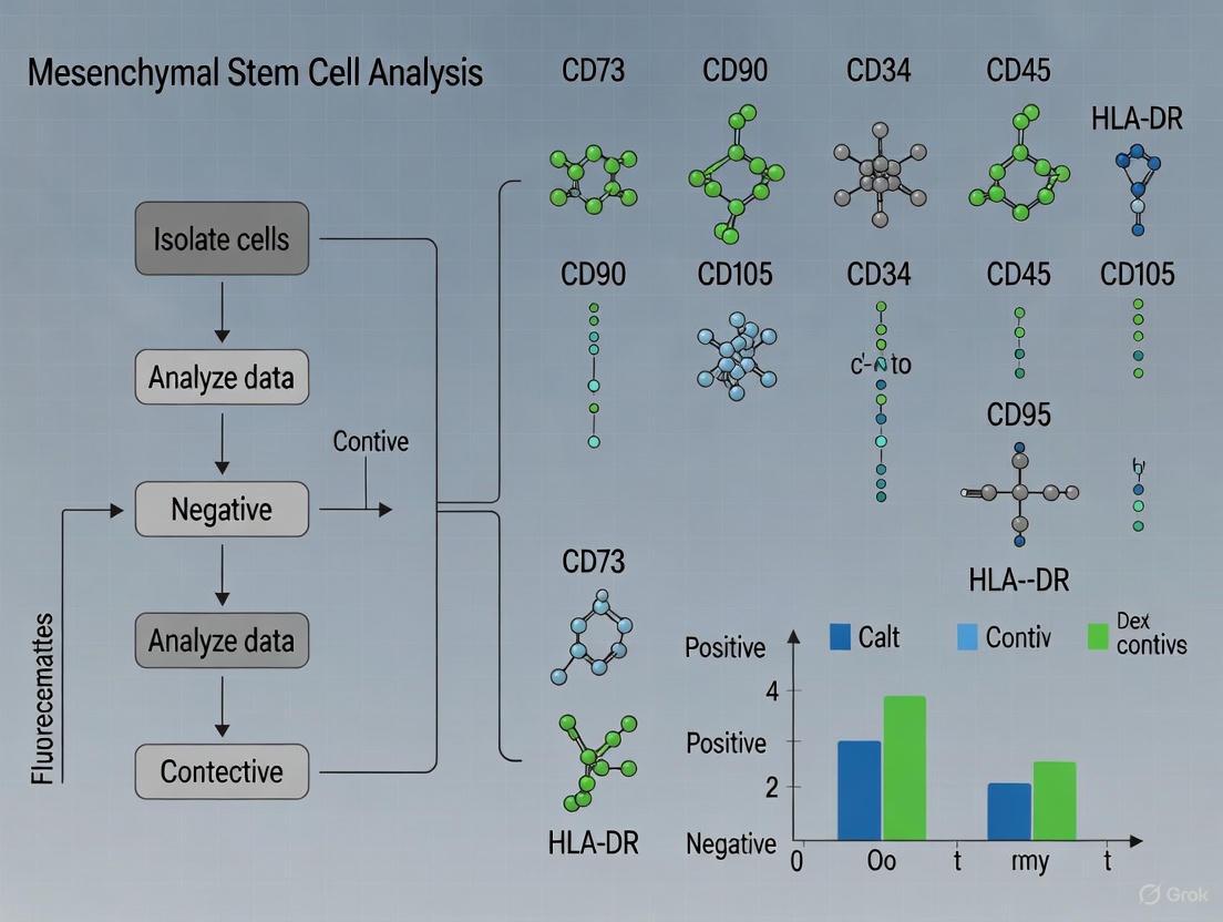
Comprehensive Guide to Mesenchymal Stem Cell CD Markers: Flow Cytometry Analysis for Research and Development
This comprehensive article provides researchers, scientists, and drug development professionals with essential knowledge and practical methodologies for mesenchymal stem cell (MSC) characterization using flow cytometry.
Comprehensive Guide to Mesenchymal Stem Cell CD Markers: Flow Cytometry Analysis for Research and Development
Abstract
This comprehensive article provides researchers, scientists, and drug development professionals with essential knowledge and practical methodologies for mesenchymal stem cell (MSC) characterization using flow cytometry. Covering foundational principles established by the International Society for Cellular Therapy (ISCT), the content details standardized protocols for CD marker analysis, troubleshooting strategies for common technical challenges, and advanced validation techniques. The guide also explores emerging biomarkers beyond classical panels and discusses the implications of MSC source variability on marker expression profiles, offering a complete framework for ensuring MSC quality and identity in preclinical and clinical applications.
Understanding MSC CD Markers: International Standards and Biological Significance
The field of mesenchymal stem cell (MSC) research has experienced exponential growth since the initial isolation and description of fibroblast-like, plastic-adherent cells from bone marrow by Friedenstein and colleagues in the 1970s [1] [2]. These cells, now universally known as mesenchymal stromal cells (or mesenchymal stem cells), possess multipotent differentiation capacity, self-renewal capability, and unique immunomodulatory properties that make them attractive candidates for regenerative medicine applications [3]. However, the rapid expansion of this field revealed a significant challenge: the inherent heterogeneity of MSC populations and the lack of standardized criteria for their identification and characterization. This heterogeneity stemmed from multiple factors, including differences in tissue sources (bone marrow, adipose tissue, umbilical cord, etc.), isolation methods, culture conditions, and donor variability [4] [5]. Without standardized characterization criteria, comparing results between laboratories and validating clinical outcomes became problematic, hindering scientific progress and clinical translation.
Recognizing this critical limitation, the International Society for Cellular Therapy (ISCT) established minimal criteria to define human MSCs in 2006, creating a foundational framework for the field [6] [1] [2]. These criteria provided three essential benchmarks for identifying MSCs, with surface antigen expression profiling through flow cytometry serving as a cornerstone for cell identification and quality control. This technical guide explores the ISCT minimum criteria with specific emphasis on surface antigen characterization, detailing the experimental protocols, technical considerations, and evolving understanding of MSC identity within the context of mesenchymal stem cell CD markers flow cytometry research.
The ISCT Minimal Criteria: A Three-Pillar Framework
The ISCT established a three-pronged approach to define human MSCs, which has been widely adopted by researchers and regulatory agencies worldwide. These criteria serve as the gold standard for verifying MSC identity and ensuring consistency across experiments and manufacturing processes [6] [2].
Plastic Adherence: Under standard culture conditions, MSCs must demonstrate adherence to plastic surfaces. This fundamental characteristic enables their selective isolation and expansion from heterogeneous tissue digests or aspirates through the removal of non-adherent cells [6] [1].
Multipotent Differentiation Potential: Validated MSCs must demonstrate the capacity to differentiate into osteoblasts, adipocytes, and chondroblasts under defined in vitro differentiation conditions. This tri-lineage differentiation potential confirms their mesenchymal origin and functional potency [6] [1] [2].
Specific Surface Antigen Expression Profile: MSCs must express a defined set of cell surface markers while lacking expression of others, as detailed in Table 1. This antigenic profile is primarily assessed using flow cytometry, which provides quantitative, single-cell resolution of marker expression [6] [7] [1].
Table 1: ISCT-Defined Positive and Negative Surface Marker Profile for Human MSCs
| Marker Category | Marker | Expression Requirement | Typical Expression | Biological Function |
|---|---|---|---|---|
| Positive Markers | CD105 (Endoglin) | ≥ 95% | >95% | Component of TGF-β receptor complex |
| CD73 (Ecto-5'-Nucleotidase) | ≥ 95% | >95% | Converts AMP to adenosine | |
| CD90 (Thy-1) | ≥ 95% | >95% | Cell-cell and cell-matrix interactions | |
| Negative Markers | CD45 | ≤ 2% | <2% | Pan-hematopoietic marker |
| CD34 | ≤ 2% | <2% | Hematopoietic progenitor marker | |
| CD14 / CD11b | ≤ 2% | <2% | Monocyte/macrophage markers | |
| CD79α / CD19 | ≤ 2% | <2% | B-cell markers | |
| HLA-DR | ≤ 2% | <2% | MHC Class II antigen |
The flow cytometry histograms below illustrate a typical analysis of bone marrow-derived MSCs, showing high expression of positive markers (CD73, CD90, CD105) and minimal expression of negative markers (CD14, CD19, CD45, HLA-DR) [6].
Diagram 1: The sequential workflow for defining MSC identity according to ISCT criteria.
Core Surface Antigens: Biological Functions and Technical Considerations
Positive Marker Biology and Significance
The ISCT-specified positive markers (CD105, CD73, CD90) are not merely identification tags; they play crucial functional roles in MSC biology. CD105 (Endoglin) functions as a coreceptor for transforming growth factor-beta (TGF-β), modulating signaling pathways involved in cellular proliferation and differentiation [2]. CD73 is an ecto-5'-nucleotidase that catalyzes the conversion of adenosine monophosphate (AMP) to adenosine, generating immunosuppressive adenosine that contributes to the renowned immunomodulatory properties of MSCs [2]. CD90 (Thy-1) is a glycophosphatidylinositol (GPI)-anchored glycoprotein involved in cell-cell and cell-matrix interactions, potentially influencing MSC migration and homing capabilities [2].
These markers collectively help distinguish MSCs from hematopoietic cell populations that contaminate initial tissue isolates. Their consistent expression (≥95% positive) across MSC populations from various donors and passages provides a reliable benchmark for quality control in both research and clinical settings [6] [1].
Negative Marker Rationale and Hematopoietic Exclusion
The negative markers specified by the ISCT primarily serve to exclude hematopoietic cell contaminants. CD45 is a pan-leukocyte marker expressed on all hematopoietic cells except erythrocytes and platelets. CD34 is typically expressed on hematopoietic stem and progenitor cells, though its expression can vary in MSCs from certain tissue sources like adipose tissue [2]. CD14 and CD11b are markers of monocytes and macrophages, while CD79α and CD19 identify B lymphocytes [6] [1]. HLA-DR, a major histocompatibility complex class II molecule, is typically absent on unstimulated MSCs but can be induced by inflammatory stimuli like interferon-gamma (IFN-γ) [1].
The ≤2% expression threshold for these negative markers ensures the exclusion of hematopoietic contaminants that could confound experimental results or pose safety risks in clinical applications. However, researchers should note that some variations can occur based on tissue source and culture conditions [2].
Experimental Protocol: Flow Cytometric Analysis of MSC Surface Antigens
Sample Preparation and Staining Protocol
Proper sample preparation is critical for obtaining accurate flow cytometry results. The following protocol has been validated for human bone marrow-derived MSCs [4] [6]:
- Cell Harvesting: Harvest subconfluent (70-80%) MSCs at passage 3-5 using 0.25% trypsin/EDTA. Neutralize trypsin activity with complete culture medium containing serum.
- Cell Washing: Wash cells twice with phosphate-buffered saline (PBS) containing 1% bovine serum albumin (BSA) to remove residual serum proteins and trypsin.
- Antibody Staining: Resuspend cell pellets (approximately 1×10^6 cells per tube) in flow cytometry staining buffer (PBS with 1% BSA and 0.1% sodium azide). Add fluorochrome-conjugated antibodies according to manufacturer recommendations. Include appropriate isotype controls for each fluorochrome to establish background staining levels.
- Incubation: Incubate stained cells for 20 minutes in the dark at 4°C to prevent internalization of surface antigens and fluorochrome degradation.
- Final Wash and Resuspension: Wash cells twice with staining buffer to remove unbound antibody. Resuspend final cell pellets in 300-500µl of staining buffer containing a viability dye (e.g., 7-AAD or propidium iodide) to exclude dead cells from analysis.
- Data Acquisition: Analyze samples using a flow cytometer calibrated with appropriate compensation controls. Acquire a minimum of 20,000 events per sample to ensure statistical significance [4].
Gating Strategy and Data Analysis
A systematic gating strategy is essential for accurate interpretation of flow cytometry data:
- Viability Gate: Exclude dead cells and debris based on forward scatter (FSC) versus side scatter (SSC) characteristics and viability dye staining.
- Singlets Gate: Select single cells using FSC-height versus FSC-area to exclude cell doublets and aggregates that could cause inaccurate marker expression quantification.
- Analysis Gate: Analyze fluorescence intensity for each marker compared to isotype controls. Set positive/negative thresholds based on isotype control staining (typically at 2% background).
- Quantification: Calculate the percentage of positive cells for each marker. Valid MSC preparations should demonstrate ≥95% expression for CD105, CD73, and CD90, and ≤2% expression for negative markers [6].
Diagram 2: Sequential gating strategy for flow cytometric analysis of MSC surface markers.
Beyond the Minimum: Limitations and Evolving Perspectives
Known Limitations of the ISCT Criteria
While the ISCT criteria provide a crucial foundational framework, extensive research has revealed several limitations. A significant concern is that the standard marker panel does not necessarily predict MSC functional potency, including differentiation capacity, proliferation potential, or secretory profile [4]. Studies have demonstrated that MSC populations satisfying all ISCT criteria can exhibit markedly different in vivo bone-forming capacity [4]. Furthermore, the criteria were originally established for bone marrow-derived MSCs and may not fully accommodate the biological variations in MSCs from other tissues. For instance, adipose-derived MSCs may initially express CD34, which is lost upon culture expansion [2].
The potential for fibroblast contamination presents another challenge, as fibroblasts share plastic adherence and similar morphology with MSCs, and can express overlapping surface markers like CD90 and CD73 [8]. Research has identified potential discriminators, such as higher expression of CD106, CD146, and CD166 in MSCs compared to fibroblasts, but these are not included in the minimal criteria [8].
Tissue-Specific Variations and Additional Markers
The tissue source significantly influences MSC surface marker expression, necessitating additional characterization beyond the core ISCT panel for certain applications. The table below summarizes key tissue-specific variations and supplemental markers documented in the literature.
Table 2: Tissue-Specific Marker Variations and Additional Characterization Markers
| Tissue Source | Marker Variations | Additional Enrichment Markers | Research Applications |
|---|---|---|---|
| Bone Marrow | Classical ISCT profile | STRO-1, CD271, CD146 | Gold standard for comparison |
| Adipose Tissue | Initial CD34+ expression | CD36 (positive), CD106 (negative) | Volume-intensive applications |
| Umbilical Cord | Standard ISCT profile | Higher immunosuppressive potential | Perinatal source applications |
| All Sources | Heterogeneous expression | SSEA-4, CD49a, PDGFR-α/β | Potency prediction |
Enhancing Potency Assessment Through Additional Markers
Research continues to identify supplemental markers that correlate with MSC functional potency. STRO-1 and platelet-derived growth factor receptor-alpha (PDGFR-α) have shown preferential expression on MSCs with high growth capacity and enhanced osteogenic potential [4]. Similarly, CD271 is considered one of the most specific markers for bone marrow-derived MSCs, identifying populations with enhanced clonogenic capacity [8] [2]. The expression of specific mRNA transcripts like TWIST-1 and DERMO-1 has also correlated with superior growth capacity and osteogenic potential in some studies [4].
These findings highlight the evolving nature of MSC characterization and the potential for future refinements to the standard criteria that incorporate potency markers alongside identity markers.
Successful characterization of MSCs according to ISCT criteria requires specific reagents and methodological approaches. The following toolkit summarizes essential resources for comprehensive MSC analysis.
Table 3: Essential Research Reagent Solutions for MSC Characterization
| Reagent Category | Specific Examples | Research Application | Technical Notes |
|---|---|---|---|
| Positive Marker Antibodies | Anti-CD105, Anti-CD73, Anti-CD90 | Flow cytometry, immunocytochemistry | Confirm species reactivity |
| Negative Marker Antibodies | Anti-CD45, Anti-CD34, Anti-CD14/CD11b, Anti-CD19/CD79α, Anti-HLA-DR | Flow cytometry, hematopoietic exclusion | Use cocktail for efficiency |
| Functional Assay Kits | Osteo-, Adipo-, Chondro-genesis kits | Trilineage differentiation confirmation | Include specific stains |
| Flow Cytometry Resources | Cell viability dyes, compensation beads, isotype controls | Instrument calibration and control | Critical for data accuracy |
| Culture Media | Defined serum-free/xeno-free media | Expansion and maintenance | Reduces batch variability |
The ISCT minimum criteria for defining MSC identity through surface antigens represent a foundational framework that has brought essential standardization to the field. The specified panel of positive (CD105, CD73, CD90) and negative (CD45, CD34, CD14/CD11b, CD19/CD79α, HLA-DR) markers provides a critical benchmark for verifying MSC identity and ensuring consistency across research and clinical applications. Flow cytometric analysis of these surface antigens remains an indispensable tool for quality control and experimental validation.
However, the evolving understanding of MSC biology continues to refine these standards. Researchers must recognize that while the ISCT criteria establish cellular identity, they do not fully predict functional potency or accommodate all tissue-specific variations. The future of MSC characterization likely involves integrated assessment panels that combine surface marker profiling with functional potency assays, molecular analyses, and perhaps physical characteristics like cell size and stiffness [4] [2]. As the field advances toward more sophisticated clinical applications, the rigorous application of these standards—while remaining open to their ongoing refinement—will be essential for generating reproducible, reliable scientific data and ensuring the safety and efficacy of MSC-based therapies.
The characterization of human Mesenchymal Stromal Cells (MSCs) fundamentally relies on a set of core positive surface markers established by the International Society for Cell Therapy (ISCT). According to the minimal criteria set forth by the ISCT, MSCs must demonstrate plastic-adherent properties under standard culture conditions, possess tri-lineage differentiation potential (osteogenic, adipogenic, and chondrogenic), and express specific surface antigens, with ≥95% of the population positive for CD73, CD90, and CD105, while lacking expression of hematopoietic markers such as CD45, CD34, CD14, CD11b, CD79α, and HLA-DR [9] [10]. These three glycoproteins—CD73, CD90, and CD105—are not merely identifiers but are functionally integral to the biology of MSCs, influencing their immunomodulatory capacity, migratory behavior, and role in tissue regeneration. In the context of flow cytometry research for drug development and advanced therapy medicinal products, precise understanding and detection of this triad is paramount for ensuring the identity, purity, and functional potency of MSC-based products. This technical guide delves into the biological functions, detection methodologies, and research applications of these core markers, providing a foundational resource for scientists and development professionals.
Detailed Biological Functions and Signaling Pathways
The following diagram illustrates the key signaling pathways and cellular processes associated with CD73, CD90, and CD105 on mesenchymal stromal cells.
CD73 (Ecto-5'-Nucleotidase)
Biological Function: CD73, also known as ecto-5'-nucleotidase, is a cell surface glycosylphosphatidylinositol (GPI)-anchored glycoprotein that catalyzes the rate-limiting step in the phosphohydrolysis of extracellular nucleotides [9]. It functions as a key enzyme in the purinergic signaling pathway, converting adenosine monophosphate (AMP) to bioactive adenosine. This enzymatic activity is a central mechanism through which MSCs exert their immunomodulatory and anti-inflammatory effects [9]. The adenosine produced binds to adenosine receptors (A1, A2A, A2B, and A3) on the surface of immune cells, such as T lymphocytes and natural killer (NK) cells, suppressing their proliferation and cytotoxic activity, thereby creating an immunosuppressive microenvironment. This function is critical for the therapeutic application of MSCs in graft-versus-host disease (GVHD) and other inflammatory conditions.
CD90 (Thy-1)
Biological Function: CD90, or Thy-1, is a GPI-anchored glycoprotein belonging to the immunoglobulin superfamily. It is one of the most abundantly expressed surface markers on MSCs, present on >95% of cultured cells [11]. CD90 is involved in a diverse array of cellular processes, including cell-cell and cell-matrix adhesion, migration, and apoptosis [9]. Its expression is strongly associated with the "stemness" and undifferentiated state of MSCs in vitro. CD90 mediates interactions with integrins and other ligands on adjacent cells and the extracellular matrix, facilitating the homing of MSCs to sites of injury. While universally expressed in vitro, it is important to note that its expression in vivo can be heterogeneous, and it is acquired during the process of in vitro culture [11].
CD105 (Endoglin)
Biological Function: CD105, also known as endoglin, is a homodimeric transmembrane glycoprotein that acts as a component of the transforming growth factor-beta (TGF-β) receptor complex, specifically binding TGF-β1 and TGF-β3 [9]. As a coreceptor, CD105 modulates TGF-β signaling, a pathway essential for angiogenesis, cardiovascular development, and extracellular matrix (ECM) synthesis. The pro-angiogenic role of CD105 makes it a critical marker for MSCs involved in vascular repair and wound healing. Its expression is a defining feature of the MSC immunophenotype and is a key parameter in flow cytometry panels for MSC identification.
Quantitative Marker Prevalence Across Tissues
The expression profile of the core positive markers is consistent across MSCs derived from various somatic and perinatal tissues, though subtle variations in prevalence have been reported. The table below summarizes the frequency of reporting for these markers in studies related to the skeletal system, as identified in a recent scoping review.
Table 1: Prevalence of Core Positive MSC Markers in Scientific Literature (Skeletal System Focus)
| Marker | Alternative Name | Reported Prevalence in Literature | Primary Functional Role |
|---|---|---|---|
| CD105 | Endoglin | 82.9% | TGF-β receptor complex; Angiogenesis |
| CD90 | Thy-1 | 75.0% | Cell adhesion, migration, and apoptosis |
| CD73 | Ecto-5'-nucleotidase | 52.0% | Purinergic signaling; Immunomodulation via adenosine production |
Data derived from a scoping review of MSC markers in the skeletal system (1994-2021) [10].
This quantitative data underscores that while all three markers are considered essential, their reported prevalence in scientific literature varies, with CD105 being the most frequently cited. This comprehensive analysis confirms the universal adoption of the ISCT criteria in defining MSC populations for research and development.
Experimental Protocols for Detection and Qualification
Accurate detection of CD73, CD90, and CD105 is essential for MSC characterization. Flow cytometry remains the gold-standard technique due to its ability to provide quantitative, single-cell analysis. The following workflow details a standardized protocol for the flow cytometric analysis of these markers.
Detailed Flow Cytometry Protocol
1. Cell Preparation and Staining
- Cell Harvesting: Culture MSCs to 70-80% confluence. Wash with phosphate-buffered saline (PBS) and detach using a non-enzymatic dissociation reagent like Accutase to preserve surface epitopes. Neutralize the enzyme with complete culture medium [12] [11].
- Cell Washing and Filtering: Pellet cells by centrifugation at 300 × g for 5 minutes. Resuspend the cell pellet in PBS and pass through a 35 μm nylon mesh filter to obtain a single-cell suspension and prevent clogging of the flow cytometer [12].
- Antibody Staining: Dispense cell aliquots (approximately 1x10^5 to 5x10^5 cells per tube) in 100 μL of staining buffer (e.g., PBS with 2% FBS). Add fluorochrome-conjugated antibodies against CD73, CD90, and CD105, as well as antibodies for negative markers (CD34, CD45, CD14, CD19, HLA-DR). Include isotype-matched control antibodies for compensation and gating. Incubate for 20-30 minutes at room temperature, shielded from light [12] [13].
- Post-staining Washing: Wash cells twice with staining buffer to remove unbound antibody. Pellet cells at 300 × g for 5 minutes and carefully aspirate the supernatant [12].
2. Data Acquisition and Analysis
- Resuspension and Acquisition: Resuspend the final cell pellet in 500 μL of flow cytometry staining buffer. Acquire data on a flow cytometer (e.g., BD FACSCalibur or spectral analyzer like Cytek Northern Lights) with a minimum of 20,000 events recorded per sample to ensure statistical significance [12] [11].
- Gating Strategy: Use forward scatter (FSC) vs. side scatter (SSC) to gate on the viable cell population. Exclude doublets using FSC-A vs. FSC-H. Analyze fluorescence in channels corresponding to the fluorochromes used. The MSC population is defined as ≥95% positive for CD73, CD90, and CD105 and ≤2% positive for any of the hematopoietic lineage markers [9].
Experimental Workflow Visualization
The following diagram summarizes the key steps in the flow cytometry workflow for MSC characterization.
The Scientist's Toolkit: Research Reagent Solutions
Successful experimentation requires a suite of validated reagents and materials. The following table catalogs essential tools for the flow cytometric analysis of core MSC markers.
Table 2: Essential Research Reagents for MSC Marker Analysis
| Reagent / Material | Specific Example | Function in Experiment |
|---|---|---|
| Anti-human CD73 Antibody | Fluorochrome-conjugated clone (e.g., AD2) | Detection of CD73 surface expression |
| Anti-human CD90 Antibody | Fluorochrome-conjugated clone (e.g., 5E10) | Detection of CD90 surface expression |
| Anti-human CD105 Antibody | Fluorochrome-conjugated clone (e.g., 266) | Detection of CD105 surface expression |
| Lineage Negative Cocktail | Anti-CD34, CD45, CD14, CD19, HLA-DR | Confirmation of absence of hematopoietic markers |
| Flow Cytometry Staining Buffer | PBS with 2% Fetal Bovine Serum (FBS) | Provides protein background to reduce non-specific antibody binding |
| Cell Dissociation Reagent | TrypLE or Accutase | Gentle detachment of adherent MSCs while preserving surface markers |
| Cell Strainer | 35 μm or 70 μm nylon mesh | Generation of a single-cell suspension for accurate flow analysis |
| Flow Cytometer | BD FACSCalibur, Cytek Northern Lights | Instrument for quantitative analysis of cell surface marker expression |
Information compiled from multiple methodological sources [12] [13] [11].
Critical Considerations for Research and Development
- Phenotypic Convergence In Vitro: A critical finding for researchers is that the uniform expression of CD73, CD90, and CD105 is largely an in vitro-acquired phenotype. A 2024 study demonstrated that primary cultures universally express CD73 and CD90 at high levels (>95%) irrespective of their expression in the original tissue (ex vivo), indicating a phenotypic convergence during plastic adherence and expansion [11]. This underscores that in vitro marker expression does not necessarily reflect the in vivo state of the source cells.
- Functional Correlation: While these markers are essential for identification, their presence alone does not guarantee functional potency. The biological functions of these molecules suggest that their expression is a prerequisite for key MSC activities like immunomodulation (CD73) and response to growth factors (CD105). Therefore, their detection can be considered a proxy for functional potential, which should be confirmed with specific potency assays (e.g., T-cell suppression for immunomodulation) [9].
- Impact of Culture Conditions: The expression of these markers can be influenced by culture conditions, including the choice of culture medium, degree of confluence, and passage number. Standardization of culture protocols is, therefore, essential for generating reproducible and reliable flow cytometry data [11].
The triad of CD73, CD90, and CD105 forms the non-negotiable core of the MSC immunophenotype as defined by the ISCT. Beyond their role as mere identifiers, each marker contributes significantly to the fundamental biological processes of immunomodulation, cell adhesion and migration, and growth factor response. For researchers and drug development professionals, robust detection of these markers via standardized flow cytometry protocols is a critical quality control step. However, it is vital to recognize the phenomenon of phenotypic convergence in vitro and to complement immunophenotypic data with functional potency assays. As the field advances toward more complex MSC-based therapeutics, a deep and nuanced understanding of these core markers remains the bedrock of rigorous research and successful product development.
The definitive identification of mesenchymal stem cells (MSCs) in research and clinical-grade manufacturing requires not only confirming the presence of positive marker expression but also rigorously excluding hematopoietic contamination. The International Society for Cellular Therapy (ISCT) established minimal criteria for defining human MSCs, which include adherence to plastic and specific differentiation potential, but crucially, also mandate the lack of expression of a panel of hematopoietic and leukocyte markers [14] [10]. This panel includes CD34, CD45, CD11b, CD19, and HLA-DR [8]. The critical application of these "negative markers" ensures the purity of the MSC population, prevents misinterpretation of experimental results, and is a fundamental safety step in therapeutic development by eliminating unwanted immune-reactive cells from the final product. This guide details the technical execution and scientific rationale behind using these markers to exclude hematopoietic contamination within the broader context of MSC flow cytometry research.
The ISCT Criteria and the Hematopoietic Exclusion Panel
The ISCT criteria serve as the foundational standard for the field, providing a benchmark for comparing MSCs from different tissue sources and laboratories. The negative marker panel is designed to identify and exclude cells of hematopoietic lineage.
- CD45: A pan-leukocyte marker, expressed on all nucleated cells of the hematopoietic system except platelets and their precursors.
- CD34: A marker primarily associated with hematopoietic stem and progenitor cells, as well as vascular endothelial cells.
- CD11b: Expressed on monocytes, granulocytes, natural killer cells, and a subset of lymphocytes.
- CD19: A specific B-lymphocyte lineage marker.
- HLA-DR: A major histocompatibility complex (MHC) class II molecule expressed on antigen-presenting cells (e.g., B-cells, monocytes, dendritic cells).
The following table summarizes the key negative markers and the specific hematopoietic cell types they target for exclusion from MSC cultures.
Table 1: Critical Negative Markers for Hematopoietic Contamination in MSC Analysis
| Marker | Primary Hematopoietic Cell Types Excluded | Significance in MSC Purity |
|---|---|---|
| CD45 | All leukocytes (e.g., lymphocytes, monocytes, neutrophils) | Pan-hematopoietic exclusion; critical for identifying overall immune cell contamination. |
| CD34 | Hematopoietic stem/progenitor cells, endothelial cells | Excludes primitive hematopoietic populations and vasculature cells. |
| CD11b | Myeloid cells (e.g., monocytes, macrophages, granulocytes) | Specifically targets the monocyte-macrophage lineage. |
| CD19 | B lymphocytes | Excludes humoral immune cell components. |
| HLA-DR | Antigen-presenting cells (e.g., B-cells, dendritic cells, monocytes) | Indicates immunologically active cells; typically negative on undifferentiated MSCs. |
Experimental Protocols for Marker Analysis
Sample Preparation and Cell Harvesting
For in vitro-expanded MSCs, subconfluent cells (typically ≤80% confluence) should be used to avoid differentiation-induced changes in marker expression [8]. Cells are harvested using a dissociation reagent such as Accutase or 0.25% trypsin-EDTA [14] [8]. Following detachment, cells must be washed with a buffer, such as PBS, and passed through a cell strainer (e.g., 70 µm) to ensure a single-cell suspension, which is critical for accurate flow cytometry analysis [14] [15].
Staining Protocol for Flow Cytometry
The following workflow outlines the key steps for staining and analysis, from sample preparation to data acquisition. Specific details on antibody cocktails and instrumentation are provided in the subsequent sections.
- Antibody Cocktail Preparation: Antibodies are diluted in an appropriate staining buffer (e.g., 2% Fetal Bovine Serum (FBS) and 1 mM EDTA in PBS) [14]. For complex panels, especially those utilizing bright fluorochromes or multiple markers, the use of a Brilliant Stain Buffer is recommended to prevent fluorochrome polymer formation and ensure accurate detection [14].
- Staining Incubation: Cell suspensions are incubated with the antibody cocktail for 20-30 minutes at 4°C, protected from light [14] [8].
- Washing and Resuspension: After incubation, cells are washed with staining buffer or PBS to remove unbound antibody. The final cell pellet is resuspended in a suitable volume of buffer for acquisition on the flow cytometer. Including a viability dye (e.g., 7-AAD or DAPI) is essential to gate out dead cells, which can cause non-specific antibody binding and inaccurate results.
Instrumentation and Data Acquisition
Spectral flow cytometers are powerful tools as they enable the simultaneous evaluation of dozens of markers from a single sample, a capability highlighted in recent studies [14]. However, conventional flow cytometers are also entirely suitable for this analysis. Researchers should perform appropriate compensation controls using compensation beads or singly stained cells to correct for spectral overlap. Data acquisition should collect a sufficient number of events (e.g., ≥10,000 events in the live, single-cell gate) for robust statistical analysis.
Data Interpretation and Analytical Considerations
Gating Strategy and Establishing Positivity Thresholds
The established ISCT threshold for negative markers is that less than 2% of the population should be positive for any of these markers, while simultaneously, over 95% of the population must express the positive markers (CD73, CD90, CD105) [14] [8]. The gating strategy must be sequential:
- Viability Gate: Select events negative for a viability dye.
- Singlets Gate: Select single cells based on FSC-A vs. FSC-H.
- MSC Phenotype Gate: From the singlets, create a final gate for cells that are positive for CD73, CD90, and CD105.
- Hematopoietic Exclusion: The population within the MSC Phenotype Gate must then be evaluated for the expression of CD34, CD45, CD11b, CD19, and HLA-DR. The final, validated MSC population will show minimal signal (typically <2%) for these negative markers.
Critical Considerations and Pitfalls
- Tissue Source Variability: While the ISCT criteria are universal, the expression profile of some markers can vary depending on the tissue source. For instance, native MSCs in adipose tissue have been reported to express CD34, which is lost upon in vitro culture [8] [15]. Therefore, analysis of freshly isolated cells versus cultured cells may show significant differences [14].
- CD34 Expression Dynamics: CD34 loss during the transition to in vitro culture is a well-documented phenomenon [14]. A study on skeletal cells found that primary cultures universally expressed CD73 and CD90 but lacked CD34, "irrespective of the expression of these markers ex vivo indicating phenotypic convergence in vitro" [14]. This highlights that in vitro marker expression may not faithfully represent the in vivo state.
- Culture Conditions: The culture microenvironment, including the medium composition (e.g., use of FBS vs. human platelet lysate) and the degree of confluence, can affect cell growth and marker expression [14] [15]. Consistency in culture conditions is vital for reproducible flow cytometry data.
Table 2: Research Reagent Solutions for Flow Cytometric Analysis of MSCs
| Reagent / Material | Function / Application | Example from Literature |
|---|---|---|
| Collagenase P / Type I | Enzymatic digestion of solid tissues (e.g., bone, cartilage, adipose) for initial cell isolation. [14] [15] | |
| StemPro Accutase | Gentle cell dissociation reagent for harvesting adherent MSCs while maintaining cell surface integrity. [14] | |
| Brilliant Stain Buffer | Prevents fluorochrome cross-talk and polymer formation in multi-color flow cytometry panels, ensuring accurate detection. [14] | |
| Human Platelet Lysate (hPL) | A xeno-free supplement for GMP-compliant clinical-grade expansion of MSCs, which can influence growth and marker expression. [15] | |
| Fluorophore-conjugated mAbs | Primary antibodies for detection of CD markers. Panels are built based on instrument configuration and required markers. [14] [8] |
The rigorous application of negative marker analysis using CD34, CD45, CD11b, CD19, and HLA-DR is a non-negotiable step in MSC characterization. It is a critical quality control checkpoint that confirms the absence of hematopoietic contaminants, thereby ensuring the identity and purity of the MSC population under investigation. As the field advances towards more complex multi-color panels and clinical-grade manufacturing, a deep and practical understanding of this analytical procedure remains fundamental for researchers, scientists, and drug development professionals working with mesenchymal stromal cells.
Mesenchymal stromal cells (MSCs) represent a cornerstone of regenerative medicine and therapeutic development due to their multipotent differentiation capacity, immunomodulatory properties, and relative ease of isolation from various tissue sources. The International Society for Cell & Gene Therapy (ISCT) has established minimal criteria for defining MSCs, including plastic adherence, specific surface marker expression, and trilineage differentiation potential [5] [10]. While these criteria provide a foundational framework, growing evidence reveals that MSCs derived from different tissue sources exhibit distinct biological properties, marker expression profiles, and functional capabilities that significantly impact their therapeutic suitability for specific clinical applications [16] [17] [18]. This technical guide provides an in-depth analysis of MSC variations across four primary sources—bone marrow, adipose tissue, umbilical cord, and placenta—framed within the context of CD marker research using flow cytometry, to inform researchers and drug development professionals in their experimental and therapeutic designs.
Standard Immunophenotypic Definition
According to ISCT criteria, human MSCs must demonstrate ≥95% expression of CD73, CD90, and CD105, and ≤2% expression of hematopoietic markers (CD45, CD34, CD14 or CD11b, CD79a or CD19, and HLA-DR) when analyzed by flow cytometry [5] [10]. These classical markers represent the minimal baseline for characterization, though they cannot distinguish between MSCs from different tissue origins [10] [18]. A scoping review of MSC markers in skeletal system research found CD105 (82.9%), CD90 (75.0%), and CD73 (52.0%) to be the most frequently used identifiers, followed by CD44 (42.1%), CD166 (30.9%), CD29 (27.6%), STRO-1 (17.7%), CD146 (15.1%), and CD271 (7.9%) [10].
Tissue-Specific Marker Variations
Beyond the classical markers, research has identified non-classical markers that exhibit variability across tissue sources and may inform functional potential. Studies of clinical-grade adipose-derived MSCs (AD-MSCs) have identified CD36, CD163, CD271, CD200, CD273, CD274, CD146, CD248, and CD140B as potentially discriminative markers for more detailed characterization [15]. Similarly, CD106 (VCAM-1) has been identified as a distinctive marker for chorionic plate-derived MSCs (81.10% ± 12.28%) with moderate expression in umbilical cord MSCs (12.07% ± 11.43%) and minimal expression in amniotic membrane (4.27% ± 4.39%) and decidua parietalis MSCs (0%) [18].
Table 1: Quantitative Expression of Primary CD Markers Across MSC Sources
| CD Marker | Bone Marrow | Adipose Tissue | Umbilical Cord | Placenta (Fetal) | Placenta (Maternal) |
|---|---|---|---|---|---|
| CD73 | ≥95% [10] | ≥95% [15] | ≥95% [18] | ≥95% [18] | ≥95% [18] |
| CD90 | ≥95% [10] | ≥95% [15] | ≥95% [18] | ≥95% [18] | ≥95% [18] |
| CD105 | ≥95% [10] | ≥95% [15] | ≥95% [18] | ≥95% [18] | ≥95% [18] |
| CD44 | 42.1% [10] | ≥95% [15] | High [19] | High [18] | High [18] |
| CD106 | Variable [18] | Low [15] | 12.07% [18] | 81.10% (CP) [18] | 0% (DP) [18] |
| CD271 | 7.9% [10] | Present [15] | Low [18] | Low [18] | Low [18] |
| CD34 | ≤2% [10] | ≤2% [15] | ≤2% [18] | ≤2% [18] | ≤2% [18] |
| CD45 | ≤2% [10] | ≤2% [15] | ≤2% [18] | ≤2% [18] | ≤2% [18] |
Table 2: Functional Characteristics and Tissue Yields of Different MSC Sources
| Parameter | Bone Marrow | Adipose Tissue | Umbilical Cord | Placenta |
|---|---|---|---|---|
| Frequency in Tissue | 0.001-0.01% [17] [15] | 1-10% [15] | Varies by compartment [5] | 0.34-1.52 million cells/g [18] |
| Proliferation Capacity | Moderate [20] | High [16] | High (PDT: 28.34±2.89h) [18] | Variable (AM:35.19±9.28h, DP:48.01±8.26h) [18] |
| Osteogenic Potential | High [21] [20] | Moderate [16] | Moderate [20] [18] | Low-Moderate [18] |
| Chondrogenic Potential | High [20] | Moderate [16] | Moderate [20] | Low-Moderate [18] |
| Adipogenic Potential | Moderate [21] | High [16] | Low [20] [18] | Low-Moderate [18] |
| Angiogenic Potential | Moderate [16] | High [16] | Moderate [16] | High (VEGF) [18] |
| Immunomodulatory Strength | Strong [17] | Strong (especially T cells) [17] | Moderate [17] [18] | Variable by source [18] |
Experimental Protocols for MSC Characterization
Flow Cytometry Analysis Protocol
Purpose: To characterize MSC surface marker expression according to ISCT criteria and identify tissue-specific signatures.
Sample Preparation:
- Harvest MSCs at 80-90% confluence using TrypLE Express Enzyme or 0.25% trypsin-EDTA [16] [19]
- Wash cells with DPBS and pass through a 70μm strainer to obtain single-cell suspension [16]
- Centrifuge at 400-450 × g for 5 minutes and resuspend in FACS buffer (DPBS with 1mM EDTA and 5% mouse serum) [16]
- Aliquot 1×10^5 cells per tube for antibody staining [16]
Antibody Staining:
- Incubate cells with fluorescently labeled antibodies (1:100 dilution) for 30-60 minutes at 4°C in the dark [16] [19]
- Wash stained cells with DPBS or FACS buffer and centrifuge at 450 × g for 4 minutes [16]
- Resuspend in FACS buffer for analysis [16]
- Include unstained and isotype controls for gating and background determination [16]
Data Acquisition and Analysis:
- Analyze samples using flow cytometer (e.g., FACS Aria II, BD FACS Calibur) [16] [19]
- Collect at least 10,000 events per sample [16]
- Set gating based on unstained/isotype controls [16]
- Express results as percentage of positive cells for each marker [19]
Multilineage Differentiation Assessment
Osteogenic Differentiation:
- Culture MSCs in 6-well plates until 80% confluent [16] [21]
- Induce with osteogenic medium (DMEM with 10% FBS, 50μg/ml ascorbic acid 2-phosphate, 10nM dexamethasone, and 10mM β-glycerol phosphate) or commercial OsteoMAX-XF medium [16] [21]
- Maintain for 21 days with medium changes every 3-4 days [16] [21]
- Fix cells with 4% paraformaldehyde for 30 minutes at room temperature [16]
- Stain calcium deposits with 2% Alizarin Red S solution (pH 4.1-4.3) for 3-10 minutes [16] [21]
- Wash with distilled water and visualize mineralized matrix [16]
Adipogenic Differentiation:
- Culture MSCs to confluence in 6-well plates [16]
- Induce with adipogenic medium (DMEM with 10% FBS, 50μg/ml indomethacin, 100nM dexamethasone, and 50μg/ml ascorbic acid 3-phosphate) or commercial StemPro adipogenesis kit [16] [21]
- Maintain for 21 days with regular medium changes [16]
- Fix cells with 4% paraformaldehyde for 30 minutes [16]
- Stain lipid vacuoles with 0.3-0.5% Oil Red O solution in isopropanol for 30 minutes [16] [21]
- Wash with distilled water and visualize lipid accumulation [16]
Chondrogenic Differentiation:
- Pellet 2.5×10^5 MSCs by centrifugation [20]
- Culture in chondrogenic medium (DMEM with 1% FBS, 50μg/ml ascorbic acid, 10nM dexamethasone, and 10ng/mL TGF-β3) [20]
- Maintain for 21-28 days with medium changes every 3-4 days [20]
- Process pellets for histology (paraffin embedding, sectioning) [20]
- Stain with Alcian Blue or Safranin O for proteoglycan detection [20]
Signaling Pathways and Functional Mechanisms
The therapeutic effects of MSCs are mediated through complex signaling pathways that vary by tissue source. Proteomic analyses have identified significant differences in pathways related to cell migration, adhesion, and Wnt signaling between MSCs from different origins [16]. Additionally, cytokine secretion profiles vary substantially, with implications for immunomodulation and tissue regeneration capacities [18].
Diagram 1: MSC Signaling Pathways and Functional Mechanisms. Signaling pathways and secretory profiles vary significantly between MSC tissue sources, influencing their functional capabilities in tissue repair, immunomodulation, and vascularization [16] [18].
MSC Characterization Workflow
The comprehensive characterization of MSCs from different tissue sources requires a systematic approach encompassing isolation, expansion, phenotypic verification, and functional validation.
Diagram 2: Comprehensive MSC Characterization Workflow. The systematic process for isolating, expanding, and characterizing MSCs from different tissue sources, including tissue-specific isolation methods and standardized validation approaches [16] [15] [5].
The Scientist's Toolkit: Research Reagent Solutions
Table 3: Essential Research Reagents for MSC Characterization
| Reagent Category | Specific Products | Application in MSC Research |
|---|---|---|
| Isolation Enzymes | Collagenase Type I/II [17] [15], TrypLE Express [16], Accutase [17] | Tissue dissociation and cell harvesting |
| Culture Media | DMEM/F12 [16] [21], α-MEM [15] | MSC expansion and maintenance |
| Serum Supplements | Fetal Bovine Serum (FBS) [16] [21], Human Platelet Lysate (hPL) [15] | Cell growth and proliferation |
| Differentiation Kits | OsteoMAX-XF [16], StemPro Adipogenesis Kit [16] | Trilineage differentiation assays |
| Flow Cytometry Antibodies | CD73, CD90, CD105, CD44, CD166 [16] [19]; CD34, CD45, CD14 [16] [19] | Immunophenotypic characterization |
| Detection Reagents | Alizarin Red S [16] [21], Oil Red O [16] [21] | Differentiation capacity validation |
| Analysis Kits | ELISA kits (HGF, VEGF, PGE2, TGF-β1) [18] | Secretory profile quantification |
Discussion and Research Implications
The systematic comparison of MSC sources reveals critical considerations for both research and therapeutic development. Bone marrow-derived MSCs remain the gold standard but present limitations in cell yield and donor morbidity [17] [15]. Adipose tissue provides abundant MSC numbers with strong immunomodulatory properties, particularly for T-cell inhibition [17] [15]. Umbilical cord and placental tissues offer fetal-derived MSCs with enhanced proliferative capacity and distinct secretory profiles, though with variations based on specific tissue compartments [20] [18].
From a technical perspective, flow cytometry panel design should incorporate both classical ISCT markers and tissue-specific identifiers (e.g., CD106 for chorionic plate MSCs, CD271 for bone marrow MSCs) to fully characterize cellular populations [10] [18] [22]. The functional correlations of these markers continue to be elucidated, with emerging evidence linking specific profiles to differential therapeutic efficacies in particular disease models [16] [18].
For drug development professionals, these source-dependent variations have significant implications for manufacturing consistency and potency assay development. The selection of MSC sources should align with target mechanisms of action—whether direct differentiation, immunomodulation, or trophic factor secretion [16] [15] [18]. Furthermore, the development of release criteria may benefit from extending beyond minimal ISCT standards to include functional markers predictive of therapeutic performance in specific clinical contexts [15] [10].
As the field advances, the integration of comprehensive CD marker profiling with functional assessments will enable more precise matching of MSC sources to clinical applications, ultimately enhancing the efficacy and reliability of MSC-based therapies.
The characterization of mesenchymal stem cells (MSCs) has undergone a remarkable evolution since their initial discovery, with CD markers serving as critical tools for identification, isolation, and functional analysis. This journey began with morphological observations and adherence properties, progressively advancing toward sophisticated multiparameter flow cytometry panels that define modern MSC research. The transition from Friedenstein's initial functional assays to today's standardized immunophenotypic profiles represents a paradigm shift in how researchers identify and validate these therapeutically promising cells. Within the broader context of mesenchymal stem cell CD markers flow cytometry research, understanding this evolutionary pathway is essential for appreciating current technical standards and future methodological developments.
The foundational work establishing MSC biology commenced decades before the discovery of cluster of differentiation (CD) markers, relying instead on functional characteristics and morphological assessment. Friedenstein's pioneering experiments in the 1960s and 1970s demonstrated that bone marrow contained adherent, fibroblast-like cells with osteogenic potential, identifying them through their capacity to form colonies (CFU-Fs) in culture [23] [24]. These early investigations revealed a population of non-hematopoietic, radio-resistant cells capable of generating bone and supporting hematopoiesis, though their precise identity remained elusive without specific surface markers [24]. This functional definition persisted for decades, with the field relying on plastic adherence, morphology, and differentiation potential as the primary characteristics of MSCs, creating challenges for standardization and comparison across laboratories.
The emergence of flow cytometry and monoclonal antibody technology catalyzed a transformation in MSC characterization, enabling researchers to move beyond functional assays to precise immunophenotypic definitions. While the term "mesenchymal stem cell" was coined by Caplan in 1991 [23] [25], the lack of specific markers continued to hamper progress. The critical turning point arrived in 2006 when the International Society for Cellular Therapy (ISCT) established minimal criteria for defining MSCs, including specific CD marker expression patterns that remain foundational to current research and clinical applications [25] [5]. These criteria provided the standardization necessary for comparative studies and clinical translation, setting the stage for the technical approaches detailed in this review.
Friedenstein's Foundational Work: The Pre-CD Marker Era
Alexander Friedenstein's pioneering research in the 1960s-1980s established the fundamental principles of MSC biology through innovative experimental approaches that predated the availability of specific surface markers. His work demonstrated that bone marrow contained osteogenic precursor cells capable of forming bone and supporting hematopoiesis when transplanted to heterotopic sites [24]. Using diffusion chambers and transplantation assays, Friedenstein provided compelling evidence for a unique population of non-hematopoietic stem cells in bone marrow that possessed self-renewal capacity and could generate multiple skeletal tissues [23] [24]. These seminal observations laid the conceptual groundwork for all subsequent MSC research, establishing the functional characteristics that would later be correlated with specific immunophenotypic profiles.
Friedenstein's key breakthrough was the identification of colony-forming unit fibroblasts (CFU-Fs) through limiting dilution assays and clonal analysis [24]. He demonstrated that these adherent, fibroblast-like cells could be expanded through multiple passages while maintaining their osteogenic potential, and that single cells could generate colonies producing bone, cartilage, and fibrous tissue in vivo [23] [24]. This clonal approach established the multipotent nature of these progenitors, though their characterization remained dependent on functional outcomes rather than surface markers. Friedenstein's experimental system using diffusion chambers revealed that these osteogenic precursors were radio-resistant compared to hematopoietic cells and could regenerate independently of hematopoietic elements [24], further distinguishing them from other marrow constituents.
The experimental approaches developed by Friedenstein established the methodological foundation for MSC research, emphasizing functional validation through in vivo transplantation. His work with syngeneic and semi-syngeneic transplants using chromosomal markers provided early evidence that CFU-Fs represented distinct cellular entities with specific differentiation potentials [24]. These carefully designed transplantation experiments, including serial transplantation studies that demonstrated the self-renewal capacity of these progenitors, created the conceptual framework for understanding MSCs as stem cells, even in the absence of specific immunophenotypic markers. This functional emphasis continues to influence modern MSC characterization, where CD marker expression must be corroborated with differentiation assays to confirm identity.
Table 1: Key Discoveries in the Pre-CD Marker Era of MSC Research
| Time Period | Key Discovery | Experimental Method | Significance |
|---|---|---|---|
| 1960s | Identification of osteogenic potential in bone marrow | Heterotopic transplantation of bone marrow fragments | Established bone marrow as source of osteogenic cells |
| 1970s | Discovery of Colony-Forming Unit Fibroblasts (CFU-Fs) | Limiting dilution culture and clonal analysis | Demonstrated clonal nature of stromal progenitors |
| 1970s-1980s | Distinction from hematopoietic cells | Radiation exposure and transplantation assays | Revealed radio-resistance and independent regeneration capacity |
| 1980s | Self-renewal capacity demonstration | Serial transplantation experiments | Established stem cell characteristics of stromal progenitors |
The Technical Evolution of MSC CD Marker Analysis
Transition to Immunophenotypic Characterization
The emergence of monoclonal antibody technology in the 1980s and 1990s enabled the transition from purely functional MSC characterization to precise immunophenotypic definition. Early efforts focused on identifying surface proteins that could distinguish MSCs from hematopoietic cells, with researchers gradually identifying patterns of marker expression that correlated with Friedenstein's functionally defined CFU-Fs [25]. This period saw the identification of numerous candidate markers, including CD44, CD29, CD106 (VCAM-1), and STRO-1, though consistency across laboratories remained challenging due to variations in tissue sources, isolation methods, and antibody specificities [25]. The gradual accumulation of data across multiple research groups eventually revealed that MSCs expressed a consistent pattern of surface markers that distinguished them from hematopoietic lineages and facilitated their isolation.
The critical milestone in standardizing MSC characterization came in 2006 when the ISCT established minimal criteria for defining human MSCs, creating a unified framework for the field [25]. These criteria specified that MSCs must express CD105, CD73, and CD90 while lacking expression of hematopoietic markers CD45, CD34, CD14/CD11b, CD79α/CD19, and HLA-DR [25] [5]. This consensus represented a watershed moment in MSC research, enabling consistent identification and comparison across laboratories and tissue sources. The ISCT criteria reflected the collective knowledge gained from decades of research since Friedenstein's initial discoveries, translating functional characteristics into standardized immunophenotypic profiles compatible with flow cytometric analysis.
Advanced Technical Standards and Contemporary Markers
Modern MSC characterization has expanded beyond the minimal ISCT criteria to incorporate additional markers that provide deeper insights into MSC biology and functional potential. While CD73, CD90, and CD105 remain foundational, researchers now routinely assess additional markers such as CD29, CD44, CD146, and CD166 to better characterize MSC populations [26] [5]. Furthermore, the recognition that MSCs may originate from perivascular niches has led to the inclusion of markers associated with pericytes (such as NG2 and α-smooth muscle actin) and adventitial cells in more sophisticated analytical panels [27]. This expanded immunophenotypic profiling enables more precise correlation between surface marker expression and functional properties such as differentiation potential, immunomodulatory capacity, and tissue-specific functions.
The technical evolution of MSC analysis has been paralleled by advances in flow cytometry instrumentation and methodologies. Modern approaches employ multiparametric analysis with sophisticated antibody panels that simultaneously assess numerous markers, enabling the identification of MSC subpopulations with distinct functional characteristics [28]. Contemporary research also leverages intracellular staining for transcription factors and functional proteins, viability markers to exclude dead cells, and proliferation dyes to track replicative capacity [28]. These technical advances have transformed flow cytometry from a simple tool for confirming basic MSC identity to a powerful platform for deep phenotypic characterization that reveals the functional heterogeneity within MSC populations—a capability unimaginable during Friedenstein's era of morphological assessment and functional assays.
Table 2: Evolution of Key CD Markers in MSC Characterization
| Marker | Friedenstein Era (1960s-1980s) | Standardization Era (1990s-2000s) | Modern Era (2010s-Present) |
|---|---|---|---|
| CD73 | Not identified | ISCT positive marker (2006) | Core identity marker; ecto-5'-nucleotidase function |
| CD90 | Not identified | ISCT positive marker (2006) | Core identity marker; thymocyte antigen |
| CD105 | Not identified | ISCT positive marker (2006) | Core identity marker; endoglin, TGF-β receptor |
| CD45 | Distinguished by functional absence | ISCT negative marker (2006) | Hematopoietic exclusion marker |
| CD34 | Not characterized | ISCT negative marker (2006) | Refined understanding of tissue-specific expression |
| CD146 | Not characterized | Emerging research | Perivascular/pericyte association; subpopulation marker |
| CD44 | Early identification | Pre-ISCT adhesion marker | Homing, migration functions; standard in expanded panels |
Modern Flow Cytometry Protocols for MSC Analysis
Standardized Staining and Analysis Procedures
Contemporary flow cytometry analysis of MSCs follows standardized protocols designed to ensure reproducibility and accuracy across different laboratories. The essential first step involves preparing a single-cell suspension of MSCs, typically through enzymatic detachment (using trypsin/EDTA or similar enzymes) from culture vessels, followed by washing and resuspension in appropriate buffer (PBS with protein) [28]. Cell concentration is adjusted to 1-5×10^6 cells/mL, and aliquots are distributed into staining tubes for antibody incubation. Proper controls including unstained cells, single-color compensation controls, and isotype controls are essential for accurate instrument setup and data interpretation [28]. The staining process typically involves incubating cells with fluorochrome-conjugated antibodies for 20-30 minutes at 4°C in the dark, followed by washing to remove unbound antibody and resuspension in buffer for analysis.
Modern flow cytometers with multiple laser lines and detection channels enable comprehensive immunophenotyping through multiparametric panels that simultaneously assess all ISCT-recommended markers alongside additional markers of interest [28]. The analytical workflow begins with gating on intact cells based on forward and side scatter properties, excluding debris and dead cells (often confirmed using viability dyes) [28]. Subsequent gating strategies focus on identifying the population of interest based on positive and negative marker expression as defined by ISCT criteria. Data analysis typically involves calculating the percentage of positive cells for each marker, with MSC populations required to demonstrate ≥95% expression for positive markers and ≤2% expression for negative markers to meet standard definitions [25] [5]. This rigorous approach ensures consistent characterization essential for comparative studies and clinical applications.
Technical Considerations and Quality Control
Robust MSC flow cytometry requires careful attention to multiple technical factors to ensure data quality and reproducibility. Antibody validation is critical, requiring verification of specificity, optimal concentration titration, and appropriate fluorochrome selection to minimize spectral overlap [28]. Instrument performance must be regularly monitored using calibration beads, with proper compensation settings established for each experiment to address fluorescence spillover [28]. Sample viability is particularly important, as dead cells can exhibit non-specific antibody binding and alter light scatter properties, potentially compromising data quality. Incorporating viability dyes such as 7-AAD or propidium iodide enables exclusion of non-viable cells from analysis, ensuring more accurate immunophenotyping [28].
The analytical process must account for potential heterogeneity in MSC populations, which may contain subpopulations with distinct marker expression profiles [26]. This heterogeneity reflects biological variation rather than technical artifact, potentially correlating with functional properties such as differentiation potential or secretory capacity. Quality control measures should include regular monitoring of reference samples to ensure consistency over time, particularly in longitudinal studies. Additionally, documentation of all methodological details—including antibody clones, fluorochromes, staining protocols, and instrument settings—is essential for experimental reproducibility and cross-laboratory comparisons [28] [26]. These rigorous approaches represent the modern implementation of the precise, careful methodology that characterized Friedenstein's original work, now enhanced with advanced technological capabilities.
Diagram 1: Standardized workflow for flow cytometric analysis of MSC CD markers, illustrating the sequential steps from sample preparation through data interpretation and quality control.
Essential Research Reagent Solutions for MSC Flow Cytometry
The following table details critical reagents and materials required for robust flow cytometric analysis of MSC CD markers, reflecting both standard practices and emerging methodologies in the field.
Table 3: Essential Research Reagent Solutions for MSC Flow Cytometry
| Reagent Category | Specific Examples | Function & Application | Technical Considerations |
|---|---|---|---|
| Core Positive Marker Antibodies | Anti-CD73, Anti-CD90, Anti-CD105 | Confirmation of MSC identity per ISCT criteria | Validate clone specificity; titrate for optimal signal-to-noise |
| Core Negative Marker Antibodies | Anti-CD45, Anti-CD34, Anti-CD14, Anti-CD19, Anti-HLA-DR | Exclusion of hematopoietic contamination | Critical for purity assessment; include in multiparameter panels |
| Viability Stains | 7-AAD, Propidium Iodide, Live/Dead Fixable Stains | Exclusion of dead cells from analysis | Essential for accuracy; dead cells cause nonspecific binding |
| Secondary Detection Reagents | Fluorochrome-conjugated secondary antibodies | Required for unconjugated primary antibodies | Use isotype controls; consider species cross-reactivity |
| Cell Preparation Reagents | Trypsin/EDTA, Collagenase, PBS/BSA, Fc Receptor Block | Single-cell suspension preparation | Optimization required for different tissue sources |
| Compensation Controls | Compensation Beads, Single-stained Cells | Instrument calibration for multicolor panels | Required for each fluorochrome in panel |
| Instrument Quality Control | Calibration Beads, Reference Cells | Daily performance verification | Ensures reproducibility across experiments |
Applications in Disease Research and Clinical Translation
MSC Analysis in Hematological Malignancies
Flow cytometric analysis of MSC CD markers has revealed significant insights into the role of these cells in hematological malignancies, particularly in myelodysplastic syndromes (MDS) and acute myeloid leukemia (AML). Recent research has identified a CD13-bright cell population that expresses classic MSC markers (CD105, CD90) while lacking hematopoietic markers, which appears enriched in patients with MDS who progress to AML [29] [30]. This MSC-like population can be identified at diagnosis using flow cytometry, with elevated levels significantly associated with earlier progression to leukemia and reduced overall survival [29]. Multivariate analysis has confirmed MSC content as an independent predictor of leukemic transformation, suggesting potential clinical utility as a prognostic biomarker [29] [30]. This application demonstrates how MSC immunophenotyping has evolved beyond basic characterization to clinically relevant assessment in disease contexts.
The technical approach for identifying disease-associated MSCs involves specialized gating strategies that focus on non-hematopoietic populations within bone marrow samples. Researchers first exclude hematopoietic cells using CD45 and other lineage markers, then identify CD13-bright cells that co-express MSC markers [29]. This population can be further characterized for additional surface proteins and functional properties correlated with disease progression. The ability to identify and quantify these cells in patient samples at diagnosis represents a significant advance in understanding the stromal contribution to hematological malignancies, illustrating how modern flow cytometric approaches build upon Friedenstein's original observations regarding the relationship between stromal elements and hematopoiesis [24].
Quality Standards for Clinical-Grade MSCs
The evolution of CD marker analysis has played a crucial role in establishing quality standards for clinical-grade MSCs used in therapeutic applications. Regulatory agencies including the FDA (U.S.), EMA (Europe), and NMPA (China) require comprehensive immunophenotypic characterization as part of Investigational New Drug (IND) applications for MSC-based products [31]. These requirements mandate that MSC products demonstrate consistent expression of defined CD markers, with strict thresholds for purity and minimal hematopoietic contamination [31] [26]. Current Good Manufacturing Practice (GMP) guidelines specify that MSC products must be manufactured under quality management systems that include in-process controls and release criteria based on CD marker profiles [31]. This regulatory framework ensures that MSC therapies meet consistent quality standards essential for patient safety and therapeutic efficacy.
Despite these standards, significant variability persists in how MSC characterization is reported in clinical trials. A comprehensive analysis of published clinical trials revealed that only 53.6% reported average values for CD marker expression across all cell lots used, while 13.1% included individual values per cell lot [26]. Alarmingly, 33.3% of studies included no characterization data whatsoever, highlighting substantial gaps in reporting standards [26]. This inconsistency presents challenges for comparing results across studies and establishing correlations between MSC characteristics and clinical outcomes. Improved standardization in reporting CD marker expression, viability, and functional assays is essential for advancing the field and realizing the full therapeutic potential of MSCs that Friedenstein's pioneering work first identified.
The evolution of CD marker analysis from Friedenstein's functional observations to modern multiparameter flow cytometry has transformed our understanding and application of MSC biology. The current ISCT criteria provide a essential foundation, yet several challenges remain in standardizing practices across different tissue sources, culture conditions, and analytical platforms. Emerging research directions focus on identifying novel marker combinations that correlate with specific functional properties such as immunomodulatory potency, tissue-specific differentiation capacity, or secretory profiles [27] [5]. Additionally, there is growing interest in developing standardized panels for MSC subpopulation identification that could enable more precise targeting of specific therapeutic applications.
Future methodological developments will likely incorporate high-dimensional technologies such as mass cytometry (CyTOF) and spectral flow cytometry that enable simultaneous assessment of dozens of parameters, providing unprecedented resolution of MSC heterogeneity [28]. These approaches may facilitate the identification of novel markers that better predict in vivo functionality and therapeutic efficacy. Furthermore, the integration of flow cytometric data with other omics technologies (transcriptomics, proteomics) promises more comprehensive MSC characterization [5]. As the field continues to evolve, the fundamental principles established by Friedenstein—rigorous functional validation and careful attention to cellular behavior—remain essential guides for navigating the increasing technical sophistication of MSC characterization. Through continued refinement of CD marker analysis and its integration with functional assessment, researchers will further unlock the therapeutic potential of these remarkable cells that have journeyed from morphological curiosity to clinical application.
Abstract In mesenchymal stem cell (MSC) research, flow cytometric analysis of CD markers is a foundational characterization technique. However, this approach alone is insufficient for definitive MSC identification. This whitepaper delineates the critical requirement for functional validation through tri-lineage differentiation—adirogenic, osteogenic, and chondrogenic—as a mandatory complement to surface marker profiling. We detail the experimental protocols, analyze quantitative differentiation outcomes, and visualize the characterization workflow, providing a comprehensive technical guide for researchers and drug development professionals to authenticate MSC identity and functional potency.
The Limitations of Surface Marker Analysis
While surface marker expression is a necessary first step in MSC characterization, reliance on this method alone presents significant pitfalls for research and therapeutic development.
- Lack of Exclusivity: The classic positive MSC markers—CD73, CD90, and CD105—are not unique to MSCs. CD73 is expressed on lymphocytes and endothelial cells, CD90 on fibroblasts and neurons, and CD105 is highly abundant in vascular endothelial cells [32]. This lack of specificity means that a population of dermal fibroblasts, for instance, can exhibit an immunophenotype nearly identical to MSCs, sharing "similar surface markers" according to comparative studies [16].
- Marker Variability and Context Dependence: The expression of certain markers can be fluid. CD34, historically used as a negative marker, is now recognized to be expressed on native MSCs in tissues like adipose tissue, though its expression is often lost in culture [32]. Furthermore, a study comparing MSCs from different sources to fibroblasts found that no single marker universally distinguishes all MSCs; rather, specific combinations (e.g., CD105/CD106/CD146 for bone marrow MSCs) are more effective [8].
- Inability to Confirm Functional Potential: The defining characteristic of MSCs is their multipotency. Surface marker analysis is a static measurement that cannot verify the dynamic, functional capacity of the cells to differentiate into multiple lineages. This creates a critical gap in quality control, as a culture may be contaminated with non-functional, fibroblast-like cells that still pass the surface marker criteria [8].
The Functional Imperative: Tri-Lineage Differentiation
Tri-lineage differentiation is the functional assay that confirms the fundamental "stemness" of MSC populations. It moves beyond phenotype to demonstrate multipotency, a core requirement set by the International Society for Cell & Gene Therapy (ISCT) [5].
The process involves exposing a confluent monolayer of MSCs to specific induction media for 2-4 weeks. Successful differentiation is confirmed through histological staining of lineage-specific products:
- Adipogenesis: Intracellular lipid vacuoles are stained with Oil Red O [16] [33].
- Osteogenesis: Calcium phosphate deposits in the extracellular matrix are stained with Alizarin Red S [16] [33].
- Chondrogenesis: Pellet cultures form cartilage-like tissue rich in proteoglycans, stained with Alcian Blue or Toluidine Blue [33].
This functional validation is not merely a checkbox exercise. Proteomic studies reveal that MSCs from different tissue sources (e.g., adipose tissue vs. dental pulp) possess distinct molecular signatures that predispose them to varied therapeutic efficacies, such as differences in angiogenesis or cell migration capacities—differences that surface marker analysis alone cannot predict [16].
Quantitative Assessment of Differentiation Potential
The efficacy of differentiation can be quantified by measuring the expression of key transcription factors and morphological markers via flow cytometry. The table below summarizes critical markers for tracking early lineage commitment.
Table 1: Key Markers for Flow Cytometric Analysis of Trilineage Differentiation
| Germ Layer | Lineage | Key Markers | Notes on Expression |
|---|---|---|---|
| Mesoderm | Osteogenic | Brachyury (T), CXCR4 (CD184) [34] | Early mesoderm transcription factor. |
| Chondrogenic | |||
| Adipogenic | |||
| Endoderm | Hepatic, Pancreatic | SOX17, CXCR4 (CD184), FOXA2 [34] | Definitive endoderm transcription factors. |
| Ectoderm | Neural, Epidermal | PAX6, Nestin [34] | Early neuroectoderm markers. |
Furthermore, tracking the dynamics of surface markers during differentiation can provide additional validation. A study on Wharton's Jelly MSCs demonstrated that the expression of standard MSC markers like CD44 and CD73 was significantly reduced following induction of tri-lineage differentiation, suggesting these can be considered markers for the undifferentiated state [33].
Table 2: Changes in Standard MSC Marker Expression Post-Differentiation
| Surface Marker | Change Post-Differentiation | Implication |
|---|---|---|
| CD44 | Reduction [33] | Associated with undifferentiated state. |
| CD73 | Reduction [33] | Associated with undifferentiated state. |
| CD90 | Differential Expression [33] | Expression varies by induced lineage. |
| CD105 | Differential Expression [33] | Expression varies by induced induced lineage. |
Experimental Protocol: Flow Cytometry for Differentiation Analysis
The following is a detailed protocol for preparing and analyzing differentiated MSCs for intracellular transcription factors via flow cytometry, based on standardized kits and procedures [34].
Sample Preparation:
- Harvesting: On day 5 (mesoderm/endoderm) or day 7 (ectoderm) of differentiation, aspirate the medium and wash wells with Dulbecco's PBS (without Ca++/Mg++).
- Single-Cell Suspension: Add 0.5 mL of ACCUTASE per well of a 24-well plate and incubate at 37°C for 8-10 minutes. Gently pipette the cell suspension to dissociate aggregates.
- Collection: Transfer the suspension to a tube containing FACS Buffer (PBS + 2% FBS), centrifuge at 300 x g for 5 minutes, and discard the supernatant [34].
Cell Staining (For Intracellular Antigens):
- Fixation: Resuspend the cell pellet in ~1 mL of 4% Paraformaldehyde (PFA) and incubate at room temperature for 15 minutes. Quench with 2-3 mL of FACS Buffer and centrifuge.
- Permeabilization: Resuspend the fixed cell pellet in 1 mL of Saponin Buffer (1 mg/mL saponin in PBS + 1% BSA) and incubate at room temperature for 15 minutes. Centrifuge.
- Antibody Labeling: During centrifugation, prepare antibody cocktails against intracellular antigens (e.g., PAX6, Nestin, Brachyury, SOX17) in Saponin Buffer.
- Incubation: Resuspend the cell pellet in 100 µL of the antibody solution and incubate in the dark at room temperature for 30-60 minutes.
- Washing: Add 1 mL of Saponin Buffer to the tube, centrifuge, and discard the supernatant. Resuspend the final pellet in FACS Buffer for analysis on the flow cytometer [34].
The Scientist's Toolkit: Essential Research Reagents
The following table catalogues critical reagents and their functions for successfully executing MSC characterization workflows that integrate both surface marker and functional analyses.
Table 3: Research Reagent Solutions for MSC Characterization
| Reagent / Kit | Function / Application | Technical Notes |
|---|---|---|
| STEMdiff Trilineage Differentiation Kit [34] | Standardized induction of ectoderm, mesoderm, and endoderm lineages. | Provides optimized media for consistent, reproducible differentiation outcomes. |
| MSC Phenotyping Cocktail Kit [35] | Multiplexed flow cytometric analysis of standard MSC surface markers (e.g., CD73, CD90, CD105). | Streamlines immunophenotyping; often includes negative markers. |
| Collagenase Type B [33] | Enzymatic digestion of tissues (e.g., Wharton's Jelly) for primary MSC isolation. | Critical for extracting cells from their native extracellular matrix. |
| OsteoMAX-XF / StemPro Adipogenesis Kit [16] | Lineage-specific differentiation media for osteogenic and adipogenic induction. | Xeno-free formulations are available for clinical-grade research. |
| ACCUTASE / TrypLE Select [34] [35] | Gentle cell detachment enzymes for creating single-cell suspensions. | Preferable to trypsin for preserving cell surface antigens. |
| Saponin-Based Permeabilization Buffer [34] | Permeabilizes cell membranes for intracellular antibody staining of transcription factors. | Essential for analyzing markers like PAX6, SOX17, and Brachyury. |
| Senescence β-Galactosidase Staining Kit [35] | Detects senescent cells in culture, a key quality control metric during expansion. | Senescence can reduce differentiation potential and therapeutic efficacy. |
Visualizing the Integrated Characterization Workflow
The following diagram illustrates the critical path for the definitive identification and validation of MSCs, which integrates both surface marker analysis and functional tri-lineage differentiation.
MSC Characterization and Validation Workflow
The path to definitive MSC identification is not a choice between surface markers or functional assays but requires their mandatory integration. As this whitepaper establishes, flow cytometry provides a necessary but incomplete snapshot of cell phenotype, one that can be mimicked by non-stem cell populations. The tri-lineage differentiation assay is the non-negotiable functional counterpart that confirms multipotency. For researchers and clinicians developing cell therapies, adhering to this dual-verification standard is paramount. It ensures that the cells at the heart of their investigations are not merely morphologically similar to MSCs but are truly multipotent stromal cells with the functional capacity to underpin rigorous, reproducible, and translatable science.
Flow Cytometry Protocols for MSC Characterization: From Panel Design to Data Interpretation
The comprehensive screening of Mesenchymal Stromal Cells (MSCs) through flow cytometry is a critical component in both basic research and clinical applications in regenerative medicine. This technical guide outlines optimized multiplexing strategies for high-dimensional immunophenotyping of MSCs, addressing the dual challenges of confirming mesenchymal identity and distinguishing these cells from contaminating fibroblasts. We present current methodologies incorporating spectral flow cytometry, detailed marker panels tailored to different tissue sources, and experimental protocols validated through recent research. The strategies detailed herein enable researchers to address MSC heterogeneity, minimize fibroblast contamination, and generate reproducible, high-quality data essential for therapeutic development. By implementing these optimized panel designs, researchers can advance the characterization of MSCs for both investigative and clinical purposes.
The accurate identification and characterization of Mesenchymal Stromal Cells (MSCs) is fundamental to their research and clinical application. The International Society for Cell Therapy (ISCT) has established minimal criteria for defining MSCs, including expression of CD73, CD90, and CD105, absence of hematopoietic markers, and tri-lineage differentiation potential [10]. However, these standard markers alone are insufficient for comprehensive screening, as they are also expressed on fibroblasts and do not capture the functional heterogeneity of MSCs from different tissue sources [36] [16]. Furthermore, MSCs constitute a remarkably small population in their native tissues—representing only 0.01 to 0.001% of bone marrow mononuclear cells—making their accurate identification and purification technically challenging [36].
Multiplexed flow cytometry panels address these limitations by enabling simultaneous assessment of dozens of parameters, providing a comprehensive immunophenotypic signature that can distinguish MSCs from contaminants, identify tissue-specific subtypes, and even predict functional potential. The advent of spectral flow cytometry has further revolutionized this field by capturing the full emission spectrum of fluorochromes, allowing superior unmixing of overlapping signals and significantly expanding multiplexing capabilities [37]. This technical guide provides researchers with current strategies and methodologies for designing optimized multiplex panels for comprehensive MSC screening.
Technological Foundations: Spectral Flow Cytometry for MSC Analysis
Spectral flow cytometry represents a significant advancement over conventional flow cytometry for high-dimensional MSC screening. Unlike conventional systems that measure peak emissions through discrete bandpass filters, spectral instruments utilize arrayed detectors to capture the entire fluorescence emission spectrum for each fluorochrome across multiple laser lines [37]. This technological difference enables several critical advantages for MSC analysis:
- Enhanced Multiplexing Capacity: Spectral unmixing algorithms can distinguish fluorochromes with highly overlapping emission spectra, permitting the simultaneous assessment of 30-40 parameters in a single tube [37]. This is particularly valuable for comprehensive MSC screening, where many markers of interest may have similar expression levels.
- Improved Signal Resolution: The ability to characterize and subtract autofluorescence signals using the same unmixing algorithms employed for fluorochrome identification significantly enhances signal-to-noise ratio and improves detection of low-abundance markers [37].
- Reduced Sample Consumption: By consolidating multiple staining panels into a single tube, spectral flow cytometry minimizes the cell numbers required for comprehensive analysis—a critical advantage when working with precious clinical samples such as bone marrow aspirates or pediatric biopsies [37].
These technological advantages make spectral flow cytometry particularly suited for MSC applications requiring deep immunophenotyping, such as distinguishing true MSCs from fibroblasts, characterizing tissue-specific subtypes, or identifying functional subpopulations with enhanced therapeutic potential.
Marker Selection Strategies: Comprehensive MSC Immunophenotyping
Core Marker Panels for MSC Identification
A strategic approach to marker selection is essential for effective MSC screening. The foundation of any MSC panel should include the ISCT-defined positive markers (CD73, CD90, CD105) combined with critical negative markers to exclude hematopoietic contaminants (CD11b, CD14, CD19, CD45, CD79a, HLA-DR) [10] [38]. However, comprehensive screening requires expansion beyond these minimal markers to capture the full immunophenotypic complexity of MSCs.
Table 1: Essential Surface Markers for Comprehensive MSC Screening
| Marker Category | Specific Markers | Biological Significance | Expression Pattern |
|---|---|---|---|
| ISCT Positive | CD73, CD90, CD105 | Ectoenzymes involved in purine metabolism; adhesion | Consistently high on MSCs |
| ISCT Negative | CD11b, CD14, CD19, CD45, CD79a | Hematopoietic lineage markers | Absent on MSCs |
| Stemness/Progenitor | CD271, STRO-1, SSEA-4 | Primitive progenitor population identification | Variable expression |
| Activation/Functional | CD106 (VCAM-1), CD146 (MCAM), CD166 (ALCAM) | Homing, adhesion, immunomodulation | Context-dependent expression |
| Tissue-Specific | CD34 (adipose), CD56 (Wharton's jelly), ROR1 (CLL) | Tissue origin signatures | Source-dependent |
Discriminating MSCs from Fibroblasts
A critical challenge in MSC culture is distinguishing these cells from contaminating fibroblasts, which share similar morphology, plastic adherence, and expression of standard MSC markers [36]. Research indicates that CD106 (VCAM-1), CD146 (MCAM), and CD271 show significantly higher expression on MSCs compared to fibroblasts, while some previously proposed fibroblast markers like CD26 lack specificity [36] [8]. The optimal markers for discrimination vary based on MSC tissue origin:
- Adipose-derived MSCs: CD79a, CD105, CD106, CD146, and CD271
- Bone marrow-derived MSCs: CD105, CD106, and CD146
- Wharton's jelly-derived MSCs: CD14, CD56, and CD105
- Placental-derived MSCs: CD14, CD105, and CD146 [36]
These discrimination markers should be incorporated into screening panels when working with heterogeneous cell populations or when validating MSC cultures for therapeutic use.
Tissue-Specific Marker Variations
MSCs from different tissue sources exhibit distinct immunophenotypic profiles that reflect their native microenvironment. Recent proteomic analyses have identified signature proteins that distinguish MSCs from different origins, with implications for their functional capabilities [16]. For instance, adipose-derived MSCs show enhanced association with angiogenesis pathways, while dental pulp stem cells demonstrate upregulated pathways involved in cell migration and adhesion [16]. These tissue-specific signatures necessitate customized panel designs for comprehensive screening of MSCs from particular sources.
Multiplex Panel Design: Practical Considerations and Strategies
Fluorochrome Selection and Panel Optimization
Effective panel design requires strategic pairing of fluorochromes with markers based on antigen density and abundance. The following hierarchy should guide these decisions:
- Brightest fluorochromes should be assigned to low-abundance markers of critical importance (e.g., CD271, tissue-specific identifiers)
- Medium-intensity fluorochromes are optimal for moderately expressed markers (e.g., activation markers like CD106)
- Dim fluorochromes are suitable for highly abundant markers (e.g., CD90, CD73)
Panel optimization should include careful assessment of spillover using compensation matrices, with particular attention to fluorochrome combinations common in spectral flow cytometry [39]. Implementation of fluorescence minus one (FMO) controls is essential for establishing accurate positive/negative boundaries, especially for dimly expressed markers and in high-dimensional panels.
Experimental Workflow for MSC Screening
The following diagram illustrates the comprehensive workflow for multiplexed MSC screening, from sample preparation through data analysis:
Quality Control and Standardization
Robust quality control measures are essential for generating reliable, reproducible MSC screening data. Key considerations include:
- Instrument Calibration: Regular calibration using standardized beads ensures consistent performance across experiments [39]
- Batch Control: Where possible, utilize single lots of reagents throughout a study to minimize batch-to-batch variability
- Reference Samples: Incorporate well-characterized control MSC samples in each run to monitor technical variation
- Standardized Protocols: Implement consistent procedures for sample processing, staining, and analysis across all experiments [39]
Recent inter-laboratory studies have demonstrated that standardized surface marker profiling approaches can significantly enhance reproducibility in MSC characterization, identifying CD44, CD73, and CD105 as robust positive markers and CD11b, CD45, and CD79a as consistent negative markers across diverse MSC preparations [38].
Experimental Protocols: Detailed Methodologies
Standardized Staining Protocol for Multiplexed MSC Screening
The following protocol has been validated for comprehensive MSC immunophenotyping:
Sample Preparation:
Antibody Staining:
- Incubate 1×10^5 cells in FACS buffer (1mM EDTA, 5% mouse serum in DPBS) for 30 minutes at 4°C to block Fc receptors [16]
- Add fluorescently conjugated antibodies at manufacturer-recommended concentrations
- Incubate for 60 minutes at 4°C in the dark
- Wash stained cells with DPBS buffer and centrifuge at 450 × g for 4 minutes [16]
- Resuspend in FACS buffer for acquisition
Data Acquisition:
- Acquire data on appropriately configured flow cytometer (spectral recommended)
- Collect a minimum of 10,000 events for analysis [16]
- Include unstained and single-color controls for compensation and gating
The Scientist's Toolkit: Essential Reagents and Materials
Table 2: Essential Research Reagents for Multiplex MSC Flow Cytometry
| Reagent/Material | Function/Purpose | Examples/Specifications |
|---|---|---|
| Dissociation Enzyme | Gentle cell detachment | TrypLE Express Enzyme [16] |
| FACS Buffer | Antibody dilution cell suspension | DPBS with EDTA and serum [16] |
| Antibody Panel | Multiplexed surface marker detection | Pre-titered conjugates against CD markers |
| Viability Dye | Exclusion of dead cells | Fixable viability dyes eFluor series |
| Compensation Beads | Spillover matrix calculation | Anti-mouse/rat Ig κ compensation beads |
| Strainers | Single-cell suspension preparation | 70μm cell strainers [16] |
| Reference Controls | Positive/Negative population identification | Unstained, FMO, isotype controls |
Data Analysis Approaches for High-Dimensional MSC Data
The complexity of multiparameter flow cytometry data requires advanced analytical approaches to fully extract biologically meaningful information. Recommended strategies include:
- Automated Population Identification: Algorithmic approaches such as FlowSOM and t-SNE/UMAP visualization enable unbiased identification of cell populations without manual gating constraints [39]. These methods are particularly valuable for detecting rare subpopulations and discovering novel MSC subsets.
- Dimensionality Reduction: Techniques such as UMAP (Uniform Manifold Approximation and Projection) facilitate visualization of high-dimensional data in two-dimensional space, revealing relationships between cell populations that might be missed in conventional sequential gating strategies [39].
- Batch Effect Correction: When analyzing data across multiple experiments or sites, implement computational methods to minimize technical variation while preserving biological signals [39].
The integration of artificial intelligence approaches, including convolutional neural networks, shows promise for automated classification of MSC samples based on raw flow cytometry data, potentially reducing subjectivity in analysis [39].
Comprehensive MSC screening through optimized multiplex flow cytometry panels provides unprecedented resolution for understanding MSC biology and ensuring cell quality for therapeutic applications. The strategies outlined in this guide—incorporating spectral technology, tailored marker selection, and standardized protocols—enable researchers to address the fundamental challenges of MSC identification, characterization, and quality control.
Future developments in MSC screening will likely include increased integration of proteomic data with functional characteristics, further refinement of tissue-specific signatures, and the implementation of artificial intelligence for automated analysis and classification. Additionally, standardized characterization frameworks such as the DOSES parameters (Donor, Origin tissue, Separation Method, Exhibited Characteristics, Site of Delivery) will be crucial for enhancing reproducibility and comparability across studies [3].
As the field advances, these optimized multiplexing strategies will play an increasingly critical role in unlocking the full therapeutic potential of MSCs while ensuring the safety and efficacy of cell-based therapies.
The isolation of high-quality mesenchymal stem cells (MSCs) is a critical first step in flow cytometry analysis, cell therapy, and regenerative medicine applications. The choice between enzymatic digestion and mechanical dissociation directly impacts cell viability, surface marker preservation, and subsequent experimental outcomes. Within the specific context of MSC research using flow cytometry, this decision becomes paramount, as the technique must effectively liberate cells from their connective tissue matrix while preserving the delicate CD markers essential for accurate immunophenotyping.
This technical guide provides an in-depth comparison of these two fundamental sample preparation techniques, focusing on their applications in MSC research for flow cytometric characterization. We present structured data comparisons, detailed experimental protocols, and analytical frameworks to guide researchers in selecting and optimizing their sample preparation strategies.
Core Principles and Technical Comparison
Enzymatic Digestion
Enzymatic digestion employs proteolytic enzymes to degrade the extracellular matrix (ECM) that binds cells within tissues. This method targets specific components like collagen, the primary structural protein in connective tissues.
- Common Reagents: Collagenase (Types I, II, IV), Trypsin, Liberase, Dispase, and Hyaluronidase are frequently used, often in combination [40] [41].
- Mechanism: These enzymes catalyze the hydrolysis of peptide bonds in collagen fibers and other ECM proteins, resulting in the release of individual cells or small cell clusters into suspension.
- Advantage: It typically yields a homogenous single-cell suspension, which is crucial for generating reproducible data in flow cytometry and for ensuring high cell viability in subsequent cultures [42].
Mechanical Dissociation
Mechanical dissociation relies on physical force to disrupt tissue architecture. This category encompasses a range of techniques from simple manual mincing to automated systems.
- Common Techniques: Manual mincing with scalpels, filtration through mesh or sieves, agitation, and the use of specialized automated devices like the Medimachine System [40].
- Mechanism: Physical shearing forces break apart the tissue structure. Automated systems like the Medimachine II process tissue pieces pressed against a metal mesh with rotating blades, standardizing the process and reducing operator-dependent variability [40].
- Advantage: It is generally faster and more cost-efficient than enzymatic methods, as it bypasses the need for expensive enzymes and lengthy incubation steps [43]. Furthermore, it better preserves the native tissue microenvironment, including cell-cell interactions and tissue-specific niches, by producing a tissue-like stromal vascular fraction (tSVF) alongside cellular SVF (cSVF) [43].
Table 1: Quantitative Comparison of Enzymatic Digestion and Mechanical Dissociation for MSC Isolation
| Parameter | Enzymatic Digestion | Mechanical Dissociation |
|---|---|---|
| Typical Cell Yield (from adipose tissue) | (0.03–26.7 \times 10^5) cells/mL [43] | (2.3–18.0 \times 10^5) cells/mL [43] |
| Cell Viability | 70%–99% [43] | 46%–97.5% [43] |
| Processing Time | 50–210 minutes [43] | 8–20 minutes [43] |
| Cost | Higher (enzyme cost) [40] | Lower and more cost-efficient [43] |
| Key Advantage | Homogenous single-cell suspension [42] | Better preservation of cell microenvironment and connections [43] |
| Key Disadvantage | Potential alteration of cell surface epitopes (CD markers) [40] | Lower cell viability in some protocols; can be operator-dependent [43] [40] |
Impact on MSC CD Marker Analysis and Cell Function
The choice of dissociation method can significantly influence the results of flow cytometric analysis by affecting the integrity and detectability of cell surface markers.
CD Marker Preservation: Studies comparing the two methods have found that cells obtained via enzyme-free automated mechanical dissociation can show better preservation of certain intracellular organelles like lysosomes and mitochondria [40]. Enzymatic treatments, particularly with trypsin, can cleave off surface proteins, potentially destroying or masking epitopes recognized by antibodies used in flow cytometry. This can lead to false negatives or underestimation of marker expression levels.
Discriminating MSCs from Fibroblasts: Accurate immunophenotyping is critical for distinguishing MSCs from contaminating fibroblasts, which can share similar morphology and some surface markers. Research has identified several CD markers that can aid in this differentiation, but their reliable detection is method-dependent [8].
- MSC-Associated Markers: CD106, CD146, and CD271 have been highlighted as more specific for MSCs from various sources (adipose tissue, bone marrow, Wharton's jelly) compared to fibroblasts [8].
- Fibroblast-Associated Markers: CD26 was previously suggested as a fibroblast-specific marker, but this has been contested, underscoring the need for careful panel validation [8].
Functional Properties: Beyond marker expression, the dissociation method can impact fundamental cell functions. Gentle enzymatic dissociation has been shown to induce a lower amount of intracellular reactive oxygen species (ROS) compared to mechanical methods, which could influence subsequent cell behavior in culture or therapeutic applications [40].
Detailed Experimental Protocols
Enzymatic Digestion Protocol for Adipose-Derived MSCs
This protocol is optimized for isolating MSCs from human lipoaspirate for flow cytometry analysis [16] [41].
Materials:
- Reagents: Collagenase Type I (or Liberase), Phosphate-Buffered Saline (PBS), Fetal Bovine Serum (FBS) or Bovine Serum Albumin (BSA), Red Blood Cell (RBC) Lysis Buffer, Culture Medium (e.g., DMEM-F12).
- Equipment: Water bath, centrifuge, sterile scalpels and forceps, cell strainer (70-100 µm).
Procedure:
- Wash: Combine equal volumes of lipoaspirate and PBS. Mix gently and let the adipose tissue float. Aspirate and discard the lower aqueous layer. Repeat.
- Digest: Mince the washed tissue finely with scalpels. Incubate with 0.1%–0.2% Collagenase Type I (or Liberase) in PBS supplemented with 1-2% BSA for 1–3 hours at 37°C with constant agitation [41].
- Neutralize: Add an equal volume of culture medium containing 10% FBS to neutralize the enzyme.
- Filter and Centrifuge: Pass the cell suspension through a 100-70 µm cell strainer to remove undigested tissue. Centrifuge the filtrate at 400–1200 × g for 10 minutes [16].
- Lyse RBCs (Optional): Resuspend the pellet in RBC lysis buffer, incubate for 5-10 minutes, then centrifuge again.
- Resuspend and Count: Resuspend the final cell pellet (the Stromal Vascular Fraction, SVF) in an appropriate buffer for immediate flow analysis or in culture medium for expansion.
Automated Mechanical Dissociation Protocol
This protocol uses the Medimachine II system for a standardized, enzyme-free approach [40].
Materials:
- Reagents: RPMI 1640 medium or PBS.
- Equipment: Medimachine II instrument, Medicons disposable dissociators.
Procedure:
- Prepare Tissue: Dissect the tissue of interest (e.g., spleen, testis) into small (~1 mm³) pieces in a Petri dish containing cold PBS.
- Load Medicons: Transfer 1 mL of medium and the tissue pieces into a Medicon chamber, which contains a steel mesh.
- Run Instrument: Insert the Medicon into the Medimachine and run for a programmed time, typically ranging from 15 seconds to several minutes, depending on tissue toughness.
- Collect Cells: Remove the Medicon, pipette the medium within it to flush out cells, and pass the entire suspension through a 50-µm filter to remove large aggregates.
- Centrifuge and Resuspend: Centrifuge the filtered suspension and resuspend the cell pellet in buffer for downstream analysis.
The Scientist's Toolkit: Essential Research Reagents
Table 2: Key Reagent Solutions for MSC Dissociation and Flow Cytometry
| Item | Function/Application | Example Specifics |
|---|---|---|
| Collagenase Type I/IV | Digests native collagen in ECM for primary MSC isolation from dense tissues [41]. | Often used at 0.1-0.2% concentration for 1-3 hours; efficiency varies by type and source. |
| Liberase | Proprietary, highly purified enzyme blend for gentle and efficient tissue dissociation. | In a bovine AD-MSC study, 0.1% Liberase for 3h provided the highest cell yield [41]. |
| Trypsin-EDTA | Cleaves peptide bonds and chelates calcium to separate adherent cells in culture. | Can be combined with collagenase for harsher tissues; may damage sensitive surface epitopes [40]. |
| Medimachine II System | Automated, standardized mechanical disaggregation system that minimizes operator bias [40]. | Uses disposable Medicons with a steel mesh and microblades; run times are short (minutes). |
| Fluorochrome-labeled Antibodies | Detection of specific CD markers on the cell surface via flow cytometry. | Critical for MSC immunophenotyping (e.g., CD105, CD73, CD90) and purity checks (e.g., CD45-) [16] [8]. |
| BD FACSLyric Flow Cytometer | Instrument for acquiring and analyzing multi-parameter flow cytometry data. | Enables sensitive detection of soluble markers and complex immunophenotyping panels [44] [45]. |
Workflow and Decision Framework
The following diagram illustrates the decision-making process for selecting a dissociation method within an MSC research workflow, culminating in flow cytometry analysis.
Diagram 1: Decision workflow for selecting a tissue dissociation method, highlighting the key questions related to research goals and tissue type that guide the choice between mechanical and enzymatic techniques. TME: Tumor Microenvironment; tSVF: tissue Stromal Vascular Fraction; cSVF: cellular Stromal Vascular Fraction.
Both enzymatic digestion and mechanical dissociation are indispensable techniques in the MSC researcher's toolkit. The choice is not a matter of one being universally superior, but rather which is optimal for the specific research context. Enzymatic digestion is often the default for obtaining high yields of single cells from dense tissues for robust flow cytometry. In contrast, mechanical methods offer a rapid, cost-effective alternative that excels at preserving native tissue microenvironments and is ideal for softer tissues or when specific surface markers are sensitive to enzymatic cleavage. Ultimately, a deep understanding of the principles, trade-offs, and protocols outlined in this guide empowers scientists to make an informed decision, ensuring that the initial sample preparation step faithfully supports the integrity and validity of all downstream MSC analyses.
In mesenchymal stem cell (MSC) research, flow cytometry serves as an indispensable tool for characterizing cell populations based on the expression of cluster of differentiation (CD) markers. The International Society for Cellular Therapy (ISCT) has established minimal criteria for defining MSCs, which include positive expression of CD73, CD90, and CD105, along with the absence of hematopoietic markers [10]. However, recent research reveals significant challenges in MSC characterization, as in vitro expression of many cell surface markers on cultured cells often does not reflect their ex vivo phenotype [11]. This discrepancy underscores the critical importance of standardized, optimized staining protocols that can generate reproducible and reliable data across different laboratories and experimental conditions.
The process of antibody titration represents a fundamental step in assay optimization, directly impacting data quality, reagent conservation, and experimental costs. Proper titration establishes the optimal antibody concentration that maximizes the signal-to-noise ratio, a crucial factor for accurately identifying MSC populations and detecting subtle differences in marker expression [46] [47]. Furthermore, standardized incubation conditions are equally vital, as factors such as temperature, duration, and blocking strategies significantly influence staining specificity, particularly in complex multicolor panels designed to comprehensively phenotype MSCs [48]. This technical guide provides detailed methodologies for antibody titration and incubation optimization, specifically framed within the context of MSC CD marker research, to empower researchers with protocols that enhance data quality and experimental reproducibility.
Theoretical Foundations: Antibody Titration Principles
The Scientific Rationale for Titration
Antibody titration is not merely a procedural formality but a scientific necessity rooted in the biochemical principles of antibody-antigen interactions. The core objective is to identify the antibody concentration that provides the highest specific signal while simultaneously minimizing non-specific background binding [47]. When antibodies are present in excess, they may bind to low-affinity, off-target epitopes, a phenomenon that becomes particularly problematic in intracellular staining where permeabilization exposes a vast array of potential binding sites [48] [47]. Conversely, insufficient antibody concentrations fail to saturate all high-affinity target antigens, resulting in suboptimal detection and potential underestimation of marker expression levels.
While many commercial antibodies are sold with vendor-recommended concentrations, these are typically determined under specific, standardized conditions that may not directly translate to all experimental setups. The cell type, specific assay conditions, and staining environment in a researcher's laboratory can differ significantly from those used by manufacturers [47]. For MSC research, this is particularly relevant as cells derived from different tissue sources—such as adipose tissue-derived MSCs (AD-MSCs), dental pulp stem cells (DPSCs), or human dermal fibroblasts (HDFa)—exhibit distinct proteomic profiles and surface marker expression patterns [16]. Consequently, a one-size-fits-all approach to antibody concentration often yields suboptimal results, making empirical determination through titration an essential practice for rigorous science.
Key Calculations for Determining Optimal Concentration
The quantitative evaluation of titration results relies on specific calculations that objectively compare the staining performance across different antibody dilutions. The most widely accepted metric is the Staining Index (SI), which provides a numerical value representing the separation between positive and negative cell populations.
The standard formula for calculating the Staining Index is: SI = (MFI Pos - MFI Neg) / (2 × rSD Neg) [49]
Where:
- MFI Pos = Median Fluorescence Intensity of the positive population
- MFI Neg = Median Fluorescence Intensity of the negative population
- rSD Neg = Robust Standard Deviation of the negative population (84th percentile minus median) [47]
An alternative calculation, the Signal-to-Noise Ratio (SNR), can also be used: SNR = MFI Pos / MFI Neg [46]
In both cases, the optimal antibody concentration is identified as the dilution that yields the highest SI or SNR value, indicating the greatest separation between positive and negative populations. This point represents the ideal balance between sufficient specific binding and minimal non-specific background [46] [47].
Experimental Protocols: Antibody Titration for MSC Markers
Comprehensive Titration Protocol
The following step-by-step protocol provides a standardized approach for titrating antibodies against MSC surface markers, such as CD73, CD90, and CD105. This procedure can be adapted for any fluorescently conjugated antibody used in flow cytometry.
Step 1: Cell Preparation Harvest MSCs in a single-cell suspension using an appropriate dissociation reagent such as TrypLE Express Enzyme [16]. For titration, it is crucial to use the same MSC source (e.g., AD-MSC, DPSC) that will be used in the final experiments, as marker expression levels may vary [16] [11]. Wash the cells and resuspend them in staining buffer (e.g., PBS with 2% FBS and 1 mM EDTA) [11] [50]. Aliquot at least 1 × 10^6 cells per titration tube [49].
Step 2: Serial Antibody Dilution Prepare a series of antibody dilutions in staining buffer. A standard approach involves 2-fold serial dilutions, typically spanning a range from 1/2x to 1/32x of the vendor's recommended concentration [46]. For example, if the recommended concentration is 4 µL per test, prepare dilutions corresponding to 4 µL, 2 µL, 1 µL, 0.5 µL, and 0.25 µL per test [49]. This serial dilution ensures a wide concentration range to identify the optimal staining point.
Step 3: Staining Incubation Add the respective antibody dilutions to the cell pellets, mixing gently by pipetting. The final staining volume should be standardized, typically 100-200 µL [49] [48]. Incubate the cells for 20-60 minutes at room temperature in the dark, maintaining consistent incubation conditions across all tubes [50]. For MSC surface markers, room temperature incubation is generally sufficient.
Step 4: Washing and Acquisition After incubation, wash the cells by adding 2-3 mL of cold staining buffer, centrifuging at 300-400 × g for 5 minutes, and carefully decanting the supernatant [49] [48]. Repeat this wash step once more to ensure removal of unbound antibody. Resuspend the final cell pellet in 200-300 µL of staining buffer, potentially supplemented with a tandem dye stabilizer if applicable [48]. Acquire data on a flow cytometer, collecting a sufficient number of events (e.g., 10,000) for robust statistical analysis [16].
Step 5: Data Analysis Analyze the acquired data by gating on the viable, single-cell population. For each dilution tube, identify the positive and negative populations for the marker of interest. Record the Median Fluorescence Intensity (MFI) for both populations and calculate the robust Standard Deviation (rSD) of the negative population. Compute the Staining Index (SI) for each dilution using the formula provided in Section 2.2. The dilution yielding the highest SI represents the optimal antibody concentration for your specific experimental conditions [49] [47].
Quantitative Data Presentation
The table below illustrates representative data from a hypothetical titration experiment for a CD90 antibody on cultured human MSCs.
Table 1: Example Titration Data for an Anti-CD90 Antibody on Cultured Human MSCs
| Antibody Dilution (µL/test) | MFI Positive | MFI Negative | rSD Negative | Staining Index (SI) |
|---|---|---|---|---|
| 4.0 (vendor rec.) | 45,200 | 850 | 210 | 105.7 |
| 2.0 | 42,500 | 720 | 185 | 112.9 |
| 1.0 | 38,100 | 650 | 170 | 110.3 |
| 0.5 | 25,400 | 580 | 155 | 80.1 |
| 0.25 | 12,500 | 520 | 145 | 41.3 |
In this example, the 2.0 µL/test dilution provides the highest Staining Index, indicating it as the optimal concentration for this specific antibody and cell system. Using this optimized concentration would provide the best separation between positive and negative populations, thereby improving data quality while conserving reagent compared to the vendor's recommended concentration.
Antibody Titration Workflow for MSC Markers
Advanced Staining Protocols: Incubation Conditions and Blocking Strategies
Comprehensive Surface Staining Protocol with Optimized Blocking
The following protocol, adapted from current best practices, provides an optimized approach for surface staining of MSC CD markers, with particular emphasis on reducing non-specific binding [48].
Step 1: Cell Preparation and Blocking Dispense MSC samples into a V-bottom 96-well plate, using standardized cell numbers to minimize batch effects [48]. Centrifuge at 300 × g for 5 minutes and carefully decant the supernatant. Prepare a blocking solution containing 30% (v/v) mouse serum, 30% (v/v) rat serum, and tandem stabilizer at a 1:1000 dilution in FACS buffer [48]. Resuspend cell pellets in this blocking solution (e.g., 20 µL) and incubate for 15 minutes at room temperature in the dark. This critical step blocks Fc receptors, reducing non-specific antibody binding, which is particularly important when using mouse antibodies against human MSC targets due to their strong interaction with human Fcγ receptors [48].
Step 2: Surface Staining Master Mix While blocking, prepare the surface staining master mix. For panels containing SIRIGEN "Brilliant" polymer dyes, include Brilliant Stain Buffer at up to 30% (v/v) of the total staining volume to prevent dye-dye interactions [48] [50]. Add tandem stabilizer (1:1000) and the titrated antibodies at their predetermined optimal concentrations. Complete the volume with FACS buffer. Add this surface staining mix (e.g., 100 µL) directly to the blocked cells without washing, mixing gently by pipetting.
Step 3: Staining Incubation and Washes Incubate the cells for 60 minutes at room temperature in the dark. The duration and temperature of this incubation are critical parameters; while room temperature for 60 minutes is a standard starting point, some antigens (particularly chemokine receptors) may benefit from shorter incubations at 37°C [50]. After incubation, wash the cells with 120 µL of FACS buffer, centrifuge, and discard the supernatant. Repeat this wash step with a larger volume (200 µL) to ensure complete removal of unbound antibody.
Step 4: Sample Acquisition Resuspend the final cell pellet in FACS buffer containing tandem stabilizer (1:1000) to preserve the integrity of tandem dyes during acquisition. Acquire the samples on a flow cytometer promptly to minimize potential signal degradation.
Special Considerations for MSC Research
Research demonstrates that MSC surface marker expression can be significantly influenced by culture conditions and differentiation status. For instance, osteogenic differentiation of skeletal MSCs leads to the loss of CD106 and CD146 expression, while CD73 and CD90 are retained in >90% of cells [11]. This underscores the importance of contextualizing staining results within the specific biological context of the MSC population being studied.
Furthermore, when establishing staining protocols for MSCs derived from different tissue sources, researchers should note that proteomic analyses have revealed distinct signaling pathways and functional capacities. For example, AD-MSCs show explicit associations with angiogenesis and vascularization pathways, while DPSCs demonstrate enhanced cell migration and adhesion capabilities compared to dermal fibroblasts [16]. These biological differences may indirectly affect marker expression and staining optimization requirements.
The Scientist's Toolkit: Essential Reagents for Flow Cytometry
The table below catalogues essential reagents used in optimized flow cytometry staining protocols for MSC research, along with their specific functions.
Table 2: Essential Research Reagent Solutions for Flow Cytometry
| Reagent | Function | Application Notes |
|---|---|---|
| FACS Buffer (PBS with 2% FBS, 1 mM EDTA) | Provides a protein-rich environment to reduce non-specific binding and prevent cell clumping. EDTA helps prevent cell adhesion. | Standard wash and resuspension buffer; compatible with most staining protocols [11] [50]. |
| Brilliant Stain Buffer | Prevents fluorescence resonance energy transfer (FRET) between Brilliant Violet and Brilliant Ultra Violet dyes. | Essential for panels containing multiple "Brilliant" polymer dyes; use at up to 30% (v/v) of staining volume [48] [50]. |
| BD Horizon Brilliant Stain Buffer Plus | Enhanced formulation that reduces required staining volume while maintaining dye stability. | Recommended for applications where total staining volume is a concern [50]. |
| Normal Serum (Mouse, Rat, etc.) | Blocks Fc receptors to reduce non-specific antibody binding via Fc-FcR interactions. | Use serum from the same species as the staining antibodies; typically used at 10-30% (v/v) [48]. |
| Tandem Dye Stabilizer | Prevents degradation of tandem fluorophores (e.g., PE-Cy7, APC-Cy7), which can dissociate into constituent fluorophores. | Should be added to both staining mixture and final resuspension buffer; typically used at 1:1000 dilution [48]. |
| Fixable Viability Dyes | Distinguishes live from dead cells; dead cells exhibit high non-specific antibody binding. | Must be used before fixation; stain in protein-free buffer, then wash with protein-containing buffer [50]. |
| BD GolgiStop/GolgiPlug | Protein transport inhibitors that block cytokine secretion, enabling intracellular cytokine detection. | Required for intracellular cytokine staining; add after 1 hour of stimulation for optimal results [50]. |
| Sodium Azide | Prevents microbial growth in antibody stocks and staining buffers. | Highly toxic; use with appropriate safety precautions; can be omitted for short-term use [48]. |
Incubation Condition Decision Guide
The implementation of standardized staining protocols for antibody titration and incubation conditions represents a fundamental requirement for generating reliable, reproducible flow cytometry data in MSC research. As the field advances toward more complex multidimensional phenotyping and functional assessments of MSCs, the foundational practices of proper antibody titration, optimized blocking, and controlled incubation conditions become increasingly critical. By adhering to the detailed methodologies outlined in this technical guide, researchers can significantly enhance the quality of their data, improve inter-laboratory reproducibility, and contribute to a more rigorous understanding of MSC biology and therapeutic potential. The ongoing refinement of these protocols, particularly as new fluorochromes and instrumentation emerge, will continue to support the evolving needs of the MSC research community in both basic science and drug development contexts.
The accurate identification and characterization of mesenchymal stem cells (MSCs) through multicolor flow cytometry is a cornerstone of modern regenerative medicine research. The therapeutic potential of MSCs derived from bone marrow, adipose tissue, dental pulp, and other sources depends heavily on their precise immunophenotypic characterization [16] [5]. This technical guide details the essential procedures for instrument setup and electronic compensation to ensure the accurate multicolor detection of MSC CD markers, providing researchers with methodologies to minimize spectral overlap and generate reproducible, high-quality data.
Multicolor flow cytometry enables researchers to simultaneously analyze multiple cell surface markers critical for identifying and characterizing MSCs. The International Society for Cell & Gene Therapy (ISCT) has established minimal criteria for defining MSCs, including specific surface antigen expression patterns that require flow cytometric verification [5] [8]. These cells must express CD105, CD73, and CD90 while lacking expression of hematopoietic markers such as CD45, CD34, CD14, CD19, and HLA-DR [8].
However, significant challenges complicate MSC analysis:
- Source-dependent variations: MSCs from different tissues (bone marrow, adipose, dental pulp, Wharton's jelly) may exhibit subtle differences in marker expression profiles [16] [8].
- Fibroblast contamination: Fibroblasts share similar morphology and some marker expression with MSCs, necessitating precise detection to avoid misinterpretation [8].
- Spectral overlap: Simultaneous detection of multiple fluorochromes leads to emission in "inappropriate" detectors, requiring electronic compensation to correct for this spillover [51].
Without proper compensation, false positive populations and artifactual histogram shapes can lead to misinterpretation of data and incorrect conclusions about MSC identity and purity [51].
Fundamentals of Multicolor Flow Cytometry Instrumentation
Fluidics and Optical Systems
The fluidics system employs hydrodynamic focusing to create a single-file stream of cells that passes through the laser interrogation point. This technique uses sheath fluid to position cells precisely within the center of the stream, ensuring consistent illumination [52] [53]. The optical system then detects two types of light signals as cells intersect with the laser: light scattering (forward and side scatter) and fluorescence emission [52].
- Forward Scatter (FSC): Correlates with cell size and surface characteristics [52]
- Side Scatter (SSC): Indicates internal complexity (granularity, organelles) [52]
- Fluorescence Detection: Emitted light from fluorochrome-conjugated antibodies is collected through a series of filters and detected by photomultiplier tubes [52]
Spectral Overlap and the Need for Compensation
Each fluorochrome emits light across a spectrum of wavelengths rather than at a single discrete wavelength. This inherent property causes spectral overlap, where emission from one fluorochrome may be detected in another detector [51] [54]. For example, FITC emission extends into the PE detector's range, while PE emission spills into the FITC detector [54].
Table 1: Common Fluorochromes and Their Emission Maxima
| Fluorochrome | Emission Maximum (nm) | Common Laser Excitation |
|---|---|---|
| FITC | 530 nm | 488 nm blue laser |
| PE | 576 nm | 488 nm blue laser |
| PerCP | 680 nm | 488 nm blue laser |
| APC | 660 nm | 640 nm red laser |
| PE-Cy5 | 670 nm | 488 nm blue laser |
If uncorrected, spectral overlap leads to inaccurate data interpretation. Electronic compensation is the process of mathematically correcting for this spillover, ensuring that fluorescence detected in each channel originates only from its intended fluorochrome [51].
Panel Design Considerations for MSC Immunophenotyping
Effective panel design requires strategic planning to overcome the unique challenges of MSC characterization.
Marker Selection for MSC Identification and Purity Assessment
MSC analysis typically requires two complementary marker categories:
- Positive markers: CD105, CD73, CD90 confirm mesenchymal lineage [8]
- Negative markers: CD45, CD34, CD14, CD19, HLA-DR exclude hematopoietic cells [8]
Additional markers help distinguish MSCs from fibroblasts or identify tissue-specific MSC subpopulations:
- Fibroblast discrimination: CD106, CD146, and CD271 show higher expression on MSCs compared to fibroblasts [8]
- Tissue-specific signatures: CD271 is particularly specific for bone marrow-derived MSCs [22]
Fluorochrome Selection and Assignment Strategy
The brightness of both fluorochrome and antigen must be matched for optimal detection:
- Bright fluorophores (PE, APC): Assign to low-abundance antigens or critical markers for distinguishing rare subpopulations [54] [55]
- Moderate/dim fluorophores (FITC, PerCP): Assign to highly expressed antigens or abundant cell populations [54]
Table 2: Fluorochrome-Antigen Pairing Strategy for MSC Markers
| Marker Expression Level | Marker Examples | Recommended Fluorochromes |
|---|---|---|
| Low abundance/critical differentiation | CD271, CD106, CD146 | PE, APC |
| Moderate expression | CD90, CD73 | FITC, PerCP |
| High abundance | CD105, CD44 | FITC, PerCP |
| Negative population markers | CD45, CD34 | Any available channel |
When designing multicolor panels, select fluorophores with minimal spectral overlap where possible. For markers that are mutually exclusive (e.g., CD3 and CD19), fluorochromes with significant spillover can be used without compromising data quality [55].
Step-by-Step Compensation Protocol
Preparation of Compensation Controls
Proper compensation requires single-stain controls for each fluorochrome used in the panel [51] [54]. Two primary approaches exist for preparing these controls:
- Bead-based controls: Commercial compensation beads provide consistent, bright staining with each antibody-fluorochrome conjugate [51]
- Cellular controls: Cells with known antigen expression can be stained with individual antibodies [54]
Controls must meet specific quality criteria:
- The positive population should be at least as bright as any sample analyzed [54]
- The control should contain both positive and negative populations for setting compensation [54]
- The same fluorochrome-antibody conjugates used in experimental panels must be used for compensation controls [54]
Instrument Setup and Compensation Procedure
Following a systematic approach ensures optimal compensation:
- Daily instrument calibration: Perform according to established laboratory protocols using calibration beads [51]
- Unstained control analysis: Adjust forward and side scatter detectors to display cells of interest on scale, then adjust fluorescence detectors to place autofluorescence background within the first decade of the log scale [51]
- Single-stain compensation: While running cells stained with each antibody-fluorochrome conjugate individually, adjust compensation settings so that positively stained cells align vertically or horizontally with negative populations on two-color dot plots [51]
- Fine-tuning with two-color controls: Run two-color stained controls (e.g., FITC/PE, PE/PE-Cy5) to verify compensation settings [51]
- Full panel verification: Check a fully stained multicolor sample to ensure no further adjustment is necessary [51]
Additional Quality Control Measures
- Dead cell exclusion: Incorporate viability dyes (propidium iodide, 7-AAD, or fixable viability dyes) to exclude dead cells that increase background autofluorescence and non-specific binding [55]
- Fc receptor blocking: Include FcR blocking reagents or use F(ab)₂ fragments to prevent non-specific antibody binding, particularly important for MSCs [55]
- Fluorescence minus one (FMO) controls: Use to establish accurate gating boundaries and identify spreading error due to residual spectral overlap [55]
MSC-Specific Methodologies and Applications
Experimental Protocol: Immunophenotyping of Mesenchymal Stem Cells
This protocol outlines the specific methodology for flow cytometric analysis of MSCs, adapted from recent studies [16] [8].
Materials and Reagents:
- MSC cultures (bone marrow, adipose, or dental pulp-derived) at passage 3-5
- Fluorochrome-conjugated antibodies against CD105, CD73, CD90, CD45, CD34, CD14
- Fc receptor blocking reagent
- Flow cytometry staining buffer (PBS with 1-5% FBS or BSA)
- Viability dye (7-AAD or fixable viability dye)
- 0.25% trypsin-EDTA for cell detachment
Procedure:
- Cell Harvesting: Harvest subconfluent cells (≤80%) using 0.25% trypsin-EDTA [8]
- Washing: Wash cells twice with cold flow cytometry staining buffer
- Counting: Determine cell concentration and viability
- Fc Receptor Blocking: Resuspend 1×10⁵ to 1×10⁶ cells in 100μL staining buffer containing FcR blocking reagent, incubate for 10-15 minutes on ice [55]
- Antibody Staining: Add fluorochrome-conjugated antibodies at manufacturer-recommended concentrations, incubate for 20-30 minutes in the dark at 4°C [8]
- Viability Staining: Wash cells, then resuspend in viability dye solution if using 7-AAD
- Fixation (optional): For delayed analysis, fix cells in 1-4% paraformaldehyde
- Data Acquisition: Analyze samples on a calibrated flow cytometer within 24 hours
Data Analysis and Interpretation
Gating strategies for MSC analysis should include:
- FSC/SSC gating to exclude debris and select intact cells
- Singlets gating (FSC-H vs FSC-A) to exclude cell aggregates
- Viability gating to select live cells
- Positive marker analysis for CD105, CD73, CD90 (should be >95% positive)
- Negative marker analysis for CD45, CD34, CD14 (should be <2% positive) [8]
The Scientist's Toolkit: Essential Research Reagents
Table 3: Key Research Reagent Solutions for Multicolor Flow Cytometry
| Reagent/Category | Specific Examples | Function and Application |
|---|---|---|
| Compensation Beads | Calibrite Beads, CompBeads | Standardized particles for instrument calibration and compensation setup [51] |
| Viability Dyes | 7-AAD, Propidium Iodide, Fixable Viability Dyes | Distinguish live from dead cells to reduce background and non-specific binding [55] |
| Fc Receptor Blocking Reagents | Human FcR Blocking Reagent, Purified Human IgG | Reduce non-specific antibody binding through Fc receptors [55] |
| Cell Preparation Reagents | Collagenase, Trypsin-EDTA, DNase I | Dissociate tissue and create single-cell suspensions without aggregates [8] |
| Tandem Dyes | PE-Cy7, APC-Cy7, Brilliant Violet Tandems | Expand panel size but require careful handling and fresh preps [55] |
| Reference Standards | Rainbow Beads, Custom Reference Cells | Monitor instrument performance over time and across experiments [51] |
Proper instrument setup and compensation are not merely technical exercises but fundamental requirements for generating reliable, reproducible flow cytometry data in MSC research. As the field advances toward more complex multidimensional immunophenotyping and the characterization of novel MSC subpopulations, rigorous attention to these foundational principles becomes increasingly critical. The methodologies outlined in this guide provide researchers with a framework to ensure data accuracy, enabling meaningful comparisons across experiments and laboratories, and ultimately supporting the development of safe and effective MSC-based therapies.
Mesenchymal stem cells (MSCs) play an increasingly vital role in regenerative medicine due to their capacity to promote tissue regeneration in diverse clinical contexts, including osteoarthritis treatment, bone regeneration, and management of conditions such as Crohn's disease and nervous system injuries [56]. The International Society for Cellular Therapy (ISCT) has established minimal criteria for defining MSCs, which include plastic adherence, tri-lineage differentiation potential, and specific surface marker expression patterns [57]. Accurate identification and quantification of MSCs within heterogeneous cell populations through flow cytometry is essential for ensuring the quality and efficacy of cell therapy products in clinical settings [56]. However, MSCs represent a rare population in bone marrow, with CD271+ CD45− cells comprising only approximately 0.03% of total cells [56], making robust gating strategies critical for distinguishing viable MSCs from debris and non-specific binding.
The gating process involves a series of sequential steps that progressively refine the population of interest, eliminating unwanted events such as debris, dead cells, and cell aggregates that can compromise data accuracy [58]. Proper gating ensures that the subsequent analysis of MSC surface markers accurately reflects the true biological characteristics of these cells. Furthermore, establishing standardized gating protocols is particularly important for MSC research due to the significant variability in cellularity among samples, which can range from 6 million cells/ml with a standard deviation of 8.7 million cells/ml [56]. This technical guide provides comprehensive strategies for flow cytometry gating specific to MSC analysis, encompassing instrument setup, control selection, and sequential gating approaches to achieve high-quality data for research and clinical applications.
MSC Surface Marker Profiles: Foundation for Gating Strategies
The ISCT has established that human MSCs must express CD105, CD73, and CD90 (≥95% expression), while lacking expression of hematopoietic markers CD45, CD34, CD14 or CD11b, CD79α or CD19, and HLA-DR (≤2% expression) [21] [57]. However, research indicates that additional markers provide further refinement for identifying MSC subpopulations with enhanced therapeutic potential. CD271 (nerve growth factor receptor) expression identifies a highly homogeneous MSC subset capable of superior differentiation into adipogenic, osteogenic, and chondrogenic lineages, along with significantly higher cytokine production compared to plastic adherence-mesenchymal stromal cells [56]. CD271+ MSCs also demonstrate higher proliferation potential and are particularly valuable for cartilage regeneration applications in orthopedics [56].
Table 1: Characteristic Surface Markers for Human Mesenchymal Stem Cells from Different Tissues
| Tissue Source | Positive Markers | Negative Markers | Tissue-Specific Additional Markers |
|---|---|---|---|
| Bone Marrow | CD105, CD73, CD90, CD44, CD166 | CD45, CD34, CD14, CD11b, CD19, HLA-DR | CD106, CD146, CD271 [56] [8] |
| Adipose Tissue | CD105, CD73, CD90, CD44 | CD45, CD34, CD14, CD11b, CD19, HLA-DR | CD79a (negative for discrimination) [8] |
| Wharton's Jelly | CD105, CD73, CD90, CD44, CD166 | CD45, CD34, CD14, CD11b, CD19, HLA-DR | CD14 (negative), CD56 (positive) [8] |
| Placental Tissue | CD105, CD73, CD90, CD44 | CD45, CD34, CD14, CD11b, CD19, HLA-DR | CD14 (negative), CD146 (positive) [8] |
It is important to note that marker expression may vary depending on the species. While human and mouse MSCs express CD44, CD90, CD105, and CD166, sheep and goat MSCs strongly express CD44 and CD166 but show weak or negative expression of CD90 and CD105 [21]. These differences highlight the importance of species-specific validation when establishing gating strategies. Furthermore, distinguishing MSCs from fibroblasts remains challenging due to overlapping marker expression, though CD106, CD146, and CD271 show promise for MSC specificity, while CD10 and CD26 have been proposed as fibroblast-specific markers [8].
Essential Flow Cytometry Controls for MSC Analysis
Compensation Controls and Spectral Overlap Management
The emission spectra of fluorophores often overlap, causing signal detection in multiple channels and contributing to background fluorescence that leads to false-positive events [58]. Reducing spectral overlap begins with selecting fluorophore combinations that have minimal emission spectrum overlap. Compensation controls consisting of cell samples individually stained with each fluorophore-conjugated antibody used in the multicolor panel are essential [58]. When dealing with low expression markers or rare cell populations, an antibody targeting a different marker but conjugated to the same fluorophore can be used, provided the control sample signal intensity is at least as bright as in the test sample [58]. Compensation beads serve as an alternative to stained cells, offering greater consistency. Particular attention is needed for antibodies labeled with tandem fluorophores due to their higher lot-to-lot variability in emission spectra, requiring use of the same antibody lot for compensation and experiments [58].
Negative Controls for Gate Setting
Appropriate negative controls are indispensable for distinguishing positive from negative populations and establishing accurate gates. Two primary types of negative controls are recommended:
Fluorescence Minus One (FMO) Controls: These samples contain all fluorophore-labeled antibodies in the panel except one [58]. FMO controls account for background signal spread in the negative population for a given fluorophore channel due to spectral overlap in multicolor panels. They are particularly crucial when analyzing markers with low expression levels. For example, an FMO control for anti-CD90-FITC would include all antibodies in the panel except CD90, allowing precise determination of the boundary between CD90-negative and CD90-positive cells [58].
Isotype Controls: These control for non-specific antibody binding and should exactly match the specific antibody in heavy and light chain subtype, fluorophore-to-antibody ratio, manufacturing process, and concentration [58]. As an alternative, biological controls (cell subsets within the test sample known not to express the target marker) can be used, providing a more relevant biological context for establishing negative populations.
Table 2: Essential Controls for MSC Flow Cytometry Analysis
| Control Type | Purpose | Composition | Application in MSC Analysis |
|---|---|---|---|
| Compensation Control | Correct for spectral overlap | Cells or beads stained with single fluorophore-conjugated antibodies | Essential for multicolor panels (CD73, CD90, CD105 combinations) |
| FMO Control | Determine positive/negative boundaries | All antibodies in panel except one | Critical for low-abundance markers (CD271) and precise phenotype definition |
| Isotype Control | Assess non-specific binding | Immunoglobulins matching primary antibodies in isotype and concentration | Control for hematopoietic marker staining (CD45, CD34) |
| Biological Control | Establish biological negative reference | Cell subset known not to express target marker | Use hematopoietic cells as negative control for CD73/CD90/CD105 |
| Unstained Control | Measure autofluorescence | Cells without any antibody staining | Baseline fluorescence for all channels |
Sequential Gating Strategy for Viable MSC Identification
Debris Exclusion and Singlet Selection
The initial gating steps focus on removing technical artifacts and ensuring analysis of single cells:
Debris Exclusion: Create a Forward Scatter (FSC) versus Side Scatter (SSC) plot and adjust the FSC threshold to eliminate most debris, air bubbles, and laser noise (typically FSC-low events) [58]. Set a region (R1) around the cell population of interest based on light scatter properties. In whole blood samples, this step also removes residual red blood cells and platelets [58].
Doublet/Multiplet Exclusion: Create an FSC-Height (FSC-H) versus FSC-Area (FSC-A) or FSC-Width (FSC-W) plot and apply the initial debris exclusion gate [58]. Multiplets exhibit higher area and width values with similar height compared to single cells. Gate the singlet population (R2) based on the proportional relationship between these parameters, excluding events that deviate from the linear singlet trajectory.
Viability Staining and Leukocyte Marker Gating
Viable Cell Selection: Stain cells with a viability dye such as propidium iodide (PI), 7-AAD, or fixable viability dyes [58]. Create an FSC versus viability stain plot and apply the singlet gate (R2). Gate the viable cell population (R3) that excludes the viability dye. Note that fixable viability dyes are preferred for intracellular staining protocols as they remain covalently bound to proteins after fixation.
Leukocyte Marker Gating (Optional but Recommended): When isolating MSC populations from heterogeneous tissues, stain with a leukocyte marker such as CD45 to gate out residual contaminating hematopoietic cells [58]. Create an FSC versus CD45 plot and apply the viable cell gate (R3). Set a region (R4) around CD45-negative populations for MSC analysis, as MSCs are defined by CD45 absence [57].
The following diagram illustrates the sequential workflow for identifying viable MSCs:
MSC Phenotype Confirmation
After applying the sequential gates (R1-R4), create necessary fluorophore analysis plots for MSC marker confirmation [58]. Apply all previous gates to these plots and use FMO controls to establish boundaries between positive and negative populations. The characteristic MSC phenotype should show high co-expression of CD73, CD90, and CD105 with minimal expression of hematopoietic markers [57]. For bone marrow-derived MSCs, additional markers such as CD271 can provide further refinement of primitive MSC populations [56] [8].
Advanced Techniques: Imaging Flow Cytometry for MSC Analysis
Imaging flow cytometry (IFC) combines the high-throughput capability of conventional flow cytometry with single-cell image acquisition, providing spatial information in addition to fluorescence intensity [59]. This technology acquires multiple images of each cell, including brightfield, darkfield (side-scatter), and fluorescent channels, enabling morphological analysis and subcellular localization assessment [59]. The spatial resolution offered by IFC is particularly valuable for MSC analysis, allowing verification of typical MSC morphology and examination of differentiation states through morphological changes.
Sample preparation for IFC follows protocols similar to conventional flow cytometry, though samples must be concentrated (20-30 million cells/ml in ≤50μl) due to slower acquisition rates [59]. IFC data requires pixel-level compensation for spectral crosstalk, a more complex process than conventional flow cytometry [59]. For MSC analysis, IFC enables advanced applications such as:
- Verification of spindle-shaped morphology characteristic of undifferentiated MSCs
- Assessment of differentiation status through morphological changes
- Analysis of marker co-localization at the subcellular level
- Examination of MSC interactions with other cell types in coculture systems
Recent technological advances have pushed IFC throughput beyond 1,000,000 events per second with sub-micron resolution using optical time-stretch imaging, though these systems are not yet widely available [60]. The rich multivariate datasets generated by IFC facilitate powerful analysis approaches, including machine learning algorithms for automated classification of MSC subpopulations [59].
The Scientist's Toolkit: Essential Research Reagent Solutions
Table 3: Essential Research Reagents for MSC Flow Cytometry Analysis
| Reagent Category | Specific Examples | Function in MSC Analysis |
|---|---|---|
| Viability Dyes | Propidium iodide, 7-AAD, Fixable viability dyes | Distinguish live/dead cells; fixable dyes preferred for intracellular staining |
| Positive Marker Antibodies | Anti-CD73, CD90, CD105, CD44, CD166, CD271 | Identify MSC populations; CD271 for primitive subsets |
| Negative Marker Antibodies | Anti-CD45, CD34, CD14, CD11b, CD19, HLA-DR | Exclude hematopoietic contaminants; verify MSC phenotype |
| Compensation Beads | Ultraviolet-compensating beads, antibody capture beads | Generate single-color controls for spectral compensation |
| Cell Preparation Reagents | Ficoll-Paque, collagenase, red blood cell lysis buffer | Isolate mononuclear cells from tissue sources |
| Staining Buffer | PBS with fetal bovine serum or BSA | Reduce non-specific antibody binding during staining |
| Fixation/Permeabilization Reagents | Formaldehyde, saponin, Triton X-100 | Intracellular staining for differentiation markers |
Experimental Protocol: CD271+ MSC Quantification in Bone Marrow Concentrate
Sample Preparation and Staining
- Bone Marrow Processing: Collect bone marrow aspirates and process using density gradient centrifugation (Ficoll-Paque) to isolate mononuclear cells [56]. Alternatively, bone marrow concentrate can be prepared using specialized systems.
- Cell Counting and Viability Assessment: Determine cell concentration and viability using a NucleoCounter or similar device [56]. Adjust concentration to 5-10×10^6 cells/ml in staining buffer.
- Viability Staining: Resuspend cells in fixable viability dye diluted in PBS according to manufacturer recommendations. Incubate for 15-20 minutes at 4°C in the dark.
- Surface Marker Staining: Wash cells to remove excess viability dye. Aliquot cells for unstained, FMO, and compensation controls. Add fluorophore-conjugated antibodies against CD271, CD45, CD73, CD90, and CD105 to test samples [56]. For FMO controls, omit one antibody from the full panel. Incubate for 20-30 minutes at 4°C in the dark.
- Washing and Fixation: Wash cells twice with staining buffer to remove unbound antibodies. Fix cells with 1-4% formaldehyde if not using fixable viability dyes. Resuspend in staining buffer for acquisition.
Instrument Setup and Data Acquisition
- Compensation Setup: Acquire single-stained compensation controls first. Use compensation beads or cells stained individually with each fluorophore-conjugated antibody [58].
- Laser and Detector Setup: Adjust photomultiplier tube voltages using unstained cells to position negative populations in the first decade of log-scale plots [61].
- FMO Control Acquisition: Acquire FMO controls to establish positive/negative boundaries for each marker [58].
- Sample Acquisition: Acquire experimental samples using the established instrument settings. Collect a minimum of 100,000 events in the live cell gate to adequately capture rare MSC populations [56]. For bone marrow samples with approximately 0.03% CD271+ CD45− cells [56], acquiring 1,000,000 total events may be necessary for reliable quantification.
Data Analysis and Interpretation
- Apply the sequential gating strategy outlined in Section 4 to identify viable, singlet, CD45− cells.
- Analyze the CD45− population for expression of CD271 and classic MSC markers (CD73, CD90, CD105).
- Use FMO controls to establish gates for positive populations, particularly for CD271 which is typically dimly expressed.
- Calculate the percentage of CD271+ CD45− cells within the total viable cell population.
- Report both the percentage and absolute count of MSCs, as cellularity shows strong correlation with MSC concentration despite not predicting MSC percentage [56].
This comprehensive gating approach enables accurate identification and quantification of viable MSCs, providing reliable data for both basic research and clinical applications in regenerative medicine.
In the field of mesenchymal stem cell (MSC) research, flow cytometry is an indispensable tool for characterizing cell populations based on the expression of specific cluster of differentiation (CD) markers. The accurate identification and purification of MSCs expanded in culture for therapeutic use is crucial for improved yield and optimal clinical results [8]. A fundamental challenge in this process is distinguishing genuine MSCs from other cell types with similar characteristics, particularly fibroblasts, which are common contaminants in MSC cultures and can affect cell yield or even cause complications after transplantation [8]. The critical step enabling this discrimination lies in establishing objective, reproducible expression thresholds that differentiate true positive marker expression from background noise and negative populations.
The process of setting thresholds is particularly vital for MSC research due to the inherent heterogeneity of these cells from different tissue sources. MSCs derived from bone marrow, adipose tissue, Wharton's jelly, and placental tissue exhibit variations in their surface marker profiles, necessitating precise, source-specific thresholds for accurate characterization [8]. Furthermore, as MSCs are increasingly used in cell-based therapies for conditions ranging from multiple sclerosis to ischemic heart disease, establishing standardized quality metrics becomes paramount for ensuring consistent, reliable results across different laboratories and clinical settings [16] [8].
Fundamental Concepts of Flow Cytometry Thresholds
Definition and Purpose
In flow cytometry, a threshold (also known as a discriminator) is a set value that an electronic signal must exceed to be recorded as an event by the flow cytometer [62]. This fundamental setting serves three primary purposes: reducing background noise, selectively recording events of interest while ignoring unwanted events, and improving overall data quality by eliminating low-amplitude signals that do not represent true cellular events [62].
The threshold operates on a designated trigger parameter, which can be based on forward scatter (FSC), side scatter (SSC), or fluorescence from a specific channel [62]. The selection of an appropriate trigger parameter depends on the experimental goals, the cell population of interest, and the level of background noise present in the system. For immunophenotyping of lymphocytes, a common practice is to trigger on CD45 to ensure the recorded events represent leukocytes, while for general analysis, setting the threshold on forward scatter effectively removes debris and sheath fluid carryover [62].
Impact of Threshold Settings
The specific threshold level chosen has substantial implications for data quality and interpretation. The table below summarizes the consequences of setting thresholds too low or too high:
| Threshold Setting | Advantages | Disadvantages |
|---|---|---|
| Low Threshold | Captures more events, detects rare cell populations | Increases noise, records unwanted events, decreases data quality |
| High Threshold | Reduces noise, records fewer unwanted events, improves data quality | Loses events, may miss rare cell populations |
For rare event detection, such as identifying antigen-specific cytokine-positive cells in intracellular staining assays, setting a threshold that is too high risks missing legitimate positive events, while a threshold that is too low increases false positives by including excessive background noise [63]. This balance is particularly crucial in MSC research when characterizing small subpopulations or detecting weakly expressed markers.
Methodologies for Establishing Expression Thresholds
Traditional Approaches and Limitations
The traditional approach to threshold setting in flow cytometry has relied on manual gating, where highly trained operators visually compare negative and test/positive control data to establish a positivity threshold [63] [64]. This method typically involves creating histogram overlays of stained samples and negative controls, then setting a threshold that separates the positive population from the negative background [64] [61]. While this approach benefits from human expertise and pattern recognition, it suffers from significant limitations including subjectivity, inconsistency across different operators, and poor scalability to large panels [63].
The manual method becomes particularly problematic when there is substantive overlap between positively and negatively stimulated samples, creating a "smear" of events without clear separation between positive and negative populations [63]. In MSC research, this challenge frequently arises when distinguishing between fibroblasts and genuine MSCs, as these cell types share many morphological characteristics and surface markers [8].
The Fβ Method for Objective Thresholding
To address the limitations of manual gating, the Fβ method provides an objective, computationally-derived approach to threshold determination [63]. This method uses both negative and positive controls to optimize the precision-recall tradeoff by maximizing an F-score metric, which balances precision (positive predictive value) and recall (sensitivity) according to the formula:
Fβ = (1 + β²) × (precision × recall) / ((β² × precision) + recall)
where β represents a tunable parameter that weights the importance of recall relative to precision [63]. Higher β values place more emphasis on recall (sensitivity), drawing the threshold closer to the negative event population, while lower β values emphasize precision (positive predictive value), moving the threshold away from the negative population [63].
The implementation of the Fβ method involves:
- Collecting appropriate controls: Including both negative controls (unstained cells or isotype controls) and positive controls (cells with known expression of the target marker).
- Generating probability density distributions: Creating normalized histograms for both negative and positive control samples.
- Calculating precision and recall: For each potential threshold value, empirically estimating precision and recall by comparing the positive control distribution to the negative control distribution.
- Optimizing the Fβ score: Identifying the threshold value that maximizes the Fβ function for the desired balance between precision and recall [63].
This method provides a standardized, reproducible approach to threshold setting that minimizes inter-operator variability and enhances consistency across different laboratories and instruments.
Staining Index for Fluorochrome Brightness Assessment
The staining index (SI) is another essential calculation that supports threshold determination by providing a standardized metric for comparing fluorochrome brightness and evaluating antibody titrations [65]. The staining index is calculated as:
SI = (Meanₚₒₛᵢₜᵢᵥₑ - Meanₙₑᵍₐₜᵢᵥₑ) / (2 × SDₙₑᵍₐₜᵢᵥₑ)
where Meanₚₒₛᵢₜᵢᵥₑ represents the mean fluorescence intensity of the positive population, Meanₙₑᵍₐₜᵢᵥₑ represents the central tendency of the negative population, and SDₙₑᵍₐₜᵢᵥₑ represents the standard deviation of the negative population [65].
The staining index serves two primary functions in MSC flow cytometry:
- Fluorochrome brightness ranking: Providing a relative measure to compare different fluorochromes on specific instruments.
- Titration optimization: Identifying the optimal antibody concentration that maximizes the separation between positive and negative populations [65].
The following workflow diagram illustrates the complete process for establishing expression thresholds in MSC research:
Experimental Protocols for MSC Marker Characterization
Sample Preparation and Staining
Proper sample preparation is fundamental to obtaining reliable flow cytometry data for MSC characterization. The following protocol outlines the key steps based on established methodologies:
- Cell Culture and Harvesting: Culture MSCs until they reach 80-90% confluence in appropriate media (typically DMEM-F12 or α-MEM supplemented with 10% FBS). Harvest cells using TrypLE Express Enzyme or 0.25% trypsin, then wash with PBS [16] [8].
- Single-Cell Suspension: Pass cells through a 70μm strainer to ensure a single-cell suspension, then centrifuge at 400-450 × g for 5-7 minutes [16] [8].
- Antibody Staining: Resuspend cell pellet at a concentration of 1×10⁵ cells in FACS buffer (PBS with 1mM EDTA and 5% mouse serum). Add fluorophore-conjugated antibodies at manufacturer-recommended concentrations and incubate for 20-60 minutes at 4°C in the dark [16] [8].
- Washing and Resuspension: Wash stained cells with PBS or FACS buffer to remove unbound antibody, centrifuge again, and resuspend in an appropriate volume of FACS buffer for analysis [16] [8].
Instrument Setup and Quality Control
Proper instrument configuration is essential for generating reproducible data:
- Threshold Configuration: Set initial threshold on forward scatter (FSC) to exclude debris and small particles, adjusting based on cell type and experimental goals [62].
- PMT Voltage Optimization: Adjust photomultiplier tube voltages to ensure target populations are within the dynamic range of detection, using unstained cells to establish baseline autofluorescence [62] [65].
- Compensation Controls: Include single-stained controls for each fluorochrome in the panel to calculate spectral overlap compensation matrix [65].
- Daily Quality Control: Run standardized calibration beads daily to ensure instrument performance remains consistent over time [66].
The following table outlines essential research reagents and their functions in MSC flow cytometry:
| Research Reagent | Function in MSC Flow Cytometry |
|---|---|
| Fluorophore-conjugated antibodies | Detection of specific CD markers and surface antigens on MSCs |
| FACS buffer (PBS with EDTA and serum) | Preservation of cell viability and reduction of non-specific binding during staining |
| TrypLE Express Enzyme / Trypsin | Gentle detachment of adherent MSCs from culture vessels for analysis |
| Density gradient media (Ficoll-Paque) | Isolation of mononuclear cells from bone marrow aspirates |
| Collagenase solutions | Digestion of adipose tissue for stromal vascular fraction isolation |
| Viability dyes (Propidium Iodide, DAPI) | Exclusion of dead cells from analysis to improve data quality |
| Compensation beads | Calculation of spectral overlap compensation matrix for multicolor panels |
| Platelet lysate | Culture supplement for MSC expansion while maintaining differentiation potential |
Quality Metrics and Data Interpretation
Marker Expression Profiles for MSC Identification
Accurate interpretation of MSC flow cytometry data requires understanding the expected marker expression profiles and how they vary between different tissue sources. The International Society for Cellular Therapy (ISCT) has established minimal criteria for defining MSCs, including positive expression of CD105, CD73, and CD90, and lack of expression of hematopoietic markers CD45, CD34, CD14 or CD11b, CD79α or CD19, and HLA-DR [8] [5]. However, research has revealed important nuances and source-specific variations in these expression patterns.
The table below summarizes key discriminatory markers for distinguishing MSCs from different sources from fibroblasts:
| MSC Source | Markers for Fibroblast Discrimination | Key Differentiating Features |
|---|---|---|
| Adipose Tissue | CD79a, CD105, CD106, CD146, CD271 | Higher expression of CD106 and CD146 compared to fibroblasts |
| Wharton's Jelly | CD14, CD56, CD105 | Distinct CD14 and CD56 expression profile |
| Bone Marrow | CD105, CD106, CD146 | CD106 expression at least tenfold higher than in fibroblasts |
| Placental Tissue | CD14, CD105, CD146 | Specific combination of CD14 with MSC-positive markers |
Recent proteomic studies have further refined our understanding of MSC characteristics, revealing that while human dermal fibroblasts (HDFa) share similar characteristics with MSCs, signaling pathways involved in cell migration, adhesion, and Wnt signaling are downregulated in HDFa compared to dental pulp stem cells (DPSCs) [16]. This molecular evidence supports the need for precise threshold setting to distinguish between these functionally distinct but phenotypically similar cell types.
Quantitative Metrics for Data Quality Assessment
Several quantitative metrics should be calculated and reported to ensure data quality and facilitate comparisons across experiments:
- Staining Index: As previously described, provides a standardized measure of signal-to-noise ratio for each fluorochrome-antibody combination [65].
- Resolution Metric (RD): Based on Fisher's Discriminant Ratio, this calculation normalizes differences between populations by accounting for variance: RD = |Mean₁ - Mean₂| / (SD₁ + SD₂) where Mean₁ and Mean₂ represent the means of two populations being compared, and SD₁ and SD₂ represent their standard deviations [65].
- Fold Change over Background: A simple but effective metric calculated as (% positive cells in sample) / (% positive cells in negative control) [65].
- Compensation Accuracy: Verification that the compensation matrix properly corrects for spectral overlap without over- or under-compensating, assessed using single-stained controls [65].
These metrics should be tracked over time as part of laboratory quality assurance programs, with established acceptance criteria for each parameter based on validation experiments and historical performance data.
Implementation in MSC Research and Drug Development
Application to Specific Research Scenarios
The establishment of precise expression thresholds enables several critical applications in MSC research and drug development:
- Quality Control for Cell Therapy Products: Implementing marker-specific thresholds allows manufacturers to release consistent, well-characterized MSC products that meet regulatory requirements [5]. This is particularly important given that fibroblasts contaminating MSC cultures could lead to tumor formation after transplantation [8].
- Potency Assay Development: As MSCs from different sources exhibit variations in therapeutic potential, threshold-based assays can help correlate specific marker expression profiles with functional outcomes [16]. For example, AD-MSCs show stronger association with angiogenesis pathways compared to DPSCs, while DPSCs demonstrate enhanced cell migration and adhesion capabilities relative to HDFa [16].
- Source-Specific Characterization: Establishing threshold panels tailored to MSCs from different tissues (bone marrow, adipose tissue, dental pulp, umbilical cord) enables more accurate characterization of source-specific attributes [16] [8].
Reporting Standards and Documentation
Comprehensive reporting of threshold methodologies is essential for scientific reproducibility and regulatory compliance. The following elements should be documented:
- Threshold Method Description: Explicit description of the method used to establish thresholds (manual, Fβ, or other automated approach), including software tools and specific parameters [63].
- Control Samples: Detailed information about negative and positive controls used for threshold determination, including source, staining procedure, and cellular targets [63] [64].
- Validation Data: Presentation of staining index values, resolution metrics, and other quality parameters that demonstrate appropriate threshold placement [65].
- Population Statistics: For each marker, report the percentage of positive cells based on the established threshold, along with mean fluorescence intensity and other relevant statistics [61].
The relationship between threshold setting and downstream data interpretation in MSC research can be visualized as follows:
Establishing robust, objective expression thresholds is not merely a technical consideration but a fundamental requirement for advancing MSC research and therapeutic development. The implementation of standardized methodologies such as the Fβ approach, complemented by quality metrics like the staining index and resolution metric, provides the foundation for reproducible, reliable characterization of MSC populations across different sources and laboratories. As the field progresses toward increasingly complex multiparameter panels and more sophisticated analytical approaches, the principles of rigorous threshold setting and quality assessment will remain essential for generating meaningful data that supports both basic research and clinical applications.
Solving Common Challenges in MSC Flow Cytometry: Artifacts, Variability, and Standardization
Addressing Marker Expression Variability Across Donors and Passages
The therapeutic application of Mesenchymal Stem Cells (MSCs) in regenerative medicine and drug development is fundamentally dependent on the precise characterization of cell populations. However, researchers face a significant challenge: the expression of critical cluster of differentiation (CD) markers demonstrates considerable variability across different donors and during in vitro expansion through successive passages. This variability poses substantial obstacles for quality control, standardization of therapeutic products, and reproducibility of experimental results. The International Society for Cellular Therapy (ISCT) has established minimal criteria for defining MSCs, including plastic adherence, specific differentiation potential, and expression of key surface markers [21] [67]. While these criteria provide a foundational framework, they do not fully account for the dynamic nature of marker expression observed in practice. This technical guide examines the sources and patterns of this variability within the context of mesenchymal stem cell CD markers flow cytometry research and provides evidence-based strategies to address these challenges methodologically.
Established MSC Markers and Documented Variability
Core Marker Panels and Their Functions
MSCs are typically identified by a combination of positive and negative surface markers. The most consistently expressed positive markers include CD90, CD73, and CD105, which must be present on ≥95% of the population, while hematopoietic markers CD45, CD34, CD14, CD19, and HLA-DR must be absent on ≤2% of cells according to ISCT standards [21] [15]. These markers serve distinct biological functions: CD44 mediates hyaluronic acid binding and cell migration; CD90 participates in cell-cell and cell-matrix interactions; CD73 functions as an ecto-5'-nucleotidase involved in purine signaling; and CD105 serves as a component of the TGF-β receptor complex [67] [68].
Quantitative Evidence of Variability Across Species and Tissues
Research consistently demonstrates that marker expression profiles are not universal across species or tissue sources. A comparative study of bone marrow-derived MSCs from human, mouse, sheep, and goat revealed significant interspecies variation: while human and mouse MSCs strongly expressed CD44, CD90, CD105, and CD166, sheep and goat MSCs showed strong expression of only CD44 and CD166, with weak expression of CD90 and CD105 [21]. Similarly, investigations of MSCs from human testis, ovary, hair follicle, and umbilical cord Wharton's jelly demonstrated markedly different expression patterns of CD19 and CD45, which are typically considered negative markers [67].
Table 1: Documented Variability in MSC Marker Expression Across Tissue Sources
| Tissue Source | Consistently Expressed Markers | Variable Markers | Unexpectedly Expressed Markers |
|---|---|---|---|
| Bone Marrow | CD44, CD90, CD73, CD105 | CD106, CD146 | CD34 (early passages) |
| Adipose Tissue | CD44, CD90, CD73 | CD36, CD140b, CD200 | CD45 (low levels in some donors) |
| Umbilical Cord | CD44, CD90, CD166 | CD105, CD73 | CD19 (fetal sources) |
| Hair Follicle | CD44, CD90 | CD105, CD106 | CD45 (low percentage) |
| Dental Pulp | CD44, CD90, CD146 | CD73, CD105 | - |
Table 2: Impact of Culture Passage on Marker Expression Stability
| Marker | Passage 1-3 Expression | Passage 4-6 Expression | Passage 7+ Expression | Reported Variability |
|---|---|---|---|---|
| CD90 | High (≥95%) | High (≥95%) | Moderate (80-95%) | Low |
| CD73 | High (≥95%) | High (≥95%) | High (≥95%) | Very Low |
| CD105 | High (≥95%) | Moderate (80-95%) | Variable (50-90%) | High |
| CD44 | High (≥95%) | High (≥95%) | High (≥95%) | Low |
| CD34 | Low (≤2%) | Low (≤2%) | May increase in some cases | Moderate |
| CD45 | Low (≤2%) | Low (≤2%) | Low (≤2%) | Low |
| CD146 | Variable (20-90%) | Variable (10-80%) | Often decreases | Very High |
Experimental Protocols for Assessing Marker Variability
Standardized Flow Cytometry Methodology
Proper assessment of marker variability requires rigorously standardized flow cytometry protocols. The following methodology has been validated for clinical-grade MSC characterization [15]:
Cell Preparation:
- Harvest MSCs at 70-80% confluence using gentle detachment reagents (e.g., 0.25% trypsin/EDTA)
- Wash cells twice with DPBS containing 1% fetal bovine serum
- Adjust cell concentration to 1×10^6 cells/mL in cold DPBS
- Aliquot 100μL cell suspension per test tube (1×10^5 cells/tube)
Staining Procedure:
- Incubate cells with primary antibodies for 45 minutes at 4°C in the dark
- Use appropriate isotype controls for each marker
- For intracellular markers, perform fixation/permeabilization after surface staining
- Wash cells twice with flow cytometry washing buffer
- Incubate with secondary conjugated antibodies if necessary for 30 minutes at 4°C in the dark
- Resuspend stained cells in 300-500μL of flow cytometry buffer for acquisition
Data Acquisition and Analysis:
- Acquire a minimum of 10,000 events per sample using standardized instrument settings
- Use fluorescence minus one (FMO) controls to establish gating boundaries
- Apply consistent gating strategies across all samples and timepoints
- Document all compensation matrices and instrument settings
Donor Screening and Passage Monitoring Framework
Implement a systematic approach to screen multiple donors and monitor passage-dependent changes:
Donor Screening Protocol:
- Establish cultures from a minimum of 5 different donors
- Analyze marker expression at passage 2 for all donors
- Include donors of different ages and sexes when possible
- Cryopreserve aliquots of early passage cells for parallel comparison
Longitudinal Passage Monitoring:
- Analyze marker expression at passages 2, 4, 6, and 8 from the same donor
- Maintain consistent culture conditions throughout expansion
- Document population doubling levels alongside marker expression
- Assess differentiation potential at critical passages to correlate with marker changes
Visualizing Variability Assessment Strategies
Diagram 1: Comprehensive workflow for assessing marker variability across donors and passages
Analytical Approaches for Variability Data
Advanced Flow Cytometry Data Analysis Techniques
Traditional manual gating approaches introduce subjectivity in the analysis of marker expression, particularly for continuous markers where gating thresholds can significantly impact population frequencies [69]. To address this, several computational approaches have been validated:
Automated Population Identification:
- Implement clustering algorithms (flowMeans, FLOCK, flowClust) that demonstrate high concordance with expert manual gating (F-measures >0.85) [70]
- Use ensemble approaches that combine multiple algorithms for improved accuracy
- Apply t-SNE visualization to identify population distributions in high-dimensional space
Statistical Framework for Variability Assessment:
- Calculate coefficient of variation (CV) for each marker across multiple donors
- Establish acceptable variability thresholds based on intended application
- Use multivariate analysis to identify correlated marker expression patterns
- Implement statistical process control for monitoring manufacturing consistency
Table 3: Research Reagent Solutions for Variability Assessment
| Reagent/Category | Specific Examples | Function in Variability Assessment |
|---|---|---|
| Flow Cytometry Antibodies | CD44, CD90, CD73, CD105, CD34, CD45 | Primary markers for phenotype characterization |
| Viability Stains | Cell-ID Cisplatin, Propidium Iodide | Exclusion of non-viable cells from analysis |
| Intracellular Staining Kits | FoxP3 Staining Buffer Set | Analysis of intracellular markers |
| Reference Controls | Isotype controls, FMO controls, Calibration beads | Standardization and gating justification |
| Cell Culture Media | DMEM/F12 with standardized FBS or hPL | Consistent expansion conditions |
| Dissociation Reagents | Gentle cell dissociation enzyme, Trypsin/EDTA | Standardized cell harvesting |
Mitigation Strategies for Technical and Biological Variability
Standardization of Technical Variables
Technical variations in sample processing significantly contribute to observed variability in marker expression. Implement these mitigation strategies:
Pre-analytical Standardization:
- Standardize cell detachment methods and incubation times
- Implement uniform antibody titration across lots
- Establish master antibody cocktails to reduce pipetting error
- Use the same flow cytometer and configuration for longitudinal studies
- Create standardized operating procedures for all technical staff
Instrument Quality Control:
- Perform daily calibration with reference beads
- Document laser power and performance metrics
- Implement rigorous cleaning schedules to prevent carryover
- Validate new reagent lots against established controls
Addressing Biological Variability
Biological variability stems from genuine differences in MSC biology across donors and passages:
Donor Selection Criteria:
- Establish donor screening protocols based on marker expression stability
- Consider age, sex, and health status in donor selection
- Maintain reference donors for internal calibration
- Implement donor pooling strategies when appropriate
Culture Conditions and Passage Management:
- Standardize culture media composition and supplement lots
- Define maximum passage numbers for therapeutic applications
- Monitor senescence markers alongside CD markers
- Control for cell density and confluence at harvesting
Diagram 2: Sources of variability and their mitigation strategies
Addressing marker expression variability across donors and passages is not merely a technical challenge but a fundamental requirement for advancing MSC-based therapies. By implementing standardized protocols, comprehensive monitoring frameworks, and appropriate analytical approaches, researchers can transform variability from a confounding factor into a measurable and manageable parameter. The strategies outlined in this guide provide a pathway to more reproducible MSC characterization, ultimately supporting the development of safer and more effective cell-based therapeutics. As the field evolves, the integration of novel markers and advanced analytical techniques will further enhance our ability to understand and control the dynamic nature of MSC biology, strengthening both basic research and clinical applications.
The cluster of differentiation 34 (CD34) is a transmembrane phosphoglycoprotein first identified on hematopoietic stem and progenitor cells. Within mesenchymal stem cell (MSC) research, a significant controversy persists: the International Society for Cellular Therapy (ISCT) defines MSCs as CD34-negative, yet accumulating evidence demonstrates that tissue-resident MSCs from various anatomical sources express CD34 in their native state [71] [8]. This discrepancy arises from a critical methodological phenomenon—the rapid loss of CD34 expression when MSCs are isolated from their tissue microenvironment and introduced to standard plastic culture conditions [71] [11]. This technical guide examines the CD34 controversy through an analytical lens, providing researchers with frameworks to reconcile conflicting data and methodologies for accurate characterization of MSC populations in both research and clinical applications.
The prevailing consensus that MSCs lack CD34 expression stems primarily from studies utilizing culture-expanded cells [71]. However, seminal research by Simmons and Torok-Storb demonstrated that in freshly isolated human bone marrow nucleated cells, greater than 95% of detectable colony-forming unit fibroblasts (CFU-F) were recovered from the CD34-positive fraction [71]. This fundamental finding has been replicated across multiple tissue sources, indicating that CD34 expression identifies progenitor cells with enhanced clonogenic capacity in their native tissue context, while the transition to plastic-adherent culture conditions triggers a phenotypic shift toward the canonical CD34-negative profile [72] [71] [11].
CD34 Expression Patterns Across Tissues
Tissue-Specific Expression Profiles
CD34 expression exhibits remarkable tissue specificity, with variations observed across different anatomical sources. Understanding these patterns is essential for accurate interpretation of experimental results and clinical applications.
Table 1: CD34 Expression Across Tissue Sources
| Tissue Source | Freshly Isolated (in vivo) | Culture-Expanded (in vitro) | Key Observations | References |
|---|---|---|---|---|
| Bone Marrow | Positive (CD34+ fraction contains >95% of CFU-F) | Negative (becomes CD34- with passaging) | CD34+ BM-MSCs exhibit higher proliferative capacity; original Stro-1 antibody generated using CD34+ cells | [71] |
| Adipose Tissue | Positive | Negative (loss during culture) | Freshly isolated ADSCs are CD34+; used to identify MSCs in capillaries and adventitia of blood vessels | [71] |
| Salivary Glands | Positive (widely expressed in stromal regions) | Not reported | CD34+ cells distributed in stroma lining acini and ducts; co-express MSC markers CD105, vimentin, and nestin | [73] |
| Skeletal Tissues (Periosteum, Cartilage) | Heterogeneous (subpopulations) | Universal loss (phenotypic convergence) | All primary cultures universally express CD73 and CD90 while lacking CD34 regardless of ex vivo expression | [11] |
The consistent pattern of CD34 loss during culture expansion suggests this phenomenon represents a fundamental adaptation to the in vitro microenvironment rather than mere selection of pre-existing CD34-negative subpopulations [11]. This has profound implications for the interpretation of MSC characterization data, particularly when applying the ISCT criteria to validate cell populations.
Temporal Dynamics of CD34 Expression
The transition from in vivo to in vitro conditions triggers rapid changes in CD34 expression patterns. Research indicates this phenotypic shift occurs relatively quickly after initial plating, with significant downregulation observed within the first few population doublings [71]. Multiple studies have documented this culture-associated loss of CD34 expression across various MSC sources, including bone marrow, adipose tissue, and other stromal vascular fractions [71] [11].
The molecular mechanisms driving CD34 downregulation remain incompletely characterized but may involve:
- Activation-induced modulation: Response to plastic adhesion and two-dimensional culture conditions
- Epigenetic reprogramming: Rapid adaptation to the in vitro microenvironment
- Signaling pathway alterations: Disruption of native tissue-specific signaling networks
- Cytoskeletal reorganization: Adaptation to mechanical properties of culture surfaces
Table 2: Temporal Dynamics of CD34 Expression Loss During Culture
| Culture Stage | CD34 Expression Status | Functional Correlates | Technical Implications |
|---|---|---|---|
| Freshly isolated tissue | CD34+ (native state) | Enhanced progenitor activity; tissue-specific functions | Flow cytometry on freshly digested tissues essential for accurate phenotyping |
| Primary culture (early passage) | Progressive loss | Decreasing clonogenic capacity; adaptation to 2D culture | Discrepancies between initial isolation and expanded cultures |
| Established culture (P2+) | CD34- (standard phenotype) | Plastic-adherent; multipotent differentiation potential | ISCT criteria applicable only to culture-expanded cells |
Experimental Evidence: Key Studies and Methodologies
Foundational Research on CD34 Expression Dynamics
The controversy surrounding CD34 expression necessitates careful examination of key experimental evidence. Multiple approaches have been employed to characterize this dynamic marker, each with specific methodological considerations.
Flow Cytometry Protocols for CD34 Characterization: Standardized flow cytometry represents the gold standard for CD34 characterization in both fresh tissues and cultured cells. The following protocol outlines a comprehensive approach:
Sample Preparation:
Antibody Staining:
- Utilize antibody cocktails including CD34-FITC/PE, CD73-PE-Cy7, CD90-APC, CD45-PerCP
- Include appropriate isotype controls and compensation beads
- Stain in 100μL antibody cocktail with Brilliant Stain buffer for 30 minutes at 4°C
Data Acquisition and Analysis:
Critical Experimental Findings:
- Kaiser et al. demonstrated that fibroblast-like colonies grew in 19/87 wells from CD34+ fraction of human bone marrow, with no growth from CD34- fraction [71]
- Sorted CD34+ cells from fetal submandibular glands showed significant upregulation of genes involved in cellular adhesion, proliferation, branching morphogenesis, and extracellular matrix remodeling [73]
- Freshly isolated adipose-derived MSCs express CD34, but this expression disappears when cells are propagated in culture [71]
Technical Considerations for Accurate CD34 Assessment
The following dot language diagram illustrates the critical procedural workflow for proper CD34 characterization, highlighting key decision points that affect experimental outcomes:
Research Reagent Solutions Toolkit
Table 3: Essential Reagents for CD34 MSC Research
| Reagent Category | Specific Examples | Research Application | Technical Considerations |
|---|---|---|---|
| Digestion Enzymes | Collagenase Type I (0.075%), Collagenase P, Dispase II | Tissue dissociation for native cell isolation | Concentration and duration affect viability and marker preservation |
| Culture Media | αMEM, DMEM/F12, Serum-free Hybridoma Medium | Cell expansion and maintenance | Media composition influences phenotypic stability |
| Supplemental Factors | IL-3, IL-6, SCF, FBS, human Platelet Lysate | Proliferation and phenotype maintenance | Growth factors can modulate CD34 expression |
| Surface Markers Antibodies | CD34, CD73, CD90, CD105, CD45, CD31 | Flow cytometry and cell sorting | Clone selection and fluorochrome combinations critical for resolution |
| Cell Separation | Ficoll-Paque density gradient, Magnetic/Flow sorting | MSC population enrichment | CD34+ selection enriches for native progenitors |
| Extracellular Matrices | Collagen I, Fibronectin, Geltrex | Alternative culture substrates | Minimal effect on preventing CD34 loss in standard culture |
Biological Significance and Functional Implications
CD34 as a Marker of Progenitor Function
Beyond the controversy surrounding its expression patterns, CD34 serves as a functionally significant marker identifying MSC subpopulations with enhanced progenitor activity. Strong evidence demonstrates CD34 is expressed not only by MSCs but by a multitude of other nonhematopoietic cell types including muscle satellite cells, corneal keratocytes, interstitial cells, epithelial progenitors, and vascular endothelial progenitors [72]. Across these diverse cell types, CD34+ cells frequently represent a distinct subset with enhanced progenitor activity and differentiation potential [72].
The functional attributes of CD34+ MSC populations include:
- Enhanced Clonogenicity: CD34+ bone marrow stromal precursors form a higher proportion of CFU-F colonies than CD34- counterparts [72] [71]
- Multilineage Potential: CD34+ cells from salivary glands demonstrate capacity for adipogenic, osteogenic, and chondrogenic differentiation [73]
- Tissue-Regenerative Capacity: CD34+ MSC populations exhibit elevated expression of genes encoding extracellular matrix, basement membrane proteins, and members of ERK, FGF and PDGF signaling pathways that play key roles in glandular development and homeostasis [73]
Molecular Pathways Associated with CD34 Expression
The molecular signature of CD34+ MSC populations provides insights into their functional properties and potential therapeutic applications. Gene expression analysis of CD34+ cells derived from fetal submandibular glands shows significant upregulation of genes involved in:
- Cellular adhesion (integrins, cadherins)
- Proliferation signaling (ERK, FGF pathways)
- Branching morphogenesis (PDGF signaling)
- Extracellular matrix remodeling (collagens, matrix metalloproteinases)
- Organ development (morphogenetic pathways) [73]
The following dot language diagram illustrates the signaling pathways and molecular networks associated with CD34+ MSC populations, highlighting potential therapeutic targets and characterization approaches:
Standardization Recommendations for CD34 Reporting
To address the CD34 controversy and enable meaningful cross-study comparisons, researchers should adopt standardized reporting practices:
Explicitly State Isolation and Culture Conditions:
- Report specific digestion protocols, including enzyme types, concentrations, and incubation times
- Document passage number and culture duration before analysis
- Specify culture medium composition and supplementations
Employ Dual Timepoint Characterization:
- Analyze marker expression on freshly isolated cells immediately after digestion
- Re-evaluate marker expression at each passage during culture expansion
- Report both datasets to illustrate phenotypic transitions
Utilize Multiparameter Flow Cytometry:
- Include minimum CD34, CD73, CD90, CD105, and hematopoietic markers (CD45, CD31)
- Use appropriate viability markers to exclude dead cells
- Report percentage positive based on validated isotype controls
Correlate Phenotype with Functional Assays:
- Link CD34 expression patterns with CFU-F efficiency
- Assess differentiation potential in relation to CD34 status
- Evaluate molecular signatures through transcriptomic analysis
The CD34 controversy underscores a fundamental principle in MSC biology: in vitro culture systems inevitably alter cellular phenotypes, potentially obscuring native biological identities. The prevailing classification of MSCs as uniformly CD34-negative represents an oversimplification based primarily on observations of culture-adapted cells. The experimental evidence comprehensively demonstrates that tissue-resident MSCs from multiple sources express CD34 in their native state, with this expression lost during adaptation to standard culture conditions.
This understanding has profound implications for both basic research and clinical applications. Researchers must exercise caution when extrapolating from in vitro phenotypes to in vivo biology, while clinical programs should consider that culture expansion fundamentally alters the cellular product from its native state. Future research directions should focus on understanding the functional consequences of CD34 loss during culture, developing culture systems that preserve native phenotypes, and exploring the therapeutic implications of CD34+ versus CD34- MSC populations for specific clinical indications.
Antibody reagents are the cornerstone of multiparametric flow cytometry analysis, and their quality performance is an absolute requirement for reproducible flow cytometry experiments in mesenchymal stem cell (MSC) research [74]. The growing awareness that many experimental results cannot be replicated—often termed the 'reproducibility crisis'—has been largely attributed to poorly validated antibodies [75]. For MSC researchers, this issue is particularly critical when characterizing cell populations according to International Society for Cellular Therapy (ISCT) guidelines, which require specific positive and negative marker expression profiles [76]. Without proper validation to ensure an antibody is actually binding to the correct target, scientific conclusions can be misdirected, costing time, resources, and potentially compromising therapeutic development [75]. This technical guide provides a comprehensive framework for antibody validation strategies specifically tailored to MSC flow cytometry analysis, encompassing specificity verification, experimental protocols, and troubleshooting approaches to ensure data accuracy and reproducibility in both basic research and clinical applications.
MSC Marker Landscape: Identity and Function
The characterization of mesenchymal stem cells by flow cytometry relies on a well-defined set of cell surface markers established by the ISCT. According to these standards, human MSCs must demonstrate ≥95% expression of CD105, CD73, and CD90, while showing ≤2% expression of hematopoietic markers CD45, CD34, CD14, CD19, and HLA-DR [76]. These criteria provide the fundamental framework for MSC identification and purity assessment across different tissue sources and laboratory environments.
Beyond these classical markers, recent single-cell transcriptomic studies have revealed further complexity in the MSC landscape. Research on human fetal bone marrow nucleated cells has identified LIFR+PDGFRB+ as specific markers for MSCs as early progenitors, with these populations demonstrating capabilities for bone tissue formation and hematopoietic microenvironment reconstitution in vivo [77]. Additionally, a subpopulation of bone unipotent progenitors expressing TM4SF1+CD44+CD73+CD45-CD31-CD235a- has been identified with osteogenic potential but limited capacity to reconstitute the hematopoietic microenvironment [77]. This evolving understanding of MSC heterogeneity underscores the importance of rigorous antibody validation to ensure accurate identification of specific MSC subpopulations.
Table 1: Essential Markers for MSC Characterization by Flow Cytometry
| Marker | Expression in MSCs | Biological Function | Validation Importance |
|---|---|---|---|
| CD73 | ≥95% [76] | Ecto-5'-nucleotidase enzyme; converts AMP to adenosine | Key positive identifier; must show high expression |
| CD90 | ≥95% [76] | Glycosylphosphatidylinositol (GPI)-anchored glycoprotein; cell-cell and cell-matrix interactions | Essential positive marker; confirms MSC identity |
| CD105 | ≥95% [76] | Component of TGF-β receptor complex; modulates signaling | Critical positive criterion; distinguishes from hematopoietic cells |
| CD44 | Positive in specific subpopulations [78] [77] | Cell surface adhesion receptor; hyaluronic acid binding | Identifies MSC-derived extracellular vesicles and specific progenitor subsets |
| CD45 | ≤2% [76] | Protein tyrosine phosphatase; hematopoietic cell marker | Exclusion marker; must show minimal expression |
| CD34 | ≤2% [76] | Hematopoietic progenitor cell marker | Critical exclusion criterion; ensures non-hematopoietic origin |
| HLA-DR | ≤2% [76] | MHC class II cell surface receptor; antigen presentation | Exclusion marker; indicates lack of activation |
| LIFR/PDGFRB | Positive in early progenitors [77] | Signaling receptors for LIF and PDGF; maintenance of stemness | Emerging markers for primitive MSC populations; requires validation |
Comprehensive Antibody Validation Strategies
The Pillars of Antibody Validation
Antibody validation for MSC flow cytometry encompasses several complementary approaches that collectively ensure reagent reliability. The validation process should address specificity, selectivity, sensitivity, and reproducibility [75]. Specificity describes the antibody's ability to discriminate between its target epitope and other similar epitopes, defined by its affinity for the intended target. Selectivity refers to whether the antibody binds exclusively to its analyte within a complex mixture of proteins such as a cell lysate or surface membrane environment. Sensitivity relates to the antibody's capacity for target detection in a given experimental setting, while reproducibility encompasses consistent antibody performance between different lots and across multiple experimental iterations [75].
For MSC research, the six complementary validation pillars proposed by leading manufacturers include genetic strategies (CRISPR/Cas9 knockout), orthogonal strategies (comparison with independent methods), independent antibody strategies (comparison with different clones targeting the same protein), biological strategies (testing in systems with known expression patterns), recombinant expression strategies (testing with overexpressed target), and immunocapture strategies (MS-based verification) [75]. These approaches used in combination provide robust evidence of antibody performance specifically for MSC applications.
Specificity Verification Methods
Specificity validation is paramount for MSC marker antibodies. Western blotting provides an initial validation step, where a single band at the known molecular weight for the target in positive control tissues indicates antibody specificity [79]. For example, testing an anti-FABP3 antibody might use heart tissue lysates as positive controls and liver, spleen, and kidney tissues as negative controls based on known expression patterns [79]. However, for monoclonal antibodies generated against purified native proteins, Western blot results using denatured proteins may not accurately reflect flow cytometry performance, necessitating additional application-specific validation [79].
Knockdown and knockout models provide the most compelling evidence of antibody specificity, especially for ubiquitously expressed proteins where negative control cell types are not readily available [75]. Genetic deletion of the target protein in MSC lines should result in complete loss of staining signal, conclusively demonstrating specificity. Similarly, siRNA-mediated knockdown should show proportional reduction in staining intensity correlated with target reduction.
Titration and Signal-to-Noise Optimization
Antibody titration is a critical yet often overlooked aspect of validation that directly impacts signal-to-noise ratio and data quality. Proper titration identifies the optimal antibody concentration that provides maximal specific signal with minimal nonspecific binding [74]. For MSC analysis, titration experiments should be performed using primary MSC cultures with known expression patterns of the target marker. A series of doubling dilutions should be tested, from concentrations below to above the manufacturer's recommendation, with staining index calculation for each concentration to objectively determine the optimal working dilution.
The staining index quantifies the separation between positive and negative populations, accounting for both the median fluorescence intensity (MFI) difference and the spread of the negative population. The optimal concentration typically falls at the plateau where further increases in antibody amount do not improve the staining index but may increase background signal. This optimization is particularly important for low-abundance markers in MSC populations, where poor signal-to-noise can lead to misinterpretation of expression levels.
Experimental Protocols for MSC Antibody Validation
Flow Cytometry Specificity Controls
Proper experimental design for MSC antibody validation must include comprehensive controls to establish specificity and minimize artifacts. The essential controls for flow cytometry include unstained cells, isotype controls, and biological controls [79].
Unstained cells (not treated with any fluorescent reagents) are used to assess cellular autofluorescence and set appropriate voltages and negative gates. Isotype controls (antibodies of the same isotype but specific for an irrelevant antigen absent from MSCs) assess background due to nonspecific antibody binding [79]. For example, in MSC characterization, an isotype control should be run in parallel with each test antibody to establish the background staining level and proper gating boundaries.
Biological controls with known expression patterns provide critical validation of antibody performance. These include cells or tissues with verified high expression of the target (positive controls) and those with confirmed absence (negative controls). For MSC markers, using cell lines with established marker expression profiles (e.g., HS-5 stromal cells for positive controls and K562 leukemia cells as negative controls for MSC markers) helps verify antibody specificity [78]. Additionally, treatment with cytokines or small molecules to modulate target expression can further demonstrate specific recognition.
Table 2: Research Reagent Solutions for MSC Flow Cytometry
| Reagent Category | Specific Examples | Function in MSC Analysis | Validation Considerations |
|---|---|---|---|
| Core Positive MSC Markers | Anti-CD73, Anti-CD90, Anti-CD105 [76] | Confirm MSC identity per ISCT criteria | Must show ≥95% expression on primary MSCs |
| Exclusion Markers | Anti-CD45, Anti-CD34, Anti-HLA-DR [76] | Exclude hematopoietic contamination | Must show ≤2% expression on purified MSCs |
| Emerging Progenitor Markers | Anti-LIFR, Anti-PDGFRB [77] | Identify primitive MSC subpopulations | Requires knockout validation for specificity |
| Extracellular Vesicle Markers | Anti-CD63, Anti-CD81 [78] | Characterize MSC-derived vesicles | Combined with MSC markers for specificity |
| Functional Antibodies | Superagonistic anti-CD28 [75] | Modulate MSC immune function | Requires functional validation beyond binding |
| Secondary Reagents | DyLight488 conjugated antibodies [79] | Signal amplification | Must validate with isotype controls |
| Viability Dyes | Propidium iodide, DAPI | Exclude dead cells | Titrate to avoid nonspecific staining |
| Intracellular Staining Reagents | Permeabilization buffers | For intracellular marker analysis | Must optimize permeabilization time |
Reproducibility Assessment Protocols
Lot-to-lot consistency is a critical aspect of antibody validation that directly impacts experimental reproducibility. To assess reproducibility, antibodies from different manufacturing lots should be tested side-by-side using the same MSC samples and experimental conditions [79]. The coefficient of variation (CV) of median fluorescence intensity between lots should be calculated, with CV values below 15-20% generally indicating acceptable consistency.
For MSC research, where longitudinal studies may span months or years, establishing consistency across antibody lots is essential for reliable data interpretation. Manufacturers should provide lot-to-lot reproducibility data, such as Western blot analyses showing consistent band patterns across multiple lots with the same cell lysates [79]. Researchers should maintain a reference MSC sample (e.g., cryopreserved aliquots of a well-characterized MSC line) that can be used to validate new antibody lots against previous established lots.
Multimodal Validation Approach
A multimodal approach that correlates flow cytometry data with complementary techniques provides the most robust validation for MSC antibodies [75]. Western blot data confirms the size of the target protein, while immunohistochemistry or immunocytochemistry demonstrates appropriate cellular localization [75]. For example, antibodies targeting MSC surface markers should show membrane localization by immunocytochemistry, while intracellular markers should demonstrate appropriate subcellular distribution.
For functional antibodies, such as superagonistic anti-CD28, validation should include functional assays demonstrating the expected biological effects [75]. In the case of CD28 superagonists, this includes inducing T cell activation and expansion without anti-CD3 co-stimulation, accompanied by up-regulation of key activation markers and increased pro-inflammatory response compared to conventional CD28 antibodies [75]. This comprehensive approach verifying both binding and function provides the highest confidence in antibody performance for MSC research applications.
Implementation in Flow Cytometry Panel Design
Conventional vs. Spectral Flow Cytometry Considerations
The choice between conventional and spectral flow cytometry significantly impacts antibody panel design and validation requirements for MSC analysis. Conventional flow cytometers operate on a "one detector–one fluorophore" principle, using optical filters to separate light emitted by fluorophores and direct it to appropriate detectors [80]. These instruments typically measure 10-20 parameters and handle spectral overlap through mathematical compensation, which subtracts confounding signals from other labels in the panel [80]. While robust for smaller panels, conventional cytometers face limitations in panel size due to ever-increasing spectral overlap with additional colors.
Spectral flow cytometry represents a significant advancement, collecting the entire emission spectrum of each fluorophore over a wide range of wavelengths using a diffraction grating or prism and an array of highly sensitive detectors [81]. This approach enables detection of 40 or more parameters simultaneously and resolves highly overlapping fluorophores that would be incompatible in conventional systems [81] [80]. The spectral unmixing process generates more accurate signal estimates by measuring the full spectral signature rather than just peak emissions, simultaneously improving autofluorescence subtraction and detection sensitivity [80]. For MSC research requiring deep immunophenotyping of heterogeneous populations, spectral cytometry offers significant advantages despite requiring more specialized expertise and analysis approaches.
Panel Design and Optimization Workflow
Designing effective flow cytometry panels for MSC analysis requires systematic planning and validation. The process begins with defining experimental objectives and selecting markers accordingly, prioritizing brightest fluorophores for lowest abundance markers while considering antigen density and co-expression patterns. Fluorophore selection must account for the specific instrument configuration—including laser wavelengths and detector availability—with particular attention to minimizing spectral overlap in conventional systems or optimizing spectral distinctness in spectral systems.
The following workflow diagram illustrates the key decision points in designing and validating a flow cytometry panel for MSC analysis:
Panel Design and Validation Workflow
Panel validation must include single-stained controls for compensation (conventional) or unmixing (spectral), biological controls verifying expected expression patterns, and titration experiments establishing optimal antibody concentrations. For MSC studies specifically, inclusion of ISCT-defined positive and negative markers validates sample quality, while experimental markers address specific research questions. Panel performance should be verified using well-characterized MSC samples before application to experimental samples, with particular attention to resolution between positive and negative populations and minimal spreading error.
Troubleshooting and Advanced Applications
Common Validation Challenges and Solutions
Antibody validation for MSC flow cytometry frequently encounters specific challenges that require targeted troubleshooting approaches. High background staining often results from insufficient antibody titration, inadequate blocking, or suboptimal fixation/permeabilization. Implementing proper Fc receptor blocking using species-matched serum or commercial blocking reagents, optimizing permeabilization conditions for intracellular targets, and verifying antibody dilutions can resolve these issues.
Unexpected staining patterns, including absence of expected signal or population heterogeneity, may indicate poor antibody specificity, target degradation, or legitimate biological variation in MSC populations. Specificity should be confirmed using knockout controls when available, while target integrity verified through positive control samples. Biological validation should include multiple MSC donors or sources to distinguish antibody artifacts from genuine heterogeneity, particularly given the established variability in MSC populations from different tissue sources [82] [77].
Lot-to-lot variability presents another common challenge, potentially altering staining intensity or background. Maintaining reference MSC samples for comparing new lots against established performers, requesting manufacturer validation data across lots, and purchasing sufficient quantities of consistent lots for longitudinal studies helps mitigate this issue. For critical applications, validating multiple clones against the same target provides insurance against lot-specific problems.
Advanced Applications: MSC-Derived Extracellular Vesicles
Antibody validation extends beyond cellular analysis to characterization of MSC-derived extracellular vesicles (EVs), which have emerged as important mediators of MSC paracrine effects [78]. Flow cytometry detection of EVs presents unique challenges due to their small size (30-1,000 nm) and low antigen density, requiring specialized protocols and rigorous validation [78].
Characterization of MSC-derived EVs by flow cytometry requires antibodies against EV-enriched tetraspanins (CD63, CD81, CD9) combined with MSC-specific markers (CD44, CD73, CD90) to confirm MSC origin [78]. Proper gating strategies using size calibration beads and isotype controls establish detection thresholds, while sample preparation without ultracentrifugation artifacts ensures accurate representation. Biological controls including EVs from non-MSC sources (e.g., K562 leukemia cells) verify specificity of MSC marker detection [78]. These rigorous validation approaches enable reliable characterization of MSC-derived EVs, facilitating studies of their biological functions and therapeutic potential.
The field continues to evolve with emerging techniques such as spectral flow cytometry, mass cytometry, and CITE-seq, each presenting unique validation requirements. As MSC research advances toward increasingly complex multidimensional analysis, implementation of thorough antibody validation protocols remains fundamental to generating reliable, reproducible data that accurately reflects MSC biology and therapeutic mechanisms.
Managing Autofluorescence and Non-Specific Binding in Stromal Cells
In the field of mesenchymal stem cell (MSC) research, flow cytometry stands as a critical tool for characterizing cell populations based on their cluster of differentiation (CD) markers. However, the accuracy of this technique is frequently compromised by two significant technical challenges: autofluorescence and non-specific binding. These phenomena introduce background noise and false positives, potentially obscuring true marker expression and leading to misinterpretation of data. Autofluorescence, the inherent light emission from intracellular molecules, becomes particularly pronounced in senescent MSCs [83]. Meanwhile, non-specific binding of antibodies complicates the definitive identification of MSCs and their distinction from contaminants like fibroblasts [8]. This whitepaper provides an in-depth technical guide for researchers navigating these challenges within the context of MSC CD marker analysis, offering evidence-based strategies for mitigation, detailed protocols, and key reagent solutions.
Understanding the Challenges
Autofluorescence in MSCs
Autofluorescence in MSCs is primarily driven by the accumulation of intracellular fluorophores. A key molecule is lipofuscin, an undegradable "age pigment" that aggregates in the cytoplasm under conditions of oxidative stress and cellular senescence [83]. Other contributors include respiratory chain proteins (NADH, FAD), structural proteins, and vitamins [83].
Crucially, autofluorescence is not a passive background signal but an active biomarker of cellular senescence. Studies have demonstrated strong and significant correlations between increased cellular autofluorescence and established senescence markers, including:
- Senescence-associated beta-galactosidase (SA-β-Gal) activity (τb = 0.672, p < 0.001) [83]
- Increased cell volume and granularity (τb = 0.776 and τb = 0.839, p < 0.001, respectively) [83]
- Secretion of senescence-associated cytokines like IL-6 and MCP-1 [83]
- Altered gene expression (e.g., increased p16INK4A and CCND2; decreased p18INK4C and CDCA7) [83]
This correlation allows autofluorescence to serve as a non-invasive, real-time indicator of MSC population fitness, which is vital for ensuring the quality of cells used in therapy [83] [84].
Non-Specific Binding and Marker Specificity
Non-specific binding in flow cytometry can arise from several sources, including antibody cross-reactivity, Fc receptor-mediated binding, and hydrophobic interactions. In MSC research, this challenge is compounded by the need to distinguish true MSCs from other cell types, particularly fibroblasts, which share similar morphology and plastic-adherence properties [8].
The International Society for Cellular Therapy (ISCT) establishes minimal criteria for MSC definition, including positive expression of CD73, CD90, and CD105, and lack of expression of hematopoietic markers such as CD34, CD45, CD14 or CD11b, CD79α or CD19, and HLA-DR [85] [10]. However, the expression profile can vary based on the MSC tissue source, and some markers once thought to be specific are also found on fibroblasts [8] [10]. For instance, CD44, CD90, and CD105 are expressed on both MSCs and pure human embryonic fibroblasts [8]. Therefore, careful selection of a marker panel is essential to avoid misidentification.
Table 1: Key Markers for Differentiating MSCs from Fibroblasts by Tissue Source
| MSC Tissue Source | Markers with Higher Expression in MSCs | Markers Useful for Exclusion (Fibroblasts) |
|---|---|---|
| Adipose Tissue | CD105, CD106, CD146, CD271 [8] | CD79a may be lower in fibroblasts [8] |
| Bone Marrow | CD105, CD106, CD146 [8] | |
| Wharton's Jelly | CD105 [8] | CD14, CD56 [8] |
| Placental Tissue | CD105, CD146 [8] | CD14 [8] |
Furthermore, the acquisition of CD45 expression, a pan-hematopoietic marker, has been identified as a consequence of MSC aging and is associated with diminished osteogenic and chondrogenic potential [86]. Its presence can therefore be a sign of an aged or dysfunctional MSC population and a source of non-specific signal if not properly accounted for in panel design.
Methodologies for Mitigation and Analysis
Experimental Protocol: Autofluorescence-based Sorting of MSCs
This protocol, adapted from Bertolo et al. (2020), describes a method to remove senescent cells from MSC cultures using fluorescence-activated cell sorting (FACS) based on endogenous autofluorescence [84].
1. Principle: Senescent MSCs exhibit significantly higher autofluorescence due to cytoplasmic accumulation of lipofuscin and other fluorophores. This property can be exploited to sort and remove this subpopulation, thereby rejuvenating the overall culture [83] [84].
2. Reagents and Equipment:
- Standard MSC culture medium (e.g., α-MEM or DMEM supplemented with platelet lysate or FBS) [8]
- Phosphate Buffered Saline (PBS)
- Flow cytometer with cell sorter (e.g., FACS Aria)
- Appropriate culture vessels for post-sort culture
3. Procedure:
- Step 1: Harvest MSC cultures at sub-confluency (e.g., ≤80%) using a standard method like trypsinization [8].
- Step 2: Wash cells with PBS and resuspend in a suitable sorting buffer (e.g., PBS with low serum).
- Step 3: Analyze cells on the flow cytometer without the addition of any fluorescent antibodies. Use a laser line such as the 488 nm blue laser and collect the autofluorescence signal in a standard FITC/GFEEN channel (e.g., 530/30 nm filter).
- Step 4: Create a dot plot of Forward Scatter (FSC) vs. Autofluorescence. Gate the population and define two subpopulations based on fluorescence intensity: Low-autofluorescence (LA) and High-autofluorescence (HA) [84].
- Step 5: Sort the LA and HA populations into separate collection tubes.
- Step 6: Culture the sorted LA, HA, and a control (unsorted) population separately. Analyze them 72 hours post-sorting for senescence markers.
4. Validation: Post-sort validation should confirm that the HA population has:
- 20% higher cell volume and autofluorescence [84]
- Over 120% more SA-β-Gal positive cells [84]
- Altered gene expression profile (e.g., differential expression of CXCL12) [84]
The following workflow diagram illustrates the key steps of this protocol:
Experimental Protocol: Managing CD45 in Aged MSCs
This protocol is based on research showing that inhibition of CD45's phosphatase activity can restore the differentiation potential of aged MSCs, effectively "rejuvenating" them [86].
1. Principle: Aging induces CD45 expression in MSCs. CD45's protein tyrosine phosphatase (PTP) activity dephosphorylates key regulatory kinases (p38, p44/42, Src, GSK3β), leading to skewed differentiation potential. Pharmacological inhibition of CD45 PTP can restore kinase phosphorylation and a youthful differentiation profile [86].
2. Key Reagent:
- CD45-specific PTP inhibitor: N-(9,10-Dioxo-9,10-dihydro-phenanthren-2-yl)-2,2-dimethyl-propionamide (Sigma-Aldrich, Cat. no. 540215) [86]. Prepare a stock solution in DMSO and use at a working concentration of 3 µM.
3. Procedure:
- Step 1: Culture aged MSCs (e.g., P3) under standard conditions [86].
- Step 2: Treat aged MSCs with 3 µM CD45 PTP inhibitor for 48 hours. Include vehicle control (DMSO) and young MSCs as controls. Replace the inhibitor-containing media every 24 hours [86].
- Step 3: After 48 hours, replace the inhibitor media with specific differentiation media (osteogenic, adipogenic, chondrogenic).
- Step 4: Culture cells in differentiation media for 3-4 weeks, replenishing media every 72 hours.
- Step 5: Quantify differentiation:
4. Expected Outcomes: Inhibitor-treated aged MSCs should show restored mineralization (osteogenesis) and chondrogenesis, and reduced lipid droplet formation (adipogenesis) compared to untreated aged controls, bringing their differentiation profile in line with young MSCs [86].
The diagram below illustrates the molecular mechanism targeted by this protocol:
The Scientist's Toolkit: Essential Research Reagents
Table 2: Key Reagents for Managing Autofluorescence and Non-Specific Binding
| Reagent / Tool | Function / Description | Application Context |
|---|---|---|
| CD45 PTP Inhibitor (e.g., N-(9,10-Dioxo-9,10-dihydro-phenanthren-2-yl)-2,2-dimethyl-propionamide) [86] | Pharmacologically inhibits CD45-specific protein tyrosine phosphatase activity. | Restoring differentiation potential in aged MSCs; studying CD45's role in senescence [86]. |
| Flow Cytometer with Sorter | Instrument for analyzing and sorting cells based on light scattering and fluorescence properties. | Autofluorescence-based sorting to remove senescent MSCs; standard immunophenotyping [83] [84]. |
| SA-β-Gal Assay Kits (using X-GAL or C12FDG substrates) [83] | Histochemical or fluorescent detection of senescence-associated beta-galactosidase activity. | Validation of cellular senescence, correlating with autofluorescence measurements [83]. |
| Antibody Panels for MSC/Fibroblast Discrimination | Carefully selected antibodies against markers like CD106, CD146, CD271 (MSC-associated) and CD14, CD56 (context-dependent exclusion) [8]. | Flow cytometric authentication of MSC populations and exclusion of fibroblast contamination [8]. |
| Osteogenic & Chondrogenic Differentiation Media [86] | Defined media containing inducters for bone and cartilage differentiation. | Functional validation of MSC potency after interventions like senescent cell removal or CD45 inhibition [86]. |
Data Presentation and Analysis
Quantitative Correlations of Autofluorescence
The data below, synthesized from research, provides a quantitative summary of the correlations between autofluorescence and established senescence markers in MSCs, offering researchers reference values for their own experiments [83].
Table 3: Correlation of MSC Autofluorescence with Senescence Markers
| Senescence Marker | Correlation with Autofluorescence (Kendall's τb) | P-value | Biological Significance |
|---|---|---|---|
| SA-β-Gal (X-GAL) | 0.672 | < 0.001 | Strong positive correlation with senescent cell abundance [83]. |
| SA-β-Gal (C12FDG) | 0.703 | < 0.001 | Strong positive correlation with enzymatic activity per cell [83]. |
| Cell Granularity (SSC) | 0.839 | < 0.001 | Very strong correlation with increased internal complexity [83]. |
| Cell Size (FSC) | 0.776 | < 0.001 | Very strong correlation with cell enlargement [83]. |
| IL-6 Secretion | 0.342 | 0.020 | Low positive correlation with SASP factor release [83]. |
| MCP-1 Secretion | 0.409 | 0.006 | Low positive correlation with SASP factor release [83]. |
| CDCA7 Gene Expression | -0.523 | < 0.001 | Moderate negative correlation with cell proliferation [83]. |
Effectively managing autofluorescence and non-specific binding is not merely a technical exercise but a fundamental requirement for generating robust and reproducible data in mesenchymal stromal cell research. As detailed in this guide, autofluorescence is a meaningful biological signal intrinsically linked to MSC senescence, while non-specific binding can be mitigated through the strategic selection of CD markers validated for distinguishing MSCs from fibroblasts. The implementation of the described protocols—autofluorescence-based cell sorting and the pharmacological management of CD45 activity—provides researchers with powerful strategies to purify MSC populations and restore their functional potency. By integrating these approaches, scientists can significantly enhance the precision of their flow cytometry data, leading to more reliable characterization of MSCs and accelerating the development of effective cell-based therapies.
Mesenchymal Stromal Cells (MSCs) have emerged as fundamental tools in regenerative medicine and cell-based therapies, with their isolation extending from various somatic and perinatal tissues. The tissue of origin is now recognized as a critical determinant influencing MSC phenotype, functionality, and consequent therapeutic efficacy [16] [87]. While the International Society for Cell and Gene Therapy (ISCT) established minimal criteria for defining MSCs—including plastic adherence, specific surface marker expression, and trilineage differentiation potential—these guidelines alone cannot capture the functional heterogeneity introduced by different tissue sources [5] [88]. This technical guide provides an in-depth analysis of optimized protocols for isolating and characterizing MSCs from three principal sources: adipose tissue, bone marrow, and perinatal tissues, with specific emphasis on flow cytometry strategies within the context of CD marker research. Recognizing the distinct biological and technical considerations for each source is paramount for researchers aiming to generate reproducible, high-quality data and therapeutic products.
Standardized Characterization and Source-Specific Marker Profiles
Core Criteria and Technical Workflow
The ISCT minimal criteria serve as the foundational framework for MSC verification. The standard workflow involves plastic adherence selection, comprehensive immunophenotyping via flow cytometry, and demonstration of multipotent differentiation into adipocytes, osteoblasts, and chondrocytes [5] [88]. Flow cytometry analysis must confirm expression (≥95% positive) of CD73, CD90, and CD105, while demonstrating lack of expression (≤2% positive) of hematopoietic markers including CD45, CD34, CD14, CD19, and HLA-DR [89] [88].
Quantitative CD Marker Expression Across Species and Tissues
The expression profiles of key markers can vary significantly based on the biological source and species, necessitating tailored flow cytometry panels.
Table 1: Comparative CD Marker Expression Profiles Across MSC Sources and Species
| CD Marker | Human BM-MSC | Human AD-MSC | Ovine/Caprine MSC | Mouse MSC | Primary Function/Identity |
|---|---|---|---|---|---|
| CD73 | ≥95% Positive [88] | ≥95% Positive [90] | Information Missing | Information Missing | 5'-Nucleotidase; MSC Positive Marker |
| CD90 | ≥95% Positive [88] | ≥95% Positive [90] | Weakly Positive [21] | Positive [21] | Thy-1; MSC Positive Marker |
| CD105 | ≥95% Positive [88] | ≥95% Positive [90] | Weakly Positive [21] | Positive [21] | Endoglin; MSC Positive Marker |
| CD44 | Positive [89] [21] | Positive [90] | Strongly Positive [21] | Positive [21] | Hyaluronan Receptor |
| CD34 | ≤2% Positive [88] | Positive (Native/SVF) [90] | Weakly Positive [21] | Negative [21] | Hematopoietic Progenitor Cell Marker |
| CD45 | ≤2% Positive [88] | ≤2% Positive [90] | Negative [21] | Negative [21] | Pan-Leukocyte Marker |
| CD146 | Variable [8] | Variable [8] | Information Missing | Information Missing | Pericyte Marker |
| CD106 | Variable (VCAM-1) [89] | Typically Negative [90] | Information Missing | Information Missing | Stromal Marker |
Figure 1: A flowchart of the standard MSC immunophenotyping criteria established by the International Society for Cell and Gene Therapy (ISCT), applicable across tissue sources with noted exceptions.
Source-Specific Isolation and Culture Protocols
Adipose-Derived MSCs (AD-MSCs)
Isolation Methodology: AD-MSCs are isolated from lipoaspirate or adipose tissue samples via enzymatic digestion, typically using collagenase type I (e.g., 0.3 PZU/mL), followed by centrifugation to separate the stromal vascular fraction (SVF) from adipocytes [91] [90]. The SVF is then plated in culture flasks. For clinical applications, the use of xeno-free enzymes like TrypLE Select is recommended [8] [16]. A critical consideration is that the native AD-MSC within the SVF is identified as a CD45-/CD235a-/CD31-/CD34+ cell population, constituting roughly 20% of the SVF [90]. However, upon culture and expansion, these cells typically lose CD34 expression and adopt the standard CD73+/CD90+/CD105+/CD44+ phenotype [90].
Culture Optimization: AD-MSCs demonstrate robust growth in specialized media such as RoosterNourish-MSC, showing rapid expansion. For GMP-compliant production, culture media should be supplemented with human platelet lysate instead of fetal bovine serum to minimize xenogenic contaminants [91].
Bone Marrow-Derived MSCs (BM-MSCs)
Isolation Methodology: BM-MSCs are traditionally isolated from bone marrow aspirates using density gradient centrifugation (e.g., with Ficoll-Paque) to separate mononuclear cells from whole marrow [29] [21]. The isolated mononuclear cells are then plated, and MSCs are selected based on their ability to adhere to plastic culture surfaces. The frequency of MSCs in bone marrow is significantly lower than in adipose tissue, estimated at 0.01% to 0.001% of total nucleated cells [90] [8].
Culture Optimization: BM-MSCs can be effectively expanded in defined, serum-free media, maintaining stable growth rates over multiple passages [88]. Their characterization often reveals a CD106 (VCAM-1) positive population, which helps distinguish them from AD-MSCs that are typically CD106 negative [89] [90] [8].
Perinatal Tissue-Derived MSCs
Isolation Methodology: Perinatal tissues, such as the umbilical cord, offer a rich source of MSCs without ethical concerns. The primary method for isolating Wharton's Jelly MSCs (WJ-MSCs) involves explant culture or enzymatic digestion of the umbilical cord matrix after removing blood vessels [5] [8]. The explant method entails mincing the Wharton's Jelly into small pieces, allowing them to adhere to the plastic surface, and letting MSCs migrate out from the tissue explants over 7-10 days [8].
Culture Optimization: WJ-MSCs can be cultured in standard media like α-MEM or DMEM supplemented with platelet lysate and antibiotics [8]. These cells generally adhere to the standard ISCT phenotype and exhibit high proliferative capacity.
Table 2: Summary of Optimized Isolation and Culture Protocols by Source
| Parameter | Adipose (AD-MSC) | Bone Marrow (BM-MSC) | Perinatal (WJ-MSC) |
|---|---|---|---|
| Starting Material | Lipoaspirate / Adipose Tissue | Bone Marrow Aspirate | Umbilical Cord Wharton's Jelly |
| Core Isolation Method | Collagenase Digestion & Centrifugation | Density Gradient Centrifugation | Explant Culture or Enzymatic Digestion |
| Key Initial Cell Population | CD45-/CD31-/CD34+ (in SVF) [90] | Plastic-adherent Mononuclear Cells | Plastic-adherent Fibroblast-like Cells |
| Recommended Culture Medium | RoosterNourish-MSC or DMEM+PL [91] [87] | RoosterNourish-MSC or Serum-free Media [88] | α-MEM + Platelet Lysate [8] |
| Relative MSC Frequency | High (~2-3% of SVF) [90] | Very Low (0.001-0.01%) [8] | High |
| Critical Notes | CD34 expression is lost in culture; Use xeno-free enzymes for GMP [91] [90] | Low initial yield requires expansion; CD106 can be a positive marker [89] [8] | Non-invasive source; Standard ISCT phenotype [5] |
Advanced Flow Cytometry and Functional Characterization
Optimized Flow Cytometry Protocols
A robust flow cytometry protocol is essential for validating MSC identity and purity. The following steps outline a standardized procedure:
- Cell Preparation: Harvest subconfluent cells (preferably passage 3-5) using a gentle dissociation enzyme like TrypLE. Pass the cell suspension through a 70 μm strainer to ensure a single-cell suspension [8] [16].
- Staining: Incubate approximately 1x10^5 cells per test in a FACS buffer (e.g., DPBS with 1 mM EDTA and 5% mouse serum) for 30 minutes at 4°C to block non-specific binding. Add fluorophore-conjugated antibodies at manufacturer-recommended concentrations and incubate for 1 hour at 4°C in the dark [29] [16].
- Analysis: Wash cells, resuspend in FACS buffer, and analyze on a flow cytometer (e.g., FACS Aria II). Collect data on at least 10,000 events per sample. Gating must be established using unstained cells and isotype controls [16].
Discriminatory Markers for Source Identification and Purity
Beyond the core ISCT panel, additional markers can help distinguish MSCs from contaminants like fibroblasts and identify the tissue of origin.
- Distinguishing MSCs from Fibroblasts: CD106, CD146, and CD271 have been proposed as more specific for MSCs, whereas CD10 and CD26 were historically considered fibroblast-specific, though this is debated [8]. One study found CD106 and CD146 expression was significantly higher in MSCs from bone marrow, adipose, and placenta compared to fibroblasts [8].
- Source-Specific Marker Variations:
- AD-MSC vs. BM-MSC: Cultured AD-MSCs can be distinguished from BM-MSCs by their CD36 positivity and CD106 negativity [90].
- Species-Specific Considerations: Antibodies against human CD markers may not consistently cross-react with MSCs from other species. For example, ovine and caprine MSCs may show weak or negative expression for CD90 and CD105 with human antibodies [21].
Figure 2: A standardized workflow for the flow cytometric analysis of Mesenchymal Stromal Cells, from cell preparation to final data interpretation against ISCT criteria.
Functional Potency and Differentiation Assays
Functional validation is a mandatory complement to immunophenotyping.
Trilineage Differentiation: Confirmation of multipotency requires inducing differentiation under standardized conditions.
- Adipogenesis: Culture in adipogenic induction medium (e.g., containing indomethacin, dexamethasone) for 21 days. Detect lipid vacuoles with Oil Red O staining [21] [88]. AD-MSCs typically differentiate more rapidly and robustly into adipocytes than BM-MSCs [87].
- Osteogenesis: Culture in osteogenic induction medium (e.g., containing ascorbate, β-glycerophosphate, dexamethasone) for 21 days. Detect calcium deposits with Alizarin Red S staining [21] [88]. BM-MSCs are often qualitatively more efficient at osteogenic differentiation than AD-MSCs [87].
- Chondrogenesis: Culture pelleted micromass in chondrogenic induction medium (e.g., containing TGF-β) for 21 days. Detect sulfated proteoglycans with Alcian Blue staining [88].
Immunomodulatory Potency Assay: A key functional assay involves priming MSCs with IFN-γ and measuring the production of kynurenine, a metabolite of the enzyme indoleamine 2,3-dioxygenase (IDO). Studies indicate that AD-MSCs can exhibit a significantly higher (e.g., ≈3.5 fold) IDO activity upon IFN-γ stimulation compared to BM-MSCs [87].
The Scientist's Toolkit: Essential Research Reagents
Table 3: Key Reagents for MSC Isolation, Culture, and Characterization
| Reagent / Kit | Function / Application | Specific Examples / Notes |
|---|---|---|
| Collagenase Type I | Enzymatic digestion of adipose tissue. | 0.3 PZU/mL concentration used for lipoaspirate digestion [91]. |
| TrypLE Select | Gentle, xeno-free cell dissociation. | Recommended for harvesting cells for flow cytometry and GMP-compliant workflows [8] [16]. |
| Ficoll-Paque | Density gradient medium for BM-MSC isolation. | Used to separate mononuclear cells from bone marrow aspirate [29] [21]. |
| Human Platelet Lysate | Serum-free culture supplement. | Preferred over FBS for clinical-grade MSC expansion [91] [8]. |
| Flow Cytometry Antibody Panels | Immunophenotyping of MSCs. | Must include CD73, CD90, CD105 (positive) and CD45, CD34, CD14, CD19, HLA-DR (negative) [89] [88]. |
| Trilineage Differentiation Kits | Functional validation of multipotency. | Commercially available kits (e.g., StemPro, OsteoMAX-XF) ensure standardized conditions [16] [88]. |
| Nanoparticle Tracking Analysis | Characterization of MSC-derived Extracellular Vesicles. | Used for concentration/size analysis of EVs in advanced therapeutic applications [91]. |
The optimization of protocols for MSCs from adipose tissue, bone marrow, and perinatal sources is not a one-size-fits-all endeavor. While the ISCT criteria provide a necessary baseline, sophisticated research and development must account for source-specific nuances in isolation techniques, dynamic CD marker expression (notably CD34 in adipose SVF), and distinct functional profiles such as enhanced adipogenesis in AD-MSCs or robust osteogenesis in BM-MSCs. The emerging use of proteomic analyses further underscores that MSCs from different sources possess unique molecular signatures, influencing their suitability for specific clinical applications, be it angiogenesis promotion with AD-MSCs or bone repair with BM-MSCs [16] [87]. As the field progresses towards more complex applications like engineered extracellular vesicles for drug delivery, the foundational principles of rigorous characterization, standardized protocols, and informed source selection detailed in this guide will be indispensable for ensuring the safety, efficacy, and reproducibility of MSC-based technologies.
In the field of mesenchymal stem cell (MSC) research, flow cytometry serves as a critical technology for identifying and characterizing cells based on their cluster of differentiation (CD) markers. However, the translational potential of MSC-based therapies is often hampered by significant inter-laboratory variability in experimental results. This variability stems from differences in sample preparation, reagent selection, instrument calibration, and data interpretation protocols. Without standardized procedures, research findings cannot be reliably validated or reproduced across different institutions, impeding scientific progress and clinical translation.
The implementation of standardized guidelines, particularly those based on International Organization for Standardization (ISO) frameworks, provides a systematic approach to overcoming these challenges. While specific ISO laboratory standards for flow cytometry were not identified in current search results, the ISO 19115 Geographic Metadata Standard illustrates the principle of comprehensive metadata documentation that ensures consistency and interoperability in complex data systems [92]. This same rigorous approach to documentation and process control can be applied to flow cytometry workflows to enhance reproducibility.
For MSC research specifically, standardization is paramount for accurately identifying genuine MSCs through their characteristic CD marker expression patterns. The International Society for Cellular Therapy (ISCT) has established minimal criteria for defining human MSCs, including positive expression (≥95%) of CD105, CD73, and CD90, and negative expression (≤2%) of CD45, CD34, CD14, CD19, and HLA-DR [78] [93]. Implementing standardized protocols across laboratories ensures consistent application of these criteria, enabling valid comparisons of MSC populations from different sources and studies.
Core Principles of Standardization Frameworks
Standardization frameworks for scientific research are built upon foundational principles that ensure reliability and reproducibility of experimental data. While specific ISO standards for flow cytometry protocols are not detailed in the available literature, the general ISO approach emphasizes comprehensive documentation, rigorous quality control, and detailed metadata reporting.
The ISO 19115 Geographic Metadata Standard provides a relevant analogy, demonstrating how standardized metadata frameworks ensure consistency and interoperability across complex systems [92]. This standard requires complete descriptions of data collections and products, providing a means to assure consistency across subsystems and support data standardization necessary for system interoperability. Similarly, effective standardization in flow cytometry requires:
- Complete procedural documentation of every step from sample preparation to data analysis
- Standardized quality control measures for instruments, reagents, and protocols
- Comprehensive metadata tracking that captures all relevant experimental parameters
- Validation against reference materials to ensure consistent performance across laboratories and over time
The International Society for Stem Cell Research (ISSCR) reinforces these principles in their Basic Stem Cell Research Standards, which establish minimum characterization and reporting criteria for scientists working with human stem cells [94]. These standards emphasize the importance of creating detailed documentation that enables other researchers to understand and replicate experimental work.
MSC CD Marker Profiles: Establishing Baseline Standards
Accurate characterization of mesenchymal stem cells requires thorough understanding of their specific surface marker profiles, which vary depending on tissue source and species. The International Society for Cellular Therapy (ISCT) has established minimal criteria for defining human MSCs, which have been widely adopted across the research community [5] [93].
Human MSC Marker Standards
According to ISCT guidelines, human MSCs must demonstrate ≥95% positive expression for the surface markers CD73, CD90, and CD105, while showing ≤2% expression of hematopoietic markers including CD45, CD34, CD14 or CD11b, CD79a or CD19, and HLA-DR [93]. These criteria serve as the foundational standard for verifying MSC identity in human populations.
Additional markers commonly expressed by MSCs include CD44, CD29, CD166, CD146, and CD271, though these are not included in the minimal ISCT criteria [93]. The BD Stemflow Human MSC Analysis Kit provides a standardized reagent system specifically designed to assess these ISCT-recommended markers in a modular format that allows inclusion of additional markers like CD44 in the open PE channel [93].
Comparative Marker Expression Across Species
Research has revealed important differences in MSC marker expression across species, highlighting the need for species-specific standardization protocols. A 2017 study compared marker profiles in sheep, goat, human, and mouse bone marrow-derived MSCs, with key findings summarized in the table below [21].
Table: Comparative MSC Surface Marker Expression Across Species
| Species | Consistently Expressed Markers | Variably Expressed Markers | Negative Markers |
|---|---|---|---|
| Human | CD44, CD90, CD105, CD166 [21] | N/A | CD34, CD45 [21] |
| Mouse | CD44, CD90, CD105 [21] | CD166 (weak expression) [21] | CD34 [21] |
| Ovine/Caprine | CD44, CD166 [21] | CD90, CD105 (low expression) [21] | CD34, CD45 [21] |
These interspecies variations underscore the importance of developing species-specific reference standards rather than applying human criteria universally. As noted in the study, "although all mesenchymal stem cells display plastic adherence and tri-lineage differentiation, not all express the same panel of surface antigens described for human mesenchymal stem cells" [21].
Standardized Flow Cytometry Protocols for MSC Analysis
Implementing standardized protocols for flow cytometry analysis of MSCs is essential for generating reproducible data across laboratories. The following section outlines detailed methodologies based on current best practices and commercially available standardized systems.
Sample Preparation and Staining Protocol
The BD Stemflow Human MSC Analysis Kit provides a validated workflow for MSC characterization that aligns with ISCT recommendations [93]. The detailed procedure includes:
- Cell Detachment: Use BD Accutase Cell Detachment Solution or similar enzymatic treatment to gently dissociate adherent MSCs while preserving surface marker integrity [93].
- Cell Washing: Centrifuge cells at 300-400 ×g for 5-10 minutes and resuspend in BD Pharmingen Stain Buffer or equivalent (PBS with 1% FBS) at a concentration of 1×10^7 cells/mL [93].
- Antibody Staining: Add 20μL of both hMSC Positive Cocktail (CD90-FITC, CD105-PerCP-Cy5.5, CD73-APC) and PE hMSC Negative Cocktail (CD34-PE, CD11b-PE, CD19-PE, CD45-PE, HLA-DR-PE) to 100μL of cell suspension [93].
- Incubation: Protect stained cells from light and incubate for 30 minutes at room temperature or on ice [93].
- Washing and Analysis: Wash cells twice with stain buffer to remove unbound antibody, then resuspend in 300-500μL buffer for flow cytometry analysis [93].
Instrument Setup and Quality Control
Proper instrument calibration is essential for reproducible flow cytometry results. The following quality control measures should be implemented:
- Daily Calibration: Perform daily calibration using standardized fluorescent particles to ensure consistent laser alignment and fluidics stability across time and between instruments [95].
- Compensation Controls: Use single-color controls (provided in the BD Stemflow kit as individual antibody conjugates) to calculate compensation for spectral overlap between fluorescent channels [93].
- Isotype Controls: Include isotype control cocktails to distinguish specific antibody binding from non-specific background fluorescence [93].
- Standardized Gating Strategy: Implement a consistent gating approach that first excludes debris based on forward and side scatter properties, then applies fluorescence thresholds based on isotype control staining [78].
Data Recording and Metadata Documentation
Comprehensive documentation is essential for experimental reproducibility. The CYTO 2025 Tutorials emphasize "essential record-keeping guidelines" that capture all technical details necessary for others to understand and repeat flow cytometry experiments [95]. Critical documentation includes:
- Sample Information: Donor/source details, passage number, culture conditions, and detachment method [96] [95].
- Staining Protocol: Antibody clones, fluorochrome conjugates, concentrations, incubation times and temperatures [95].
- Instrument Settings: Laser powers, voltage settings, compensation matrices, and gating strategies [95].
- Analysis Parameters: Software version, analysis algorithms, and positivity thresholds [95].
Figure 1: Standardized workflow for MSC flow cytometry analysis
Essential Reagents and Materials for Standardized MSC Analysis
Consistent reagent quality is fundamental to reproducible MSC characterization. The following table outlines key research reagent solutions and their functions in standardized flow cytometry workflows.
Table: Essential Research Reagent Solutions for MSC Flow Cytometry
| Reagent/Material | Function | Standardized Example |
|---|---|---|
| Defined Culture Medium | Supports MSC expansion while maintaining phenotype and differentiation potential | MSC-Brew GMP Medium [96] |
| Cell Detachment Solution | Gently dissociates adherent MSCs while preserving surface marker integrity | BD Accutase Cell Detachment Solution [93] |
| Staining Buffer | Provides optimal pH and protein content for antibody binding while minimizing non-specific staining | BD Pharmingen Stain Buffer [93] |
| Validated Antibody Cocktails | Multiparametric analysis of positive and negative MSC markers in standardized combinations | BD Stemflow Human MSC Analysis Kit [93] |
| Isotype Controls | Distinguish specific antibody binding from non-specific background fluorescence | Included in BD Stemflow kit [93] |
| Compensation Controls | Enable accurate calculation of spectral overlap between fluorescent channels | Individual antibody conjugates in BD Stemflow kit [93] |
| Reference Standard Cells | Provide consistent positive and negative populations for instrument calibration and protocol validation | Cultured MSCs characterized per ISCT criteria [78] |
Recent studies have demonstrated the importance of standardized culture conditions, with research showing that MSC-Brew GMP Medium supported enhanced proliferation rates and maintained MSC marker expression compared to standard media [96]. Such defined, animal component-free media eliminate batch-to-batch variability inherent in fetal bovine serum-based systems, contributing to more reproducible experimental outcomes.
Implementing Quality Control and Validation Measures
Robust quality control systems are essential components of standardized flow cytometry workflows. These measures ensure consistent performance across experiments, instruments, and laboratories.
Process Validation and Stability Testing
Comprehensive validation should demonstrate that MSC characteristics remain stable throughout culture expansion and cryopreservation cycles. A 2025 study on GMP-compliant protocols for infrapatellar fat pad-derived MSCs (FPMSCs) established rigorous validation criteria, showing that post-thaw cells maintained >95% viability (requirement: >70%) and stable marker expression even after extended storage (up to 180 days) [96]. This demonstrates the robustness of properly implemented isolation and storage protocols.
Sterility testing including Bact/Alert for microbial contamination and Mycoplasma assays should be incorporated into quality control workflows, with endotoxin levels monitored to ensure final product safety [96]. These measures are particularly critical for MSCs intended for clinical applications.
Cross-Laboratory Proficiency Testing
Regular participation in proficiency testing programs allows laboratories to:
- Compare results against reference standards and other laboratories
- Identify systematic deviations in methodology or instrumentation
- Validate new protocols before implementation
- Document performance metrics for quality assurance records
While not explicitly detailed in the available literature, such programs align with the ISO philosophy of continuous quality improvement through comparative assessment [92] [97].
Analytical Frameworks for Data Interpretation and Reporting
Standardized analysis methods are equally as important as consistent experimental procedures for ensuring reproducible research outcomes.
Gating Strategies and Population Identification
Effective MSC analysis requires a systematic approach to data interpretation:
- Viability Gating: Exclude dead cells using viability dyes not included in the core staining panel
- Singlets Selection: Gate on single cells using FSC-H versus FSC-A to avoid aggregation artifacts
- Positive/Negative Thresholds: Set markers based on isotype control staining to distinguish true positive populations
- Hierarchical Gating: Apply sequential gates to identify populations of interest while preserving population relationships
For MSC-derived extracellular vesicles, researchers have successfully adapted similar principles, defining positive populations as particles <0.9μm that are positive for MSC markers (CD90, CD44, CD73) and negative for hematopoietic markers (CD34, CD45) [78].
Quantitative Assessment and Statistical Analysis
Rigorous statistical methods should be applied to flow cytometry data to ensure robust conclusions:
- Replicate Measurements: Include appropriate biological and technical replicates to account for variability
- Longitudinal Monitoring: Track marker expression across passages to identify phenotypic drift
- Statistical Testing: Employ appropriate methods such as one-way ANOVA with Tukey's Multiple Comparisons for evaluating differences between experimental groups [96]
Table: Key Quantitative Parameters for MSC Characterization
| Parameter | Acceptance Criterion | Assessment Method |
|---|---|---|
| Viability | >95% [96] | Trypan Blue exclusion [96] |
| Positive Markers | ≥95% expression [78] [93] | Flow cytometry with standardized antibodies [93] |
| Negative Markers | ≤2% expression [78] [93] | Flow cytometry with standardized antibodies [93] |
| Doubling Time | Varies by source and media; lower indicates better proliferation [96] | Calculation across passages [96] |
| Sterility | No microbial contamination [96] | Bact/Alert, Mycoplasma assays [96] |
Figure 2: Standardized data analysis workflow for MSC characterization
Implementation of standardized protocols across laboratories is not merely a technical formality but a fundamental requirement for advancing MSC research toward reliable clinical applications. By adopting the comprehensive framework outlined in this guide—encompassing standardized reagents, validated protocols, rigorous quality control measures, and consistent analytical approaches—researchers can significantly enhance the reproducibility and reliability of flow cytometry data characterizing MSC CD markers.
The integration of these standardized practices requires commitment at both institutional and individual levels but yields substantial returns through accelerated discovery, improved collaboration, and more successful translation of MSC-based therapies from bench to bedside. As the field continues to evolve, ongoing refinement of these standards will further enhance their utility in supporting robust, reproducible MSC research across the global scientific community.
Advanced MSC Characterization: Quality Control, Potency Assays, and Emerging Biomarkers
The International Society for Cell & Gene Therapy (ISCT) established minimal criteria for defining mesenchymal stem/stromal cells (MSCs) that have served as the foundation for two decades of research and clinical application. These criteria encompass plastic adherence, specific surface antigen expression (CD105, CD73, CD90 positivity with CD45, CD34, CD14/CD11b, CD79α/CD19, and HLA-DR negativity), and tri-lineage differentiation potential [98]. While these standards have provided essential phenotypic characterization, significant limitations have emerged as MSC research has advanced toward clinical translation. Classical molecular markers frequently fail to predict the true functional capacity and therapeutic efficacy of MSCs in vivo, creating an urgent need for more robust characterization methods [99].
The disconnect between marker expression and functional potency represents a critical challenge in MSC therapeutics. Research reveals that MSCs satisfying all ISCT criteria may still exhibit considerable variability in migration capacity, paracrine factor secretion, immunomodulatory activity, and overall therapeutic impact after transplantation [99]. This discrepancy is particularly problematic in clinical manufacturing, where lot-to-lot consistency and predictable potency are essential for regulatory approval and patient outcomes. Furthermore, the classical panel proves insufficient for distinguishing between MSC subpopulations with different differentiation potentials, tissue origins, or functional states [15] [10]. This whitepaper examines emerging markers and characterization strategies that transcend conventional ISCT standards to address these critical gaps in MSC characterization for research and therapeutic development.
Emerging Non-Classical Surface Markers
Expanded Marker Panels for Enhanced Characterization
Research has identified numerous non-classical surface markers that provide additional layers of characterization beyond the ISCT minimums. These markers offer insights into functional subpopulations, tissue-specific signatures, and potency-related attributes. The table below summarizes key non-classical markers with demonstrated utility in refined MSC characterization.
Table 1: Non-Classical Surface Markers for Enhanced MSC Characterization
| Marker | Expression/Function | Utility in MSC Characterization | Reference |
|---|---|---|---|
| CD44 | Hyaluronic acid receptor involved in cell adhesion and migration | Highly expressed in human and mouse BM-MSCs; shows variable expression in ovine and caprine MSCs [21]. | [21] |
| CD166 | Cell adhesion molecule | Expressed in human, mouse, ovine, and caprine BM-MSCs; potential marker for stemness [21]. | [21] |
| CD146 | Pericyte marker, cell adhesion | Present in 15.1% of studies on skeletal system MSCs; associated with vascular niche and migration [10]. | [10] |
| CD271 | Low-affinity nerve growth factor receptor | Identified in 7.9% of skeletal MSC studies; marks primitive MSC populations [10]. | [10] |
| CD29 | Integrin subunit β1, mediates cell-matrix adhesion | Used in 27.6% of studies on skeletal system MSCs [10]. | [10] |
| STRO-1 | Cell surface antigen | Used in 17.7% of studies on skeletal system MSCs; identifies clonogenic progenitor cells [10]. | [10] |
| CD36 | Scavenger receptor, fatty acid transport | Identified on clinical-grade adipose-derived MSCs (AMSCs) as a non-classical marker with variable donor expression [15]. | [15] |
| CD200 | Immunoregulatory type I membrane protein | Identified on clinical-grade AMSCs; may modulate immune response [15]. | [15] |
| CD273 | Immunomodulatory ligand | Identified on clinical-grade AMSCs; potential role in T-cell regulation [15]. | [15] |
| CD274 | Programmed death-ligand 1 (PD-L1) | Identified on clinical-grade AMSCs; involved in immune checkpoint regulation [15]. | [15] |
| CD140B | Platelet-derived growth factor receptor β | Identified on clinical-grade AMSCs; may influence proliferation and migration [15]. | [15] |
Species-Specific and Tissue-Specific Variations
The expression patterns of surface markers demonstrate significant variation across species and tissue sources, complicating comparative analyses and translational efforts. While human and mouse bone marrow-derived MSCs (BM-MSCs) consistently express CD44, CD90, CD105, and CD166, ovine and caprine MSCs show strong CD44 and CD166 expression but weak or negative expression of CD90 and CD105 [21]. This interspecies variation underscores the necessity of validating species-specific marker panels rather than universally applying human-centric standards.
Tissue-specific marker profiles are equally important. Adipose-derived MSCs (AMSCs) represent a particularly promising source for clinical translation due to their abundance and accessible harvest. Research on clinical-grade AMSCs expanded in human platelet lysate (hPL) has identified a distinct surface marker profile including both classical markers and non-classical markers such as CD36, CD163, CD271, CD200, CD273 (PD-L2), CD274 (PD-L1), CD146, CD248, and CD140B (PDGFRβ) [15]. These markers exhibit variability across donors and culture conditions, potentially reflecting functional differences that could inform potency-based quality control.
Cellular Deformability as a Functional Biomarker
Biomechanical Properties as Predictors of Therapeutic Potential
Beyond biochemical markers, cellular deformability—the ability of cells to deform under mechanical stress—has emerged as a robust, integrative biomarker of MSC biological quality and therapeutic potency [99]. Unlike static surface markers, deformability represents a dynamic, functional property that integrates the contributions of multiple cellular components including the actin cortex, cytoskeletal architecture, nuclear stiffness, and membrane fluidity. This mechanical phenotype, or "mechanotype," strongly correlates with critical therapeutic attributes including stemness, homing efficiency, differentiation potential, and replicative age [99].
The relationship between deformability and MSC function is particularly evident in homing and migration capabilities. More deformable MSCs demonstrate enhanced capacity to traverse narrow endothelial gaps and basement membrane barriers to reach sites of tissue injury, a process essential for therapeutic efficacy [99]. Conversely, increased cellular stiffness—often associated with differentiation commitment, replicative senescence, or prolonged culture—correlates with impaired migration and reduced secretory activity [99]. These findings position deformability as both a passive physical trait and an active, functional indicator of regenerative capacity that may surpass surface markers in predicting in vivo performance.
Molecular Determinants of MSC Deformability
The deformability of MSCs is governed by an interconnected network of structural components that define their mechanical phenotype:
- Cytoskeleton: The actin cortex provides primary resistance to deformation, with F-actin reorganization directly influencing mechanical properties. Microtubules provide compressive resistance, while vimentin intermediate filaments facilitate force transmission to the nucleus [99].
- Cell Nucleus: As the largest and stiffest organelle, the nucleus often represents the limiting factor during transmigration. Nuclear stiffness, determined by lamin A/C expression and chromatin organization, directly impacts overall cellular deformability [99].
- Cell Membrane: Lipid composition, cholesterol content, and cytoskeletal interactions create local variations in membrane fluidity and bending rigidity that modulate deformation capacity [99].
These structural elements integrate to establish a mechanical phenotype that reflects the functional state of MSCs more accurately than surface markers alone.
Methodologies for Advanced MSC Characterization
Experimental Protocols for Surface Marker Analysis
Comprehensive Flow Cytometry Profiling Protocol Objective: To quantitatively characterize classical and non-classical surface marker expression on clinical-grade MSCs using multiparametric flow cytometry. Sample Preparation: Human adipose-derived MSCs are expanded in GMP-compliant human platelet lysate (hPL) under standardized conditions. At passage 3-5, cells are harvested at 70-80% confluence using gentle detachment methods. A single-cell suspension is prepared in cold DPBS at 10⁶ cells/mL [21] [15]. Antibody Staining: Cell aliquots are incubated with fluorochrome-conjugated antibodies against target markers (CD90, CD73, CD105, CD44, CD166, CD146, CD271, CD200, CD36, CD274) and appropriate isotype controls for 30 minutes at 4°C. Antibody panels should include classical ISCT markers plus investigational non-classical markers based on research objectives [15]. Data Acquisition and Analysis: Samples are analyzed using a high-sensitivity flow cytometer calibrated with compensation controls. Data from ≥10,000 events should be collected, with analysis gates set using appropriate isotype and fluorescence-minus-one (FMO) controls. Expression levels are reported as percentage positive cells and median fluorescence intensity (MFI) relative to controls [15]. Quality Considerations: Include replicate samples from multiple donors (recommended n≥15 for clinical-grade validation). Assess batch-to-batch variability and establish acceptance criteria based on intended application [15].
Techniques for Mechanophenotype Assessment
Real-Time Deformability Cytometry (RT-DC) Principle: Cells are suspended in viscous medium and passed through a constrictive microfluidic channel at constant flow rate. High-speed imaging captures cell deformation, with analysis software quantifying deformation index and other mechanical parameters [99]. Protocol Details: Prepare MSC suspension at 0.5-1×10⁶ cells/mL in appropriate buffer. Calibrate system using reference particles of known stiffness. Acquire data for ≥1,000 cells per condition across multiple biological replicates. Analyze deformation index, cross-sectional area, and cell velocity [99]. Applications: High-throughput screening of MSC mechanophenotype for quality control; correlation of deformability with functional attributes like migration capacity; sorting subpopulations based on mechanical properties [99].
Atomic Force Microscopy (AFM) for Nanomechanical Properties Principle: A cantilever with precisely defined tip geometry applies controlled force to individual cells while monitoring deflection, enabling calculation of Young's modulus and other nanomechanical properties [99]. Protocol Details: Plate MSCs at low density on appropriate substrates. Perform measurements in physiological buffer at multiple locations per cell (membrane, cytoplasm, nuclear region). Maintain consistent loading rates and indentation depths. Analyze force-distance curves to derive elastic modulus and viscoelastic parameters [99]. Applications: Detailed single-cell mechanical characterization; investigation of subcellular mechanical heterogeneity; correlation of local mechanical properties with structural components [99].
Research Reagent Solutions
Table 2: Essential Research Reagents for Advanced MSC Characterization
| Reagent/Category | Specific Examples | Research Application | Functional Role |
|---|---|---|---|
| Flow Cytometry Antibodies | Anti-CD105, Anti-CD73, Anti-CD90, Anti-CD44, Anti-CD166, Anti-CD146, Anti-CD271, Anti-CD200 | Surface marker phenotyping beyond ISCT criteria | Identification of MSC subpopulations with distinct functional properties [15] [10] |
| Cell Culture Supplements | GMP-grade Human Platelet Lysate (hPL) | Clinical-grade MSC expansion | Defined, xeno-free culture conditions that maintain MSC functionality and marker expression [15] |
| Microfluidic Systems | Real-time Deformability Cytometry (RT-DC) chips | High-throughput mechanophenotyping | Assessment of cellular deformability as a functional biomarker [99] |
| Molecular Biology Assays | RNA-sequencing, Quantitative PCR | Surface marker transcriptome analysis | Comprehensive characterization of MSC surface proteome at transcriptional level [15] |
Visualization of Advanced Characterization Workflows
Integrated MSC Characterization Pipeline
Diagram 1: Integrated workflow combining surface marker profiling with functional characterization for comprehensive MSC quality assessment.
Determinants of MSC Deformability
Diagram 2: Key cellular components determining MSC deformability and their relationship to functional therapeutic properties.
Implementation in GMP-Compliant Manufacturing
The integration of novel markers and functional characterization into Good Manufacturing Practice (GMP)-compliant production requires careful consideration of scalability, reproducibility, and regulatory acceptance. For Advanced Therapy Medicinal Products (ATMPs), quality control must evolve beyond basic phenotypic characterization to include parameters that better predict therapeutic efficacy [99] [100].
A proposed framework for implementation includes:
- Tiered Characterization Approach: Implement classical ISCT markers as minimum release criteria, supplemented with investigational non-classical markers for batch consistency monitoring.
- Functional Potency Assays: Incorporate deformability measurements via RT-DC as a non-destructive, predictive biomarker for homing capacity and overall potency [99].
- Donor Stratification: Utilize expanded marker panels and mechanophenotyping to identify optimal donor characteristics and screen for suboptimal cells before manufacturing commitment.
- Stability Monitoring: Apply functional assays throughout production and post-cryopreservation to ensure consistent product quality.
Standardization remains a critical challenge. Emerging technologies like AI-based image analysis show promise for predicting deformability and functional traits directly from brightfield images, potentially offering scalable alternatives to conventional measurement techniques [99]. International collaborative efforts through organizations like ISCT are essential to establish standardized protocols and validation frameworks for these advanced characterization methods [100].
The field of MSC research stands at a pivotal transition from phenotypic definitions to functional characterization. While ISCT criteria provide a necessary foundation, they represent the starting point rather than the destination for comprehensive MSC characterization. The integration of non-classical surface markers with functional biomarkers like cellular deformability creates a multidimensional assessment framework that better predicts therapeutic performance.
For researchers and drug development professionals, these advanced characterization strategies offer the potential to reduce heterogeneity in MSC preparations, enrich for therapeutically potent subpopulations, and ultimately improve clinical outcomes. The future of MSC therapeutics will likely involve personalized, mechanotype-guided manufacturing where cell products are tailored to specific clinical applications based on their functional attributes rather than surface markers alone. As standardization improves and technologies mature, these approaches promise to enhance both the efficacy and consistency of MSC-based therapies, accelerating their successful translation to clinical practice.
The therapeutic application of Mesenchymal Stem Cells (MSCs) relies heavily on their dual capabilities: multilineage differentiation potential and potent immunomodulatory capacity. These functional attributes are intrinsically linked to their surface phenotype, a connection that must be precisely understood for therapeutic development. The International Society for Cellular Therapy (ISCT) established minimal criteria for defining MSCs, including plastic adherence, specific differentiation potential, and a surface phenotype positive for CD105, CD73, and CD90 while lacking hematopoietic markers [101] [88]. However, growing evidence indicates that surface marker expression extends far beyond these minimal criteria, correlating strongly with functional potency and tissue origin [36] [14] [102]. This technical guide explores the functional correlations between MSC surface phenotype, differentiation potential, and immunomodulatory mechanisms, providing researchers and drug development professionals with experimental frameworks for comprehensive MSC characterization.
Surface Marker Profiles and Tissue-Specific Variations
Core Marker Expression and Functional Significance
The ISCT-defined positive markers (CD105, CD73, CD90) represent fundamental components of MSC identity, each contributing to functional capabilities. These markers are acquired in vitro and demonstrate high expression (>95%) in plastic-adherent cultures regardless of tissue origin, indicating phenotypic convergence during culture expansion [14]. Beyond these core markers, numerous other surface antigens provide insights into MSC function and origin, creating a complex phenotypic landscape that researchers must navigate.
Table 1: Core and Extended Surface Markers for MSC Characterization
| Marker | Expression in MSCs | Functional Role | Therapeutic Significance |
|---|---|---|---|
| CD105 | ≥95% (ISCT criterion) | Component of TGF-β receptor complex; angiogenesis | Required for MSC definition; implicated in immunomodulation |
| CD73 | ≥95% (ISCT criterion) | Ecto-5'-nucleotidase; produces adenosine | Immunosuppressive via adenosine pathway |
| CD90 | ≥95% (ISCT criterion) | Thy-1; cell-cell and cell-matrix interactions | Adhesion, migration, potentially tumorigenic if dysregulated |
| CD44 | Acquired during in vitro culture | Hyaluronic acid receptor; migration and adhesion | Upregulated with HAS1/HAS2 during culture expansion [102] |
| CD146 | Variable by source and passage | Perivascular marker; endothelial adhesion | Correlates with osteogenic potential [36] [14] |
| CD106 | Variable by source | VCAM-1; adhesion molecule | Higher in MSCs vs fibroblasts; lost during osteogenesis [36] [14] |
| CD271 | Bone marrow MSCs | Nerve growth factor receptor | Specific for BM-MSCs; high clonogenic/osteogenic capacity [36] [102] |
Tissue-Specific Marker Variations
MSCs isolated from different tissues demonstrate distinct surface marker profiles that reflect their tissue origin and functional specializations. These variations persist despite the phenotypic convergence observed during in vitro culture and have significant implications for therapeutic selection.
Table 2: Tissue-Specific Surface Marker Expression Profiles
| Tissue Source | Highly Expressed Markers | Markers for Discrimination from Fibroblasts | Functional Correlations |
|---|---|---|---|
| Bone Marrow | CD271, CD146 | CD105, CD106, CD146 [36] | Enhanced osteogenic potential [102] |
| Adipose Tissue | CD36 (variable) | CD79a, CD105, CD106, CD146, CD271 [36] | Superior hematopoiesis support and angiogenesis [102] |
| Wharton's Jelly | CD56 (variable) | CD14, CD56, CD105 [36] | Stable proliferation; potent T cell suppression [102] |
| Placental Tissue | CD14 (subset) | CD14, CD105, CD146 [36] | |
| Periosteum | PDPN (>95%) | Enhanced skeletal regeneration capacity [14] |
These tissue-specific signatures enable researchers to select optimal MSC sources for particular applications and authenticate cell populations by origin. The dynamic nature of surface marker expression during culture expansion and differentiation necessitates ongoing characterization throughout experimental workflows.
Surface Markers and Differentiation Potential
Marker Correlations with Lineage Commitment
Surface antigen expression provides predictive value for MSC differentiation capacity toward osteogenic, adipogenic, and chondrogenic lineages. Specific markers correlate with enhanced potential for particular lineages, while others are lost during differentiation commitment. Understanding these correlations enables researchers to select optimal cell populations for tissue-specific applications.
The osteogenic differentiation capacity of MSCs strongly correlates with expression of CD146 and CD271. CD271+ bone marrow MSCs demonstrate higher clonogenic and osteogenic capacities compared to their CD271- counterparts [102]. During osteogenic differentiation, MSCs consistently downregulate CD106 and CD146, while maintaining expression of the core markers CD73 and CD90 in >90% of cells even after differentiation [14]. This pattern suggests these markers are associated with undifferentiated states rather than committed lineages.
For adipogenic differentiation, the marker CD26 (DPP4) identifies highly proliferative multipotent progenitors in developing adipose tissue that give rise to committed preadipocytes marked by ICAM-1 and CD142 [102]. Additionally, Thy-1 (CD90) expression level influences lineage commitment decisions—CD90-/- MSCs demonstrate reduced capacity for osteogenesis and increased propensity for adipogenic differentiation [102].
Dynamic Phenotypic Changes During Differentiation
The surface phenotype of MSCs undergoes significant alterations during differentiation commitment, reflecting changes in cellular function and identity. Osteogenic differentiation not only reduces CD106 and CD146 expression but also induces upregulation of inflammatory mediators like lipocalin-2, which augments transcription of osteogenic genes [102]. These phenotypic shifts provide valuable indicators of differentiation progression and can be monitored through longitudinal flow cytometric analysis.
Surface Phenotype and Immunomodulatory Capacity
Mechanisms of Immunomodulation
MSCs exert potent immunomodulatory effects primarily through paracrine activity and direct cell contact with immune cells [101]. These interactions regulate both innate and adaptive immunity, maintaining homeostasis during excessive or insufficient immune responses. The immunomodulatory capacity correlates with specific surface antigens that facilitate cellular crosstalk and environmental sensing.
MSCs interact with innate immune cells including natural killer (NK) cells, macrophages, and dendritic cells (DCs). With NK cells, MSCs suppress proliferation, cytotoxicity, and pro-inflammatory cytokine secretion through PGE2, IDO, and HLA-G5, while downregulating activating receptors NKG2D, NKp30, and NKp44 [101]. For macrophages, MSCs promote polarization toward the anti-inflammatory M2 phenotype through PGE2, IDO, IL-10, and TGF-β secretion, while inducing production of TSG-6 that interacts with macrophage CD44 to inhibit NF-κβ signaling [101]. Regarding dendritic cells, MSCs inhibit differentiation, maturation, and antigen-presenting capacity by reducing IL-12 secretion and downregulating MHC I/II, CD83, and costimulatory molecules [101].
Surface Markers in Immune Interactions
Specific surface markers facilitate MSC interactions with adaptive immune cells. The CD40-CD40L interaction is vital for priming CD4+ T cells by dendritic cells [103], while CD73-generated adenosine contributes to immunosuppressive signaling. Expression patterns of these markers directly influence immunomodulatory potency, with variations observed across different tissue sources. For instance, Wharton's Jelly-derived MSCs demonstrate particularly strong T lymphocyte suppression through cell cycle arrest and apoptosis induction [102].
The immunomodulatory capacity of MSCs is dynamically regulated by inflammatory cytokines in the microenvironment. Preconditioning with IFN-γ enhances MSC tolerance to NK cell cytotoxicity by downregulating ULBP3 and upregulating MHC class I molecules [101]. This plasticity enables context-dependent immune regulation, with surface markers serving both functional roles and indicator status for immunomodulatory potential.
Experimental Protocols for Functional Correlation Studies
Flow Cytometry Characterization Protocol
Comprehensive surface marker profiling requires standardized flow cytometry approaches. The following protocol details a robust methodology for quantitative MSC phenotyping:
Sample Preparation:
- Harvest subconfluent cells (≤80%) at Passage 3 using 0.25% trypsin [36]
- Wash cells with PBS containing 1% penicillin/streptomycin
- Incubate with fluorophore-conjugated monoclonal antibodies for 20 minutes in the dark using manufacturer-recommended quantities [36]
- Centrifuge at 350g for 5 minutes and resuspend in PBS for analysis
Panel Design:
- Include ISCT minimum markers: CD105, CD73, CD90 (positive) and CD45, CD34, CD14/CD11b, CD19/CD79α, HLA-DR (negative) [88]
- Incorporate tissue-specific markers: CD106, CD146, CD271 for bone marrow; CD56 for Wharton's Jelly
- Consider functional markers: CD26, CD106, CD146 for differentiation potential assessment
Instrument Standardization:
- Implement quantitative expression profiling using standardized backbone reagents [104]
- Utilize spectral flow cytometry for high-parameter analysis (10+ markers) [14]
- Include compensation controls and fluorescence-minus-one (FMO) controls
Data Analysis:
- Establish positivity thresholds using isotype controls
- Report percentage positive cells and mean fluorescence intensity
- Analyze ≥95% expression for positive markers and ≤2% for negative markers per ISCT criteria [88]
Differentiation Potential Assessment
Osteogenic Differentiation:
- Culture cells in αMEM with 5% FBS, 50μg/mL ascorbate-2-phosphate, 5mM β-glycerophosphate, and 10⁻⁸M dexamethasone [14]
- Differentiate for 21 days with medium changes every 2-3 days
- Assess mineralization via von Kossa staining with 1.25% silver nitrate [14]
- Monitor surface marker changes (CD106, CD146 reduction) at day 9 [14]
Adipogenic Differentiation:
- Use induction cocktails containing glucocorticoids, insulin, and indomethacin
- Differentiate for 14-21 days
- Visualize lipid accumulation with Oil Red O staining
- Assess preadipocyte markers (ICAM-1, CD142) via flow cytometry [102]
Chondrogenic Differentiation:
- Culture as micropellets in high-glucose DMEM with TGF-β3, dexamethasone, and ascorbate-2-phosphate [14]
- Differentiate for 14-21 days at 5% oxygen
- Analyze proteoglycan deposition with Alcian blue staining [14]
Immunomodulatory Function Assays
Lymphocyte Proliferation Assay:
- Co-culture MSCs with peripheral blood mononuclear cells (PBMCs) activated with mitogens or alloantigens
- Measure T cell proliferation via CFSE dilution or ³H-thymidine incorporation
- Assess cell cycle arrest in G0/G1 phase [102]
Macrophage Polarization Assay:
- Co-culture MSCs with monocyte-derived macrophages
- Evaluate M2 polarization via CD206 expression and IL-10 production [101]
- Measure PGE2 and IDO involvement using specific inhibitors
NK Cell Cytotoxicity Modulation:
- Co-culture MSCs with IL-2/IL-15 activated NK cells
- Assess NK cell cytotoxicity against target cells
- Measure expression of activating receptors (NKG2D, NKp30, NKp44) [101]
The Scientist's Toolkit: Essential Research Reagents
Table 3: Essential Research Reagents for MSC Characterization
| Reagent Category | Specific Examples | Application Purpose | Technical Notes |
|---|---|---|---|
| Flow Cytometry Antibodies | CD105-PE, CD73-FITC, CD90-APC, CD45-PerCP | Surface phenotyping per ISCT criteria | Validate clones for consistent results [104] |
| Hematopoietic Exclusion Panel | CD45, CD34, CD14/CD11b, CD19/CD79α, HLA-DR | Confirm mesenchymal lineage | ≤2% expression required [88] |
| Tissue-Specific Antibodies | CD106, CD146, CD271, CD56, CD14 | Discriminate MSC tissue origin | Expression patterns vary by source [36] |
| Culture Media | α-MEM, DMEM with platelet lysate or FBS | Cell expansion and maintenance | Serum-free alternatives available [36] [88] |
| Dissociation Reagents | Trypsin (0.25%), TrypLE Select, Accutase | Cell harvesting for analysis | Enzyme selection affects surface antigen integrity |
| Differentiation Kits | Osteo: Ascorbate-2-phosphate, β-glycerophosphate, Dexamethasone | Trilineage differentiation capacity | Quality control for lineage commitment [14] |
| Cytokine Cocktails | IFN-γ, TNF-α for preconditioning | Enhance immunomodulatory function | Mimic inflammatory microenvironment [101] |
The functional correlations between MSC surface phenotype, differentiation potential, and immunomodulatory capacity form a critical knowledge foundation for therapeutic development. While the ISCT minimum criteria provide essential standardization parameters, the extended marker profiles offer predictive insights for functional potency. Tissue-specific variations in markers like CD271 (bone marrow), CD56 (Wharton's Jelly), and CD106 (adipose tissue) enable researchers to select optimal cell sources for specific applications. The dynamic nature of surface antigen expression during differentiation and immune interaction underscores the importance of comprehensive characterization throughout the research workflow. By integrating robust flow cytometry panels with functional assays, researchers can establish predictive correlations that enhance MSC-based therapeutic development and manufacturing consistency.
Mesenchymal Stromal Cells (MSCs) stand at the forefront of regenerative medicine, yet their inherent heterogeneity presents a significant challenge for therapeutic standardization. The tissue source from which MSCs are isolated is a critical determinant of their molecular signature and functional capabilities [16]. This whitepaper provides an in-depth comparative analysis of marker expression profiles across common MSC sources—adipose tissue, dental pulp, bone marrow, and umbilical cord—framed within the critical context of CD marker analysis via flow cytometry. By synthesizing recent proteomic and transcriptomic findings, we aim to equip researchers with the technical knowledge to select the optimal MSC source for specific therapeutic applications and to authenticate cell identity with greater precision.
Core Markers and Tissue-Specific Signatures
Universal MSC Marker Profile
The International Society for Cell & Gene Therapy (ISCT) has established minimum criteria for defining MSCs, which include plastic adherence, trilineage differentiation potential (osteogenic, adipogenic, and chondrogenic), and a specific surface marker phenotype [5]. This phenotype is characterized by the positive expression (≥95% positive) of CD73, CD90, and CD105, and the negative expression (≤2% positive) of hematopoietic markers such as CD11b or CD14, CD19 or CD79α, CD34, CD45, and HLA-DR [5] [16]. These core markers are consistently used to qualify MSCs regardless of their tissue of origin.
Distinguishing MSCs from Stem Cells
Recent single-cell transcriptomic analyses have revealed fundamental genetic distinctions between MSCs and true stem cells (e.g., embryonic stem cells - ESCs, induced pluripotent stem cells - iPSCs, and adult stem cells - ASCs). MSCs do not express critical self-renewal and differentiation genes found in stem cells, including SOX2, NANOG, POU5F1, SFRP2, DPPA4, SALL4, ZFP42, and MYCN [105]. Conversely, MSCs express a set of functional genes—TMEM119, FBLN5, KCNK2, CLDN11, and DKK1—that are not expressed in stem cells, providing a clear molecular signature for differentiation [105].
Quantitative Proteomic and Functional Variations by Tissue Source
While MSCs from different sources share the core ISCT profile, advanced proteomic and functional analyses reveal significant quantitative differences. The following table synthesizes key distinctions identified across recent studies.
Table 1: Comparative Marker Expression and Functional Profiles of MSCs from Different Tissues
| Tissue Source | Key Positive Markers | Key Negative/ Low Markers | Differentiation Potential | Secretory & Functional Specialization |
|---|---|---|---|---|
| Adipose-Derived (AD-MSC) | High CD36 [16], Angiogenesis-associated proteins [16] | — | Strong adipogenic, osteogenic, chondrogenic [16] | Highly pro-angiogenic [16]; Broader regulatory gene expression profile [105] |
| Dental Pulp (DPSC) | — | Low CD36 [16] | Strong osteogenic [16] | Enhanced cell migration and adhesion pathways; Upregulated Wnt signaling [16] |
| Bone Marrow (BM-MSC) | Standard ISCT Profile [5] | — | Standard trilineage [5] | Considered the "gold standard" but isolation is invasive [5] [16] |
| Umbilical Cord (UC-MSC) | Standard ISCT Profile [5] | — | Standard trilineage [5] | Fetal origin; WJ-MSCs are a prominent subtype [5] |
| Human Dermal Fibroblasts (HDFa) | Shares many standard MSC markers [16] | — | Demonstrated adipogenic & osteogenic potential [16] | Proposed as MSC alternative; Weaker migration/adhesion vs. DPSCs [16] |
Experimental Protocols for Marker Analysis
Standardized Flow Cytometry for Immunophenotyping
Flow cytometry remains the gold standard for validating MSC surface marker profiles. The following detailed protocol is adapted from validated methods for MSC qualification [16].
Cell Preparation and Staining:
- Harvesting: Culture MSCs until 80% confluent. Harvest using a cell dissociation enzyme like TrypLE Express.
- Single-Cell Suspension: Pass the cell suspension through a 70 μm strainer to ensure a monodisperse suspension and prevent clogging the flow cytometer.
- Staining: Resuspend ~1x10^5 cells in a FACS buffer (e.g., DPBS with 1 mM EDTA and 5% mouse serum). The serum helps block non-specific antibody binding.
- Incubation: Add 5 μL of a fluorescently conjugated antibody (e.g., CD73-BV421, CD90-FITC, CD105-APC) to the cell pellet and incubate for 1 hour at 4°C in the dark.
- Washing: Centrifuge the cells (400-450 x g, 4-5 minutes) and wash with DPBS to remove unbound antibody. Resuspend the final pellet in FACS buffer for analysis.
Instrumentation and Data Acquisition:
- Flow Cytometer: Use an instrument equipped with lasers and filters compatible with your fluorochromes (e.g., FACS Aria II).
- Controls: Include unstained cells and cells stained with isotype control antibodies to set up gating and determine background fluorescence.
- Acquisition: Acquire a minimum of 10,000 events per sample. Consistent gating strategies across all samples are crucial for reliable comparative analysis [16].
Proteomic Profiling via Mass Spectrometry
For deep profiling beyond surface markers, liquid chromatography-mass spectrometry (LC-MS) can identify tissue-specific signatures.
- Cell Lysis: Lyse cultured MSCs in a appropriate buffer supplemented with protease inhibitors.
- Protein Quantification: Determine total protein concentration using an assay like micro BCA.
- Digestion and Preparation: Digest proteins with trypsin and desalt the resulting peptides.
- LC-MS Analysis: Analyze peptides using nano-liquid chromatography coupled to a high-resolution mass spectrometer.
- Data Analysis: Identify and quantify proteins using bioinformatics software. Statistical analysis (e.g., ANOVA) reveals differentially accumulated proteins between MSC sources, as demonstrated by the identification of 86 such proteins in a recent study comparing AD-MSCs, DPSCs, and HDFa [16].
Functional Differentiation Assays
Confirming multilineage potential is a mandatory release criterion. The standard protocol involves:
- Osteogenic Differentiation: Culture MSCs in commercial osteogenic medium (e.g., OsteoMAX-XF) for 21 days. Fix cells and stain calcium phosphate deposits with Alizarin Red S.
- Adipogenic Differentiation: Culture MSCs in adipogenic differentiation medium (e.g., StemPro kit) for 21 days. Fix cells and stain accumulated lipid vacuoles with Oil Red O [16].
Signaling Pathways and Functional Correlations
The functional specializations of MSCs from different tissues are driven by underlying differences in their signaling pathway activity. Gene Ontology (GO) and Gene Set Enrichment Analysis (GSEA) of proteomic data highlight these key distinctions.
Diagram: Tissue-Specific Signaling Pathway Activation. Pathways downregulated in HDFa compared to DPSCs are indicated with a blue arrow [16].
The Scientist's Toolkit: Essential Research Reagents
Successful research into MSC marker profiles relies on a suite of well-validated reagents and instruments.
Table 2: Essential Reagents and Tools for MSC Marker Analysis
| Category | Specific Item / Instrument | Critical Function in MSC Research |
|---|---|---|
| Culture & Preparation | TrypLE Express Enzyme | Generates single-cell suspensions for flow cytometry without damaging surface antigens. |
| DMEM-F12 / FBS | Standardized culture medium to ensure consistent cell growth and phenotype before analysis. | |
| Flow Cytometry | Fluorescently conjugated Antibodies (e.g., from BioLegend) | Tools for detecting specific CD markers (e.g., CD73, CD90, CD105) and other surface proteins. |
| FACS Aria II (BD Biosciences) | High-performance cell sorter/analyzer for multiparameter immunophenotyping. | |
| FACS Buffer (DPBS, EDTA, Serum) | Maintains cell viability and prevents clumping during analysis; serum blocks non-specific binding. | |
| Functional Assays | OsteoMAX-XF / StemPro Adipogenesis Kit | Specialized media to induce osteogenic and adipogenic differentiation for functional validation. |
| Alizarin Red S / Oil Red O | Histochemical stains to visualize calcium deposits (osteogenesis) and lipid droplets (adipogenesis). | |
| Advanced Profiling | Nano LC-MS System | For deep, unbiased proteomic profiling to identify tissue-specific protein signatures. |
| Proteome Profiler Array (R&D Systems) | Targeted protein array for quantifying specific sets of proteins, such as pluripotency factors. |
The comparative analysis of MSC marker profiles unequivocally demonstrates that tissue origin imparts a distinct molecular and functional signature on these cells. While the core ISCT markers provide a essential baseline for identification, they are insufficient to capture the functional heterogeneity between MSC sources. The advanced profiling techniques detailed in this whitepaper—including high-parameter flow cytometry and proteomic analysis—reveal that AD-MSCs are predisposed for angiogenic therapies, DPSCs for repair involving osteogenesis and cell migration, and that HDFa, while similar, are functionally distinct. For the field to advance, researchers must move beyond minimal criteria and incorporate tissue-specific markers and functional assays into their validation workflows. This precision medicine approach will ensure the selection of the most appropriate MSC source for specific clinical indications, ultimately enhancing the efficacy and reproducibility of cell-based regenerative therapies.
The clinical translation of Mesenchymal Stromal Cell (MSC)-based therapies represents a frontier in regenerative medicine and immunomodulation, with over 950 registered clinical trials investigating their potential [3]. However, variability in biological sources, manufacturing processes, and characterization methods has complicated the path to standardized clinical application and regulatory approval [106] [100]. The establishment of robust, reproducible release criteria is therefore not merely a regulatory formality but a fundamental requirement for ensuring product quality, safety, and ultimately, therapeutic efficacy. This technical guide provides a comprehensive framework for establishing quality control parameters for MSC-based therapies, with particular emphasis on flow cytometric characterization of CD markers as a central release criterion, framed within the context of advanced research into MSC biology and manufacturing.
The International Society for Cell & Gene Therapy (ISCT) has provided foundational criteria for defining MSCs, including plastic adherence, trilineage differentiation potential, and specific surface marker expression profiles [5] [8]. While these criteria establish a baseline for identity, they are insufficient alone for predicting therapeutic potency or manufacturing consistency in clinical settings. This document expands upon these foundational requirements by integrating recent research on non-classical markers, manufacturing parameters, and analytical methods that collectively form a more comprehensive quality control system for MSC-based investigational medicinal products.
Core Identity: Classical and Non-Classical Surface Marker Profiles
ISCT-Defined Classical Markers
The minimal criteria proposed by the ISCT for human MSCs specify that ≥95% of the cell population must express CD105, CD73, and CD90, while ≤2% must lack expression of hematopoietic markers CD45, CD34, CD14 or CD11b, CD79α or CD19, and HLA-DR [8]. These markers represent the foundational phenotype for MSC identification and are essential release criteria for any clinical product.
Table 1: Classical Surface Markers for MSC Identification Based on ISCT Criteria
| Marker | Expression | Function | Clinical Significance |
|---|---|---|---|
| CD105 (Endoglin) | Positive (≥95%) | TGF-β receptor component; angiogenesis | Core identity marker [8] |
| CD73 (Ecto-5'-nucleotidase) | Positive (≥95%) | Converts AMP to adenosine; immunomodulation | Core identity marker [8] |
| CD90 (Thy-1) | Positive (≥95%) | Cell-cell and cell-matrix interactions | Core identity marker [8] |
| CD45 | Negative (≤2%) | Pan-leukocyte marker | Exclusion of hematopoietic cells [29] [8] |
| CD34 | Negative (≤2%) | Hematopoietic progenitor marker | Exclusion of hematopoietic cells [8] |
| CD14/CD11b | Negative (≤2%) | Monocyte/macrophage markers | Exclusion of hematopoietic cells [8] |
| CD79α/CD19 | Negative (≤2%) | B-cell markers | Exclusion of hematopoietic cells [8] |
| HLA-DR | Negative (≤2%) | MHC Class II antigen | Exclusion of activated immune cells [8] |
Non-Classical and Source-Specific Markers
Beyond the classical panel, research has identified additional markers that provide greater discrimination between MSC sources and potentially reflect functional properties. These non-classical markers offer opportunities for enhanced quality control and may correlate with specific functional attributes.
Table 2: Non-Classical and Source-Specific MSC Markers with Potential Functional Relevance
| Marker | Expression Pattern | Potential Functional Significance | Tissue Source Specificity |
|---|---|---|---|
| CD271 (LNGFR) | Variable | Neural growth factor receptor; primitive MSC marker [22] | Bone marrow (specific marker) [8] |
| CD146 (MCAM) | Variable | Pericyte marker; migration and homing potential [106] [8] | Adipose tissue, bone marrow [8] |
| CD106 (VCAM-1) | Variable | Cell adhesion; immunomodulation [8] | Higher in MSCs vs. fibroblasts [8] |
| CD36 | Variable | Fatty acid transporter; metabolic activity [106] [15] | Adipose-derived MSCs [106] [15] |
| CD200 | Variable | Immunomodulatory functions [106] [15] | Adipose-derived MSCs [106] [15] |
| CD274 (PD-L1) | Variable | Immune checkpoint regulation [106] [15] | Adipose-derived MSCs [106] [15] |
| CD140B (PDGFRβ) | Variable | Platelet-derived growth factor receptor [106] [15] | Adipose-derived MSCs [106] [15] |
For adipose-derived MSCs specifically, Camilleri et al. validated nine non-classical markers (CD36, CD163, CD271, CD200, CD273, CD274, CD146, CD248, and CD140B) that exhibit variability among donors and may provide novel information for quality control during manufacturing [106] [15]. Importantly, certain markers help distinguish MSCs from fibroblasts—a common concern in culture purity. CD106, CD146, and CD271 show significantly higher expression in MSCs compared to fibroblasts, while CD10 and CD26 were previously suggested as fibroblast-specific (though CD26 specificity has been contested) [8].
Comprehensive Quality Control Framework: Beyond Surface Markers
Integrated Release Criteria Framework
A robust quality control system for clinical-grade MSCs extends beyond surface marker characterization to include multiple critical quality attributes (CQAs) that collectively ensure product safety, identity, purity, and potency.
Table 3: Comprehensive Release Criteria for Clinical-Grade MSC Therapies
| Parameter Category | Specific Test | Release Criteria | Methodology |
|---|---|---|---|
| Identity | Surface marker profile | ≥95% CD105, CD73, CD90 positive; ≤2% CD45, CD34, CD14, CD19, HLA-DR positive | Flow cytometry [106] [8] |
| Identity | Trilineage differentiation | Demonstrated adipogenic, osteogenic, chondrogenic potential | Histochemical staining after in vitro induction [16] [5] |
| Safety | Sterility | No microbial, fungal, or mycoplasma contamination | BacT/ALERT, culture methods, PCR |
| Safety | Endotoxin | <5 EU/kg body weight | LAL assay |
| Purity | Viability | ≥80-90% viability | Trypan blue, flow cytometry, NucleoCounter [22] |
| Potency | Functional assay | Mechanism-based activity (e.g., immunomodulation) | Mixed lymphocyte reaction, IDO activity, angiogenesis assays [16] [100] |
| Manufacturing | Cellularity | Target-dependent | NucleoCounter, hemocytometer [22] |
| Genetic Stability | Karyotype | Normal diploid karyotype | G-banding, FISH |
The Scientist's Toolkit: Essential Research Reagents and Materials
Successful implementation of quality control protocols requires specific reagents and materials validated for MSC characterization. The following table details essential components for establishing a robust QC pipeline.
Table 4: Essential Research Reagent Solutions for MSC Characterization
| Reagent/Material | Specific Function | Application Context |
|---|---|---|
| Flow Cytometry Antibodies | Detection of classical (CD105, CD73, CD90) and non-classical (CD271, CD146, CD36) markers | MSC identity verification and purity assessment [106] [8] |
| Human Platelet Lysate (hPL) | Xeno-free culture supplement for clinical-grade expansion | GMP-compliant MSC manufacturing [106] [15] |
| Collagenase Type I | Enzymatic digestion of adipose tissue for MSC isolation | Isolation of adipose-derived MSCs from lipoaspirate [15] |
| Density Gradient Medium | Separation of mononuclear cells from bone marrow | Isolation of bone marrow-derived MSCs [5] [22] |
| OsteoMAX-XF Differentiation Medium | Induction of osteogenic differentiation | Functional potency assessment - osteogenesis [16] |
| StemPro Adipogenesis Differentiation Kit | Induction of adipogenic differentiation | Functional potency assessment - adipogenesis [16] |
| WHO International Reference Reagent | Standardization of flow cytometry across laboratories | Instrument calibration and inter-laboratory comparison [107] |
| TrypLE Express Enzyme | Gentle cell detachment for flow cytometry | Harvesting cells while maintaining surface marker integrity [16] |
Experimental Protocols: Detailed Methodologies for Key QC Assays
Flow Cytometry Surface Marker Analysis Protocol
Principle: This protocol provides a standardized method for the quantitative analysis of MSC surface markers using flow cytometry, enabling verification of identity and purity according to ISCT criteria and extended marker panels.
Materials:
- Single-cell suspension of MSCs (passage 3-5 recommended)
- Fluorescently conjugated antibodies against target markers (e.g., CD105-FITC, CD73-PE, CD90-APC, CD45-PerCP)
- FACS buffer (DPBS with 1 mM EDTA and 5% mouse serum)
- Flow cytometer with appropriate laser and filter configuration
- 70 μm cell strainer
Procedure:
- Cell Preparation: Harvest MSCs at 80-90% confluence using TrypLE Express Enzyme. Pass cells through a 70 μm strainer to ensure single-cell suspension [16].
- Cell Counting: Determine cell concentration and viability using NucleoCounter or trypan blue exclusion. Adjust concentration to 1 × 10^6 cells/100 μL in FACS buffer [22].
- Antibody Staining: Aliquot 1 × 10^5 cells per tube. Add fluorescently conjugated antibodies at manufacturer-recommended concentrations. Include isotype controls and single-color compensations controls [106] [29].
- Incubation: Incubate cells with antibodies for 30 minutes at 4°C in the dark [16].
- Washing: Centrifuge at 400 × g for 5 minutes, discard supernatant, and resuspend in FACS buffer. Repeat twice [16].
- Acquisition: Resuspend final cell pellet in 300-500 μL FACS buffer. Acquire data on flow cytometer, collecting a minimum of 10,000 events per sample [16] [29].
- Analysis: Set gating based on unstained and isotype controls. Determine percentage positive populations for each marker using appropriate statistical gates [29].
Technical Notes:
- Maintain consistent passage number for comparisons, as marker expression may change with extended culture
- Include a viability dye (e.g., 7-AAD) to exclude dead cells from analysis
- Validate antibody clones and concentrations using appropriate controls
- For clinical applications, consider implementing the WHO International Reference Reagent for MSC identity to standardize inter-laboratory comparisons [107]
MSC Isolation from Adipose Tissue Protocol
Principle: This protocol describes the isolation of MSCs from adipose tissue through enzymatic digestion and density separation, yielding the stromal vascular fraction (SVF) for subsequent expansion and characterization.
Materials:
- Lipoaspirate samples (with appropriate donor consent and IRB approval)
- Collagenase Type I (0.075% solution)
- Phosphate-buffered saline (PBS)
- Erythrocyte lysis buffer (154 mM NH4Cl, 10 mM KHCO3, 0.1 mM EDTA)
- Culture medium (α-MEM supplemented with 5% platelet lysate)
- 70 μm cell strainer
Procedure:
- Sample Processing: Combine equal volumes of lipoaspirate and collagenase solution. Incubate at 37°C for 1.5 hours with continuous shaking [15].
- Separation: Centrifuge at 400 × g for 5 minutes to separate the stromal vascular fraction (pellet) from adipocytes (floating layer) and collagenase solution (supernatant) [15].
- Filtration: Discard supernatant and resuspend pellet in PBS. Pass through a 70 μm cell strainer to remove undigested tissue fragments [15].
- Erythrocyte Lysis: Incubate cell suspension with erythrocyte lysis buffer for 10 minutes at room temperature to remove red blood cells [15].
- Plating: Centrifuge at 400 × g for 5 minutes, discard supernatant, and resuspend in complete culture medium. Plate cells at appropriate density for expansion [15].
- Expansion: Culture at 37°C in a humidified atmosphere of 5% CO2 until 80% confluence, typically 7-10 days for initial outgrowth [15].
Technical Notes:
- The yield of MSCs from adipose tissue is typically 1-10% of the stromal vascular fraction, significantly higher than the 0.001-0.01% yield from bone marrow [15] [3]
- Culture in human platelet lysate (hPL) instead of fetal bovine serum (FBS) provides a GMP-compliant, xeno-free alternative that may enhance growth rates [106] [15]
- Document population doubling levels and monitor morphology throughout expansion to identify signs of senescence or differentiation
Visualization of Quality Control Workflows
MSC Quality Control Pathway
The following diagram illustrates the comprehensive quality control pathway for clinical-grade MSC manufacturing, from tissue sourcing through final product release:
Flow Cytometry Gating Strategy for MSC Characterization
The following diagram outlines a standardized gating strategy for the flow cytometric analysis of MSC surface markers, essential for identity confirmation:
Regulatory Considerations and Clinical Translation
The progression of MSC therapies from research to clinical application requires careful attention to regulatory frameworks and manufacturing standards. Recent initiatives by the ISCT emphasize the need for standardized reporting in clinical trials, particularly for autoimmune diseases, to enhance interpretability and enable meaningful meta-analyses [100]. Key considerations include:
Manufacturing Consistency: Donor-related biological variability significantly impacts MSC characteristics and potency. Strategies to address this include rigorous donor screening, establishment of master cell banks, and implementation of in-process controls during expansion [106] [15].
Potency Assays: While surface markers confirm identity, they may not directly correlate with therapeutic potency. Mechanism-based potency assays tailored to the specific clinical application (e.g., immunomodulation for autoimmune diseases, angiogenesis for ischemic conditions) should be developed alongside identity testing [16] [100].
Product Characterization Across Manufacturing: MSC characteristics may change during expansion and after cryopreservation. Quality control should therefore be implemented at multiple stages: initial isolation, master cell banking, pre-cryopreservation, and post-thaw (if applicable) [106].
Establishing robust release criteria for MSC-based therapies requires a multifaceted approach that integrates classical surface marker profiling with novel biomarkers, functional potency assays, and rigorous safety testing. While the ISCT criteria provide a essential foundation, advancing research supports the inclusion of additional markers such as CD271, CD146, and CD36 for enhanced characterization and potential correlation with functional attributes. The implementation of standardized protocols, reference materials, and comprehensive quality control frameworks will accelerate the clinical translation of MSC therapies, ultimately fulfilling their potential to address significant unmet medical needs across diverse therapeutic areas. As the field evolves, quality control paradigms must similarly advance to incorporate new knowledge while maintaining the fundamental principles of safety, identity, purity, and potency that underpin all effective cellular therapies.
The defining of human mesenchymal stem or stromal cells (MSCs) has historically relied on criteria established by the International Society for Cellular Therapy (ISCT), which include adherence to plastic, specific surface marker expression (CD105, CD73, CD90), lack of hematopoietic markers (CD45, CD34, CD14/CD11b, CD79α/CD19, HLA-DR), and tri-lineage differentiation potential [98]. However, recent evidence reveals significant limitations in relying solely on surface marker expression, as in vitro culture conditions can dramatically alter phenotypic profiles, with markers like CD73 and CD90 being acquired during culture rather than representing native states [14]. This discrepancy between in vivo identity and in vitro phenotype has driven the development of integrated technologies that combine the single-cell resolution of flow cytometry with the deep molecular insights of transcriptomics.
The emergence of sophisticated spectral flow cytometers capable of simultaneously evaluating dozens of markers [14], coupled with advances in single-cell RNA sequencing (scRNA-seq), now enables researchers to deconstruct the profound heterogeneity within MSC populations. This technical guide explores the methodologies, applications, and implementation strategies for combining these powerful technologies to achieve unprecedented resolution in MSC characterization, with direct implications for both basic research and clinical therapeutic development.
Technological Foundations
Advanced Flow Cytometry for MSC Characterization
Modern flow cytometry has evolved beyond basic immunophenotyping to become a high-parameter discovery tool. Spectral flow cytometry now enables simultaneous evaluation of 15 or more cell surface markers, providing comprehensive immunophenotypic profiles from limited sample material [14]. This capability is crucial for identifying rare subpopulations and understanding marker co-expression patterns.
For MSC analysis, the standard positive marker panel (CD73, CD90, CD105) and negative hematopoietic markers (CD45, CD34, CD14/CD11b, CD79α/CD19, HLA-DR) form the foundation [98]. However, researchers are increasingly incorporating additional markers to identify functional subpopulations, including:
- CD106 (VCAM-1) and CD146 (MCAM): Associated with perivascular localization and diminished during osteogenic differentiation [14] [108].
- CD271: Proposed as a highly specific marker for bone marrow-derived MSCs [8].
- CD26: Investigated for its potential to distinguish between fibroblasts and MSCs [8].
Critical considerations for flow cytometry panel design include accounting for culture-induced changes, as markers like CD73 and CD90 are consistently acquired in vitro regardless of tissue origin, while CD34 is typically lost during culture adaptation [14]. Furthermore, differentiation status significantly impacts marker expression, with osteogenic differentiation leading to decreased CD106 and CD146 [14].
Single-Cell RNA Sequencing Platforms
Single-cell RNA sequencing (scRNA-seq) has revolutionized our understanding of cellular heterogeneity by enabling comprehensive transcriptomic profiling of individual cells. When applied to MSCs, scRNA-seq has revealed previously unappreciated subpopulation structures and lineage relationships.
The 10x Genomics platform has been successfully used to profile large numbers of MSCs (over 60,000 cells) from both bone marrow and Wharton's jelly, identifying distinct subpopulations with varying functional capacities [109]. This high-throughput approach provides not only gene expression data but also enables the reconstruction of developmental trajectories and the identification of novel biomarkers.
Recent studies have demonstrated that MSCs contain subpopulations with specialized functions, including stem-like active proliferative cells (APCs) expressing perivascular markers (CSPG4, MCAM/CD146, NES) and a distinct prechondrocyte subpopulation that highly expresses immunomodulatory genes [109]. These findings explain the diverse functional capacities observed in bulk MSC cultures and provide targets for future purification strategies.
Integrated Methodologies: Protocol for Combined Analysis
Experimental Workflow for Correlative Analysis
The power of combined flow cytometry and transcriptomic analysis is maximized when experiments are designed to enable direct correlation between protein surface markers and gene expression profiles. The following workflow outlines a standardized approach for simultaneous analysis:
Table 1: Key Research Reagent Solutions for Combined Profiling
| Reagent Category | Specific Examples | Primary Function |
|---|---|---|
| Dissociation Reagents | Accutase, Collagenase P, TrypLE Select | Generate single-cell suspensions while preserving surface epitopes [14] [110] |
| Flow Cytometry Antibodies | CD73-BV785, CD90-BV711, CD105-FITC, CD45-APC/Cy7 | Multiparameter immunophenotyping (compatible with transcriptomics) [14] |
| Cell Hashing Antibodies | TotalSeq-C/B/A antibodies (BioLegend) | Sample multiplexing and doublet detection in scRNA-seq |
| Single-Cell Platform | 10x Genomics Chromium Controller | Partitioning single cells for barcoded library preparation [109] |
| Cell Culture Media | αMEM with 10% FBS, DMEM/F12 with platelet lysate | Maintain MSC phenotype during expansion [14] [8] |
Implementation of Cell Tagging and Multiplexing
To track clonal histories and differentiation trajectories across multiple timepoints, CellTagging technology provides a powerful approach. This method uses lentiviral delivery of heritable barcodes (CellTags) that are captured during scRNA-seq, enabling parallel analysis of cell identity and lineage history [111].
Implementation protocol:
- CellTag Lentivirus Production: Generate replication-incompetent lentivirus containing CellTag barcode libraries.
- CellTagging of MSCs: Transduce early-passage MSCs at low MOI (<0.3) to ensure most cells receive a single, unique barcode combination.
- Expansion and Differentiation: Culture tagged cells for multiple passages and subject to differentiation protocols.
- Single-Cell Analysis: Harvest cells at multiple timepoints for simultaneous flow cytometry and scRNA-seq with CellTag recovery.
This approach has revealed that MSC heterogeneity increases with passage, and that differentiation trajectories are determined in early stages of lineage commitment [111]. Furthermore, it enables retrospective tracking of successfully differentiated cells back to their progenitor states, identifying predictive markers of differentiation potential.
Data Integration and Analytical Approaches
Bioinformatics Pipeline for Multi-Omic Integration
The integration of flow cytometry and scRNA-seq data requires specialized computational approaches. The following pipeline has been successfully applied to MSC analysis:
Preprocessing and Quality Control:
- Flow cytometry data: normalization using bead standards, compensation, and transformation
- scRNA-seq data: alignment, quantification, and quality filtering (mitochondrial content, unique genes)
Joint Dimensionality Reduction:
- Utilize methods like MOFA+ (Multi-Omics Factor Analysis) to identify shared sources of variation
- Apply weighted-nearest neighbor analysis (Seurat v4+) to integrate protein and RNA measurements
Cross-Modality Validation:
- Validate surface protein expression from scRNA-seq using reference transcript-protein pairs
- Identify discordances between transcript and protein levels (e.g., CD90 showing strong protein but variable transcript expression [112])
Trajectory Inference:
- Apply algorithms (PAGA, Monocle3, Slingshot) to reconstruct differentiation trajectories
- Correlate surface marker expression with pseudotime and lineage commitment
Table 2: MSC Subpopulations Identified Through Integrated Analysis
| Subpopulation | Defining Markers | Transcriptomic Features | Functional Characteristics |
|---|---|---|---|
| Stem-like APCs | CD146+, CD271+, CD90+ | CSPG4, MCAM, NES, cell cycle genes | High proliferation, self-renewal capacity [109] |
| Multipotent MPCs | CD106+, CD90+ | Mixed lineage priming genes | Osteogenic, adipogenic, chondrogenic potential [109] |
| Pre-chondrocytes | CD73+, CD105+ | Immunomodulatory genes (TSG-6, PGE2) | T-cell suppression, immunoregulation [109] |
| Osteo-primed | CD106-, CD146- | SERPINE2, SFRP1, KRT7 | Enhanced osteogenic differentiation [111] |
Resolving MSC Heterogeneity Through Integrated Clustering
Integrated analysis has consistently revealed that traditionally defined MSC populations contain at least five distinct subpopulations with varying functional properties [109]. The developmental trajectory typically progresses from stem-like active proliferative cells (APCs) to multipotent progenitor cells (MPCs), followed by branching into unipotent preadipocytes or bipotent prechondro-osteoblasts [109].
This refined understanding explains why bulk MSC cultures exhibit variable differentiation efficiency and immunomodulatory capacity, as the relative abundance of these subpopulations varies based on tissue source, donor characteristics, and culture conditions. For instance, Wharton's jelly-derived MSCs demonstrate higher proliferation rates and different subpopulation distributions compared to bone marrow-derived MSCs [109] [112].
Applications in MSC Research and Therapeutic Development
Predicting Differentiation Potential and Lineage Commitment
Integrated analysis has identified specific subpopulations and markers predictive of differentiation outcomes. For osteogenic differentiation, cells expressing SERPINE2, SFRP1, KRT7, PI16, and CPE demonstrate enhanced osteogenic potential [111]. Functional validation has confirmed that SERPINE2 overexpression promotes osteogenic differentiation, identifying it as a key regulatory gene.
Similarly, the immunomodulatory capacity of MSCs has been attributed to a specific prechondrocyte subpopulation that highly expresses genes encoding secreted immunomodulators [109]. This subpopulation demonstrates superior ability to suppress activated CD3+ T-cell proliferation, providing a target for purification of MSCs with enhanced immunotherapeutic potential.
Biomarker Discovery for Disease Progression
In clinical applications, integrated technologies have identified MSC-related biomarkers with prognostic value. In myelodysplastic syndrome (MDS), a CD13-bright/CD45-low population enriched for MSC markers (CD105, CD90) was significantly associated with progression to acute myeloid leukemia (AML) [29]. Patients with elevated levels of these MSC-like cells experienced earlier progression and reduced overall survival, with multivariate analysis confirming MSC content as an independent predictor of leukemic transformation.
This application demonstrates the clinical translation potential of combined profiling approaches, enabling risk stratification and early intervention for high-risk patients.
Distinguishing MSCs from Fibroblast Contaminants
A persistent challenge in MSC research is distinguishing true MSCs from contaminating fibroblasts, which share similar plastic adherence and morphology. Integrated analysis has identified marker combinations that differentiate these populations:
- For adipose-derived MSCs: CD79a, CD105, CD106, CD146, and CD271 [8]
- For bone marrow-derived MSCs: CD105, CD106, and CD146 [8]
- For Wharton's jelly-derived MSCs: CD14, CD56, and CD105 [8]
Contrary to some previous reports, CD26 was not found to be fibroblast-specific, highlighting the importance of validated, context-specific marker panels [8].
Future Directions and Implementation Considerations
The integration of flow cytometry with transcriptomic technologies represents a new paradigm in MSC characterization, moving beyond surface marker checklists to functional, mechanism-based definitions. As these technologies continue to evolve, several emerging trends promise to further enhance their capabilities:
Spatial Transcriptomics Integration: Adding spatial context to single-cell data will elucidate niche-specific subpopulations and cell-cell interactions critical for MSC function in native tissues.
Live Cell Tracking and Sorting: Combining the CellTagging approach with high-parameter flow cytometry enables isolation of specific subpopulations for functional validation, creating a closed loop between phenotype, genotype, and function.
Standardization and Quality Control: As MSC-based therapies advance toward clinical application, integrated profiling provides robust quality control metrics for manufacturing consistency and potency assessment.
Implementation of these technologies requires careful experimental design, appropriate controls, and multidisciplinary collaboration. However, the insights gained into MSC biology, heterogeneity, and function justify the investment, promising to unlock the full therapeutic potential of these versatile cells through precise characterization and subpopulation isolation.
The pathway to regulatory approval for Mesenchymal Stromal Cell (MSC)-based therapies demands rigorous characterization and validation of cell products. Among the various quality control measures, comprehensive analysis of cell surface CD markers via flow cytometry stands as a cornerstone requirement for defining MSC identity, purity, and potency. This technical guide examines successful validation strategies implemented in approved MSC products and clinical trials, providing a framework for researchers navigating the complex regulatory landscape. The International Society for Cell & Gene Therapy (ISCT) has established minimal criteria for defining MSCs, including specific surface marker expression profiles that must be demonstrated for regulatory compliance [113] [98]. These criteria require MSCs to express CD105, CD73, and CD90 at ≥95% prevalence while lacking expression of hematopoietic markers (CD45, CD34, CD14/CD11b, CD79α/CD19, and HLA-DR) at ≤2% [98] [25]. This foundation of marker characterization provides the baseline from which successful regulatory submissions have been built.
Recent analyses of MSC clinical trials reveal significant variability in characterization depth, with approximately 33% of studies including no characterization data and only 13% providing individual values per cell lot [114]. This guidance addresses these gaps by detailing standardized approaches that have successfully supported regulatory approvals, including the first FDA-approved MSC product, Ryoncil, which gained approval in December 2024 for pediatric steroid-refractory acute graft-versus-host disease [115]. By examining these case studies and their technical frameworks, researchers can implement validation strategies that meet evolving regulatory expectations for MSC-based therapeutics.
Regulatory Framework and Characterization Standards
Evolution of MSC Characterization Requirements
The regulatory framework for MSC products has evolved significantly since the ISCT first established minimal criteria in 2006. Current requirements extend beyond simple marker presence/absence to include quantitative assessment, functional correlation, and manufacturing consistency. Regulatory agencies including the FDA and EMA now expect comprehensive characterization data that demonstrates product consistency across manufacturing batches and establishes links between marker profiles and therapeutic mechanisms [100].
Recent workshops convened by ISCT have emphasized the need for standardized reporting of MSC clinical trials, particularly for autoimmune diseases, where characterization details are essential for interpreting clinical outcomes [100]. These initiatives aim to establish minimal reporting criteria that enhance transparency and reproducibility across the field. The 2021-2025 period has seen increased regulatory alignment on characterization requirements, with published standards for specific MSC types including ISO/TS 24651:2022 for bone marrow-derived MSCs and ISO/TS 22859-1:2022 for umbilical cord-derived MSCs [98].
Analysis of Characterization in Published Clinical Trials
A comprehensive analysis of 84 MSC clinical trials published between 2010-2019 revealed significant gaps in characterization reporting [114]. The table below summarizes the key findings from this analysis:
Table 1: Characterization Reporting in MSC Clinical Trials (2010-2019)
| Characterization Element | Studies Reporting (%) | Reporting Quality Notes |
|---|---|---|
| Surface Marker Expression | 66.7% | 45 studies reported average values; only 11 reported individual lot values |
| Cell Viability | 57% | Often reported without standardized methodology |
| Osteogenic Differentiation | 29% | Most commonly reported differentiation capacity |
| Adipogenic Differentiation | 27% | Frequently omitted in non-adipose derived MSCs |
| Chondrogenic Differentiation | 20% | Least reported standard differentiation capacity |
| Functional/Potency Assays | 8% | Rarely correlated with proposed mechanism of action |
This analysis underscores the critical need for improved characterization strategies throughout clinical development [114]. Successful regulatory submissions have consistently exceeded these baseline reporting practices, implementing comprehensive marker panels that extend beyond minimal ISCT criteria.
Case Studies of Successfully Validated MSC Products
Ryoncil (Remestemcel-L): Bone Marrow-Derived MSCs for GVHD
Ryoncil's approval in December 2024 marked a significant milestone as the first FDA-approved MSC therapy, specifically indicated for pediatric steroid-refractory acute graft-versus-host disease (SR-aGVHD) [115]. Its successful validation approach provides a template for MSC characterization:
Product Profile:
- Source: Bone marrow-derived allogeneic MSCs
- Indication: Pediatric SR-aGVHD
- Approval Date: December 2024 [115]
Characterization Strategy: The validation framework for Ryoncil implemented a comprehensive marker panel that expanded upon ISCT minimal criteria. In addition to standard positive (CD73, CD90, CD105) and negative (CD45, CD34, CD11b, CD19, HLA-DR) markers, the characterization included functional markers relevant to its immunomodulatory mechanism of action [115] [25]. These potentially included:
- CD274 (PD-L1) and CD273 (PD-L2): Critical immunomodulatory ligands that inhibit T-cell responses
- CD200: An immunoregulatory membrane glycoprotein
- HLA-G: A non-classical MHC class I molecule with potent immunosuppressive properties
This expanded panel established not only MSC identity but also provided evidence of relevant biological function aligned with the proposed mechanism of action in GVHD [15].
Manufacturing Consistency: A critical factor in Ryoncil's approval was the demonstration of consistent marker expression across manufacturing batches and donor sources. Validation data showed minimal lot-to-lot variability in marker profiles, establishing product reliability for allogeneic application [115].
CYP-001: iPSC-Derived MSCs for Graft-Versus-Host Disease
The Cymerus iMSC platform (CYP-001) represents a novel approach to MSC manufacturing through induced pluripotent stem cell (iPSC) derivation. While not yet FDA-approved, this product has advanced to clinical trials for High-Risk Acute Graft-Versus-Host Disease (HR-aGvHD) in combination with corticosteroids (NCT05643638) [115].
Product Profile:
- Source: iPSC-derived MSCs (iMSCs)
- Indication: HR-aGvHD (clinical stage)
- Trial Reference: NCT05643638 [115]
Characterization Strategy: The Cymerus platform validation emphasized scalability and consistency advantages over primary MSC sources. Characterization data demonstrated that iMSCs maintain stable marker expression profiles through extensive population doublings, addressing a key regulatory concern about product senescence and phenotypic drift [115] [98].
Marker Stability Validation: Comprehensive studies established that iMSCs retain characteristic CD marker profiles across multiple passages while demonstrating reduced donor-to-donor variability compared with primary tissue-derived MSCs. This consistency represents a significant advantage for manufacturing standardization and regulatory approval [98].
Table 2: Commercially Available MSC Products and Their Characterization Profiles
| Product Name | MSC Source | Indication | Key Characterization Markers | Regulatory Status |
|---|---|---|---|---|
| Ryoncil | Bone Marrow | Pediatric SR-aGVHD | CD73, CD90, CD105, CD45-, CD34-; Expanded panel: CD274, CD200 | FDA Approved (2024) [115] |
| CYP-001 | iPSC-derived | HR-aGvHD | Standard ISCT markers with stability across passages | Phase I Trial (NCT05643638) [115] |
| Multiple Approved Products | Various (10 BM, 3 UC, 2 AT, 1 UCB) | Various | Adherence to ISCT minimal criteria with source-specific markers | 16 approved products worldwide [98] |
Technical Framework for CD Marker Validation
Flow Cytometry Panel Design for MSC Characterization
Comprehensive flow cytometry panel design is fundamental to successful MSC validation. The multicolor panel must address spectral overlap, antigen density, and instrument configuration while capturing identity, purity, and potential potency markers.
Instrument-Specific Optimization: Before panel design, researchers must determine:
- Laser configuration and available wavelengths
- Detector number and filter specifications
- Fluorophore compatibility with existing instrument setup [54]
Marker Selection Strategy:
- High Abundance Antigens (CD90, CD73, CD105): Assign to dimmer fluorophores
- Low Abundance/Sensitive Targets (Immunomodulatory markers): Assign to brightest fluorophores (PE, APC)
- Critical Negative Markers (CD45, CD34): Include in multiple fluorescence channels for confirmation [54] [15]
Spectral Overlap Management: Implement fluorescence compensation controls for each fluorophore using:
- Single-stained compensation particles or cells
- Positive populations at least 10% of total sample
- Brightness matching experimental conditions [54]
Expanded Marker Panels for Enhanced Characterization
While ISCT minimal criteria provide a foundation, successful regulatory submissions increasingly include expanded marker panels that offer additional product characterization:
Table 3: Expanded Marker Panels for Comprehensive MSC Characterization
| Marker Category | Specific Markers | Biological Significance | Validation Role |
|---|---|---|---|
| Classical ISCT Markers | CD73, CD90, CD105, CD45-, CD34-, CD14/CD11b-, CD19/CD79α-, HLA-DR- | Definitive MSC identity | Product definition and purity |
| Non-Classical Identity Markers | CD29, CD44, CD146, CD166 | Adhesion, migration potential | Additional identity confirmation [15] |
| Functional/ Potency Markers | CD274 (PD-L1), CD273 (PD-L2), CD200, HLA-G, CD106 (VCAM-1) | Immunomodulatory capacity | Mechanism-relevant characterization [15] |
| Tissue-Specific Markers | CD271 (BM-MSC), CD36 (AD-MSC) | Source-specific attributes | Manufacturing consistency |
| Senescence/Safety Markers | CD248, p16, p21 | Replicative history, safety | Product quality and safety |
This comprehensive approach to marker validation supports both product characterization and lot-release criteria, providing regulators with confidence in product consistency and relevant biological properties [15].
Experimental Protocols for MSC Validation
Comprehensive Flow Cytometry Protocol for MSC Characterization
Sample Preparation:
- Harvest MSCs at 70-80% confluence using non-enzymatic dissociation agents when possible
- Wash cells with PBS containing 1% FBS or human platelet lysate
- Adjust cell concentration to 1-5×10^6 cells/mL in staining buffer
- Aliquot 100μL cell suspension per test tube [54] [15]
Antibody Staining:
- Prepare antibody cocktail in staining buffer
- Include viability dye (e.g., 7-AAD) for live/dead discrimination
- Add appropriate isotype controls for each fluorophore
- Incubate for 30 minutes at 4°C in dark
- Wash twice with staining buffer
- Resuspend in 300-500μL staining buffer for acquisition [54]
Instrument Setup and Quality Control:
- Perform daily calibration using fluorescent beads
- Establish photomultiplier tube (PMT) voltages using unstained cells
- Validate compensation using single-stain controls
- Acquire minimum of 10,000 events per sample
- Include system suitability samples for longitudinal consistency [54]
Data Analysis and Interpretation:
- Gate on viable, single cells for analysis
- Set marker positivity thresholds using isotype controls
- Report percentage positive and median fluorescence intensity (MFI)
- Document any population heterogeneity or bimodal distribution [54] [15]
Validation of Method Robustness and Reproducibility
Successful regulatory submissions include comprehensive method validation demonstrating:
- Inter-assay precision: ≤15% CV for marker expression across multiple runs
- Intra-assay precision: ≤10% CV for replicate samples
- Sample stability: Consistent results post-preparation (0, 4, 24 hours)
- Operator-to-operator reproducibility: ≤20% CV between trained personnel
- Reagent lot consistency: ≤15% CV between different antibody lots
This validation framework ensures characterization data supporting regulatory submissions is both reliable and reproducible [15].
Advanced Technical Considerations
Addressing MSC Heterogeneity in Validation Strategies
MSC heterogeneity presents significant challenges for regulatory validation. Successful approaches include:
Donor-to-Donor Variability Mitigation:
- Establish acceptable ranges for marker expression across multiple donors
- Implement reference standards for normalization between assays
- For allogeneic products, validate consistency across manufacturing-scale donor pools [114] [15]
Population Heterogeneity Analysis:
- Include assessment of subpopulation distribution within products
- Evaluate presence of primitive subpopulations (STRO-1+, CD146+, CD271+)
- Correlative analysis between subpopulation frequency and potency [15]
Process-Related Changes Monitoring:
- Monitor marker expression changes through population doublings
- Assess impact of cryopreservation on marker profiles
- Validate consistency between early and late passage cells [15]
Correlation of Marker Profiles with Functional Potency
Advanced validation strategies increasingly link marker expression with functional activity:
Immunomodulatory Potential:
- Correlation of CD274, CD273, and HLA-G expression with T-cell suppression
- Association of CD200 levels with macrophage polarization capacity
- Relationship between CD106 and MSC migration to inflammatory sites [15]
Trophic Factor Production:
- Association of specific marker profiles with angiogenic factor secretion
- Correlation of CD146 expression with perivascular functionality
- Relationship between CD90 levels and matrix remodeling capacity [25] [15]
These correlations strengthen regulatory submissions by demonstrating that marker-based characterization reflects biologically relevant product properties.
The Scientist's Toolkit: Essential Reagents and Materials
Table 4: Research Reagent Solutions for MSC Characterization
| Reagent Category | Specific Examples | Function in MSC Validation | Technical Considerations |
|---|---|---|---|
| Flow Cytometry Antibodies | Anti-CD73, CD90, CD105, CD45, CD34 | Definitive phenotypic characterization | Validate clone specificity for human MSCs; check reactivity in specific media formulations [54] [15] |
| Viability Stains | 7-AAD, Propidium Iodide, DAPI | Discrimination of live/dead cells | Titrate for optimal concentration; validate compatibility with fixation protocols [54] |
| Compensation Controls | Compensation beads, single-stained cells | Spectral overlap correction | Match positive control brightness to experimental samples [54] |
| Cell Separation Reagents | Collagenase, trypsin alternatives, Ficoll gradient | MSC isolation with surface marker preservation | Validate enzyme impact on target epitopes; optimize digestion conditions [113] [15] |
| Culture Media Components | Human platelet lysate, FBS alternatives, defined media supplements | Maintain native marker expression during expansion | Screen lots for consistent performance; document impact on phenotype [15] |
| Reference Standards | Well-characterized MSC lines, stabilized cell controls | Inter-assay standardization and qualification | Establish acceptance criteria for system suitability [15] |
Visualizing the Validation Workflow
The following diagram illustrates the comprehensive validation workflow for MSC CD marker analysis, integrating both technical and regulatory considerations:
The successful regulatory approval of MSC products hinges on comprehensive, well-validated CD marker characterization that extends beyond minimal ISCT criteria. Implementation of robust flow cytometry methods, expanded marker panels with functional relevance, and rigorous validation frameworks provides the foundation for regulatory confidence. As the field advances toward increasingly complex MSC products, including iPSC-derived and engineered variants, characterization strategies must evolve to address novel quality attributes while maintaining regulatory alignment. By adopting the principles and practices outlined in this guide, researchers can navigate the complex validation requirements and accelerate the development of safe, efficacious MSC-based therapies for patients in need.
Conclusion
Flow cytometry analysis of CD markers remains the cornerstone of MSC characterization, providing essential quality control for research and clinical applications. The established ISCT criteria offer a foundational framework, yet emerging evidence demonstrates the need for tissue-specific considerations and additional markers to fully assess MSC functionality and potency. As MSC therapies advance through clinical trials, standardized flow cytometry protocols and comprehensive validation strategies will be crucial for ensuring product consistency, meeting regulatory requirements, and ultimately demonstrating therapeutic efficacy. Future directions include the identification of potency-specific markers, development of automated analysis platforms, and integration of multi-omics approaches to establish more predictive correlations between surface phenotype and clinical outcomes in regenerative medicine.
References
- 1. Characteristics of Mesenchymal Stem Cells - PubMed Central [https://pmc.ncbi.nlm.nih.gov/articles/PMC3083283/]
- 2. Mesenchymal Stromal Cells: Markers, Isolation and Culture ... [https://www.stemcell.com/mesenchymal-cells-lp.html]
- 3. Mesenchymal stem cell perspective: cell biology to clinical ... [https://www.nature.com/articles/s41536-019-0083-6]
- 4. Establishing Criteria for Human Mesenchymal Stem Cell ... [https://pmc.ncbi.nlm.nih.gov/articles/PMC5363381/]
- 5. Validated methods for isolation and qualification of ... [https://translational-medicine.biomedcentral.com/articles/10.1186/s12967-025-06972-8]
- 6. Mesenchymal Stem Cell Characterization [https://www.sigmaaldrich.com/US/en/technical-documents/technical-article/cell-culture-and-cell-culture-analysis/imaging-analysis-and-live-cell-imaging/mesenchymal-stem-cell-characterization?srsltid=AfmBOoqonRYsFFR5W_LcnotqhRa6ELCzgtME69IbxXEEQqoHZJ9OzLl1]
- 7. Mesenchymal Stem Cell (MSC) Markers [https://www.novusbio.com/research-areas/stem-cells/mesenchymal-stem-cell-markers?srsltid=AfmBOoq-l8zYmCSO6SQnvRVWTvZtbFsMqc5DJexmQaSKAwee17xCclCD]
- 8. Flow cytometric characterization of cell surface markers to ... [https://www.archivesofmedicalscience.com/Flow-cytometric-characterization-of-cell-surface-markers-to-differentiate-between,131088,0,2.html]
- 9. Mesenchymal Stem Cell (MSC) Markers [https://www.novusbio.com/research-areas/stem-cells/mesenchymal-stem-cell-markers?srsltid=AfmBOor_pnoUmbmpWpTMF4P5gCS_pT0lw0enU5VuzQyAIc2ZxR6njo4M]
- 10. Cell surface markers for mesenchymal stem cells related ... [https://www.sciencedirect.com/science/article/pii/S2405844023006710]
- 11. In Vitro Cell Surface Marker Expression on Mesenchymal ... [https://link.springer.com/article/10.1007/s12015-024-10743-1]
- 12. CD73, CD90, CD105 and Cadherin-11 RT-PCR Screening ... [https://pmc.ncbi.nlm.nih.gov/articles/PMC5984056/]
- 13. 125 Mesenchymal Stem/Stromal Cells SCALABLE ... [https://www.sciencedirect.com/science/article/abs/pii/S1465324925001896]
- 14. In Vitro Cell Surface Marker Expression on Mesenchymal ... [https://pmc.ncbi.nlm.nih.gov/articles/PMC11319515/]
- 15. Identification and validation of multiple cell surface markers of ... [https://stemcellres.biomedcentral.com/articles/10.1186/s13287-016-0370-8]
- 16. Proteomic Study Revealed a Distinction Between Human ... [https://pmc.ncbi.nlm.nih.gov/articles/PMC12408728/]
- 17. Mesenchymal stem cells from umbilical cord matrix, adipose ... [https://pmc.ncbi.nlm.nih.gov/articles/PMC3854702/]
- 18. Comparison of the Biological Characteristics ... [https://www.nature.com/articles/s41598-018-23396-1]
- 19. Comparison of Mesenchymal Stem Cell Markers in Multiple ... [https://pmc.ncbi.nlm.nih.gov/articles/PMC4249894/]
- 20. Comparison of infant bone marrow- and umbilical cord ... [https://www.sciencedirect.com/science/article/pii/S235232042400172X]
- 21. Characterization and Classification of Mesenchymal Stem ... [https://pmc.ncbi.nlm.nih.gov/articles/PMC5766460/]
- 22. “Cellularity as a predictive tool for mesenchymal stem cell ... [https://www.sciencedirect.com/science/article/pii/S2352187224000871]
- 23. Mesenchymal Stem Cells: Revisiting History, Concepts, ... [https://pmc.ncbi.nlm.nih.gov/articles/PMC2613570/]
- 24. Alexander Friedenstein, Mesenchymal Stem Cells, Shifting ... [https://www.mdpi.com/2306-5354/11/6/534]
- 25. Mesenchymal stem cells in treating human diseases [https://www.nature.com/articles/s41392-025-02313-9]
- 26. Characterisation of mesenchymal stromal cells in clinical trial ... [https://stemcellres.biomedcentral.com/articles/10.1186/s13287-021-02435-1]
- 27. Mesenchymal stem cells — where do they come from and is it ... [https://bmcbiol.biomedcentral.com/articles/10.1186/s12915-015-0212-7]
- 28. An Update on Flow Cytometry Analysis of Hematological ... [https://pmc.ncbi.nlm.nih.gov/articles/PMC12190783/]
- 29. Flow Cytometry Analysis of Mesenchymal Stem Cells [https://pmc.ncbi.nlm.nih.gov/articles/PMC12118590/]
- 30. Flow Cytometry Analysis of Mesenchymal Stem Cells [https://pubmed.ncbi.nlm.nih.gov/40438705/]
- 31. Development and investigational new drug application ... [https://pmc.ncbi.nlm.nih.gov/articles/PMC8560202/]
- 32. Commonly Used Mesenchymal Stem Cell Markers and ... [https://pmc.ncbi.nlm.nih.gov/articles/PMC3839663/]
- 33. Multi-Lineage Differentiation of Human Umbilical Cord ... [https://journals.plos.org/plosone/article?id=10.1371/journal.pone.0122465]
- 34. Assessing Differentiation to Three Germ Lineages [https://www.stemcell.com/the-stemdiff-trilineage-differentiation-kit-assessing-differentiation-to-three-germ-lineages.html]
- 35. Batch variability and anti-inflammatory effects of iPSC ... [https://www.frontiersin.org/journals/bioengineering-and-biotechnology/articles/10.3389/fbioe.2025.1536843/full]
- 36. Flow cytometric characterization of cell surface markers to ... [https://pmc.ncbi.nlm.nih.gov/articles/PMC10507789/]
- 37. Beyond the Limits: How Is Spectral Flow Cytometry ... [https://pmc.ncbi.nlm.nih.gov/articles/PMC12248732/]
- 38. Inter-laboratory multiplex bead-based surface protein ... [https://pubmed.ncbi.nlm.nih.gov/38868945/]
- 39. High-dimensional flow cytometry best practices [https://www.bdbiosciences.com/en-eu/learn/science-thought-leadership/blogs/high-dimensional-flow-cytometry-best-practices]
- 40. Automated—Mechanical Procedure Compared to Gentle ... [https://www.mdpi.com/2218-273X/12/5/701]
- 41. Evaluation of enzymatic protocols to optimize efficiency ... [https://www.nature.com/articles/s41538-024-00313-7]
- 42. The pros and cons of mechanical dissociation ... [https://www.sciopen.com/article/10.26599/CO.2024.9410009]
- 43. Comparing mechanical and enzymatic isolation procedures to ... [https://pmc.ncbi.nlm.nih.gov/articles/PMC11584359/]
- 44. Flow cytometry-based validation of soluble biomarker detection [https://pmc.ncbi.nlm.nih.gov/articles/PMC12648499/]
- 45. Enhancing diagnostics through flow cytometry [https://www.frontiersin.org/journals/oncology/articles/10.3389/fonc.2025.1604295/full]
- 46. Flow Cytometry Antibody Titration Protocol [https://www.fn-test.com/news/product-news/flow-cytometry-antibody-titration-protocol/]
- 47. Importance of Antibody Titration in Flow cytometry [https://bitesizebio.com/22374/importance-of-antibody-titration-in-flow-cytometry/]
- 48. Optimization of the Blocking and Signal Preservation ... [https://pmc.ncbi.nlm.nih.gov/articles/PMC12462741/]
- 49. Titration of Antibody Protocol | Flow Cytometry Core | ECU [https://medicine.ecu.edu/flow-core/titration-of-antibody-protocol/]
- 50. 7 Tips for Optimizing Your Flow Cytometry Experiments [https://www.bdbiosciences.com/en-us/learn/science-thought-leadership/blogs/optimizing-tips-flow-cytometry-experiments]
- 51. Setting Compensation Multicolor Flow [https://www.bdbiosciences.com/en-us/resources/protocols/setting-compensation-multicolor-flow]
- 52. | PPTX Flow cytometry [https://www.slideshare.net/slideshow/flow-cytometry-46618943/46618943]
- 53. Guidelines for the use of flow cytometry and cell sorting in ... [https://pmc.ncbi.nlm.nih.gov/articles/PMC9165548/]
- 54. Designing a multicolor flow cytometry protocol [https://www.abcam.com/en-us/technical-resources/guides/flow-cytometry-guide/designing-a-multicolor-protocol]
- 55. Ten tips for the design of multi-color flow panels [https://www.labclinics.com/2019/05/21/ten-tips-for-the-design-of-multi-color-flow-panels/?lang=en]
- 56. Cellularity as a predictive tool for mesenchymal stem cell ... [https://pmc.ncbi.nlm.nih.gov/articles/PMC11650265/]
- 57. Mesenchymal Stem Cell (MSC) Markers [https://www.novusbio.com/research-areas/stem-cells/mesenchymal-stem-cell-markers?srsltid=AfmBOoqraji9ADQX50x597B1Llrhmk4i8NdWkEWMlbRrI_2hndVYCeCH]
- 58. Considerations for Flow Cytometry Gating | FACS Analysis [https://www.stemcell.com/considerations-for-facs-gating.html]
- 59. Imaging flow cytometry: a primer - PMC - PubMed Central - NIH [https://pmc.ncbi.nlm.nih.gov/articles/PMC10468826/]
- 60. Imaging flow cytometry with a real-time throughput beyond ... [https://www.nature.com/articles/s41377-025-01754-9]
- 61. Data analysis in flow cytometry [https://www.abcam.com/en-us/technical-resources/guides/flow-cytometry-guide/data-analysis-in-flow-cytometry]
- 62. Threshold/Discriminator | Cytometry [https://cytometry.mlsascp.com/thresholddiscriminator.html]
- 63. Setting objective thresholds for rare event detection in flow ... [https://pmc.ncbi.nlm.nih.gov/articles/PMC4138273/]
- 64. How to Interpret Flow Cytometry Data [https://www.fortislife.com/resources/antibody-resources/how-to-interpret-flow-cytometry-data]
- 65. 5 Essential Calculations For Accurate Flow Cytometry Results [https://expertcytometry.com/essential-calculations-for-accurate-flow-cytometry-results/]
- 66. ICCS Quality & Standards Committee Modules [https://www.cytometry.org/web/quality.php]
- 67. Comparison of Mesenchymal Stem Cell Markers in Multiple Human Adult Stem Cells - PMC [https://www.ncbi.nlm.nih.gov/pmc/articles/PMC4249894/]
- 68. Phenotypic Characterization of Mesenchymal Stem Cells from Various Tissues - PMC [https://www.ncbi.nlm.nih.gov/pmc/articles/PMC3083285/]
- 69. Quantitative Comparison of Conventional and t-SNE- ... [https://www.frontiersin.org/journals/immunology/articles/10.3389/fimmu.2019.01194/full]
- 70. Critical assessment of automated flow cytometry data ... [https://www.nature.com/articles/nmeth.2365]
- 71. Is CD34 Truly a Negative Marker for Mesenchymal Stem ... [https://pmc.ncbi.nlm.nih.gov/articles/PMC3846603/]
- 72. Concise Review: Evidence for CD34 as a Common Marker ... [https://pmc.ncbi.nlm.nih.gov/articles/PMC4260088/]
- 73. Identification and characterization of a rich population ... [https://www.nature.com/articles/s41598-017-03681-1]
- 74. Relevance of Antibody for Validation Flow Cytometry [https://pubmed.ncbi.nlm.nih.gov/31577065/]
- 75. of Validation for Antibodies - FluoroFinder Flow Cytometry [https://fluorofinder.com/antibody-validation/]
- 76. Mesenchymal Stem Cell Characterization [https://www.sigmaaldrich.com/US/en/technical-documents/technical-article/cell-culture-and-cell-culture-analysis/imaging-analysis-and-live-cell-imaging/mesenchymal-stem-cell-characterization?srsltid=AfmBOorWA_fBhNd1q9mJEEFtYxs-f7oowfjPzcH2k4nGGX5zvCgyPWAD]
- 77. Characterization of mesenchymal stem cells in human fetal ... [https://www.nature.com/articles/s41392-023-01338-2]
- 78. MSC surface markers (CD44, CD73, and CD90) can identify ... [https://biosignaling.biomedcentral.com/articles/10.1186/s12964-015-0124-8]
- 79. Bosterbio's Antibodies Information Validation [https://www.bosterbio.com/antibodies-validation-information]
- 80. Blog Post - Seeing in Full Color – Selecting the Right ... [https://www.cerbaresearch.com/article/blog-post-seeing-in-full-color-selecting-the-right-reagents-and-instrumentation-for-your-flow-cytometry-assay/]
- 81. Spectral Flow Cytometry: The Current State and Future of ... [https://pmc.ncbi.nlm.nih.gov/articles/PMC12193525/]
- 82. A Reproducible Immunopotency Assay to Measure ... [https://pmc.ncbi.nlm.nih.gov/articles/PMC4297551/]
- 83. Autofluorescence is a Reliable in vitro Marker of Cellular ... [https://pmc.ncbi.nlm.nih.gov/articles/PMC6376004/]
- 84. Autofluorescence-based sorting removes senescent cells ... [https://www.merckmillipore.com/NI/es/tech-docs/paper/1457920]
- 85. Mesenchymal Stem Cell (MSC) Markers [https://www.novusbio.com/research-areas/stem-cells/mesenchymal-stem-cell-markers?srsltid=AfmBOopDLUBCSYIS_82xk_zZHpr5dJcMg5nts2Kem4VNt7vVw-SJD5pt]
- 86. Inhibition of CD45-specific phosphatase activity restores the ... [https://biolres.biomedcentral.com/articles/10.1186/s40659-025-00603-8]
- 87. Adipose & Bone Marrow-Derived hMSCs: What's the ... [https://www.roosterbio.com/blog/adipose-and-bone-marrow-derived-hmscs-whats-the-difference/]
- 88. Mesenchymal Stem Cell Characterization [https://www.sigmaaldrich.com/US/en/technical-documents/technical-article/cell-culture-and-cell-culture-analysis/imaging-analysis-and-live-cell-imaging/mesenchymal-stem-cell-characterization]
- 89. Mesenchymal Stem Cell Markers: R&D Systems [https://www.rndsystems.com/resources/cell-markers/stem-cells/mesenchymal-stem-cell/mesenchymal-stem-cell-markers]
- 90. Adipose-derived mesenchymal stromal/stem cells [https://pmc.ncbi.nlm.nih.gov/articles/PMC4131268/]
- 91. Extracellular vesicles isolated from adipose tissue-derived ... [https://pmc.ncbi.nlm.nih.gov/articles/PMC12168270/]
- 92. ISO 19115 Geographic Metadata Information [https://www.earthdata.nasa.gov/es/about/esdis/esco/standards-practices/iso-19115-geographic-metadata-information]
- 93. BD Stemflow™ Human MSC Analysis Kit [https://www.bdbiosciences.com/en-us/products/reagents/flow-cytometry-reagents/research-reagents/panels-multicolor-cocktails-ruo/human-msc-analysis-kit.562245]
- 94. Basic Stem Cell Research Standards [https://www.isscr.org/basic-research-standards]
- 95. CYTO 2025 Tutorials: Ensuring Rigor and Reproducibility ... [https://learning.isac-net.org/products/cyto-2025-tutorials-ensuring-rigor-and-reproducibility-in-flow-cytometry-experiments-essential-record-keeping-guidelines]
- 96. Optimizing mesenchymal stem cell therapy: from isolation ... [https://pmc.ncbi.nlm.nih.gov/articles/PMC12054297/]
- 97. ISO Geospatial Metadata Standards [https://www.fgdc.gov/metadata/iso-standards]
- 98. Mesenchymal stem cells: opening a new chapter in the ... [https://pmc.ncbi.nlm.nih.gov/articles/PMC12465317/]
- 99. Beyond Molecular Markers: The Therapeutic Significance ... [https://www.mdpi.com/2073-4409/14/19/1516]
- 100. Defining minimal criteria for peer-reviewed reporting of ... [https://www.sciencedirect.com/science/article/pii/S1465324925007443]
- 101. Immunomodulatory Mechanisms of Mesenchymal Stem ... [https://pmc.ncbi.nlm.nih.gov/articles/PMC9456387/]
- 102. The secretion profile of mesenchymal stem cells and ... [https://www.nature.com/articles/s41392-022-00932-0]
- 103. Flow cytometry markers guide [https://www.abcam.com/en-us/knowledge-center/flow-cytometry/flow-cytometry-markers]
- 104. Standardization of Workflow and Flow Cytometry Panels for ... [https://pubmed.ncbi.nlm.nih.gov/35222411/]
- 105. Unveiling distinctions between mesenchymal stromal cells ... [https://www.sciencedirect.com/science/article/pii/S2405844025006917]
- 106. Identification and validation of multiple cell surface markers of ... [https://pubmed.ncbi.nlm.nih.gov/27515308/]
- 107. WHO/BS/2019.2376 Collaborative Study to assess the ... [https://www.who.int/publications/m/item/WHO-BS-2019.2376]
- 108. Phenotypic Characterization of Mesenchymal Stem Cells ... [https://pmc.ncbi.nlm.nih.gov/articles/PMC3083285/]
- 109. Single- cell Analysis Reveals the Transcriptomic ... Cellular [https://pmc.ncbi.nlm.nih.gov/articles/PMC9510874/]
- 110. Alternating 2D and 3D culture reduces cell size and ... [https://www.frontiersin.org/journals/bioengineering-and-biotechnology/articles/10.3389/fbioe.2025.1632810/full]
- 111. Combining single- cell and CellTagging to identify... transcriptomics [https://stemcellres.biomedcentral.com/articles/10.1186/s13287-023-03237-3]
- 112. Insights into the human mesenchymal stromal/stem cell ... [https://bmcgenomics.biomedcentral.com/articles/10.1186/s12864-016-3230-0]
- 113. Validated methods for isolation and qualification of ... [https://pmc.ncbi.nlm.nih.gov/articles/PMC12403454/]
- 114. Characterisation of mesenchymal stromal cells in clinical ... [https://pmc.ncbi.nlm.nih.gov/articles/PMC8220718/]
- 115. Current Landscape of FDA Stem Cell Approvals and Trials ... [https://www.reprocell.com/blog/current-landscape-of-fda-stem-cell-approvals-and-trials-2023-2025]