Chromatin Accessibility and gRNA Efficiency: A Foundational Guide for CRISPR Researchers
This article provides a comprehensive analysis of how local chromatin accessibility fundamentally influences the efficiency of CRISPR-Cas9 gene editing.
Chromatin Accessibility and gRNA Efficiency: A Foundational Guide for CRISPR Researchers
Abstract
This article provides a comprehensive analysis of how local chromatin accessibility fundamentally influences the efficiency of CRISPR-Cas9 gene editing. Tailored for researchers, scientists, and drug development professionals, it synthesizes foundational evidence, explores advanced single-cell methodologies for screening, offers practical strategies for optimizing gRNA design and delivery, and compares validation techniques for assessing editing outcomes. By integrating the latest research, this guide serves as a critical resource for improving experimental success rates in both basic research and therapeutic development, ultimately enabling more reliable and efficient genome engineering.
The Chromatin Barrier: How DNA Packaging Governs CRISPR-Cas9 Access
Frequently Asked Questions (FAQs)
Q1: What is chromatin accessibility and why is it important for CRISPR-Cas9 gene editing?
Chromatin accessibility refers to the physical permissibility of nuclear macromolecules to contact chromatinized DNA. It is primarily determined by the distribution and occupancy of nucleosomes, which are the basic structural units of chromatin consisting of DNA wrapped around a histone core [1] [2]. The genome can be divided into two main types of regions based on this property:
- Open Chromatin (Euchromatin): Characterized by less nucleosome occupancy, these regions are more accessible to DNA-binding proteins. Active regulatory elements like enhancers and promoters are typically found in these open regions [1] [2].
- Closed Chromatin (Heterochromatin): These regions are more compressed, with dense nucleosome packing that restricts access to the DNA [1].
For CRISPR-Cas9, this distinction is critical because the Cas9-sgRNA complex must physically access the target DNA sequence to perform its editing function. Numerous studies have confirmed that gene editing is more efficient in open, euchromatic regions than in closed, heterochromatic regions [3] [4] [5]. When a target site is buried within a nucleosome or located in heterochromatin, the ability of Cas9 to bind and cleave the DNA is significantly reduced [3] [4].
Q2: What experimental methods can I use to profile chromatin accessibility in my cell type?
Several high-throughput methods have been developed to profile chromatin accessibility genome-wide. The table below summarizes the most commonly used techniques.
Table 1: Methods for Profiling Chromatin Accessibility
| Method | Core Principle | Key Features | References |
|---|---|---|---|
| ATAC-seq (Assay for Transposase-Accessible Chromatin using sequencing) | Uses a hyperactive Tn5 transposase to simultaneously fragment and tag accessible DNA with sequencing adapters. | Fast, high signal-to-noise ratio, requires very few cells (compatible with single-cell protocols). | [1] [6] |
| DNase-seq (DNase I hypersensitive sites sequencing) | Relies on the enzyme DNase I to cleave accessible regions of the genome. | Well-established; effectively maps hyper-accessible regions like enhancers and promoters. | [1] [6] |
| MNase-seq (Micrococcal Nuclease sequencing) | Uses MNase to digest DNA, with a preference for linker DNA between nucleosomes. Can map both nucleosome positions and accessible regions depending on enzyme dosage. | Can define nucleosome positioning and occupancy; has a known cleavage bias based on DNA sequence. | [7] [1] |
| FAIRE-seq (Formaldehyde-Assisted Isolation of Regulatory Elements) | Based on the differential solubility of cross-linked chromatin after sonication; accessible DNA is enriched in the supernatant. | Nuclease-free method. | [1] [6] |
Q3: My gRNA has high predicted efficiency in silico, but editing in cells is poor. Could chromatin accessibility be the issue?
Yes, this is a common experimental hurdle. Computational tools for gRNA design primarily consider sequence composition and specificity, but often do not fully account for the local chromatin environment of the target cell type [7] [5]. A gRNA targeting a sequence with high GC content or one that forms strong secondary structures may already have reduced efficiency [3] [4]. If that target is also located in a nucleosome-dense or heterochromatic region, the editing efficiency can be further diminished to near-undetectable levels [5]. Therefore, it is highly recommended to consult chromatin accessibility data (e.g., from ATAC-seq or DNase-seq) from a relevant cell type or tissue when designing your gRNAs.
Troubleshooting Guide: Low CRISPR-Cas9 Editing Efficiency
Table 2: Troubleshooting Low Editing Efficiency Related to Chromatin Context
| Problem | Potential Cause | Solutions | References |
|---|---|---|---|
| Low mutation rate at multiple targets within a gene. | Target gene is located in a transcriptionally silent, heterochromatic region with low inherent accessibility. | 1. Consult chromatin maps: Check available ATAC-seq/DNase-seq data for your cell type to select gRNAs in accessible regions.2. Use chromatin-modulating agents: Co-deliver CRISPR with small molecule inhibitors of histone deacetylases (HDACs) or DNA methyltransferases (DNMTs) to open chromatin.3. Target permissive domains: If possible, design gRNAs for essential exons located in more permissive chromatin domains. | [3] [7] [8] |
| High variation in efficiency between gRNAs targeting the same genomic locus. | Local nucleosome occupancy or specific histone modifications are blocking access for some gRNAs but not others. | 1. Use nucleosome positioning data: If available, use MNase-seq data to avoid gRNA targets where the PAM site is directly within a well-positioned nucleosome.2. Test multiple gRNAs: Always design and empirically test 3-4 gRNAs per target to identify one with high activity.3. Employ engineered Cas9 variants: Use high-fidelity or eSpCas9 variants, which have been shown in some studies to have different interactions with chromatin. | [3] [7] [4] |
| Efficient editing in cell line A, but no editing in cell line B for the same gRNA. | Differences in the epigenetic landscape and chromatin accessibility between the two cell lines. | 1. Validate accessibility: Profile or find existing chromatin accessibility data for the specific target site in the recalcitrant cell line (cell line B).2. Employ transcriptional activators: Fuse a transcriptional activation domain (e.g., VP64) to a nuclease-deficient dCas9 and target it near your cutting site to help open the chromatin.3. Use histone acetyltransferase activators: Co-treatment with compounds like YF-2 can increase global chromatin accessibility and boost Cas9 editing efficiency. | [3] [8] |
Experimental Protocols for Investigating Chromatin Effects
Protocol 1: Co-Treatment with Chromatin-Modulating Agents
This protocol outlines a pharmacological approach to increase chromatin accessibility and potentially improve CRISPR editing efficiency [8].
- Reagent Preparation: Prepare a solution of your chromatin-modulating agent. A common example is the histone deacetylase (HDAC) inhibitor Trichostatin A (TSA), typically used at concentrations ranging from 50 nM to 500 nM.
- Cell Transfection: Deliver the CRISPR-Cas9 components (e.g., Cas9-gRNA ribonucleoprotein complex or plasmid) into your target cells using your standard method (e.g., electroporation, lipofection).
- Drug Application: Approximately 4-6 hours post-transfection, add the prepared chromatin-modulating agent to the cell culture medium.
- Incubation and Analysis: Incubate the cells for 24-72 hours. Then, harvest the cells and assess editing efficiency using your preferred method (e.g., T7E1 assay, TIDE analysis, or next-generation sequencing).
Protocol 2: Validating Chromatin Accessibility by ATAC-seq
This is a simplified overview of the ATAC-seq workflow to generate genome-wide accessibility profiles for your cell type of interest [1] [6].
- Nuclei Isolation: Harvest and wash your cells. Lyse the cell membrane using a mild detergent to isolate intact nuclei. Critical step: avoid over-lysis which can damage nuclei.
- Tagmentation: Resuspend the nuclei in a buffer containing the Tn5 transposase. The Tn5 enzyme will simultaneously cut accessible DNA and insert sequencing adapters into these regions. The reaction is incubated at 37°C for 30 minutes.
- DNA Purification: Purify the tagmented DNA using a standard DNA clean-up kit (e.g., SPRI beads).
- PCR Amplification & Sequencing: Amplify the purified DNA with primers complementary to the adapters to create the sequencing library. The library is then sequenced on a high-throughput platform.
- Data Analysis: The resulting sequencing reads are aligned to the reference genome. Accessible regions are identified as genomic loci with a high density of aligned reads.
Signaling Pathways and Logical Workflows
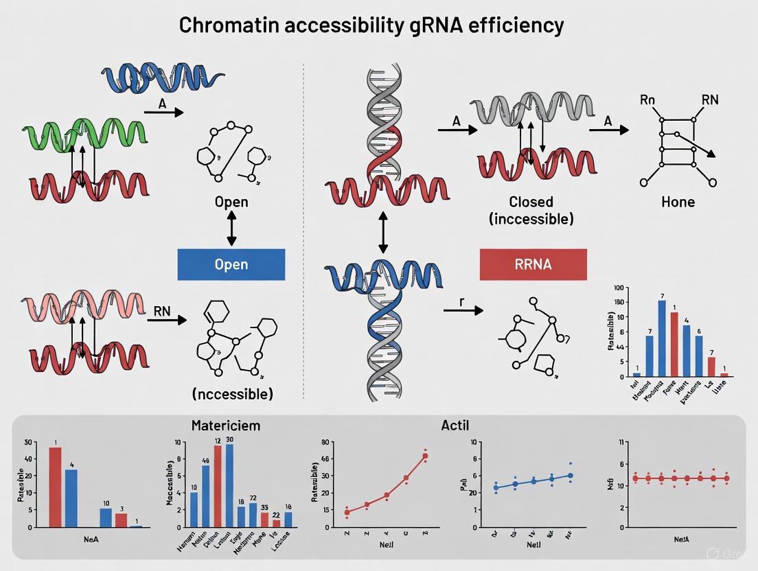
The following diagram illustrates the logical relationship between chromatin states and their impact on the CRISPR-Cas9 editing workflow, highlighting potential intervention points.
The Scientist's Toolkit: Key Research Reagents
Table 3: Essential Reagents for Investigating Chromatin and CRISPR Efficiency
| Reagent / Tool | Function / Description | Example Use Case |
|---|---|---|
| Tn5 Transposase | The core enzyme used in ATAC-seq to tag accessible genomic DNA with sequencing adapters. | Generating genome-wide chromatin accessibility maps from your experimental cell line [1] [6]. |
| HDAC Inhibitors (e.g., Trichostatin A - TSA) | Small molecules that inhibit histone deacetylases, leading to increased histone acetylation and a more open chromatin state. | Co-treatment with CRISPR-Cas9 to improve editing efficiency at refractory heterochromatic targets [8]. |
| dCas9-Activator Fusions (e.g., dCas9-VP64) | A nuclease-deficient Cas9 fused to a transcriptional activation domain. | Targeted chromatin opening at a specific genomic locus by recruiting transcriptional machinery to the site [8] [9]. |
| Engineered Chromatin Remodeling Proteins (E-ChRPs) | Designed proteins that can be programmed to reposition nucleosomes to a specific genomic location. | A research tool for precisely controlling nucleosome positioning to directly study its impact on Cas9 efficiency [9]. |
| Nucleosome Positioning Data (MNase-seq) | Sequencing data that maps the precise locations and occupancy of nucleosomes across the genome. | In silico screening of gRNA targets to avoid designing guides where the PAM site is occluded by a nucleosome [7] [1]. |
Frequently Asked Questions (FAQs)
1. Does heterochromatin really affect my CRISPR-Cas9 editing efficiency? Yes, numerous studies have confirmed that heterochromatin (condensed, transcriptionally inactive DNA) can significantly impede CRISPR-Cas9 mutagenesis. The compact structure physically hinders the access and binding of the Cas9 complex to its target DNA site. Research using imprinted genes as an internal control showed that repressed maternal alleles had up to 7-fold fewer mutations than active paternal alleles when Cas9 exposure was brief or its intracellular concentration was low [10]. Another study reported that TALEN editors could be up to five times more efficient than Cas9 in heterochromatin regions [11].
2. Does chromatin state affect the final outcome or just the speed of editing? Evidence suggests it primarily affects the kinetics, not the final endpoint, of mutagenesis. While heterochromatin slows down the initial rate of cleavage, given sufficient time and high enough levels of Cas9, the mutation frequencies on active and repressed alleles can become similar. The outcome of DNA repair (the spectrum of insertions, deletions, or efficiency of homology-directed repair) appears largely unaffected by the initial chromatin state [10].
3. Are some genome editors better than others for heterochromatic targets? Yes. TALEN has been shown to outperform Cas9 when editing heterochromatin target sites. Single-molecule imaging reveals that Cas9 tends to get encumbered by prolonged local searches on non-specific sites within heterochromatin, reducing its efficiency. TALEN does not face this same limitation to the same degree, making it a more effective choice for these hard-to-reach regions [11].
4. How can I measure the chromatin accessibility of my target site? Several established methods can profile chromatin accessibility genome-wide. The table below summarizes key techniques [1].
Table: Key Methods for Profiling Chromatin Accessibility
| Method | Description | Key Feature |
|---|---|---|
| ATAC-seq | Uses the Tn5 transposase to cut and tag accessible DNA regions. | Fast, highly sensitive, works with single cells. |
| DNase-seq | Relies on the DNase I enzyme to digest accessible DNA. | Well-established; biases towards hyper-accessible sites like promoters. |
| MNase-seq | Uses Micrococcal Nuclease to digest unprotected DNA. Maps nucleosome positions. | Can map both accessible regions and nucleosome occupancy; has sequence bias. |
| FAIRE-seq | Isoles nucleosome-depleted DNA based on differential solubility after fixation and sonication. | Nuclease-free approach. |
Troubleshooting Guide: Low Editing Efficiency in Heterochromatic Regions
Problem: Consistently low mutation rates in a target site suspected to be in a heterochromatic region.
Solution: A multi-pronged strategy involving target site re-assessment, experimental optimization, and the use of alternative tools is recommended.
Table: Quantitative Evidence of Heterochromatin Impact on Cas9
| Experimental System | Quantitative Finding | Key Parameter | Citation |
|---|---|---|---|
| Mouse Embryonic Stem Cells (Imprinted Genes) | Up to 7-fold reduction in mutagenesis on repressed alleles. | Low Cas9 concentration, brief exposure. | [10] |
| Live-Cell Imaging in Mammalian Cells | TALEN editing efficiency was up to 5-fold higher than Cas9 in heterochromatin. | Direct comparison in constrained chromatin. | [11] |
| HEK293T, HeLa, and Human Fibroblasts | Editing was consistently more efficient in euchromatin than heterochromatin. | Chromatin accessibility measured by DNase-seq. | [3] |
Actionable Steps:
- Verify Chromatin Status: Use public databases like the Encyclopedia of DNA Elements (ENCODE) to find DNase-seq or ATAC-seq data for your cell type. This will confirm if your target site is in an inaccessible region [5].
- Optimize Delivery and Dosage:
- Use modified, synthetic guide RNAs (with 2’-O-methyl modifications) to improve stability and efficiency [12].
- Deliver pre-assembled Cas9-gRNA Ribonucleoproteins (RNPs) via electroporation. This ensures high intracellular concentration and can reduce off-target effects [12].
- Consider using a strong, cell-type-appropriate promoter (e.g., EF1α, CAG, or a synthetic promoter) to drive high Cas9 expression, helping to overcome the chromatin barrier [10].
- Consider Alternative Editors: If Cas9 continues to fail, switch to TALEN for that specific target, as it is less hindered by heterochromatin [11].
The Scientist's Toolkit: Key Research Reagents
Table: Essential Reagents for Studying Chromatin Effects on Genome Editing
| Reagent / Tool | Function in Experiment |
|---|---|
| Cas9-gRNA RNP Complex | Direct delivery of editing machinery; improves kinetics and reduces toxicity. |
| Modified sgRNAs | Chemically synthesized guides with stability modifications (e.g., 2'-O-methyl) to enhance editing efficiency. |
| Chromatin Modifying Enzymes | Enzymes like DNase I (for DNase-seq) or Tn5 transposase (for ATAC-seq) to probe DNA accessibility. |
| dCas9 Fusion Proteins | Catalytically "dead" Cas9 fused to effector domains to manipulate or mark chromatin without cutting DNA. |
| TALEN Plasmids | An alternative nuclease system that can be used when Cas9 efficiency is low due to chromatin. |
Experimental Deep Dive: Key Supporting Protocols
1. Protocol: Quantifying Allelic Editing Bias in Imprinted Genes
This method, derived from a key study, uses the natural epigenetic differences in imprinted genes to isolate the effect of chromatin [10].
- Principle: In a diploid cell, the maternal and paternal alleles of an imprinted gene have identical DNA sequences but distinct chromatin states (e.g., one euchromatic and active, the other heterochromatic and silenced). Any difference in editing efficiency between the alleles can be unequivocally attributed to epigenetics.
- Workflow:
- Cell Line Selection: Use F1 hybrid mouse embryonic stem cells (mESCs) from reciprocal crosses (e.g., C57BL/6J and JF1 strains) to have genetic markers (SNPs) that distinguish maternal and paternal alleles.
- Transfection: Co-transfect cells with a Cas9/sgRNA expression plasmid (e.g., pX459) and a single-stranded oligodeoxynucleotide (ssODN) donor template.
- Selection and Harvest: Select transfected cells (e.g., with puromycin) and harvest as a pool ~96 hours post-transfection.
- Amplification and Sequencing: PCR-amplify the target region from genomic DNA and perform deep sequencing (e.g., Illumina).
- Allele-Specific Analysis: Use the known SNPs to bioinformatically separate sequencing reads into maternal and paternal alleles. Calculate the frequency and spectrum of mutations on each allele independently.
The diagram below illustrates this experimental logic and workflow.
2. Protocol: Live-Cell Single-Molecule Imaging of Editor Search Dynamics
This technique visualizes how genome-editing proteins like Cas9 and TALEN move and search for their targets in different chromatin environments [11].
- Principle: Fuse a halo-tag to a nuclease-deficient editor (dCas9 or TALE). Label it with a bright, cell-permeable fluorescent dye (e.g., JF549) for single-molecule tracking in live mammalian cells.
- Workflow:
- Design and Labeling: Design TALE or dCas9 proteins targeting euchromatic (e.g., CFTR gene) or heterochromatic (e.g., repetitive Alu elements) regions. Express the halo-tagged proteins in cells and label them with the fluorescent dye.
- Microscopy under Two Conditions:
- Short Exposure (10-20 ms): Captures fast diffusion kinetics, distinguishing between global search (fast diffusion/hopping) and local search (slow diffusion/sliding).
- Long Exposure (500 ms): Blurs out fast-moving molecules, allowing visualization and tracking of only the DNA-bound proteins to measure their residence times.
- Data Analysis:
- Calculate diffusion coefficients from short-exposure trajectories.
- Fit residence time histograms from long-exposure movies to a two-component exponential decay model to determine the lifetimes of non-specifically and specifically bound molecules.
The following diagram visualizes the search behaviors and analysis methods.
The Role of Active Transcription in Stimulating Cas9 Activity and Release
Within the nucleus, the efficiency of CRISPR-Cas9 gene editing is not solely determined by the guide RNA (gRNA) sequence. The local chromatin environment presents a significant physical and regulatory barrier. Chromatin, the complex of DNA and proteins, exists in various states of compaction. Heterochromatin, a tightly packed form, can impede Cas9 access to its target DNA sequence, while active transcription is associated with a more open chromatin configuration and has been shown to directly stimulate DNA cleavage by Cas9 [13]. This guide addresses common challenges and questions researchers face when considering the impact of transcription and chromatin state on their CRISPR experiments.
Frequently Asked Questions & Troubleshooting
FAQ: Why does the same gRNA have different editing efficiencies in different cell types?
- Cause: The primary cause is often differences in the chromatin accessibility and transcriptional status of the target locus between cell types. A locus that is actively transcribed in one cell line may be silent and packaged into heterochromatin in another [14] [13].
- Troubleshooting Steps:
- Consult Epigenomic Maps: Before designing experiments, check publicly available chromatin accessibility data (e.g., from ATAC-seq or DNase-seq) for your specific cell type. Targeting regions confirmed to be in open chromatin will yield more predictable results [14].
- Measure Local Chromatin: If no data exists, consider performing a pilot assay to measure accessibility at your target site.
- Design Multiple gRNAs: If possible, design several gRNAs targeting the same gene but in different genomic regions (e.g, promoter, intron, exon) to increase the likelihood of one residing in an accessible locus.
FAQ: My in vitro cleavage assay works perfectly, but editing fails in my cellular model. Why?
- Cause: In vitro assays use purified, protein-free DNA, while cellular DNA is packaged into chromatin. Nucleosomes, the basic units of chromatin, can completely block Cas9 access to DNA, as demonstrated in cell-free assays [13].
- Troubleshooting Steps:
- Verify Chromatin State: Confirm that your target site is not located in a nucleosome-dense region. Nucleosome positioning data can help with this.
- Time Your Experiment: For ex vivo editing of primary cells (like T cells), the chromatin landscape can change with activation status. Electroporation of Cas9-RNP complexes is often most effective shortly after cell stimulation when the genome is more receptive [14].
- Consider Epigenetic Pretreatment: In some cases, pretreatment with cytokines like IL-7 has been shown to enhance gene-editing efficiency in unstimulated T cells, potentially by altering chromatin accessibility [14].
FAQ: How does active transcription directly influence the Cas9 enzyme mechanism?
- Cause: Recent evidence suggests that active transcription does more than just open chromatin. The act of transcription itself can directly stimulate DNA cleavage by influencing Cas9 release rates [13].
- Explanation & Solution: The passage of RNA polymerase along the DNA template can act as a "molecular motor" that actively displaces Cas9 from the DNA after cleavage, facilitating enzyme turnover. This effect is strand-specific. To leverage this, if the target strand is known, design gRNAs so that the non-target strand is the same as the transcribed strand, potentially allowing the transcription machinery to aid in Cas9 dissociation [13].
Experimental Protocols for Assessing Transcription Impact
Protocol 1: Correlating Endogenous Editing with Transcription Status
This protocol outlines how to determine if the transcriptional activity of a locus affects Cas9 editing efficiency in your cell model.
Key Research Reagent Solutions
| Reagent/Material | Function in the Protocol |
|---|---|
| Isogenic Cell Lines | Clonal cell lines derived from a single progenitor, ensuring genetic identity and minimizing variability. |
| dCas9-Expresssing Line | A cell line stably expressing catalytically "dead" Cas9, used to measure binding accessibility without cleavage. |
| ATAC-seq Kit | A kit to perform Assay for Transposase-Accessible Chromatin using sequencing, which maps open chromatin regions genome-wide. |
| gRNA Design Tool (e.g., CRISPRon) | An AI-based tool to predict gRNA on-target activity based on sequence, helping to control for sequence-intrinsic effects [15] [16]. |
| RT-qPCR Reagents | Reagents for Reverse Transcription Quantitative PCR to measure mRNA levels and quantify transcriptional activity at the target locus. |
Methodology:
- Cell Line Selection: Obtain two isogenic cell lines where your gene of interest is either actively transcribed or silenced.
- gRNA Design & Transfection: Design a panel of 3-5 high-scoring gRNAs (using a tool like CRISPRon [16]) targeting the same locus. Transfert each gRNA with Cas9 into both cell lines.
- Efficiency Quantification: 72 hours post-transfection, harvest genomic DNA and use targeted next-generation sequencing (NGS) to quantify the indel percentage at the target site for each gRNA-cell line combination.
- Chromatin & Transcription Assessment:
- Perform ATAC-seq on both cell lines to map genome-wide chromatin accessibility.
- Use RT-qPCR to measure the baseline mRNA expression level of your target gene in both cell lines.
- (Optional) Use dCas9 ChIP-qPCR in a stable cell line to directly measure Cas9 binding efficiency at the target site in both transcriptional states.
- Data Analysis: Correlate the measured indel percentages with the transcriptional activity (mRNA level) and chromatin accessibility (ATAC-seq signal) at the target site.
Protocol 2: High-Throughput Screening of gRNA Efficiency
This method uses a pooled gRNA library to systematically evaluate how chromatin features and sequence context govern editing outcomes.
Methodology:
- Library Design: Clone a pooled library of thousands of gRNAs into a lentiviral vector. The library should include gRNAs targeting genomic sites with diverse epigenetic contexts [16].
- Viral Transduction: Transduce your target cells (e.g., HEK293T) with the lentiviral gRNA library at a low multiplicity of infection (MOI ~0.3) to ensure most cells receive only one gRNA.
- Editing & Enrichment: Allow sufficient time (e.g., 8-10 days) for Cas9 editing and enrichment of edited cells, often facilitated by a selectable marker on the vector [16].
- Sequencing & Analysis: Harvest genomic DNA and perform deep sequencing of the target sites. The resulting indel frequency for each gRNA is its measured on-target activity.
- Integration with Epigenetic Data: Integrate the gRNA efficiency data with cell-type-specific epigenetic data (e.g., ATAC-seq, histone modification ChIP-seq). This allows you to build a model that predicts efficiency based on both sequence and chromatin features [14].
The workflow for this high-throughput approach is summarized in the following diagram:
Data Presentation: Quantitative Impact of Chromatin
The table below summarizes key quantitative findings from research on how chromatin state and transcription influence Cas9 activity.
Table 1: Quantitative Effects of Chromatin State on CRISPR-Cas9 Efficiency
| Experimental Observation | Quantitative Impact / Correlation | Experimental Context | Source |
|---|---|---|---|
| Chromatin Accessibility | Positive correlation (R values vary by cell type) | ATAC-seq signal vs. measured indel percentage in human T cells. | [14] |
| Active Transcription | Directly stimulates cleavage; strand-specific effect on Cas9 release. | Observation from controlled allele-specific studies. | [13] |
| Nucleosome Occupancy | Can completely block Cas9 access in vitro; major hurdle in vivo. | Cell-free cleavage assays with reconstituted nucleosomes. | [13] |
| DNA Methylation (CpG) | Does not directly hinder Cas9 binding or cleavage. | In vitro cleavage assays comparing methylated vs. unmethylated DNA substrates. | [13] |
| gRNA Efficiency Prediction | Model performance (Spearman's R) improved by adding epigenetic features. | Combining sequence-based prediction with ATAC-seq data in T cells. | [14] |
Visualization of the Core Mechanism
The following diagram illustrates the proposed mechanistic relationship between active transcription and enhanced Cas9 activity and release, which is central to troubleshooting the issues described in the FAQs.
This technical support center provides guidance for researchers investigating how temporal chromatin states impact genome editing efficiency. Chromatin modifications are dynamic regulators of DNA accessibility, directly influencing the efficacy of CRISPR-based systems [17]. A thorough understanding of these states is crucial for predicting and optimizing guide RNA (gRNA) performance in diverse cellular contexts.
Frequently Asked Questions (FAQs)
What are chromatin states and why are they important for genome editing? Chromatin states are combinatorial patterns of histone modifications (e.g., methylation, acetylation) that functionally annotate the genome into regions such as active promoters, enhancers, and repressed heterochromatin [18]. These states determine the physical openness or compactness of chromatin, thereby controlling the accessibility of the DNA strand to editors like Cas9. Inaccessible, closed chromatin is a major cause of low editing efficiency [17].
How can I identify the chromatin state of my target locus? You must use computational tools to analyze existing epigenomic data (e.g., ChIP-seq, ATAC-seq) for your cell type.
- ChromHMM is the most widely used tool for this purpose. It uses a multivariate hidden Markov model to segment the genome into discrete states based on histone mark data [19] [18].
- Alternative Tools: Other tools like Segway or EpiCSeg may offer advantages for specific data types, such as direct read count modeling [19]. Procedure:
- Obtain epigenomic datasets (e.g., from ENCODE or Roadmap Epigenomics) for your specific cell type.
- Run ChromHMM to learn the chromatin state model for your cell type.
- Annotate your target genomic region with the learned states to determine if it resides in an open (active) or closed (repressed) chromatin context.
My gRNA has high predicted on-target efficiency but performs poorly in my experiment. Could chromatin be the cause? Yes, this is a common issue. Computational on-target scores often do not fully account for the local chromatin environment.
- Troubleshooting Steps:
- Annotate Chromatin State: As described above, check the chromatin state of your target site.
- Correlate with Accessibility: Use ATAC-seq or DNase-seq data to confirm the physical accessibility of the region. A low signal suggests closed chromatin.
- Design Alternative gRNAs: If the target is in closed chromatin, design new gRNAs that target nearby regions in open chromatin, even if their raw efficiency score is slightly lower.
- Use Chromatin Modulators: Consider co-delivering chromatin-modulating agents, such as histone deacetylase (HDAC) inhibitors, to transiently open the chromatin structure at your target site.
Are there computational tools that incorporate chromatin data into gRNA design? While most standard gRNA design tools focus on sequence-based rules for on-target and off-target activity, the chromatin context must be assessed separately. The recommended workflow is to first use chromatin state maps to select a target region that is accessible, and then use tools like CRISPick or CRISPOR to design the specific gRNA sequence within that region [20].
Troubleshooting Guides
Problem: Low Editing Efficiency in a Closed Chromatin Region
Issue: Your target site is confirmed to be in a repressed chromatin state (e.g., marked by H3K9me2/3 or H3K27me3), leading to poor Cas9 binding and cutting.
Solution 1: Epigenome Editing Pre-conditioning A powerful strategy is to use epigenome editing to open the chromatin before introducing the editing machinery.
- Experimental Protocol:
- Select an Activator: Use a programmable editor (e.g., dCas9 fused to the p300 core domain for H3K27ac deposition) to install an activating histone mark at the target site [17].
- Pre-treat Cells: Transfert cells with the dCas9-activator and a specific gRNA targeting your locus.
- Wait for Chromatin Remodeling: Incubate for 24-48 hours to allow for stable establishment of the active chromatin mark.
- Perform Main Edit: Introduce the active Cas9 nuclease and your therapeutic gRNA. The pre-conditioned, more open chromatin should yield higher editing efficiency.
Solution 2: Co-delivery with Chromatin-opening Agents For a simpler approach, co-deliver chemical inhibitors of chromatin-repressing enzymes.
- Reagents: HDAC inhibitors (e.g., Sodium Butyrate, Trichostatin A) or DNA methyltransferase inhibitors (e.g., 5-Azacytidine).
- Procedure: Treat cells with a low, non-toxic concentration of the inhibitor 24 hours before and during the CRISPR editing transfection. Titrate the concentration carefully, as high doses can cause widespread epigenetic dysregulation and cell toxicity.
Problem: High Cell Toxicity During Epigenome Editing
Issue: Over-expression of powerful chromatin modifiers, especially transcriptional activators like p300, can lead to significant cell death and indirect transcriptional changes [17].
Mitigation Strategies:
- Use Inducible Systems: Use a doxycycline (DOX)-inducible system to control the timing and duration of epigenome editor expression, limiting prolonged exposure [17].
- Titrate Expression: Use a lower concentration of the inducer (e.g., 20-fold lower DOX for p300) to reduce expression levels to a functional but less toxic range [17].
- Catalytic Domains: Use only the catalytic domain (CD) of the chromatin modifier, fused to dCas9, rather than the full-length protein. This minimizes non-catalytic, confounding effects that can contribute to toxicity [17].
Chromatin State Analysis Tools and Data
The table below summarizes key computational tools for chromatin state discovery and annotation, which are essential for planning your experiments [19].
| Tool | Modeling Strategy | Key Features | Best For |
|---|---|---|---|
| ChromHMM | Multivariate HMM + EM | Fast, interpretable, widely adopted | Standard, cell type-specific chromatin segmentation [19] [18]. |
| TreeHMM | Tree-structured HMM | Models lineage relationships among cell types | Analyzing related cell types with a known developmental hierarchy [19]. |
| GATE | Graph-aware HMM | Integrates spatial proximity data (e.g., Hi-C) | Accounting for 3D chromatin structure in state annotation [19]. |
| EpiCSeg | HMM + Count data | Uses actual ChIP-seq read counts instead of binarization | More accurate modeling of weak/moderate histone signals [19]. |
| Chromswitch | Peak-based clustering | Uses ChIP-seq peaks and statistical summaries | Comparing chromatin states in a specific genomic region across conditions [18]. |
Essential Research Reagent Solutions
The table below lists key reagents used in advanced epigenome editing studies, which can be integrated into your troubleshooting workflows [17].
| Research Reagent | Function in Experiment |
|---|---|
| dCas9GCN4 System | Programmable scaffold that amplifies effector recruitment via GCN4 motif array [17]. |
| CDscFV Effectors | Catalytic domains of chromatin writers (e.g., for H3K4me3, H3K27ac, H3K9me2) fused to scFV. Isolates the function of the mark itself [17]. |
| mut-CDscFV | Catalytic point mutants of effectors. Critical controls to confirm phenotypic effects are due to chromatin mark deposition and not just protein tethering [17]. |
| Doxycycline (DOX)-Inducible System | Allows dynamic control of epigenome editor expression, mitigating toxicity and enabling memory studies [17]. |
| CUT&RUN-qPCR | A low-background method to quantitatively validate the deposition of chromatin marks at the target locus after editing [17]. |
Experimental Workflow Diagrams
Workflow for Editing in Dynamic Chromatin
Modular Epigenome Editing Platform
FAQs: Core Concepts and Experimental Design
Q1: What is the fundamental relationship between ATAC-seq signals and CRISPR gRNA efficiency? Research consistently shows a positive correlation between chromatin accessibility and gRNA efficiency. Genomic regions with higher ATAC-seq signals, indicating more open chromatin, generally enable more efficient CRISPR-Cas9 gene editing. This relationship has been validated across multiple systems, including human T cells and zebrafish embryos [14] [7] [3].
Q2: Why does chromatin accessibility impact CRISPR editing efficiency? Chromatin accessibility reflects how "open" or "closed" a genomic region is. In closed chromatin (heterochromatin), DNA is tightly wrapped around nucleosomes and less accessible to CRISPR machinery, reducing editing efficiency. In open chromatin (euchromatin), the Cas9-gRNA complex can more easily bind target DNA sequences, resulting in higher efficiency [3] [5].
Q3: Can I design efficient gRNAs for targets in epigenetically closed regions? Yes. While closed regions typically show reduced efficiency, studies demonstrate that designing two gRNAs targeting adjacent regions in closed chromatin can improve gene-editing outcomes. Pretreating cells with cytokines like IL-7 can also enhance efficiency in these challenging contexts [14].
Q4: My ATAC-seq data shows low TSS enrichment. How does this affect gRNA design? Low TSS (Transcription Start Site) enrichment suggests poor signal-to-noise ratio or suboptimal library preparation. This can compromise the reliability of chromatin accessibility data for gRNA prediction. A TSS enrichment score below 6 is a warning sign; you should optimize your ATAC-seq protocol before using the data to inform gRNA design [21].
Troubleshooting Guides
Troubleshooting ATAC-seq Data Quality for gRNA Prediction
Poor ATAC-seq data quality is a primary source of failed gRNA efficiency predictions.
Problem: Strange ATAC-seq fragment size distribution.
- Expected: Clear peaks at ~50 bp (nucleosome-free), ~200 bp, and ~400 bp [21].
- Symptom: Missing or unclear nucleosomal patterning.
- Solution:
- Check for over-tagmentation, which can digest chromatin excessively and mask nucleosomal features [21].
- Verify cell viability and ensure no DNA degradation occurred during sample preparation.
- Optimize transposition reaction time and input cell number.
Problem: Low correlation between predicted and actual gRNA efficiency.
- Symptom: gRNAs selected using ATAC-seq data perform poorly in editing assays.
- Solution:
- Combine epigenetic data with sequence-based prediction tools. Using ATAC-seq data alone is insufficient; integration with algorithms significantly improves performance [14].
- Ensure ATAC-seq data is from a relevant cell type, as chromatin states are cell-specific [14].
- For critical experiments, validate chromatin accessibility at the target locus by examining ATAC-seq signal tracks in a genome browser.
Problem: Peaks called in ATAC-seq data do not agree with biological expectation.
Troubleshooting Low gRNA Efficiency Despite High Chromatin Accessibility
- Problem: gRNA targets an open chromatin region but editing efficiency remains low.
- Solution:
- Investigate gRNA secondary structure. The guide sequence itself can form secondary structures that inhibit its function, independent of chromatin context [3].
- Verify sequence-based features. Ensure the gRNA follows established sequence rules (e.g., preference for guanine at positions -1 and -2 upstream of the PAM) [7].
- Check for genetic variation. Ensure no SNPs or indels in the target sequence are preventing gRNA binding.
- Solution:
Table 1: Summary of Key Studies on ATAC-seq and gRNA Efficiency Correlation
| Study System | Key Finding | Quantitative Improvement | Citation |
|---|---|---|---|
| Human T Cells | Combining ATAC-seq with prediction tools improves gRNA design. | Significantly improved gene-editing efficiency over sequence-based tools alone. | [14] |
| Zebrafish Embryos | Chromatin accessibility positively correlates with CRISPR/Cas9 efficiency. | Open chromatin regions showed higher mutation rates in F0 embryos. | [7] |
| HEK293T, HeLa, Human Fibroblasts | Gene editing is more efficient in euchromatin vs. heterochromatin. | Confirmed in multiple human cell types. | [3] |
| Computational Analysis (GUIDE-seq) | Correlation between sequence and cleavage frequency is altered by DNA accessibility. | Abolished in less accessible regions. | [5] |
Table 2: Performance of Different gRNA Prediction Models Incorporating Chromatin Data
| Prediction Model | Features Used | Correlation Coefficient | Citation |
|---|---|---|---|
| CINDEL | Hand-crafted features | 0.61 | [23] |
| DeepCpf1 | One-hot encoding + Chromatin Accessibility | 0.71 | [23] |
| Cas-FM (RNA-FM) | Pretrained RNA foundation model embedding | 0.76 | [23] |
| Cas-FM-CA (RNA-FM) | Foundation model + Chromatin Accessibility | 0.78 | [23] |
Experimental Protocols
Key Materials:
- Primary human T cells (e.g., from PBMCs)
- Tn5 transposase (commercially available)
- RPMI-1640 culture medium with IL-2 (100 IU/ml)
- NEPA 21 electroporator
- Equipment for standard molecular biology and sequencing
Methodology:
- T Cell Culture and Stimulation: Stimulate human PBMCs with mitomycin C-treated K562 cells expressing anti-CD3 scFv and CD80.
- Nuclei Preparation and Transposition: Lyse cells to isolate nuclei. Incubate nuclei with the Tn5 transposase to simultaneously fragment and tag accessible DNA with adapters.
- Library Preparation and Sequencing: Purify transposed DNA and amplify via PCR to create sequencing libraries. Perform paired-end sequencing on an Illumina platform.
- Bioinformatic Analysis:
- Alignment: Trim adapters and align reads to the reference genome (e.g., hg38) using Bowtie2 [14].
- Peak Calling: Identify regions of significant chromatin accessibility using MACS2 [14].
- Data Integration: Overlay potential gRNA target sequences with ATAC-seq peaks. Prioritize gRNAs that bind within accessible regions.
Key Materials:
- Designed gRNAs targeting adjacent sites
- Alt-R CRISPR-Cas9 system (IDT)
- Recombinant IL-7 (10 ng/ml)
Methodology:
- Dual gRNA Design: For a target in a closed chromatin region, design two gRNAs that bind to closely spaced adjacent sites.
- Cell Pretreatment: For unstimulated T cells, pretreat with IL-7 (10 ng/ml) to enhance basal editing efficiency.
- RNP Complex Electroporation: Form ribonucleoprotein (RNP) complexes by combining Cas9 nuclease with the two annealed gRNAs. Introduce the RNP complexes into T cells via electroporation.
Signaling Pathways and Workflow Diagrams
Workflow for ATAC-seq Guided gRNA Design
ATAC-seq Data Troubleshooting for gRNA Design
The Scientist's Toolkit: Research Reagent Solutions
Table 3: Essential Research Reagents and Resources
| Item / Resource | Function / Application | Example / Source |
|---|---|---|
| Hyperactive Tn5 Transposase | Enzymatic tagmentation of open chromatin for ATAC-seq library prep. | Commercially available (e.g., Illumina Nextera). |
| Alt-R CRISPR-Cas9 System | Pre-complexed Cas9-gRNA RNP for highly efficient editing in primary cells. | Integrated DNA Technologies (IDT) [14]. |
| Recombinant IL-7 | Cytokine pretreatment to enhance gene-editing efficiency in unstimulated/naïve T cells [14]. | PeproTech [14]. |
| NEPA 21 Electroporator | Efficient delivery of RNP complexes into hard-to-transfect cells like primary T cells [14]. | Nepa Gene [14]. |
| ENCODE Blacklist Regions | Genomic regions with anomalous signals; should be filtered out during ATAC-seq analysis [24]. | ENCODE Consortium [24]. |
| MACS2 | Standard peak calling software for identifying accessible chromatin regions from ATAC-seq data [14] [24]. | Open-source tool. |
| Bowtie2 / BWA-MEM | Sequence aligners for mapping ATAC-seq reads to a reference genome [24]. | Open-source tools. |
Mapping the Epigenome: Advanced Tools to Link Perturbations and Chromatin State
Core Concepts and FAQs
What are single-cell CRISPR screens with epigenetic readouts?
These are advanced functional genomics methods that combine multiplexed CRISPR perturbations with single-cell epigenomic profiling. Unlike traditional screens that measure survival or surface markers, these techniques directly link genetic perturbations to changes in the chromatin landscape within individual cells, revealing how specific genes regulate epigenetic states [25] [26].
How do chromatin accessibility and structure impact CRISPR editing efficiency?
Chromatin accessibility significantly influences Cas9 cutting efficiency. Multiple studies demonstrate that closed, silenced chromatin states inhibit Cas9 binding and editing, while open chromatin regions permit more efficient editing [3] [27] [5].
Table: Experimental Evidence of Chromatin Impact on CRISPR Efficiency
| Chromatin State | Effect on Editing Efficiency | Experimental System | Key Finding |
|---|---|---|---|
| Heterochromatin (Closed) | Reduced by ~30-40% at most target sites [27] | HEK293T inducible silencing system | Editing efficiency negatively correlated with repressive histone mark H3K27me3 [27] |
| Euchromatin (Open) | Higher efficiency [3] | HEK293T, HeLa, and human fibroblasts | Editing more efficient in euchromatin versus heterochromatin [3] |
| Variable Accessibility | Abolished correlation between sequence match and cleavage in less accessible regions [5] | GUIDE-seq and CIRCLE-seq computational analysis | DNA accessibility modulates the relationship between guide-target complementarity and cleavage frequency [5] |
Why do different sgRNAs targeting the same gene show variable performance?
Variability arises from both sgRNA-intrinsic properties and local chromatin context:
- sgRNA sequence determinants: The secondary structure formation of the guide RNA itself affects Cas9 activity [3].
- Chromatin environment: Even different target sites within the same gene locus can experience varying levels of chromatin compaction, leading to divergent editing outcomes [27].
- Best practice: Design 3-4 sgRNAs per gene to mitigate the impact of individual sgRNA performance variability [28].
What are the solutions for low signal-to-noise in epigenetic CRISPR screens?
- Increase selection pressure: For negative screens where the goal is to identify essential genes, applying stronger selective pressure can enhance the depletion signal of effective sgRNAs [28].
- Ensure sufficient cell coverage: Plan for approximately 100 cells per perturbed gene to achieve adequate statistical power for detecting chromatin accessibility changes [29].
- Verify chromatin perturbation: Include positive control sgRNAs targeting known epigenetic regulators and confirm expected chromatin changes at their target loci [25] [26].
How can I determine if my single-cell epigenetic CRISPR screen was successful?
- Positive control validation: Include well-validated positive-control sgRNAs (e.g., targeting known chromatin regulators). Successful perturbation should recapitulate expected chromatin accessibility changes [28] [26].
- Data quality metrics: Assess single-cell library complexity, percentage of reads in peaks, and fragment size distribution, which should resemble high-quality bulk ATAC-seq data [25].
- Phenotypic concordance: Verify that perturbations of known factors produce the expected chromatin phenotypes (e.g., SPI1/PU.1 perturbation reduces accessibility at its motif sites) [25].
Key Methodologies and Experimental Protocols
Perturb-ATAC Workflow
The Perturb-ATAC method enables simultaneous detection of CRISPR guide RNAs and genome-wide chromatin accessibility profiling in single cells [25] [26].
Protocol Steps:
- Single-cell capture: Cells are captured in microfluidic chambers (e.g., Fluidigm IFC) and lysed [25].
- Tagmentation: Genomic DNA is fragmented and tagged with sequencing adapters using the Tn5 transposase, which preferentially targets open chromatin regions [25].
- sgRNA detection: CRISPR sgRNAs or their identifying barcodes (GBCs) are reverse transcribed using target-specific primers within each reaction chamber [25].
- Whole-cell amplification: All contents from each chamber are amplified by PCR [25].
- Library separation: sgRNA and ATAC amplicons are separately amplified with cell-specific barcoded primers, then pooled for sequencing [25].
- Multi-modal analysis: Sequencing data is processed to assign chromatin accessibility profiles to specific genetic perturbations in individual cells [25].
CROP-seq for Single-Cell Transcriptional and Perturbation Profiling
While primarily for transcriptome analysis, CROP-seq principles are adaptable to epigenetic readouts and address key design considerations [30].
Table: Comparison of Single-Cell CRISPR Screening Approaches
| Feature | Perturb-ATAC [25] [26] | CROP-seq [30] | 10x Genomics 5' Screening [29] |
|---|---|---|---|
| Primary Readout | Chromatin accessibility (ATAC-seq) | Gene expression (RNA-seq) | Gene expression + surface proteins |
| sgRNA Detection | Guide barcode reverse transcription | Polyadenylated sgRNA transcript | Direct capture of guide sequence |
| Key Advantage | Direct measurement of TF binding, nucleosome positioning | No viral recombination concerns | Compatible with existing libraries; multiomic capacity |
| Compatibility | Custom CRISPRi/CRISPRko libraries | Requires modified vector (CROPseq-Guide-Puro) | Works with most existing Cas9 libraries |
The Scientist's Toolkit: Essential Research Reagents
Table: Key Reagents for Single-Cell Epigenetic CRISPR Screens
| Reagent / Tool | Function | Example/Reference |
|---|---|---|
| Modified sgRNA Vectors | Expresses guide RNA in detectable format for single-cell assays | CROPseq-Guide-Puro [30], Perturb-ATAC GBC vectors [25] |
| Tn5 Transposase | Fragments and tags accessible genomic DNA for ATAC-seq | Hyperactive Tn5 [25] |
| Chromatin State Model Systems | Controls for chromatin effects on editing | Doxycycline-inducible Gal4EED system [27] |
| Bioinformatic Tools | Analyzes single-cell multi-modal perturbation data | MAGeCK [28], SCENIC [31] |
| Validated Epigenetic Regulators | Positive controls for screening | SRCAP, KAT5 (identified in fibrosis screen) [31] |
Advanced Troubleshooting: Addressing Complex Scenarios
Interpreting epistatic relationships between chromatin regulators
Combinatorial Perturb-ATAC can reveal how multiple epigenetic regulators interact hierarchically:
- Experimental design: Perform pairwise co-deletion of transcription factors or chromatin modifiers and measure the resulting chromatin accessibility landscape [25] [26].
- Analysis approach: Identify synergistic (greater than additive) or antagonistic effects on chromatin accessibility at shared regulatory elements [26].
- Biological insight: Genomic co-localization of transcription factors often predicts synergistic interactions in chromatin regulation [26].
Optimizing for specific chromatin phenotypes
When screening for regulators of specific epigenetic states:
- FACS enrichment: Sort cells based on chromatin accessibility reporters or specific histone modifications before single-cell library preparation [28].
- Trajectory analysis: In developmental systems, order cells along differentiation trajectories and identify perturbations that disrupt normal chromatin state progression [26].
- Motif analysis: Compute differential transcription factor motif accessibility across perturbations to infer regulatory hierarchies [25].
Troubleshooting Guide: Addressing Common Experimental Challenges
This section provides solutions to common issues encountered during CRISPR-sciATAC experiments, helping researchers achieve high-quality data on how genetic perturbations alter the epigenetic landscape.
Table 1: Troubleshooting Common CRISPR-sciATAC Issues
| Problem | Possible Cause | Recommended Solution |
|---|---|---|
| Low single-cell recovery or capture rate | Nuclear envelope integrity compromised during processing [32] | Optimize fixation and lysis conditions to preserve nuclear envelope [33] [32] |
| Low gRNA detection/assignment rate | Inefficient capture of gRNA sequences; low gRNA transcript abundance [34] | Use a custom, easy-to-purify transposase [33] [32]; Flank lentiviral sgRNA with pre-integrated Nextera adapters [34] |
| High species collision rate (in control experiments) | Inefficient single-cell barcoding or nuclear partitioning [33] | Use a unique, easy-to-purify transposase and optimize combinatorial indexing steps [33] |
| Variable chromatin accessibility profiles for the same gRNA | Biological variability or insufficient cell count [33] | Ensure adequate cell numbers per perturbation; Downsampling analysis shows some perturbations (e.g., EZH2) need only ~5 cells, while others (e.g., TET2) may need ~225 [33] |
| No cleavage band or low editing efficiency | Chromatin inaccessibility at target site; gRNA secondary structure [3] [5] | Design gRNAs targeting accessible regions (euchromatin) [3]; Check for and avoid gRNA sequences prone to secondary structure formation [3] |
| Unexpected cell proliferation phenotypes | Targeting essential genes or genes affecting growth [33] | Perform early-timepoint bulk gRNA amplification to distinguish essential gene drop-out from technical capture failure [33] |
Frequently Asked Questions (FAQs)
Q1: What is the core innovation of CRISPR-sciATAC compared to previous methods?
CRISPR-sciATAC is a novel integrative platform that combines pooled CRISPR screening with single-cell combinatorial indexing ATAC-seq (sciATAC-seq). Its key innovation is the ability to simultaneously capture CRISPR guide RNAs (gRNAs) and genome-wide chromatin accessibility profiles from tens of thousands of single cells in a single, scalable experiment. This creates a direct causal link between a genetic perturbation and its consequent changes in genome-wide chromatin organization within a uniform genetic background [33] [32] [35].
Q2: How does chromatin accessibility directly impact CRISPR-Cas9 gene editing efficiency?
Research confirms that CRISPR-Cas9-mediated gene editing is significantly more efficient in euchromatin (open, accessible DNA) than in heterochromatin (closed, inaccessible DNA). The level of DNA accessibility at the target locus is a major cellular factor that influences cleavage efficiency. Furthermore, the secondary structure of the gRNA sequence itself is an independent determinant of Cas9 activity [3] [5].
Q3: My single-cell data shows high variability after a perturbation. Is this technical noise or biological reality?
It can be both. Technical noise should be minimized by optimizing protocols. However, CRISPR-sciATAC has revealed that the loss of certain chromatin modifiers can intrinsically increase cell-to-cell variability in chromatin accessibility. For example, disrupting the TET2 gene creates more variable chromatin states than non-targeting controls, requiring more single cells (~225) to accurately represent the population average. In contrast, perturbations like EZH2 loss show consistent changes with as few as 5 cells [33].
Q4: Can I use CRISPR-sciATAC to study nucleosome positioning, not just accessibility?
Yes. Beyond measuring accessibility, CRISPR-sciATAC is equipped with computational methods to map dynamic movements of nucleosomes. For instance, knocking out ARID1A led to tighter nucleosome spacing and reduced accessibility at specific transcription factor binding sites, demonstrating the method's power to reveal nucleosome-level reorganization [33] [32].
Q5: What is the throughput and cost advantage of CRISPR-sciATAC over similar methods like Perturb-ATAC?
CRISPR-sciATAC is designed for high throughput and lower cost. It does not require microfluidic devices for single-cell isolation. In contrast, methods like Perturb-ATAC need multiple runs on a Fluidigm C1 to process about 96 cells per run. CRISPR-sciATAC can process up to ~30,000 cells in a single study. One analysis showed that Perturb-ATAC cost about $9.80 per cell, whereas the Spear-ATAC method (a similar droplet-based approach) reduced the cost to about $0.46 per cell [33] [34].
Essential Experimental Protocols & Workflows
Core CRISPR-sciATAC Workflow
The following diagram illustrates the key steps in the CRISPR-sciATAC protocol, from cell preparation to sequencing.
Detailed Protocol: From Library Design to Data Generation
gRNA Library Design & Cloning:
- Design multiple gRNAs (typically 3-5) per target gene to ensure robust results [36].
- Clone gRNA sequences into a lentiviral vector (e.g., CROP-seq vector) where the gRNA is embedded within a longer RNA Polymerase II transcript for co-expression with the perturbation [33].
- Include a sufficient number of non-targeting control gRNAs (e.g., 3-5) for baseline comparison [33].
Cell Preparation & Transduction:
- Use a cell line expressing Cas9 (e.g., K562-Cas9 for knockout screens or K562-dCas9-KRAB for CRISPRi screens) [33] [34].
- Transduce cells with the lentiviral gRNA library at a low multiplicity of infection (MOI < 0.3) to ensure most cells receive a single gRNA [36].
- Apply antibiotic selection (e.g., puromycin) for several days to enrich for successfully transduced cells [33] [37].
Nuclei Preparation & Combinatorial Indexing:
- After about 7-10 days of selection, harvest and fix cells. Gently lyse cells to isolate intact nuclei, being careful to preserve the nuclear envelope [33] [32].
- Distribute nuclei across a 96-well plate. In each well, perform tagmentation using a hyperactive Tn5 transposase (e.g., the unique, easy-to-purify transposase from Vibrio parahemolyticus mentioned in the study). This step simultaneously fragments accessible DNA and adds well-specific barcode #1 [33].
- Perform in situ reverse transcription to capture the gRNA sequences and tag them with the same well-specific barcode #1 [33].
Library Construction & Sequencing:
- Pool all nuclei and then redistribute them into a new 96-well plate.
- In each new well, perform a PCR reaction to add a second barcode (barcode #2) to both the ATAC fragments and the gRNA sequences. The combination of barcode #1 and barcode #2 creates a unique "cell barcode" for each single cell [33].
- Perform a final PCR to add sequencing adapters.
- Sequence the final library using paired-end sequencing on an Illumina platform to capture both the chromatin accessibility fragments and the gRNA sequences [33].
The Impact of Chromatin on Editing: A Conceptual Diagram
The diagram below visualizes the relationship between chromatin state and CRISPR-Cas9 efficiency, a key concept underlying the need for assays like CRISPR-sciATAC.
The Scientist's Toolkit: Key Research Reagents & Materials
Table 2: Essential Reagents and Materials for CRISPR-sciATAC
| Item | Function / Role in the Experiment | Example / Note |
|---|---|---|
| Hyperactive Tn5 Transposase | Fragments accessible chromatin and simultaneously adds sequencing adapters. The heart of ATAC-seq. | A unique, easy-to-purify transposase from Vibrio parahemolyticus was developed for CRISPR-sciATAC [33] [32]. |
| Lentiviral gRNA Library | Delivers genetic perturbations (knockout, CRISPRi/a) into a pool of cells. | Use a vector like the CROP-seq vector, which allows gRNA capture as part of a Pol II transcript [33]. Include controls. |
| Cas9-Expressing Cell Line | Provides the nuclease (or inactive variant) for targeted genomic perturbation. | K562 leukemia cells were used in the foundational study [33]. Can be adapted to other cell lines relevant to your research. |
| Combinatorial Indexing Primers | Unique barcodes added in two rounds to tag fragments from individual cells. | Critical for pooling and splitting nuclei to achieve high-throughput single-cell resolution [33]. |
| DNase I or MNase | (For validation) Confirms chromatin accessibility patterns from ATAC-seq independently. | Used in parallel methods (e.g., DNase-seq) by ENCODE to map open chromatin [5]. |
| Bioinformatics Pipelines | Process raw sequencing data, assign gRNAs to cells, call accessibility peaks, and identify differential accessibility. | Custom computational methods were developed for mapping nucleosome positioning and TF dynamics [33] [32]. |
Spear-ATAC (Single-cell perturbations with an accessibility read-out using scATAC-seq) represents a significant methodological advancement that enables high-throughput single-cell chromatin accessibility CRISPR screening. This innovative approach allows researchers to simultaneously capture single-guide RNA (sgRNA) sequences and chromatin accessibility profiles from thousands of individual cells in parallel, providing unprecedented resolution for studying epigenetic responses to regulatory perturbations [34] [38].
This technology addresses a critical gap in functional genomics by enabling the assessment of how transcription factor perturbations affect chromatin states at single-cell resolution. Unlike previous methods limited to 96 cells per run, Spear-ATAC can process up to 80,000 nuclei in a single 10x Genomics Chromium Controller run, dramatically reducing costs from $9.80 per cell to just $0.46 per cell while increasing throughput [34]. This breakthrough is particularly valuable for cancer research, where understanding heterogeneous regulatory networks can reveal mechanisms driving oncogenesis and potential therapeutic vulnerabilities.
Key Research Reagent Solutions
Table 1: Essential Research Reagents for Spear-ATAC Experiments
| Reagent Category | Specific Product/Sequence | Function in Protocol |
|---|---|---|
| Lentiviral Vector | BstXI/BlpI-flanked sgRNA construct with Nextera adapters [39] | Ensures sgRNA detection independent of local chromatin accessibility |
| Specialized Oligos | oSP1735, oSP2053 (/Bio/), MCB1672 [39] | Enables exponential amplification of sgRNA fragments alongside linear ATAC-seq amplification |
| Cell Line | K562;dCas9-KRAB cells [34] | Provides inducible CRISPRi platform for transcription factor knockdown |
| Transposase | Hyperactive Tn5 [34] | Fragments open chromatin regions for sequencing library preparation |
| Barcoded Beads | 10x Gel Beads with nucleus-specific barcodes [38] | Enables single-cell partitioning and barcoding in GEMs |
| Sequencing Primers | Illumina Nextera Read 1/Read 2, i7 Sample Index Plate [39] | Facilitates library amplification and multiplexed sequencing |
Technical FAQs & Troubleshooting Guides
sgRNA Detection Issues
Q: What can cause low sgRNA detection rates in Spear-ATAC experiments?
Low sgRNA detection typically stems from suboptimal amplification or capture efficiency. The Spear-ATAC protocol addresses this through several key modifications:
- Solution: Implement the specialized oligo system with biotin-tagged primers during targeted sgRNA amplification to enrich specific fragments while minimizing background from scATAC-seq reads [34].
- Solution: Increase in-GEM linear amplification cycles from 12 to 15, which subsequently adds three rounds of exponential sgRNA amplification without compromising scATAC-seq quality [34].
- Solution: Flank lentiviral sgRNA spacers with pre-integrated Nextera Read1 and Read2 adapters to ensure amplification regardless of local chromatin context, improving sgRNA detection by approximately 4-fold [34].
- Troubleshooting Note: Consistently low detection across all samples may indicate issues with the reverse oligo specific to the sgRNA backbone, which should be verified for sequence accuracy.
Q: How specific are sgRNA-to-cell assignments in Spear-ATAC?
The original Spear-ATAC publication demonstrated high assignment specificity (>80%), with 48% of captured nuclei (3,045 of 6,390) successfully linked to their corresponding sgRNA in a pilot experiment targeting GATA1 and GATA2 [34]. Specificity can be confirmed through:
- UMAP visualization clearly distinguishing cells harboring different sgRNAs
- Expected chromatin accessibility changes at target transcription factor binding sites
- Validation using multiple sgRNAs targeting the same gene yielding consistent profiles
Experimental Design Considerations
Q: What factors influence CRISPR-Cas9 editing efficiency in chromatin context?
Chromatin accessibility significantly impacts CRISPR-Cas9 efficiency, which is particularly relevant for Spear-ATAC studies investigating epigenetic regulation:
- Chromatin State: Editing is more efficient in euchromatin than heterochromatin [3]. Closed, polycomb-mediated chromatin can reduce Cas9 binding and editing efficiency by 30-40% at certain target sites [27].
- gRNA Secondary Structure: Guide sequence self-folding can impede Cas9 binding and reduce cleavage activity [3] [40].
- Binding Free Energy: Optimal CRISPR-Cas9 activity occurs within a narrow binding free energy range that excludes extremely weak or strong bindings [40].
- PAM Context: Local Cas9 "sliding" on overlapping PAM sequences can either increase (upstream PAM) or decrease (downstream PAM) editing efficiency by approximately 12% [40].
Q: What timepoints are appropriate for assessing dynamic epigenetic responses?
Spear-ATAC enables temporal monitoring of epigenetic changes. The original study profiled responses at 3, 6, 9, and 21 days post-knockdown [34]. Selection of timepoints should consider:
- Early responses (3-6 days): Direct effects on transcription factor binding and immediate chromatin changes
- Intermediate responses (9 days): Stabilized regulatory network adaptations
- Late responses (21 days): Cell fate decisions and potential population selection effects
Protocol Optimization
Q: How can I optimize nuclei preparation for Spear-ATAC?
Proper nuclei preparation is critical for success:
- Isolation Method: Use gentle detergent-based lysis to preserve nuclear membrane integrity while removing cytoplasmic components
- Quality Control: Assess nuclei integrity and concentration before transposition using fluorescence-based counting methods
- Transposition Optimization: Titrate Tn5 enzyme concentration to balance fragment length distribution and library complexity
- Storage Considerations: Process fresh nuclei when possible; if necessary, flash-freeze in appropriate cryopreservation media
Q: What sequencing depth is recommended for Spear-ATAC libraries?
While the original publication doesn't specify exact sequencing metrics, based on standard scATAC-seq recommendations and the additional sgRNA reads:
- Target 25,000-50,000 read pairs per cell for chromatin accessibility profiling
- Ensure sufficient coverage for sgRNA detection (typically requiring less sequencing depth)
- Include additional cycles to fully sequence sgRNA barcodes and spacer regions
Workflow Visualization
Diagram 1: Spear-ATAC experimental workflow highlighting key protocol modifications.
Quantitative Performance Metrics
Table 2: Performance Comparison Between Spear-ATAC and Alternative Methods
| Performance Metric | Spear-ATAC | Perturb-ATAC (Previous Method) | Improvement Factor |
|---|---|---|---|
| Cells per Run | Up to 80,000 nuclei [34] | 96 cells per run [34] | ~833x |
| Cost per Cell | $0.46 [34] | $9.80 [34] | ~21x reduction |
| sgRNA Detection | ~40-fold increase vs standard scATAC-seq [34] | Baseline | 40x |
| Cell-sgRNA Assignments | 48% of captured nuclei (3,045/6,390) [34] | Requires multiple batches | Single run sufficient |
| Run Time | 7 minutes on 10x Controller [34] | 4 hours per 96 cells on Fluidigm C1 [34] | ~34x faster |
| Multiplexing Capacity | 414 sgRNA knock-down populations in 104,592 cells [34] | Limited by throughput | Massive parallelization |
Application Example: GATA1 Perturbation Analysis
The foundational Spear-ATAC study demonstrated the technology's power through GATA1 perturbation in K562 leukemia cells, revealing how chromatin accessibility changes drive lineage decisions:
Experimental Protocol:
- Cell Engineering: K562 cells expressing dCas9-KRAB were transduced with lentiviral sgRNAs targeting GATA1 (3 guides), GATA2 (3 guides), and non-targeting controls (3 guides) [34]
- Culture Duration: Cells were expanded for 6 days post-transduction to allow epigenetic remodeling
- Cell Sorting: sgRNA+ cells were isolated using FACS before nuclei preparation
- Spear-ATAC Processing: Following the optimized protocol with modified amplification conditions
- Bioinformatic Analysis: sgRNA assignment followed by differential accessibility testing using chromVAR for motif deviation scores [34]
Key Findings:
- GATA1 knockdown identified 14,262 peaks (14.76%) increasing and 14,026 peaks (14.52%) decreasing in accessibility [34]
- Decreased accessibility regions were enriched for erythroid-specific genes, while increased accessibility regions associated with megakaryocyte-specific genes [34] [38]
- This suggested GATA1 knockdown pushes erythro-megakaryocytic progenitors toward megakaryocyte lineage, explaining clinical observations in GATA1 deficiency disorders [34]
Advanced Experimental Applications
Temporal Dynamics Assessment
Spear-ATAC enables unprecedented monitoring of epigenetic changes over time. The scalability of the method allows researchers to profile the same perturbation across multiple timepoints to distinguish direct regulatory effects from secondary adaptations:
Protocol for Timecourse Experiments:
- Introduce sgRNA library into target cells and collect samples at 3, 6, 9, and 21 days post-transduction
- Process all samples using standardized Spear-ATAC conditions
- Analyze both sgRNA representation (proliferation effects) and chromatin accessibility changes
- Construct temporal regulatory networks showing how perturbations propagate through epigenetic landscapes
Chromatin Accessibility Impact on gRNA Efficiency
Understanding how chromatin context affects CRISPR efficiency is fundamental for interpreting Spear-ATAC results and designing effective sgRNAs:
Diagram 2: Relationship between chromatin accessibility and CRISPR-Cas9 efficiency, with potential optimization strategies.
Spear-ATAC represents a transformative methodology that enables high-throughput functional epigenomics by linking CRISPR-mediated perturbations to chromatin accessibility outcomes at single-cell resolution. The technical solutions embedded in this protocol—including specialized adapter designs, optimized amplification conditions, and efficient sgRNA capture strategies—address previous limitations in scale and cost that hindered comprehensive epigenetic screening.
For researchers investigating gene regulatory networks in cancer and developmental systems, Spear-ATAC provides an unprecedented window into how transcription factor perturbations reshape the epigenetic landscape. The technology's compatibility with temporal analysis further enables dissection of direct versus indirect regulatory effects, offering powerful insights for both basic biology and therapeutic development.
Frequently Asked Questions (FAQs)
FAQ 1: What are the primary factors that cause variations in gRNA efficiency in my CRISPR screens? gRNA efficiency is influenced by a combination of sequence-specific features, structural elements, and cellular context. Key factors include:
- Sequence Features: Nucleotide composition is critical; for example, adenine (A) counts and specific dinucleotides (AG, CA) are associated with higher efficiency, while excessive guanine (G) or uracil (U) counts and poly-N sequences (e.g., GGGG) are detrimental [41]. The presence of specific nucleotides at certain positions also matters; a 'G' in position 20 of the gRNA and a 'C' in the PAM (CGG) are associated with higher efficiency, while a 'T' in the PAM (TGG) is inefficient [41].
- gRNA Secondary Structure: The formation of secondary structures in the gRNA sequence itself can inhibit its function. Stable structures with low minimum folding energies (MFE < -7.5 kcal/mol) are unfavorable for Cas9 activity [3] [16].
- Chromatin Accessibility: This is a major determinant. Gene editing is consistently more efficient in open chromatin (euchromatin) compared to closed, silenced chromatin (heterochromatin) [3] [27]. In K562 cells, knocking down the transcription factor GATA1 in closed chromatin regions led to a significant reduction in editing efficiency [27].
FAQ 2: My screen in K562 cells identified a transcription factor hit. How can I distinguish its direct regulatory targets from indirect effects? To map direct regulatory targets, you need a method that can simultaneously capture the perturbation and a readout of the direct epigenetic consequences. The Spear-ATAC method is designed for this purpose [34]. It combines pooled CRISPR perturbations with single-cell ATAC-seq, allowing you to:
- Link individual sgRNAs to the chromatin accessibility profile of the cell in which it was expressed.
- Unbiasedly identify changes in transcription factor (TF) motif accessibility specific to your TF perturbation.
- Identify direct binding sites, as a TF's direct targets will show the most immediate and significant changes in accessibility upon its knockdown. For example, GATA1 knockdown in K562 cells directly decreased accessibility at peaks containing the GATA motif [34].
FAQ 3: Why do my validation experiments fail to replicate the strong phenotype from my primary CRISPR screen in K562 cells? This common issue often stems from poor gRNA efficiency or specificity during validation.
- Solution: Always use a state-of-the-art deep learning tool like CRISPRon for gRNA design. These models, trained on large-scale gRNA activity datasets, provide significantly more accurate predictions of on-target efficiency than older tools [16].
- Best Practice: For critical validation experiments, use synthetic sgRNAs in a ribonucleoprotein (RNP) format for delivery. This format enables the highest editing efficiencies and the most reproducible results, minimizing false negatives [42].
FAQ 4: How does chromatin state not only affect efficiency but also the outcome of Cas9 editing? Evidence suggests that closed chromatin does more than just reduce the frequency of indels; it can also alter the spectrum of mutations. One study found that while the types of mutations (primarily small deletions) were similar in open and closed chromatin, the frequency and range of affected nucleotides were higher in open chromatin. However, the most common mutation (a 1-bp deletion) was the same in both states, indicating that the fundamental mechanism of repair is not changed, but the accessibility influences the efficiency of the process [27].
Troubleshooting Guides
Problem: Low Editing Efficiency in Heterochromatic Regions
Issue: Your target site is located in a region with low chromatin accessibility, leading to poor Cas9 binding and editing.
Solutions:
- Predict and Avoid: Before experimentation, use chromatin accessibility data (e.g., from public ATAC-seq or DNase-seq datasets for K562 cells) to inform your gRNA design. Prioritize targets in open chromatin regions.
- Modulate Chromatin State:
- Protocol: Co-express chromatin-modulating proteins with your CRISPR components. For example, co-deliver a plasmid encoding a transcriptional activator or a histone demethylase specific to your target region.
- Rationale: Artificially opening the chromatin landscape can restore editing efficiency. Research has shown that artificial reversal of a silenced state restores Cas9-mediated editing [27].
- Considerations: This approach adds complexity and requires careful controls to ensure the modulator itself does not cause confounding phenotypic effects.
Problem: High Off-Target Effects in Genome-Wide Screens
Issue: Your screen results have an unacceptably high number of false positives, likely due to gRNAs with low specificity.
Solutions:
- Optimize gRNA Design:
- Use modern prediction tools (e.g., CRISPRon) that incorporate both on-target efficiency and off-target specificity scores during the design phase [16].
- Avoid gRNAs with significant homology to multiple genomic sites, even with 1-2 mismatches.
- Use High-Fidelity Cas9 Variants: Employ engineered Cas9 nucleases (e.g., eSpCas9(1.1)) that have been designed to reduce off-target cleavage while maintaining high on-target activity. One study found that eSpCas9(1.1) showed editing efficiency advantages in heterochromatin, though the main challenge remained chromatin accessibility itself [3].
- Utilize RNP Delivery: Delivering pre-complexed, synthetic sgRNAs with Cas9 protein (RNP format) reduces the time the nuclease is active in the cell, which can decrease off-target effects compared to plasmid-based delivery [42].
The tables below summarize key quantitative findings from research on factors affecting CRISPR screen outcomes in K562 and other cell models.
Table 1: Impact of Chromatin State on Cas9 Editing Efficiency
| Chromatin State | Experimental System | Effect on Editing Efficiency | Key Evidence |
|---|---|---|---|
| Fully Silenced (Closed) | GAL4EED HEK293 Model [27] | Significant reduction (editing undetectable near TSS) | Editing at TSS-proximal sites dropped from 7.5-55.1% in open chromatin to below detection limits in closed chromatin. |
| Partially Silenced | GAL4EED HEK293 Model [27] | Moderate reduction | Editing efficiency was intermediate between open and fully silenced states. |
| Open (Euchromatin) | Multiple cell lines (HEK293T, HeLa, fibroblasts) [3] | Highly efficient | Gene editing is consistently more efficient in euchromatin compared to heterochromatin. |
Table 2: Key Sequence and Structural Features Influencing gRNA Efficiency [41] [16]
| Feature Category | Associated with Higher Efficiency | Associated with Lower Efficiency |
|---|---|---|
| Nucleotide Content | High 'A' count; Specific dinucleotides (AG, CA, AC, UA) | High 'U' and 'G' count; Poly-N sequences (e.g., GGGG); Specific dinucleotides (UU, GC) |
| Positional Nucleotides | 'G' at position 20; 'A' at position 20; 'C' in PAM (CGG) | 'C' at position 20; 'U' at positions 17-20; 'T' in PAM (TGG) |
| Structural & Energetic | GC content between 40-60% | GC content >80%; Stable gRNA secondary structure (MFE < -7.5 kcal/mol) |
Experimental Protocols
Application: This protocol is used to perform high-throughput, single-cell CRISPR screens with a chromatin accessibility readout, ideal for identifying direct targets of transcription factors in cancer cells like K562.
Workflow Diagram:
Key Steps:
- sgRNA Library Design and Cloning: Design sgRNAs with flanking Nextera Read1 and Read2 adapters to ensure efficient amplification alongside ATAC-seq fragments. Clone into a lentiviral vector.
- Cell Transduction: Transduce your K562 cells (e.g., K562;dCas9-KRAB) with the lentiviral sgRNA library at a low multiplicity of infection (MOI ~0.3) to ensure most cells receive a single sgRNA. Expand cells for the desired duration (e.g., 6-21 days).
- Nuclei Isolation and Transposition: Isolate nuclei from the pooled, transduced cells. Perform tagmentation with the Tn5 transposase to fragment and tag regions of open chromatin.
- Single-Cell Partitioning and Library Construction: Load nuclei into a droplet-based system (e.g., 10x Genomics). The modified protocol uses a custom reverse primer to exponentially amplify sgRNA fragments during the initial amplification. A biotinylated primer is then used to specifically enrich sgRNA fragments.
- Sequencing and Data Analysis: Sequence the libraries. Bioinformatic pipelines are used to:
- Assign each cell's chromatin accessibility profile to a specific sgRNA.
- Perform differential accessibility analysis between sgRNA-targeted cells and non-targeting controls.
- Calculate transcription factor motif accessibility scores (e.g., chromVAR) to identify downstream regulatory networks.
Application: This method uses a controlled system to directly test how chromatin openness at a specific locus affects Cas9 binding and editing efficiency.
Key Steps:
- Establish a Controllable System: Use a cell line (e.g., GAL4EED HEK293) with a drug-inducible system to recruit repressive complexes (PRCs) to a specific, stably integrated reporter locus (e.g., luciferase).
- Induce Chromatin States: Treat cells with doxycycline to induce the formation of closed, silenced chromatin at the target locus. Use untreated cells as an open chromatin control.
- Transfect with Cas9/gRNA: Transfect cells in both open and closed chromatin states with plasmids expressing Cas9 and sgRNAs targeting sites within the controlled locus. Include a fluorescent reporter (e.g., EGFP) on the plasmid for normalization.
- Quantify Editing and Binding:
- Editing Efficiency: Measure indel frequency at the target site using a method like SURVEYOR assay or next-generation sequencing. Normalize results based on transfection efficiency (e.g., percentage of GFP-positive cells).
- Binding (Optional): Perform Chromatin Immunoprecipitation (ChIP) with an antibody against Cas9 to directly measure its binding to the target site in open versus closed chromatin states.
The Scientist's Toolkit
Table 3: Essential Research Reagents and Solutions
| Item | Function in the Experiment | Specific Example/Application |
|---|---|---|
| dCas9-KRAB Cell Line | Enables stable, inducible transcriptional knockdown (CRISPRi) without DNA cleavage, ideal for functional screens. | K562;dCas9-KRAB cells for Spear-ATAC screens to perturb transcription factors [34]. |
| Arrayed Synthetic sgRNA Libraries | Provides pre-arrayed, chemically modified sgRNAs for high-throughput screens, ensuring high editing efficiency and easy data deconvolution. | Synthego's Arrayed CRISPR Libraries for confident screening with minimal off-targets [42]. |
| Synthetic sgRNA (RNP Format) | The complex of purified Cas9 protein and synthetic guide RNA; the preferred delivery method for high efficiency and reproducibility in validation experiments. | Used for validating screen hits outside of a library context [42]. |
| Chromatin Accessibility Prediction Tools | Computational tools that predict open and closed chromatin regions from DNA sequence or existing data, aiding gRNA design. | Using public DNase-seq or ATAC-seq data from K562 (ENCODE) to select gRNAs in accessible regions. |
| gRNA Efficiency Predictor (CRISPRon) | A deep learning model that accurately predicts on-target gRNA activity, incorporating sequence and thermodynamic properties. | https://rth.dk/resources/crispr/ for designing highly active gRNAs [16]. |
| ChIP-Validated Antibodies | Antibodies for Chromatin Immunoprecipitation to validate chromatin states or protein binding. | Anti-H3K27me3 antibody to confirm the presence of Polycomb-mediated repressive chromatin [27]. |
Conceptual Diagrams
Diagram: Mechanism of Chromatin Impact on Cas9 Function [3] [27]
Frequently Asked Questions (FAQs)
FAQ 1: Why does my gRNA show high predicted on-target efficiency but low actual knockout in primary T cells? The chromatin accessibility of the target region in your specific cell type is a major determining factor. In silico prediction tools, often trained on data from cell lines, may not account for the closed chromatin state in primary T cells. To resolve this, perform ATAC-seq on your cultured T cells to identify epigenetically open regions and select gRNAs that target these accessible sites [14].
FAQ 2: How can I improve CRISPR/Cas9 editing efficiency in difficult-to-edit T cell subsets, such as naïve T cells? Pretreating unstimulated T cells with interleukin-7 (IL-7) before electroporation of the Cas9-gRNA ribonucleoprotein (RNP) complex can enhance gene-editing efficiency. This pretreatment can help make the chromatin more permissive to editing [14].
FAQ 3: Can I target a gene that resides in a largely closed chromatin region? Yes, one strategy is to use two gRNAs designed to target adjacent sites in the closed region. The use of dual gRNAs can cooperatively improve the editing efficiency at these challenging, epigenetically closed loci [14].
FAQ 4: What is the best method to map the chromatin accessibility landscape of my experimental T cells? ATAC-seq (Assay for Transposase-Accessible Chromatin using sequencing) is a rapid and sensitive method. It uses a hyperactive Tn5 transposase to insert sequencing adapters into accessible regions of the genome, providing a genome-wide map of open chromatin in as few as 50,000 cells [43].
FAQ 5: Besides nucleases, what other factors regulate chromatin accessibility? Chromatin accessibility is dynamically regulated by several mechanisms, including:
- ATP-dependent nucleosome remodeling: Complexes like SWI/SNF and ISWI use ATP to slide or evict nucleosomes [2] [44].
- Histone modifications: The presence of histone acetyltransferases (HATs) and histone deacetylases (HDACs) can influence accessibility. For example, the HDAC inhibitor Romidepsin has been shown to promote accessible chromatin dynamics [45].
- DNA-binding proteins: Factors like CTCF can function as insulators or help maintain accessibility for specific genes during cellular reprogramming [44].
Troubleshooting Guides
Issue 1: Low Gene Knockout Efficiency in Primary Human T Cells
Potential Causes and Solutions:
| Potential Cause | Diagnostic Step | Recommended Solution | Relevant Data / Observation |
|---|---|---|---|
| Low intrinsic chromatin accessibility at the gRNA target site. | Perform or consult ATAC-seq data from your specific T-cell type (e.g., cultured CAR-T cells). | Select gRNAs that target peaks in the ATAC-seq profile, indicating open chromatin [14]. | Combining epigenetic data with sequence-based prediction tools significantly improves editing efficiency [14]. |
| Suboptimal delivery or function of the CRISPR/Cas9 system. | Check RNP complex transfection efficiency using a fluorescently labeled tracer RNA. | Optimize electroporation parameters and ensure the use of high-quality, purified Cas9 protein and gRNAs [14]. | Transient RNP electroporation is an efficient delivery method for cultured T cells [14]. |
| Cell state not permissive for editing. | Analyze the activation status (e.g., naïve vs. stimulated) of your T-cell population. | Pretreat unstimulated T cells with IL-7 (e.g., 10 ng/ml) before electroporation to enhance editing [14]. | Pretreatment with IL-7 can enhance the gene-editing efficiency of unstimulated T cells [14]. |
Issue 2: High Cell Death Following CRISPR/Cas9 Electroporation
Potential Causes and Solutions:
| Potential Cause | Diagnostic Step | Recommended Solution | Relevant Data / Observation |
|---|---|---|---|
| Electroporation toxicity. | Titrate the voltage and pulse length of the electroporation protocol. | Use a commercially available Cas9 electroporation enhancer and optimize cell numbers per electroporation reaction [14]. | The Alt-R Cas9 Electroporation Enhancer can be used to improve viability and editing efficiency [14]. |
| Off-target activity or oncogenic stress from excessive DNA damage. | Use targeted sequencing methods to assess off-target cleavage. | Design and use high-specificity gRNAs; consider using a high-fidelity Cas9 variant. | Available prediction tools alone were insufficient to accurately predict outcomes in T cells, underscoring the need for empirical validation [14]. |
| Toxic knockout of the target gene. | Assess viability in a control (non-targeting gRNA) group. | If the gene is essential, consider using an inducible knockout system or knocking in a reporter gene instead. | Gene knockout is used to enhance the functions of antitumor T cells, but the target gene's function must be considered [14]. |
Table 1: Strategies to Improve gRNA Efficiency Based on Chromatin Environment
| Strategy | Experimental Approach | Typical Improvement / Outcome | Key Reference |
|---|---|---|---|
| ATAC-seq Guided gRNA Design | Select gRNAs whose target sites overlap with peaks in ATAC-seq data from the same cell type. | Significant improvement in gene-editing efficiency compared to sequence-only prediction tools. | [14] |
| Dual gRNA Targeting | Design two gRNAs to target adjacent sites within an epigenetically closed region. | Enables effective gene editing in otherwise inaccessible closed chromatin regions. | [14] |
| Cytokine Pretreatment (IL-7) | Pretreat unstimulated T cells with IL-7 (10 ng/ml) before RNP electroporation. | Enhanced gene-editing efficiency in naïve/unstimulated T cell populations. | [14] |
| HDAC Inhibitor Treatment | Treat cells with Romidepsin (10 nM, 8 hours) to increase overall chromatin accessibility before editing. | Promotes accessible chromatin dynamics and can increase accessibility at apoptotic genes. | [45] |
Experimental Protocols
Protocol 1: ATAC-seq on Primary Human T Cells
This protocol is adapted from the seminal ATAC-seq method and is used to map genome-wide chromatin accessibility in your T-cell samples [43].
Key Reagents:
- Phosphate Buffered Saline (PBS)
- Lysis buffer (10 mM Tris-HCl, pH 7.4, 10 mM NaCl, 3 mM MgCl₂, 0.1% IGEPAL CA-630)
- TD Buffer (Illumina)
- Tn5 Transposase (TDE1, Illumina)
- Qiagen MinElute PCR Purification Kit
- NEBNext High-Fidelity 2x PCR Master Mix
- Custom Nextera PCR Primers
Step-by-Step Method:
- Cell Preparation: Harvest and wash 50,000 intact, homogenous T cells. Centrifuge at 500 x g for 5 minutes at 4°C. Carefully resuspend the cell pellet in 50 µl of cold lysis buffer to generate crude nuclei. Centrifuge immediately at 500 x g for 10 minutes at 4°C and discard the supernatant [43].
- Tagmentation Reaction: Resuspend the nuclei pellet in a transposition reaction mix containing TD Buffer and Tn5 Transposase. Incubate the reaction at 37°C for 30 minutes. Immediately purify the DNA using a MinElute PCR Purification Kit and elute in 10 µl of Elution Buffer [43].
- Library Amplification: Amplify the transposed DNA using a limited-cycle PCR program. To determine the optimal cycle number and avoid over-amplification, set up a parallel qPCR reaction. Calculate the additional number of cycles needed by identifying the cycle number that corresponds to one-quarter of the maximum fluorescent intensity. Amplify the main PCR reaction for this number of cycles [43].
- Library Purification: Purify the final amplified library using a MinElute PCR Purification Kit. Elute in 20 µl of Elution Buffer. The library can be quantified and sequenced on an appropriate high-throughput sequencing platform [43].
Protocol 2: CRISPR/Cas9 RNP Electroporation in T Cells
This protocol outlines the process for knocking out a target gene in human T cells using Cas9-gRNA RNP complexes, a method shown to be effective in multiple studies [14].
Key Reagents:
- Pre-complexed Alt-R Cas9 Ribonucleoprotein (RNP) (e.g., from Integrated DNA Technologies)
- Alt-R Cas9 Electroporation Enhancer
- Electroporation Buffer (appropriate for your system)
- NEPA 21 electroporator (or similar)
Step-by-Step Method:
- gRNA Complex Formation: Chemically synthesize crRNA and tracrRNA. Mix and anneal them at 95°C for 5 minutes, followed by gradual cooling to room temperature.
- RNP Complex Assembly: Incubate the Alt-R Cas9 nuclease with the annealed guide RNA(s) at 37°C for 10-15 minutes to form the RNP complex. For dual gRNA editing, mix the Cas9 nuclease with two separately prepared gRNAs.
- Electroporation: Add the Alt-R Cas9 Electroporation Enhancer to the RNP complex at a final concentration of 2 µM. Electroporate the T cells using optimized parameters. An example setting is:
- Poring pulse: Voltage 275 V, Pulse length 1 ms, Pulse interval 50 ms, Number of pulses 2, Decay rate 10%, Polarity +.
- Transfer pulse: Voltage 20 V, Pulse length 50 ms, Pulse interval 50 ms, Number of pulses 5, Decay rate 40%, Polarity +/- [14].
- Validation: After 48-72 hours, extract genomic DNA. Amplify the target region by PCR and submit for Sanger sequencing. Use the Inference of CRISPR Edits (ICE) tool to calculate the indel percentage and editing efficiency [14].
Signaling Pathways and Workflows
Chromatin Accessibility and gRNA Efficiency Workflow
Epigenetic Regulation of CRISPR Efficiency
The Scientist's Toolkit: Research Reagent Solutions
Table 2: Essential Reagents for Chromatin Accessibility and CRISPR Studies in T Cells
| Reagent / Kit | Function | Example Use Case |
|---|---|---|
| ATAC-seq Kit | Maps genome-wide chromatin accessibility by inserting sequencing adapters into open chromatin regions. | Profiling the epigenetic landscape of your experimental CAR-T cells to inform gRNA design [43]. |
| Alt-R CRISPR-Cas9 System | Provides synthetic crRNA, tracrRNA, and high-purity Cas9 nuclease for RNP complex formation. | Performing consistent and efficient gene knockout in primary human T cells via electroporation [14]. |
| Recombinant IL-7 | A cytokine used to pretreat unstimulated T cells to make their chromatin more permissive to editing. | Enhancing CRISPR editing efficiency in naïve or quiescent T cell populations [14]. |
| HDAC Inhibitors (e.g., Romidepsin) | Chemical inhibitors that increase global histone acetylation and chromatin accessibility. | Experimentally opening closed chromatin domains to study the effect on gRNA efficiency or gene expression [45]. |
| Nucleofector System | Specialized electroporation device optimized for transfection of hard-to-transfect cells like primary T cells. | High-efficiency delivery of Cas9 RNP complexes into primary human T cells with maintained cell viability [14]. |
Boosting Your Editing Success: Practical Strategies to Overcome Chromatin Limitations
The relationship between chromatin architecture and CRISPR efficiency represents a pivotal frontier in genome editing. The cellular epigenetic landscape exerts a profound influence on CRISPR activity. DNA methylation, for instance, can impair Cas9 binding and reduce editing efficiency, particularly when target sites reside within highly methylated CpG islands. Similarly, histone modifications modulate chromatin accessibility: repressive marks such as H3K27me3 compact chromatin and hinder Cas9 access, whereas acetylated histones often correlate with enhanced editing outcomes [46]. This creates a fundamental challenge—even perfectly designed gRNAs may fail if their target sites are buried within inaccessible heterochromatin.
Quantitative studies demonstrate that integrating chromatin accessibility data significantly improves gRNA efficacy predictions. Algorithms such as EPIGuide show that incorporating epigenetic features, including chromatin accessibility and histone modification states, can improve sgRNA efficacy prediction by 32–48% over sequence-based models alone [46]. This substantial improvement underscores why chromatin context must become a standard parameter in gRNA design pipelines, especially for therapeutic applications where efficiency and precision are paramount.
Essential Tools and Reagents for Chromatin-Informed gRNA Design
Research Reagent Solutions
Table 1: Key experimental reagents and computational tools for chromatin-aware CRISPR workflows.
| Item Name | Type | Primary Function |
|---|---|---|
| ATAC-Seq | Experimental Assay | Maps genome-wide chromatin accessibility using hyperactive Tn5 transposase [43]. |
| DNase-Seq | Experimental Assay | Identifies accessible regulatory regions via DNase I enzyme sensitivity [46]. |
| ChIP-Seq | Experimental Assay | Maps histone modifications (e.g., H3K27ac) and transcription factor binding sites [46]. |
| CRISPRware | Software Tool | Designs contextual gRNA libraries that can incorporate NGS data like ATAC-Seq [47]. |
| GuideScan2 | Software Tool | Enumerates gRNA off-targets and enables design focusing on accessible genomic regions [48]. |
| CRISPRon | Software Tool | AI-powered predictor integrating sequence and epigenetic features for on-target efficiency [15] [49]. |
| EPIGuide | Software Tool | Predictive model that uses epigenetic features to score gRNA efficacy [46]. |
Computational Design Platforms
Table 2: Software for designing gRNAs with chromatin accessibility data.
| Tool Name | Key Features | Chromatin Data Integration |
|---|---|---|
| CRISPRware | Designs gRNAs for coding and noncoding regions; uses RNA-seq/Ribo-seq for context [50] [47]. | Can use ATAC-Seq signal tracks (bigwig) or called peaks (BED) to target accessible windows [47]. |
| GuideScan2 | Memory-efficient, genome-wide gRNA design and specificity analysis [48]. | Allows construction of gRNA libraries focused on the active portion of the genome, defined by chromatin state [47]. |
| CRISPRon | Deep learning model predicting Cas9 efficiency [15] [49]. | Integrates epigenomic information (e.g., local chromatin accessibility) with sequence features [49]. |
Frequently Asked Questions (FAQs) and Troubleshooting Guides
Experimental Design and gRNA Selection
Q1: My gRNAs have perfect sequence scores but consistently fail to edit. Could chromatin be the culprit?
Yes, this is a classic symptom of targeting inaccessible chromatin. Repressive chromatin states physically block Cas protein access. To resolve this:
- Solution A: Perform ATAC-Seq on your specific cell type to identify genuinely open chromatin regions near your target.
- Solution B: Consult public epigenomic databases (e.g., ENCODE) for chromatin accessibility data from your cell type or a close model.
- Solution C: Use computational tools like CRISPRware or CRISPRon that explicitly integrate chromatin features into their gRNA selection algorithms [47] [49]. Avoid targets within heterochromatin marked by H3K9me3 or dense DNA methylation [46].
Q2: How much does chromatin accessibility quantitatively impact editing efficiency?
The impact is substantial. Studies quantifying this relationship show that gRNAs targeting open chromatin can have dramatically higher success rates. Machine learning models demonstrate that including chromatin accessibility data improves the prediction of gRNA on-target activity by 32–48% compared to models using sequence information alone [46]. Furthermore, gRNAs targeting open chromatin regions enriched with H3K27ac facilitate efficient editing, while those in repressive regions (H3K27me3) are hindered [46].
Q3: Can I use the same gRNA library across different cell types?
This is highly discouraged. Chromatin accessibility is highly cell-type-specific. A promoter might be open in one cell line and closed in another.
- Best Practice: Design context-specific gRNA libraries using RNA-Seq and ATAC-Seq data from the exact cell type you are editing [47]. CRISPRware is specifically designed for this purpose, allowing you to filter gRNAs based on cell-type-specific chromatin accessibility data [47].
Data Analysis and Interpretation
Q4: What is a simple workflow to incorporate ATAC-Seq data into my gRNA design?
The following diagram outlines a streamlined workflow for using ATAC-Seq data to inform gRNA design, from sample preparation to final selection.
Q5: My CRISPR screen yielded confusing hits. Could low-specificity gRNAs interacting with chromatin be a factor?
Absolutely. This is a recognized confounding effect. gRNAs with low specificity can have multiple off-target sites across the genome. If these off-targets fall in accessible chromatin, dCas9 (in CRISPRi/a screens) can be diluted across these sites, reducing its concentration at the primary target and leading to inefficient perturbation [48]. Analysis of published screens shows that genes targeted by low-specificity gRNAs are systematically under-called as hits [48].
- Troubleshooting: Use GuideScan2 to analyze the specificity of your library gRNAs. For future screens, employ high-specificity libraries like those provided by GuideScan2, which are designed to minimize this effect [48].
Advanced Applications and Future Directions
Q6: How is AI transforming chromatin-aware gRNA design?
Artificial Intelligence, particularly deep learning, is a key driver of gRNA Design 2.0. AI models address the complexity of predicting how sequence and chromatin context jointly determine editing outcomes.
- Function: These models leverage large-scale datasets to learn the hidden rules linking gRNA sequence, epigenetic features (like chromatin accessibility), and editing efficiency [15] [49].
- Tools: Models like CRISPRon integrate gRNA sequence features with epigenomic information to achieve more accurate efficiency rankings than sequence-only predictors [15] [49]. DeepSpCas9 is another deep learning model that has shown high accuracy in predicting gRNA activity [15].
Q7: Beyond basic editing, how are chromatin and CRISPR interacting in more complex applications?
The interaction is bidirectional, forming a "CRISPR-Epigenetics Regulatory Circuit" [46]. This means:
- Epigenetics → CRISPR: The epigenetic landscape influences CRISPR efficiency.
- CRISPR → Epigenetics: CRISPR systems can be used to rewrite the epigenetic state. Using nuclease-deactivated Cas9 (dCas9) fused to epigenetic effector domains (e.g., methyltransferases, acetyltransferases), researchers can actively program the epigenome at specific loci [46]. This approach, known as Epi-CRISPR, allows for precise gene regulation without altering the underlying DNA sequence.
Scientific Rationale: Why Chromatin Accessibility Matters for CRISPR Efficiency
How does chromatin structure influence CRISPR-Cas9 editing efficiency?
Chromatin structure significantly impacts CRISPR-Cas9 efficiency because the Cas9 nuclease and its guide RNA must physically access the target DNA sequence to create a double-strand break. In eukaryotic cells, DNA is packaged into chromatin, which exists in two primary states:
- Euchromatin (Open Chromatin): Characterized by loose DNA packaging and high accessibility, these regions allow transcription factors and other DNA-binding proteins, including CRISPR-Cas9 complexes, to bind more readily.
- Heterochromatin (Closed Chromatin): Features tightly packed DNA, making it difficult for proteins to access the underlying DNA sequence [51].
Research has consistently demonstrated that gene editing is more efficient in euchromatin than in heterochromatin [52]. The compact nature of heterochromatic regions creates a physical barrier that impedes the Cas9-sgRNA complex from binding to its target site, leading to reduced editing efficiency. Therefore, selecting gRNAs that target open chromatin regions, as identified by ATAC-seq, can substantially improve knockout rates.
Integrated Experimental Protocol
This section provides a detailed methodology for integrating ATAC-seq data with CRISPR gRNA design to enhance editing efficiency.
Phase 1: ATAC-Seq to Map Open Chromatin Regions
The Assay for Transposase-Accessible Chromatin with sequencing (ATAC-seq) identifies regions of open chromatin by using a hyperactive Tn5 transposase to insert adapters into accessible DNA regions, which are then amplified and sequenced [51].
Step 1: Nuclei Isolation from Target Cells/Tissues
- Use fresh tissue or cells whenever possible, as preservation methods can affect chromatin integrity. For cell cultures, harvest cells during logarithmic growth.
- Lyse cell membranes using a mild detergent while keeping nuclear membranes intact. Critical factors include buffer osmolarity, pH, and the presence of Mg²⁺ ions.
- Filter the nuclei suspension through a flow-through cell strainer to remove aggregates and obtain a single-nuclei suspension.
- Quality Checkpoint: Count nuclei and assess integrity using trypan blue staining. Aim for a high percentage of intact nuclei.
Step 2: Tagmentation with Tn5 Transposase
- Incubate the isolated nuclei with the Tn5 transposase. The reaction conditions (time, temperature) are crucial and may require optimization for your specific cell type.
- The Tn5 enzyme simultaneously fragments and tags accessible genomic DNA with sequencing adapters.
Step 3: Library Preparation and Sequencing
- Purify the tagmented DNA and amplify it via PCR using barcoded primers to create the final sequencing library.
- The amplified libraries are sequenced on a high-throughput platform (e.g., Illumina).
Step 4: Bioinformatic Analysis of ATAC-seq Data
- Sequence Alignment: Map the sequenced reads to a high-quality reference genome.
- Peak Calling: Identify statistically significant regions of enriched signal (open chromatin peaks) using tools like MACS2. These regions are called Open Chromatin Regions (OCRs) [53].
- Differential Analysis (Optional): If comparing multiple conditions, use tools like DESeq2 or edgeR to identify Differential Accessibility Regions (DARs). Note: Batch effects are common and should be corrected to improve sensitivity [53].
Phase 2: gRNA Design and Selection Prioritizing Open Chromatin
Step 1: Define Your Genomic Target
- Identify the gene or regulatory element you intend to edit.
Step 2: Generate a List of Candidate gRNAs
- Use established algorithms (e.g., CCTop, Benchling) to design gRNAs targeting your region of interest, typically focusing on early exons for gene knockouts [54].
- The algorithms will provide a list of candidate gRNAs with predicted efficiency scores and off-target potential.
Step 3: Integrate ATAC-seq Data
- Overlap the candidate gRNA target sites with your ATAC-seq OCRs.
- Prioritization Strategy: Assign the highest priority to gRNAs whose target sequences fall within strong ATAC-seq peaks, indicating high chromatin accessibility.
Step 4: Final gRNA Selection
- From the high-priority list, select the gRNA(s) with the best combination of predicted on-target efficiency (as per algorithms like Benchling, which was found to provide accurate predictions [54]) and minimal off-target sites.
Workflow Diagram: From Sample to Optimized gRNA
The diagram below illustrates the integrated workflow for selecting gRNAs based on chromatin accessibility data.
Troubleshooting Guide: FAQs on gRNA Selection and Chromatin Accessibility
1. We have high-quality ATAC-seq data, but our CRISPR editing efficiency remains low. What are other potential factors?
While chromatin accessibility is critical, other factors can significantly impact efficiency. The table below summarizes key considerations and solutions.
| Factor | Description | Troubleshooting Action |
|---|---|---|
| gRNA Secondary Structure | The gRNA itself can form internal structures that hinder its binding to the Cas9 protein or the target DNA [52]. | Use tools that predict gRNA secondary structure during design. Opt for gRNAs with minimal self-complementarity. Chemically modified gRNAs can enhance stability [54]. |
| Cas9 Delivery Method | The form in which Cas9 and the gRNA are delivered (DNA, mRNA, or Ribonucleoprotein - RNP) affects kinetics and precision. | Shift from plasmid DNA to RNP delivery. RNP complexes are immediately active, reduce off-target effects, and can show improved efficiency, especially in hard-to-transfect cells [55]. |
| Cell Health & Transfection | The health of your cell line and the efficiency of your transfection/nucleofection protocol are fundamental. | Optimize nucleofection parameters and cell-to-sgRNA ratios. Ensure high cell viability post-transfection. For stem cells, systems like inducible-Cas9 (iCas9) can improve performance [54]. |
2. Our target site is in a closed chromatin region, but we must edit it. Are there any strategies to overcome this?
Yes, several advanced strategies can be employed to edit refractory heterochromatic regions:
- Epigenetic Pre-conditioning: Use CRISPR-based epigenetic editors (e.g., dCas9 fused to transcriptional activators like VP64 or chromatin looseners) to first open the target region. After pre-conditioning, perform the actual edit with a standard CRISPR-Cas9 system. This approach is part of an emerging "CRISPR-Epigenetics Regulatory Circuit" model [56] [57].
- Cell Cycle Synchronization: Chromatin accessibility varies during the cell cycle. Synchronizing cells to the S/G2 phase, when chromatin is generally more accessible, can temporarily increase editing efficiency at challenging sites.
- Cas9 Variants: Explore high-fidelity or engineered Cas9 variants (e.g., eSpCas9) that may have different chromatin engagement properties [57].
3. How many biological replicates are needed for a reliable ATAC-seq experiment to guide gRNA design?
For differential analysis, more replicates increase statistical power. However, for the purpose of mapping OCRs to inform gRNA design in a single cell type or condition:
- Minimum: 2 high-quality biological replicates are standard and considered acceptable by consortia like ENCODE [53].
- Recommended: 3 or more replicates will provide a more robust and reproducible map of open chromatin, helping to distinguish true accessible regions from technical noise.
4. We detected high INDEL rates via sequencing, but the target protein is still expressed. What happened?
This indicates you may have selected an "ineffective sgRNA." High INDEL percentages do not always guarantee a functional knockout. Some INDELs (insertions or deletions) can be in-frame, resulting in a protein that is slightly altered but still functional. This underscores the importance of prioritizing gRNAs that target crucial protein domains and, critically, validating knockout success at the protein level (e.g., via Western blot) rather than relying solely on genomic DNA assays [54].
Research Reagent Solutions Toolkit
The table below lists key reagents and their functions for the experiments described in this guide.
| Research Reagent | Function & Application Notes |
|---|---|
| Tn5 Transposase | Core enzyme for ATAC-seq library construction; fragments and tags accessible genomic DNA [51]. |
| 4D-Nucleofector System | Instrumentation for high-efficiency delivery of CRISPR components (RNP or plasmids) into a wide range of cell types, including primary and stem cells [54]. |
| Chemically Modified sgRNA | sgRNAs with 2'-O-methyl-3'-thiophosphonoacetate modifications at their ends; confer enhanced nuclease resistance and stability within cells, improving editing efficiency [54]. |
| Doxycycline-inducible Cas9 System | Allows controlled, temporal expression of Cas9 (e.g., in hPSCs-iCas9 lines); improves cell viability and can enhance editing efficiency by minimizing prolonged Cas9 exposure [54]. |
| Lipid Nanoparticles (LNPs) | A non-viral delivery vehicle for in vivo CRISPR cargo delivery; can encapsulate and protect CRISPR RNPs or mRNA for targeted organ delivery [55]. |
Leveraging Public Epigenomic Data (e.g., ENCODE) for Informed gRNA Design
The success of CRISPR-Cas9 genome editing is heavily dependent on the selection of guide RNAs (gRNAs) with high on-target activity. A major challenge in gRNA design is that target sites are not equally accessible; they exist within a complex chromatin landscape where DNA is wrapped around histone proteins. Chromatin accessibility—the degree to which genomic DNA is physically open for binding by macromolecules like the Cas9 nuclease—has emerged as a critical factor influencing gRNA efficiency [58]. Regions of tightly packed, inaccessible heterochromatin present a significant barrier to Cas9 binding and cleavage, often resulting in low editing efficiency.
Public epigenomic data, particularly from consortia like the ENCODE (Encyclopedia of DNA Elements) Project, provide a powerful resource for quantifying this accessibility and integrating it into gRNA design pipelines [59]. This technical guide explores how to leverage these datasets to design better gRNAs, troubleshoot common experimental failures, and ultimately improve the success rates of your genome editing experiments.
FAQ: Core Concepts and Troubleshooting
Q1: What is chromatin accessibility, and why does it directly impact my CRISPR experiments?
Chromatin accessibility refers to the physical permissibility of nuclear macromolecules to contact DNA that is packaged into chromatin [58]. The basic structural unit of chromatin is the nucleosome, where ~147 base pairs of DNA are wrapped around a histone protein core. Accessible regions, often located in euchromatin, have less nucleosome occupancy and are more amenable to processes like transcription and, crucially, Cas9 binding. Inaccessible regions are found in densely packed heterochromatin.
The direct impact on your CRISPR experiments is straightforward: a gRNA designed to target a sequence buried within a nucleosome or in a closed chromatin region will have great difficulty recruiting Cas9 for cleavage, leading to low editing efficiency. Integrating chromatin accessibility data into your design process allows you to prioritize target sites with a higher probability of success.
Q2: How can I find and download chromatin accessibility data for my cell type of interest from ENCODE?
The ENCODE Portal is the primary repository for this data. Below is a standard protocol for data retrieval.
- Experimental Protocol: Accessing ENCODE ATAC-seq and DNase-seq Data
- Navigate to the ENCODE Portal: Go to https://www.encodeproject.org.
- Search for Experiments: Click on "Data" in the header and select "Search." You will be directed to the Experiment search page.
- Apply Filters: Use the facets in the sidebar to narrow down results. Essential filters include:
- Assay term: Select "ATAC-seq" (Assay for Transposase-Accessible Chromatin using sequencing) or "DNase-seq" (DNase I hypersensitive sites sequencing). ATAC-seq is a modern, widely used assay.
- Biosample term: Enter your cell type of interest (e.g., "K562," "HEK293").
- Status: Prefer "released" datasets, which have been reviewed for quality [59].
- Assess Data Quality: Before downloading, check for any audit flags (indicated by colored icons) and review quality control (QC) metrics available on the experiment page.
- Download Files: You can download files individually from the experiment page or use the batch download functionality. For analysis, you typically want the processed data in "bigWig" (signal track) or "BED" (peak calls) format [59].
Q3: My gRNAs were predicted to be highly efficient by standard tools, but my editing rates were low. How can chromatin data help me troubleshoot?
This is a common issue. Standard on-target prediction tools primarily rely on the gRNA sequence itself. If your experiment failed despite high predicted scores, low chromatin accessibility at your target site is a likely culprit.
- Troubleshooting Guide: Diagnosing Low Editing Efficiency
- Check Accessibility Post-Hoc: Map your failed gRNA target sites against the chromatin accessibility data (e.g., ATAC-seq peaks) for your experimental cell type. If the target falls outside of called peaks or in a region with low signal, accessibility is likely the problem.
- Verify Cell Type Concordance: Ensure the epigenomic data you are consulting is from a cell type that is biologically relevant to your experiment. Chromatin states can vary dramatically between cell types.
- Use Integrated Design Tools: For your next design, use tools that explicitly incorporate chromatin features. For example, the CRISPRon model is a deep learning tool that integrates sequence features with epigenomic information like chromatin accessibility to provide more accurate efficiency predictions [49] [16].
Q4: Which computational tools can directly use epigenomic data to improve gRNA design?
Several modern software packages and web servers are designed for this purpose. The table below summarizes key tools and their capabilities.
Table 1: Computational Tools for Contextual gRNA Design
| Tool Name | Type | Key Feature Related to Epigenomics | Reference/Source |
|---|---|---|---|
| CRISPRon | Deep Learning Model | Integrates gRNA sequence with epigenomic information (e.g., chromatin accessibility) for on-target prediction. | [49] [16] |
| CRISPRware | Software Package | Uses NGS data (e.g., ATAC-seq) to design context-specific gRNAs, filtering for accessible regions. | [47] |
| Alt-R HDR Design Tool | Web Server (IDT) | Provides design for HDR donor templates; consider accessibility when selecting the Cas9 guide RNA. | [60] |
| WashU Epigenome Browser | Visualization Tool | Critical for visualizing your candidate gRNA sequences in the context of public chromatin accessibility tracks. | [61] |
Q5: Are there specific experimental strategies to modulate chromatin accessibility and improve editing efficiency?
Yes, this is an emerging and advanced strategy. Researchers are exploring the co-delivery of chromatin-modulating agents to open up closed genomic regions temporarily. For instance, targeting components of the SWI/SNF or INO80 chromatin remodeling complexes can actively alter the local chromatin landscape to make a target site more accessible to Cas9 [58]. However, these approaches are complex and can have widespread effects, requiring careful experimental controls.
Table 2: Key Research Reagent Solutions for Epigenome-Informed Editing
| Item | Function/Explanation | Example Use Case |
|---|---|---|
| ENCODE ATAC-seq Datasets | Provides genome-wide map of open chromatin regions for specific cell types. | Defining accessible regulatory elements for CRISPRa/i experiments in K562 cells. |
| CRISPRware Software | Python package for designing gRNA libraries using processed NGS data. | Generating a gRNA library that specifically targets active promoters in a primary cell type. |
| Alt-R HDR Donor Oligos | Optimized donor templates for homology-directed repair (HDR). | Introducing a point mutation into an open genomic region identified via DNase-seq data. |
| WashU Epigenome Browser | Web-based tool for visualizing genomic data, including chromatin tracks. | Overlaying candidate gRNA positions with public chromatin accessibility tracks for final selection. |
| Cas9 Chromatin Variants | Engineered Cas9 versions with altered PAM specificities. | Increasing the number of potential target sites within a narrow, accessible genomic window. |
Workflow and Data Integration Diagrams
The following diagram illustrates the recommended workflow for integrating public epigenomic data into your gRNA design process.
Diagram 1: Epigenome-Informed gRNA Design Workflow. This flowchart outlines the key steps from data retrieval to final gRNA selection, highlighting stages where chromatin data is integrated.
The integration of sequence-based and chromatin-based features in AI models is a key advancement. The following diagram conceptualizes this process within a deep learning framework like CRISPRon.
Diagram 2: Multi-Modal AI for gRNA Efficiency Prediction. This diagram shows how modern predictors combine sequence and epigenetic features to generate more accurate gRNA efficacy scores.
For researchers investigating the relationship between chromatin accessibility and gRNA efficiency, controlling off-target effects is a fundamental experimental challenge. Standard CRISPR-Cas9 nucleases can induce unintended DNA breaks at sites with sequence similarity to the target, potentially confounding results. High-fidelity Cas9 variants are engineered proteins designed to robustly reduce these off-target effects while maintaining strong on-target activity. This guide provides troubleshooting and methodological support for integrating these specificity-enhanced tools into your research on chromatin architecture.
FAQ: High-Fidelity Cas9 Variants and Experimental Design
What are high-fidelity Cas9 variants and how do they work?
High-fidelity Cas9 variants are engineered versions of the native Cas9 enzyme from Streptococcus pyogenes (SpCas9) with specific amino acid substitutions that reduce non-specific interactions with DNA.
The widely characterized SpCas9-HF1 (High Fidelity 1) incorporates four key amino acid substitutions (N497A, R661A, Q695A, and Q926A). These mutations replace long amino acid side chains that bind to the DNA backbone with shorter ones that cannot make these connections [62]. This reduces the overall binding energy between Cas9 and the DNA, making the enzyme more dependent on perfect guide RNA:DNA complementarity and less tolerant of mismatches, thereby "abolishing the unwanted, off-target DNA breaks" [62].
Why should I use a high-fidelity variant in my research on chromatin accessibility?
Chromatin compaction can influence the kinetics of Cas9 binding. In closed chromatin regions, the Cas9 complex may spend more time searching for the target site, potentially increasing the opportunity for off-target binding at more accessible, off-target sites with similar sequences [63]. Using a high-fidelity variant mitigates this risk by reducing the enzyme's affinity for these imperfect matches, ensuring that your data on gRNA efficiency is not skewed by undetected off-target editing events. This leads to more reliable correlations between editing outcomes and chromatin accessibility metrics.
I'm not seeing satisfactory on-target efficiency with SpCas9-HF1. What can I do?
While SpCas9-HF1 retains on-target activity for most gRNAs, some guides can show reduced efficiency. Several strategies can address this:
- gRNA Optimization: Ensure your gRNA is designed with high predicted efficiency. Use updated design tools that account for high-fidelity variant behavior.
- Explore Alternative Variants: The research community has developed multiple high-fidelity variants. If one variant underperforms with your specific gRNA, testing another (e.g., eSpCas9(1.1)) can yield better results [63].
- Validate Chromatin Context: Confirm that your target site is indeed accessible. If the region is highly compacted, even the most active Cas9 will show low efficiency, and your experimental focus may need to shift.
How do I definitively confirm that off-target effects have been reduced in my experiment?
Relying on standard targeted sequencing is insufficient, as it will not detect off-target events at unpredicted genomic locations. You must employ unbiased, genome-wide methods [62]:
- GUIDE-Seq: A highly sensitive method that integrates a short, double-stranded oligodeoxynucleotide tag into double-strand breaks (DSBs) in cells, allowing for the subsequent amplification and sequencing of all DSB sites across the genome [62].
- Circle-Seq: An in vitro method that uses circularized genomic DNA and Cas9 nuclease to identify potential off-target sites.
- Targeted Deep Sequencing: While not unbiased, this method is crucial for validating potential off-target sites identified by other methods with extremely high read depth [62].
Quantitative Comparison of High-Fidelity Cas9 Variants
The table below summarizes key engineered variants. Note that performance can be gRNA-dependent, and empirical testing is recommended.
| Variant Name | Key Mutations | Reported On-Target Efficiency | Reported Reduction in Off-Target Effects | Primary Source |
|---|---|---|---|---|
| SpCas9-HF1 | N497A, R661A, Q695A, Q926A | Comparable to wild-type SpCas9 for >85% of gRNAs tested [62] | No detectable off-targets with GUIDE-Seq for 6/7 gRNAs tested [62] | [62] |
| eSpCas9(1.1) | Not specified in search results | High | Significant reduction | [63] |
| OpenCRISPR-1 | AI-designed, ~400 mutations from natural Cas9 [64] | Comparable or improved relative to SpCas9 [64] | Improved specificity relative to SpCas9 [64] | [64] |
Experimental Protocol: Validating Specificity with GUIDE-Seq
This protocol provides a detailed methodology for using GUIDE-Seq to assess the off-target profile of your high-fidelity Cas9 variant in the context of your target cells.
1. Transfection with GUIDE-Seq Oligo
- Culture your target cells (e.g., HEK293T) according to standard protocols.
- Co-transfect the cells with the following using your preferred method (e.g., lipofection):
- Plasmid expressing your high-fidelity Cas9 variant (e.g., SpCas9-HF1).
- Plasmid expressing your target-specific gRNA.
- GUIDE-Seq dsODN (double-stranded oligodeoxynucleotide), a ~50-100 nM final concentration of a blunt-ended, phosphorothioate-modified duplex that will be captured in DSBs.
2. Genomic DNA Harvesting and Shearing
- Allow editing to proceed for 48-72 hours post-transfection.
- Harvest genomic DNA using a standard kit (e.g., DNeasy Blood & Tissue Kit).
- Shear the genomic DNA to an average fragment size of 500 bp using a focused-ultrasonicator.
3. Library Preparation and Sequencing
- Repair the ends of the sheared DNA and ligate sequencing adaptors.
- Perform a primer extension using a biotinylated primer specific to the GUIDE-Seq dsODN. This enriches for DNA fragments that contain the integrated tag.
- Capture the biotinylated products using streptavidin-coated magnetic beads.
- Perform a second PCR to add full Illumina sequencing adaptors and sample barcodes.
- Purify the final library and sequence on an Illumina MiSeq or HiSeq platform.
4. Data Analysis
- Process the sequencing data using the original GUIDE-Seq computational pipeline or subsequent tools to map all genomic integration sites of the dsODN, which correspond to DSBs induced by your CRISPR system.
- Compare the off-target profile of your high-fidelity variant directly to that of wild-type SpCas9 using the same gRNA to quantify the improvement in specificity.
The Scientist's Toolkit: Essential Reagents
| Item | Function/Description | Example Use Case |
|---|---|---|
| SpCas9-HF1 Plasmid | Engineered nuclease with reduced non-specific DNA binding [62]. | The core reagent for achieving high-specificity editing in mammalian cells. |
| GUIDE-Seq dsODN | A short, modified double-stranded DNA oligo that integrates into DSBs. | Essential reagent for unbiased, genome-wide detection of off-target cleavage sites [62]. |
| Lipid Nanoparticles (LNPs) | Synthetic nanoparticles for delivering CRISPR cargo (RNP, mRNA) [55]. | Enables in vivo delivery of high-fidelity editors; allows for re-dosing [65]. |
| Synthesized gRNA | High-purity guide RNA for complex formation. | Used with purified Cas9 protein to form Ribonucleoprotein (RNP) complexes for reduced off-target effects and immediate activity [55]. |
| AI-Designed Editors (e.g., OpenCRISPR-1) | Cas proteins designed by large language models, highly divergent from natural sequences [64]. | Provides novel editing platforms with potentially superior activity and specificity profiles for challenging targets [64]. |
Mechanism of High-Fidelity Cas9 Variants
The diagram below illustrates the conceptual mechanism by which high-fidelity mutations enhance specificity.
For researchers investigating the impact of chromatin accessibility on gRNA efficiency, a fundamental challenge persists: even meticulously designed CRISPR experiments often yield variable and suboptimal editing rates. This technical hurdle frequently stems from inefficient delivery of editing components and inadequate expression levels that cannot overcome refractory chromatin states. This guide provides targeted solutions, focusing on systemic optimizations of promoter choice and delivery methods to significantly enhance editing efficiency in challenging genomic contexts.
Frequently Asked Questions (FAQs)
Q1: How does chromatin accessibility directly impact my CRISPR editing efficiency, and what evidence supports this?
Chromatin accessibility significantly correlates with CRISPR-Cas9 editing efficiency, as target sites within open chromatin regions consistently demonstrate higher modification rates. Research in rice plants demonstrated that editing efficiency was substantially higher in DNase I hypersensitive (open chromatin) sites compared to closed regions, with differences reaching up to 13.4-fold in pairwise comparisons of identical spacer sequences targeting different chromatin contexts [66]. In zebrafish embryos, the likelihood of successful mutagenesis also correlated with transcript levels during early development and chromatin openness, further supporting this relationship [67]. Computational analyses of human functional genomics data confirm that correlations between sequence similarity and CRISPR-induced cleavage frequency are altered by cellular factors modulating DNA accessibility, with this correlation essentially abolished when cleavage sites are located in less accessible regions [5].
Q2: What specific promoter choices can help overcome chromatin-mediated repression of CRISPR components?
Selecting robust, ubiquitous promoters can dramatically enhance the expression levels of CRISPR components, thereby increasing the probability of successful editing at refractory sites. Research indicates that replacing the standard CMV promoter with the CAG promoter (a hybrid cytomegalovirus immediate-early enhancer-chicken β-actin promoter) drives higher expression levels of prime editing components, contributing to editing efficiencies reaching up to 80% across multiple cell lines [68]. The CAG promoter is particularly valuable for achieving strong, sustained expression in diverse cell types, including challenging systems like human pluripotent stem cells.
Q3: Which delivery methods provide the most reliable editing efficiency for chromatin-refractory targets?
Ribonucleoprotein (RNP) delivery consistently demonstrates superior editing efficiency with reduced off-target effects compared to plasmid-based methods. The RNP approach, involving pre-complexed Cas9 protein and guide RNA, avoids issues caused by inconsistent expression levels of individual CRISPR components and provides high editing efficiency while decreasing off-target mutations relative to plasmid transfection [12]. For persistent expression needs, the piggyBac transposon system facilitates stable genomic integration of prime editors, enabling sustained expression that achieves >50% editing efficiency even in human pluripotent stem cells in both primed and naïve states [68].
Q4: Are there specialized CRISPR systems designed to edit closed chromatin regions?
Yes, engineered Cas9 variants fused with chromatin-modulating domains demonstrate enhanced capability for editing refractory regions. The Cas9-TV system, which fuses a synthetic transcription activation domain (containing six TALE and eight VP16 activation domains) to Cas9, improves editing efficiency in both open and closed chromatin regions by 1.44 to 1.87-fold compared to standard Cas9 [66]. When combined with proximally binding dead sgRNAs (dsgRNAs) that further improve target site accessibility, this system enhances editing efficiency up to severalfold, providing a specialized solution for nuclease-refractory target sites [66].
Troubleshooting Guides
Problem: Consistently Low Editing Efficiency at Multiple Genomic Loci
Potential Cause: Suboptimal delivery method and insufficient expression of CRISPR components.
Solution: Implement a combined optimization strategy:
- Utilize Enhanced Promoters: Replace standard promoters (e.g., CMV) with stronger alternatives like CAG for more robust expression [68].
- Adopt RNP Delivery: Use preassembled Cas9-gRNA ribonucleoprotein complexes for immediate activity and reduced toxicity [12].
- Ensure Adequate Component Concentration: Verify guide RNA concentrations and maintain proper guide:nuclease ratios to maximize editing while minimizing cellular toxicity [12].
Problem: Variable Editing Efficiency Across Different gRNAs Targeting the Same Gene
Potential Cause: Position-dependent chromatin effects and inherent gRNA activity variation.
Solution:
- Design Multiple gRNAs: Always test 3-4 gRNAs per gene to account for performance variability [28].
- Employ Chromatin-Modulating Systems: Implement Cas9-TV or similar chromatin-opening fusions to create a more permissive editing environment [66].
- Test gRNA Activity: Perform pilot experiments in your specific experimental system, as gRNA performance can differ in vitro versus cellular contexts and across distinct cell lines [12].
Problem: Inefficient Editing in Therapeutically Relevant but Chromatin-Dense Stem Cell Models
Potential Cause: Inadequate sustained expression for challenging cell types.
Solution: Implement stable integration systems:
- Apply Transposon Technology: Use the piggyBac transposon system for stable genomic integration of editing components, ensuring persistent expression throughout differentiation processes [68].
- Select High-Expressing Clones: Isolate and utilize single-cell clones with verified high editor expression levels [68].
- Combine with Lentiviral gRNA Delivery: Ensure sustained gRNA expression for extended periods (up to 14 days) to maximize editing opportunities in slow-dividing cells [68].
Table 1: Editing Efficiency Improvements Achieved Through Systemic Optimizations
| Optimization Strategy | Experimental System | Efficiency Improvement | Reference |
|---|---|---|---|
| CAG Promoter + piggyBac System | Multiple cell lines | Up to 80% editing efficiency | [68] |
| CAG Promoter + piggyBac System | Human pluripotent stem cells | >50% editing efficiency | [68] |
| Cas9-TV System | Rice protoplasts | 1.44-1.87x higher than standard Cas9 | [66] |
| Cas9-TV + Proximal dsgRNA | Rice protoplasts | 2.5x higher than standard Cas9 | [66] |
| Optimized iCas9 System | Human pluripotent stem cells | 82-93% INDELs for single-gene KO | [54] |
Table 2: Impact of Chromatin Context on Editing Efficiency
| Chromatin Context | Experimental System | Relative Efficiency | Reference |
|---|---|---|---|
| Open vs. Closed Chromatin | Rice cells | Up to 13.4x higher in open regions | [66] |
| High vs. Low Expression Genes | Zebrafish embryos | More efficient in highly expressed genes | [67] |
| DNase I Hypersensitive Sites | Rice plants | Significantly higher at DH sites | [66] |
Experimental Protocols
Protocol 1: Implementing the piggyBac Transposon System for Stable Editor Integration
This protocol enables sustained, high-level expression of prime editors through genomic integration.
Materials:
- pB-pCAG-PEmax-P2A-hMLH1dn vector (editor construct)
- pCAG-hyPBase plasmid (transposase source)
- Appropriate cell line
- Selection antibiotics (if applicable)
Method:
- Construct Preparation: Clone your prime editor into a piggyBac transposon vector, ideally using the CAG promoter for enhanced expression [68].
- Co-transfection: Co-deliver the transposon vector and hyPBase transposase plasmid at optimal ratios (typically 1:1 to 3:1 transposon:transposase) [68].
- Single-Cell Cloning: After 48-72 hours, isolate single cells by FACS or limiting dilution to establish monoclonal populations [68].
- Clone Validation: Expand clones and validate editor expression via Western blot or fluorescence (if tagged with reporter like mCherry) [68].
- Editing Assessment: Transduce validated clones with lentiviral epegRNAs and measure editing efficiency after 7-14 days via targeted deep sequencing [68].
Protocol 2: Evaluating Chromatin Accessibility Impact on gRNA Efficiency
This systematic approach quantifies how chromatin context affects your specific gRNAs.
Materials:
- 3-4 gRNAs per target gene
- Cas9 nuclease (protein or expression construct)
- DNase I hypersensitivity or ATAC-seq data for your cell type
- Targeted deep sequencing platform
Method:
- gRNA Design and Selection: Design multiple gRNAs for each target gene using algorithms like CCTop or Benchling (which was found to provide the most accurate predictions in one study) [54].
- Chromatin Context Annotation: Map each gRNA target site to chromatin accessibility data (e.g., ENCODE DNase-seq or in-house ATAC-seq) [5] [66].
- Parallel Transfection: Deliver all gRNAs in parallel to your cell system using consistent conditions and RNP delivery for maximal comparability [12].
- Efficiency Quantification: Extract genomic DNA 72-96 hours post-transfection and analyze editing efficiency via targeted amplicon sequencing [54].
- Correlation Analysis: Statistically correlate indel frequencies with chromatin accessibility metrics to establish locus-specific barriers [67] [66].
Research Reagent Solutions
Table 3: Essential Reagents for Optimizing CRISPR Efficiency
| Reagent / Tool | Function | Application Note |
|---|---|---|
| CAG Promoter | Drives high-level, ubiquitous expression | Superior to CMV for sustained editor expression [68] |
| piggyBac Transposon System | Enables stable genomic integration | Ideal for difficult-to-transfect cells; large cargo capacity [68] |
| Chemically Modified gRNAs | Enhances stability and reduces immune response | 2'-O-methyl modifications improve editing efficiency [12] |
| Cas9-TV Fusion | Increases chromatin accessibility at target sites | Synthetic activator domain remodels local chromatin [66] |
| Proximal dsgRNAs | Improves target site accessibility via binding competition | 14-15bp spacers enable binding without cleavage [66] |
| Lentiviral epegRNA Vectors | Provides sustained gRNA expression | Critical for editing in slow-dividing cell populations [68] |
System Workflow and Relationships
Measuring Outcomes: A Comparative Guide to CRISPR Analysis Methods
Targeted Next-Generation Sequencing (NGS) has established itself as the gold standard for detecting insertion and deletion variants (indels), capable of accurately assessing reportable indels as long as 68 base pairs (bp) [69]. In the context of chromatin accessibility research, particularly in studies utilizing CRISPR-Cas9 systems, precise indel detection is paramount for understanding how chromatin landscape influences gRNA efficiency. This technical support center provides comprehensive troubleshooting guides and detailed protocols to help researchers overcome common challenges in NGS-based indel detection workflows, ensuring data of the highest quality for robust scientific conclusions.
Troubleshooting Guides and FAQs
Common NGS Library Preparation Issues and Solutions
Table 1: Troubleshooting Common NGS Library Preparation Problems Affecting Indel Detection
| Problem Category | Typical Failure Signals | Common Root Causes | Corrective Actions |
|---|---|---|---|
| Sample Input / Quality | Low starting yield; smear in electropherogram; low library complexity | Degraded DNA/RNA; sample contaminants (phenol, salts); inaccurate quantification [70] | Re-purify input sample; use fluorometric quantification (Qubit) instead of UV absorbance alone; check 260/280 and 260/230 ratios [70]. |
| Fragmentation & Ligation | Unexpected fragment size; inefficient ligation; sharp ~70-90 bp peak (adapter dimers) [70] | Over- or under-shearing; improper buffer conditions; suboptimal adapter-to-insert ratio [70] | Optimize fragmentation parameters (time, energy); titrate adapter:insert molar ratios; ensure fresh ligase and buffer [70]. |
| Amplification / PCR | Overamplification artifacts; high duplicate rate; bias [70] | Too many PCR cycles; inefficient polymerase due to inhibitors; primer exhaustion [70] | Reduce the number of PCR cycles; use technical replicates; employ master mixes to reduce pipetting error [70]. |
| Purification & Cleanup | Incomplete removal of adapter dimers; high sample loss; carryover of salts [70] | Wrong bead:sample ratio; over-dried beads; inadequate washing steps [70] | Precisely follow bead cleanup protocols; avoid over-drying beads; ensure wash buffers are fresh [70]. |
Frequently Asked Questions (FAQs) on Indel Detection
Q1: Is Sanger sequencing confirmation necessary for indels detected by NGS? A: Provided variants meet appropriate coverage and allele frequency thresholds, Sanger sequencing confirmation is usually unnecessary for indels assessed by NGS. Studies have demonstrated 100% concordance between NGS and Sanger sequencing for 492 indels ranging from 1 to 68 bp [69].
Q2: What are the key quality metrics for validating NGS indel detection? A: Key metrics include:
- Median depth of coverage: High coverage (e.g., 2,562x in validation studies) is crucial [69].
- Variant allele frequency: Accurate detection for heterozygous (11.4% to 67.4%) and homozygous (85.1% to 100%) variants [69].
- TSS enrichment score and fragment size distribution: Critical for chromatin accessibility assays like ATAC-seq [51] [71].
Q3: How does chromatin accessibility impact CRISPR gRNA efficiency and indel detection? A: Accessible chromatin regions, where DNA is more loosely packed in euchromatic domains, allow regulatory proteins like Cas9 to bind more efficiently. ATAC-seq can identify these putative regulatory regions. gRNA efficiency is higher in accessible regions, which in turn affects the spectrum and efficiency of induced indels [51].
Q4: What are the major sources of indel errors in prime editing, and how can they be minimized? A: Recent advances have engineered precise prime editors (pPE) with strikingly low indel errors. Combining error-suppressing strategies with efficiency-boosting architecture has resulted in a next-generation prime editor (vPE) with comparable efficiency yet up to 60-fold lower indel errors, enabling edit:indel ratios as high as 543:1 [72].
Experimental Protocols for Key Applications
Protocol 1: Optimized ATAC-seq for Chromatin Accessibility Profiling in Emerging Model Organisms
This protocol is crucial for understanding the chromatin context that influences gRNA efficiency.
Key Reagent Solutions:
- Tn5 Transposase: Enzyme that simultaneously fragments and tags accessible DNA.
- Nuclei Isolation Buffer: Must be optimized for the specific tissue or cell type.
- Proteinase K: Used in FFPE-ATAC-seq to break protein-DNA crosslinks [71].
- Tris-EDTA Buffer (pH 9.0): Optimal for target retrieval in spatial FFPE-ATAC-seq [71].
Detailed Methodology:
- Nuclei Isolation: Use fresh tissue if possible. For FFPE tissues, implement target retrieval using Tris-EDTA buffer (pH 9.0) at 65°C with proteinase K digestion (10 ng/µl for 45 minutes) [71].
- Tagmentation: Incubate nuclei with Tn5 transposase. Optimization of incubation time and temperature is species-specific [51].
- Library Amplification: Use a minimal number of PCR cycles to avoid bias. Purify using size selection beads to remove primer dimers [51].
- Quality Control: Check fragment size distribution. Expect a periodical pattern indicative of nucleosome positioning. Calculate TSS enrichment score [51] [71].
ATAC-seq Experimental Workflow
Protocol 2: High-Fidelity Prime Editing and Indel Detection
Key Reagent Solutions:
- Prime Editor Plasmids: Use engineered versions like pPE (K848A–H982A) for minimal indel errors [72].
- pegRNA + ngRNA: Design both primary pegRNA and nicking gRNA for optimal efficiency.
- MMR Inhibitors: Can be used to reduce specific classes of indel errors [72].
Detailed Methodology:
- Design: Design pegRNA to encode the genomic target and intended edit. Include nicking gRNA (ngRNA) for enhanced efficiency.
- Delivery: Transfect prime editor components (pPE plasmid, pegRNA, ngRNA) into cells.
- Harvest: Extract genomic DNA 72 hours post-transfection.
- Amplification: PCR-amplify the target locus.
- Sequencing: Prepare NGS libraries and sequence with sufficient coverage (>1000x).
- Analysis: Use bioinformatics tools to quantify editing efficiency and indel errors. Calculate edit:indel ratio [72].
Data Analysis and Quality Control
Table 2: Essential Quality Metrics for NGS-Based Indel Detection
| Quality Metric | Target Value | Importance for Indel Detection |
|---|---|---|
| Sequencing Depth | >1000x median coverage [69] | Ensures sufficient read coverage for confident variant calling, especially for heterozygous indels. |
| Variant Allele Frequency | Heterozygous: ~50%, Homozygous: ~100% [69] | Validates expected variant frequency; significant deviations may indicate artifacts. |
| Mapping Rate | >85% [71] | Indicates the proportion of reads aligned to the reference genome; low rates suggest contamination or poor library quality. |
| TSS Enrichment Score | >4 (for ATAC-seq) [71] | Critical for chromatin accessibility data; confirms library complexity and specificity. |
| Edit:Indel Ratio | As high as 543:1 (for prime editing) [72] | Key metric for genome editing applications; indicates the specificity of the desired edit versus errors. |
NGS Data Analysis Pipeline for Indel Detection
For scATAC-seq data analysis, be aware of unique challenges such as extreme data sparsity (over 90% zeros in the count matrix). Standard normalization methods like TF-IDF may be ineffective in removing library size effects. Consider specialized tools and count strategies like Paired Insertion Counts (PIC) for more accurate quantification [73].
Frequently Asked Questions (FAQs)
1. What are ICE and TIDE, and how do they differ from NGS for CRISPR analysis?
ICE (Inference of CRISPR Edits) and TIDE (Tracking of Indels by Decomposition) are computational methods that use standard Sanger sequencing data to quantitatively assess the outcomes of CRISPR genome editing experiments [74] [75] [76]. They analyze sequence chromatograms to determine the spectrum and frequency of inserted or deleted nucleotides (indels) introduced at the Cas9 cut site. The primary difference from Next-Generation Sequencing (NGS) is the technology used. While NGS is the gold standard for sensitivity and comprehensive detail, it is also time-consuming, costly, and requires complex bioinformatics support [76]. ICE and TIDE offer a rapid and cost-effective alternative (a 100-fold cost reduction) that provides NGS-quality analysis from accessible Sanger sequencing, making detailed editing analysis practical for most labs [75] [76].
2. When should I use TIDE versus TIDER? Your choice depends on the type of CRISPR experiment you conducted [74]:
- TIDE is designed for non-templated editing, such as gene knockouts achieved via the non-homologous end joining (NHEJ) repair pathway. It requires two Sanger sequence traces: one from an edited sample and one from a control (non-edited) sample [74].
- TIDER should be used for template-directed editing, such as precise gene knock-ins achieved via homology-directed repair (HDR). In addition to quantifying non-templated indels, it estimates the frequency of the desired templated mutation. It requires three sequencing traces [74].
3. My ICE analysis results in a low R² value. What does this mean and how can I improve it? The R² value, or Model Fit score in ICE, indicates how well the sequencing data fits the predicted model for indel distribution. A low R² score suggests potential issues with the quality of your sequencing data or sample preparation [75]. To address this:
- Verify Sample Quality: Ensure your genomic DNA is clean and your PCR amplification is specific. Contaminants or non-specific amplification can lead to mixed traces and poor model fits [77].
- Check Template Concentration: Sanger sequencing is sensitive to template concentration. Too much or too little DNA can cause early termination or noisy data, respectively [77].
- Confirm gRNA Sequence: Double-check that the correct gRNA target sequence was entered into the ICE software.
4. How does chromatin accessibility influence my choice of gRNA and the expected editing efficiency? Chromatin accessibility is a pivotal determinant of Cas9 editing activity [8]. Euchromatin (open, transcriptionally active chromatin) is generally more accessible and allows for higher editing efficiency. In contrast, heterochromatin (closed, tightly packed chromatin) presents a barrier to Cas9 binding and results in lower efficiency [3]. Therefore, when designing your experiment:
- Prioritize gRNA targets in open chromatin regions.
- Consider strategies to increase chromatin openness, such as using Cas9 fused to transcriptional activators (e.g., VP64) or treating cells with histone acetyltransferase activators (e.g., YF-2), to improve editing efficiency at otherwise difficult-to-edit loci [8].
Troubleshooting Guides
Troubleshooting Sanger Sequencing for CRISPR Analysis
Poor-quality Sanger sequencing data will lead to unreliable ICE/TIDE results. Below are common issues and their solutions [77].
| Problem | Identification in Chromatogram | Possible Causes & Solutions |
|---|---|---|
| Failed Reaction | Messy trace with no discernable peaks, mostly N's [77]. | Low template concentration or poor quality DNA [77]: Re-measure concentration with a Nanodrop, ensure 260/280 ratio ≥1.8, and re-clean DNA [77]. |
| High Background Noise | Discernable peaks but with high baseline noise; low quality scores [77]. | Low signal intensity [77]: Check template concentration. Can also be caused by poor primer binding efficiency; re-design primer if necessary [77]. |
| Early Termination | Good quality sequence that stops abruptly [77]. | Secondary structures (e.g., hairpins) in the template DNA block polymerase progression [77]. Solution: Use a "difficult template" sequencing chemistry or design a new primer that sits on or just beyond the problematic region [77]. |
| Mixed Sequence | Single, clean peaks become double or multiple peaks from a certain point [77]. | Mixed template [77]: Often caused by colony contamination (picking more than one bacterial clone) or a toxic DNA sequence that causes rearrangements in E. coli. Ensure single-colony picking and use low-copy vectors if toxicity is suspected [77]. |
Troubleshooting Low Editing Efficiency in the Context of Chromatin
If your ICE/TIDE analysis confirms low editing efficiency, consider these advanced factors.
| Problem | Underlying Cause | Investigative Steps & Solutions |
|---|---|---|
| Inefficient gRNA | Poor gRNA-DNA binding energy or stable gRNA self-folding [40]. | Re-analyze gRNA design: Use energy-based models for design, not just GC content. Avoid gRNAs with strong self-secondary structures. Ensure a "sweet spot" of binding free energy—not too weak and not too strong [40]. |
| Low Chromatin Accessibility | The target site is in a closed chromatin (heterochromatin) region [3] [8]. | Check chromatin state: Use public datasets (e.g., ATAC-seq, DNase-seq) for your cell type to confirm local accessibility. Employ chromatin modulation: Co-express Cas9 with a transcriptional activator (e.g., VP64) or use small molecule epigenetic modulators like YF-2 to open chromatin [8]. |
| Local Cas9 "Sliding" | Competition from overlapping Protospacer Adjacent Motifs (PAMs) near your target can sequester Cas9 [40]. | Analyze the PAM context: Check for the presence of overlapping canonical PAMs (NGG) upstream or downstream of your intended target. An upstream PAM can increase efficiency, while a downstream PAM can decrease it. Re-design gRNA to a location with fewer competing PAMs if necessary [40]. |
Comparative Methodologies
Quantitative Comparison of CRISPR Analysis Methods
The table below summarizes the key characteristics of popular methods for analyzing CRISPR editing efficiency [76].
| Method | Throughput | Cost | Key Outputs | Best For |
|---|---|---|---|---|
| Next-Generation Sequencing (NGS) | High (many samples) | High | Comprehensive sequence-level data; precise indel identity and frequency [76]. | Projects requiring maximum detail, with bioinformatics support and budget [76]. |
| ICE (Synthego) | Medium | Low ( |
ICE Score (indel %), KO/KI Scores, full indel spectrum, model fit (R²) [75]. | Most knockout/knock-in experiments; users needing NGS-like data from Sanger sequencing [75] [76]. |
| TIDE | Low | Low | Indel frequency, P-value, basic indel spectrum [74] [76]. | Basic knockout efficiency analysis without complex edits [74] [76]. |
| T7E1 Assay | Low | Very Low | Presence/absence of indels; non-quantitative [76]. | Quick, low-cost confirmation that editing occurred; not for precise quantification [76]. |
Research Reagent Solutions
Essential materials and reagents for conducting and analyzing CRISPR-Cas9 experiments.
| Item | Function in Experiment |
|---|---|
| SpCas9 Nuclease | The core enzyme that creates double-strand breaks in DNA at a location specified by the gRNA [41]. |
| Guide RNA (gRNA) | A synthetic RNA that combines the crRNA (targeting sequence) and tracrRNA (scaffold). Directs Cas9 to the specific genomic locus [41]. |
| Histone Acetyltransferase Activator (e.g., YF-2) | A small molecule used to increase chromatin accessibility by promoting histone acetylation, thereby boosting Cas9 editing efficiency in heterochromatin [8]. |
| Cas9 Transcriptional Activator (e.g., Cas9-VP64) | A fusion protein that both cuts DNA and recruits activation machinery to open chromatin locally, enhancing editing at difficult sites [8]. |
Experimental Protocols
Workflow Diagram: From Cells to ICE Analysis
Diagram: Factors Influencing gRNA Efficiency
Step-by-Step Protocol for ICE Analysis of CRISPR Knockouts
Sample Preparation (Post-Editing):
- After delivering CRISPR components (Cas9 and gRNA) to your cells, extract genomic DNA using a standard method.
- Design primers that flank the CRISPR target site and perform PCR to amplify a region of approximately 500-800 base pairs surrounding the cut site.
- Purify the PCR product to remove primers, salts, and enzymes. Verify amplification success and purity via agarose gel electrophoresis.
Sanger Sequencing:
- Submit the purified PCR amplicon for capillary (Sanger) sequencing. It is critical to also sequence a control sample (un-edited) using the same primer.
- Ensure you request the raw sequencing trace files (
.ab1format) from the sequencing facility, as these are required for ICE analysis.
ICE Web Tool Analysis:
- Navigate to the Synthego ICE website (https://ice.synthego.com).
- Select the appropriate nuclease used in your experiment (e.g., SpCas9).
- Enter the 20-nucleotide gRNA target sequence (excluding the PAM).
- Upload the control (un-edited)
.ab1file and the experimental (edited).ab1file. - Run the analysis. The tool will automatically decompose the mixed sequencing trace from the edited sample.
Interpreting ICE Results:
- ICE Score (Indel %): The overall percentage of DNA sequences that contain an insertion or deletion. This is the primary measure of editing efficiency.
- Knockout (KO) Score: The proportion of edits that are likely to result in a functional gene knockout (i.e., frameshifts or large indels of 21+ bp).
- Model Fit (R²): A statistical score indicating how well the data fits the model. A value above 0.9 is excellent, while a low value may indicate poor-quality sequencing or a complex editing outcome.
- Indel Spectrum: A detailed breakdown of each specific insertion and deletion detected, along with its relative frequency in the population [75].
Frequently Asked Questions (FAQs)
1. What is the T7E1 assay and what is its primary role in CRISPR validation? The T7 Endonuclease I (T7E1) assay is a mismatch cleavage assay used for the initial, or "first-pass," validation of CRISPR-Cas9-mediated gene editing [78]. Its primary role is to quickly and inexpensively confirm that a guide RNA (gRNA) has successfully induced a double-strand break at the target locus and to provide an initial estimate of gene editing efficiency in a pool of cells [78] [79]. It is particularly useful for screening multiple gRNA designs before committing to more costly and time-consuming sequencing-based methods.
2. How does chromatin accessibility impact the effectiveness of the T7E1 assay? Chromatin accessibility directly influences the initial efficiency of the CRISPR-Cas9 edit that the T7E1 assay seeks to detect. Research consistently shows that gene editing is more efficient in open chromatin regions (euchromatin) compared to closed regions (heterochromatin) [3] [14]. Therefore, a negative or weak result from a T7E1 assay could be due to low gRNA efficiency caused by a closed chromatin environment at the target site, and not necessarily a failure of the assay itself. This underscores the importance of considering epigenetic profiles when designing gRNAs.
3. What are the main advantages and limitations of the T7E1 assay?
Table 1: Advantages and Limitations of the T7E1 Assay
| Advantages | Limitations |
|---|---|
| Cost-effective and uses standard lab equipment [78] | Cannot determine the specific sequence of the induced mutation [78] [79] |
| Provides same-day results [78] | Lower sensitivity and dynamic range compared to NGS; often underestimates high editing efficiency [80] |
| Easy to analyze without complex software [78] | May produce false positives from naturally occurring polymorphisms [78] |
| Moderately high-throughput for screening samples [79] | Efficiency is affected by reaction conditions, requiring optimization [79] |
4. My T7E1 assay shows weak or no cleavage. What could be wrong? A weak result can stem from issues at the editing or detection stage:
- Low Editing Efficiency: Your gRNA may have low activity. This is often due to a closed chromatin state at your target site [3] [14] or suboptimal gRNA secondary structure [3].
- Assay Optimization: The T7E1 reaction conditions (temperature, time, enzyme amount) may need optimization [79].
- Amplicon Issues: The PCR product size or quality may be poor. Ensure your amplicon is between 400-800 bp, with the target site positioned to generate cleavage products larger than 100 bp [79].
5. The T7E1 result looks good, but my functional knockout is inefficient. Why? The T7E1 assay confirms the presence of indels but does not reveal the nature of the mutation [78]. It is possible that the edits are in-frame, producing a truncated or partially functional protein rather than a complete knockout [78]. Confirmation of protein loss requires downstream methods like Western blot [78].
Troubleshooting Guides
Problem: Inconsistent or Faint Cleavage Bands in Gel
Potential Causes and Solutions:
Table 2: Troubleshooting Weak T7E1 Results
| Symptoms | Potential Cause | Recommended Solutions |
|---|---|---|
| Faint or no cleavage bands | Low editing efficiency due to chromatin inaccessibility [3] [14]. | • Check chromatin accessibility data (e.g., ATAC-seq) for your target region [14]. • Re-design gRNA to target a more accessible region. |
| Suboptimal gRNA activity due to sequence or structure [3]. | • Use AI-based gRNA design tools that integrate epigenetic data for better prediction [81] [16]. • Design and test multiple gRNAs. | |
| Inefficient T7E1 digestion. | • Optimize T7E1 reaction conditions (incubation temperature/time) [79]. • Add MnCl₂ to the digestion reaction to enhance efficiency [79]. • Ensure the PCR product is heteroduplexed properly by careful denaturation/annealing [78]. | |
| High background noise on gel | Non-specific PCR amplification or enzyme activity. | • Use a high-fidelity DNA polymerase to prevent PCR-introduced errors [78]. • Titrate the amount of T7E1 enzyme used. |
Problem: Discrepancy Between T7E1 and Sequencing Results
Potential Cause: The T7E1 assay has a limited dynamic range and can be inaccurate for quantifying high editing efficiencies. Studies comparing T7E1 with next-generation sequencing (NGS) show that T7E1 often underestimates efficiency, particularly when NGS reveals indel frequencies above 30-40% [80]. For example, an sgRNA showing 28% activity by T7E1 might actually have 92% activity as measured by NGS [80].
Solution: Use the T7E1 assay for qualitative (yes/no) assessment and initial screening. For accurate, quantitative measurement of editing efficiency, especially for high-activity guides, confirm with a sequencing-based method like TIDE, IDAA, or targeted NGS [78] [80].
Experimental Protocol: T7E1 Assay Workflow
The following diagram illustrates the key steps in the T7E1 validation workflow.
Detailed Step-by-Step Methodology [78] [79]:
- Isolate Genomic DNA: Harvest cells and extract genomic DNA 3-4 days after transfection/nucleofection with CRISPR-Cas9 components.
- PCR Amplification: Amplify the genomic region surrounding the gRNA target site.
- Denature and Anneal: Purify the PCR product and subject it to a denaturation and re-annealing program (e.g., 95°C for 5 min, ramp down to 25°C at 0.1°C/sec). This allows wild-type and mutant strands to hybridize, forming heteroduplexes with mismatches at the site of indels.
- T7E1 Digestion: Incubate the re-annealed DNA with T7 Endonuclease I enzyme.
- Optimization: This step may require optimization of enzyme amount, incubation time (typically 15-60 mins), and temperature (typically 37°C) for maximum cleavage efficiency [79].
- Analysis by Gel Electrophoresis: Run the digestion products on an agarose gel. The presence of cleavage bands (in addition to the full-length parent band) indicates successful gene editing. Editing efficiency can be estimated by comparing the band intensities of the cleaved and uncleaved products.
The Scientist's Toolkit: Key Research Reagent Solutions
Table 3: Essential Reagents and Kits for T7E1-based CRISPR Validation
| Item | Function/Description | Example/Note |
|---|---|---|
| T7 Endonuclease I | The core enzyme that recognizes and cleaves mismatched DNA in heteroduplexes. | Available as a standalone enzyme or as part of a complete detection kit [78]. |
| High-Fidelity DNA Polymerase | For accurate PCR amplification of the target locus without introducing errors. | Critical to prevent false positives; e.g., AccuTaq LA DNA Polymerase [78]. |
| T7E1 Detection Kit | A complete kit providing the enzyme, buffers, and controls for the assay. | Streamlines the workflow; e.g., Sigma-Aldrich T7E1 kit [78]. |
| Genomic DNA Extraction Kit | For rapid and pure isolation of genomic DNA from edited cells. | Ensures high-quality template for PCR. |
| Electroporation System | For delivering Cas9-gRNA Ribonucleoprotein (RNP) complexes into cells, especially primary T cells. | e.g., NEPA 21 electroporator; a common method for efficient editing [14]. |
| Alt-R CRISPR-Cas9 System | A synthetic, high-purity system for forming Cas9-gRNA RNP complexes. | From Integrated DNA Technologies (IDT); widely used for RNP electroporation [14]. |
Frequently Asked Questions
Q1: What is the most decisive factor when choosing a CRISPR validation method for a high-throughput screen? For high-throughput screens, throughput and scalability are the most critical factors. Next-Generation Sequencing (NGS) is the gold standard in this context because it can concurrently analyze thousands of uniquely indexed samples from a pooled screen, providing comprehensive sequence data for every target site [82]. While other methods like T7E1 or TIDE are suitable for low-throughput work, their manual and sample-by-sample nature makes them impractical for genome-wide studies [76] [83].
Q2: Why might my validation results be inconsistent between technical replicates? Inconsistent replicates often stem from low editing efficiency or suboptimal sample quality. First, confirm that your CRISPR reagents were efficiently delivered into your cells using fluorophore expression or antibiotic selection [83]. Then, ensure your DNA sample quality and PCR amplification are optimal. For sequencing-based methods, low read depth can lead to poor data quality and irreproducible results [84]. Using a method with high sensitivity, such as NGS or ICE, can provide a more accurate and consistent picture of the editing outcomes [76].
Q3: How can I accurately detect low-frequency editing events in a mixed cell population? Detecting low-frequency edits requires a method with high sensitivity. The T7E1 assay is not quantitative and is poor at detecting rare events [76] [83]. Sanger sequencing combined with advanced decomposition tools like ICE (Inference of CRISPR Edits) is highly accurate and can detect a wider spectrum of indels, including large insertions or deletions, at lower frequencies [76]. For the highest sensitivity, targeted NGS is the best option, as it can detect very rare editing events in a heterogeneous population [82] [83].
Q4: My gRNA was highly efficient in one cell type but not in another. Could chromatin accessibility be a factor? Yes, this is a well-documented challenge. Chromatin accessibility is a major determinant of gRNA efficiency because the CRISPR-Cas9 complex must physically access the target DNA sequence [15]. Dense, closed chromatin (heterochromatin) can significantly reduce editing efficiency. If you are working with a difficult-to-edit cell type, consult existing chromatin accessibility data (e.g., from ATAC-seq or DNase-seq assays) for your target region [84]. Furthermore, AI-driven tools are now being developed to design more effective gRNAs by incorporating epigenetic features into their prediction models [15].
Troubleshooting Guides
Problem: Inconclusive or No Editing Detected
Potential Causes and Solutions:
Inefficient delivery of CRISPR reagents:
- Solution: Always include a positive control (e.g., a validated gRNA) and confirm delivery. Use methods like fluorescence microscopy or FACS for fluorophore-tagged reagents, or antibiotic selection for vectors containing resistance genes [83].
Low gRNA efficiency:
- Solution: Utilize AI-powered gRNA design tools (e.g., Rule Set 2, DeepSpCas9) that consider sequence context and epigenetic features like chromatin accessibility to select highly active guides [15].
Insufficient sensitivity of the detection method:
Problem: High Background Noise or Off-Target Effects Detected
Potential Causes and Solutions:
gRNA off-target activity:
Methodological limitations:
- Solution: The T7E1 assay can mistake natural polymorphisms for CRISPR-induced indels [83]. Sequencing-based validation (Sanger or NGS) is required to confirm the exact sequence change and distinguish true editing events from background noise.
Problem: Discrepancy Between Genetic Validation and Functional Phenotype
Potential Causes and Solutions:
In-frame mutations:
- Solution: Confirm successful protein knockout. Even with a detected indel, the mutation might not cause a frameshift. Always perform a downstream functional assay, such as Western blot, to check for loss of protein expression [83].
Heterozygous or mosaic editing:
Comparison of Validation Techniques
The table below summarizes the key characteristics of common CRISPR validation methods to guide your selection.
Table 1: Comparison of Key CRISPR Validation Techniques
| Method | Principle | Sensitivity | Relative Cost | Throughput | Key Advantages | Key Limitations |
|---|---|---|---|---|---|---|
| T7E1 Assay [76] [83] | Enzyme cleavage of heteroduplex DNA | Low | Very Low | Low | Fast, inexpensive, uses standard lab equipment | Not quantitative; cannot identify specific indel sequences. |
| TIDE [76] [83] | Decomposition of Sanger sequencing chromatograms | Moderate | Low | Moderate | More sensitive than TIDE; provides indel sequences. | Struggles with complex edits and rare alleles. |
| ICE [76] | Decomposition of Sanger sequencing chromatograms | High | Low | Moderate | High accuracy comparable to NGS; detects large indels; user-friendly. | Based on Sanger sequencing, which has a lower ceiling than NGS. |
| NGS [82] [76] [83] | Massively parallel sequencing of target amplicons | Very High | High | Very High | Gold standard; highly sensitive and comprehensive; ideal for multiplexing. | Expensive; requires bioinformatics expertise. |
Experimental Protocols
Protocol 1: Validation using the ICE Tool with Sanger Sequencing
This protocol provides a cost-effective method to achieve NGS-like quality data for analyzing indel spectra [76].
- DNA Extraction: Isolate genomic DNA from CRISPR-edited and wild-type control cells.
- PCR Amplification: Design primers to amplify the genomic region spanning the target site. Perform PCR on both edited and control DNA.
- Purification: Purify the PCR products to remove primers and enzymes.
- Sanger Sequencing: Submit the purified PCR products for Sanger sequencing.
- Data Analysis:
- Upload the Sanger sequencing trace files (.ab1) from both the control and edited samples to the ICE web tool.
- Input the target sequence and the gRNA sequence.
- The software will align the sequences and output an ICE score (indel frequency), a knockout score, and a detailed breakdown of the types and proportions of indels present.
Protocol 2: Validation using Targeted Next-Generation Sequencing (NGS)
This is the gold-standard protocol for comprehensive, high-throughput validation [82].
- DNA Extraction & PCR #1: Isolate gDNA and perform the first PCR (PCR #1) using gene-specific primers that have partial Illumina adapter overhangs. This amplifies the region of interest.
- PCR #2 (Indexing PCR): Use the PCR #1 product as a template for a second PCR with primers that contain unique index sequences and the remainder of the Illumina adapters. This step allows for multiplexing different samples in one sequencing run.
- Pooling and Sequencing: Pool the final PCR products from all samples and sequence them on an Illumina platform (e.g., MiSeq) using paired-end reads.
- Data Analysis with CRIS.py:
- Demultiplex the sequenced data using the unique indexes to assign reads to each sample.
- Run the CRIS.py software on all FastQ files in the directory.
- CRIS.py aligns the reads to a reference sequence and compiles the results into summary files, detailing the identity and frequency of all modifications (indels, HDR) for each sample [82].
Experimental Workflow Diagram
The following diagram illustrates the decision workflow for selecting and applying the appropriate validation technique, from experimental setup to final analysis.
The Scientist's Toolkit
Table 2: Essential Research Reagents and Solutions
| Item | Function in Validation | Brief Explanation |
|---|---|---|
| gDNA Isolation Kit | To obtain high-quality template DNA. | Essential for all subsequent PCR-based validation methods; poor gDNA yield or quality leads to failed PCRs and unreliable results. |
| High-Fidelity PCR Master Mix | To accurately amplify the target locus. | Reduces PCR-introduced errors, ensuring that the sequenced product reflects the true genomic edit. |
| T7 Endonuclease I | To detect mismatches in heteroduplex DNA. | The key enzyme for the T7E1 assay; it cleaves at sites of base pair mismatches caused by indels [76]. |
| CRIS.py Software | To analyze NGS data from editing experiments. | A Python-based program that automates the analysis of hundreds of samples, summarizing editing outcomes (indels, knock-in) into consolidated files [82]. |
| Validated Control gRNAs | To serve as positive and negative controls. | A positive control (known active gRNA) confirms the system is working; a negative control (non-targeting gRNA) identifies off-target effects [83]. |
| Antibody for Western Blot | To confirm loss of protein expression. | Genetic validation alone is insufficient. A well-validated antibody, ideally against an N-terminal epitope, confirms the functional knockout of the protein [83]. |
Why is chromatin accessibility a critical factor in predicting CRISPR off-target effects?
Chromatin dynamics directly interfere with the CRISPR-Cas9 system's ability to access and edit genomic DNA. Closed, gene-silencing-associated chromatin, characterized by features such as Polycomb Repressive Complexes (PRCs) and histone marks like H3K27me3, acts as a physical barrier. Direct experimental evidence shows that this closed state can significantly inhibit both Cas9 binding and its DNA cleavage activity [27].
The inhibition is not uniform; the level of interference depends on the specific Cas9/sgRNA complex and the location within the locus. Editing efficiency is often most reduced at sites proximal to where chromatin compaction is initiated [27]. Furthermore, genome-wide studies using catalytically inactive dCas9 (ChIP-seq) have demonstrated that Cas9 binding is heavily skewed towards open chromatin regions with high DNase hypersensitivity, leaving off-target sites in closed chromatin under-detected by methods that only consider sequence similarity [85] [27].
Troubleshooting Guide: Common Challenges in Chromatin-Aware Off-Target Assessment
Problem 1: My in silico off-target predictions do not match my experimental results.
Potential Cause: Standard in silico prediction tools primarily rely on sequence alignment and do not account for the cellular context, particularly chromatin accessibility and epigenetic states [85] [27]. An off-target site with high sequence similarity to your sgRNA but located in closed heterochromatin may be under-predicted, while a site with lower similarity in open chromatin may be over-predicted.
Solutions:
- Integrate Epigenetic Data: Use advanced prediction tools like DeepCRISPR that incorporate both sequence and epigenetic features into their models [85].
- Employ Cell-Based Empirical Methods: Move from purely computational predictions to experimental detection methods performed in your specific cell type. Techniques like GUIDE-seq or DISCOVER-seq are conducted in a cellular context and thus naturally capture the impact of chromatin accessibility on off-target editing [85] [86].
- Validate with Relevant Cell Types: The chromatin landscape is cell-type-specific. Always perform off-target validation in the same cell type used for your primary editing experiments, as predictions from one cell type may not transfer to another [27].
Problem 2: Low editing efficiency at the on-target site despite high predicted gRNA activity.
Potential Cause: The on-target site itself may be situated within a region of closed or repressive chromatin, physically blocking Cas9 access [27].
Solutions:
- Check Chromatin Status: Use available epigenomic datasets (e.g., DNase-seq, ATAC-seq, H3K27me3 ChIP-seq) for your cell type to determine the chromatin state of your target locus.
- Consider Chromatin Modulators: Research shows that artificially reversing the silenced state can restore editing efficiency [27]. While not always practical for all experiments, the use of small molecules or dCas9-activators to open chromatin at a specific site is an area of active investigation.
- Select an Alternative gRNA: If possible, design a gRNA that targets a more accessible region within your gene of interest, even if it requires a longer homology arm for knock-in strategies.
Problem 3: Unwanted chromosomal translocations or large deletions are detected after editing.
Potential Cause: The simultaneous generation of multiple double-strand breaks (DSBs) from on-target and off-target activity can lead to chromosomal rearrangements. The risk may be elevated if off-target sites are in active genomic regions.
Solutions:
- Use High-Fidelity Cas9 Variants: Engineered Cas9 variants like HypaCas9, eSpCas9, or evoCas9 are designed to be more specific, reducing the number of DSBs at off-target sites and thereby lowering the probability of translocations [87].
- Employ a Dual-Guide Nickase Strategy: Use a Cas9 nickase (Cas9n) with two sgRNAs that target opposite DNA strands near each other. A DSB is only formed when both nickases bind in close proximity, drastically reducing the likelihood of off-target DSBs [87].
- Detect with Appropriate Methods: Utilize assays like LAM-HTGTS that are specifically designed to detect DSB-caused chromosomal translocations by sequencing bait-prey DSB junctions [85].
Comparison of Key Off-Target Assessment Methods
The table below summarizes the advantages and disadvantages of various methods for identifying off-target effects, helping you select the most appropriate one for your experiment.
| Method | Type | Key Principle | Advantages | Disadvantages |
|---|---|---|---|---|
| In Silico Prediction (e.g., Cas-OFFinder, CCTop) [85] [87] | Computational | Algorithmic search for genomic sites with sequence similarity to the sgRNA. | Fast, inexpensive, convenient for initial screening. | Biased toward sgRNA-dependent effects; ignores chromatin context [85]. |
| GUIDE-seq [85] [86] | Cell-Based (Empirical) | Captures DSBs via integration of a tagged double-stranded oligodeoxynucleotide (dsODN). | Highly sensitive; genome-wide; low false-positive rate. | Limited by transfection efficiency; requires NHEJ repair. |
| CIRCLE-seq [85] [86] | Biochemical (Empirical) | Highly sensitive in vitro screening using circularized genomic DNA. | Extremely high sensitivity; low background; does not require living cells. | Performed in a cell-free system, so it may over-predict off-targets in closed chromatin. |
| DISCOVER-seq [85] [86] | Cell-Based / In Vivo (Empirical) | Uses the DNA repair protein MRE11 as a biomarker for DSBs via ChIP-seq. | Can detect off-targets in vivo; high precision in cells. | May have some false positives; relies on the DNA repair response. |
| Digenome-seq [85] [86] | Biochemical (Empirical) | Cas9 digestion of purified genomic DNA followed by whole-genome sequencing (WGS). | Highly sensitive; no reference genome needed. | Expensive; requires high sequencing coverage; cell-free system. |
| ChIP-seq (using dCas9) [85] | Cell-Based (Empirical) | Maps the binding sites of catalytically inactive Cas9 across the genome. | Identifies binding sites genome-wide, including those without cleavage. | Low validation rate; binding does not always lead to cleavage; affected by antibody specificity and chromatin [85] [27]. |
Workflow for Integrating Chromatin Data in Off-Target Assessment
Experimental Protocol: Validating Off-Target Effects with DISCOVER-seq
DISCOVER-seq is an effective method for identifying off-target effects in vivo by leveraging the native cellular DNA repair machinery [85] [86].
Principle: The protocol utilizes the DNA repair protein MRE11, which is recruited to the sites of CRISPR-Cas9-induced double-strand breaks (DSBs). MRE11 is used as a bait for Chromatin Immunoprecipitation (ChIP), followed by sequencing to map off-target cleavage events genome-wide.
Step-by-Step Methodology:
Cell Preparation and Transfection:
- Culture the cells of interest (can be applied in vivo).
- Transfect with your CRISPR-Cas9 system (e.g., RNP, plasmid). A negative control (e.g., non-targeting gRNA) is essential.
In Vivo Editing and MRE11 Recruitment:
- Allow the CRISPR-Cas9 system to induce DSBs for a sufficient time (e.g., 6-24 hours) for the DNA repair machinery to be recruited.
Chromatin Cross-Linking and Immunoprecipitation (ChIP):
- Cross-link cells with formaldehyde to fix protein-DNA interactions.
- Lyse cells and shear chromatin by sonication to an average fragment size of 200-500 bp.
- Perform immunoprecipitation using a validated antibody against MRE11.
Library Preparation and Sequencing:
- Reverse cross-links, purify the immunoprecipitated DNA.
- Prepare a next-generation sequencing (NGS) library from the purified DNA and the input control (non-immunoprecipitated, sheared DNA).
- Sequence the libraries using a high-throughput platform.
Bioinformatic Analysis:
- Align sequencing reads to the reference genome.
- Call peaks (sites of significant MRE11 enrichment) in the ChIP sample compared to the input control.
- These MRE11-enriched peaks represent potential DSB sites, both on-target and off-target.
- Filter and annotate the peaks to nominate off-target sites for downstream validation.
The Scientist's Toolkit: Essential Reagents for Off-Target Analysis
| Research Reagent / Tool | Function / Explanation |
|---|---|
| High-Fidelity Cas9 Variants (e.g., eSpCas9, SpCas9-HF1) [87] | Engineered versions of Cas9 with reduced tolerance for mismatches between the sgRNA and DNA, thereby lowering off-target cleavage while maintaining on-target activity. |
| Ribonucleoprotein (RNP) Complexes [12] | Pre-complexed Cas9 protein and sgRNA. RNP delivery leads to high editing efficiency with a short cellular exposure time, which can reduce off-target effects compared to plasmid-based delivery. |
| Chemically Modified sgRNAs [12] | Synthetic guide RNAs with chemical modifications (e.g., 2'-O-methyl analogs). These modifications enhance gRNA stability against cellular nucleases, improve editing efficiency, and can reduce immune stimulation. |
| Chromatin Immunoprecipitation (ChIP) Kits | Essential for methods like DISCOVER-seq [86] and dCas9 ChIP-seq [85]. Used to pull down DNA fragments bound by specific proteins (e.g., MRE11, dCas9) to map binding/cleavage sites. |
| dsODN Tag (for GUIDE-seq) [85] [86] | A short, double-stranded oligodeoxynucleotide that is integrated into DSBs via the NHEJ pathway. Its integration site allows for PCR amplification and sequencing to identify off-target loci genome-wide. |
| Polycomb Repressive Complex (PRC) Inhibitors [27] | Small molecules or genetic tools used to experimentally reverse closed chromatin states. Useful for probing the direct impact of chromatin opening on Cas9 editing efficiency at a specific locus. |
Chromatin State Directly Impacts CRISPR Efficiency
Conclusion
The intricate relationship between chromatin accessibility and gRNA efficiency is no longer a peripheral concern but a central factor in designing successful CRISPR experiments. A comprehensive strategy that integrates foundational knowledge of local chromatin structure, utilizes advanced screening methodologies like CRISPR-sciATAC, applies practical gRNA design and delivery optimizations, and employs rigorous validation is paramount. For the future, the development of more sophisticated predictive models that combine sequence-based gRNA scoring with cell-type-specific epigenetic maps will be crucial. This holistic approach will significantly advance the precision and reliability of CRISPR applications, accelerating their translation from basic research into transformative clinical therapies.
References
- 1. Chromatin accessibility: methods, mechanisms, and biological ... [https://pmc.ncbi.nlm.nih.gov/articles/PMC9683059/]
- 2. Chromatin accessibility: biological functions, molecular ... [https://www.nature.com/articles/s41392-024-02030-9]
- 3. Chromatin accessibility and guide sequence secondary ... [https://pubmed.ncbi.nlm.nih.gov/28580607/]
- 4. Factors affecting the cleavage efficiency of the CRISPR- ... [https://pmc.ncbi.nlm.nih.gov/articles/PMC10911232/]
- 5. Computational Analysis Concerning the Impact of DNA ... [https://www.sciencedirect.com/science/article/pii/S1525001619304629]
- 6. Chromatin accessibility profiling methods [https://www.nature.com/articles/s43586-020-00008-9]
- 7. Using local chromatin structure to improve CRISPR/Cas9 ... [https://pmc.ncbi.nlm.nih.gov/articles/PMC5553855/]
- 8. Increasing Genome Editing Efficiency of Cas9 Nucleases ... [https://pubmed.ncbi.nlm.nih.gov/36374245/]
- 9. Engineered Chromatin Remodeling Proteins for Precise ... [https://www.sciencedirect.com/science/article/pii/S2211124719313609]
- 10. Heterochromatin delays CRISPR-Cas9 mutagenesis but does ... [https://pmc.ncbi.nlm.nih.gov/articles/PMC6306241/]
- 11. TALEN outperforms Cas9 in editing heterochromatin target ... [https://www.nature.com/articles/s41467-020-20672-5]
- 12. 5 tips when starting a CRISPR-Cas9 experiment | IDT [https://www.idtdna.com/page/support-and-education/decoded-plus/quick-tips-when-starting-a-crispr-experiment/]
- 13. The influence of eukaryotic chromatin state on CRISPR ... [https://www.sciencedirect.com/science/article/abs/pii/S095816691830051X]
- 14. Epigenetic profiles guide improved CRISPR/Cas9-mediated ... [https://pmc.ncbi.nlm.nih.gov/articles/PMC10783505/]
- 15. Revolutionizing CRISPR technology with artificial intelligence [https://www.nature.com/articles/s12276-025-01462-9]
- 16. Enhancing CRISPR-Cas9 gRNA efficiency prediction by ... [https://www.nature.com/articles/s41467-021-23576-0]
- 17. Systematic epigenome editing captures the context ... [https://www.nature.com/articles/s41588-024-01706-w]
- 18. Computational methods to explore chromatin state dynamics [https://pmc.ncbi.nlm.nih.gov/articles/PMC9677473/]
- 19. [BI] Tools for chromatin-state discovery and annotation [https://keun-hong.github.io/bioinformatics/chromatin-state-tool/]
- 20. A Guide to Efficient CRISPR gRNA Design: Principles and ... [https://www.genscript.com/a-guide-to-efficient-crispr-grna-design-principles-and-design-tools.html]
- 21. Troubleshooting ATAC-seq, CUT&Tag, ChIP-seq & More [https://www.accurascience.com/blogs_16_0.html]
- 22. ATAC-seq normalization method can significantly affect ... [https://epigeneticsandchromatin.biomedcentral.com/articles/10.1186/s13072-020-00342-y]
- 23. Multimodal Modeling of CRISPR-Cas12 Activity Using ... [https://arxiv.org/html/2506.11182v1]
- 24. From reads to insight: a hitchhiker's guide to ATAC-seq data ... [https://genomebiology.biomedcentral.com/articles/10.1186/s13059-020-1929-3]
- 25. Coupled single-cell CRISPR screening and epigenomic ... [https://pmc.ncbi.nlm.nih.gov/articles/PMC6329648/]
- 26. Coupled Single-Cell CRISPR Screening and Epigenomic ... [https://www.sciencedirect.com/science/article/pii/S0092867418315149]
- 27. The Impact of Chromatin Dynamics on Cas9-Mediated ... [https://pmc.ncbi.nlm.nih.gov/articles/PMC5357160/]
- 28. CRISPR Screen Analysis: Expert Troubleshooting Q&A [https://www.ubigene.us/application/crispr-screening-data-analytics-faq.html]
- 29. Answering your questions about Single Cell 5' CRISPR ... [https://www.10xgenomics.com/blog/answering-your-questions-about-single-cell-5-crispr-screening]
- 30. Pooled CRISPR screening with single-cell transcriptome ... [https://pmc.ncbi.nlm.nih.gov/articles/PMC5334791/]
- 31. Identification of epigenetic regulators of fibrotic ... [https://www.nature.com/articles/s41467-025-66597-9]
- 32. Single-Cell CRISPR Technology Deciphers Role of ... [https://www.nyu.edu/about/news-publications/news/2021/april/sanjana-nature-biotechnology.html]
- 33. Profiling the genetic determinants of chromatin accessibility ... [https://pmc.ncbi.nlm.nih.gov/articles/PMC8516442/]
- 34. High-throughput single-cell chromatin accessibility ... [https://www.nature.com/articles/s41467-021-23213-w]
- 35. Profiling the genetic determinants of chromatin accessibility with... [https://pubmed.ncbi.nlm.nih.gov/33927415/]
- 36. CRISPR screening in hematology research: from bulk to ... [https://jhoonline.biomedcentral.com/articles/10.1186/s13045-023-01495-5]
- 37. -Based Genome Editing Support— CRISPR Troubleshooting [https://www.thermofisher.com/de/de/home/technical-resources/technical-reference-library/genome-editing-support-center/crispr-based-genome-editing-support/crispr-based-genome-editing-support-troubleshooting.html]
- 38. Understanding cancer gene regulation with single cell ... [https://www.10xgenomics.com/blog/understanding-cancer-gene-regulation-with-single-cell-chromatin-accessibility-crispr-screens]
- 39. Spear-ATAC [https://teichlab.github.io/scg_lib_structs/methods_html/Spear-ATAC.html]
- 40. New Study Explains Why gRNA Efficiency Varies at On- ... [https://crisprmedicinenews.com/news/new-study-explains-why-grna-efficiency-varies-at-on-and-off-target-sites/]
- 41. CRISPR–Cas9 gRNA efficiency prediction: an overview of ... [https://pmc.ncbi.nlm.nih.gov/articles/PMC9023298/]
- 42. RNAi vs. CRISPR: Guide to Selecting the Best Gene ... [https://www.synthego.com/blog/rnai-vs-crispr-guide/]
- 43. ATAC-seq: A Method for Assaying Chromatin Accessibility ... [https://pmc.ncbi.nlm.nih.gov/articles/PMC4374986/]
- 44. CTCF functions as an insulator for somatic genes and a ... [https://www.sciencedirect.com/science/article/pii/S2211124722003746]
- 45. Visualization and Sequencing of Accessible Chromatin ... [https://www.sciencedirect.com/science/article/pii/S0022283620304800]
- 46. CRISPR-epigenetic crosstalk: From bidirectional regulation ... [https://pmc.ncbi.nlm.nih.gov/articles/PMC12595283/]
- 47. CRISPRware: a software package for contextual gRNA library ... [https://bmcgenomics.biomedcentral.com/articles/10.1186/s12864-025-11775-8]
- 48. Genome-wide CRISPR guide RNA design and specificity ... [https://genomebiology.biomedcentral.com/articles/10.1186/s13059-025-03488-8]
- 49. Artificial Intelligence for CRISPR Guide RNA Design [https://arxiv.org/html/2508.20130v1]
- 50. New software promises to make precision genome editing ... [https://news.ucsc.edu/2025/07/crisprware/]
- 51. ATAC‐seq in Emerging Model Organisms: Challenges and ... [https://pmc.ncbi.nlm.nih.gov/articles/PMC12576391/]
- 52. Chromatin accessibility and guide sequence secondary ... [https://www.scienceopen.com/document?vid=f3025acf-6bfd-419e-ab15-36754be70c77]
- 53. Comparison of differential accessibility analysis strategies ... [https://www.nature.com/articles/s41598-020-66998-4]
- 54. Optimization of gene knockout approaches and sgRNA ... [https://www.nature.com/articles/s41598-025-24184-4]
- 55. CRISPR Delivery Methods: Cargo, Vehicles, and Challenges [https://www.synthego.com/blog/delivery-crispr-cas9/]
- 56. CRISPR-epigenetic crosstalk: from bidirectional regulation ... [https://www.sciencedirect.com/science/article/pii/S2001037025004325]
- 57. Unraveling the future of genomics: CRISPR, single-cell ... [https://www.frontiersin.org/journals/genome-editing/articles/10.3389/fgeed.2025.1565387/full]
- 58. Chromatin accessibility: biological functions, molecular ... [https://pmc.ncbi.nlm.nih.gov/articles/PMC11615378/]
- 59. FAQ [https://www.encodeproject.org/help/faq/]
- 60. Alt-R CRISPR HDR Design Tool | IDT [https://www.idtdna.com/pages/tools/alt-r-crispr-hdr-design-tool]
- 61. WashU Epigenome Browser update 2025 - Wang Lab [https://wang.wustl.edu/research/2025-daofeng-nar/]
- 62. High-fidelity CRISPR [https://hms.harvard.edu/news/high-fidelity-crispr]
- 63. Enhancing Specificity, Precision, Accessibility, Flexibility ... [https://pmc.ncbi.nlm.nih.gov/articles/PMC12302564/]
- 64. Design of highly functional genome editors by modelling ... [https://www.nature.com/articles/s41586-025-09298-z]
- 65. CRISPR Clinical Trials: A 2025 Update [https://innovativegenomics.org/news/crispr-clinical-trials-2025/]
- 66. Modulating chromatin accessibility by transactivation and ... [https://genomebiology.biomedcentral.com/articles/10.1186/s13059-019-1762-8]
- 67. Chromatin accessibility is associated with CRISPR-Cas9 ... [https://pmc.ncbi.nlm.nih.gov/articles/PMC5912780/]
- 68. Systematic optimization of prime editing for enhanced ... [https://pmc.ncbi.nlm.nih.gov/articles/PMC12069386/]
- 69. Next-Generation Sequencing Accurately Detects Indels [https://myadlm.org/cln/articles/2023/novdec/next-generation-sequencing-accurately-detects-indels]
- 70. How to Troubleshoot Sequencing Preparation Errors (NGS ... [https://www.cd-genomics.com/resource-sequencing-preparation-troubleshooting.html]
- 71. Spatial profiling of chromatin accessibility in formalin-fixed ... [https://www.nature.com/articles/s41467-025-60882-3]
- 72. Engineered prime editors with minimal genomic errors [https://www.nature.com/articles/s41586-025-09537-3]
- 73. A hierarchical, count-based model highlights challenges in ... [https://genomebiology.biomedcentral.com/articles/10.1186/s13059-025-03735-y]
- 74. TIDE [https://tide.nki.nl/]
- 75. How To Use ICE: A Detailed Guide for Analyzing CRISPR ... [https://www.synthego.com/guide/how-to-use-crispr/ice-analysis-guide/]
- 76. How to Pick the Best CRISPR Data Analysis Method for ... [https://www.synthego.com/guide/how-to-use-crispr/analysis-methods/]
- 77. Sanger DNA Sequencing: Troubleshooting [https://dnacore.mgh.harvard.edu/new-cgi-bin/site/pages/sequencing_pages/seq_troubleshooting.jsp]
- 78. Validating CRISPR/Cas9-mediated Gene Editing [https://www.sigmaaldrich.com/US/en/technical-documents/technical-article/genomics/advanced-gene-editing/validating-crispr-cas9-mediated-gene-editing?srsltid=AfmBOorKZzyO5VuHNvjWa-EXebPA__5PcEpYQyc9To1gxJ0kyMAgpJ9T]
- 79. Genome Editing Validation using T7 Endonuclease I [https://www.goldbio.com/blogs/articles/genome-editing-validation-using-t7-endonuclease?srsltid=AfmBOoogEOsDZUO6uMYJphNB7ST205FV7xnerkXm2x5vVbSwGhh0JNbi]
- 80. A Survey of Validation Strategies for CRISPR-Cas9 Editing [https://www.nature.com/articles/s41598-018-19441-8]
- 81. Artificial Intelligence for CRISPR Guide RNA Design [https://arxiv.org/html/2508.20130]
- 82. High Throughput Analysis of CRISPR-Cas9 Editing ... [https://pmc.ncbi.nlm.nih.gov/articles/PMC10427880/]
- 83. Validating CRISPR: How to Confirm Successful Editing [https://bitesizebio.com/47275/validate-crispr-experiment/]
- 84. Quality assessment and refinement of chromatin accessibility ... [https://pmc.ncbi.nlm.nih.gov/articles/PMC9907136/]
- 85. Off-target effects in CRISPR/Cas9 gene editing - PMC [https://pmc.ncbi.nlm.nih.gov/articles/PMC10034092/]
- 86. Identifying CRISPR editing off-target sites | IDT [https://www.idtdna.com/pages/support/faqs/how-do-i-identify-crispr-editing-off-target-sites]
- 87. CRISPR 101: Off-Target Effects [https://blog.addgene.org/crispr-101-off-target-effects]