Advanced Flow Cytometry Techniques for Stem Cell Analysis: From Basic Characterization to AI-Powered Applications
This article provides a comprehensive guide for researchers and drug development professionals on the application of flow cytometry in stem cell research.
Advanced Flow Cytometry Techniques for Stem Cell Analysis: From Basic Characterization to AI-Powered Applications
Abstract
This article provides a comprehensive guide for researchers and drug development professionals on the application of flow cytometry in stem cell research. It covers foundational principles for characterizing pluripotent, hematopoietic, and mesenchymal stem cells, detailed protocols for intracellular and surface marker staining, and advanced methods like imaging flow cytometry and mass cytometry. The content includes essential troubleshooting for sample preparation and staining, explores the integration of artificial intelligence for data analysis, and discusses the critical role of flow cytometry in validating stem cell quality for clinical applications, including CAR-T cell therapy and regenerative medicine.
Understanding Stem Cell Markers and Flow Cytometry Fundamentals
Pluripotency is the defining characteristic of stem cells that possess the capacity to differentiate into all derivatives of the three primary germ layers: ectoderm, mesoderm, and endoderm. Two primary types of pluripotent stem cells are fundamental to biomedical research and therapeutic development: embryonic stem cells (ESCs), which are isolated from the inner cell mass of the blastocyst, and induced pluripotent stem cells (iPSCs), which are generated by reprogramming somatic cells through the forced expression of specific transcription factors [1] [2]. The emergence of iPSC technology, pioneered by Shinya Yamanaka, has revolutionized regenerative medicine by providing a patient-specific cell source that bypasses the ethical concerns associated with ESCs [2]. Both ESC and iPSC populations are characterized by inherent heterogeneity, which affects their fate decisions and necessitates rigorous characterization through defined molecular markers [3].
Accurate identification and validation of pluripotent stem cells is critical for quality control in basic research, disease modeling, drug screening, and clinical applications. The characterization of these cells relies heavily on the detection of specific molecular markers through techniques including immunocytochemistry, flow cytometry, and gene expression analysis [1]. This application note provides a comprehensive overview of key surface and intracellular markers for defining pluripotency in ESCs and iPSCs, with particular emphasis on protocols optimized for flow cytometric analysis within stem cell research.
Key Pluripotency Markers: Surface and Intracellular Profiles
Pluripotent stem cells express a distinctive set of markers that can be categorized as surface antigens and intracellular transcription factors. The coordinated expression of these molecules maintains the self-renewal capacity and undifferentiated state of these cells.
Surface Markers of Pluripotency
Surface markers are particularly valuable for the identification and isolation of live pluripotent stem cells through techniques such as fluorescence-activated cell sorting (FACS) without requiring cell fixation [4] [5]. The most significant surface markers include:
Stage-Specific Embryonic Antigens (SSEAs): These carbohydrate-associated molecules are crucial for controlling cell surface interactions during development [4]. The expression patterns of SSEAs differ notably between human and mouse ESCs, which is critical for species-specific identification:
- SSEA-1 (CD15/Lewis x): Expressed in murine embryos, mouse ES cells, and germ cells, but absent in human ESCs and human embryonic carcinoma cells. Notably, SSEA-1 expression increases upon differentiation in human cells but decreases in mouse cells [4].
- SSEA-3 and SSEA-4: Synthesized during oogenesis and present on primate ESC, human embryonic germ cells, human teratocarcinoma stem cells, and human ESCs. SSEA-4 is absent in murine ESCs but appears following differentiation [4] [6]. Conventional human ESCs typically exhibit an SSEA-4 positive/SSEA-1 negative phenotype, as demonstrated by flow cytometry showing 88.5% of BG01V human embryonic stem cells positive for SSEA-4 and negative for SSEA-1 [6].
Tumor Recognition Antigens (TRA-1-60 and TRA-1-81): These glycoprotein antigens are highly specific for human pluripotent stem cells, including human ESCs, teratocarcinoma cells, and embryonic germ cells [4] [1]. They serve as excellent indicators of the undifferentiated state and are commonly used for quality assessment during cell culture.
Cluster of Differentiation (CD) Antigens: Several CD antigens characterize pluripotent stem cells and facilitate their isolation through immunomagnetic separation or FACS:
- CD326 (EpCAM): Functions as a growth factor receptor or adhesion molecule and is expressed on both human and mouse ESCs [4].
- CD9 (MRP-1): A cell surface marker involved in cell adhesion, migration, and T-cell costimulation, present on both human and mouse ESCs [4].
- CD24 (HAS): Expressed on human and mouse ESCs and functions as a CD62P receptor [4].
- CD49f (Integrin α6): Forms a complex with CD29 (β1 integrin) and serves as a critical receptor for laminin, playing important roles in cell adhesion, signaling, and migration [4].
Table 1: Key Surface Markers for Human and Mouse Pluripotent Stem Cells
| Marker | Classification | Human ESC | Mouse ESC | Function |
|---|---|---|---|---|
| SSEA-1 (CD15) | Carbohydrate-associated | Absent (appears upon differentiation) | Present | Cell surface interactions during development |
| SSEA-3 | Carbohydrate-associated | Present | Absent | Present on oocytes, zygotes, early embryos |
| SSEA-4 | Carbohydrate-associated | Present (88.5% of cells) | Absent (appears upon differentiation) | Present on oocytes, zygotes, early embryos |
| TRA-1-60 | Glycoprotein | Present | Not reported | Specific marker for human pluripotency |
| TRA-1-81 | Glycoprotein | Present | Not reported | Specific marker for human pluripotency |
| CD326 (EpCAM) | Surface glycoprotein | Present | Present | Growth factor receptor, adhesion molecule |
| CD9 | Transmembrane protein | Present | Present | Cell adhesion, migration, T-cell costimulation |
| CD24 | Glycosylphosphatidylinositol-anchored protein | Present | Present | T-cell costimulation, CD62P receptor |
| CD49f (Integrin α6) | Integrin receptor | Present | Present | Laminin receptor, cell adhesion, signaling |
Intracellular Transcription Factors
The core transcriptional regulatory network that governs pluripotency centers on several key transcription factors that maintain self-renewal and suppress differentiation. These factors are typically assessed in fixed, permeabilized cells through immunocytochemistry or intracellular flow cytometry [1] [7].
OCT4 (POU5F1): A POU-family transcription factor that plays an indispensable role in maintaining pluripotency. OCT4 expression must be maintained within a precise range, as its downregulation leads to trophectoderm differentiation, while overexpression promotes differentiation into primitive endoderm and mesoderm [7] [2]. It serves as one of the primary reprogramming factors for iPSC generation and is a critical quality attribute for monitoring pluripotent stem cell populations [3] [2].
SOX2: A high-mobility group (HMG) box transcription factor that partners with OCT4 to regulate numerous target genes involved in self-renewal. SOX2 collaborates with OCT4 to activate genes encoding other pluripotency-related transcription factors while repressing genes associated with differentiation [7] [2].
NANOG: A homeodomain-containing transcription factor named after the mythical Celtic land of eternal youth (Tír na nÓg). NANOG plays a crucial role in maintaining pluripotency by suppressing alternative gene expression programs that would lead to differentiation. It works in concert with OCT4 and SOX2 to activate the regulatory network that sustains the pluripotent state [1] [7].
LIN28: An RNA-binding protein that influences pluripotency by regulating miRNA processing and mRNA translation. LIN28 is particularly prominent in human ESCs and was identified as a replacement for c-MYC in one of the original reprogramming factor combinations for human iPSC generation [1] [2].
Table 2: Key Intracellular Transcription Factors for Pluripotent Stem Cells
| Marker | Family | Function in Pluripotency | Localization | Reprogramming Role |
|---|---|---|---|---|
| OCT4 (POU5F1) | POU-domain transcription factor | Master regulator of pluripotency; maintains undifferentiated state | Nuclear | Essential factor (Yamanaka factor) |
| SOX2 | HMG-box transcription factor | Partners with OCT4 to co-regulate target genes | Nuclear | Essential factor (Yamanaka factor) |
| NANOG | Homeodomain transcription factor | Suppresses differentiation signals; maintains self-renewal | Nuclear | Enhances reprogramming efficiency |
| LIN28 | RNA-binding protein | Regulates miRNA processing and translation | Cytoplasmic | Alternative reprogramming factor |
Diagram 1: Core transcriptional network regulating pluripotency. Key transcription factors OCT4, SOX2, and NANOG form an interconnected auto-regulatory loop that maintains the pluripotent state in response to external signaling pathways.
Experimental Protocols for Flow Cytometric Analysis
Flow cytometry provides a powerful quantitative approach for analyzing pluripotency markers at single-cell resolution, enabling researchers to assess population heterogeneity and identify distinct cellular states within cultures.
Multiplex Flow Cytometry for Surface and Intracellular Antigens
This protocol enables simultaneous detection of surface markers and intracellular antigens, allowing for comprehensive characterization of pluripotent stem cell populations [5] [3].
Sample Preparation:
- Culture pluripotent stem cells under standard conditions (e.g., on Matrigel-coated plates with essential 8 medium or similar defined medium) [8] [3].
- Harvest cells using gentle dissociation reagent (e.g., Accumax or EDTA) to preserve surface antigen integrity.
- Wash cells with phosphate-buffered saline (PBS) and resuspend in flow cytometry buffer (PBS with 1-2% fetal bovine serum or BSA) at a concentration of 1-5×10^6 cells/mL.
Surface Antigen Staining:
- Aliquot 100 μL cell suspension into flow cytometry tubes.
- Add fluorochrome-conjugated antibodies against surface markers (e.g., SSEA-4, TRA-1-60, CD9) at manufacturer-recommended concentrations.
- Include appropriate isotype controls for each antibody to establish background staining levels.
- Incubate for 30 minutes at 4°C protected from light.
- Wash cells twice with flow cytometry buffer to remove unbound antibody.
Fixation and Permeabilization:
- Fix cells with 4% paraformaldehyde in PBS for 10 minutes at room temperature.
- Wash cells twice with flow cytometry buffer.
- Permeabilize cells with 0.1% Triton X-100 in PBS for 30 minutes at room temperature OR use saponin-based permeabilization buffers for better preservation of some surface epitopes [5].
- Wash cells twice with flow cytometry buffer.
Intracellular Antigen Staining:
- Incubate cells with fluorochrome-conjugated antibodies against intracellular markers (e.g., OCT4, SOX2, NANOG) for 30-60 minutes at 4°C protected from light.
- Wash cells twice with flow cytometry buffer.
- Resuspend in flow cytometry buffer for analysis.
Flow Cytometric Analysis:
- Analyze samples using a flow cytometer equipped with appropriate lasers and filters.
- Collect a minimum of 10,000 events per sample to ensure statistical significance.
- Use forward scatter (FSC) and side scatter (SSC) to gate on viable single cells, excluding debris and doublets.
- Analyze fluorescence using logarithmic amplification.
- Determine positive populations using fluorescence minus one (FMO) controls or isotype controls.
Cell Cycle Analysis with Pluripotency Marker Assessment
This protocol enables simultaneous analysis of cell cycle status and pluripotency marker expression, providing insights into the relationship between proliferation and pluripotency [3].
EdU Incorporation and Staining:
- Culture cells for 48 hours prior to analysis to ensure logarithmic growth.
- Pulse cells with 10 μM EdU (5-ethynyl-2'-deoxyuridine) for 60 minutes.
- Harvest cells and process for surface and intracellular staining as described in section 3.1.
- Perform EdU detection using Click-iT chemistry according to manufacturer's protocol (e.g., Click-iT EdU Alexa Fluor 647 Flow Cytometry Assay Kit) [3].
DNA Staining and Cell Cycle Analysis:
- After completing antibody staining, incubate cells with 4 μg/mL Hoechst 33342 for 15 minutes at room temperature protected from light [3].
- Analyze samples on a flow cytometer equipped with UV (355 nm) or violet (405 nm) laser for Hoechst detection.
- Use pulse processing (width vs. area) to discriminate single cells from aggregates.
- Analyze cell cycle distribution (G0/G1, S, G2/M) based on DNA content while assessing pluripotency marker expression in each cell cycle phase.
Data Analysis and Interpretation
For accurate quantification of pluripotency markers:
- Gating Strategy: Begin by gating on single cells based on FSC-A vs. FSC-H, then gate on viable cells based on scatter properties or viability dye exclusion.
- Compensation Controls: Use single-stained controls for proper compensation of spectral overlap.
- Population Analysis: Determine the percentage of positive cells for each marker and assess co-expression patterns.
- Quantitative Assessment: Use median fluorescence intensity (MFI) to compare expression levels between samples and conditions.
- Statistical Analysis: Perform experiments in triplicate and use appropriate statistical tests (e.g., Student's t-test for comparing two groups, ANOVA for multiple groups).
Diagram 2: Sequential workflow for multiplex flow cytometry analyzing surface and intracellular pluripotency markers.
Advanced Quantitative Approaches: Population Balance Modeling
Recent advances in stem cell analytics have moved beyond simple population averages to account for the inherent heterogeneity in isogenic pluripotent stem cell populations. Population balance equation (PBE) modeling represents a sophisticated framework that captures the distribution of critical quality attributes rather than relying on bulk measurements [3].
Principles of Population Balance Modeling
PBE modeling treats cell populations as distributions of physiological states rather than homogeneous entities. This approach is particularly relevant for pluripotent stem cells, where subtle variations in transcription factor expression can significantly impact differentiation potential. The model incorporates physiological state functions (PSFs) that represent distributions of rates of cellular processes including:
- Synthesis rates of pluripotency markers (e.g., OCT4)
- Division rates
- Differentiation rates
These PSFs are calculated based on experimental analysis of stem cell ensembles, including mitotic and newborn subpopulations identified through multiplex flow cytometry [3].
Implementation for Pluripotency Assessment
Experimental Framework:
- Cell Culture: Maintain hESCs (e.g., H9 line) and hiPSCs (e.g., IMR90-4 line) in defined, feeder-free conditions using Matrigel-coated surfaces and commercial stem cell media [3].
- Subpopulation Identification: Use multiplex flow cytometry to identify and analyze newborn cells (EdU+ cells with 1x DNA content) and dividing cells (pHH3+ cells with 2x DNA content) [3].
- OCT4 Quantification: Measure intracellular OCT4 levels in these subpopulations using antibody staining and flow cytometry.
- Data Acquisition: Collect distributions of OCT4 expression in newborn and dividing cells across multiple replicates.
Mathematical Formulation: The PBE for a stem cell population can be expressed as: ∂n(x,t)/∂t + ∂/[n(x,t)∙r(x,t)]/∂x = 2∫₀^∞ b(x',t)ω(x,x',t)n(x',t)dx' - b(x,t)n(x,t) - d(x,t)n(x,t)
Where:
- n(x,t) is the cell number density distribution over state vector x (e.g., OCT4 content)
- r(x,t) is the rate of increase of x
- b(x,t) is the division intensity
- d(x,t) is the differentiation intensity
- ω(x,x',t) is the partition probability density function
Application Example: In a recent study, PSFs were derived for OCT4 content in hESCs and hiPSCs. The PSFs followed a unimodal distribution over OCT4 cargo, with exogenous lactate suppressing the PSF range and revealing notable differences across stem cell lines [3]. This approach demonstrated that intracellular OCT4 levels follow distinct rate distributions rather than fixed values, providing insights into how environmental factors influence pluripotency at single-cell resolution.
Research Reagent Solutions
Table 3: Essential Research Reagents for Pluripotency Marker Analysis
| Reagent Category | Specific Examples | Application | Key Considerations |
|---|---|---|---|
| Cell Culture Matrix | Matrigel, Geltrex, Synthetic thermoresponsive scaffolds [9] | Provides substrate for pluripotent stem cell growth | Synthetic scaffolds offer reduced batch variability; natural matrices may enhance certain differentiation pathways |
| Culture Media | StemMACS iPS-Brew XF, Essential 8 Medium [3] | Maintains pluripotent state in culture | Defined, xeno-free formulations enhance reproducibility |
| Dissociation Reagents | Accumax, EDTA solutions [3] | Gentle cell harvesting | Preserves surface antigen integrity for flow cytometry |
| Fixation Reagents | 4% Paraformaldehyde [5] [3] | Cell fixation for intracellular staining | Standard concentration preserves epitopes while maintaining cell morphology |
| Permeabilization Agents | Triton X-100, Saponin [5] | Enables intracellular antibody access | Saponin may better preserve some surface epitopes after permeabilization |
| Flow Cytometry Antibodies | Anti-OCT4, Anti-SSEA-4, Anti-TRA-1-60 [1] [3] | Marker detection | Conjugates with different fluorochromes enable multiplex analysis |
| DNA Stains | Hoechst 33342 [3] | Cell cycle analysis | Compatible with antibody staining for multiparameter analysis |
| Proliferation Markers | EdU (5-ethynyl-2'-deoxyuridine) [3] | Cell division tracking | Click-iT chemistry enables flexible fluorochrome conjugation |
| Mitotic Markers | Anti-phospho-histone H3 (pHH3) [3] | Identification of dividing cells | Specific for cells in M phase of cell cycle |
Comprehensive characterization of pluripotency through surface and intracellular markers remains fundamental to stem cell research and its therapeutic applications. The integration of robust flow cytometry protocols with advanced computational approaches like population balance modeling provides researchers with powerful tools to quantify and understand the inherent heterogeneity of pluripotent stem cell populations. As the field advances toward clinical applications, standardized marker analysis will be essential for quality control, validation of pluripotent stem cell lines, and monitoring of differentiation efficiency. The protocols and markers detailed in this application note provide a foundation for rigorous pluripotency assessment that can be adapted to various research and development contexts.
Within the fields of regenerative medicine and translational research, the precise identification and functional characterization of adult stem cells are paramount. Hematopoietic Stem Cells (HSCs) and Mesenchymal Stem Cells (MSCs) represent two critically important adult stem cell populations, each with distinct roles in homeostasis, immunity, and tissue repair. Flow cytometry serves as the cornerstone technique for phenotyping these cells, enabling researchers to isolate and analyze rare stem cell populations based on cell surface marker expression. This Application Note provides a consolidated resource of the defining phenotypic markers for HSCs and MSCs and details standardized protocols for their flow cytometric analysis, framed within the context of advanced research and drug development.
Phenotypic Marker Profiles
Hematopoietic Stem and Progenitor Cell (HSPC) Markers
HSCs are rare cells responsible for the lifelong production of all blood cell lineages. Their identification and functional characterization rely heavily on a combination of cell surface markers, which allow for the distinction between long-term HSCs and various multipotent and lineage-committed progenitors [10] [11]. The definitive identification of HSCs is functional, measured by their ability to reconstitute the entire hematopoietic system upon transplantation, but flow cytometry provides a powerful tool for phenotypic isolation of these populations [10].
Table 1: Human Hematopoietic Stem and Progenitor Cell Subsets and Markers
| Cell Subset | Phenotypic Marker Profile | Functional Significance |
|---|---|---|
| Hematopoietic Stem Cell (HSC) | Lin⁻ CD34⁺ CD38⁻ CD45RA⁻ CD90⁺ CD49f⁺ [10] | Possesses long-term, self-renewing multipotent capacity to reconstitute the entire hematopoietic system. |
| Multipotent Progenitor (MPP) | Lin⁻ CD34⁺ CD38⁻ CD45RA⁻ CD90⁻ CD49f⁻ [10] | Has limited self-renewal capacity but maintains multipotent differentiation potential. |
| Multipotent Lymphoid Progenitor (MLP) | Lin⁻ CD34⁺ CD38⁻ CD45RA⁺ CD90⁻ [10] | Primarily committed to the lymphoid cell lineages (T, B, NK cells). |
| Common Myeloid Progenitor (CMP) | Lin⁻ CD34⁺ CD38⁺ CD45RA⁻ [10] | Gives rise to all myeloid lineages, including granulocytes, monocytes, megakaryocytes, and erythrocytes. |
| Common Lymphoid Progenitor (CLP) | Lin⁻ CD34⁺ CD38⁻/lo CD45RA⁺ CD90⁻ [10] | A progenitor population committed to lymphoid differentiation. |
The "Lin⁻" designation refers to the absence of markers associated with mature hematopoietic lineages, such as CD2, CD3, CD11b, CD11c, CD14, CD16, CD19, CD24, CD56, CD66b, and CD235a [10]. CD34 is a key glycoprotein marker for the vast majority of human HSPCs [10] [11]. The advent of multicolor flow cytometry has been instrumental in dissecting this hierarchy, as the HSPC population is heterogeneous, and subsets with different reconstitution potentials can be distinguished based on the combination of markers like CD38, CD45RA, CD90 (Thy-1), and CD49f [10].
Mesenchymal Stem Cell (MSC) Markers
MSCs are multipotent stromal cells with immunomodulatory properties and the capacity to differentiate into mesodermal lineages such as osteoblasts, adipocytes, and chondrocytes [12] [13]. The International Society for Cellular Therapy (ISCT) has established minimal criteria for defining MSCs, which include plastic adherence, tri-lineage differentiation potential, and a specific surface marker profile [14] [13]. Unlike HSCs, MSCs are identified by a consistent set of positive markers and the absence of hematopoietic and endothelial markers.
Table 2: Markers for Human Mesenchymal Stem Cells
| Marker Category | Markers | Significance |
|---|---|---|
| Positive Markers | CD90, CD73, CD105, CD44 [14] [13] [15] | Classical set of markers used to define MSCs according to ISCT criteria. |
| Negative Markers | CD45, CD34, CD31, HLA-DR [14] [13] [15] | Absence of these markers helps rule out hematopoietic (CD45, CD34), endothelial (CD31), and activated immune cell (HLA-DR) contamination. |
| Non-Classical / Novel Markers | CD36, CD163, CD271, CD200, CD273 (PD-L2), CD274 (PD-L1), CD146, CD248, CD140b (PDGFRβ) [14] | These markers may provide additional information on MSC source, potency, and functional state, and can be used for more refined quality control during manufacturing. |
The expression of classical positive markers is consistently high on MSCs, allowing for the identification of a homogeneous cell population. Multiparameter flow cytometry has demonstrated that a vast majority (~94.5%) of cells in a bone marrow-derived MSC culture express the classic phenotype of CD73⁺/CD90⁺/CD105⁺/HLA-DR⁻/CD34⁻ [13]. It is important to note that marker expression can vary depending on the tissue source (e.g., bone marrow vs. adipose tissue), culture conditions, and donor variability [14].
Experimental Protocols
Protocol 1: Immunophenotyping of Human HSPCs by Flow Cytometry
This protocol is designed for the detailed analysis of HSPC subsets from sources such as bone marrow or cord blood.
Materials:
- Research Reagent Solutions: See Table 4 for a detailed list.
- Biological Sample: Human bone marrow mononuclear cells or cord blood.
- Buffers: Flow cytometry staining buffer (e.g., PBS with 1-2% FBS), red blood cell lysis buffer.
- Equipment: Flow cytometer equipped with blue (488 nm) and red (640 nm) lasers, capable of detecting a minimum of 4 fluorochromes.
Procedure:
- Cell Preparation: Isolate mononuclear cells from bone marrow or cord blood using density gradient centrifugation (e.g., Ficoll-Paque). Perform red blood cell lysis if necessary. It is recommended to start with pre-enriched CD34⁺ cells or lineage-depleted cells to increase the accuracy of analysis of these rare populations [10].
- Viability Staining: Resuspend cells in staining buffer. Include a viability dye (e.g., fixable viability stain) to exclude dead cells from the analysis.
- Antibody Staining: Add fluorochrome-conjugated antibodies to the cell suspension. A recommended panel is detailed in Table 3. Incubate for 20-30 minutes at 4°C in the dark.
Table 3: Example HSPC Staining Panel
Cell Surface Marker Fluorochrome Clone Purpose CD34 FITC 581 Identifies HSPC population CD38 APC HIT2 Distinguishes HSCs/MPPs (CD38⁻) from progenitors (CD38⁺) CD90 PE 5E10 Brightest fluorochrome for dimly expressed CD90 on HSCs CD45RA APC-Cy7 HI100 Distinguishes lymphoid-primed progenitors CD49f Pacific Blue GoH3 Further refines identification of long-term HSCs - Wash and Resuspend: Wash cells twice with staining buffer to remove unbound antibody. Resuspend the final cell pellet in staining buffer for acquisition.
- Flow Cytometry Acquisition: Acquire data on the flow cytometer. Collect a sufficient number of events to robustly analyze the rare HSPC populations.
- Data Analysis: Use the gating strategy outlined in Figure 1 to identify viable HSPC subsets.
Protocol 2: Immunophenotyping of Human MSCs by Flow Cytometry
This protocol outlines a multiparameter flow cytometry assay to characterize human MSCs, confirming their identity and purity.
Materials:
- Research Reagent Solutions: See Table 4.
- Biological Sample: Culture-expanded MSCs (e.g., from bone marrow or adipose tissue).
- Buffers: Flow cytometry staining buffer, cell dissociation reagent (e.g., trypsin-EDTA or enzyme-free solution).
- Equipment: Flow cytometer capable of multiparameter analysis.
Procedure:
- Cell Harvesting: Wash adherent MSCs with PBS and detach them using a standard cell dissociation method. Neutralize the reaction with complete medium and collect the cells.
- Cell Counting and Washing: Count the cells and wash them once with staining buffer.
- Antibody Staining: Distribute cells into staining tubes and add the antibody cocktail. A typical panel includes antibodies against CD90, CD73, CD105, CD44, CD34, CD45, and HLA-DR. Incubate for 20-30 minutes at 4°C in the dark.
- Wash and Resuspend: Wash cells twice with staining buffer and resuspend in a fixed volume for acquisition.
- Flow Cytometry Acquisition and Analysis: Acquire data on the flow cytometer. Analyze the data to determine the percentage of cells expressing the positive MSC markers (CD90, CD73, CD105, CD44) and the absence of negative markers (CD34, CD45, HLA-DR). The population should be highly homogeneous for the positive markers [13].
Workflow Visualization
HSPC Phenotyping Workflow
The following diagram illustrates the logical sequence and gating strategy for identifying hematopoietic stem and progenitor cell subsets from a starting population of mononuclear cells.
MSC Phenotyping Workflow
This diagram outlines the key steps and decision points in the multiparameter flow cytometry analysis for characterizing a mesenchymal stem cell population.
The Scientist's Toolkit: Research Reagent Solutions
Successful phenotyping is dependent on high-quality, well-validated reagents. The following table lists essential materials for flow cytometric characterization of stem cells.
Table 4: Essential Research Reagents for Stem Cell Phenotyping
| Reagent / Material | Function / Application | Example Specifics |
|---|---|---|
| Fluorochrome-Conjugated Antibodies | Detection of specific cell surface markers. | Antibodies against CD34, CD38, CD90, CD45RA, CD49f for HSPCs; CD73, CD90, CD105, CD44, CD34, CD45 for MSCs [10] [13]. |
| Viability Dye | Distinguishing live from dead cells to ensure analysis of healthy populations. | Fixable viability stains (e.g., near-IR) that can be used prior to antibody staining. |
| Cell Staining Buffer | Provides an optimal medium for antibody binding and washing steps. | Phosphate-buffered saline (PBS) containing 1-2% fetal bovine serum (FBS). |
| Lineage Cell Depletion Kit | Negative selection to remove mature lineage-positive cells, enriching for rare HSPCs. | Immunomagnetic kits for the removal of cells expressing CD2, CD3, CD11b, CD11c, CD14, CD16, CD19, CD24, CD56, CD66b, CD235a [10]. |
| CD34 Positive Selection Kit | Positive selection to highly enrich for CD34⁺ HSPCs from a starting population. | Immunomagnetic kits for the isolation of CD34+ cells from cord blood or bone marrow [15]. |
| Flow Cytometer | Instrument for acquiring multiparameter data from single cells in suspension. | A cytometer equipped with blue (488 nm) and red (640 nm) lasers and multiple fluorescence detectors is essential for the panels described [10]. |
The Evolving Role of Flow Cytometry in Stem Cell Biology and Therapy
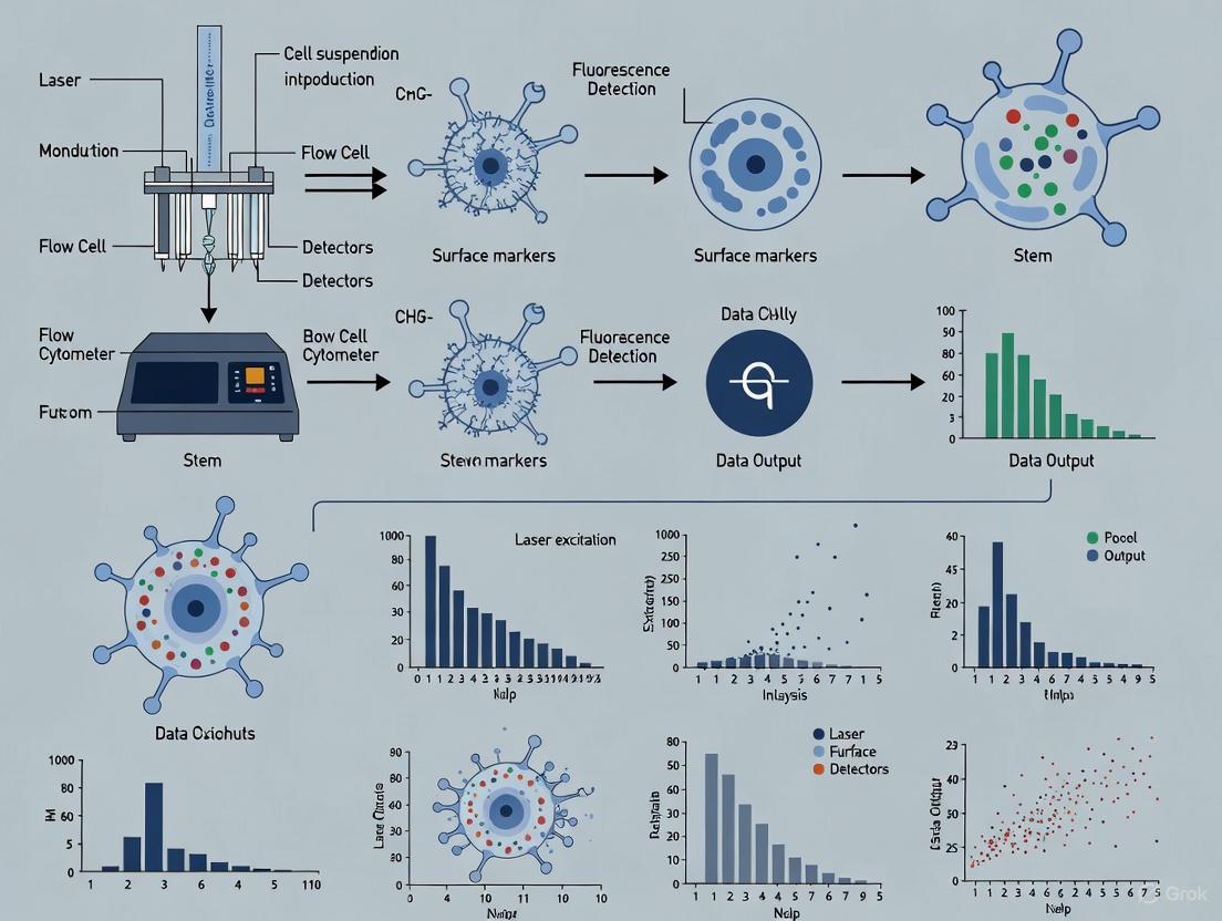
Flow cytometry has established itself as an indispensable technology in stem cell research and therapy development. By enabling rapid, multiparameter analysis of physical and chemical characteristics at the single-cell level, flow cytometry provides unprecedented resolution for identifying and characterizing rare stem cell populations within heterogeneous mixtures [16]. The fundamental principle of this technology relies on measuring light scattered by particles and the fluorescence emitted from fluorochrome-conjugated antibodies as cells pass in a stream through a laser beam [16]. The major strength of flow cytometry lies in its ability to perform highly multiplexed quantitative measurements on single cells, making it ideally suited for stem cell research where the cell types of interest are often extremely rare [17]. This application note examines the technological evolution of flow cytometry and details standardized protocols that leverage these advances for advanced stem cell analysis.
Applications in Stem Cell Research
Flow cytometry serves multiple critical functions in stem cell biology, from basic phenotyping to preparatory isolation for therapeutic applications. The table below summarizes key application areas:
Table 1: Key Applications of Flow Cytometry in Stem Cell Research
| Application Area | Specific Uses | Stem Cell Types |
|---|---|---|
| Immunophenotyping | Identification and enumeration of stem/progenitor cells using surface and intracellular markers [17] [18]. | Hematopoietic Stem Cells (HSCs), Mesenchymal Stem Cells (MSCs), Neural Stem Cells (NSCs) [17]. |
| Cell Cycle Analysis | DNA content quantification using propidium iodide to distinguish G0/G1, S, and G2/M phases [19]. | Pluripotent Stem Cells, Cancer Stem Cells [19]. |
| Functional Analysis | Measurement of mitochondrial parameters, reactive oxygen species, and apoptosis [20]. | Induced Pluripotent Stem Cells (iPSCs) and their derivatives [20]. |
| Cell Sorting | Physical isolation of pure stem cell populations for downstream analysis or therapy [16] [18]. | All stem cell types, particularly HSCs for transplantation [18]. |
| Disease Modeling | Characterization of patient-specific stem cell derivatives for disease mechanisms and drug screening [17] [21]. | iPSC-derived neurons, glial cells, cardiomyocytes [17] [21]. |
The applications extend across diverse stem cell types. For hematopoietic stem cells (HSCs), flow cytometry enables precise immunophenotyping for transplantation biology, using markers like CD34 to identify hematopoietic reconstituting cells [17]. In mesenchymal stem cells (MSCs) from bone marrow and adipose tissue, flow cytometry facilitates characterization using markers such as CD45−/CD34−/CD73+/CD105+/CD90+ [17]. For neural stem cells, specific surface antigen combinations (CD15/CD24/CD29 or CD133) allow isolation and quantification of neural populations [22]. The technology also plays a crucial role in cancer stem cell (CSC) research, enabling the identification and isolation of cancer stem-like cells for understanding tumorigenesis and treatment resistance [17].
Technological Advances in Flow Cytometry
The evolution of flow cytometry from basic 2-3 color analysis to sophisticated polychromatic platforms has dramatically enhanced its utility in stem cell research. These advances synergize improvements in hardware, reagents, and analytical software [18].
Hardware Innovations
Modern flow cytometers feature multiple laser systems and enhanced detection capabilities. Key developments include physically smaller air-cooled lasers, new designs in optics, and highly sensitive photomultiplier tubes (PMTs) [18]. These innovations have enabled higher parameter analysis while reducing the operational footprint and cost. For stem cell applications, the introduction of high-quality multilaser platforms has been particularly valuable for techniques like side population (SP) analysis, which requires violet laser excitation to detect Hoechst 33342 dye efflux - a hallmark of certain stem cell populations [18].
Expanded Fluorophore Repertoire
The commercial availability of monoclonal antibodies conjugated to fluorochromes with excitation maxima across multiple laser lines has been pivotal for polychromatic panels. Violet-excitable dyes (Pacific Blue, Alexa 405, quantum dots), blue-light excited fluorophores (FITC, PE, PerCP), and red-excited dyes (APC, Alexa 647) now provide researchers with an extensive palette [18]. Tandem dyes that combine energy transfer between donor and acceptor fluorochromes further expand possibilities, though they require careful validation due to potential instability and lot-to-lot variation [18].
Analytical Software Capabilities
As flow cytometry panels have grown in complexity, software capable of managing numerous intra- and inter-laser fluorochrome compensation calculations has become essential [18]. Modern digital software applies compensation matrixes post-acquisition and utilizes bi-exponential scaling to visualize data with broad dynamic ranges. However, analytical software remains a developing field, with "data-mining" of complex polychromatic datasets still presenting usability challenges [18].
Standardized Protocols for Stem Cell Analysis
Protocol 1: Surface and Intracellular Antigen Analysis for Neural Cell Types
This protocol enables comprehensive immunophenotyping of neural stem cells and their derivatives through simultaneous surface and intracellular antigen detection [22].
Table 2: Key Reagent Solutions for Neural Antigen Analysis
| Reagent | Function | Application Notes |
|---|---|---|
| CD Antibodies (e.g., CD24, CD54) | Surface antigen detection for cell population identification [22]. | Use bright fluorophores (PE, APC) for low-abundance antigens [23]. |
| CFSE | Fluorescent cell labeling for tracking and comparative analysis [22]. | Enables comparison of two conditions in one tube, reducing variance [22]. |
| Zenon Labeling Kit | Non-covalent Fab fragment labeling for intracellular targets [22]. | Reduces cell manipulation steps compared to secondary antibodies [22]. |
| Paraformaldehyde | Cross-linking fixative | Preserves fluorescent proteins and surface markers; requires subsequent permeabilization [19]. |
| Permeabilization Buffer (Triton X-100) | Enables antibody access to intracellular epitopes [19]. | Necessary after aldehyde fixation for intracellular staining [19]. |
Experimental Workflow:
- Cell Harvesting: Gently wash adherent neural cultures with Mg²⁺/Ca²⁺-free PBS. Detach cells using appropriate enzymatic (trypsin) or non-enzymatic means [22].
- Viability and Cell Counting: Assess cell viability and concentration using trypan blue exclusion or similar methods [22].
- Optional CFSE Labeling: For comparative experiments, label one cell population with CFSE (5-10 μM, 10 minutes at 37°C) followed by serum-containing medium to quench the reaction [22].
- Surface Antigen Staining: Resuspend cells in staining buffer with fluorochrome-conjugated CD antibodies. Incubate for 20-30 minutes at 4°C in the dark [22].
- Fixation and Permeabilization: Fix cells with 2-4% paraformaldehyde for 15 minutes. Permeabilize with 0.1% Triton X-100 for intracellular antigen access [19].
- Intracellular Staining: Apply Zenon-labeled primary antibodies or standard primary/secondary antibody combinations for intracellular targets [22].
- Flow Cytometric Analysis: Resuspend cells in appropriate buffer and analyze using configured flow cytometer. Use forward scatter (FSC) vs. side scatter (SSC) to identify single cells and exclude debris [22].
Flowchart for Neural Cell Antigen Analysis
Protocol 2: Multiparameter Mitochondrial Functional Analysis in iPSCs
This protocol enables comprehensive assessment of mitochondrial function in pluripotent stem cells and their derivatives, which is crucial for modeling neurodegenerative diseases [20].
Table 3: Reagents for Mitochondrial Function Analysis
| Reagent | Function | Detection Parameter |
|---|---|---|
| MitoTracker Green (MTG) | Labels mitochondrial mass/volume regardless of membrane potential [20]. | Mitochondrial Volume |
| Tetramethylrhodamine Ethyl Ester (TMRE) | Accumulates in active mitochondria based on membrane potential [20]. | Mitochondrial Membrane Potential (MMP) |
| MitoSox Red | Selective detection of mitochondrial superoxide [20]. | Mitochondrial Reactive Oxygen Species (ROS) |
| Antibodies to MRC subunits | Target specific mitochondrial respiratory chain complexes [20]. | Respiratory Chain Composition |
| Anti-TFAM | Binds mitochondrial transcription factor A [20]. | mtDNA Copy Number (indirect) |
Experimental Workflow:
- Cell Preparation: Culture iPSCs or iPSC-derived neural/glial cells on matrix-coated plates using appropriate maintenance media [20].
- Live-Cell Staining for Functional Parameters: For mitochondrial volume, membrane potential, and ROS detection, stain live cells with MTG (50-100 nM), TMRE (50-200 nM), and MitoSox Red (2-5 μM) for 30 minutes at 37°C [20].
- Fixation for Immunostaining: For respiratory chain and mtDNA analysis, fix cells with 4% paraformaldehyde for 15 minutes [20].
- Intracellular Staining: Permeabilize fixed cells and stain with antibodies against MRC complex subunits and TOMM20 (mitochondrial mass marker), or TFAM and TOMM20 [20].
- Flow Cytometric Analysis: Analyze cells using appropriate laser lines and detectors. Relate functional parameters to mitochondrial content by comparing fluorescence intensities [20].
- Data Interpretation: Calculate ratios such as TFAM/TOMM20 to estimate mtDNA copy number per mitochondrial unit [20].
Flowchart for Mitochondrial Function Analysis
Panel Design and Optimization for Polychromatic Flow Cytometry
Designing effective multicolor panels requires systematic planning to overcome spectral overlap challenges. Follow these key principles for optimal panel design:
- Instrument Configuration: Determine the number and type of lasers, number of detectors, and available filters on your flow cytometer before selecting fluorophores [23].
- Antigen-Fluorophore Matching: Assign bright fluorophores (PE, APC) to low-abundance antigens or rare cell populations. Use dimmer fluorophores for highly expressed antigens [23].
- Spectral Overlap Minimization: Select fluorophores with minimal emission spectrum overlap. Where overlap exists, ensure it can be adequately compensated using single-stained controls [23].
- Compensation Controls: Include compensation controls for each fluorophore using beads or cells with positive and negative populations. The positive population should be at least as bright as experimental samples and comprise ≥10% of the control sample [23].
- Validation: Always validate antibody panels using known positive and negative control cell populations to confirm specific staining patterns [21].
Future Perspectives
The evolving role of flow cytometry in stem cell biology continues to expand with emerging technological capabilities. Mass cytometry (CyTOF) represents one frontier, allowing simultaneous analysis of over 30 parameters using metal-conjugated antibodies instead of fluorochromes [22]. Imaging flow cytometry combines the high-throughput capability of conventional flow cytometry with morphological information from fluorescence microscopy [16]. Additionally, sophisticated bioinformatics tools for high-dimensional data analysis are enhancing our ability to extract meaningful biological insights from complex stem cell datasets [18].
These technological advances synergize with the growing importance of stem cells in disease modeling, drug screening, and cellular therapy. The ability to rigorously characterize stem cell populations and their derivatives using standardized flow cytometric protocols ensures the reliability and reproducibility essential for both basic research and clinical applications [17] [21]. As the field progresses, flow cytometry will undoubtedly remain a cornerstone technology in stem cell biology and therapy development.
Flow cytometry has evolved far beyond simple cell identification and sorting. In stem cell research, this technology provides a powerful platform for interrogating fundamental cellular processes, offering unparalleled insights into cell cycle status, proliferation kinetics, and functional heterogeneity within complex populations. While immunophenotyping remains crucial for identifying stem cell populations based on surface markers, true understanding of stem cell behavior requires integration of these identification methods with functional and cell cycle analyses [24] [25]. This integrated approach enables researchers to decipher the complex mechanisms controlling hematopoietic stem cell (HSC) cycling, self-renewal, and differentiation—critical processes for both basic research and therapeutic development [24]. For drug development professionals, these analyses provide essential tools for assessing how potential therapeutics influence stem cell fate decisions, proliferation dynamics, and ultimately, functional outcomes in both normal and diseased states.
Core Scientific Principles and Methodologies
DNA Content Analysis for Cell Cycle Profiling
Cell cycle analysis by flow cytometry typically utilizes fluorescent dyes that bind stoichiometrically to DNA, enabling discrimination of cells in different cell cycle phases based on DNA content [19] [26]. Propidium iodide (PI) represents one of the most widely employed dyes for this application, intercalating with double-stranded DNA and emitting red fluorescence when excited by a 488nm laser [19]. The fundamental principle underpinning this technique is that DNA content doubles during S-phase, with cells in G0/G1 phase exhibiting half the DNA content of cells in G2/M phase, while S-phase cells display intermediate DNA content [19] [26].
A critical consideration for DNA content analysis is the requirement for cell permeabilization to allow dye access to nuclear DNA. Ethanol fixation effectively permeabilizes cells while maintaining structural integrity for DNA analysis, though alternative approaches including detergent-based permeabilization or cross-linking fixatives like paraformaldehyde may be employed when simultaneous analysis of surface markers or intracellular proteins is required [19]. Equally important is the inclusion of RNase treatment during sample preparation, as PI binds to both DNA and RNA, and RNA digestion is essential to eliminate background signal and ensure specific DNA quantification [19].
Figure 1: Workflow for DNA Content Analysis Using Propidium Iodide Staining
Advanced Multiparametric Functional Assays
Beyond static DNA content measurement, flow cytometry enables dynamic assessment of stem cell function through multiparametric approaches. BrdU (5-bromo-2'-deoxyuridine) incorporation provides a powerful method for tracking DNA synthesis over time, allowing researchers to distinguish actively cycling cells from those in quiescence [26]. When combined with DNA content dyes, BrdU detection facilitates detailed analysis of cell cycle progression kinetics, identifying cells that have entered S-phase during a specific labeling window [26].
Mitochondrial profiling has emerged as particularly valuable in stem cell research, as mitochondrial content and function often correlate with stemness and differentiation potential [27]. Studies in planarian stem cells demonstrated that pluripotent stem cells exhibit lower mitochondrial content compared to specialized progenitors, enabling purification of pluripotent populations using dyes like MitoTracker Green in combination with DNA stains [27]. Similarly, functional assays measuring calcium flux, intracellular pH, and mitochondrial membrane potential provide insights into metabolic status and signaling dynamics within stem cell populations [25].
For comprehensive stem cell analysis, integration of cell surface immunophenotyping with these functional assessments is essential. The well-established LSK (Lin-Sca1+c-Kit+) phenotype for murine hematopoietic stem and progenitor cells can be further refined using functional markers, with advanced phenotypes like LSK/SLAM (CD150+CD48-) and ESLAM (CD45+EPCR+CD150+CD48-) providing enhanced resolution of primitive stem cell subsets with distinct functional properties [24].
Experimental Protocols
DNA Content Analysis Using Propidium Iodide
Materials Required:
- 70% Ethanol (prepared with distilled water, not PBS)
- Propidium iodide stock solution (50 µg/mL)
- Ribonuclease I stock solution (100 µg/mL)
- Phosphate-buffered saline (PBS)
- Flow cytometer with 488nm excitation and appropriate emission filters (e.g., 605nm bandpass)
Procedure:
- Cell Harvesting: Harvest cells using appropriate methods (e.g., trypsin for adherent cells) and wash in PBS. Concentrate cells to ensure optimal sample stream formation during analysis [19].
- Fixation: Gently resuspend cell pellet in cold 70% ethanol added drop-wise while vortexing to minimize clumping. Fix for 30 minutes at 4°C [19].
- Washing: Centrifuge at 850 × g for 5-10 minutes and carefully discard supernatant. Wash cell pellet twice in PBS to remove residual ethanol [19].
- RNase Treatment: Resuspend cell pellet in PBS containing RNase (final concentration approximately 5 µg/mL) and incubate for 15-30 minutes to digest RNA [19].
- DNA Staining: Add propidium iodide to a final concentration of 50 µg/mL and incubate for 30 minutes protected from light [19].
- Flow Cytometric Analysis: Analyze samples using a flow cytometer equipped with a 488nm laser. Collect forward scatter (FSC) and side scatter (SSC) parameters to identify single cells, followed by PI fluorescence detection using appropriate emission filters [19].
Data Analysis:
- Use pulse processing (pulse area vs. pulse width) to exclude cell doublets from analysis [19] [26].
- Gate on single cell population using FSC vs. SSC, then apply this gate to PI histogram.
- Analyze DNA content histogram using curve-fitting algorithms to quantify percentage of cells in G0/G1, S, and G2/M phases [19].
Table 1: Critical Steps in DNA Content Analysis Protocol
| Step | Key Parameter | Optimization Tips | Potential Issues |
|---|---|---|---|
| Fixation | Ethanol concentration | Add drop-wise while vortexing | Cell clumping with rapid addition |
| RNase Treatment | Concentration & time | Include in staining solution | RNA contamination without proper treatment |
| Doublet Exclusion | Pulse processing | Use area vs. width/height | G2/M misidentification without discrimination |
| Analysis | Curve-fitting model | Validate with control samples | Poor model fitting with high debris |
Integrated Stem Cell Phenotyping and Functional Analysis
Materials Required:
- Fluorescently-conjugated antibodies against surface markers (e.g., lineage cocktail, Sca1, c-Kit, CD150, CD48)
- Functional dyes (e.g., MitoTracker Green, BrdU, viability dyes)
- Staining buffer (PBS with 1-5% FBS)
- Fc receptor blocking antibody (e.g., anti-CD16/32)
- Permeabilization buffers if intracellular staining required
Procedure:
- Cell Preparation: Isolate bone marrow cells by flushing femora and tibiae with PBS containing 5mM EDTA and 1% fetal calf serum. Generate single-cell suspension by gentle trituration and filter through 40µm strainer [24].
- Viability Staining: Include viability dye (e.g., propidium iodide or alternative viability markers) to exclude dead cells from analysis [19] [27].
- Fc Receptor Blocking: Incubate cells with Fc block (anti-CD16/32 antibody) or serum from same species as detection antibodies to reduce non-specific binding [24].
- Surface Marker Staining: Incubate cells with fluorescently-conjugated antibody cocktail against lineage markers (CD3, CD11b, CD45R, Gr-1, Ter119), Sca1, c-Kit, and additional markers of interest (CD150, CD48, EPCR) for 20-30 minutes on ice protected from light [24].
- Functional Staining: For mitochondrial content analysis, incubate cells with MitoTracker Green (diluted in staining buffer) for 15-30 minutes at 37°C [27].
- Fixation/Permeabilization: If intracellular staining required, fix cells with 2-4% paraformaldehyde followed by permeabilization with 0.1% Triton X-100 [19].
- BrdU Staining: For proliferation analysis, incubate cells with BrdU prior to harvest, then detect incorporated BrdU using fluorescent anti-BrdU antibodies after DNA denaturation [26].
- Flow Cytometric Analysis: Acquire data on flow cytometer configured for multiple fluorochromes. Include appropriate controls (unstained, single stains, fluorescence-minus-one) for proper compensation and gating [24] [28].
Data Analysis:
- Begin with FSC vs. SSC gating to identify intact cells, followed by viability dye exclusion [29].
- Apply lineage negative gate to exclude mature hematopoietic cells [24].
- Identify LSK population (Lin-Sca1+c-Kit+) and further subset using SLAM markers (CD150+CD48-) or other refinement markers [24].
- Analyze functional parameters (mitochondrial content, BrdU incorporation) within defined immunophenotypic subsets [27].
Table 2: Multicolor Panel for Murine Hematopoietic Stem Cell Analysis
| Marker | Fluorochrome | Population Identified | Expression Pattern |
|---|---|---|---|
| Lineage Cocktail | FITC | Differentiated cells | Positive on mature cells |
| Sca1 | APC or Biotin | Primitive cells | Positive on stem/progenitor cells |
| c-Kit | PE | Stem/progenitor cells | Positive on stem/progenitor cells |
| CD150 | PE-Cy7 | LT-HSC enrichment | Positive on long-term HSCs |
| CD48 | APC | Differentiated progenitors | Negative on primitive HSCs |
| Viability Dye | e.g., PI or DAPI | Dead cells | Positive on dead cells |
Data Presentation and Analysis
Data Acquisition and Quality Control
Proper data acquisition and quality control are essential for generating reliable flow cytometric data, particularly when analyzing rare stem cell populations. Instrument calibration using fluorescent beads ensures consistent performance across experiments, while appropriate compensation corrects for spectral overlap between fluorochromes, preventing misinterpretation of marker expression [28]. For rare population analysis, acquiring sufficient event counts is critical—Poisson statistics dictate that precise quantification of populations representing <0.1% of total cells requires acquisition of hundreds of thousands to millions of events [28].
Control samples represent another crucial component of quality flow cytometry data. Fluorescence-minus-one (FMO) controls, which contain all antibodies except the one being evaluated, establish background fluorescence and proper gating boundaries, particularly important for dimly expressed markers and complex multicolor panels [24] [28]. While single-color controls facilitate compensation setup, biological controls (e.g., wild-type vs. knockout cells) often provide more meaningful expression references than isotype controls [24] [28].
Data Visualization and Interpretation
Effective data presentation employs multiple visualization strategies to convey different aspects of flow cytometric data. Histograms optimally display single-parameter data, enabling clear comparison of fluorescence intensity distributions between samples [29]. Overlaying histograms of experimental and control conditions (e.g., stained vs. unstained, different treatment groups) facilitates direct visualization of expression differences and calculation of relative fluorescence intensity [29].
Scatter plots (dot plots, density plots, contour plots) enable multiparameter visualization, displaying the relationship between two measured parameters for each cell [29]. The standard FSC vs. SSC plot provides initial population discrimination, while fluorescence vs. fluorescence plots (e.g., CD4 vs. CD8) enable identification of distinct immunophenotypic subsets [29]. Gating strategies should be clearly documented, beginning with intact cell selection, followed by single-cell gating (using pulse width vs. pulse area), viability gating, and successive marker gates to define populations of interest [19] [28].
Figure 2: Comprehensive Gating Strategy for Stem Cell Analysis
For publication, comprehensive documentation of gating strategies, instrument configuration, and analytical methods is essential [28] [30]. Journals increasingly require inclusion of this information as supplemental data to ensure reproducibility and proper interpretation [28]. Statistical analysis should be applied appropriately to either fluorescence intensity (typically reported as mean or median) or population frequency, with clear indication of replicate number and statistical tests employed [28] [30].
Table 3: Essential Controls for Flow Cytometry Experiments
| Control Type | Purpose | Composition | Application |
|---|---|---|---|
| Unstained | Autofluorescence | No antibodies | Instrument setup |
| Single Stains | Compensation | Individual antibodies | Multi-color compensation |
| FMO | Gating boundaries | All antibodies minus one | Defining positive populations |
| Biological | Expression reference | Wild-type/KO cells | Biological context |
| Compensation Beads | Standardized compensation | Antibody-coated beads | Alternative to cells |
The Scientist's Toolkit: Research Reagent Solutions
Table 4: Essential Reagents for Flow Cytometric Stem Cell Analysis
| Reagent Category | Specific Examples | Function/Application | Considerations |
|---|---|---|---|
| DNA Binding Dyes | Propidium iodide, DAPI, Hoechst 33342 | DNA content quantification, cell cycle analysis | PI requires permeabilization; Hoechst penetrates live cells [19] [26] |
| Viability Indicators | Propidium iodide, DAPI, LIVE/DEAD dyes | Dead cell exclusion | Membrane-impermeant DNA dyes [19] [27] |
| Functional Probes | MitoTracker Green, BrdU, Ca²⁺ indicators | Mitochondrial content, proliferation, signaling | MitoTracker Green reflects mass not membrane potential [27] |
| Surface Antibodies | Lineage cocktail, Sca1, c-Kit, CD150, CD48 | Immunophenotypic identification | Titration essential for signal-to-noise optimization [24] |
| Intracellular Antibodies | Phospho-histone H3, Ki-67, cyclins | Cell cycle stage, proliferation status | Requires fixation/permeabilization [26] |
| Compensation Beads | Anti-mouse/rat Ig beads | Compensation controls | Consistent fluorescence for instrument setup [24] |
Integration of cell cycle and functional analyses with traditional immunophenotyping represents a powerful approach for comprehensive stem cell characterization in research and drug development contexts. The methodologies outlined in this application note—from fundamental DNA content analysis to sophisticated multiparametric assessments of function and metabolism—provide researchers with robust tools for interrogating stem cell behavior at unprecedented resolution. As flow cytometry technology continues to advance, with innovations in spectral analysis, increased parameter capacity, and enhanced computational tools, these integrated approaches will undoubtedly yield deeper insights into stem cell biology and accelerate the development of stem cell-based therapeutics.
Practical Protocols for Staining and Analyzing Stem Cells
In stem cell research, high-quality flow cytometry data is essential for accurately identifying distinct stem cell types, monitoring differentiation, and isolating rare populations like cancer stem cells. The foundation of any successful flow cytometry experiment is the preparation of a viable single-cell suspension that preserves cell surface antigens and minimizes artifacts. This guide details standardized protocols for obtaining single-cell suspensions from diverse biological sources, framed within the context of flow cytometry for stem cell analysis.
Core Principles of Single-Cell Suspension Preparation
Preparing a high-quality single-cell suspension is a critical initial step that profoundly influences all downstream flow cytometry results. The primary goals are to achieve a suspension with high cell viability, minimal cell debris, and an absence of cell aggregates, all while preserving the antigenic properties of the cells [31] [32].
Key Considerations:
- Viability Maintenance: Adding protein (e.g., 2% FBS, 1% BSA) to buffers and media at all stages of cell processing improves cell viability. Gentle resuspension of fragile cells is essential [32].
- Clump Prevention and Removal: Clumps can cause instrument blockages and uneven staining. Strategies include using DNase to break down DNA from dead cells, adding EDTA to chelate cations involved in cell adhesion, gentle but thorough pipetting, and filtering the suspension through cell strainers (e.g., 70 µm nylon mesh) before analysis [32].
- Antigen Preservation: The methods used for tissue dissociation, especially enzymatic digestion, can destroy antibody epitopes. Careful selection of enzymes and validation of surface marker integrity after processing are crucial [33].
The method for creating a single-cell suspension must be tailored to the starting material. The table below summarizes the core approaches for different sample types.
Table 1: Overview of Single-Cell Preparation Methods for Various Sources
| Sample Source | Primary Dissociation Method | Key Considerations | Common Applications in Stem Cell Research |
|---|---|---|---|
| Lymphoid Tissues (Spleen, Lymph Nodes) [33] | Mechanical Disruption | Generally requires only mechanical teasing; gentle yet effective. | Analysis of hematopoietic stem/progenitor cells from bone marrow [24]. |
| Solid Tissues / Tumors [31] [33] | Enzymatic Digestion & Mechanical Dissociation | Enzyme choice (collagenase, trypsin, accutase) is critical to preserve target antigens. | Isolation of mesenchymal stem cells or cancer stem cells from solid tissues [34] [35]. |
| Adherent Cell Cultures [33] [32] | Enzymatic or Non-Enzymatic Detachment | Scraping can damage cells; enzymes like Accutase are gentler on surface markers than trypsin. | Culture and analysis of pluripotent stem cells (ESCs, iPSCs) or mesenchymal stromal cells [34]. |
| Peripheral Blood / Bone Marrow [33] [32] | Density Gradient Centrifugation (for PBMCs) or RBC Lysis | Minimizes manipulation, preserving rare and fragile cell types. | Immunophenotyping of hematopoietic stem cells (HSCs) from blood or bone marrow [34] [24]. |
Universal Workflow for Sample Processing
The following diagram outlines the general logical workflow for processing various sample types into a single-cell suspension ready for flow cytometry analysis.
Detailed Step-by-Step Protocols
Protocol A: Adherent Cell Cultures (e.g., MSC, iPSC cultures)
This protocol is suitable for adherent stem cell cultures, such as mesenchymal stem cells (MSCs) or induced pluripotent stem cells (iPSCs) [33] [32].
Materials:
- Accutase Enzyme Cell Detachment Medium or EDTA (e.g., 10 mM in PBS) [33] [32].
- Phosphate-buffered saline (PBS) without Ca2+/Mg2+.
- Flow Cytometry Staining Buffer (PBS with 1% BSA or 2% FBS).
- Centrifuge tubes.
Experimental Procedure:
- Remove Culture Medium: Aspirate and wash the cell layer gently with PBS.
- Detach Cells:
- Add a sufficient volume of pre-warmed Accutase or EDTA to cover the cell layer.
- Incubate at 37°C for 5-10 minutes (or until cells detach). Monitor under a microscope.
- Neutralize & Recover: Gently pipette the detached cells and transfer them to a conical tube. If using trypsin, neutralize with serum-containing medium.
- Wash: Centrifuge the cell suspension at 300-400 x g for 4-5 minutes. Discard the supernatant.
- Resuspend & Count: Resuspend the cell pellet in a known volume of Flow Cytometry Staining Buffer. Perform a cell count and viability analysis (e.g., using trypan blue exclusion).
- Final Preparation: Adjust the cell concentration to 1 x 10^7 cells/mL (or as required for your staining protocol) in Flow Cytometry Staining Buffer [33].
Protocol B: Lymphoid Tissues (e.g., Spleen, Bone Marrow)
This method uses mechanical disruption and is ideal for generating single-cell suspensions from murine spleen or bone marrow for hematopoietic stem cell (HSC) analysis [33] [24].
Materials:
- Frosted glass microscope slides or the plunger of a 3-mL syringe.
- Nylon cell strainer (70 µm).
- Flow Cytometry Staining Buffer.
- Cold PBS supplemented with 2% FBS and 2 mM EDTA.
Experimental Procedure:
- Harvest Tissue: Place the freshly harvested tissue (e.g., spleen) into a culture dish containing 5-10 mL of cold staining buffer.
- Mechanical Disruption:
- Option 1 (Slides): Place the tissue between the frosted ends of two glass slides and gently grind them together to release cells.
- Option 2 (Plunger): Press the tissue with the plunger of a 3-mL syringe.
- Filter Suspension: Transfer the cell suspension through a 70 µm cell strainer into a 15 mL or 50 mL conical tube to remove debris and clumps.
- Wash & Count: Centrifuge at 300-400 x g for 5 minutes. Discard supernatant, resuspend in fresh buffer, and perform a cell count and viability analysis.
- Final Preparation: Centrifuge again and resuspend at the desired concentration (e.g., 1 x 10^7 cells/mL) for staining [33] [24].
Protocol C: Solid Tissues and Tumors (e.g., for CSC isolation)
This protocol, combining mechanical and enzymatic dissociation, is critical for processing solid tumors to isolate cancer stem cells (CSCs) or other stem cells from connective tissues [31] [33] [35].
Materials:
- Surgical scissors or scalpel.
- Appropriate enzymes (e.g., collagenase, liberase).
- DNase I.
- Cell strainer (70-100 µm).
- Staining buffer with protein and EDTA.
Experimental Procedure:
- Mince Tissue: Harvest the tissue and finely mince it into 2-4 mm pieces using scissors or a scalpel in a small volume of buffer.
- Enzymatic Digestion: Add the selected enzyme cocktail (e.g., Collagenase IV + DNase I) dissolved in PBS. Incubate at 37°C with gentle agitation for 15-45 minutes. The optimal time and temperature must be determined empirically [31].
- Dissociate & Stop: Periodically triturate the tissue digest during incubation using a pipette. After digestion, add excess cold buffer with serum to stop enzyme activity.
- Filter: Pass the crude cell suspension through a cell strainer to remove undigested fragments.
- Wash & Purify: Centrifuge the filtrate at 300-400 x g for 5 minutes. Resuspend the pellet in buffer. For tissues with high red blood cell content, perform RBC lysis at this stage.
- Count & Prepare: Perform a cell count and viability analysis. The suspension is now ready for staining or further processing [31] [33].
The Scientist's Toolkit: Essential Reagents and Materials
The following table catalogs key reagents and materials essential for preparing high-quality single-cell suspensions.
Table 2: Essential Reagents and Materials for Single-Cell Suspension Preparation
| Item | Function & Application | Example Use-Case |
|---|---|---|
| Accutase | Enzyme blend for detaching adherent cells; gentler on surface epitopes than trypsin. | Detaching mesenchymal stem cells (MSCs) or pluripotent stem cells without cleaving critical surface receptors [33] [32]. |
| Collagenase | Enzyme degrading collagen in the extracellular matrix for solid tissue dissociation. | Digesting solid tumors or connective tissues to isolate cancer stem cells (CSCs) or other resident stem cells [31] [35]. |
| DNase I | Degrades free DNA released by dead cells, reducing viscous clumping and cell aggregation. | Added during and after dissociation of fragile tissues (e.g., tumors) to improve single-cell yield and prevent instrument clogs [32]. |
| EDTA | Chelates cations (Ca2+); inhibits cell-cell adhesion and acts as a non-enzymatic cell detachment agent. | Used in dissociation buffers for adherent cells and in wash buffers to prevent re-aggregation of single cells [33] [32]. |
| Ficoll-Paque | Density gradient medium for isolating peripheral blood mononuclear cells (PBMCs) from whole blood. | Isolation of mononuclear cells, including progenitor cells, from peripheral blood or bone marrow aspirates [33]. |
| Cell Strainer (70 µm) | Physically removes cell clumps and tissue debris from the single-cell suspension. | Final filtration step for lymphoid tissue or solid tumor dissociates before staining or flow cytometric analysis [33] [32]. |
| Flow Cytometry Staining Buffer (PBS + Protein) | Protects cell viability, reduces nonspecific antibody binding, and maintains cells in suspension. | Used for all cell washing and resuspension steps after dissociation and as the base for antibody cocktails [33]. |
Application in Stem Cell Research: Marker Panels and Quality Control
Stem Cell Marker Panels for Flow Cytometry
Once a single-cell suspension is prepared, accurate immunophenotyping is crucial. The table below outlines common surface marker combinations used to identify major stem cell types.
Table 3: Common Flow Cytometry Markers for Identifying Stem Cell Populations
| Stem Cell Type | Positive Markers | Negative Markers | Primary Research Application |
|---|---|---|---|
| Hematopoietic Stem Cells (HSC) | CD34, CD49f, CD90, c-Kit (CD117), Sca-1 (mouse) | CD38, CD45RA | Purification and analysis of HSCs from bone marrow or cord blood for transplantation studies [34] [24]. |
| Mesenchymal Stem Cells (MSC) | CD73, CD90, CD105 | CD11b, CD19, CD45, HLA-DR | Identification and isolation of MSCs from bone marrow or adipose tissue for regenerative medicine [34] [36]. |
| Pluripotent Stem Cells (PSC) | SSEA-3, SSEA-4, TRA-1-60 | SSEA-1 | Characterizing embryonic stem cells (ESCs) and induced pluripotent stem cells (iPSCs) [34]. |
| Mouse HSC (LSK/SLAM Phenotype) | Lin-, Sca-1+, c-Kit+, CD150+ | CD48- | High-purity isolation of long-term hematopoietic stem cells (LT-HSCs) from mouse bone marrow [24]. |
Quality Assessment and Troubleshooting
Before proceeding to antibody staining, rigorously assess the quality of your single-cell suspension.
Assessment Methods:
- Viability Analysis: Use trypan blue exclusion or automated cell counters. High viability (>90% is ideal) is critical for reducing non-specific binding and background noise [31] [32].
- Microscopic Inspection: Visually check for the presence of clumps and overall cell condition [32].
- Flow Cytometry Pre-Scan: Use forward scatter (FSC) vs. side scatter (SSC) plots to identify and gate out debris and doublets. A sharp, distinct population of single cells indicates a high-quality prep.
Common Pitfalls and Solutions:
- Poor Viability: Ensure solutions contain protein, processing is done quickly on ice, and harsh mechanical force is avoided.
- Excessive Clumping: Increase the concentration of DNase I or EDTA; perform additional filtration steps; avoid over-trituration, which can damage cells and release more DNA [32].
- Low Yield from Solid Tissues: Optimize enzyme type, concentration, and incubation time. Consider using a combination of enzymes (e.g., collagenase + dispase) for complex tissues [31].
- Loss of Surface Antigen: If an epitope is lost, switch to a gentler enzyme (e.g., from trypsin to Accutase) or a non-enzymatic method (e.g., EDTA-based). Always validate your dissociation protocol for the markers of interest [33] [32].
Mastering the preparation of a high-quality single-cell suspension is a foundational skill in stem cell research. The protocols detailed here for adherent cultures, lymphoid tissues, and solid tissues provide a reliable starting point. By adhering to best practices in tissue dissociation, clump removal, and viability maintenance, and by rigorously applying quality control measures, researchers can ensure that their flow cytometry data accurately reflects the biology of rare and valuable stem cell populations, thereby enabling advancements in both basic research and therapeutic development.
Optimized Staining for Surface Antigens and Intracellular Markers (e.g., Transcription Factors)
Flow cytometry serves as a cornerstone of modern stem cell research, enabling the detailed characterization of complex populations at a single-cell level. While immunophenotyping based on surface antigens is well-established for hematopoietic lineages, stem cell biology often requires the simultaneous analysis of both surface markers and intracellular proteins, such as transcription factors, to definitively identify stem and progenitor cell states [37] [5]. This combined approach is crucial for isolating well-defined cell subsets for downstream applications in regenerative medicine, disease modeling, and drug development [5]. However, the fixation and permeabilization steps required for intracellular staining can compromise surface antigen detection and cell viability, presenting a significant technical challenge [38] [5]. This application note provides optimized, detailed protocols for the simultaneous flow cytometric analysis of surface and intracellular antigens, with a specific focus on stem cell research applications.
Critical Considerations for Experimental Design
Successful multicolor flow cytometry hinges on careful upfront planning. The following considerations are paramount for generating high-quality, reproducible data.
- Antibody Validation and Titration: Always use antibodies validated for flow cytometry, particularly for intracellular targets. Perform titration experiments for each new antibody lot to determine the optimal signal-to-noise ratio [38].
- Appropriate Controls: Include the following controls in every experiment:
- Viability Staining: The fixation and permeabilization process can increase non-specific binding. The use of fixable viability dyes (FVDs) is strongly recommended to exclude dead cells from the analysis, which is critical for data accuracy [38] [39].
- Antigen Localization: The subcellular location of the intracellular target (cytoplasmic, secreted, or nuclear) dictates the choice of fixation and permeabilization reagents [38]. For example, transcription factors like FoxP3 require a specific buffer set for optimal detection [38].
Detailed Staining Protocols
Protocol A: Simultaneous Staining of Surface Antigens and Intracellular Cytoplasmic Proteins
This two-step protocol is optimized for cytoplasmic proteins, cytokines, and other secreted factors. It is widely used for assessing the functional state of stem and progenitor cells, such as cytokine production in hematopoietic stem and progenitor cells (HSPCs) [40].
Table 1: Key Reagents for Staining Cytoplasmic Proteins
| Reagent | Function | Example Product |
|---|---|---|
| Intracellular Fixation Buffer | Stabilizes cell membranes and proteins; cross-links proteins. | Intracellular Fixation & Permeabilization Buffer Set [38] |
| Permeabilization Buffer | Creates pores in membranes allowing antibody access to the interior of the cell. | 1X Permeabilization Buffer (10X concentrate diluted in dH₂O) [38] |
| Protein Transport Inhibitors | Blocks protein secretion, allowing cytokines to accumulate inside the cell. | Brefeldin A, Monensin [38] [40] |
| Stimulation Cocktail | Activates cells to induce production of proteins like cytokines. | Cell Stimulation Cocktail (plus protein transport inhibitors) [38] |
| Flow Cytometry Staining Buffer | Provides a protein-rich solution for antibody dilution and washing to minimize background. | Flow Cytometry Staining Buffer [38] |
Experimental Procedure (in 12 x 75 mm Tubes):
- Prepare a single-cell suspension. Harvest and wash cells gently to preserve viability. For tissues, this may involve enzymatic digestion and/or filtration [37] [39]. Determine cell count and viability.
- [Optional] Stain with a fixable viability dye. Resuspend cells in PBS and incubate with the recommended amount of FVD for 20-30 minutes on ice in the dark. Wash cells with 2 mL of staining buffer [38] [39].
- Stain cell surface markers. Resuspend the cell pellet in 100 µL of staining buffer. Add pre-titrated antibodies against surface antigens. Incubate for 20-30 minutes on ice or at 4°C in the dark. Wash cells with 2 mL of staining buffer and centrifuge at 400-600 x g for 5 minutes. Discard the supernatant [38] [22].
- Fix the cells. Resuspend the cell pellet thoroughly by pulse vortexing. Add 100 µL of IC Fixation Buffer and mix immediately. Incubate for 20-60 minutes at room temperature in the dark [38].
- Permeabilize the cells. Add 2 mL of 1X Permeabilization Buffer and centrifuge at 400-600 x g for 5 minutes at room temperature. Discard the supernatant. Repeat this wash step once [38].
- Stain intracellular antigens. Resuspend the cell pellet in 100 µL of 1X Permeabilization Buffer. Add directly conjugated antibodies against the intracellular target(s) of interest. Incubate for 20-60 minutes at room temperature in the dark [38].
- Wash and resuspend. Add 2 mL of 1X Permeabilization Buffer and centrifuge. Discard the supernatant. Repeat the wash. Resuspend the final cell pellet in an appropriate volume of Flow Cytometry Staining Buffer for acquisition on the flow cytometer [38].
The following workflow diagram outlines the key steps of this protocol.
Protocol B: Staining of Surface Antigens and Intracellular Nuclear Proteins (e.g., Transcription Factors)
This one-step protocol is specifically optimized for nuclear antigens, such as transcription factors (e.g., FoxP3, Nanog, Oct4). It uses a combined fixation/permeabilization solution that better preserves the integrity of nuclear epitopes [38].
Experimental Procedure (in 12 x 75 mm Tubes):
- Steps 1-3: Identical to Protocol A (Prepare single-cell suspension, optional viability dye, and stain surface markers) [38].
- Fix and permeabilize the cells. After the final wash from surface staining, discard the supernatant. Resuspend the cell pellet in 1 mL of freshly prepared Foxp3 Fixation/Permeabilization working solution. Incubate for 30-60 minutes at room temperature in the dark [38].
- Wash the cells. Add 2 mL of 1X Permeabilization Buffer (from the Foxp3 buffer set) and centrifuge at 400-600 x g for 5 minutes. Discard the supernatant. Repeat this wash step once [38].
- Stain intracellular nuclear antigens. Resuspend the cell pellet in 100 µL of 1X Permeabilization Buffer. Add directly conjugated antibodies against the nuclear target(s). Incubate for 30-60 minutes at room temperature in the dark. Note: Some protocols suggest an optional blocking step with 2% normal serum before adding the intracellular antibody to reduce non-specific binding [38].
- Wash and resuspend. Add 2 mL of 1X Permeabilization Buffer and centrifuge. Discard the supernatant. Repeat the wash. Resuspend the final cell pellet in Flow Cytometry Staining Buffer for analysis [38].
Optimization and Troubleshooting
Even with standardized protocols, optimization for specific cell types and targets is often necessary. The table below summarizes critical parameters and common challenges.
Table 2: Optimization and Troubleshooting Guide
| Parameter | Consideration | Troubleshooting Tip |
|---|---|---|
| Fixation | Over-fixation can destroy epitopes; under-fixation results in poor structure preservation. | Titrate fixation time and concentration of paraformaldehyde (1-4%). For nuclear factors, use the dedicated Foxp3 buffer set [38] [39]. |
| Permeabilization | The detergent must be matched to the target's location. | Use saponin for cytoplasmic/secreted proteins; Triton X-100 for nuclear proteins. Note that permeabilization alters light scatter properties [38] [39]. |
| Antibody Cloning | Not all antibody clones recognize their epitope after fixation/permeabilization. | Use antibodies that are specifically validated for intracellular staining. Consult manufacturer datasheets for clone-specific performance data [38]. |
| Cell Harvesting | Enzymatic digestion (e.g., trypsin) can cleave and destroy surface epitopes. | Use gentle enzyme blends like Accutase and minimize incubation time. Always validate the impact of harvesting on your target antigens [37] [22]. |
| High Background | Can be caused by dead cells, over-fixation, or insufficient blocking. | Include a viability dye. Optimize fixation. Block Fc receptors with normal serum or specific blocking antibodies [38] [39]. |
Recent research highlights the importance of optimizing the entire intracellular staining workflow. For instance, an optimized protocol for detecting cytokines in rare HSPCs involved testing different combinations of pharmacologic stimuli (PMA, Ionomycin), protein transport inhibitors (Brefeldin A, Monensin), and culture media. The study found that the optimal restimulation condition for assessing GM-CSF, IL-6, and TNF-α in human and murine HSPCs was culture in IMDM supplemented with SCF, TPO, FLT3L, PMA, Ionomycin, and Brefeldin A for 6 hours [40].
The Scientist's Toolkit: Essential Research Reagents
Table 3: Key Reagent Solutions for Combined Surface and Intracellular Staining
| Reagent / Kit | Function | Application Note |
|---|---|---|
| Fixable Viability Dyes (FVDs) | Covalently labels amines in dead cells; stain is retained after fixation. | Crucial for excluding false positives from dead cells. Choose a dye with an emission spectrum that does not overlap with your antibody panel [38] [39]. |
| Intracellular Fixation & Permeabilization Buffer Set | Provides optimized buffers for fixing and permeabilizing cells for cytoplasmic targets. | Ideal for cytokines, chemokines, and other cytoplasmic proteins. Requires continuous presence of permeabilization buffer during intracellular steps [38]. |
| Foxp3/Transcription Factor Staining Buffer Set | A combined fixation/permeabilization solution optimized for nuclear antigens. | The gold standard for staining transcription factors and other nuclear proteins. Not compatible with all cytoplasmic targets [38]. |
| Fc Receptor Blocking Reagent | Blocks non-specific binding of antibodies to Fc receptors on immune cells. | Reduces background staining. Use species-specific reagents (e.g., mouse anti-CD16/CD32) or normal serum [40] [39]. |
| Cell Stimulation Cocktail / Protein Transport Inhibitors | Activates cells and blocks protein secretion for cytokine detection. | Essential for intracellular cytokine staining assays. Stimulation conditions (time, reagents) must be optimized for the cell type and cytokine [38] [40]. |
Application in Stem Cell Research: A Conceptual Workflow
The combined analysis of surface and intracellular markers is a powerful strategy for identifying novel surface marker signatures for specific stem cell populations. This approach is particularly valuable in lineages like the neural lineage, where surface markers are less defined compared to the hematopoietic system [5].
The conceptual workflow involves: harvesting a heterogeneous differentiated cell population (e.g., from neural stem cells); staining for a panel of surface antigen candidates (CD markers); fixing and permeabilizing the cells; and then co-staining with well-characterized intracellular lineage markers (e.g., nestin for stem/progenitor cells, MAP2 for mature neurons) [5]. Flow cytometric analysis of the co-expression patterns allows researchers to identify which surface antigens are uniquely expressed on the target population. These surface markers can then be used in subsequent experiments to isolate live, viable target cells using fluorescence-activated cell sorting (FACS) for further functional studies or therapeutic applications [37] [5].
The ability to simultaneously interrogate surface antigens and intracellular markers dramatically expands the analytical power of flow cytometry in stem cell research. The protocols detailed herein, emphasizing proper fixation/permeabilization strategies, rigorous controls, and target-specific optimization, provide a robust foundation for researchers. By implementing these methods, scientists can better define stem cell heterogeneity, identify novel purification strategies, and generate highly purified cell populations crucial for advancing regenerative medicine and drug discovery.
Within the rigorous demands of stem cell research and therapy development, flow cytometry stands as an indispensable tool for identifying, characterizing, and isolating rare stem and progenitor cell populations [17]. The integrity of this analysis is paramount, as the viability and intrinsic properties of these cells directly dictate their therapeutic potential and experimental reliability. This application note details the critical wet-lab procedures of viability dye staining, fixation, and permeabilization, framed within the context of stem cell analysis. These steps are essential for generating high-quality, reproducible data by preserving cell state, excluding compromised cells, and enabling access to intracellular targets, thereby supporting advanced applications from immunophenotyping to the analysis of signaling networks.
The Scientist's Toolkit: Essential Reagents and Materials
The following table catalogs key reagents required for the protocols described in this note.
Table 1: Essential Research Reagent Solutions for Flow Cytometry Sample Preparation
| Reagent/Material | Function/Description | Example Catalog Numbers/References |
|---|---|---|
| Propidium Iodide (PI) | DNA-binding viability dye that is excluded by live cells; used for dead cell exclusion in surface staining protocols [41]. | Cat. no. 00-6990 [41] |
| 7-AAD | DNA-binding viability dye; an alternative to PI for dead cell exclusion [41]. | Cat. no. 00-6993 [41] |
| Fixable Viability Dyes (FVD) | Amine-reactive dyes that covalently label dead cells; compatible with fixation, permeabilization, and long-term storage [41]. | eFluor 450, 506, 520, 660, 780 [41] |
| FoxP3/Transcription Factor Staining Buffer Set | Commercial kit containing optimized buffers for fixation and permeabilization for nuclear and transcription factor staining [42]. | #43481 (Kit) [42] |
| Paraformaldehyde (PFA) | Common cross-linking fixative; preserves cellular structure. Typical working concentrations are 1-4% [39]. | - |
| Methanol | A precipitating fixative and permeabilizing agent; effective for many intracellular and nuclear antigens [39]. | - |
| Triton X-100 | A harsh detergent for permeabilization; suitable for nuclear antigen staining [39]. | - |
| Saponin | A mild detergent for permeabilization; suitable for cytoplasmic and some nuclear antigens; allows pores to reseal [39]. | - |
| FcR Blocking Reagent | Reduces non-specific antibody binding by blocking Fc receptors on cells. | e.g., Human IgG, Mouse anti-CD16/CD32 [39] |
Viability Dye Staining: A Critical First Step
Accurate dead cell exclusion is non-negotiable in stem cell analysis. Dead cells and cellular debris can bind antibodies non-specifically, compromising data integrity and leading to false positives, especially critical when analyzing rare populations like cancer stem cells or hematopoietic reconstituting cells [39] [17]. The choice of viability dye is dictated by the experimental workflow.
Table 2: Viability Dye Selection Guide Based on Experimental Goals
| Dye Type | Mechanism | Compatibility | Staining Timeline | Key Considerations |
|---|---|---|---|---|
| Propidium Iodide (PI) / 7-AAD | Membrane integrity; enters dead cells and intercalates into DNA/RNA [41]. | Surface staining only; not compatible with fixation/permeabilization [41]. | Add post-surface stain; incubate 5-15 min; do not wash [41]. | Must be present during acquisition; analyze samples within 4 hours [41]. |
| Fixable Viability Dyes (FVD) | Covalently binds amine groups on compromised dead cells; irreversibly fixed [41]. | Fully compatible with fixation, permeabilization, and intracellular staining [41]. | Stain before fixation; incubate 30 min at 2-8°C; wash before next step [41]. | Recommended to stain in azide- and protein-free PBS for brightest signal [41]. |
Fixation and Permeabilization Methods
For stem cell research, analyzing intracellular markers such as transcription factors (e.g., FoxP3), cytokines, and phospho-proteins (e.g., pERK) is essential for understanding cell state and function [43]. This requires fixation to preserve cellular architecture, followed by permeabilization to allow antibody access to intracellular epitopes.
Table 3: Comparison of Fixation and Permeabilization Methods
| Method | Fixative | Permeabilization Agent | Incubation Conditions | Best Suited For | Considerations for Stem Cell Analysis |
|---|---|---|---|---|---|
| Standard Formaldehyde & Mild Detergent | 1-4% PFA [39] | Saponin (0.1-0.5%) [39] | Fix: 15-20 min on ice. Perm: 10-15 min at RT [39]. | Cytoplasmic antigens, soluble nuclear antigens [39]. | Maintains light scatter properties well; ideal for concurrent surface and intracellular staining. |
| Formaldehyde & Strong Detergent/Alcohol | 4% Formaldehyde [43] | Triton X-100 (0.1%) followed by Methanol (50-90%) [43]. | Fix: Whole blood fixed briefly. Perm: Sequential with Triton then MeOH [43]. | Phospho-epitopes (e.g., pERK); requires epitope unmasking [43]. | Methanol unmasks phospho-epitopes but degrades light scatter at high concentrations; requires optimization [43]. |
| Transcription Factor Buffer Set | Proprietary formaldehyde-based fixative [42] | Proprietary detergent buffer [42]. | Fix/Perm: Combined 30-60 min at RT. Washes with perm buffer [42]. | Nuclear antigens, transcription factors (e.g., FoxP3) [42]. | Provides standardized, reliable results for challenging nuclear targets. |
| Alcohol-Based | 90% Methanol [39] | (Self-permeabilizing) [39]. | Fix: 10 min at -20°C [39]. | Nuclear antigens, cell cycle analysis [39]. | Can destroy some epitopes; alters light scatter profiles significantly [39]. |
Experimental Protocols
Protocol A: Staining with Fixable Viability Dyes for Fixed-Cell Applications
This protocol is essential for any workflow involving intracellular staining, such as analyzing pluripotency factors in iPS cells or cytokine production in immune cells [41] [39].
- Sample Preparation: Prepare a single-cell suspension from your stem cell culture or tissue. For whole blood, lyse red blood cells using an appropriate lysis buffer prior to fixation [42] [39]. Wash cells 2x in azide- and protein-free PBS.
- Viability Staining: Resuspend the cell pellet at 1–10 x 10^6 cells/mL in ice-cold, azide- and protein-free PBS. Add 1 µL of Fixable Viability Dye (FVD) stock solution per 1 mL of cells and vortex immediately [41].
- Incubation: Incubate the cells for 30 minutes at 2–8°C. Protect from light.
- Washing: Wash cells 1-2 times with an excess of Flow Cytometry Staining Buffer or PBS containing protein (e.g., 5-10% FCS) to remove unbound dye.
- Proceed to Staining: Cells can now be stained for surface markers or proceed to fixation and permeabilization for intracellular targets.
Protocol B: Whole Blood Fixation and Permeabilization for Phospho-epitopes
This protocol, adapted from a cited study, is designed for sensitive signaling studies in heterogeneous samples like peripheral blood, minimizing artifactual changes during processing [43].
- Stimulation & Fixation: Stimulate whole blood samples as required (e.g., with PMA). Immediately add an equal volume of 4% formaldehyde to the whole blood sample to fix. Mix well to dissociate the cell pellet [43].
- Incubate: Incubate for a brief period at room temperature (specific time to be optimized, but typically 10-15 minutes).
- Lysis and Permeabilization: Add Triton X-100 to a final concentration of 0.1% to lyse red blood cells and permeabilize leukocytes. Mix thoroughly.
- Denaturation: Add methanol to a final concentration of 50%. This step is critical for unmasking phospho-epitopes like pERK [43].
- Washing: Wash cells 2x with an appropriate buffer (e.g., PBS with 1% BSA). The sample is now ready for immunostaining.
Protocol C: Intracellular Staining for Transcription Factors
This protocol is critical for characterizing stem cell populations, such as evaluating the differentiation state by analyzing key transcription factors [42].
- Surface Staining & Viability: Begin with a single-cell suspension. Stain for surface antigens and a fixable viability dye if desired (Protocol A). Wash cells.
- Fixation/Permeabilization: Resuspend the cell pellet in 1 mL of freshly prepared 1X FoxP3/Transcription Factor Fixation/Permeabilization working solution. Mix thoroughly.
- Incubate: Incubate for 30-60 minutes at room temperature. Protect from light.
- Wash: Centrifuge and discard the supernatant. Wash cells twice with 1x FoxP3/Transcription Factor Permeabilization Buffer.
- Intracellular Staining: Resuspend the fixed/permeabilized cell pellet in 100 µL of intracellular antibody diluted in 1X Permeabilization Buffer.
- Incubate and Wash: Incubate for 30-60 minutes at room temperature, protected from light. Wash twice with 1X Permeabilization Buffer.
- Acquisition: Resuspend cells in Flow Cytometry Staining Buffer and analyze on the flow cytometer.
Experimental Workflow Visualization
The following diagram illustrates the key decision points and procedural pathways for integrating viability staining with fixation and permeabilization in a flow cytometry experiment.
Decision Workflow for Viability Staining and Permeabilization
The meticulous execution of viability staining, fixation, and permeabilization forms the foundation of robust and reliable flow cytometry data in stem cell research. The choice of protocol and reagents must be carefully tailored to the specific biological question, whether it involves isolating pure populations of hematopoietic stem cells based on surface immunophenotypes or probing the intricate signaling networks of induced pluripotent stem cells (iPS). By adhering to these standardized protocols and understanding the principles behind them, researchers and drug development professionals can ensure the generation of high-quality, reproducible data that accelerates both basic discovery and clinical translation.
Panel Design for Multicolor Flow Cytometry in Heterogeneous Populations
Flow cytometry has emerged as an indispensable tool in stem cell research, enabling researchers to characterize and isolate rare cellular subpopulations within complex heterogeneous mixtures. The technology's capacity for single-cell analysis and multiparametric measurements makes it uniquely suited for probing the complexity of stem cell populations, their differentiation states, and functional characteristics [17]. Modern flow cytometers can simultaneously measure upwards of 20 parameters, dramatically expanding our ability to resolve subtle cellular differences that define stem cell identity and function [44].
The application of multicolor flow cytometry to heterogeneous populations presents unique challenges and opportunities, particularly in stem cell research where target cells may represent rare subpopulations within a complex cellular milieu. Properly designed multicolor panels must account for numerous factors including spectral overlap, antigen density, instrument configuration, and biological context to generate reliable, reproducible data [45]. This application note provides a comprehensive framework for designing, validating, and implementing multicolor flow cytometry panels specifically tailored to the analysis of heterogeneous stem cell populations, with detailed protocols and strategic considerations for researchers and drug development professionals.
Strategic Planning for Panel Design
Foundational Principles
Successful multicolor panel design begins with careful strategic planning that aligns experimental objectives with technical capabilities. The fit-for-purpose approach requires defining the specific research question and determining which cellular populations must be resolved to answer it effectively [46]. This foundational step influences every subsequent decision in panel design, from marker selection to fluorophore combination.
When investigating heterogeneous stem cell populations, researchers must consider both surface markers and intracellular antigens depending on experimental needs. Surface markers enable live cell identification and sorting for downstream functional assays or culture, while intracellular targets including transcription factors and structural proteins provide additional resolution for defining cellular identity and state [34]. For fixed cell applications, a combination approach often yields the most comprehensive understanding of population heterogeneity.
The biological context of the target antigens significantly impacts panel design decisions. Researchers should investigate which markers are co-expressed on the same cell populations and their expected expression levels [45]. Understanding these relationships helps guide fluorophore assignment decisions, ensuring that dim markers are paired with bright fluorophores and highly expressed antigens with less bright detection channels.
Instrument-Specific Considerations
Panel design must be tailored to the specific configuration of the flow cytometer available, including the number of lasers, laser wavelengths, and detection filters [45]. Modern spectral flow cytometers offer enhanced capabilities for large panels by measuring the full emission spectrum of each fluorophore and applying unmixing algorithms to resolve overlapping signals [47]. However, conventional flow cytometers require more strategic management of spectral overlap through careful fluorophore selection and compensation.
The optical configuration of the instrument directly determines which fluorophores can be effectively excited and detected. Researchers should consult instrument specifications and core facility managers to understand available laser lines (e.g., 355nm, 405nm, 488nm, 561nm, 640nm) and the filter sets for each detector. This information is critical for selecting fluorophores that can be excited by available lasers and detected within the configured emission ranges.
Figure 1: Instrument configuration fundamentally determines fluorophore selection and overall panel performance in multicolor flow cytometry.
Technical Considerations for Multicolor Panels
Fluorophore Selection and Management of Spectral Overlap
The core challenge in multicolor flow cytometry is managing spectral overlap, which occurs when the emission spectrum of one fluorophore spills into the detection channels of others [45]. This phenomenon necessitates compensation, a mathematical correction process that accounts for spillover and ensures that signal in each detector is accurately attributed to its intended fluorophore [48]. As panel complexity increases, so does the complexity of spectral overlap and the importance of proper compensation.
Tandem dyes present both opportunities and challenges for multicolor panels. These dyes combine a fluorescent donor with a fluorescent acceptor through Fluorescence Resonance Energy Transfer (FRET), effectively creating new fluorescence combinations that expand the usable spectrum [45]. However, tandem dyes are susceptible to batch-to-batch variability and degradation that can compromise data quality. Some tandems also exhibit non-specific binding to certain cell types, such as PE-Cy5, PerCP-Cy5, and APC-Cy7 binding to monocytes and macrophages via the CD64 receptor [45].
To optimize fluorophore selection, researchers should:
- Consult online spectra viewers to visualize potential spectral overlaps
- Consider the Stokes shift for each fluorophore (the difference between excitation and emission maxima)
- Assign bright fluorophores to dimly expressed antigens and less bright fluorophores to highly expressed markers
- Reserve the most well-separated fluorophores for markers that must be measured simultaneously on the same cells
- Verify manufacturer-provided antibody affinities through independent testing
Spillover Spreading Matrix and Panel Optimization
The Spillover Spreading Matrix (SSM) has emerged as a critical tool for evaluating and optimizing multicolor panels [45]. The SSM quantifies the amount of spillover between every fluorophore-parameter combination in a panel, enabling researchers to identify problematic overlaps that might compromise data quality. By analyzing the SSM, researchers can strategically arrange fluorophores to minimize spillover into channels where critical low-abundance markers will be detected.
Advanced panel design strategies include:
- Placing fluorophores with significant spectral overlap on markers that are not co-expressed on the same cells
- Using the same fluorophore for markers that are expressed on mutually exclusive cell populations
- Assigning dim fluorophores to brightly expressed markers and bright fluorophores to dim markers
- Avoiding combinations where a bright fluorophore spills over into the channel of a dim marker that will be measured on the same cells
Table 1: Key Stem Cell Markers for Panel Design
| Cell Type | Positive Markers | Negative Markers | Intracellular Markers |
|---|---|---|---|
| Pluripotent Stem Cells | SSEA-3, SSEA-4, TRA-1-60 | SSEA-1 | Nanog, Oct4, Sox2 |
| Hematopoietic Stem Cells | CD34, CD49f, CD90 | CD38, CD45RA | Runx1, GATA2 |
| Mesenchymal Stem Cells | CD73, CD90, CD105 | CD11b, CD19, HLA-DR | - |
| Neural Stem Cells | CD24, CD29, CD184 | CD44, CD271 | Nestin, SOX1, SOX2 |
| Cancer Stem Cells | Varies by cancer type | Varies by cancer type | - |
Implementation Workflow
Step-by-Step Panel Design Process
Designing a robust multicolor flow cytometry panel requires a systematic approach that balances biological questions with technical constraints. The following step-by-step process ensures comprehensive panel development:
Define experimental objectives: Clearly articulate the biological question and determine which cell populations must be resolved. Identify the key markers required to answer the research question, distinguishing between essential and desirable targets [46].
Research marker expression patterns: Investigate the literature to understand which cell populations express the targets of interest, their expected expression levels, and whether they are co-expressed on the same cells [45]. This information guides fluorophore assignment strategies.
Inventory available resources: Document the specific flow cytometer configuration, including laser lines and detection filters. Identify available antibodies and their known performance characteristics.
Select fluorophores: Assign fluorophores to markers based on brightness, antigen density, and spectral overlap considerations. Use online spectra viewers to model potential overlaps [45].
Design controls: Include appropriate controls such as unstained cells, fluorescence minus one (FMO) controls, biological controls, and compensation controls [48].
Titrate antibodies: Determine optimal antibody concentrations through titration experiments to achieve the best signal-to-noise ratio [46].
Validate panel performance: Test the full panel on relevant biological samples and evaluate resolution, spillover, and compensation accuracy. Refine as necessary based on empirical results.
Figure 2: Systematic workflow for developing and validating multicolor flow cytometry panels, from initial planning to experimental implementation.
Antibody Titration and Validation
Antibody titration is a critical but often overlooked step in panel development that directly impacts data quality. Proper titration identifies the antibody concentration that provides optimal signal-to-noise ratio, maximizing separation between positive and negative populations while minimizing background staining [46]. Using excessive antibody not only wastes reagents but can increase background fluorescence and exacerbate spectral spillover, while insufficient antibody may fail to detect legitimate positive populations.
The titration process involves staining cells with a series of antibody dilutions, typically spanning at least four two-fold dilutions above and below the manufacturer's recommended concentration. The optimal concentration is identified as the point that provides the greatest staining index (the difference between positive and negative median fluorescence intensities divided by twice the standard deviation of the negative population) [46].
Validation of antibody specificity is equally important, particularly for intracellular targets or when working with novel cell types. Validation strategies include:
- Using genetic approaches (knockdown/knockout) to confirm loss of signal
- Comparing staining patterns across cell types with known expression profiles
- Testing multiple clones against the same target
- Correlating flow cytometry results with other detection methods (Western blot, immunofluorescence)
Experimental Protocols
Sample Preparation for Heterogeneous Stem Cell Populations
Proper sample preparation is essential for accurate flow cytometric analysis, particularly when working with complex heterogeneous populations such as stem cell cultures or primary tissues. The following protocol outlines sample preparation for fixed intracellular staining, adapted from established methodologies for human pluripotent stem cell derivatives [46].
Materials
- HyPure WFI Quality Water (sterile water)
- Dulbecco's phosphate buffered saline, Ca2+/Mg2+ free (1xDPBS −/−)
- Liberase-TH
- DNase 1
- TrypLE express enzyme
- Accutase
- 16% Formaldehyde (w/v), methanol-free
- Bovine serum albumin
- Saponin
- Flow buffer 1 (1% BSA, 0.1% saponin in DPBS)
- Flow buffer 2 (1% BSA in DPBS)
- Round bottom tubes (5 mL)
- Filter top round bottom tubes
Cell Collection Protocol
hPSC-Derived Cell Collection:
- Gently wash cells with 2 mL of DPBS−/− and aspirate.
- Add 1 mL of Liberase/DNase solution and incubate at 37°C for 30 minutes.
- Add 1 mL of TrypLE and incubate at 37°C for 3 minutes.
- Disrupt the monolayer by tapping the plate and triturate gently using a P1000 pipet.
- Incubate for an additional 2 minutes at 37°C, tap again, and triturate.
- Collect cells into 8 mL of growth media, centrifuge at 200 × g for 5 minutes.
- Aspirate supernatant and resuspend in 6 mL DPBS−/− for cell counting [46].
hPSC Collection:
- Wash cells with 2 mL of DPBS−/− and aspirate.
- Add 1 mL of Accutase and incubate for 4-6 minutes.
- Tap plate to disrupt monolayer and collect cells by gentle trituration.
- Transfer to 15 mL conical tube with 1 mL stem cell basal media, centrifuge at 200 × g for 5 minutes.
- Aspirate supernatant and resuspend in 4 mL DPBS−/− for cell counting [46].
Cell Labeling and Staining Protocol
Fixation and Permeabilization:
- Place 1×10^6 cells in each 5 mL round bottom tube.
- Centrifuge at 200 × g for 5 minutes, remove supernatant.
- Resuspend cell pellets in 100 μL of fixation solution (4% formaldehyde in DPBS) with gentle vortexing.
- Incubate with gentle agitation for 20 minutes.
- Wash twice with DPBS−/−.
- Resuspend pellets in 100 μL of flow buffer 1 (permeabilization buffer) and incubate on rocker for 15 minutes [46].
Antibody Labeling:
- Add titrated primary antibodies to each tube.
- Incubate for 30 minutes at room temperature protected from light.
- Wash twice with flow buffer 1.
- If using secondary antibodies, add appropriate dilution in flow buffer 1 and incubate for 20 minutes protected from light.
- Wash twice with flow buffer 1 followed by one wash with flow buffer 2.
- Resuspend in 200-500 μL of flow buffer 2 for acquisition [46].
Controls and Data Acquisition
Appropriate controls are essential for accurate interpretation of multicolor flow cytometry data, particularly when analyzing heterogeneous populations where positive signals may be subtle or rare. The following controls should be included in every experiment:
- Unstained cells: Assess cellular autofluorescence and background signals.
- Fluorescence minus one (FMO) controls: Contain all fluorophores except one, helping to set boundaries for positive populations and identify spillover effects [48].
- Isotype controls: Match the immunoglobulin subclass and concentration of primary antibodies to assess non-specific binding.
- Biological controls: Include cells with known expression patterns (positive and negative) to verify antibody specificity.
- Compensation controls: Use singly stained samples or compensation beads for each fluorophore in the panel to calculate compensation matrices.
During data acquisition, collect a sufficient number of events to ensure statistical significance, particularly for rare populations. For populations representing less than 1% of total cells, aim to acquire at least 100 target events to achieve reasonable precision [48]. Document all instrument settings including laser powers, voltages, and compensation matrices to ensure reproducibility between experiments.
Table 2: Essential Research Reagent Solutions for Flow Cytometry
| Reagent Category | Specific Examples | Function | Considerations |
|---|---|---|---|
| Digestion Enzymes | Liberase-TH, Accutase, TrypLE | Dissociate cells from culture surface | Varying specificity and gentleness; optimize for cell type |
| Fixation Reagents | 16% Formaldehyde (methanol-free) | Preserve cellular structure and antigenicity | Methanol-free formulations better preserve some epitopes |
| Permeabilization Agents | Saponin, Triton X-100 | Enable intracellular antibody access | Saponin provides reversible permeabilization |
| Blocking Reagents | BSA, FBS, Fc Receptor Blocking | Reduce non-specific antibody binding | Critical for intracellular targets and specific cell types |
| Viability Dyes | Propidium iodide, DAPI, Live/Dead fixable dyes | Distinguish live from dead cells | Fixable dyes allow staining before fixation |
| Antibody Diluents | Flow buffer (BSA + saponin) | Maintain antibody stability and function | Preserve antibody affinity while reducing background |
Applications in Stem Cell Research
Resolving Heterogeneity in Stem Cell Populations
Multicolor flow cytometry has proven particularly valuable for resolving the inherent heterogeneity within stem cell populations, enabling researchers to identify distinct subpopulations with unique functional characteristics. For example, a 23-color spectral flow cytometry panel designed to investigate placental mesenchymal heterogeneity successfully identified four distinct subsets: CD73+CD90+ mesenchymal cells, CD146+CD271+ perivascular cells, podoplanin+CD36+ stromal cells, and CD26+CD90+ myofibroblasts [47]. This high-resolution analysis revealed that in vitro culture conditions induce phenotypic convergence, with distinct native populations adopting more homogeneous surface marker profiles during expansion [47].
In cancer stem cell research, multicolor panels enable the identification and isolation of rare cancer stem cells (CSCs) believed to drive tumor initiation, progression, and therapeutic resistance [17]. These panels typically combine markers associated with stemness, differentiation, and tissue-specific patterns to resolve hierarchical organization within tumors. The debate continues as to whether CSCs represent a distinct cell type or a transient cell state that can be adopted by various cancer cells under certain conditions [17].
Tracking Differentiation and Lineage Commitment
Flow cytometry panels designed to track stem cell differentiation typically combine markers of pluripotency with lineage-specific antigens, enabling researchers to monitor the emergence of committed progenitors and mature cell types over time. For example, during hematopoietic differentiation from pluripotent stem cells, panels might include markers such as CD34, CD43, CD45, and CD235a to resolve distinct stages of blood development [17].
Similar approaches apply to monitoring neuronal, cardiac, hepatic, and other lineage differentiation protocols, providing quality control metrics for assessing differentiation efficiency and population purity. Intracellular transcription factors often provide the earliest indicators of lineage commitment, while surface markers typically appear later in differentiation, necessitating panel designs that incorporate both detection strategies [34].
Data Analysis and Reporting Standards
Gating Strategies and Data Interpretation
Robust data analysis begins with appropriate gating strategies that systematically exclude artifacts while preserving biological information. A standard gating hierarchy should include:
- Light scatter gates: Remove debris based on forward scatter (FSC) and side scatter (SSC) properties.
- Doublet discrimination: Exclude cell aggregates using FSC-height versus FSC-area or FSC-width versus FSC-area.
- Viability gating: Exclude dead cells using viability dyes.
- Lineage exclusion: Remove unwanted cell types using lineage markers.
- Analytical gates: Identify populations of interest based on target antigen expression.
When analyzing heterogeneous populations, researchers should use bi-exponential scaling to visualize both positive and negative populations that span decades of fluorescence intensity [48]. For rare event analysis, collecting sufficient cells is critical, and the precision of frequency estimates follows Poisson statistics [48].
Reporting Standards for Publication
Comprehensive reporting of flow cytometry methods and results is essential for ensuring reproducibility and proper interpretation. Minimum reporting standards include:
- Experimental design: Number of independent experiments, replicates, and samples within each experiment.
- Sample preparation: Specific proteases, filtration methods, fixation, permeabilization, and blocking procedures.
- Reagent details: Antibody clones, fluorochromes, vendors, catalog numbers, and dilutions in table format.
- Instrument configuration: Manufacturer, model, software version, laser wavelengths, and filter specifications.
- Data acquisition: Instrument settings, compensation methods, and number of events collected.
- Data analysis: Gating strategies, statistical methods, and software used.
Table 3: Flow Cytometry Data Reporting Requirements
| Category | Essential Information | Purpose |
|---|---|---|
| Sample Information | Tissue source, processing method, cell concentration | Interpret isolation artifacts and cell recovery |
| Antibody Panel | Clone, fluorochrome, vendor, catalog number, dilution | Enable experimental replication |
| Instrument Settings | Laser powers, PMT voltages, compensation matrix | Ensure consistent data collection between runs |
| Gating Strategy | Sequential gating hierarchy with thresholds | Document population identification methodology |
| Data Presentation | Scale type, event count, percentage in gates | Facilitate accurate interpretation of results |
Effective multicolor flow cytometry panel design for heterogeneous stem cell populations requires integration of biological knowledge, technical expertise, and practical experience. By following a systematic approach to panel design, validation, and implementation, researchers can maximize the information content of their flow cytometry data while minimizing technical artifacts. The protocols and guidelines presented here provide a framework for developing robust, reproducible assays that take full advantage of modern flow cytometry capabilities.
As flow cytometry technology continues to evolve, with instruments capable of measuring increasingly numerous parameters, the importance of careful panel design only grows. The principles outlined in this application note will remain relevant regardless of technical advancements, enabling researchers to design fit-for-purpose assays that generate biologically meaningful insights into stem cell heterogeneity, function, and therapeutic potential.
Fluorescence-activated cell sorting (FACS), a specialized form of flow cytometry, has emerged as an indispensable tool in modern stem cell research, enabling the precise identification and isolation of rare stem cell populations from heterogeneous mixtures for downstream experimental applications [49]. This technology serves as a critical driving force for stem cell research by allowing highly multiplexed quantitative measurements on single cells within complex populations [17]. The major strength of this approach lies in its ability to rapidly isolate viable stem cells based on their physical and fluorescent characteristics, facilitating subsequent culture, molecular analysis, and therapeutic development [49].
For stem cell biologists, FACS provides unprecedented capabilities for resolving cellular heterogeneity, a common challenge when working with stem cell cultures and primary tissues [22]. As the field progresses toward clinical applications, including regenerative medicine and cell-based therapies, the precision and reproducibility of stem cell isolation have become increasingly critical [17] [18]. Current advanced applications leverage sophisticated multi-parameter sorting strategies that combine cell surface markers, intracellular antigens, and functional dyes to isolate highly purified stem cell populations with defined functional characteristics [27] [18].
Stem Cell Marker Panels for Identification and Isolation
The accurate identification of stem cells requires carefully designed antibody panels targeting specific surface and intracellular markers that define each stem cell type. These marker panels typically combine positive selectors (markers expressed on target cells) with negative selectors (markers absent on target cells but present on contaminating populations) to achieve precise isolation.
Table 1: Marker Panels for Major Stem Cell Types
| Stem Cell Type | Positive Markers | Negative Markers | Primary Sources |
|---|---|---|---|
| Hematopoietic Stem Cells (HSCs) | CD34, CD49f, CD90, CD150, EPCR | CD38, CD45RA | Bone marrow, umbilical cord blood, peripheral blood [50] [34] [51] |
| Mesenchymal Stem Cells (MSCs) | CD73, CD90, CD105, CD271 | CD11b, CD19, CD45, HLA-DR | Bone marrow, adipose tissue, umbilical cord [17] [50] [34] |
| Pluripotent Stem Cells (PSCs) | SSEA-3, SSEA-4, TRA-1-60 | SSEA-1 | Embryonic stem cells, induced pluripotent stem cells [50] [34] |
| Neural Stem Cells | CD15, CD24, CD29, CD184 | CD44, CD271 | Brain, spinal cord, neural crest [22] [34] |
| MUSE Cells | SSEA-3, CD105, CD90 | Lineage markers | Mesenchymal tissues, peripheral blood [17] [50] |
| Cancer Stem Cells | Marker profiles vary by cancer type (e.g., ErbB2/Her2 for breast cancer) | Various tumors [17] [34] |
Beyond surface markers, functional properties can also be exploited for stem cell isolation. The side population (SP) phenomenon, identified through Hoechst 33342 dye efflux mediated by ATP-binding cassette (ABC) transporters, enables the identification of stem cells based on their dye efflux capability [51]. This property is particularly valuable for hematopoietic stem cell isolation, though it's important to note that SP is minimal in young mice and not detectable in fetal liver, indicating developmental regulation of these transporters [51].
Experimental Design and Workflow
Successful stem cell sorting requires meticulous experimental planning, from panel design through sample preparation to instrument configuration. The workflow involves sequential steps that must be optimized for each specific stem cell type and tissue source.
Figure 1: Comprehensive workflow for stem cell sorting experiments, highlighting critical optimization points that significantly impact sorting efficiency and cell viability.
Sample Preparation and Tissue Dissociation
The initial sample preparation phase is particularly critical when working with solid tissues, as enzymatic dissociation procedures can significantly impact cell viability, surface antigen preservation, and downstream functionality [52]. For neural tissues and other complex structures, the dissociation process must be carefully optimized to balance cell yield with preservation of epitopes of interest [22]. A standardized protocol for solid tissues includes:
- Mechanical disruption: Using wide-bore pipettes to minimize shear stress during tissue dissociation [27]
- Enzymatic digestion: Tissue-specific enzyme cocktails with strict time and temperature control
- Viability preservation: Use of protein-containing buffers like bovine serum albumin (BSA) to maintain cell stability during processing [27]
- Debris removal: Filtration through 40μm strainers and centrifugation gradients to eliminate aggregates and dead cells [27] [22]
For planarian stem cell isolation, researchers have developed optimized dissociation protocols that maintain cell viability for transplantation assays, demonstrating the importance of tissue-specific adaptations [27].
Staining Strategies and Controls
Comprehensive staining strategies must account for both surface and intracellular markers, depending on experimental requirements:
- Surface antigen staining: Preferred for live cell sorting applications where downstream culture or transplantation is required [22] [34]
- Intracellular staining: Requires fixation and permeabilization, compatible with molecular analysis but not viable cell culture [22]
- Vital dye staining: Functional probes like Hoechst 33342 (for side population identification) [51] or MitoTracker Green (for mitochondrial content assessment) [27] provide additional discrimination power
Critical controls include fluorescence-minus-one (FMO) controls to establish gating boundaries, isotype controls for non-specific binding, and compensation controls for polychromatic panels [49] [52]. For intracellular staining, appropriate fixation and permeabilization reagents must be selected, with saponin or Triton X-100 providing optimal balance between epitope access and fluorescence preservation [49].
Instrument Configuration and Sorting Parameters
Modern flow cytometers for stem cell applications feature multiple laser lines and sophisticated detection systems capable of resolving numerous parameters simultaneously. Proper instrument configuration is essential for achieving high purity and viability in sorted stem cell populations.
Table 2: Key Instrument Parameters for Stem Cell Sorting
| Parameter | Importance for Stem Cell Sorting | Recommended Settings |
|---|---|---|
| Nozzle Size | Larger diameters (70-100μm) reduce shear stress on sensitive stem cells | 70-100μm for most stem cells; 100μm for large or fragile cells [52] |
| Sheath Pressure | Lower pressures maintain cell viability | 20-25 psi for most applications [52] |
| Laser Power | Affects fluorescence sensitivity and potential cell damage | Optimize for dimmest markers while minimizing photodamage [18] |
| Sort Mode | Purity vs. yield trade-offs | Purity mode for highest purity at potential yield cost [49] |
| Collection Medium | Maintains viability during and after sorting | Ice-cold, protein-rich medium (e.g., FBS-containing buffer) [27] |
Advanced cytometric platforms now incorporate up to five lasers and numerous fluorescence detectors, enabling unprecedented resolution of stem cell subpopulations [18]. The development of violet (∼405nm) and ultraviolet lasers has facilitated the use of new fluorochromes including Pacific Blue, Alexa 405, and various quantum dots, significantly expanding the possibilities for polychromatic panels [18].
The Scientist's Toolkit: Essential Reagents and Materials
Successful stem cell sorting requires carefully selected reagents and materials optimized for preserving cell viability and marker integrity.
Table 3: Essential Research Reagent Solutions for Stem Cell Sorting
| Reagent Category | Specific Examples | Function and Application |
|---|---|---|
| Dissociation Reagents | Collagenase, Trypsin-EDTA, Accutase | Tissue-specific enzymes for generating single-cell suspensions while preserving surface epitopes [22] [52] |
| Viability Dyes | Propidium iodide, DAPI, 7-AAD | Distinguish live/dead cells; critical for eliminating false positives from dead cells [49] [27] |
| Surface Staining Antibodies | CD34-FITC, CD90-PE, CD45-APC | Fluorochrome-conjugated antibodies targeting stem cell surface markers [49] [34] |
| Functional Probes | Hoechst 33342, MitoTracker Green, Rhodamine123 | Identify stem cells based on functional characteristics like dye efflux (SP) or mitochondrial content [27] [51] |
| Sorting Buffers | PBS with BSA/FBS, EDTA-containing buffers | Maintain cell stability, prevent clumping, and support viability during extended sorting procedures [49] [27] |
| Collection Media | IPM + 10% FBS, specialized stem cell media | Preserve viability and functionality of sorted cells for downstream culture or transplantation [27] |
Gating Strategies and Data Analysis
Robust gating strategies are essential for accurate stem cell identification and isolation, particularly when targeting rare populations within heterogeneous samples. A hierarchical gating approach ensures elimination of confounding elements while preserving the target population.
Figure 2: Recommended gating hierarchy for stem cell isolation, emphasizing the importance of documenting exclusion percentages at each step to ensure reproducibility and accurate population quantification.
The gating approach should include:
- Singlets selection: Based on forward scatter height versus area to exclude cell doublets and aggregates
- Viability gating: Using dyes like propidium iodide or DAPI to eliminate dead cells [27] [52]
- Lineage exclusion: A "dump channel" containing antibodies against markers expressed on irrelevant populations (e.g., CD45 for hematopoietic cells, CD31 for endothelial cells) [51] [52]
- Target population identification: Based on positive expression of stem cell markers and potentially functional characteristics
For planarian stem cells, researchers have successfully employed a combination of DNA dyes (SiR-DNA) and mitochondrial dyes (MitoTracker Green) to distinguish pluripotent stem cells based on their low mitochondrial content, demonstrating how functional parameters can enhance traditional marker-based approaches [27].
Downstream Applications and Functional Validation
Sorted stem cell populations enable diverse downstream applications that advance both basic research and therapeutic development:
Culture and Expansion
FACS-isolated stem cells can be cultured under defined conditions to expand populations or induce differentiation along specific lineages. For example, feeder-free methods exist for the differentiation of human pluripotent stem cells along hematopoietic and vascular lineages, recapitulating orderly hematopoiesis similar to human yolk sac development [17].
Transplantation and Regeneration Assays
The functional potential of sorted stem cells is often validated through transplantation models. In planaria, stem cells isolated using MitoTracker Green staining demonstrated significantly higher transplantation efficiency (~65% for MTG Low cells vs ~30% for MTG High cells), confirming the functional importance of this separation method [27].
Molecular Analysis
Sorted populations enable precise molecular characterization through techniques including RNA sequencing, proteomic analysis, and epigenetic profiling. The reduced cellular heterogeneity in sorted samples significantly enhances the resolution of these analyses [22].
Disease Modeling
Patient-specific induced pluripotent stem cells (iPSCs) generated through reprogramming and isolated via FACS provide powerful models for studying disease mechanisms and developing personalized medicine strategies [17].
Quality Control and Standardization
Reproducible stem cell sorting requires rigorous quality control measures and standardized reporting practices. Inconsistent data reporting styles for flow cytometric analyses create challenges in comparing results across publications, particularly for solid tissues where enzymatic dissociation introduces additional variability [52].
Essential quality control measures include:
- Post-sort purity analysis: Re-analysis of a portion of sorted cells to determine sort efficiency [52]
- Viability assessment: Using dye exclusion or functional assays to confirm cell health after sorting
- Functional validation: Colony-forming assays, differentiation potential, or transplantation capacity to confirm stem cell identity [51] [52]
- Molecular verification: PCR or other molecular methods to confirm expression of stemness genes
Standardized reporting should include detailed antibody clone information, staining concentrations, instrument settings, gating hierarchies, and compensation procedures to enable experimental replication [52]. These practices are particularly crucial as stem cell research progresses toward clinical applications, where reproducibility and validation are paramount.
Solving Common Problems and Optimizing Data Quality
Troubleshooting Weak Signal and High Background in Stem Cell Staining
Flow cytometry serves as an indispensable tool in stem cell research, enabling the identification, characterization, and isolation of rare stem and progenitor cell populations for both basic research and clinical applications [17]. However, researchers often encounter significant technical challenges with weak specific signal and high background staining during flow cytometric analysis of stem cells. These issues are particularly problematic when working with rare stem cell populations, such as hematopoietic stem cells (HSCs) or mesenchymal stem cells (MSCs), where accurate identification is crucial for downstream functional analyses [17] [24]. This application note provides a systematic framework for diagnosing and resolving these common staining problems within the context of stem cell research, offering detailed protocols and optimization strategies to enhance data quality and experimental reproducibility.
Diagnostic Tables: Identifying Staining Problems
Effective troubleshooting requires systematic identification of the underlying causes of staining problems. The tables below categorize common issues, their potential causes, and recommended solutions specific to stem cell analysis.
Table 1: Troubleshooting Weak or Absent Staining in Stem Cell Analysis
| Possible Cause | Solution |
|---|---|
| Epitope masking from fixation procedures [53] | Use different antigen retrieval methods (HIER or PIER); shorten fixation time [53] |
| Insufficient antibody penetration for nuclear proteins [53] | Add a permeabilizing agent (e.g., Triton X-100) to blocking and antibody dilution buffers [53] |
| Low abundance of target antigen on stem cells [24] | Include an amplification step; increase antibody concentration; incubate overnight at 4°C [53] |
| Antibody not validated for flow cytometry or specific stem cell type [53] | Check antibody datasheet for validation in flow cytometry and relevant applications (e.g., native conformation) [53] |
| Incompatible primary and secondary antibody pair [53] | Use a secondary antibody raised against the species of the primary antibody host [53] |
| Sample degradation during storage [53] | Prepare fresh slides/samples; store at 4°C; avoid baking slides before storage [53] |
Table 2: Resolving High Background Staining in Stem Cell Experiments
| Possible Cause | Solution |
|---|---|
| Insufficient blocking of nonspecific binding sites [53] | Increase blocking incubation period; use 10% normal serum (1 hour) for sections or 1-5% BSA (30 minutes) for cultures [53] |
| Primary antibody concentration too high [53] | Titrate antibody to determine optimal concentration; incubate at 4°C to reduce nonspecific binding [53] |
| Non-specific binding by secondary antibody [53] | Use a secondary antibody pre-adsorbed against the immunoglobulin of the sample species; include a no-primary control [53] |
| Active endogenous enzymes [53] | Quench endogenous peroxidase with H₂O₂/methanol; inhibit phosphatase with Levamisole [53] |
| Insufficient washing steps [53] | Increase wash time and frequency between antibody incubation steps [53] |
| Sample dryness during processing [53] | Ensure samples remain covered in liquid throughout the entire experiment [53] |
Critical Experimental Protocols
Optimized Staining Protocol for Rare Stem Cell Populations
This protocol is specifically adapted for staining rare stem cell populations, such as those found in bone marrow, and incorporates steps to minimize background while maximizing signal.
Materials:
- Flow Cytometry Staining Buffer (PBS containing 1-5% BSA or serum)
- Fc Receptor Blocking Solution (e.g., anti-CD16/32 antibody for mouse cells)
- Fluorochrome-conjugated primary antibodies (titrated)
- Viability dye (e.g., propidium iodide or DAPI)
- Fixation solution (1–4% formaldehyde if required)
Procedure:
- Prepare Single-Cell Suspension: Generate a high-viability (>95%) single-cell suspension from your stem cell source (e.g., by flushing murine long bones with PBS containing 5 mM EDTA and 1% fetal calf serum) [54].
- Cell Count and Viability Assessment: Count cells and assess viability. High cell viability is critical to reduce background from dead cells.
- Fc Receptor Blocking: Resuspend up to 1x10⁷ cells in 100 µL of staining buffer. Add Fc receptor blocking antibody and incubate on ice for 10–15 minutes [24]. This step is crucial for preventing nonspecific antibody binding.
- Surface Antigen Staining: Add titrated, fluorochrome-conjugated primary antibodies directly to the cell suspension. Mix gently and incubate for 30–60 minutes in the dark at 4°C.
- Washing: Add 2–3 mL of staining buffer, centrifuge (300–400 x g for 5 minutes), and carefully decant the supernatant. Repeat this wash step twice to remove unbound antibody thoroughly [53].
- Viability Staining (Optional): Resuspend cells in a viability dye solution prepared in PBS or buffer, as per manufacturer's instructions. Incubate for 5–30 minutes in the dark at 4°C.
- Fixation (Optional): If required for biosafety or time delays, resuspend cells in a mild fixation solution (e.g., 1% formaldehyde). Fixation is not recommended if cell sorting is intended.
- Resuspension and Analysis: Resuspend the final cell pellet in an appropriate volume of staining buffer or PBS for flow cytometric analysis. Process samples promptly.
Fluorescence Minus One (FMO) Controls
For multicolor stem cell panels (e.g., LSK - Lin⁻Sca1⁺c-Kit⁺), FMO controls are essential for accurate gate placement and identifying spectral spread background [24].
Procedure:
- For each fluorochrome in your panel, prepare one control tube containing all antibodies except the one of interest.
- Stain the cells with these FMO cocktails in parallel with the fully stained test sample.
- During analysis, use the FMO control to set the positive/negative boundary for the omitted fluorochrome. This correctly accounts for any background signal from the other fluorochromes in the panel [24].
The Scientist's Toolkit: Essential Research Reagents
Successful stem cell staining relies on a set of key reagents, each serving a specific function to ensure optimal results.
Table 3: Essential Reagents for Stem Cell Flow Cytometry
| Reagent | Function | Application Notes |
|---|---|---|
| Fc Block (anti-CD16/32) | Blocks Fc receptors on cells to prevent nonspecific antibody binding [24] | Critical for myeloid cells and stem cells from bone marrow; pre-incubate before antibody staining. |
| Viability Dye (e.g., Propidium Iodide, DAPI) | Distinguishes live from dead cells; dead cells cause high background [24] | Use a viability dye in every experiment to gate out dead cells and improve signal-to-noise ratio. |
| Compensation Beads | Used for creating single-color controls to calculate spectral overlap compensation [24] | Essential for multicolor panels; provide a bright, uniform signal superior to cellular controls. |
| Titrated Antibodies | Antibodies optimized for specific concentration to maximize signal and minimize background [53] | Always titrate new antibody batches; avoid using antibodies at "standard" or manufacturer-suggested concentrations without validation. |
| Lineage Cocktail (e.g., CD3, CD11b, Gr-1, etc.) | A mixture of antibodies against markers of mature lineages; used to negatively gate for primitive stem cells [24] | Allows enrichment of rare Lin⁻ populations like HSCs; frees up fluorescence channels for other markers. |
Workflow Diagram: A Systematic Troubleshooting Strategy
The following diagram outlines a logical, step-by-step strategy for diagnosing and resolving staining issues in stem cell experiments.
Systematic Troubleshooting for Stem Cell Staining
Advanced Considerations for Stem Cell Research
Fluorochrome Selection for Rare Populations
The choice of fluorochrome is critical when analyzing rare stem cell populations. High-sensitivity instruments like spectral flow cytometers can resolve more parameters by capturing the full emission spectrum of fluorophores, allowing the use of dyes with overlapping spectra that would be problematic on conventional cytometers [55]. When designing panels, assign the brightest fluorophores (e.g., PE, Brilliant Violet 421) to markers expressed at low levels on your target stem cell population and to antibodies with weak staining indices [24]. Weaker fluorophores (e.g., FITC) should be reserved for highly expressed antigens or for lineage cocktail markers used for negative gating [24].
The Role of Proper Gating Controls
Multiparameter flow cytometry of stem cells requires rigorous gating strategies. The use of Fluorescence Minus One (FMO) controls is considered best practice for setting boundaries for positive staining, especially for markers with continuous expression or when analyzing populations that are closely spaced in multidimensional space [24]. FMO controls contain all antibodies in a panel except one, allowing researchers to distinguish true positive signal from background and spectral overlap. While isotype controls were historically common, they often provide little value as they can differ significantly from specific antibodies in their nonspecific binding characteristics [24].
Optimizing Fluorochrome Selection and Titration for Rare Cell Populations
In stem cell research, the accurate identification and isolation of rare populations—such as hematopoietic stem cells (HSCs), cancer stem cells, or specific progenitor subsets—is paramount for understanding development, disease progression, and therapeutic potential. Flow cytometry serves as a cornerstone technology for these investigations, yet the analysis of rare cell populations, often defined as representing 0.01% or less of the total cell population, presents unique challenges [56]. The cornerstone of success in these endeavors lies in the meticulous optimization of fluorochrome selection and titration. An improperly designed panel can obscure these rare events with background noise and spectral overlap, leading to inaccurate data and failed experiments. This application note provides a detailed framework for optimizing these critical parameters within the context of stem cell research, ensuring the highest data quality and reliability for researchers and drug development professionals.
The Criticality of Rare Event Analysis
Detecting rare events is a statistical challenge. To reliably identify a population representing 0.01% with a coefficient of variation (CV) below 5%, acquisition of approximately four to five million events is necessary [56]. This requirement places significant demands on sample material and instrument time. Furthermore, the signal-to-noise ratio must be maximized; faint positive signals can easily be lost within the background fluorescence of negative cells or compromised by spectral spillover from brighter fluorochromes in the panel. In stem cell research, where samples can be limited (e.g., clinical bone marrow aspirates or purified cord blood), and target populations are intrinsically scarce, every optimization step is crucial to avoid false negatives or inaccurate frequency estimates.
Strategic Fluorochrome Selection
The process of panel design begins with strategic fluorochrome selection, which must align with both the instrument's capabilities and the biological characteristics of the markers.
Know Your Instrument and Marker Expression
First, a thorough understanding of the available flow cytometer is essential. The number and type of lasers and the specific filter sets for detectors determine which fluorochromes can be excited and detected efficiently [23]. Panel design must be tailored to this specific configuration. Second, the expression level of each cellular target must be considered. A fundamental rule is to pair the brightest fluorochromes (e.g., PE, APC) with markers that have low or unknown antigen expression levels or that identify the rare population itself. Conversely, dimmer fluorochromes (e.g., FITC, PerCP) are suitable for highly expressed, abundant antigens [23]. This strategy ensures that the dimmest signals, which are often the most biologically informative in rare cell analysis, are amplified as much as possible.
Minimize Spectral Overlap
Multicolor panels inevitably involve spectral overlap, where the emission of one fluorochrome is detected in the channel of another. To minimize the need for compensation and reduce background noise, select fluorochromes with well-separated emission spectra [23]. For instance, combining FITC and PE results in significant spillover, whereas FITC and APC are a much better combination due to minimal spectral overlap [23]. Tandem dyes, while expanding panel possibilities, can be unstable and increase complexity; their use requires careful validation. The goal is to "spread out" the fluorochromes across the instrument's detectable spectrum to create the cleanest possible signal for each parameter.
Optimizing Fluorochrome Titration
Antibody titration is a critical, yet frequently overlooked, step for maximizing the sensitivity of rare cell detection. Using a manufacturer's recommended volume or a standard 1:100 dilution is suboptimal, as it may lead to either under-staining (reduced signal) or over-staining (increased background).
Titration Protocol
The following protocol ensures that the optimal antibody concentration is determined for each specific reagent and cell type.
- Step 1: Prepare Cell Sample. Use a cell type that expresses the target antigen. For rare population markers, this may require a positive control cell line or a pre-enriched sample. Ensure cell viability is high (>95%).
- Step 2: Serial Dilution. Prepare a series of doubling dilutions of the antibody in FACS buffer (e.g., PBS with 0.5-1% BSA). A typical range might span from 1:25 to 1:800, depending on the antibody.
- Step 3: Stain Cells. Aliquot a constant number of cells (e.g., 0.5-1 x 10^6) into several tubes. Add an equal volume of each antibody dilution to its respective tube. Include a negative control (FACS buffer only) and a fluorescence-minus-one (FMO) control for complex panels.
- Step 4: Analyze by Flow Cytometry. After staining and washing, acquire data on the flow cytometer. Plot the fluorescence intensity for the channel of interest.
- Step 5: Determine Optimal Concentration. Identify the dilution that provides the best Stain Index. The Stain Index is calculated as (Median Positive - Median Negative) / (2 × SD of Negative), where the negative refers to the unstained or isotype control [54]. The optimal dilution is at the plateau of the fluorescence intensity just before the background (as measured by the SD of the negative) begins to increase significantly. This point delivers the highest possible signal-to-noise ratio.
Advanced Strategy: Marker Combination for Expanded Analysis
An innovative approach to maximize the information gained from limited fluorochrome channels is to combine multiple antibodies conjugated to the same fluorochrome. This strategy, as demonstrated in a protocol for discriminating seven immune subsets with only two fluorochromes, relies on the constant and differential expression of lineage markers [57].
The principle is that each cell population has a unique combination of markers. For example, in a panel designed to identify T cells, B cells, NK cells, and monocytes, antibodies against CD3, CD56, and TCRγδ can be combined in one fluorochrome, while antibodies against CD4, CD8, CD14, and CD19 are combined in another [57]. Careful titration is used to position the fluorescence intensity of each subset uniquely on the two-fluorochrome plot. CD4+ T cells, for instance, can be distinguished from CD8+ T cells by maximizing the CD8 signal while using titration to place the CD4 signal at an intermediate level [57]. This method is exceptionally valuable for extending the capabilities of instruments with a limited number of detectors or for reserving channels for functional assays in stem cell research.
Workflow for Advanced Marker Combination
The following diagram illustrates the logical decision process for implementing this advanced strategy in a stem cell research context.
The Scientist's Toolkit: Essential Reagents and Materials
The following table details key reagents and materials essential for successfully executing the optimization of fluorochrome panels for rare cell analysis.
- Research Reagent Solutions
| Item | Function & Application in Rare Cell Analysis |
|---|---|
| Fluorochrome-Conjugated Antibodies | Target-specific probes for cell surface and intracellular markers. Critical for identifying and characterizing rare stem cell populations. |
| Viability Dye (e.g., Fixable Viability Stain) | Distinguishes live from dead cells. Essential for excluding dead cells which cause nonspecific antibody binding and increase background. |
| Compensation Beads | Uniform particles used with individual antibodies to calculate spectral overlap compensation matrices accurately. |
| Fc Receptor Blocking Reagent | Reduces nonspecific antibody binding via Fc receptors, lowering background and improving signal-to-noise ratio. |
| Cell Staining Buffer | Protein-based buffer used to wash and resuspend cells, maintaining viability and reducing nonspecific binding. |
| Density Gradient Medium (e.g., Ficoll) | Isolates peripheral blood mononuclear cells (PBMCs) from whole blood, a common first step in sample preparation. |
| Collagenase I | Enzymatically digests tissues like bone to liberate rare stromal and stem cells for analysis [54]. |
| Cell Strainer (40 µm) | Filters single-cell suspensions to remove clumps and debris that can clog the flow cytometer and create artifacts. |
A Practical Protocol for Rare Stem Cell Analysis
This integrated protocol provides a step-by-step guide for applying the above principles to the analysis of a rare stem cell population, such as HSCs in human peripheral blood or bone marrow.
Sample Preparation and Staining
- Sample Acquisition: Obtain peripheral blood in sodium heparin tubes or bone marrow aspirates. For bone, follow a partial digestion protocol using Collagenase I to liberate stromal cells [54].
- PBMC Isolation: Dilute blood 1:1 with PBS. Carefully layer over density gradient medium. Centrifuge at 400 x g for 30 min at room temperature with no brake. Collect the PBMC layer at the interface [57].
- Wash and Count: Wash cells twice in PBS, then resuspend in FACS buffer. Determine cell count and viability.
- Viability Staining: Resuspend up to 1x10^7 cells in 1 mL of PBS. Add a fixable viability dye, incubate for 10-30 minutes in the dark, then wash with FACS buffer.
- Fc Block: Resuspend cell pellet in FACS buffer containing an Fc receptor blocking reagent. Incubate for 10-15 minutes on ice.
- Surface Staining: Without washing, add the pre-titrated, optimized antibody cocktail directly to the cell suspension. Vortex gently and incubate for 30 minutes in the dark at 4°C.
- Wash and Fix: Add 2-3 mL of FACS buffer, centrifuge, and decant the supernatant. Resuspend the cell pellet in 200-500 µL of FACS buffer or a suitable fixation buffer (e.g., 1% formaldehyde) for acquisition [54].
Data Acquisition and Analysis
- Instrument Setup: Before acquiring experimental samples, run single-stained compensation controls to set up the compensation matrix on the flow cytometer.
- Acquisition for Rare Events: Based on the expected frequency of your target population, calculate the total number of events needed to achieve a low CV (see Table 1). Adjust the flow rate to a slower setting to minimize coincidence events (doublets), which is critical for accurate rare event detection [56].
- Gating Strategy:
- Plot 1 (FSC-A vs. SSC-A): Gate on the main cell population, excluding debris.
- Plot 2 (FSC-H vs. FSC-W): Gate on singlets to exclude cell doublets.
- Plot 3 (Viability Dye vs. SSC-A): Gate on viability dye-negative cells (live cells).
- Subsequent Plots: Use a sequential gating strategy to progressively narrow down on the rare population using your selected markers (e.g., for HSCs: Lineage-/CD34+/CD38-/CD90+/CD45RA-). Always use FMO controls to accurately set gates for dim populations.
The reliable detection and analysis of rare stem cell populations by flow cytometry is an achievable goal that demands a rigorous, systematic approach. By strategically selecting fluorochromes based on instrument configuration and antigen density, meticulously titrating every antibody to maximize the stain index, and employing advanced strategies like marker combination, researchers can push the limits of sensitivity. The protocols and guidelines outlined in this application note provide a roadmap for optimizing these critical steps, ultimately yielding high-quality, publishable data that drives discovery and innovation in stem cell research and therapeutic development.
Addressing Cell Clumping and Instrument Blockage during Acquisition
Cell clumping and subsequent instrument blockage are significant challenges in flow cytometry, particularly in stem cell research where rare cell populations are often analyzed. The presence of cell aggregates can compromise data quality by causing inaccurate scatter and fluorescence measurements, leading to the potential loss of rare and valuable stem cell populations [32]. For researchers investigating hematopoietic stem cells, mesenchymal stem cells, or induced pluripotent stem cells, where target populations may represent less than 0.1% of the total cell suspension, preventing and addressing cell clumping is essential for obtaining statistically significant and biologically relevant data [17] [24]. This application note provides detailed protocols and strategies to minimize cell clumping during sample preparation and acquisition, with specific considerations for stem cell research applications.
The Critical Importance of Single-Cell Suspensions
A high-quality single-cell suspension is the foundation of successful flow cytometry experiments. The detrimental effects of cell clumping are multifaceted and particularly problematic in stem cell research:
- Instrument Blockages: Cell aggregates can obstruct the flow cell or fluidic system, halting data acquisition and potentially requiring professional maintenance to resolve [32].
- Measurement Inaccuracy: Clumps passing through the sensing area disrupt the laminar flow, causing inaccurate scatter and fluorescence measurements that may lead to misinterpretation of cell characteristics [32].
- Uneven Staining: Cells within clumps may have limited access to staining reagents, resulting in heterogeneous staining patterns and compromised data quality [32].
- Loss of Rare Populations: Preferential loss of fragile or adherent cell types during clump removal can significantly impact the analysis of rare stem cell populations, potentially skewing experimental results [32] [24].
For stem cell researchers, these issues are compounded by the fact that many stem cell types, including mesenchymal stem cells and hematopoietic stem cells, have inherent characteristics that promote aggregation, such as high adhesion properties and the tendency to form colonies [17] [24].
Preventive Strategies During Sample Preparation
Mechanical and Enzymatic Dissociation Techniques
The appropriate sample preparation method depends on the starting material. The table below summarizes optimized approaches for different sample types relevant to stem cell research:
Table 1: Sample Preparation Techniques for Different Starting Materials
| Sample Type | Recommended Technique | Specific Considerations | References |
|---|---|---|---|
| Lymphoid Tissue | Mechanical disruption | Gentle teasing with syringe plunger or frosted slides; filter through cell strainer | [33] |
| Non-Lymphoid Tissue | Enzymatic digestion (e.g., collagenase, accutase) followed by mechanical dispersal | Optimize enzyme concentration and incubation time; minimize epitope damage | [32] [33] |
| Adherent Cell Cultures | Enzymatic (accutase) or non-enzymatic (EDTA) detachment | Avoid trypsin for surface marker preservation; accutase preferred for stem cells | [32] [33] |
| Pre-existing Suspensions | Gentle pipetting and filtration | Use protein-containing buffers to maintain viability | [32] [33] |
For solid tissues, mechanical disruption combined with enzymatic digestion typically yields the best results. The gentleMACS Dissociator system provides standardized, reproducible tissue dissociation with pre-installed programs optimized for various tissues [32]. When processing tissues or adherent cultures, it is crucial to select enzymes that effectively dissociate cells while preserving cell surface epitopes critical for stem cell identification. For instance, Accutase is generally preferred over trypsin for mesenchymal stromal cells as it better preserves chemokine receptors and other functionally important surface markers [32].
Chemical and Additive Strategies
The strategic use of specific additives throughout sample preparation can significantly reduce clumping:
Table 2: Chemical Additives to Prevent Cell Clumping
| Additive | Concentration | Mechanism of Action | Application Notes | |
|---|---|---|---|---|
| DNase | 25 µg/mL final concentration | Degrades free DNA from damaged cells that acts as "glue" | Add to isolation and resuspension buffers; especially useful for fragile tissues | [32] |
| EDTA | 2 mM final concentration | Chelates divalent cations required for cell adhesion | Avoid with cation-dependent applications; add to buffers and media | [32] |
| Protein (FBS/BSA) | 1-2% in buffers | Reduces non-specific adhesion and improves cell viability | Add after dead cell staining steps; use human AB serum for sensitive primary cells | [32] |
The implementation of these additives should be tailored to the specific cell type. For example, hematopoietic stem cells from bone marrow may benefit from DNase treatment due to the high frequency of erythroid precursors and associated DNA release, while mesenchymal stem cells from adipose tissue may respond better to EDTA to disrupt calcium-dependent adhesion mechanisms [32] [24].
Comprehensive Protocol for Single-Cell Suspension Preparation
Sample Preparation Workflow
The following diagram illustrates the complete workflow for preparing single-cell suspensions from various starting materials, incorporating critical quality control checkpoints:
Detailed Step-by-Step Protocol
Sample Harvesting
- Tissue Samples: Harvest tissue into a culture dish containing 10 mL of cold flow cytometry staining buffer supplemented with 2% FBS and 2 mM EDTA. Mince tissue into 2-4 mm pieces using sterile scissors or a scalpel blade [33].
- Adherent Cultures: Remove culture medium and wash with PBS without Ca²⁺/Mg²⁺. Add appropriate detachment solution (Accutase recommended for stem cells) and incubate at 37°C until cells detach (typically 5-10 minutes) [32] [33].
- Suspension Cultures: Proceed directly to step 4.
Cell Dissociation
- Tissue Samples: For enzymatic digestion, add appropriate enzyme (e.g., collagenase, liberase) diluted in PBS and incubate at optimal temperature according to manufacturer instructions. Gently pipette periodically to dissociate cells [33].
- Adherent Cultures: Neutralize enzymatic reaction with complete medium containing serum. Gently pipette to achieve single-cell suspension.
Filtration and Clump Removal
- Place a 70 µm cell strainer on top of a 15- or 50-mL conical tube.
- Pass cell suspension through the strainer to remove clumps and debris.
- For problematic samples, consider sequential filtration through 100 µm followed by 70 µm strainers [32].
Additive Treatment
- Centrifuge cells at 300-400 × g for 4-5 minutes at 2-8°C.
- Resuspend pellet in appropriate volume of staining buffer containing DNase (25 µg/mL) and EDTA (2 mM).
- Incubate for 5-10 minutes at room temperature with occasional gentle mixing.
Washing and Quality Control
- Wash cells twice with staining buffer.
- Perform cell count and viability assessment.
- Check suspension quality visually and by microscopy: place 10 µL on a slide, cover with coverslip, and examine at 10-20× magnification for remaining clumps [32].
Final Preparation
- Centrifuge and resuspend at appropriate concentration (typically 1 × 10⁷ cells/mL for staining) [33].
- If fixation is required, add fixative drop-wise while vortexing to prevent aggregation.
- Mix samples immediately before acquisition by vortexing or rigorous flicking.
Stem Cell-Specific Considerations
Unique Challenges in Stem Cell Analysis
Stem cells present particular challenges for single-cell suspension preparation due to their biological characteristics and rarity:
- Adhesion Properties: Many stem cells, including mesenchymal stem cells and hematopoietic stem cells in niche-mimicking cultures, exhibit strong adherence and require optimized detachment protocols [17] [24].
- Size Heterogeneity: Stem cell populations often contain cells of varying sizes, making uniform processing difficult. Very small stem cells (VSELs) may be lost during standard filtration steps [28].
- Sensitivity to Processing: Stem cells may be more susceptible to processing-induced stress, affecting viability and function. This is particularly relevant for primary stem cells intended for functional assays or transplantation [17] [24].
- Rarity: Target stem cell populations can be extremely rare (<0.1% of total cells), making the loss of even a small number of cells during processing statistically significant [17] [28].
Specialized Staining Panels for Stem Cell Analysis
The table below shows examples of fluorochrome panels used for identifying mouse hematopoietic stem cells, demonstrating the complexity that necessitates high-quality single-cell suspensions:
Table 3: Example Fluorochrome Panels for Mouse Hematopoietic Stem Cell Identification
| Marker Panel | Target Population | Fluorochrome Combinations | Purity of Target Population | References |
|---|---|---|---|---|
| LSK | Lin⁻Sca1⁺c-Kit⁺ | Lineage-FITC, Sca1-APC, c-Kit-PE | ~10% | [24] |
| LSK/SLAM | LSK CD150⁺CD48⁻ | Lineage-FITC, Sca1-Biotin, c-Kit-PE, CD48-APC, CD150-PECy7 | ~40% | [24] |
| ESLAM | CD45⁺EPCR⁺CD150⁺CD48⁻ | CD45-Alexa Fluor 488, EPCR-PE, CD48-APC, CD150-PECy7 | Highest purity | [24] |
These multi-parameter panels highlight the critical need for minimal spectral overlap and compensation issues, which can be exacerbated by cell clumping [24]. Proper single-cell suspensions ensure uniform staining and accurate resolution of these rare populations.
Troubleshooting Instrument Blockage During Acquisition
Pre-Acquisition Quality Control
Implement rigorous quality control measures before starting acquisition:
- Visual Inspection: Examine sample for visible clumps or particulates. Cloudiness or granular appearance may indicate poor suspension quality [32].
- Microscopy Check: Examine a small aliquot (10 µL) under a microscope to assess single-cell suspension quality and identify residual aggregates [32].
- Strainer Use: If clumps are detected, pass sample through an appropriate cell strainer (35-70 µm) immediately before loading onto the cytometer [32] [33].
- Sample Mixing: Vortex or pipette samples thoroughly immediately before acquisition to resuspend any settled cells or recently formed aggregates [32].
Addressing Acquisition Problems
If pressure fluctuations or inconsistent flow are observed during acquisition:
- Immediate Response: Stop acquisition and run backflush or clean cycle according to instrument manufacturer recommendations.
- Sample Salvaging: Remove sample from instrument, filter through appropriate cell strainer, and resume acquisition.
- Preventive Maintenance: Implement regular instrument cleaning protocols and use in-line filters when analyzing challenging samples.
Essential Research Reagent Solutions
The following table compiles key reagents for preventing cell clumping in flow cytometry samples:
Table 4: Research Reagent Solutions for Preventing Cell Clumping
| Reagent | Function | Application Specifics | References |
|---|---|---|---|
| Accutase | Enzyme-based cell detachment | Preserves surface markers; preferred for stem cells over trypsin | [32] [33] |
| Cell Strainers | Physical removal of aggregates | 70 µm standard; use sequentially (100→70 µm) for difficult samples | [32] [33] |
| DNase I | Degrades extracellular DNA | Critical for tissues with high cell death; add to buffers at 25 µg/mL | [32] |
| EDTA | Cation chelator | Reduces cation-dependent adhesion; use at 2 mM in buffers | [32] |
| Fetal Bovine Serum | Protein source | Reduces non-specific adhesion; use at 1-2% in buffers | [32] |
| Polypropylene Tubes | Low-adherence surfaces | Reduces cell loss to tube walls; especially important for adherent cells | [32] |
Successful flow cytometry acquisition without cell clumping or instrument blockage requires a systematic approach to sample preparation, incorporating mechanical, enzymatic, and chemical strategies tailored to specific stem cell types. By implementing the protocols and quality control measures outlined in this application note, researchers can significantly improve data quality, particularly when working with rare stem cell populations where every cell counts. The integration of appropriate detachment methods, strategic use of clump-reducing additives, and rigorous quality assessment at multiple stages of sample preparation provides a robust framework for reliable flow cytometry analysis in stem cell research.
Best Practices for Gating and Analyzing Rare Stem Cell Subsets
The phenotypic characterization of rare stem cell subsets, such as hematopoietic stem cells (HSCs), represents a significant technical challenge in flow cytometry due to their extremely low frequency in tissues, often constituting less than 0.01% of the total cell population [24] [28] [58]. Accurately identifying and isolating these cells is critical for advancing our understanding of stem cell biology, regeneration, and therapeutic development. The inherent functional heterogeneity within stem cell compartments and the absence of unique, single specific markers further complicate their analysis, necessitating multicolor panels that can detect combinations of cell surface and intracellular proteins [24] [28]. This application note details established and emerging best practices for designing robust flow cytometry panels, executing sequential gating strategies, and implementing appropriate controls to ensure the accurate and reproducible analysis of rare stem cell subsets.
Phenotyping Panels for Stem Cell Identification
Established Mouse Hematopoietic Stem Cell Phenotypes
No single marker is sufficient to identify pure stem cell populations. Researchers instead rely on combinations of markers to enrich for these rare cells. The tables below summarize three commonly used phenotypic panels for identifying mouse hematopoietic stem and progenitor cells (HSPCs) in C57Bl/6 bone marrow [24].
Table 1: Mouse LSK Phenotyping Panel [24]
| Cell Surface Marker | Fluorochrome | Clone | Function/Population |
|---|---|---|---|
| Lineage Cocktail | FITC | Various | Exclusion of mature blood cells |
| CD3 | FITC | 145-2C11 | T lymphocytes |
| CD11b | FITC | M1/70 | Monocytes, Granulocytes |
| CD45R | FITC | RA3/6B2 | B lymphocytes |
| Gr-1 | FITC | RB6-8C5 | Granulocytes |
| Ter119 | FITC | Ter119 | Erythroid cells |
| c-Kit | PE | 2B8 | Enrichment for progenitor cells |
| Sca1 | APC | E13-161.7 | Enrichment for stem/progenitor cells |
Table 2: Mouse LSK/SLAM Phenotyping Panel [24]
| Cell Surface Marker | Fluorochrome | Clone | Function/Population |
|---|---|---|---|
| Lineage Cocktail | FITC | Various | Exclusion of mature blood cells |
| c-Kit | PE | 2B8 | Enrichment for progenitor cells |
| Sca1 | Biotin | E13-161.7 | Enrichment for stem/progenitor cells |
| CD48 | APC | HM48-1 | Exclusion; not expressed on HSCs |
| CD150 | PE-Cy7 | TC15-12F12.2 | Enrichment; expressed on HSCs |
Table 3: Mouse ESLAM Phenotyping Panel [24]
| Cell Surface Marker | Fluorochrome | Clone | Function/Population |
|---|---|---|---|
| CD45 | Alexa Fluor 488 | 30-F11 | Pan-hematopoietic marker |
| CD48 | APC | HM48-1 | Exclusion; not expressed on HSCs |
| CD150 | PE-Cy7 | TC15-12F12.2 | Enrichment; expressed on HSCs |
| EPCR | PE | RMEPCR1560 | Enrichment; expressed on HSCs |
The evolution from the LSK (Lin⁻Sca1⁺c-Kit⁺) to the LSK/SLAM (Lin⁻Sca1⁺c-Kit⁺CD150⁺CD48⁻) and ESLAM (CD45⁺EPCR⁺CD150⁺CD48⁻) phenotypes demonstrates a continuous improvement in purity. While the LSK population contains only about 1 in 10 true HSCs, the LSK/SLAM population enriches this to nearly 1 in 2.5, and the ESLAM phenotype further increases purity by incorporating EPCR (endothelial cell protein C receptor) and using CD45 to exclude contaminating non-hematopoietic cells [24].
Metabolic and Organellar Markers for Pluripotency
Beyond surface immunophenotyping, functional and metabolic properties can be leveraged to isolate stem cell subsets. For example, in planarian stem cells (neoblasts), pluripotent stem cells (PSCs) are associated with low mitochondrial content, a property that can be exploited for purification using the mitochondrial dye MitoTracker Green (MTG) in combination with a nuclear dye like SiR-DNA or Hoechst [27]. This method has shown that PSCs with low MTG signal possess significantly higher transplantation efficiency (~65%) compared to high MTG cells (~30%), confirming their functional primitiveness [27]. This principle of using organellar and metabolic dyes is broadly applicable to other stem cell systems, including mammalian HSCs, which also exhibit distinct metabolic states.
Experimental Protocol: Isolation and Staining of Mouse Bone Marrow HSPCs
Sample Preparation and Staining
The following protocol is adapted for the analysis of rare HSPCs from adult mouse bone marrow [24].
- Cell Isolation: Euthanize mouse according to approved institutional guidelines. Isolate femora and tibiae. Flush the bone marrow cavities with 1-3 mL of ice-cold phosphate-buffered saline (PBS) without Mg²⁺ or Ca²⁺, supplemented with 5 mM EDTA and 1% fetal calf serum (FCS), using a 21-26G needle. Maintaining samples on ice throughout the procedure is critical for preserving cell viability and surface epitopes.
- Single-Cell Suspension: Gently triturate the flushed marrow to generate a single-cell suspension. Pass the suspension through a 40 μm cell strainer to remove clumps and debris. Centrifuge the suspension at ~290-350 × g for 10 minutes at 4°C and discard the supernatant [24] [27].
- Red Blood Cell Lysis: Resuspend the cell pellet in an appropriate ammonium-chloride-based red blood cell lysis buffer for 5-10 minutes on ice to eliminate erythrocytes. Stop the reaction with excess PBS/EDTA/FCS and centrifuge again.
- Cell Counting and Viability Assessment: Resuspend the pellet in cold staining buffer (PBS with 1-5% FCS). Count cells and assess viability using trypan blue or an automated cell counter.
- Fc Receptor Blocking: To minimize non-specific antibody binding, incubate the cell suspension (approximately 1-5 × 10⁶ cells per tube) with an anti-CD16/32 antibody (e.g., clone 2.4G2) or serum from the same species as the staining antibodies for 10-15 minutes on ice [24].
- Antibody Staining: Add pre-titrated fluorochrome-conjugated antibodies to the cell suspension. Mix well and incubate for 20-30 minutes in the dark at 4°C.
- Washing and Resuspension: Wash the cells with 2-3 mL of cold staining buffer and centrifuge. Carefully decant the supernatant. Resuspend the final cell pellet in an appropriate volume of staining buffer (e.g., 200-500 μL) containing a viability dye, such as propidium iodide (PI) or 7-AAD, to exclude dead cells during analysis. Keep samples on ice and protected from light until acquisition on the flow cytometer.
Essential Controls for Rare Event Analysis
- Unstained Control: Cells processed without any fluorescent antibodies to assess autofluorescence.
- Single-Stained Controls: Cells or compensation beads stained individually with each antibody in the panel. These are mandatory for setting up fluorescence compensation on the cytometer to correct for spectral overlap [24] [28].
- Fluorescence Minus One (FMO) Controls: Samples stained with all antibodies in the panel except one. FMO controls are crucial for accurately setting positive/negative gates, especially for dimly expressed markers and in complex multicolor panels, as they account for background fluorescence and spillover from all other fluorochromes [24] [59].
- Biological Controls: Include samples from knockout mice or known negative tissues, if available, to confirm antibody specificity.
Gating Strategy and Data Analysis Workflow
A hierarchical gating strategy is essential to sequentially refine the population of interest from the complex starting mixture, effectively eliminating debris, dead cells, and irrelevant cell types.
Step-by-Step Gating Procedure
- Gate on Intact Cells (P1): Plot Forward Scatter-Area (FSC-A) versus Side Scatter-Area (SSC-A). Draw a gate (P1) around the population of intact cells, excluding events with very low FSC and SSC that represent cellular debris and fragments [60] [59].
- Exclude Doublets (P2): From the P1 population, plot FSC-A versus FSC-Width (FSC-W). Single cells will form a diagonal cluster where the pulse width is proportional to the pulse area. Draw a gate (P2) around this population to exclude cell doublets and aggregates, which have a larger width for a given area [59].
- Select Live Cells (P3): From the single-cell gate (P2), use a viability dye (e.g., PI, 7-AAD) to distinguish live from dead cells. Plot the viability dye versus FSC-A or SSC-A. Gate on the viability dye-negative population (P3) to exclude dead cells, which can non-specifically bind antibodies and increase background fluorescence [59].
- Exclude Lineage-Positive Cells: From the live, single cells (P3), display the fluorescence of the lineage cocktail (e.g., FITC channel). Draw a gate to select the lineage-negative (Lin⁻) population (P4). This critical step removes the majority of mature hematopoietic cells [24].
- Identify LSK Population: From the Lin⁻ gate (P4), create a bivariate plot of Sca1 versus c-Kit. The Lin⁻Sca1⁺c-Kit⁺ (LSK) population is identified in the upper right quadrant (P5). This gate contains all multipotent hematopoietic stem and progenitor cells [24].
- Refine to Rare HSC Subsets: From the LSK population (P5), further resolution is achieved by plotting CD150 versus CD48. True HSCs are highly enriched within the CD150⁺CD48⁻ fraction (P6) [24]. For the ESLAM phenotype, this gating is performed directly on CD45⁺ cells without using the LSK strategy.
Data Presentation and Quantification
When presenting data, especially for publication, it is vital to include the gating hierarchy and proper graphical representations [28]. Use pseudocolor or contour plots for bivariate displays to better visualize population densities. Always report the percentage of cells in each gate and, for rare populations, back-calculate the frequency as a percentage of the total live, single-cell population to provide context for its rarity [61]. Statistical analysis should be performed on the final, gated population frequencies or median fluorescence intensities, with the number of replicate experiments (n) clearly stated [28].
The Scientist's Toolkit: Essential Reagent Solutions
Table 4: Key Research Reagent Solutions for Stem Cell Flow Cytometry
| Reagent / Tool | Function / Application | Example(s) |
|---|---|---|
| Lineage Depletion Cocktail | Negative selection to exclude mature blood cell lineages, enriching for primitive cells. | Antibodies against CD3, CD11b, CD45R/B220, Gr-1, Ter119 [24]. |
| SLAM Family Antibodies | Positive (CD150) and negative (CD48) selection for high-purity enrichment of HSCs. | Anti-CD150 (clone TC15-12F12.2), Anti-CD48 (clone HM48-1) [24]. |
| Viability Dyes | Critical for excluding dead cells during analysis, which reduces background and improves data quality. | Propidium Iodide (PI), 7-AAD [59]. |
| Metabolic/Organellar Dyes | Isolate stem cell subsets based on functional state, such as low mitochondrial membrane potential or content. | MitoTracker Green (MTG), SiR-DNA for DNA content [27]. |
| Compensation Beads | Used with single-stained controls to calculate compensation matrices accurately, correcting for spectral overlap. | Commercial anti-mouse/anti-rat Ig beads [24]. |
| Fc Receptor Block | Reduces non-specific antibody binding by blocking Fc receptors on immune cells. | Anti-CD16/32 antibody (clone 2.4G2) [24]. |
Technological Advances: Spectral Flow Cytometry
The advent of spectral flow cytometry represents a significant advancement for the analysis of rare stem cell populations. Unlike conventional cytometry, which uses optical filters to direct specific wavelength ranges to individual detectors, spectral cytometry collects the full emission spectrum of every fluorophore using a prism or diffraction grating and an array of detectors [62]. The key advantage is a dramatic increase in the number of parameters that can be analyzed simultaneously (up to 40+ colors) without a corresponding increase in optical complexity. This is particularly beneficial for stem cell research because it allows for the dissection of ever-more refined subsets within heterogeneous populations and improves the resolution of dimly expressed markers through more accurate unmixing of overlapping fluorophore spectra [62]. This technology, combined with the best practices outlined above, will continue to enhance our ability to identify and characterize rare stem cell subsets with high precision.
In the field of stem cell research, flow cytometry serves as an indispensable tool for identifying, characterizing, and isolating rare stem and progenitor cell populations. The technology's power to perform highly multiplexed quantitative measurements on single cells within heterogeneous populations has revolutionized our understanding of stem cell biology [17]. However, this analytical power is entirely dependent on the proper implementation of experimental controls. Without appropriate controls, the identification of rare populations such as hematopoietic stem cells (<1 in 10,000 bone marrow cells) becomes unreliable and non-reproducible [28] [24]. This application note delineates the essential roles of Fluorescence Minus One (FMO), isotype, and biological controls within the context of stem cell analysis, providing detailed methodologies to ensure data accuracy and interpretation.
The Critical Importance of Controls in Stem Cell Research
Stem cell analysis presents unique challenges that necessitate rigorous control strategies. These rare populations often exhibit dim antigen expression and exist within complex mixtures of differentiated cells, creating significant analytical hurdles [17] [28]. Measurement artifacts can arise from multiple sources, including spectral overlap between fluorochromes, autofluorescence, non-specific antibody binding, and instrument variability [63] [28]. These challenges are particularly pronounced in stem cell research because conclusions often rely on detecting small differences in marker expression that define functional subsets.
The consequences of inadequate controls are far-reaching. Misidentification of stem cell populations can lead to erroneous scientific conclusions, problematic reproducibility between laboratories, and ultimately, failed translation to clinical applications [28]. As flow cytometry panels expand to include more parameters—with polychromatic analysis now routinely measuring 15-30 markers simultaneously—the potential for compensation errors and spectral spillover increases exponentially [28] [29]. Thus, a comprehensive control strategy is not merely advisable but fundamental to generating scientifically valid data.
Fluorescence Minus One (FMO) Controls
Principle and Application
Fluorescence Minus One (FMO) controls are specifically designed to establish accurate gating boundaries in multicolor flow cytometry experiments. An FMO control contains all fluorochrome-conjugated antibodies in a panel except one, hence the name "fluorescence minus one" [63]. These controls are essential for distinguishing positive populations from negative populations, particularly when analyzing dimly expressed antigens or continuous staining distributions commonly encountered in stem cell immunophenotyping [63] [24].
The fundamental purpose of FMO controls is to account for "spreading error" or background fluorescence that occurs due to spectral overlap from other fluorochromes in the panel [63]. This spread becomes more pronounced as the number of fluorescence parameters increases, making FMO controls indispensable for polychromatic panels used in stem cell identification, such as those characterizing LSK (Lin⁻Sca1⁺c-Kit⁺) populations in mouse bone marrow [24].
When FMO Controls Are Essential
FMO controls are particularly critical in the following scenarios common to stem cell research:
- Detection of dimly expressed markers such as transcription factors or cytokine receptors
- Analysis of continuous expression patterns where distinct positive and negative populations are not easily separable
- Identification of rare populations including hematopoietic stem cells, cancer stem cells, or side populations
- High-parameter panels where spectral overlap is complex and unavoidable
- Novel marker characterization where expression patterns are not previously established [63] [24]
Not all channels in an experiment necessarily require FMO controls. Researchers should prioritize markers where accurate discrimination between positive and negative populations is challenging or where the population of interest displays a smear rather than distinct separation from negative cells [63].
Detailed FMO Control Protocol
Materials Required
- Single-cell suspension from tissue of interest (e.g., bone marrow, blood, solid tissue)
- Complete antibody panel for the experiment
- Staining buffer (PBS with 2-10% fetal calf serum)
- Flow cytometry tubes
- Centrifuge capable of 300-400 × g
- Ice bucket or refrigerated centrifuge
Procedure
Prepare single-cell suspension using appropriate methods for your tissue source. For mouse bone marrow, flush femora and tibiae with PBS supplemented with 5 mM EDTA and 1% fetal calf serum, then gently triturate to create a single-cell suspension [24].
Aliquot cells into staining tubes. For each FMO control, aliquot approximately 0.5-1 × 10⁶ cells in 100 μL of staining buffer [64].
Prepare FMO mixtures by combining all antibodies from your panel except the one being controlled. For example, for a CD150 FMO control in an LSK/SLAM panel, combine all antibodies (Lineage-FITC, c-Kit-PE, Sca1-Biotin, CD48-APC) except CD150-PECy7 [24].
Add FMO antibody mixtures to the corresponding cell aliquots. Incubate at 4°C (on ice) for 30 minutes in the dark.
Wash cells once with ice-cold PBS by centrifuging at 300-400 × g for 5 minutes at 4°C. Carefully aspirate supernatant.
Resuspend cells in 100-200 μL of FACS buffer or fixing buffer (1-4% paraformaldehyde). Acquire data preferably within 24 hours, storing samples at 4°C in darkness [64].
Repeat for each fluorochrome in your panel that requires FMO control, typically focusing on markers with dim expression or critical gating importance.
Data Interpretation
When analyzing FMO controls, set gating boundaries so that the negative population in the FMO control contains approximately 99% of cells [63]. This establishes the threshold for positive staining in your experimental samples. The following table summarizes key applications of FMO controls in stem cell analysis:
Table 1: FMO Control Applications in Stem Cell Markers
| Stem Cell Population | Critical Markers for FMO Controls | Rationale |
|---|---|---|
| Mouse Hematopoietic Stem Cells (LSK) | Sca1, c-Kit, CD150, CD48 | Dim expression, continuous expression patterns, rare populations [24] |
| Human Mesenchymal Stem Cells | CD73, CD90, CD105 | Distinguishing positive from negative populations in heterogeneous isolates [17] |
| Cancer Stem Cells | CD44, CD133, ALDH | Critical gating for rare tumor-initiating cells [17] |
| Neural Crest Stem Cells | p75, SOX10, HNK-1 | Dim intracellular markers requiring precise gating [17] |
Isotype Controls
Principle and Limitations
Isotype controls are antibodies that share the same immunoglobulin class (e.g., IgG1, IgG2a) as the primary antibody but have no specific binding to the target antigen. Historically, these controls were used to distinguish specific antibody binding from non-specific Fc receptor binding or other non-specific interactions [24].
However, current expert consensus considers isotype controls of limited value for multicolor flow cytometry in stem cell research. The fundamental limitation lies in the fact that isotype controls differ from specific antibodies in their background staining characteristics, providing an inaccurate baseline for comparison [24]. Non-specific binding is influenced by multiple factors including antibody conjugation efficiency, fluorochrome characteristics, and protein concentration—all of which typically differ between specific antibodies and their supposed isotype matches.
Appropriate Applications
Despite their limitations, isotype controls retain value in certain specific scenarios:
- Pilot experiments to estimate non-specific binding when establishing new protocols
- Detection of intracellular cytokines where background signals may be elevated
- Single-color analysis where more sophisticated controls are impractical
- Verification of staining problems when abnormal patterns are observed [24]
For most stem cell immunophenotyping applications, particularly those involving rare populations, FMO controls provide substantially more accurate gating guidance than isotype controls [24].
Isotype Control Protocol
Materials Required
- Isotype control antibodies matched to primary antibodies (same species, immunoglobulin class, and conjugation)
- Experimental antibody panel
- Single-cell suspension
- Staining buffer
Procedure
Prepare cells as described in the FMO control protocol.
Aliquot cells into two tubes: one for the specific antibody and one for the isotype control.
Stain cells with the specific antibody or corresponding isotype control using identical concentrations and incubation conditions.
Process samples in parallel through washing and data acquisition steps.
Compare fluorescence intensity between specifically stained and isotype control samples. Specific binding should demonstrate a distinct population with higher fluorescence intensity than the isotype control.
Data Interpretation
When using isotype controls, the threshold for positive staining is typically set so that ≤1-2% of cells in the isotype control tube appear positive [24]. However, researchers should note that this approach may overestimate or underestimate true positive populations depending on the actual non-specific binding characteristics of the specific antibody.
Biological Controls
Types and Applications
Biological controls account for variability inherent in experimental biological systems and are particularly crucial in stem cell research where phenotypic markers can change with developmental stage, activation status, and environmental factors.
Table 2: Biological Controls in Stem Cell Flow Cytometry
| Control Type | Application | Examples in Stem Cell Research |
|---|---|---|
| Unstained Control | Determines cellular autofluorescence and background signal | Baseline for all stem cell immunophenotyping [24] |
| Compensation Control | Corrects for spectral overlap between fluorochromes | Essential for multicolor panels identifying HSPC subsets [28] [24] |
| Viability Control | Excludes dead cells that bind antibodies non-specifically | Critical for accurate analysis of fragile stem cell populations [39] [64] |
| Positive/Negative Biological Controls | Verify staining protocol and antibody functionality | Known positive and negative cell lines for stem cell markers [24] |
| Process Controls | Account for effects of tissue dissociation, activation, or culture | Important for analysis of solid tissue stem cells (e.g., neural, mesenchymal) [17] |
Detailed Viability Staining Protocol
Viability controls are particularly critical in stem cell analysis as dead cells exhibit non-specific antibody binding that can obscure rare populations.
Materials Required
- DNA binding dye (7-AAD, DAPI, or TOPRO3) or amine-reactive viability dye
- Single-cell suspension
- Staining buffer
Procedure
Harvest cells and wash once with PBS.
Resuspend cells in staining buffer at 10⁷ cells/mL.
Add viability dye according to manufacturer's instructions. For 7-AAD, add 5 μL per 100 μL of cell suspension [64].
Incubate in the dark for 1 minute (for DNA dyes) or according to manufacturer's protocol.
Wash out dye (if recommended) and resuspend in staining buffer.
Acquire data immediately as viability staining may be time-sensitive.
Data Interpretation
Viability dyes should be plotted against light scatter parameters. Live cells typically exhibit low viability dye fluorescence, while dead cells show high fluorescence. Gate specifically on live cells before proceeding to immunophenotypic analysis [39].
Integrated Control Strategy for Stem Cell Analysis
Comprehensive Workflow
Implementing a systematic approach to controls ensures robust identification and characterization of stem cell populations. The following workflow visualization illustrates the integrated control strategy for a typical stem cell immunophenotyping experiment:
Control Implementation by Experimental Stage
Different controls serve specific purposes throughout the flow cytometry workflow:
Table 3: Control Implementation Timeline
| Experimental Stage | Essential Controls | Purpose |
|---|---|---|
| Panel Design | Compensation controls, FMO controls | Establish fluorophore compatibility and spillover correction [23] [24] |
| Sample Preparation | Viability controls, unstained controls | Determine background fluorescence and exclude non-viable cells [39] [64] |
| Data Acquisition | Compensation controls, biological positive/negative controls | Ensure instrument performance and staining validity [28] [24] |
| Data Analysis | FMO controls, viability-gated populations | Establish accurate gating boundaries and population identification [63] [24] |
| Data Publication | Full gating strategy, control examples | Enable reproducibility and scientific rigor [28] [30] |
Data Presentation and Publication Standards
Proper presentation of flow cytometry data, including controls, is essential for scientific reproducibility. When publishing stem cell flow cytometry data:
Include full gating strategies showing all light scatter gates, live/dead gates, doublet discrimination gates, and fluorescence-detecting gates [28] [30].
Present representative plots from both control and experimental samples to demonstrate how gates were set [30].
Label axes clearly with the antibody and fluorochrome rather than instrument-specific parameters (e.g., "CD45-FITC" rather than "FL1-height") [28].
Display percentages within gates and indicate the total number of events analyzed [28].
Use appropriate scales that avoid piling up events on the axis; consider biexponential scales for displaying compensated data with negative values [28] [30].
Essential Research Reagent Solutions
Table 4: Key Reagents for Flow Cytometry Controls
| Reagent Type | Specific Examples | Application in Stem Cell Research |
|---|---|---|
| Viability Dyes | 7-AAD, DAPI, TOPRO3, fixable viability dyes | Live/dead discrimination in fragile stem cell populations [39] [64] |
| Compensation Beads | Anti-mouse/anti-rat κ capture beads | Consistent compensation controls for multicolor panels [24] |
| Fc Blocking Reagents | Anti-CD16/32, species-matched serum | Reduce non-specific antibody binding [24] |
| Lineage Cocktail Antibodies | CD3, CD11b, CD45R, Gr-1, Ter119 | Exclusion of mature cells in HSPC analysis [24] |
| Intracellular Staining Kits | Fixation/Permeabilization solutions | Transcription factor and cytokine analysis in stem cells [39] [64] |
| Reference Control Cells | Known positive/negative cell lines | Protocol validation and staining verification [24] |
The rigorous implementation of FMO, isotype, and biological controls forms the foundation of reliable stem cell analysis by flow cytometry. As stem cell research advances toward increasingly complex polychromatic panels and rare population detection, comprehensive control strategies become progressively more critical. By integrating these controls into standardized workflows and adhering to data presentation guidelines, researchers can ensure the accuracy, reproducibility, and scientific validity of their flow cytometric analyses in stem cell research and drug development applications.
Leveraging Advanced Cytometry and AI for Stem Cell Validation
Imaging Flow Cytometry (IFC) represents a revolutionary bioanalytical technology that integrates the high-throughput, multi-parameter capabilities of conventional flow cytometry with the high-resolution morphological detail of microscopy [65]. This synergy enables the simultaneous collection of quantitative data and visual information from thousands of individual cells per second, providing unprecedented insight into cellular heterogeneity and function [65]. Within stem cell research, where identifying and characterizing rare populations like pluripotent stem cells or cancer stem cells is paramount, IFC offers a powerful tool for analyzing these cells without losing morphological context [17]. This document outlines key applications, detailed protocols, and essential analytical workflows for employing IFC in stem cell research and therapy development.
Technical Principles and Advantages
The core architecture of an IFC system consists of four main components: a fluidic system to hydrodynamically focus and align cells into a single-file stream; an optical system with lasers and optical filters to excite fluorescent labels and collect specific wavelengths; an imaging system, often employing high-precision cameras or fluorescence imaging via radiofrequency-tagged emission (FIRE), to capture high-resolution images; and an electronic system for signal processing and data acquisition [65].
The unique value of IFC lies in its ability to provide morpho-functional integration, combining quantitative fluorescence data with visual confirmation of cellular features such as size, shape, intracellular granularity, and the subcellular localization of targets [65]. This is crucial for applications like distinguishing true stem cell populations from false positives based on morphological cues, analyzing cell-cell interactions, and monitoring subcellular dynamics in a high-throughput manner [65] [17]. Furthermore, advanced software and machine learning algorithms automate image analysis, reducing reliance on subjective manual gating and minimizing human bias, which is particularly beneficial for complex datasets encountered in stem cell biology [65].
Application Notes in Stem Cell Research
IFC facilitates critical investigations in stem cell biology, from basic characterization to clinical application. The following table summarizes primary use cases, and the subsequent sections provide detailed experimental approaches.
Table 1: Key Applications of Imaging Flow Cytometry in Stem Cell Research
| Application Area | Specific Use Case | Measurable Parameters & Outcomes |
|---|---|---|
| Rare Cell Detection & Characterization | Identification of circulating tumor cells (CTCs), very small embryonic-like stem cells (VSELs), and side population (SP) cells [17] [66]. | Higher detection sensitivity and specificity; quantification of population frequency; morphological analysis to reduce false positives [66]. |
| Stem Cell Phenotyping & Identity | Immunophenotyping of hematopoietic stem cells (HSCs), mesenchymal stem cells (MSCs), and neural crest stem cells [17]. | Simultaneous measurement of surface (e.g., CD34, CD45, CD90, CD105) and intracellular markers; correlation of marker expression with cell size and structure [17]. |
| Cancer Stem Cell (CSC) Analysis | Isolation and functional analysis of cancer stem-like cells [17]. | Analysis of self-renewal markers; evaluation of heterogeneity and plasticity (cell state vs. cell type); response to cytotoxic therapies [17]. |
| Cell Therapy & Quality Control | Quality control in stem cell and CAR-T manufacturing [66]. | Assessment of cell viability, purity, and morphological consistency; validation of product before clinical use [66]. |
| Differentiation & Lineage Tracing | Monitoring differentiation of induced pluripotent stem cells (iPSCs) into hematopoietic, vascular, or neuronal lineages [17]. | High-throughput tracking of morphological changes and marker expression over time; quantification of differentiation efficiency. |
Protocol 1: Characterization of Rare Stem Cell Populations
This protocol details the use of IFC to identify and characterize a rare stem cell population, such as VSELs or CSCs, from a heterogeneous sample.
A. Workflow Overview
The following diagram illustrates the key stages of the experimental and analytical workflow for characterizing rare stem cell populations.
B. Materials and Reagents Table 2: Essential Research Reagent Solutions for Rare Cell Characterization
| Item | Function & Description | Example(s) / Notes |
|---|---|---|
| Single-Cell Suspension | Starting material for analysis. | Bone marrow, cord blood, disaggregated solid tissue (e.g., lung tumor) [17] [39]. |
| Viability Dye | Distinguishes live from dead cells to exclude the latter from analysis. | 7-AAD, DAPI, TOPRO³ (for live cells); amine-reactive fixable dyes (if fixation is required) [39]. |
| Fluorochrome-Conjugated Antibodies | Tag specific cell surface and intracellular antigens for detection. | Antibodies against CD45, CD34, GR-1, Thy1.2; use clones with validated specificity [17] [48]. |
| FcR Blocking Reagent | Prevents non-specific antibody binding via Fc receptors. | 2-10% goat serum, human IgG, or specific anti-CD16/CD32 antibodies [39]. |
| Fixative | Preserves cell structure and stabilizes antibody binding for intracellular staining. | 1-4% Paraformaldehyde (PFA) [39]. |
| Permeabilization Buffer | Disrupts cell membrane to allow antibody access to intracellular targets. | Mild (Saponin) for cytoplasmic antigens; Harsh (Triton X-100) for nuclear antigens [39]. |
| Wash/Suspension Buffer | Medium for washing and resuspending cells. | Phosphate-Buffered Saline (PBS) with 5-10% Fetal Calf Serum (FCS) [39]. |
C. Step-by-Step Methodology
- Sample Preparation: Generate a high-viability (>90%) single-cell suspension from the tissue of interest using enzymatic or mechanical disaggregation, followed by filtration and optional red blood cell lysis [17] [39]. Maintain cells at 4°C in an ice-cold suspension buffer at a concentration of 0.5–1 x 10⁶ cells/mL.
- Viability Staining: Resuspend the cell pellet in a buffer containing a DNA-binding viability dye (e.g., 7-AAD) and incubate in the dark at 4°C according to the manufacturer's instructions. Perform two wash steps to remove unbound dye [39].
- Surface Marker Staining: Resuspend the cell pellet in FcR blocking buffer and incubate for 30-60 minutes at 4°C. Without washing, add the pre-titrated cocktail of fluorochrome-conjugated antibodies against surface markers (e.g., CD45, CD34). Incubate for 30 minutes in the dark at 4°C. Wash cells twice to remove unbound antibodies [39] [48].
- Fixation and Permeabilization (for intracellular antigens): For intracellular targets (e.g., transcription factors), fix cells with 1-4% PFA for 15-20 minutes on ice. Wash twice, then permeabilize with a suitable detergent (e.g., Saponin for cytoplasmic antigens) for 10-15 minutes at room temperature [39].
- Intracellular Staining: Incubate cells with antibodies against intracellular targets in permeabilization buffer. Wash twice and resuspend in a cold suspension buffer for acquisition [39].
- Data Acquisition on IFC: Calibrate the IFC instrument according to manufacturer guidelines. Set up the experiment using the appropriate laser lines and emission filters for the fluorochromes used. Acquire data for a statistically significant number of events (e.g., >1-10 million cells to adequately capture rare populations) [65] [48].
- Data Analysis: Using IFC analysis software (e.g., IDEAS or similar), create a sequential gating strategy:
- Gate on focused cells based on gradient RMS.
- Gate on single cells using aspect ratio vs. area of the brightfield channel.
- Gate on viable cells (viability dye-negative).
- Within the live, single cell population, apply fluorescence gates to identify the rare population of interest (e.g., CD45-/CD34+ for VSELs).
- On the final gated population, perform morphological analysis (size, shape, texture, intensity of fluorescence in subcellular compartments) and calculate population frequency [65] [48].
Protocol 2: Monitoring Induced Pluripotent Stem Cell (iPSC) Differentiation
This protocol describes how IFC can be used to monitor the differentiation of iPSCs into specific lineages, such as hematopoietic progenitors.
A. Workflow Overview
The logical flow for monitoring stem cell differentiation via IFC involves tracking the loss of pluripotency markers and the gain of lineage-specific markers over time, coupled with morphological analysis.
B. Key Materials
- iPSC Culture: Maintained on feeder-free conditions [17].
- Differentiation Kit/Chemicals: Specific to the desired lineage (e.g., hematopoietic) [17].
- Antibodies: Against pluripotency markers (e.g., Oct4, Nanog) and lineage-specific markers (e.g., CD31, CD45 for hematopoietic/endothelial lineages) [17].
C. Step-by-Step Methodology
- Differentiation Induction: Initiate differentiation of iPSCs toward the target lineage using a standardized, feeder-free protocol [17].
- Time-Point Sampling: Harvest cells at critical time points during the differentiation process (e.g., days 0, 4, 7, 10).
- Cell Staining: For each time point, prepare a single-cell suspension and perform viability and antibody staining as described in Protocol 1. A combination of surface (e.g., CD34, CD45) and intracellular (e.g., Oct4) markers is typically used.
- IFC Data Acquisition: Acquire data for each time point sample, ensuring consistent instrument settings across all runs to allow for direct comparison.
- Data Analysis:
- Quantify the percentage of cells positive for pluripotency markers and lineage-specific markers at each time point.
- Use bivariate plots to identify double-positive transitional populations (e.g., Oct4+/CD34+).
- Apply morphological features to characterize differentiating cells. For instance, track changes in cell size (area) and nuclear-to-cytoplasmic ratio, which are indicative of differentiation state.
- Generate kinetic profiles of marker expression and morphological parameters to visualize the differentiation trajectory.
Instrumentation and Data Analysis
The performance and configuration of IFC systems can vary. The table below summarizes key specifications for selected commercial platforms to aid in comparative evaluation.
Table 3: Representative Imaging Flow Cytometry Systems and Specifications
| Instrument / Platform | Key Technological Features | Reported Throughput & Imaging | Primary Application Areas in Stem Cell Research |
|---|---|---|---|
| Amnis ImageStream (Luminex) | High-resolution morphological imaging; multiple cameras [65]. | Captures thousands of cells per second [65]. | Rare cell analysis (VSELs, CSCs); immunophenotyping; subcellular localization [65] [17]. |
| Thermo Fisher Attune CytPix | Acoustic focusing for high-speed; provides morphological indices [65]. | High-speed morphological imaging [65]. | General cell analysis, viability, and basic phenotyping. |
| BD FACSDiscover S8 | "Cell Sorter" with "spectral flow cytometry" and "focusless imaging" (S8) for real-time visualization during sorting [65]. | Real-time cellular visualization during high-throughput analysis [65]. | Sorting of rare stem cell populations with image-based verification. |
| Standard BioTools Helios (CyTOF) | Mass cytometry; uses metal-isotope labels detected by time-of-flight mass spectrometry [67]. | Simultaneous detection of >50 parameters; no fluorescence spectral overlap [67]. | Deep, high-dimensional immunophenotyping of complex stem cell populations. |
Advanced Analysis: Integrating Machine Learning
The high-content data generated by IFC is ideally suited for machine learning (ML) algorithms. ML can be trained to automatically identify and classify complex cell states based on a combination of morphological and fluorescence features, moving beyond traditional manual gating [65] [68]. This is particularly powerful for identifying subtle, heterogeneous subpopulations within differentiating stem cell cultures or for classifying CSCs based on a complex set of visual phenotypes that may be difficult for the human eye to consistently define [65].
Imaging Flow Cytometry has fundamentally expanded the toolbox for stem cell researchers. By seamlessly combining quantitative, high-throughput data with rich morphological information, it provides a more comprehensive and intuitive understanding of stem cell identity, heterogeneity, and function. The detailed application notes and protocols provided here serve as a foundation for deploying IFC in diverse research and pre-clinical scenarios, from fundamental studies of pluripotency to quality control for next-generation cell therapies. As the technology continues to evolve with advancements in optics, fluorochromes, and AI-driven analysis, its role in accelerating stem cell research and therapeutic development is poised to grow even further [65] [68].
Mass Cytometry (CyTOF) for High-Parameter Profiling of Stem Cell States
Mass Cytometry by Time-of-Flight (CyTOF) represents a transformative technological advancement in single-cell analysis, offering unprecedented capabilities for profiling complex stem cell populations. By replacing fluorochromes with heavy metal isotopes and detecting cells using time-of-flight mass spectrometry, CyTOF overcomes the spectral overlap limitations inherent in conventional flow cytometry [69] [70]. This enables the simultaneous measurement of over 40 parameters from a single sample, providing a high-resolution view of cellular heterogeneity that is particularly valuable for characterizing rare stem cell populations and their developmental intermediates [17] [71].
The application of CyTOF in stem cell research has created new opportunities to dissect the complexity of stem cell systems, from embryonic development and tissue maintenance to cancer and regenerative medicine [17]. For researchers investigating basic stem cell biology or developing cellular therapies, CyTOF offers the ability to deeply phenotype stem cells, analyze signaling networks, track differentiation trajectories, and identify novel subpopulations—all with the limited sample material often available in these research contexts [71] [72]. This technical note outlines practical methodologies and applications of mass cytometry for comprehensive analysis of stem cell states, with protocols optimized for various stem cell types including pluripotent, hematopoietic, mesenchymal, and cancer stem cells.
Fundamental Principles
Mass cytometry operates on principles that combine flow cytometry with mass spectrometry. Cells are labeled with antibodies conjugated to stable heavy metal isotopes rather than fluorophores [70]. The labeled single-cell suspension is nebulized into droplets, which are then ionized in an argon plasma. This process converts each cell into a cloud of atomic ions, with a quadrupole removing biological background ions (mass below 75 Da) [69]. The remaining metal ions from the antibody tags are separated by their mass-to-charge ratio in a time-of-flight chamber and detected, with ion counts converted to digital data representing marker expression for each cell [69].
This fundamental difference in detection methodology provides CyTOF with significant advantages for high-parameter experimentation. Whereas fluorescence-based flow cytometry suffers from broad emission spectra and significant overlap between fluorochromes, mass cytometry features minimal overlap between metal isotope channels, virtually eliminating the need for compensation [72] [70]. The concise and discrete mass spectra of the metal labels enable the simultaneous use of many more parameters—typically 40-60 compared to 20-40 in spectral flow cytometry—making CyTOF particularly suited for comprehensive stem cell immunophenotyping [69] [70].
Comparative Advantages for Stem Cell Research
For stem cell researchers, several CyTOF-specific advantages are particularly noteworthy. The absence of biological background from cells (autofluorescence) that plagues fluorescence cytometry results in exceptionally clean data with high signal-to-noise ratios [72]. Metal-tagged antibodies also demonstrate superior stability compared to fluorescent conjugates, allowing preparation of master mixes that can be frozen into aliquots, thus reducing batch-to-batch variability—a crucial consideration for longitudinal studies of stem cell differentiation or therapeutic responses [70]. Furthermore, the ability to store already-labeled samples for acquisition at a later time or different location enhances experimental flexibility, especially valuable for multi-center clinical trials involving stem cell therapies [70].
Table 1: Comparison of Cytometry Platforms for Stem Cell Analysis
| Feature | Conventional Flow Cytometry | Spectral Flow Cytometry | Mass Cytometry (CyTOF) |
|---|---|---|---|
| Maximum Parameters | 20-30 | Up to 40-50 | 40-60+ |
| Label Type | Fluorophores | Fluorophores | Heavy metal isotopes |
| Spectral Overlap | Significant, requires compensation | Significant, requires unmixing | Minimal, minimal compensation |
| Background | Autofluorescence present | Autofluorescence present | No biological background |
| Acquisition Speed | >10,000 cells/second | >10,000 cells/second | 300-500 cells/second |
| Sample Throughput | High | High | Lower, enhanced by barcoding |
| Cell Loss | Low | Low | High during staining/acquisition |
| Best Applications | Rapid sorting, high-throughput screening | High-parameter screening without CyTOF | Maximum parameter depth, rare cell analysis |
Experimental Design and Panel Configuration
Panel Design Strategy
Effective CyTOF panel design requires careful strategic planning to address specific stem cell research questions. The first consideration involves defining study endpoints: whether the goal is discovery of novel stem cell subpopulations, tracking differentiation trajectories, comparing healthy and disease states, or monitoring therapeutic responses [69]. For stem cell applications, panels should include markers that cover broad lineage assignment, stemness status, developmental potential, functional states, and signaling activities.
A critical step in panel design is the appropriate pairing of antibodies with metal isotopes. High-abundance markers should be conjugated to isotopes with minimal background and high detection sensitivity, while low-abundance targets may require the most sensitive isotopes available [69]. For stem cell research, key pluripotency markers (OCT4, SOX2, NANOG, TRA-1-60) should be assigned to high-sensitivity channels, as their expression levels provide crucial information about stem cell states [71]. Similarly, important surface markers used for stem cell identification (CD34 for hematopoietic stem cells, CD73/CD90/CD105 for mesenchymal stem cells, SSEA markers for pluripotent stem cells) warrant priority channel assignment [17] [34].
Isotope Selection and Barcoding
The CyTOF platform utilizes various metal isotopes for antibody conjugation, primarily from the lanthanide series but also including cadmium, palladium, indium, platinum, and bismuth [69] [70]. Commercially available Maxpar X8 antibody labeling kits enable custom conjugation of lanthanides to antibodies, while Maxpar MCP9 kits are designed for cadmium conjugation [69]. This expanded isotope palette facilitates comprehensive panel design while maintaining minimal channel crosstalk.
Sample barcoding represents a powerful strategy for enhancing experimental rigor in stem cell studies. The Cell-ID 20-Plex Pd barcoding kit uses 6 distinct palladium isotopes in a 6-choose-3 combination to label up to 20 samples simultaneously, which are then combined for staining and acquisition as a single sample [69] [70]. This approach minimizes staining variability, reduces inter-sample contamination, and enables the inclusion of internal controls across all samples—particularly valuable when comparing multiple stem cell lines, differentiation timepoints, or treatment conditions [69]. For live cell barcoding, antibodies against ubiquitous markers like CD45 (for immune cells) or CD298 (when including non-hematopoietic cells) can be conjugated to different metals to label distinct samples before pooling [70].
Table 2: Essential Metal Isotopes and Their Applications in Stem Cell Panels
| Metal Isotope | Mass | Typical Application | Stem Cell Marker Examples |
|---|---|---|---|
| 141Pr | 141 | High-sensitivity channel | OCT4, NANOG |
| 142Nd | 142 | Medium-sensitivity channel | SOX2, CD34 |
| 145Nd | 145 | Medium-sensitivity channel | CD73, CD90 |
| 148Nd | 148 | Medium-sensitivity channel | CD105, SSEA-4 |
| 153Eu | 153 | High-sensitivity channel | TRA-1-60, transcription factors |
| 158Gd | 158 | Medium-sensitivity channel | Lineage markers |
| 165Ho | 165 | DNA intercalator | Cell identification |
| 169Tm | 169 | Medium-sensitivity channel | Signaling proteins |
| 175Lu | 175 | Low-sensitivity channel | High-abundance markers |
Sample Preparation and Staining Protocols
Sample Preparation for Different Stem Cell Types
Proper sample preparation is critical for successful CyTOF analysis of stem cells. The protocol varies depending on the stem cell type and source material:
Human Induced Pluripotent Stem Cells (hiPSCs): For reprogramming studies, hiPSCs can be generated using non-integrating episomal vectors to minimize interference with cell cycle checkpoints [71]. Cells are typically harvested at multiple timepoints during reprogramming (e.g., days 10, 20, and 30) to capture intermediate states, with untransformed fibroblasts and fully reprogrammed hiPSCs serving as negative and positive controls, respectively [71]. For analysis, cells are dissociated to single-cell suspensions using enzyme-free dissociation buffers when possible to preserve surface epitopes.
Tissue-Derived Stem Cells: Solid tissues require mechanical disruption and enzymatic digestion to generate single-cell suspensions. A Standard Operating Procedure (SOP) for tissue processing should include meticulous mincing followed by enzymatic digestion using collagenase D (for virus-infected samples) or collagenase type IV combined with elastase (for tumor samples) at 37°C for 30-60 minutes [73] [17]. The resulting suspension is filtered through cell strainers, subjected to red blood cell lysis if necessary, and washed before staining.
Primary Hematopoietic and Mesenchymal Stem Cells: Bone marrow, cord blood, or adipose tissue-derived stem cells require careful processing to preserve viability and surface markers. Density gradient centrifugation is commonly used to isolate mononuclear cells, followed by careful washing and resuspension in staining buffer [17] [34].
Staining Protocol for Mass Cytometry
The following detailed protocol ensures consistent staining for CyTOF analysis:
Cell Counting and Viability Assessment: Determine cell concentration and viability using trypan blue or automated cell counters. Aim for ≥1×10^6 viable cells per sample, though fewer cells may be used for rare populations with appropriate protocol adjustments.
Viability Staining: Resuspend cells in cisplatin (Fluidigm) to label dead cells. Incubate for 5 minutes at room temperature, then quench with cell staining media [73].
Fc Receptor Blocking: Incubate cells with Fc receptor blocking antibody (e.g., 2.4G2) for 20 minutes to reduce non-specific antibody binding [73].
Surface Marker Staining:
- Add titrated metal-conjugated antibody cocktail for surface markers.
- Incubate for 15 minutes at 37°C followed by 15 minutes at 22°C [73].
- Wash cells with cell staining media.
Intracellular Staining (if required):
- Fix and permeabilize cells using appropriate buffers (e.g., eBioscience FoxP3 Fix/Perm buffer).
- Incubate with metal-conjugated intracellular antibodies for 2 hours at 4°C [73].
- Wash with permeabilization buffer, then with cell staining media.
DNA Labeling: Resuspend cells in intercalator solution (Cell-ID Intercalator-Ir, Fluidigm) containing 1.6% paraformaldehyde to fix cells and label DNA [73].
Acquisition Preparation:
- Wash cells and resuspend in water or acquisition buffer containing EQ normalization beads.
- Filter through a 35-μm mesh cap tube immediately before acquisition to remove clumps [73].
Data Acquisition and Preprocessing
Instrument Acquisition
Samples are acquired on a Helios or similar CyTOF instrument following manufacturer's guidelines. Key acquisition parameters include:
- Cell Concentration: Optimize to 0.5-1×10^6 cells/mL to achieve event rates of 300-500 cells/second [70].
- Instrument Tuning: Daily tuning with EQ beads ensures proper alignment and detector calibration.
- Normalization: Incorporate EQ normalization beads in each sample to correct for instrument sensitivity drift during acquisition [73].
- Data Collection: Acquire a minimum of 100,000-500,000 events per sample, with higher numbers beneficial for rare stem cell population analysis.
Following acquisition, normalized data files are exported in FCS format for downstream analysis. The normalization process corrects for temporal drift in instrument sensitivity during the run, ensuring comparable signal intensities across samples [73].
Data Preprocessing and Quality Control
Raw FCS files require preprocessing before analysis:
- Debarcoding: For barcoded samples, apply appropriate debarcoding algorithms to assign events to individual samples and remove doublets [69].
- Gating Strategy:
- Transformations: Apply arc-hyperbolic sine (asinh) transformation with cofactor of 5 to normalize variance and stabilize signal across markers [71].
Quality control metrics should include viabilities >85% for most samples, minimum event numbers for populations of interest, and consistent expression of invariant markers across samples.
Computational Analysis Approaches
Dimensionality Reduction and Clustering
The high-dimensional data generated by CyTOF requires specialized computational approaches for interpretation. Several algorithms have been developed specifically for mass cytometry data:
viSNE (t-SNE): This algorithm visualizes high-dimensional data in two dimensions while preserving local structure, allowing identification of distinct cell populations [73] [71]. viSNE is particularly valuable for observing continuum states during stem cell differentiation and for identifying novel subpopulations. In practice, viSNE analysis is performed on 35-40 transformed parameters with equal sampling of events across samples to ensure comparable representation [73].
PhenoGraph: This clustering algorithm partitions cells into phenotypically similar communities, automatically identifying distinct cell populations without prior gating strategies [73] [71]. PhenoGraph has proven effective for delineating intermediate states during hiPSC reprogramming, revealing distinct clusters representing transitional phenotypes [71]. The resulting clusters can be further analyzed using metaclustering approaches to group phenotypically similar populations across samples or timepoints.
SPADE (Spanning-tree Progression Analysis of Density-normalized Events): SPADE creates minimum spanning trees to visualize cellular hierarchy and relationships, particularly useful for understanding differentiation trajectories [71]. The algorithm applies density-dependent down-sampling to preserve rare populations, then builds a tree structure where branches represent related cellular states. SPADE analysis of hiPSC reprogramming has successfully demonstrated the population shift from fibroblast-like to iPSC-like cells through intermediate states [71].
Advanced Analytical Approaches
For stem cell research, several advanced analytical methods provide additional biological insights:
Diffusion Maps: These capture transitional states and temporal processes, ideal for modeling differentiation trajectories and reprogramming pathways [71]. Diffusion mapping can order cells along pseudo-temporal trajectories, revealing the sequence of molecular events during cellular transitions.
Citrus (Cluster Identification, Characterization, and Regression): This algorithm identifies statistically significant clusters associated with experimental conditions, useful for comparing different stem cell lines, treatments, or disease states [73]. Citrus can correlate cellular phenotypes with external outcomes, such as differentiation efficiency or therapeutic potential.
X-shift: A density-based clustering algorithm that automatically determines the number of clusters, effective for discovering novel stem cell subpopulations without predefined population definitions [73].
Table 3: Computational Algorithms for CyTOF Data Analysis in Stem Cell Research
| Algorithm | Method Type | Key Strengths | Stem Cell Applications |
|---|---|---|---|
| viSNE/t-SNE | Dimensionality reduction | Visualizes high-dimensional data in 2D, preserves local structure | Identifying novel subpopulations, visualizing continua |
| PhenoGraph | Clustering | Automatic community detection, no predefined populations | Comprehensive cataloging of cellular states in heterogeneous samples |
| SPADE | Clustering & visualization | Tree structure shows relationships, preserves rare cells | Differentiation hierarchies, developmental pathways |
| Diffusion Maps | Trajectory inference | Models transitions, pseudotemporal ordering | Reprogramming pathways, differentiation timecourses |
| Citrus | Supervised analysis | Identifies clusters associated with outcomes | Biomarker discovery for stem cell quality or therapeutic potential |
| X-shift | Density-based clustering | Automatic determination of cluster number | Discovery of novel stem cell subtypes without bias |
Applications in Stem Cell Research
Pluripotent Stem Cell Reprogramming and Characterization
Mass cytometry has provided remarkable insights into the process of cellular reprogramming. In studies of human induced pluripotent stem cell (hiPSC) generation, CyTOF analysis with computational approaches has revealed several distinct intermediate cell clusters along the reprogramming route [71]. SPADE analysis clustered by pluripotency markers (OCT4, SOX2, NANOG, TRA-1-60) and the fibroblast marker CD44 has demonstrated a progressive population shift from fibroblast-like cells to hiPSCs through intermediate states that dominate at mid-reprogramming timepoints [71].
Notably, correlation analysis of pluripotency markers in hiPSCs has revealed that TRA-1-60 behaves differently from other pluripotency markers, suggesting distinct regulatory mechanisms [71]. Furthermore, the expression pattern of OCT4 was found to be distinctive in the pHistone-H3high population (M phase) of the cell cycle, highlighting the connection between pluripotency regulation and cell cycle progression in stem cells [71]. These findings demonstrate how CyTOF can uncover novel aspects of stem cell biology through simultaneous assessment of multiple regulatory layers.
Hematopoietic and Mesenchymal Stem Cell Analysis
In hematopoietic stem cell (HSC) research, CyTOF enables deep immunophenotyping of rare populations in terms of both phenotypic markers and functional potential [17]. High-resolution immunophenotyping following transplantation of CD34+ hematopoietic reconstituting cells has revealed that increased HoxB4 expression enhances proliferation but reduces capacity for short-term differentiation, providing a molecular marker for assessing cell suitability for transplantation following myeloablation [17].
For mesenchymal stem cells (MSCs), multidimensional cytometry plays a critical role in characterizing cellular products used in clinical trials [17]. CyTOF analysis of the stromal-vascular fraction of adipose tissue has identified multiple cell types with multipotent differentiation potential, revealing phenotypic similarities to CD45−/CD342−/CD73+/CD105+/CD90+ bone marrow-derived MSCs while also uncovering unique subpopulations including CD34−/CD146+ pericytes and transitional CD34+/CD146+ populations [17].
Cancer Stem Cell and Developmental Biology Applications
Cancer stem cell (CSC) biology has become an integral part of cancer research, with CyTOF enabling identification and isolation of cancer stem-like cells based on surface marker expression and functional properties [17]. Interestingly, CyTOF analyses have challenged the conventional view of CSCs as a fixed entity, suggesting instead that "stemness" may be an inducible cell state rather than a cell type [17]. This concept, drawn from the de-differentiation potential of iPSCs, proposes that CSCs may not be a unique cell type but rather an interchangeable cell state that can be conditionally re-expressed in response to environmental cues [17].
In developmental biology, CyTOF has been applied to study neural crest stem cells, with compendiums of markers used for identification and isolation across human, chick, and murine tissues [17]. Similarly, studies of neurotrophins and growth factors in neurogenesis have benefited from the ability to simultaneously analyze multiple signaling pathways and their effects on fate determination in neural stem and progenitor cells [17].
Essential Reagents and Research Solutions
Table 4: Essential Research Reagent Solutions for CyTOF Stem Cell Analysis
| Reagent Category | Specific Examples | Function & Importance |
|---|---|---|
| Metal Conjugation Kits | Maxpar X8 Antibody Labeling Kit (lanthanides), Maxpar MCP9 Kit (cadmium) | Enable custom conjugation of metal isotopes to antibodies specific for stem cell markers |
| Cell Staining Reagents | Cell-ID Intercalator-Ir, cisplatin viability stain, Fc receptor blocking antibody | Fundamental reagents for sample preparation, viability assessment, and reducing non-specific binding |
| Barcoding Reagents | Cell-ID 20-Plex Pd Barcoding Kit, CD45-barcoding antibodies | Allow sample multiplexing, reduce technical variability, and enable internal controls |
| Pluripotency Markers | OCT4, SOX2, NANOG, TRA-1-60, SSEA-3, SSEA-4 | Critical for identifying and characterizing pluripotent stem cell populations |
| HSC Markers | CD34, CD49f, CD90 (positive); CD38, CD45RA (negative) | Define hematopoietic stem cells and distinguish from more differentiated progenitors |
| MSC Markers | CD73, CD90, CD105 (positive); CD11b, CD19, HLA-DR (negative) | Identify mesenchymal stem cells from various tissue sources |
| Neural Stem Cell Markers | CD24, CD29, CD184 (positive); CD44, CD271 (negative) | Characterize neuronal stem cells and their differentiation states |
| Cell Cycle Markers | pHistone H3, Ki67, Cyclin B1, pRB | Interrogate cell cycle status and proliferation dynamics in stem cells |
| Data Normalization Standards | EQ Four Element Calibration Beads | Enable signal normalization across samples and acquisition sessions |
Mass cytometry represents a powerful platform for high-parameter single-cell analysis of stem cell states, offering unparalleled depth of characterization for complex cellular systems. The methodologies outlined in this technical note provide researchers with a comprehensive framework for implementing CyTOF in stem cell research, from experimental design and sample preparation through computational analysis. As stem cell biology continues to advance toward clinical applications, the ability to deeply phenotype cells at the single-cell level will be increasingly important for ensuring product quality, understanding differentiation mechanisms, and developing safe and effective therapies. The integration of CyTOF with other single-cell technologies and the continued development of analytical approaches will further enhance our ability to decipher stem cell heterogeneity and function in health and disease.
Spectral flow cytometry represents a paradigm shift in flow cytometric analysis, offering a powerful alternative to conventional flow cytometry for complex stem cell research. This technology enables researchers to perform high-dimensional single-cell analysis, simultaneously investigating a vast number of cellular parameters in a single experiment [74]. Unlike conventional flow cytometry, which relies on a 1:1 detector-to-fluorochrome ratio and compensation to correct for spectral overlap, spectral flow cytometry captures the full emission spectrum of every fluorophore across multiple detectors [75]. This comprehensive spectral data enables advanced unmixing algorithms to distinguish between fluorochromes with highly similar emission profiles, a capability particularly valuable for characterizing heterogeneous stem cell populations and identifying rare subpopulations [75] [74].
The fundamental advantage of spectral flow cytometry lies in its ability to overcome the pervasive challenge of spectral spillover, which has traditionally limited the complexity of flow cytometry panels. Where conventional panels typically max out at approximately 28 colors, spectral flow cytometry enables researchers to design panels exceeding 50 colors, providing unprecedented depth in cellular phenotyping [75]. For stem cell researchers, this technological advancement translates to enhanced ability to resolve complex cellular hierarchies, track differentiation pathways, and identify novel stem cell subpopulations with functional significance in development, disease, and regeneration.
Key Technological Differences: Spectral vs. Conventional Flow Cytometry
Understanding the core technological distinctions between spectral and conventional flow cytometry is essential for effectively leveraging spectral technology in stem cell applications. The fundamental difference lies in data acquisition and analysis. Conventional flow cytometry uses optical filters to direct specific wavelength ranges to dedicated detectors, with compensation mathematically correcting for spillover between adjacent channels. In contrast, spectral flow cytometry captures the entire emission spectrum for each fluorophore across a wide array of detectors, then uses reference spectra to "unmix" the contributions of each fluorophore in a polychromatic sample [75].
The table below summarizes the critical differences between these two approaches:
Table 1: Fundamental Differences Between Conventional and Spectral Flow Cytometry
| Feature | Spectral Flow Cytometry | Conventional Flow Cytometry |
|---|---|---|
| Detector/Fluorochrome Relationship | More detectors than fluorochromes [75] | 1:1 ratio [75] |
| Spillover Management | Unmixing algorithm [75] | Compensation [75] |
| Autofluorescence Handling | Can be extracted and removed [75] | Contributes to background signal [75] |
| Resolution of Similar Fluorochromes | Yes (e.g., FITC vs. AF488) [75] | Limited [75] |
| Typical Maximum Panel Size | 50+ colors [75] | ~28 colors [75] |
| Fluorochrome Choice Flexibility | Primarily dependent on laser configuration [75] | Dependent on laser and filter configuration [75] |
A key advantage for stem cell research is the ability of spectral flow cytometry to resolve highly similar fluorochromes that would be indistinguishable on conventional instruments [75]. Furthermore, the capacity for autofluorescence unmixing is particularly beneficial when working with tissue-derived stem cells, which often exhibit significant autofluorescence that can obscure dim markers. The technology mathematically separates this cellular autofluorescence from specific antibody-associated signals, thereby improving detection sensitivity for low-abundance antigens critical for identifying stem cell subpopulations [75].
Panel Design Principles for Spectral Flow Cytometry
Fundamental Rules for Spectral Panel Design
While spectral flow cytometry offers greater flexibility, designing a robust multicolor panel requires careful planning and adherence to fundamental principles. The core workflow of sample preparation, staining, and acquisition remains consistent with conventional flow cytometry [75]. However, the approach to fluorochrome selection and spillover management differs significantly.
The first critical principle is to match antigen abundance with fluorophore brightness, while considering the staining index rather than brightness alone. The staining index is a measure of signal-to-background that accounts for the spread of the negative population and autofluorescence [76]. For stem cell markers with low expression (e.g., certain cytokine receptors or transcription factors), assign your brightest fluorophores (e.g., PE, BV421) [76] [77]. For highly abundant antigens (e.g., CD45 in hematopoietic cells), less bright fluorophores can be used effectively.
The second principle is to avoid combinations of markers conjugated to fluorophores with heavy spectral overlap that co-express on the same cell population [76]. When highly overlapping fluorophores are used for antigens expressed on the same cell, the resulting "spillover spreading error" can distort data and make population boundaries unclear. This is quantified by the complexity index, a measure of the total spectral overlap within a panel that increases as more reagents are added [76]. Modern panel design software often calculates this index to help researchers optimize their fluorophore combinations.
The third principle is to always employ a viability dye and appropriate blocking buffers. Dead cells non-specifically bind antibodies and have altered autofluorescence profiles, which can lead to errors during the unmixing process [76] [77]. Fc receptor blocking remains essential, particularly for stem cell populations like mesenchymal stem cells or hematopoietic stem cells that may express Fc receptors [74]. Additionally, for certain fluorophore families like Brilliant Violet polymers, specific blocking buffers are required to prevent polymer-associated non-specific binding [76].
Practical Steps for Panel Building
- Define Biological Question and Gating Strategy: Start by outlining your anticipated gating scheme to identify which markers will be co-expressed [76] [77]. This pre-planning is crucial for avoiding high complexity index combinations on co-expressed markers.
- Select Core Markers: Identify the non-negotiable markers for defining your stem cell populations of interest. Consult literature and online databases to understand their expression levels and patterns.
- Assign Fluorophores Strategically: Use a spectral viewer specific to your instrument (e.g., BD Spectrum Viewer) to visualize potential spillover [75]. Leverage automated panel design tools like IntelliPanel, which can suggest optimal fluorophore combinations to minimize the complexity index [76].
- Iterate and Validate: Panel design is an iterative process. Test new antibody-fluorophore combinations one by one to assess their performance within the full panel context [77].
Essential Protocols for Spectral Flow Cytometry
Staining Protocol for Complex Spectral Panels
The following protocol is adapted for high-parameter spectral analysis of stem cell populations, incorporating best practices for sample preparation, viability staining, surface staining, and intracellular staining.
Table 2: Key Reagent Solutions for Spectral Flow Cytometry
| Reagent Category | Specific Examples | Function in Experiment |
|---|---|---|
| Viability Dyes | Zombie UV, Fixable Viability Dyes e.g., 7-AAD [39] [64] | Distinguishes live from dead cells to exclude cells with nonspecific binding [76]. |
| Fc Blocking Reagent | Human TruStain FcX, mouse anti-CD16/CD32, species-specific serum [39] [74] | Blocks Fc receptors to prevent antibody non-specific binding [76]. |
| Brilliant Stain Buffer | Brilliant Stain Buffer Plus (BD) [74] | Prevents non-specific polymer aggregation between certain dyes (e.g., Brilliant Violet dyes) [76]. |
| Fixation Reagent | 1-4% Paraformaldehyde (PFA) [39] | Preserves cell structure and fixes antibody binding. |
| Permeabilization Reagent | Methanol, Saponin, Triton X-100, Commercial Kits (e.g., FoxP3 Buffer Set) [39] [74] | Disrupts cell membrane to allow intracellular antibody access. Choice depends on target antigen [39]. |
| Cell Stimulation Cocktails | PMA/Ionomycin, LPS, Cytokines (e.g., GM-CSF, IL-3) [64] | Induces production of intracellular proteins like cytokines or phosphorylation of signaling molecules for functional assays. |
Sample Preparation (Approx. 20 minutes)
- Prepare a single-cell suspension from your stem cell culture or tissue. For tissues, use gentle dissociation methods to maximize viability and preserve surface epitopes.
- Transfer the suspension to a suitable container (e.g., 96-well U-bottom plate or FACS tube). Wash cells with ice-cold PBS containing 0.5-10% FCS (FACS buffer) by centrifuging at ~200-400 x g for 5 minutes at 4°C [39] [64].
- Resuspend the cell pellet in FACS buffer at a concentration of 0.5–1 x 10^7 cells/mL [39] [64]. Determine cell count and ensure viability is >90% for optimal results [39].
Viability Staining
- Resuspend cells in the recommended buffer and add a fixable viability dye (e.g., Zombie UV) at the manufacturer's suggested concentration.
- Incubate in the dark at 4°C for the recommended time (typically 15-30 minutes) [74].
- Wash cells twice with excess FACS buffer to remove unbound dye [39].
Surface Staining (Direct)
- Fc Receptor Blocking: Resuspend the cell pellet in Fc blocking buffer (e.g., Human TruStain FcX) and incubate for 5-15 minutes at room temperature [74]. Do not wash.
- Antibody Incubation: Add the pre-titrated surface antibody cocktail prepared in Brilliant Stain Buffer (if using polymer dyes) or FACS buffer. Incubate for 15-30 minutes in the dark at 4°C [74].
- Wash: Wash cells once with FACS buffer and once with PBS to prepare for fixation or viability staining [74].
Intracellular Staining (Optional)
- Fixation: After surface staining, fix cells using 1-4% PFA for 15-20 minutes on ice, or according to your intracellular antibody manufacturer's protocol [39] [74].
- Wash: Wash out the fixative with FACS buffer.
- Permeabilization: Permeabilize cells by incubating with a suitable detergent (e.g., 100% methanol for transcription factors, 0.1% saponin for cytokines) for 10-30 minutes [39] [64].
- Intracellular Antibody Staining: Add the pre-titrated intracellular antibody cocktail prepared in permeabilization buffer. Incubate for 30 minutes in the dark at 4°C.
- Final Wash: Wash cells once with permeabilization buffer and once with FACS buffer [74].
- Acquisition: Resuspend the final cell pellet in FACS buffer or fixing buffer (e.g., 1-4% PFA). Acquire data on the spectral flow cytometer, preferably within 24 hours [64].
Controls for Spectral Experiments
- Unstained Controls: Establish baseline cellular autofluorescence [77].
- Single-Stain Controls: Essential for building the spectral unmixing matrix. Use compensation beads or cells for each fluorophore in the panel [75] [77].
- FMO Controls: Critical for setting accurate gating boundaries, especially for dim markers and complex panels [77].
- Biological Controls: Use known positive and negative cell populations to confirm antibody specificity [77].
Data Analysis Workflow for High-Dimensional Spectral Data
The high-dimensional nature of spectral flow cytometry data necessitates analysis strategies that move beyond traditional manual gating. A typical workflow for analyzing spectral data involves several key steps to ensure valid and reproducible results [74].
The following workflow diagram outlines the key stages in preparing and analyzing spectral flow cytometry data:
Spectral Flow Data Analysis Workflow
- Quality Control and Data Cleaning: The first step involves assessing data quality, removing low-quality events, and potentially gating out doublets and debris based on light scatter characteristics [74].
- Data Transformation: Applying mathematical transformations (e.g., logicle or arcsinh) is crucial to properly visualize and analyze the data, especially for negative populations and dim signals [74].
- Batch Effect Correction: When integrating data from multiple experiments or time points, technical variations must be identified and corrected to allow for valid biological comparisons [74].
- Subsampling: For large datasets, subsampling is often performed to reduce computational time during exploratory analysis steps [74].
- Automated Clustering and Visualization: Using algorithms like FlowSOM or PhenoGraph, cells are automatically grouped into clusters based on their marker expression profiles [74]. Dimensionality reduction tools such as t-SNE or UMAP are then used to visualize these high-dimensional clusters in two dimensions [74].
- Data Integration and Interpretation: The final step involves annotating the computationally derived clusters based on known marker expression, comparing cluster abundances between experimental conditions, and performing downstream biological interpretation.
Application in Stem Cell Research: A 50-Color Case Study
The power of spectral flow cytometry is demonstrated by the development of advanced panels for in-depth immune analysis, which can be adapted for complex stem cell characterization. In one collaboration with the Fred Hutchinson Cancer Center, a 50-color spectral flow cytometry panel was developed on the BD FACSDiscover S8 Cell Sorter for the comprehensive analysis of the immune compartment in human blood and tissues [75]. This panel evaluates all major immune cell subsets with a specific emphasis on phenotyping markers focused on the activation and differentiation status of T cells and dendritic cells [75].
For stem cell researchers, this approach can be translated to dissect the complex heterogeneity within stem cell populations. A similar strategy could be employed to:
- Identify novel stem cell subpopulations and transitional states during differentiation.
- Simultaneously analyze cell surface markers, intracellular transcription factors, and phosphorylated signaling proteins in single cells.
- Correlate phenotypic markers with functional outputs in complex cultures.
The integration of real-time imaging with spectral sorting, as featured in the BD FACSDiscover S8 Cell Sorter, opens further possibilities for stem cell research by allowing sorting decisions based not only on fluorescence but also on morphological characteristics [75]. This is particularly valuable for isolating cells based on spatial protein localization or specific morphological features that correlate with stemness or early differentiation.
Spectral flow cytometry, with its enhanced capacity for spillover management and high-parameter analysis, represents a transformative technology for stem cell research. By enabling the design of panels with 50 or more colors, it allows for an unprecedented depth of cellular phenotyping that can unravel the complexity of stem cell populations, their niches, and their differentiation trajectories. Adherence to optimized panel design principles, robust staining protocols, and sophisticated data analysis workflows is essential to fully leverage this powerful technology. As spectral instruments and reagents continue to evolve, their application in stem cell biology and drug development promises to yield novel insights with the potential to accelerate regenerative medicine and therapeutic discovery.
Integrating AI and Machine Learning for Automated Analysis and Diagnosis
Flow cytometry is an indispensable tool in stem cell research, enabling the high-throughput, multi-parametric analysis of individual cells essential for characterizing complex stem cell populations [78]. The integration of Artificial Intelligence (AI) and Machine Learning (ML) is revolutionizing this field by transforming flow cytometry from a primarily manual, expert-dependent technology into an automated, reproducible, and data-rich analytical platform [79]. This evolution is critical for advancing stem cell analysis, where precise identification, functional assessment, and monitoring of cell populations are paramount for both basic research and clinical applications in regenerative medicine [78] [79].
The traditional method of "manual gating" for identifying cell populations is inherently subjective, labor-intensive, and poorly reproducible, particularly with the increasing complexity of high-parameter panels [80]. AI and ML overcome these limitations by providing objective, automated computational pipelines that can rapidly and consistently analyze high-dimensional data, uncover subtle patterns, and identify rare cell subsets that might be missed by manual analysis [80] [79]. For stem cell researchers and drug development professionals, this translates to enhanced precision in characterizing mesenchymal stem cells (MSCs), monitoring differentiation, and evaluating therapeutic potency, thereby accelerating the translation of stem cell therapies from the laboratory to the clinic [78] [79].
Market and Technological Context
The flow cytometry market is experiencing robust growth, significantly fueled by technological advancements in AI and an expanding application base in stem cell research and regenerative medicine. The tables below summarize key market data and trends directly relevant to this technological integration.
Table 1: Global Flow Cytometry Market Size and Growth Projections [78] [81] [82]
| Source | Market Size (2024/2025) | Projected Size (2030/2035) | CAGR |
|---|---|---|---|
| Mordor Intelligence | USD 6.75 billion (2024) | USD 9.78 billion (2030) | 7.69% |
| Future Market Insights | USD 6.83 billion (2025) | USD 14.82 billion (2035) | 7.1% |
| Research and Markets | USD 3.39 billion (2024) | USD 7.37 billion (2035) | 7.40% |
Table 2: Key Market Trends Driving AI Integration in Flow Cytometry [78] [81] [79]
| Trend | Impact on Stem Cell Research |
|---|---|
| Integration of AI and ML | Automates data analysis, reduces manual effort from hours to minutes, and improves diagnostic accuracy for cell characterization. |
| Expansion in Stem Cell Therapy | Over 320 clinical trials for stem cell therapy in Germany alone (as of 2021) drive demand for precise flow cytometry. |
| Portable & Point-of-Care Devices | Enables decentralized stem cell analysis with palm-sized cytometers for on-site leukocyte detection. |
| High-Parameter Multiplexing | Modern cytometers support up to 40 parameters simultaneously, enabling deep phenotyping of complex stem cell populations. |
The Asia-Pacific region is projected to be the fastest-growing market, with a CAGR of up to 9.04%, attributed to rising healthcare investments and increasing R&D activities [78] [82]. This global growth is supported by continuous product innovation from key industry players, as evidenced by recent launches of advanced systems like the Cytek Aurora Evo Flow Cytometer and BD's integrated spectral and imaging cell analyzers in 2025 [81] [82].
Automated Analysis Protocols and Methodologies
Transitioning to AI-driven analysis requires a foundational shift from purely manual gating to a workflow that incorporates computational tools for data processing and interpretation. The following section outlines the core protocol and a specific application for stem cell analysis.
Core Computational Workflow for Automated Gating
The general pipeline for automated analysis of flow cytometry data involves sequential steps to transform raw data into biologically meaningful insights. The following diagram illustrates this integrated workflow.
Diagram 1: Automated Analysis Workflow. This workflow integrates data standards at each computational step to ensure reproducibility [80].
The workflow involves several stages. First, raw data in FCS format is collected from the flow cytometer [80]. The data then undergoes pre-processing, which includes file format manipulation, compensation, normalization, and removal of outlier events or samples to correct for technical batch effects [80]. The core automated gating and clustering step uses unsupervised or supervised algorithms (e.g., FLOCK, FlowSOM, PhenoGraph) to identify cell populations in high-dimensional space without manual intervention, a critical step for objective analysis [80] [79]. Following this, relevant features (e.g., cell population percentages, mean fluorescence intensity) are extracted from the identified clusters for downstream biological interpretation and statistical analysis [80]. Finally, the entire process is supported by data standards and database resources, which ensure that data is AI-ready and analysis is reproducible, consistent, and sharable across different laboratories and studies [80] [83].
Protocol: AI-Assisted Characterization of Mesenchymal Stem Cells (MSCs)
This protocol details the procedure for using an AI-powered computational pipeline to characterize MSC populations from a heterogeneous cell suspension, such as bone marrow or adipose tissue.
Objective: To objectively identify and characterize MSC populations (CD73+/CD90+/CD105+) and quantify their purity and activation state from a primary cell suspension using automated gating and analysis.
Materials and Reagents:
- Single-cell suspension from tissue source (e.g., bone marrow aspirate).
- Staining buffer (PBS with 2-10% Fetal Calf Serum).
- Viability dye: 7-AAD or similar DNA-binding dye [64] [39].
- Antibody panel: Fluorochrome-conjugated antibodies against CD73, CD90, CD105, CD45, CD34, and a viability marker. CD45 and CD34 serve as negative markers for MSCs.
- Fixation buffer (1-4% Paraformaldehyde, PFA).
- FcR Blocking reagent: e.g., human IgG or commercial blocking buffer [39].
- Pre-optimized computational pipeline (e.g., using FlowSOM or PhenoGraph algorithms accessible through platforms like FlowJo or custom R/Python scripts) [80] [79].
Procedure:
Sample Preparation:
Viability Staining:
Fc Receptor Blocking:
- Resuspend the cell pellet in an appropriate FcR blocking buffer (e.g., 2-10% goat serum) and incubate for 30-60 minutes in the dark at 4°C to prevent non-specific antibody binding [39].
Cell Surface Staining:
- Dispense 100 µl of cell suspension into staining tubes for unstained, single-color compensation, and fully stained test samples.
- Add the optimized dilution of the antibody cocktail (CD73, CD90, CD105, CD45, CD34) to the test sample. Incubate at 4°C (on ice) for 30 minutes in the dark [64].
- Wash the cells once with ice-cold staining buffer at 300-400 x g to remove unbound antibodies.
Fixation:
- Resuspend the cell pellet in 200 µl of 1-4% PFA fixing buffer for stability.
- Store at 4°C in darkness and acquire data on the flow cytometer preferably within 24 hours [64].
Data Acquisition and AI Analysis:
- Acquire data on a flow cytometer, collecting a sufficient number of events (e.g., 50,000-100,000 live cell events).
- Export the data as FCS files and input them into the pre-defined computational pipeline.
- The AI/ML pipeline will automatically:
- Pre-process the data and perform quality control.
- Use clustering algorithms (e.g., FlowSOM, PhenoGraph) to identify all distinct cell populations in the high-dimensional space created by the antibody panel.
- Map the identified clusters to known cell phenotypes based on marker expression (e.g., identifying the triple-positive CD73+/CD90+/CD105+ cluster as MSCs and confirming it is negative for CD45 and CD34).
- Output metrics such as the percentage of MSCs in the total live cell population and the intensity of key markers.
Troubleshooting Note: The performance of the computational pipeline is dependent on the quality of the initial sample and staining. Ensure compensation is correctly set during acquisition and validate the automated gating results against a manual gating strategy for the first few runs to build confidence in the pipeline's accuracy [80] [79].
Experimental Validation and Diagnostic Applications
The robustness of AI/ML frameworks for flow cytometry is demonstrated by their performance in critical diagnostic applications, which provides a validation blueprint for their use in stem cell analysis.
Case Study: Automated Diagnosis of Acute Myeloid Leukemia (AML)
A 2025 study developed a machine learning framework for distinguishing AML from non-neoplastic conditions using flow cytometry data from multiple institutions with different panel configurations [84]. The framework utilized a Gaussian Mixture Model (GMM) for initial data representation and a Support Vector Machine (SVM) for final classification, leveraging 16 common parameters present across diverse panel designs [84].
Table 3: Performance Metrics of the AML Machine Learning Framework [84]
| Metric | Model Training Set (215 samples) | Independent Validation Set (196 samples) |
|---|---|---|
| Accuracy | 98.15% | 93.88% |
| Area Under Curve (AUC) | 99.82% | 98.71% |
| Sensitivity | 97.30% | Information not specified |
| Specificity | 99.05% | Information not specified |
This study validates that ML models can achieve high diagnostic accuracy across different instruments and staining panels, a concept directly transferable to standardizing MSC analysis across multiple research or clinical sites [84]. The ability to maintain over 93% accuracy on an independent validation set underscores the generalizability and robustness of a well-designed computational pipeline [84].
Protocol: Minimal Residual Disease (MRD) Monitoring in Cell Therapy
The sensitivity of AI-enhanced flow cytometry makes it ideal for monitoring minimal residual disease in oncology, a paradigm that can be adapted to track specific stem cell populations in vivo post-transplantation.
Objective: To detect and quantify rare, target stem cells (e.g., infused MSCs) within a complex biological sample to monitor engraftment and persistence.
Materials and Reagents:
- Patient sample (e.g., peripheral blood or bone marrow aspirate) post-therapy.
- Staining buffer, viability dye, and fixation buffer.
- High-sensitivity antibody panel: Designed to uniquely identify the target stem cell population (e.g., using a combination of common MSC markers and a unique tag applied to the therapeutic cells).
- Automated analysis pipeline optimized for rare event detection (e.g., incorporating algorithms like FLOCK, which has demonstrated high sensitivity in identifying rare plasma cell and mast cell populations) [80].
Procedure:
- Sample Staining: Follow the staining protocol outlined in Section 3.2, using the high-sensitivity panel designed to distinguish the target cells from the background.
- High-Throughput Data Acquisition: Acquire a very high number of events (e.g., 1-5 million total cells) to ensure sufficient statistical power for detecting rare cell populations.
- Computational Rare Event Detection:
- The computational pipeline is trained or configured to recognize the specific high-dimensional signature of the target stem cell.
- It will systematically analyze all acquired events, using clustering and dimensionality reduction techniques (like UMAP) to visualize and isolate the rare population of interest from the vast majority of normal cells.
- The pipeline provides a precise count and percentage of the detected target cells, with a sensitivity that can be superior to manual analysis, as demonstrated in studies on systemic mastocytosis where FLOCK achieved 97% sensitivity for rare plasma cell neoplasms [80].
Key Consideration: The extreme sensitivity of this application requires meticulous protocol optimization and stringent controls to minimize background noise and false positives. The use of an objective, automated pipeline is crucial for achieving the reproducibility and consistency required for reliable MRD monitoring [80].
The Scientist's Toolkit: Research Reagent Solutions
The successful implementation of AI-driven flow cytometry relies on a foundation of high-quality reagents and tools. The following table details essential materials for these experiments.
Table 4: Essential Research Reagents and Materials for AI-Enhanced Flow Cytometry [64] [39]
| Item | Function / Explanation |
|---|---|
| Viability Dyes (7-AAD, DAPI) | DNA-binding dyes that distinguish live from dead cells by penetrating compromised membranes, critical for excluding dead cells that cause nonspecific binding and data artifacts. |
| FcR Blocking Reagent | Blocks Fc receptors on cells to prevent non-specific antibody binding, significantly reducing background fluorescence and improving signal-to-noise ratio. |
| Fixation Buffer (PFA) | Preserves cell structure and stabilizes the antibody-antigen complexes, allowing for delayed acquisition while maintaining cell integrity. |
| Permeabilization Reagent (Saponin) | Creates pores in the cell membrane to allow antibodies to access intracellular targets (e.g., cytokines, transcription factors) for comprehensive cellular profiling. |
| Fluorochrome-conjugated Antibodies | Antibodies tagged with fluorescent dyes are the primary detection tools that bind to specific cell antigens, creating the multi-parameter data cloud for AI analysis. |
| Compensation Beads | Used to accurately calculate and correct for spectral overlap between fluorescent channels, a crucial pre-processing step for high-quality data. |
| AI/ML Analysis Software | Software platforms (e.g., with integrated FlowSOM, UMAP) that provide the computational tools for automated gating, clustering, and visualization of high-dimensional data. |
The integration of AI and machine learning with flow cytometry marks a transformative advancement for stem cell research and drug development. This synergy moves analysis beyond subjective manual interpretation to an objective, data-driven discipline capable of extracting profound insights from cellular complexity. The standardized protocols and validated frameworks for automated analysis and diagnosis provide researchers with the tools to achieve unprecedented levels of reproducibility, sensitivity, and efficiency. As these technologies continue to mature, with growing market support and increasing accessibility, AI-powered flow cytometry is poised to become the cornerstone of precision medicine in regenerative medicine, enabling more robust characterization of therapeutic stem cells and accelerating the development of novel cell-based therapies.
Chimeric Antigen Receptor T-cell (CAR-T) therapy has emerged as a transformative treatment for relapsed or refractory hematologic malignancies. The monitoring of these engineered cells is crucial for understanding therapeutic efficacy, managing toxicities, and ensuring long-term patient safety [85]. Flow cytometry has established itself as an indispensable tool in this monitoring landscape, providing a multiparameter approach to track CAR-T cell expansion, persistence, and phenotypic characteristics in clinical settings [86]. This application note details the validated methodologies and protocols for implementing flow cytometry in CAR-T monitoring and potency assays, providing a framework for researchers and drug development professionals engaged in stem cell and cellular therapy research.
The critical importance of flow cytometric monitoring is underscored by its ability to provide real-time insights into CAR-T cell kinetics. Studies have demonstrated that early expansion profiles correlate with both therapeutic activity and inflammatory toxicities, such as cytokine release syndrome (CRS) and immune effector cell-associated neurotoxicity syndrome (ICANS) [87]. Furthermore, long-term monitoring of CAR-T persistence, often through the surrogate marker of B-cell aplasia, provides valuable pharmacodynamic information about continued functional activity [85].
Flow Cytometric Assay Validation for CAR-T Cell Monitoring
Comprehensive Assay Validation Parameters
Robust validation of flow cytometric assays is essential for generating reliable clinical data. A systematic validation approach assesses multiple performance characteristics to ensure accurate CAR-T cell detection and quantification in patient samples.
Table 1: Validation Parameters for Flow Cytometric CAR-T Cell Detection Assays
| Validation Parameter | Experimental Approach | Acceptance Criterion | Reported Performance |
|---|---|---|---|
| Limit of Detection (LOD) | Serial dilution in CAR-T negative whole blood; calculation of Limit of Blank (LOB) | LOB + 1.645 SD | 13 events [85] |
| Lower Limit of Quantification (LLOQ) | Serial dilution with triplicate measurements | CV < 30% | 0.05% of T cells or 22 CAR-T events [85] |
| Linearity | Serial dilution experiments across expected concentration range | Precise and linear quantification | Demonstrated from LLOQ to upper quantification limit [85] |
| Precision | Intra-assay, inter-assay, and inter-instrument comparisons | CV < 30% for low abundance populations | Established across multiple instruments [85] |
| Specificity | Testing CAR-T negative patient specimens | Minimal false positive events | Verified with negative controls [85] |
| Sample Stability | Daily analysis of unstabilized samples at ambient temperature | RPD within acceptable limits | Diminished values after 24 hours [85] |
| Inter-method Comparison | Correlation with real-time PCR | Appreciable correlation | Demonstrated good correlation [85] |
Key Considerations for Assay Implementation
The validation data highlights several critical factors for successful implementation. Sample stability testing revealed significantly diminished CAR-T cell values just 24 hours after sample collection, emphasizing the necessity for rapid processing [85]. This stability limitation must be considered when designing multi-center trials or when samples require transportation.
The accuracy of CAR-T cell quantification is highly dependent on acquiring sufficient T-cell events. For patients with severe lymphopenia, increasing the acquisition volume may be necessary to achieve statistically robust cell counts [85]. Furthermore, the optimal antibody concentration determined through titration experiments – typically identified as the volume providing the highest signal-to-noise ratio – is crucial for assay sensitivity and reproducibility.
Protocol: Validated Flow Cytometric Detection of Circulating CAR-T Cells
Staining and Acquisition Protocol
This protocol details the validated method for detecting CD19-directed CAR-T cells in peripheral blood, utilizing a commercial CD19 CAR Detection Reagent [85].
Materials:
- EDTA-anticoagulated peripheral blood
- NH4Cl-based erythrocyte lysing solution
- Phosphate-buffered saline (PBS) with 0.5% Human Serum Albumin (HSA)
- CD19 CAR Detection Reagent (biotinylated CD19 antigen)
- Anti-Biotin-PE antibody
- 7-AAD viability dye
- CD3-APC antibody
- CD45-KrO antibody
- Flow cytometer with appropriate configuration (e.g., NAVIOS, DxFLEX, or MACSQuant Analyzer 10)
Procedure:
- Sample Preparation: Add 200 μL of whole blood to 2 mL of NH4Cl-based erythrocyte lysing solution. Incubate for 10 minutes at room temperature.
- Washing: Centrifuge and decant supernatant. Wash cell pellet with PBS containing 0.5% HSA. Repeat centrifugation and carefully remove supernatant, leaving approximately 200 μL.
- CAR Staining: Resuspend cells and transfer 100 μL to a new flow cytometry tube. Add 1 μL of CD19 CAR Detection Reagent. Incubate for 15 minutes at room temperature, protected from light.
- Antibody Staining: Wash cells twice to remove unbound reagent. Add the following cocktail: 1 μL Anti-Biotin-PE, 10 μL 7-AAD, 5 μL CD3-APC, and 5 μL CD45-KrO. Incubate for 15 minutes at room temperature, protected from light.
- Final Wash and Acquisition: Perform a final wash step, resuspend in appropriate buffer, and acquire immediately on a flow cytometer.
Gating Strategy:
- Exclude cellular debris based on forward and side scatter properties.
- Identify viable lymphocytes as 7-AAD-negative, CD45-positive events.
- Gate on CD3-positive T cells within the viable lymphocyte population.
- Identify CAR-T cells as the CD19 CAR-positive (Anti-Biotin-PE positive) population within the CD3-positive gate.
Diagram 1: CAR-T Cell Gating Strategy
Critical Protocol Notes
- Sample Freshness: Acquire samples immediately after staining. Delayed acquisition can impact data quality and quantification accuracy.
- Antibody Titration: Prior to implementation, titrate all antibodies, including the CD19 CAR Detection Reagent, to determine the optimal volume for your specific instrument setup. The optimal volume provides the highest mean fluorescence intensity (MFI) ratio between positive and negative fractions [85].
- Instrument Quality Control: Perform daily quality control on the flow cytometer using calibration beads to ensure consistent laser alignment and fluidics performance.
- Controls: Include a CAR-T negative control sample (from a patient without CAR-T treatment) to establish background signal and set appropriate positive gates.
Protocol: Flow Cytometric Potency Assay via Killing Assay
Validated Cytotoxicity Assay
Potency assays are required by regulatory guidelines to measure the biological activity of CAR-T products. This validated killing assay evaluates cytotoxic function, a key mechanism of action [88].
Materials:
- CAR-T cells (effector cells)
- Target cell line (e.g., CD19+ REH cell line for anti-CD19 CAR-T)
- Control cell line (e.g., CD19− MOLM-13 cell line)
- Non-transduced T-cells (from the same donor, background control)
- RPMI culture medium with 10% FBS
- 7-AAD viability dye
- Anti-CD3 Viogreen antibody
- Anti-CD19 APC-Vio770 antibody
- 24-well cell culture plates
Table 2: Key Research Reagent Solutions for CAR-T Monitoring & Potency Assays
| Reagent/Kit | Function | Application Context |
|---|---|---|
| CD19 CAR Detection Reagent | Detection of CD19-specific CAR via biotinylated CD19 protein | Monitoring circulating CAR-T cells in patient blood [85] |
| Stain Express CART-T Transduction Cocktail | Immunophenotyping of CAR-T cells, includes anti-idiotype antibody | Determining transduction efficiency in final product [88] |
| 7-AAD Viability Dye | Exclusion of dead cells during flow cytometric analysis | Essential for accurate immunophenotyping and killing assays [85] [88] |
| Lentiviral Vector (e.g., CD19 CAR SF) | Genetic modification of T-cells to express CAR | CAR-T cell product manufacturing [88] |
| CliniMACS CD4/CD8 Reagents | Immunomagnetic selection of T-cell subsets | Manufacturing process for generating allogenic or autologous products [88] |
Procedure:
- Cell Preparation:
- Thaw and wash CAR-T cells, non-transduced T-cells (background control), and target cells.
- Determine viability and count using 7-AAD staining.
- Confirm CD19 expression on target cells (>70% for REH) and absence on control cells.
Co-culture Setup (in 24-well plate):
- Seed target cells (REH) at a density of 1×10^5 to 5×10^5 cells/well.
- Add effector cells (CAR-T or non-transduced T-cells) at an Effector:Target (E:T) ratio of 1:1. Adjust for CAR-T cell transduction efficiency.
- Include a target cell-only well as a negative control (CTR−).
- Culture for 24 hours at 37°C and 5% CO2.
Post-culture Staining and Analysis:
- Harvest cells from each well.
- Stain with anti-CD3 Viogreen, anti-CD19 APC-Vio770, and 7-AAD for 10 minutes at 4°C in the dark.
- Acquire on a flow cytometer (e.g., MACSQuant Analyzer 10), collecting a minimum of 10,000 events.
Data Analysis:
- Identify target cells as CD3−/CD19+.
- Quantify target cell death as the percentage of 7-AAD+ events within the CD3−/CD19+ gate.
- Calculate specific cytotoxicity using the formula: % Specific Killing = (% Dead Target Cells in Test Well) - (% Dead Target Cells in Background Well)
Validation Parameters for Potency Assay [88]:
- Specificity: Demonstrated significantly higher killing by CAR-T cells compared to non-transduced T-cells (p < 0.05).
- Linearity: r² ≥ 0.97 across the tested range.
- Accuracy: Average relative error ≤ 10%.
- Precision: Intra-assay, inter-assay, and inter-analyst precision established (Intra-class Correlation Coefficient > 0.4).
- Robustness: The assay is robust between 23-25 hours of co-culture.
Diagram 2: Potency Killing Assay Workflow
Clinical Correlations and Phenotypic Analysis
Longitudinal monitoring of CAR-T cells provides critical insights into their clinical behavior. Flow cytometric analysis has revealed distinct phenotypic profiles and expansion kinetics that correlate with clinical outcomes.
Expansion and Toxicity: Early and robust CAR-T cell expansion is associated with both therapeutic efficacy and the development of toxicities such as CRS and ICANS [87]. Patients with grade 2 CRS displayed substantially higher expansion levels than those without CRS, highlighting the value of flow cytometry for toxicity risk assessment.
Persistence and B-Cell Aplasia: Long-term CAR-T cell detectability and concurrent B-cell aplasia, indicating ongoing functional activity, were observed in most patients [85]. However, a subset of patients experienced B-cell recovery despite the coexistence of CAR-T cells, suggesting potential functional exhaustion or antigen escape.
Phenotypic Characterization: Comparative analysis of CAR-T cell subsets has revealed a significantly higher percentage of effector memory T cells and a significantly lower percentage of naïve T cells and terminally differentiated effector (TEMRA) cells among CAR-T cells compared to their counterparts in the overall T-cell population [85]. This skewed phenotypic composition may influence long-term persistence and functionality.
Flow cytometry provides a robust, reproducible, and information-rich platform for the clinical monitoring and potency assessment of CAR-T cell therapies. The validated protocols outlined in this application note enable researchers and clinicians to track cellular kinetics, evaluate functional potency, and characterize phenotypic subsets. As the field of cellular therapy continues to evolve, flow cytometry will remain an essential tool for correlating product attributes with clinical outcomes, ultimately guiding the development of safer and more effective treatments.
Conclusion
Flow cytometry remains an indispensable and rapidly evolving tool in stem cell research and therapy. By mastering foundational staining techniques, applying rigorous troubleshooting, and integrating advanced platforms like imaging flow cytometry and CyTOF, researchers can achieve unprecedented resolution in characterizing stem cell populations. The future of the field lies in the synergy between high-dimensional cytometry data and artificial intelligence, which promises to unlock new biomarkers, refine disease models, and accelerate the development of reliable cellular therapies for regenerative medicine and oncology. Adopting these optimized and validated protocols is crucial for ensuring the quality, safety, and efficacy of stem cell-based applications in both research and clinical settings.
References
- 1. An Overview of Pluripotent and... | Thermo Fisher Scientific - UK [https://www.thermofisher.com/uk/en/home/life-science/antibodies/antibodies-learning-center/antibodies-resource-library/antibody-methods/pluripotent-multipotent-stem-cell-targets.html]
- 2. Induced pluripotent stem cell - Wikipedia [https://en.wikipedia.org/wiki/Induced_pluripotent_stem_cell]
- 3. Quantifying human pluripotent stem cell attributes with population balance modeling | Stem Cell Research & Therapy | Full Text [https://stemcellres.biomedcentral.com/articles/10.1186/s13287-025-04668-w]
- 4. Embryonic Stem Cell Markers - PMC [https://pmc.ncbi.nlm.nih.gov/articles/PMC6268870/]
- 5. Combined Flow Cytometric Analysis of Surface and Intracellular Antigens Reveals Surface Molecule Markers of Human Neuropoiesis | PLOS One [https://journals.plos.org/plosone/article?id=10.1371/journal.pone.0068519]
- 6. Embryonic Stem Cell Markers [https://www.bio-techne.com/cn/research-areas/stem-cells/embryonic-stem-cell-markers]
- 7. Stem Cell & Lineage Markers | Cell Signaling Technology [https://www.cellsignal.com/pathways/stem-cell-and-lineage-markers-pathways]
- 8. Comparing the mitochondrial signatures in ESCs and iPSCs and their... [https://pmc.ncbi.nlm.nih.gov/articles/PMC9518993/]
- 9. Synthetic thermoresponsive scaffolds for the expansion and... [https://pubs.rsc.org/en/content/articlehtml/2025/ra/d5ra04674b]
- 10. Hematopoietic Stem and Progenitor Cell Markers and Phenotyping Panels [https://www.stemcell.com/human-hematopoietic-stem-and-progenitor-cell-phenotyping-panels.html]
- 11. Markers for human haematopoietic stem cells: The disconnect between an identification marker and its function - PMC [https://pmc.ncbi.nlm.nih.gov/articles/PMC9564379/]
- 12. : Differences Mesenchymal Stem Cells vs Hematopoietic Stem Cell [https://www.edhacare.com/blogs/mesenchymal-stem-cells-vs-hematopoietic-stem-cell/]
- 13. Multiparameter flow cytometry for the characterisation of extracellular markers on human mesenchymal stem cells | Biotechnology Letters [https://link.springer.com/article/10.1007/s10529-013-1422-0]
- 14. Identification and validation of multiple cell surface markers of clinical-grade adipose-derived mesenchymal stromal cells as novel release criteria for good manufacturing practice-compliant production | Stem Cell Research & Therapy | Full Text [https://stemcellres.biomedcentral.com/articles/10.1186/s13287-016-0370-8]
- 15. Characterization of menstrual stem : angiogenic effect, migration... cells [https://pmc.ncbi.nlm.nih.gov/articles/PMC4404686/]
- 16. -Definition, Principle, Parts, Steps, Types, Uses Flow Cytometry [https://microbenotes.com/flow-cytometry/]
- 17. in Cytometry and Therapy - PMC Stem Cell Research [https://pmc.ncbi.nlm.nih.gov/articles/PMC4293101/]
- 18. Advances in Complex Multiparameter Flow Cytometry Technology: Applications in Stem Cell Research - PMC [https://www.ncbi.nlm.nih.gov/pmc/articles/PMC2728153/]
- 19. with PI staining | Abcam Flow cytometry [https://www.abcam.com/en-us/technical-resources/protocols/flow-cytometry-with-propidium-iodide-staining]
- 20. Analysis of Multiple Mitochondrial Flow in... Cytometric Parameters [https://www.jove.com/t/63116/flow-cytometric-analysis-multiple-mitochondrial-parameters-human]
- 21. Reliable Protocols for Flow of Intracellular... Cytometry Analysis [https://pubmed.ncbi.nlm.nih.gov/31479597/]
- 22. for Surface and Intracellular Antigen... Flow Cytometry Protocols [https://www.jove.com/t/52241/flow-cytometry-protocols-for-surface-intracellular-antigen-analyses]
- 23. Designing a multicolour flow cytometry protocol | Abcam [https://www.abcam.com/en-us/technical-resources/guides/flow-cytometry-guide/designing-a-multicolor-protocol]
- 24. of Mouse Hematopoietic Flow : Beyond LSK Cytometry Stem Cells [https://www.stemcell.com/flow-cytometry-methods-for-identifying-mouse-hematopoietic-stem-and-progenitor-cells.html]
- 25. How is Flow Used in Biotechnology? Cytometry [https://www.azolifesciences.com/article/Flow-Cytometry-in-Biotechnology.aspx]
- 26. Using Cell | News-Medical Cycle Analysis Flow Cytometry [https://www.news-medical.net/life-sciences/Cell-Cycle-Analysis-Using-Flow-Cytometry.aspx]
- 27. of Planarian Flow Using DNA and... Cytometry Analysis Stem Cells [https://bio-protocol.org/en/bpdetail?id=4299&type=0]
- 28. Publishing flow cytometry data - PMC [https://www.ncbi.nlm.nih.gov/pmc/articles/PMC2822558/]
- 29. How to Interpret Flow Cytometry Data | Fortis Life Sciences [https://www.fortislife.com/resources/antibody-resources/how-to-interpret-flow-cytometry-data]
- 30. Guidelines for the Presentation of Flow Cytometric Data - ScienceDirect [https://www.sciencedirect.com/science/article/abs/pii/S0091679X04750104]
- 31. Best Practices for Preparing a Single Cell Suspension from Solid Tissues for Flow Cytometry - PubMed [https://pubmed.ncbi.nlm.nih.gov/30523671/]
- 32. How To Prepare a Good Single Cell Suspension [https://www.appliedcytometry.com/articles/good-single-cell-suspension/]
- 33. BestProtocols: Cell Preparation for Flow Cytometry Protocols | Thermo Fisher Scientific - US [https://www.thermofisher.com/us/en/home/references/protocols/cell-and-tissue-analysis/protocols/cell-preparation-flow-cytometery.html]
- 34. in Flow Research and Therapy - FluoroFinder Cytometry Stem Cell [https://fluorofinder.com/flow-cytometry-stem-cell-research/]
- 35. Flow cytometry in cancer stem cell analysis and separation - PubMed [https://pubmed.ncbi.nlm.nih.gov/22311742/]
- 36. Flow Cytometry Analysis of Mesenchymal Stem Cells: A Predictive Biomarker for Leukemia Transformation in Myelodysplastic Syndrome - PubMed [https://pubmed.ncbi.nlm.nih.gov/40438705/]
- 37. Protocols for Flow and Cytometry ... Surface Intracellular Antigen [https://pmc.ncbi.nlm.nih.gov/articles/PMC4396953/]
- 38. BestProtocols: Staining for Intracellular Antigens Flow Cytometry [https://www.thermofisher.com/us/en/home/references/protocols/cell-and-tissue-analysis/protocols/staining-intracellular-antigens-flow-cytometry.html]
- 39. Protocol | Abcam Flow Cytometry [https://www.abcam.com/en-us/technical-resources/protocols/flow-cytometry-for-intracellular-and-extracellular-targets]
- 40. Optimized Intracellular Staining Reveals Heterogeneous Cytokine Production Ability of Murine and Human Hematopoietic Stem and Progenitor Cells - PMC [https://pmc.ncbi.nlm.nih.gov/articles/PMC8079767/]
- 41. BestProtocols: Viability for Staining Protocol Flow Cytometry [https://www.thermofisher.com/us/en/home/references/protocols/cell-and-tissue-analysis/protocols/viability-staining-flow-cytometry.html]
- 42. , FoxP3/Transcription Factor Flow ... Cytometry Fixation and [https://www.cellsignal.com/learn-and-support/protocols/protocol-foxp3-transcritption]
- 43. Whole blood fixation with red blood... and permeabilization protocol [https://pubmed.ncbi.nlm.nih.gov/16080188/]
- 44. Seventeen-colour flow cytometry: unravelling the immune system | Nature Reviews Immunology [https://www.nature.com/articles/nri1416]
- 45. | Tips for Building Multicolor Flow Cytometry Flow Cytometry Panels [https://www.sigmaaldrich.com/KE/en/technical-documents/technical-article/protein-biology/flow-cytometry/multicolor-flow-cytometry-tips]
- 46. Reliable Protocols for Flow of Intracellular... Cytometry Analysis [https://pmc.ncbi.nlm.nih.gov/articles/PMC6727984/]
- 47. Full spectrum flow reveals mesenchymal cytometry in... heterogeneity [https://pmc.ncbi.nlm.nih.gov/articles/PMC9371602/]
- 48. Publishing flow cytometry data - PMC [https://pmc.ncbi.nlm.nih.gov/articles/PMC2822558/]
- 49. Fluorescence-activated cell : An overview | Abcam sorting [https://www.abcam.com/en-us/knowledge-center/flow-cytometry/fluorescence-activated-cell-sorting-overview]
- 50. Detecting Stem Cells with Flow-Cytometry — Apeiron Research Center [https://www.apeironresearch.org/learn/detecting-stem-cells-with-flow-cytometry]
- 51. Detection of Hematopoietic Stem Cells by Flow Cytometry - ScienceDirect [https://www.sciencedirect.com/science/article/abs/pii/B9780123854933000024]
- 52. Separating Stem Cells By Flow Cytometry: Reducing Variability for Solid Tissues - PMC [https://pmc.ncbi.nlm.nih.gov/articles/PMC3076188/]
- 53. IHC Troubleshooting Guide | Common Issues & Fixes | Boster Bio [https://www.bosterbio.com/protocol-and-troubleshooting/ihc-troubleshooting]
- 54. A New Method of Bone Stromal Cell Characterization by Flow ... [https://pmc.ncbi.nlm.nih.gov/articles/PMC8981709/]
- 55. Spectral Flow Cytometry: The Current State and Future of the Technology - PMC [https://pmc.ncbi.nlm.nih.gov/articles/PMC12193525/]
- 56. Detection... | Thermo Fisher Scientific - US Flow Cytometry Rare Cell [https://www.thermofisher.com/us/en/home/life-science/cell-analysis/flow-cytometry/flow-cytometry-learning-center/flow-cytometry-resource-library/flow-cytometry-methods/flow-cytometry-rare-cell-analysis.html]
- 57. Discrimination of Seven Immune Cell Subsets by Two- fluorochrome ... [https://www.jove.com/t/58955/discrimination-seven-immune-cell-subsets-two-fluorochrome-flow]
- 58. Metabolomic profiling of rare isolated by cell ... populations flow [https://elifesciences.org/articles/61980]
- 59. Comprehensive Guide to Gating in Strategies Flow Cytometry [https://www.bosterbio.com/blog/post/comprehensive-guide-to-gating-strategies-in-flow-cytometry]
- 60. for Gating Strategies Flow Cytometry [https://www.news-medical.net/life-sciences/Gating-Strategies-for-Flow-Cytometry.aspx]
- 61. Data analysis in flow cytometry | Abcam [https://www.abcam.com/en-us/technical-resources/guides/flow-cytometry-guide/data-analysis-in-flow-cytometry]
- 62. Spectral Flow Cytometry: The Current State and Future of the Technology [https://www.mdpi.com/1422-0067/26/12/5911]
- 63. What are Fluorescence Minus One ( FMO ) Controls ? [https://www.news-medical.net/life-sciences/What-are-Fluorescence-Minus-One-(FMO)-Controls.aspx]
- 64. : Step-by-Step Guide for FACS... | Boster Bio Flow Cytometry Protocol [https://www.bosterbio.com/protocol-and-troubleshooting/flow-cytometry-protocol]
- 65. Frontiers | Imaging : from high - resolution... flow cytometry [https://www.frontiersin.org/journals/bioengineering-and-biotechnology/articles/10.3389/fbioe.2025.1580749/full]
- 66. in the Real World: 5 Uses You'll Actually... Imaging Flow Cytometry [https://www.linkedin.com/pulse/imaging-flow-cytometry-real-world-5-uses-youll-actually-emuff]
- 67. Flow Cytometry Core // Sue & Bill Gross Stem Cell Research Center // UC Irvine [https://stemcell.uci.edu/Resources/Research/cytometry.php]
- 68. 6 Channels Imaging Market - Global Forecast... Flow Cytometry [https://www.researchandmarkets.com/reports/6132960/6-channels-imaging-flow-cytometry-market]
- 69. ® for the CyTOF - PMC Masses [https://pmc.ncbi.nlm.nih.gov/articles/PMC9047695/]
- 70. as a suitable tool for stratification and monitoring of cancer... CyTOF [https://translational-medicine.biomedcentral.com/articles/10.1186/s12967-025-06764-0]
- 71. Mass cytometry–based single-cell analysis of human stem cell reprogramming uncovers differential regulation of specific pluripotency markers - PMC [https://www.ncbi.nlm.nih.gov/pmc/articles/PMC6901311/]
- 72. Yale CyTOF Facility [https://medicine.yale.edu/internal-medicine/research/cytof/]
- 73. A Beginner’s Guide To Analyzing and Visualizing Mass Cytometry Data - PMC [https://pmc.ncbi.nlm.nih.gov/articles/PMC5765874/]
- 74. How to Prepare Spectral Flow Cytometry Datasets for High Dimensional Data Analysis: A Practical Workflow - PMC [https://pmc.ncbi.nlm.nih.gov/articles/PMC8640183/]
- 75. Spectral Flow Cytometry [https://www.bdbiosciences.com/en-ie/learn/applications/multicolor-flow-cytometry/spectral-flow-cytometry/]
- 76. - FluoroFinder Spectral Panel Design [https://fluorofinder.com/spectral-panel-design-2/]
- 77. Expert Tips for Flow | Technology Networks Cytometry Panel Design [https://www.technologynetworks.com/analysis/articles/building-successful-and-complex-panels-in-flow-cytometry-396730]
- 78. Market Trends Flow : Growth Forecasts & Cytometry ... 2025 AI [https://www.accio.com/business/flow_cytometry_market_trends]
- 79. How AI is driving the future of flow and regenerative medicine cytometry [https://www.selectscience.net/article/clinical24-how-ai-is-driving-the-future-of-flow-cytometry-and-regenerative-medicine]
- 80. Automated Analysis of Clinical Flow Cytometry Data: A Chronic Lymphocytic Leukemia (CLL) Illustration - PMC [https://pmc.ncbi.nlm.nih.gov/articles/PMC5766345/]
- 81. Flow Cytometry Market Analysis 2025 to 2035 [https://www.futuremarketinsights.com/reports/flow-cytometry-market]
- 82. Flow Cytometry Market Analysis and Forecast, 2025-2035: Offerings, Applications, Methods, End Users and Regions [https://www.globenewswire.com/news-release/2025/11/17/3189316/28124/en/Flow-Cytometry-Market-Analysis-and-Forecast-2025-2035-Offerings-Applications-Methods-End-Users-and-Regions.html]
- 83. and AI flow cytometry [https://pubmed.ncbi.nlm.nih.gov/41212078/]
- 84. A machine learning framework for cross-institute standardized analysis of flow cytometry in differentiating acute myeloid leukemia from non-neoplastic conditions - PubMed [https://pubmed.ncbi.nlm.nih.gov/40403631/]
- 85. of Circulating Monitoring Cells: CAR of a T ... Validation Flow [https://pubmed.ncbi.nlm.nih.gov/35309367/]
- 86. Poster – Development And Validation Of Flow Assays For... Cytometry [https://www.cerbaresearch.com/poster/poster-development-and-validation-of-flow-cytometry-assays-for-autologous-and-allogenic-car-t-in-global-clinical-programs-3/]
- 87. Tracks Flow -Cell Therapy Persistence in... | AJMC Cytometry CAR T [https://www.ajmc.com/view/flow-cytometry-tracks-car-t-cell-therapy-persistence-in-aggressive-lbcl]
- 88. In vitro CAR - T cell killing: validation of the potency - PMC assay [https://pmc.ncbi.nlm.nih.gov/articles/PMC11219661/]