A Strategic Guide to Choosing Your NGS Method for Microbiome Analysis
Selecting the optimal Next-Generation Sequencing (NGS) method is critical for successful microbiome research and clinical application.
A Strategic Guide to Choosing Your NGS Method for Microbiome Analysis
Abstract
Selecting the optimal Next-Generation Sequencing (NGS) method is critical for successful microbiome research and clinical application. This guide provides researchers, scientists, and drug development professionals with a structured framework for navigating the complex landscape of NGS methodologies. We cover foundational principles, compare the applications and performance of 16S rRNA sequencing, shotgun metagenomics (mNGS), and targeted NGS (tNGS), and delve into the emerging role of long-read sequencing. The article also addresses common troubleshooting and optimization strategies, supported by recent comparative data on diagnostic accuracy, turnaround time, and cost-effectiveness to empower informed, project-specific decision-making.
Understanding the Core NGS Technologies in Microbiome Science
Defining the Microbiome and the Need for Culture-Independent NGS
The human microbiome, comprising trillions of microorganisms inhabiting various body sites, plays crucial roles in health and disease. Traditional microbiology, reliant on culturing techniques, fails to characterize the vast majority of microbial diversity. This whitepaper defines the microbiome and establishes why culture-independent next-generation sequencing (NGS) is indispensable for its comprehensive analysis. We compare the fundamental NGS methodologies—16S rRNA amplicon sequencing and shotgun metagenomics—detailing their experimental protocols, analytical pipelines, and applications. Framed within the broader context of selecting appropriate NGS methods for research, this guide provides a foundational resource for researchers and drug development professionals to navigate the technical landscape of microbiome science.
The term microbiome refers to the complex community of microorganisms—including bacteria, archaea, fungi, viruses, and other microbes—inhabiting a particular environment, along with their structural elements, genomes, and surrounding environmental conditions [1]. In humans, these microbiomes are essential for physiological processes, including nutrient metabolism, immune system modulation, and protection against pathogens. Dysbiosis, or an imbalance in this microbial community, has been linked to a wide array of diseases, from inflammatory bowel disease and diabetes to cancer and neurological disorders [2] [3].
For over a century, the study of microbes was dominated by culture-dependent techniques, pioneered by Robert Koch. While foundational, these methods are inherently biased, as they only capture microorganisms that can proliferate under specific laboratory conditions [4]. This approach has led to a significant knowledge gap known as the "great plate count anomaly," which describes the discrepancy where microscopic counts from environmental samples are orders of magnitude higher than the number of colonies that can be cultured on artificial media. It is estimated that only 0.01–1% of environmental microorganisms are culturable, leaving the vast majority of microbial diversity unexplored [4]. This uncultured majority is often referred to as microbial "dark matter" [1]. Causes for this anomaly include the lack of essential nutrients in growth media, dependence on symbiotic relationships with other species, and mismatches between laboratory growth conditions and an organism's natural habitat [4].
The Revolution of Culture-Independent NGS
The advent of culture-independent NGS has revolutionized microbial ecology by allowing researchers to sequence genetic material directly from environmental or clinical samples, bypassing the need for cultivation [3]. This paradigm shift has enabled the comprehensive sampling of all genes from all organisms present in a complex sample, providing unprecedented insights into the taxonomic composition and functional potential of microbiomes [5].
Two primary NGS methodologies are employed for microbiome analysis:
- Targeted Amplicon Sequencing (Metataxonomics): This approach involves PCR amplification and sequencing of specific phylogenetic marker genes, most commonly the bacterial 16S ribosomal RNA (rRNA) gene [2] [1].
- Shotgun Metagenomic Sequencing (Metagenomics): This method involves randomly shearing and sequencing all DNA fragments from a sample, enabling reconstruction of whole-genome sequences and functional profiling [5] [1].
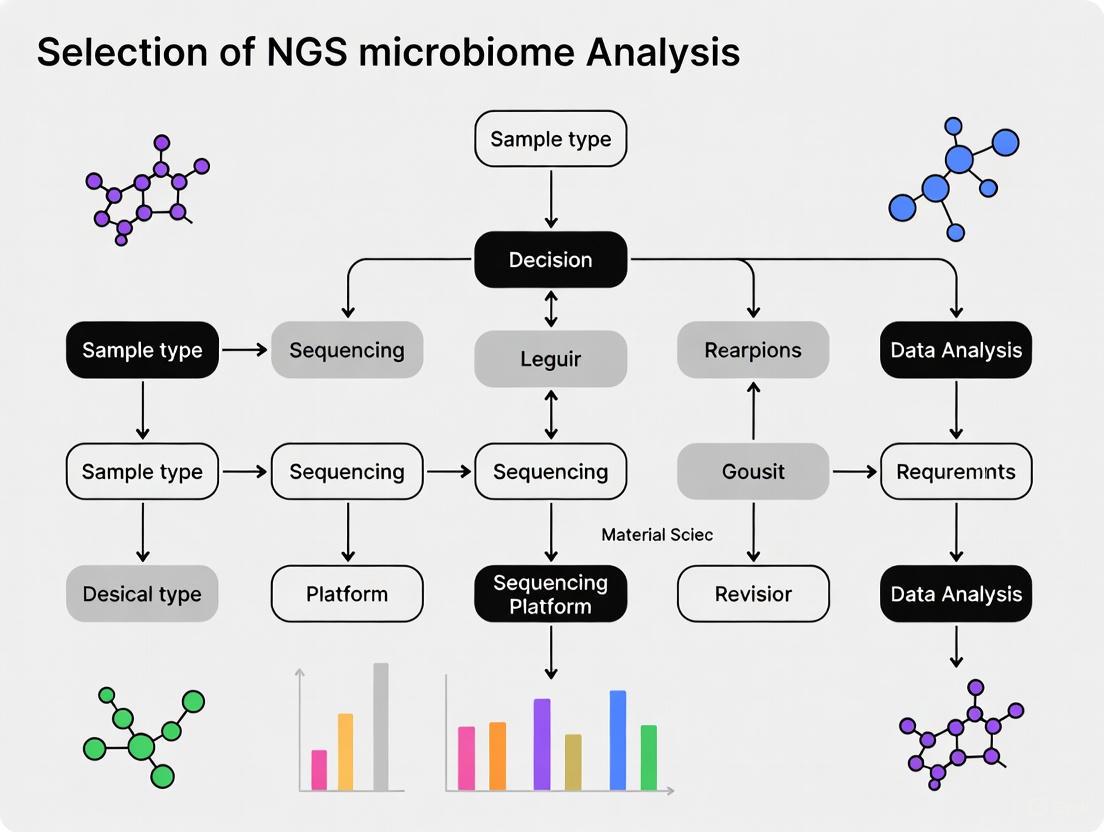
The following diagram illustrates the core decision-making workflow for selecting an NGS methodology for microbiome analysis.
Comparative Analysis of NGS Methodologies
Choosing between 16S rRNA sequencing and shotgun metagenomics is a critical decision that depends on research objectives, budget, and desired analytical depth. The table below summarizes the core characteristics of each method.
Table 1: Core Methodological Comparison: 16S rRNA vs. Shotgun Metagenomic Sequencing
| Feature | 16S rRNA Amplicon Sequencing | Shotgun Metagenomic Sequencing |
|---|---|---|
| Sequencing Target | Specific hypervariable regions of the 16S rRNA gene [2] | All genomic DNA in the sample [5] |
| Taxonomic Scope | Primarily Bacteria and Archaea [2] | All domains (Bacteria, Archaea, Viruses, Fungi) and plasmids [2] [1] |
| Typical Taxonomic Resolution | Genus-level (species-level with full-length sequencing) [6] [2] | Species-level and strain-level resolution [2] [7] |
| Functional Insight | Indirectly inferred from taxonomy [7] | Direct assessment of functional genes and pathways [5] [7] |
| Relative Cost | Lower cost per sample [7] | Higher cost per sample [5] [3] |
| Computational Demand | Lower | High (data-intensive assembly and binning) [3] [1] |
| Primary Applications | Microbial community profiling, diversity studies, population-level surveys [1] | Functional potential discovery, strain tracking, gene cataloging, MAG recovery [5] [4] |
Beyond this foundational comparison, the performance of each method has been quantitatively evaluated in direct comparative studies. Key findings on detection power and abundance correlation are summarized below.
Table 2: Empirical Performance Metrics from Comparative Studies
| Performance Metric | 16S rRNA Sequencing | Shotgun Metagenomics | Context & Notes |
|---|---|---|---|
| Detection Power (Genera) | Identifies a subset of the community [7] | Detects more, less abundant taxa with sufficient reads (>500,000) [7] | In one study, shotgun sequencing identified 152 significant changes between conditions that 16S missed [7]. |
| Abundance Correlation | Good agreement for common taxa (avg. r = 0.69) [7] | Good agreement for common taxa [7] | Discrepancies often due to genera being near the detection limit of 16S sequencing [7]. |
| Error Rate | Low (Illumina: <0.1%) [6] | Low (Illumina: <0.1%) [6] | Higher error rates historically associated with long-read technologies (e.g., ONT: 5-15%), though improving [6]. |
Detailed Experimental Protocols and Best Practices
Workflow for 16S rRNA Amplicon Sequencing
The standard protocol for 16S rRNA sequencing involves sample collection, DNA extraction, library preparation, sequencing, and bioinformatics analysis [6] [1].
- Sample Collection and DNA Extraction: Samples are collected (e.g., stool, saliva, skin swab) and immediately frozen, typically at -80°C. DNA is extracted using specialized kits, with the choice of kit significantly impacting yield and community representation [6]. DNA quality and concentration are assessed using spectrophotometry and fluorometry [6].
- Library Preparation: The 16S rRNA gene is amplified via PCR using primers that bind to conserved regions and flank hypervariable regions (e.g., V3-V4). Barcodes and sequencing adapters are added in a subsequent PCR step to allow for sample multiplexing [6]. The use of a positive control, such as a synthetic DNA standard, is recommended to monitor library construction [6].
- Sequencing: Amplified libraries are pooled and sequenced on a high-throughput platform like the Illumina NextSeq to generate paired-end reads (e.g., 2x300 bp) [6].
- Bioinformatics Analysis:
- Quality Control & Denoising: Raw reads are processed for quality using tools like FastQC and MultiQC. Primer sequences are trimmed, and low-quality reads are filtered. Denoising algorithms like DADA2 are used to correct errors and infer exact Amplicon Sequence Variants (ASVs) [6] [2].
- Taxonomic Assignment: ASVs are classified by comparison to reference databases (e.g., SILVA, Greengenes) to assign taxonomic identities from phylum to genus or species level [6] [2].
- Downstream Analysis: Diversity metrics (alpha and beta diversity) are calculated using packages like
phyloseqandveganin R. Differential abundance analysis can be performed with tools like ANCOM-BC [6].
Workflow for Shotgun Metagenomic Sequencing
Shotgun metagenomics provides a more comprehensive but complex alternative [5] [1].
- Sample Collection and DNA Extraction: This step is similar but often requires higher-quality, high-molecular-weight DNA to facilitate robust library preparation and assembly [1].
- Library Preparation and Sequencing: DNA is randomly fragmented (sheared), and adapters are ligated without target-specific amplification. Libraries are sequenced on platforms like the Illumina NovaSeq for high depth or long-read platforms like PacBio and Oxford Nanopore for enhanced resolution [6] [8].
- Bioinformatics Analysis:
- Quality Control and Host Depletion: Reads are quality-filtered, and reads originating from the host (e.g., human) are removed.
- Taxonomic Profiling: Reads can be directly aligned to reference databases (e.g., RefSeq) using tools like the DRAGEN Metagenomics pipeline for taxonomic classification [5] [2].
- Assembly and Binning: High-quality reads are assembled into contigs, which are then grouped into Metagenome-Assembled Genomes (MAGs) representing individual microbial populations [4] [8].
- Functional Annotation: Genes are predicted from contigs or MAGs and annotated against functional databases (e.g., KEGG, COG) to determine the metabolic capabilities of the community [8].
The Scientist's Toolkit: Essential Research Reagents and Materials
Successful microbiome sequencing relies on a suite of specialized reagents, kits, and computational tools.
Table 3: Essential Research Reagents and Solutions for Microbiome NGS
| Item | Function | Example Products / Tools |
|---|---|---|
| DNA Extraction Kit | Isolation of high-quality, inhibitor-free microbial DNA from complex samples. | Sputum DNA Isolation Kit (Norgen Biotek) [6] |
| 16S Library Prep Kit | Amplification and barcoding of target hypervariable regions for multiplexed sequencing. | QIAseq 16S/ITS Region Panel (Qiagen) [6] |
| Shotgun Library Prep Kit | Fragmentation, adapter ligation, and amplification of total genomic DNA for untargeted sequencing. | Illumina DNA Prep kits [5] |
| Long-Read Library Prep Kit | Preparation of libraries for long-read sequencing platforms. | ONT 16S Barcoding Kit (Oxford Nanopore) [6] |
| Positive Control | Synthetic DNA standard to monitor efficiency and bias in library preparation and sequencing. | QIAseq 16S/ITS Smart Control (Qiagen) [6] |
| Bioinformatics Pipelines | Automated workflows for processing raw data into taxonomic and functional profiles. | nf-core/ampliseq [6], DRAGEN Metagenomics [5], EPI2ME [6] |
| Reference Databases | Curated collections of genomic or gene sequences for taxonomic and functional classification. | SILVA [6], RefSeq [2], GenBank [2] |
The definition and study of the microbiome are inextricably linked to the development of culture-independent NGS technologies. While 16S rRNA amplicon sequencing remains a powerful, cost-effective tool for large-scale taxonomic surveys, shotgun metagenomics provides a superior and comprehensive view of the microbiome's taxonomic and functional landscape. The choice between them should be dictated by the specific research question, with 16S suitable for broad ecological studies and shotgun metagenomics essential for mechanistic insights and discovering uncultured microorganisms.
The field continues to evolve rapidly, driven by technological advancements. Long-read sequencing from Oxford Nanopore and PacBio is overcoming previous limitations in accuracy, enabling full-length 16S sequencing and more complete metagenome assembly for superior resolution [6] [8]. Furthermore, the development of integrated, user-friendly bioinformatics platforms is making sophisticated data analysis more accessible, promoting reproducibility and collaboration [9]. As the global microbiome sequencing market expands, projected to reach $3.7 billion by 2029 [10], these innovations will undoubtedly deepen our understanding of microbial communities and unlock new diagnostic and therapeutic avenues in human health and beyond.
Next-generation sequencing (NGS) has revolutionized microbiome research by enabling comprehensive, culture-independent analysis of microbial communities [11] [2]. The choice of sequencing method profoundly influences the depth, breadth, and clinical applicability of microbiome data, making method selection a critical first step in research design. This guide provides an in-depth technical overview of the three principal NGS approaches used in microbiome analysis: 16S rRNA gene sequencing, shotgun metagenomic sequencing (mNGS), and targeted next-generation sequencing (tNGS). Framed within the context of how to choose an NGS method for microbiome research, this document synthesizes current methodologies, performance characteristics, and practical considerations to equip researchers, scientists, and drug development professionals with the knowledge needed to align their technical approach with specific research objectives.
Core NGS Methodologies in Microbiome Research
16S Ribosomal RNA (rRNA) Gene Sequencing
The 16S rRNA gene is a cornerstone of microbial phylogenetics and taxonomy. This ~1500 bp gene contains nine hypervariable regions (V1-V9) interspersed with conserved regions [11] [2]. The conserved regions allow for the design of universal PCR primers, while the hypervariable regions provide the sequence diversity necessary for taxonomic classification [3].
Experimental Protocol and Workflow:
- DNA Extraction: Microbial DNA is extracted from samples (e.g., stool, saliva, skin swabs). The extraction method, including the use of bead-beating for robust cell lysis, can significantly impact the microbial profile obtained [12] [13].
- PCR Amplification: Specific hypervariable regions (e.g., V1-V2, V3-V4, V4) are amplified using universal primers targeting the flanking conserved regions [11] [12]. The selection of the hypervariable region is a critical consideration, as it influences taxonomic resolution and can introduce biases; for instance, the V4 region may perform poorly for species-level classification compared to the V1-V3 region or full-length sequencing [12] [14].
- Library Preparation: Amplified products (amplicons) are prepared for sequencing with the addition of platform-specific adapters and sample barcodes (indexes) to enable multiplexing [12].
- Sequencing: Typically performed on Illumina platforms (e.g., MiSeq) [12] [15].
- Bioinformatic Analysis:
- Quality Filtering: Removal of low-quality reads, adapters, and chimeric sequences [12] [2].
- Clustering/Denoising: Sequences are clustered into Operational Taxonomic Units (OTUs) based on a sequence similarity threshold (e.g., 97% for species-level) or denoised into Amplicon Sequence Variants (ASVs) [11] [2].
- Taxonomic Assignment: OTUs/ASVs are classified by comparison to reference databases such as SILVA, Greengenes, or the RDP database [2].
Figure 1: 16S rRNA Gene Sequencing Workflow. The process involves wet lab procedures from sample to sequencing, followed by bioinformatic analysis for taxonomic classification and community profiling.
Shotgun Metagenomic Sequencing (mNGS)
Shotgun metagenomics moves beyond a single gene to sequence the entire complement of DNA extracted from a microbial community [5] [3]. This approach allows for simultaneous assessment of taxonomic composition and the functional potential of the microbiome.
Experimental Protocol and Workflow:
- DNA Extraction: Similar to 16S, but with potential use of host DNA depletion kits (e.g., MolYsis) for samples with high host contamination, such as bronchoalveolar lavage fluid (BALF) [16].
- Library Preparation: Total DNA is randomly fragmented (sheared), and adapters are ligated without target-specific PCR amplification. This creates a library representing all genomic material in the sample [5] [2].
- High-Throughput Sequencing: Requires deeper sequencing (millions to billions of reads) to achieve adequate coverage of complex communities. Platforms like Illumina NovaSeq or BGISEQ are commonly used [16].
- Bioinformatic Analysis:
- Host Depletion: Reads aligning to a host reference genome (e.g., hg38) are removed [16].
- Taxonomic Profiling: Non-host reads are aligned to comprehensive genomic databases (e.g., RefSeq, GenBank) to identify microorganisms from all domains of life (bacteria, archaea, viruses, fungi) and achieve species- or strain-level resolution [16] [2].
- Functional Profiling: Reads are assembled into contigs or mapped to functional databases (e.g., KEGG, COG) to identify genes and metabolic pathways [5].
Targeted Next-Generation Sequencing (tNGS)
tNGS is a hypothesis-driven approach that uses targeted enrichment techniques to sequence specific genomic regions or a pre-defined set of pathogens. It bridges the gap between 16S and shotgun mNGS [16] [17]. Two primary enrichment methods are used:
- Amplification-based tNGS: Utilizes multiple pairs of primers in a multiplex PCR to amplify targeted pathogen sequences [17].
- Capture-based tNGS: Uses labeled oligonucleotide probes (baits) to hybridize and capture genomic regions of interest from a total DNA library [17].
Experimental Protocol and Workflow (Amplification-based):
- Nucleic Acid Extraction: DNA and/or RNA can be co-extracted. RNA is reverse-transcribed to cDNA [16] [17].
- Target Enrichment: For amplification-based tNGS, a multiplex PCR with dozens to hundreds of pathogen-specific primers is performed to enrich target sequences [17].
- Library Preparation & Sequencing: The amplified products are prepared for sequencing, often requiring fewer sequencing reads than mNGS [16] [17].
- Bioinformatic Analysis: Processed reads are aligned to a curated database of target pathogens to determine presence and abundance [16] [17].
Comparative Analysis of NGS Approaches
The choice between 16S, shotgun mNGS, and tNGS involves trade-offs between resolution, scope, cost, and analytical complexity. The tables below summarize key comparative metrics and recent clinical performance data.
Table 1: Technical and Practical Comparison of Core NGS Methods
| Feature | 16S rRNA Sequencing | Shotgun Metagenomics (mNGS) | Targeted NGS (tNGS) |
|---|---|---|---|
| Target | 16S rRNA gene hypervariable regions | Entire microbial DNA | Pre-defined pathogens/genomic regions |
| Taxonomic Resolution | Genus-level, limited species/strain [2] | Species- and strain-level possible [2] | Species- and strain-level [16] |
| Scope of Detection | Bacteria and Archaea | All domains (Bacteria, Archaea, Viruses, Fungi, Parasites) [2] | Customizable panel (e.g., bacteria, viruses, fungi) [16] [17] |
| Functional Insights | Inferred from taxonomy | Direct assessment of genes and pathways [5] | Limited to targeted genes (e.g., AMR/virulence factors) [17] |
| Cost | Low | High | Moderate [17] |
| Turnaround Time | Shorter | Longer (~20 hours) [17] | Shorter than mNGS [17] |
| Bioinformatic Complexity | Moderate | High ("big data" challenges) [11] | Lower (simplified analysis) |
| Human Host Read Interference | Low (due to targeted amplification) | High (requires depletion steps) [16] | Low (enrichment reduces host background) [16] |
| Ideal Application | Microbial community profiling, diversity studies | Discovering novel organisms, functional metagenomics | Clinical diagnostics, pathogen detection, AMR profiling [16] [17] |
Table 2: Performance Comparison from Recent Clinical Studies (2024-2025)
| Study Context | mNGS Performance | tNGS Performance | Key Findings |
|---|---|---|---|
| 85 BALF specimens [16] | Detected 55 species. Similar performance for bacteria/fungi. | Detected 49 species. Higher detection rate for DNA viruses (e.g., HHV-4, -5, -6, -7). | Overall concordance was 86.75%. tNGS superior for DNA virus detection. |
| 205 LRTI patients [17] | Identified 80 species. High cost (\$840) and long TAT (20h). | Capture-based: 71 species, 93.17% accuracy.Amplification-based: 65 species, lower sensitivity for some bacteria. | Capture-based tNGS recommended for routine diagnostics; mNGS for rare pathogens; amplification-based for resource-limited settings. |
Figure 2: NGS Method Selection Decision Framework. A flowchart to guide the choice of NGS method based on research goals, required resolution, and practical constraints.
Successful microbiome research relies on a suite of carefully selected reagents, kits, and bioinformatic resources.
Table 3: Research Reagent Solutions and Essential Resources
| Item | Function | Example Products/Citations |
|---|---|---|
| Host DNA Depletion Kit | Reduces human host background in samples rich in human cells (e.g., BALF, tissue). | MolYsis Basic5 [16] |
| Nucleic Acid Extraction Kit | Isolates total genomic DNA (and RNA) from complex samples. | DNeasy PowerSoil Kit (QIAGEN) [12], Isolate II Genomic DNA Kit (Bioline) [15] |
| 16S rRNA PCR Primers | Amplifies specific hypervariable regions for 16S sequencing. | 27Fmod/338R (V1-V2) [12], 341F/805R (V3-V4) [12] |
| Library Prep Kit | Prepares amplicon or fragmented DNA for NGS sequencing. | NEBNext Ultra II DNA Library Prep Kit [15] |
| tNGS Enrichment Kit | Enriches for specific pathogen sequences via multiplex PCR or probe capture. | Respiratory Pathogen Detection Kit (KingCreate) [17] |
| Bioinformatics Pipelines | Processes raw sequencing data for quality control, taxonomic assignment, and diversity analysis. | QIIME2 [12] [15], DRAGEN Metagenomics [5] |
| Reference Databases | Essential for taxonomic classification and functional annotation. | Greengenes [12], SILVA [2], RefSeq [16] [2] |
The landscape of NGS-based microbiome analysis offers a powerful suite of tools, each with distinct strengths and optimal applications. 16S rRNA sequencing remains a cost-effective method for high-throughput microbial community profiling and diversity analysis. Shotgun metagenomics provides the most comprehensive view, enabling functional insights and high-resolution taxonomic assignment across all domains of life, albeit at a higher cost and computational burden. Targeted NGS is emerging as a robust, sensitive, and efficient solution for clinical diagnostics and specific hypothesis testing.
The decision on which methodology to employ should be guided by a clear alignment between the technical capabilities of each platform and the primary research question, whether it is broad ecological discovery, functional characterization, or precise pathogen detection. As databases expand and workflows become more standardized, the integration of these NGS approaches will continue to deepen our understanding of the microbiome's role in health and disease and advance drug development.
The choice of DNA sequencing technology is a foundational decision in microbiome research, directly impacting the resolution, accuracy, and depth of microbial community analysis. Next-Generation Sequencing (NGS) technologies are broadly categorized into short-read and long-read platforms, each with distinct technical principles and performance characteristics [2] [18]. Short-read sequencing, dominated by Illumina platforms, generates massive volumes of reads typically 50-600 bases in length, offering high per-base accuracy at a low cost [19] [20]. Conversely, long-read sequencing, represented by Pacific Biosciences (PacBio) and Oxford Nanopore Technologies (ONT), produces reads that can span thousands to tens of thousands of bases, which simplifies genome assembly and resolves complex genomic regions [21] [20].
The selection between these methodologies is crucial and must be aligned with the specific research objectives—whether the goal is to broadly profile microbial diversity, reconstruct whole genomes from complex samples, or understand functional potential [22]. This guide provides an in-depth technical comparison of these platforms, framed within the context of choosing an NGS method for microbiome analysis, to equip researchers and drug development professionals with the information needed to design robust and informative microbiome studies.
Technology Comparison: Performance and Characteristics
The performance of short-read and long-read sequencing technologies differs across several key metrics that are critical for experimental design in microbiome research.
Sequencing Performance Metrics
| Performance Metric | Short-Read Sequencing (e.g., Illumina) | Long-Read Sequencing (PacBio) | Long-Read Sequencing (ONT) |
|---|---|---|---|
| Typical Read Length | 35–600 bases [18] [19] [20] | Several kilobases to >10 kb [23] [20] | Several kilobases to 10s of kb [21] [20] |
| Per-Base Raw Accuracy | >99.9% [19] | >99.9% (HiFi mode) [23] | ~99% (with latest R10.4.1 flow cells) [23] |
| Primary Advantage | High throughput, low per-base cost, high accuracy [2] [19] | High accuracy for long reads, excellent for assembly [24] [20] | Very long reads, fast turnaround, portability [19] [20] |
| Primary Limitation | Limited resolution in repetitive regions, fragmented assemblies [19] [20] | Higher DNA input requirements, higher cost [24] [20] | Historically higher error rate, though improving [23] [19] |
| Ideal Microbiome Application | High-density population profiling, 16S rRNA amplicon studies [2] [22] | High-quality metagenome-assembled genome (MAG) recovery [21] [24] | Rapid pathogen identification, full-length 16S sequencing, complex MAGs [23] [19] |
A Researcher's Guide to Sequencing Method Selection
Choosing the right sequencing method depends on the research question. The table below outlines the optimal technologies for common microbiome research goals.
| Research Goal | Recommended Method | Rationale |
|---|---|---|
| Microbial Diversity & Composition (Genus Level) | 16S Amplicon Sequencing (Short-read) [22] | Cost-effective for large sample sets, provides robust genus-level taxonomy [2] [22]. |
| Microbial Diversity & Composition (Species Level) | Full-Length 16S Amplicon Sequencing (Long-read) [23] [20] | Full-length 16S gene sequencing provides superior species-level resolution [23]. |
| Functional Potential (Gene Content) | Shotgun Metagenomics (Short-read or Long-read) [22] | Profiles all genes in a community. Short-read is cost-effective; long-read provides better genomic context [24] [20]. |
| Recovery of High-Quality Genomes (MAGs) | Shotgun Metagenomics (Long-read preferred) [21] [24] | Long reads span repetitive regions, enabling complete, uncontaminated genome assemblies [21]. |
| Rapid, On-Site Pathogen Detection | Shotgun Metagenomics (ONT) [19] [20] | Nanopore's portability and fast turnaround enable real-time analysis in field or clinical settings [20]. |
Experimental Protocols for Microbiome Sequencing
Standardized protocols are essential for generating reproducible and reliable microbiome data. The following workflows are widely adopted in the field.
Workflow for 16S rRNA Amplicon Sequencing
This protocol is used for taxonomic profiling of bacterial and archaeal communities [2].
Step-by-Step Protocol:
- Sample Collection: Collect sample (e.g., soil, feces, water) using sterile techniques and immediately preserve it. Flash-freezing at -80°C or using a commercial microbiome preservation buffer is critical to maintain microbial composition integrity [25].
- DNA Extraction: Extract total genomic DNA using a kit designed for microbial lysis (e.g., Zymo Research Quick-DNA Fecal/Soil Microbe Microprep kit). This step often combines chemical and mechanical lysis to ensure efficient recovery from tough-to-lyse microorganisms like Gram-positive bacteria [23] [25].
- PCR Amplification: Amplify the target hypervariable region of the 16S rRNA gene (e.g., V3-V4 for gut and general samples, V4 for environmental samples) using universal primers [23] [22]. The PCR conditions typically involve 25-30 cycles of denaturation (e.g., 95°C for 30s), annealing (e.g., 57°C for 30s), and extension (e.g., 72°C for 60s) [23].
- Library Preparation: Purify the PCR amplicons and ligate sample-specific barcodes (indices) to allow for multiplexing of multiple samples in a single sequencing run [23] [25].
- Sequencing: Pool the barcoded libraries and sequence on a short-read platform (e.g., Illumina MiSeq or NovaSeq) to a depth of 50,000-100,000 reads per sample for complex communities [23] [2].
- Bioinformatic Analysis: Process raw sequences through a pipeline (e.g., QIIME 2, mothur) for quality filtering, chimera removal, and clustering into Operational Taxonomic Units (OTUs) or Amplicon Sequence Variants (ASVs) for taxonomic assignment and diversity analysis [2].
Workflow for Shotgun Metagenomic Sequencing
This protocol sequences all DNA in a sample, enabling functional profiling and genome reconstruction [22].
Step-by-Step Protocol:
- Sample Collection: Identical to the 16S protocol, proper preservation is key [25].
- DNA Extraction: Extract high-quality, high-molecular-weight DNA. For long-read sequencing, this is particularly critical as the protocol requires long, intact DNA fragments to leverage its advantages [20].
- Library Preparation (Platform-Specific):
- For Illumina (Short-read): Fragment the purified DNA mechanically, then repair ends, ligate adapters, and perform size selection [2] [25].
- For PacBio (Long-read): For HiFi sequencing, create SMRTbell libraries by ligating hairpin adapters to double-stranded DNA, creating a circular template. No PCR amplification is needed [23] [8].
- For ONT (Long-read): Use a native barcoding kit (e.g., SQK-NBD109) to ligate barcodes directly to the DNA fragments. The library preparation is often faster and requires no PCR [23] [20].
- Sequencing: Sequence the library on the chosen platform. Required depth varies significantly: 20-100 Gbp of data may be needed per sample for deep metagenomic analysis, especially with complex samples like soil [21] [24].
- Bioinformatic Analysis: This is a complex, multi-step process. For long-read data, specialized workflows like
mmlong2have been developed for complex environmental samples. The process generally involves quality control, de novo assembly of reads into contigs, binning of contigs into Metagenome-Assembled Genomes (MAGs) using coverage and composition information, and finally, functional and taxonomic annotation of the assembled data [21].
The Scientist's Toolkit: Key Reagents and Materials
Successful microbiome sequencing relies on a suite of specialized reagents and kits. The following table details essential solutions for key steps in the workflow.
| Item | Function/Application | Examples / Key Features |
|---|---|---|
| DNA Preservation Buffer | Stabilizes microbial community at point of collection; prevents shifts. | CosmosID collection kits; ZymoBIOMICS DNA/RNA Shield [25]. |
| Bead-Based Lysis Kit | Mechanical & chemical cell lysis; efficient for Gram-positive bacteria. | Kits with bead-beating step (e.g., Zymo Research Quick-DNA kits) [23] [25]. |
| 16S rRNA PCR Primers | Amplifies specific hypervariable regions for amplicon sequencing. | 27F/1492R for full-length; V3-V4 or V4-specific primers for short-read [23] [22]. |
| SMRTbell Prep Kit 3.0 | Prepares circularized DNA templates for PacBio HiFi sequencing. | Pacific Biosciences library prep kit [23]. |
| Native Barcoding Kit 96 | Adds barcodes to DNA for multiplexed ONT sequencing without PCR. | Oxford Nanopore kit (SQK-NBD109.24) [23]. |
| Metagenomic Assembly & Binning Tool | Assembles sequences and groups them into putative genomes (MAGs). | mmlong2 workflow for complex long-read datasets [21]. |
The choice between short-read and long-read sequencing is not a matter of one being universally superior, but rather which is fit-for-purpose for a specific research question, budget, and sample type [24] [22].
Short-read sequencing remains the workhorse for large-scale, high-throughput profiling studies where the goal is to compare microbial community structures (beta-diversity) across hundreds of samples or to conduct genus-level association studies [2] [22]. Its high accuracy and low cost per sample make it ideal for this application.
Long-read sequencing is transformative for applications that require higher taxonomic resolution or complete genomic context. It is the preferred choice for: achieving species- and strain-level discrimination via full-length 16S sequencing [23] [20]; recovering high-quality, complete Metagenome-Assembled Genomes (MAGs) from complex environments like soil [21]; resolving repetitive genomic elements and mobile genetic elements like plasmids [18] [20]; and providing rapid diagnostic results in clinical or outbreak settings due to its real-time sequencing capabilities [19] [20].
As sequencing technologies continue to evolve, the accuracy of long-read platforms is increasing while costs are decreasing, making them an increasingly accessible and powerful tool. For the most comprehensive insights, a hybrid approach, using both short- and long-read technologies, can sometimes offer the optimal balance of depth, accuracy, and genomic completeness [24]. Researchers are advised to base their final platform selection on a careful consideration of their primary objectives, required resolution, and available resources.
The Critical Role of Library Preparation in NGS Success
In next-generation sequencing (NGS) for microbiome analysis, library preparation is the critical bridge between a raw biological sample and actionable genomic insights. This process transforms extracted nucleic acids into a format compatible with sequencing platforms, directly determining the accuracy, reproducibility, and depth of microbial community characterization. Within the specific context of microbiome research, the choice between 16S rRNA gene sequencing and shotgun metagenomics is one of the earliest and most consequential decisions, guided by the research objectives [3]. 16S sequencing, targeting specific hypervariable regions, is a cost-effective method for taxonomic profiling and is widely used in bacterial population studies [3]. In contrast, shotgun metagenomics sequences all DNA in a sample, enabling functional analysis and the discovery of unculturable microorganisms but at a higher cost and computational expense [3]. The library preparation protocols for these two paths diverge significantly, and variations within each method—such as the choice of 16S hypervariable regions or the fragmentation technique for shotgun libraries—can introduce specific biases that impact downstream results [6] [26]. Therefore, a meticulously optimized library preparation protocol is not merely a preliminary step but a fundamental determinant of data integrity, influencing all subsequent biological interpretations in microbiome research.
Core Principles and Methodologies of NGS Library Preparation
The process of NGS library preparation consists of a series of standardized yet adaptable steps designed to fragment the genetic material and attach platform-specific oligonucleotide adapters. The general workflow involves nucleic acid extraction, fragmentation, adapter ligation, and library quantification [27].
Key Steps in Library Construction
- Nucleic Acid Extraction: The first step in every sample preparation protocol is isolating DNA or RNA from a variety of biological samples, such as blood, cultured cells, or tissue [27]. The quality and quantity of the extracted nucleic acids are crucial for the success of subsequent steps.
- Fragmentation: The targeted DNA sequences are fragmented to a desired length. This can be achieved through physical, enzymatic, or chemical methods. Enzymatic fragmentation continues to dominate due to its simplicity and compatibility with diverse sample types [28].
- Adapter Ligation: Specialized adapters are attached to the ends of the fragmented DNA. These adapters often include barcode sequences that permit sample multiplexing, allowing multiple libraries to be sequenced simultaneously in a single run [27].
- Amplification: This optional but common step uses polymerase chain reaction (PCR) to increase the amount of DNA library, which is essential for samples with small amounts of starting material [27].
- Purification and Quality Control: A final "clean-up" step removes unwanted material, such as adapter dimers or fragments that are too large or too small. Quality control confirms the quality and quantity of the final library before sequencing [27].
Common Challenges and Biases
Library preparation is prone to several challenges that can introduce bias and compromise data quality. A primary concern is amplification bias, where certain sequences are preferentially amplified over others during PCR, leading to an inaccurate representation of the original microbial community [27]. This is often reflected in a high PCR duplication rate. Inefficient library construction, characterized by a low percentage of fragments with correct adapters, can decrease data yield and increase the formation of chimeric reads [27]. Sample contamination is an inherent risk, particularly when many libraries are prepared in parallel. Finally, the large costs associated with laboratory equipment, trained personnel, and reagents can be a significant constraint [27].
Figure 1: Generalized NGS Library Preparation Workflow. The process transforms raw nucleic acids into a sequencer-ready format, with each stage being a potential source of bias.
Platform-Specific Considerations for Microbiome Analysis
The selection of a sequencing platform is a strategic decision that dictates the required library preparation approach and ultimately shapes the taxonomic resolution of a microbiome study. The choice often centers on the trade-off between read length, accuracy, throughput, and cost [18].
Short-Read vs. Long-Read Sequencing
Short-read platforms, such as Illumina, generate highly accurate reads (error rate < 0.1%) but are typically limited to a few hundred base pairs [6] [29]. This makes them suitable for sequencing specific hypervariable regions of the 16S rRNA gene (e.g., V3-V4) for reliable genus-level classification [6]. However, their limited read length restricts the ability to resolve closely related bacterial species [6]. In contrast, long-read platforms from Oxford Nanopore Technologies (ONT) and Pacific Biosciences (PacBio) can generate reads spanning thousands of base pairs, enabling full-length 16S rRNA gene sequencing (~1,500 bp) [6] [26]. This long-read capability provides higher taxonomic resolution, often enabling species-level and even strain-level identification [18]. Historically, long-read technologies were associated with higher error rates (5–15%), but recent advancements have significantly improved their accuracy [6].
Impact on Microbiome Characterization
Comparative studies highlight how these technical differences translate into varied biological outcomes. A 2025 comparative analysis of Illumina and ONT for respiratory microbiome profiling found that while Illumina captured greater species richness, ONT exhibited improved resolution for dominant bacterial species [6]. Another 2025 study on rabbit gut microbiota showed that ONT and PacBio offered superior species-level classification rates (76% and 63%, respectively) compared to Illumina (48%) [26]. However, it also noted that a significant portion of species-level assignments were labeled as "uncultured_bacterium," indicating a limitation of reference databases rather than the technology itself [26]. Furthermore, differential abundance analysis can reveal platform-specific biases; for example, ONT may overrepresent certain taxa (e.g., Enterococcus, Klebsiella) while underrepresenting others (e.g., Prevotella, Bacteroides) [6]. These findings emphasize that platform selection should align with study objectives: Illumina is ideal for broad microbial surveys, whereas ONT excels in applications requiring species-level resolution [6].
Table 1: Comparative Analysis of Sequencing Platforms for Microbiome Studies
| Platform | Read Length | Key Strengths | Key Limitations | Ideal Microbiome Application |
|---|---|---|---|---|
| Illumina [6] [29] | Short-read (~300 bp) | High accuracy (error rate <0.1%), high throughput, low cost per gigabase | Limited species-level resolution | Large-scale population studies, genus-level profiling |
| Oxford Nanopore (ONT) [6] [26] | Long-read (~1,500 bp to >10,000 bp) | Species-level resolution, real-time data streaming, portability | Historically higher error rate, though improving | In-field sequencing, pathogen identification, haplotype resolution |
| PacBio [26] [29] | Long-read (average 10,000–25,000 bp) | High-fidelity (HiFi) reads with high accuracy | Higher cost, lower throughput | High-quality genome assembly, discovering novel microbes |
Best Practices for Optimal Library Preparation
Adhering to rigorous laboratory practices is essential for generating high-quality, reproducible NGS libraries, which is the foundation of robust microbiome data.
Technical Optimization and Quality Control
- Optimize Adapter Ligation: Use freshly prepared adapters and control ligation temperature and duration. Blunt-end ligations are typically performed at room temperature for 15–30 minutes, while ligations with cohesive ends are often performed at 12–16°C for longer durations [30].
- Handle Enzymes with Care: Maintain enzyme stability by avoiding repeated freeze-thaw cycles and storing them at recommended temperatures. Accurate pipetting is crucial to ensure consistent results [30].
- Normalize Libraries Accurately: Before pooling, ensure each library is normalized to contribute equally to the final sequencing pool. This prevents under- or over-representation of samples, which can bias sequencing depth and results [30].
- Implement Quality Control Checkpoints: Establish QC checkpoints at multiple stages: post-ligation, post-amplification, and post-normalization. Techniques like fragment analysis, qPCR, and fluorometry assess library quality and allow for early issue detection [30].
Automation and Contamination Prevention
Automation is a powerful strategy for mitigating the risks of manual library preparation. Automated systems standardize workflows, reduce human error and variability, and improve reproducibility, especially across large sample batches [28] [30]. They also provide traceability by logging every step of the workflow, which is vital for regulatory compliance and data reliability [30]. To minimize contamination, dedicate a pre-PCR room or area separate from post-amplification steps [27]. This reduces the risk of cross-contamination from amplified DNA products, a common pitfall in sensitive NGS workflows.
Figure 2: Essential Quality Control Checkpoints in the NGS workflow. Implementing QC at multiple stages ensures the integrity of the final library before sequencing.
The Scientist's Toolkit: Essential Reagents and Materials
Successful library preparation relies on a suite of specialized reagents and tools. The following table details key components and their critical functions in the workflow.
Table 2: Key Research Reagent Solutions for NGS Library Preparation
| Reagent / Material | Function | Considerations for Microbiome Analysis |
|---|---|---|
| Nucleic Acid Extraction Kit [6] [26] | Isolates DNA/RNA from complex biological samples. | Yield and purity are critical; protocols may need optimization for different sample types (e.g., soil vs. human gut). |
| DNA Library Prep Kit [31] [32] | Contains enzymes and buffers for fragmentation, end-repair, A-tailing, and adapter ligation. | Platform-specific (e.g., Illumina, ONT); PCR-free kits are available to reduce amplification bias. |
| Index Adapters [27] [31] | Short, unique DNA sequences ligated to fragments; enable sample multiplexing. | Use unique dual indexes to improve demultiplexing accuracy and detect index hopping. |
| Bead-Based Cleanup Kits [28] [30] | Purify nucleic acids by size selection and remove enzymes, salts, and adapter dimers. | Crucial for removing primer dimers after 16S rRNA PCR amplification. |
| PCR Enzymes [27] | Amplify the adapter-ligated library to generate sufficient material for sequencing. | High-fidelity polymerases minimize amplification bias and errors. |
| Quality Control Assays [27] [30] | Quantify and qualify the final library (e.g., Fragment Analyzer, Qubit, qPCR). | qPCR provides the most accurate quantification of amplifiable libraries for loading onto the sequencer. |
In conclusion, library preparation is a critically dynamic and influential phase in the NGS workflow for microbiome analysis. There is no universal "best" protocol; instead, the optimal approach is dictated by a strategic alignment between the research question, the chosen sequencing technology, and the sample type. The decision to use 16S rRNA gene sequencing for cost-effective taxonomic census or shotgun metagenomics for functional potential and strain-level resolution will define the library construction path [3]. As sequencing technologies evolve, with long-read platforms closing the accuracy gap with short-read platforms, library preparation methods will continue to adapt [18]. Embracing automation, adhering to rigorous quality control, and understanding the inherent biases of each method are non-negotiable practices for generating reliable, reproducible data. By investing time and resources into optimizing this foundational step, researchers can ensure that their microbiome studies are built upon a solid experimental foundation, leading to more meaningful and trustworthy biological insights.
Essential Bioinformatic Considerations for Data Analysis
The selection of an appropriate Next-Generation Sequencing (NGS) method serves as the foundational decision that dictates all subsequent bioinformatic workflows in microbiome analysis. This choice creates a cascade of technical requirements that span experimental design, computational infrastructure, and analytical methodologies. The growing diversity of available NGS platforms—from short-read Illumina systems to long-read PacBio and Oxford Nanopore technologies—has transformed microbiome research capabilities while introducing significant complexity to the bioinformatic landscape [33]. Within this context, bioinformatic considerations must evolve from secondary concerns to primary design criteria, as they directly determine the feasibility, accuracy, and biological relevance of research outcomes.
The critical importance of these bioinformatic considerations extends beyond technical implementation to impact the very scientific questions that can be addressed. As research transitions from descriptive catalogs of microbial communities to mechanistic investigations of ecosystem function, the integration of multi-omics data and advanced analytical approaches becomes essential [33] [34]. This guide provides a comprehensive framework for navigating the bioinformatic ecosystem surrounding NGS method selection, with particular emphasis on the computational strategies that underpin robust, reproducible, and biologically insightful microbiome research.
NGS Technology Landscape: Implications for Bioinformatic Workflows
The core sequencing technologies available for microbiome research present distinct advantages and limitations that directly shape subsequent bioinformatic requirements. Understanding these technical characteristics is essential for matching sequencing platforms to specific research objectives and ensuring that analytical workflows are appropriately designed.
Short-read technologies (e.g., Illumina) generate massive volumes of data (typically millions to billions of 35-700 bp reads) with low per-base error rates (~0.1-15%) but face limitations in taxonomic resolution, variant detection, and genome assembly contiguity due to their fragmentary nature [33]. These platforms produce data that excels for quantitative abundance measurements but struggles with resolving repetitive regions, structural variants, and complex genomic architectures.
Long-read technologies (e.g., PacBio, Oxford Nanopore) address these limitations through read lengths that can span entire genes, operons, or even small genomes, enabling more complete genome assemblies and direct detection of structural variants [33]. The trade-offs historically included higher error rates and lower throughput, though these limitations have substantially improved in recent platform iterations. The bioinformatic implications include reduced assembly complexity but increased computational demands for error correction and base-calling.
The emerging field of targeted NGS approaches further expands this landscape, with capture-based and amplification-based methods enabling focused investigation of specific microbial groups or functional elements. Recent clinical comparisons demonstrate that capture-based tNGS achieves 93.17% accuracy and 99.43% sensitivity in pathogen identification, outperforming both mNGS and amplification-based approaches for routine diagnostics [17]. These methods reduce sequencing costs and computational burdens while introducing their own bioinformatic considerations around hybridization efficiency, amplification bias, and reference database completeness.
Table 1: Sequencing Platform Characteristics and Bioinformatic Implications
| Platform/Technology | Read Length | Accuracy | Throughput | Primary Bioinformatic Challenges |
|---|---|---|---|---|
| Illumina | 35-700 bp | ~99.9% | 10GB-1.8TB | Genome assembly fragmentation; GC bias correction |
| PacBio | 10-25 kb | ~99.9% (HiFi) | 5-50 Gb | Computational resource requirements; data storage |
| Oxford Nanopore | Up to 2+ Mb | ~99% (duplex) | 10-50+ Gb | Basecalling optimization; error profile modeling |
| Capture-based tNGS | Varies | High | Targeted | Hybridization efficiency normalization; off-target analysis |
| Amplification-based tNGS | Varies | Variable | Targeted | PCR bias correction; primer dimers filtering |
Experimental Design and Data Generation Considerations
Robust bioinformatic analysis begins during experimental design, where choices about sample collection, library preparation, and sequencing depth establish fundamental parameters that either enable or constrain subsequent computational approaches. The integration of culturomics with metagenomics exemplifies how wet-laboratory and computational approaches can be synergistically combined to overcome the limitations of either method alone.
Culture-enriched metagenomic sequencing (CEMS) represents a powerful hybrid approach that leverages high-throughput culturing across diverse media conditions followed by metagenomic sequencing of the entire cultured community. This strategy recently demonstrated remarkably low overlap between culture-dependent and culture-independent methods, with CEMS and direct metagenomic sequencing (CIMS) identifying only 18% shared species, while 36.5% and 45.5% of species were unique to each method respectively [35]. This profound methodological complementarity highlights how experimental design directly shapes the observable biological reality in microbiome studies.
Library preparation protocols further dictate bioinformatic requirements through their influence on data structure and quality. For bacterial RIBO-seq analysis, which precisely maps ribosome positions on transcripts to monitor protein synthesis, critical experimental steps include rapid translation inhibition through flash-freezing in liquid nitrogen, mechanical cell disruption using mortar grinding with alumina to prevent RNA shearing, and careful buffer formulation with magnesium ions to preserve ribosomal integrity [36]. The resulting data enables transcriptome-wide measurement of translation dynamics but requires specialized preprocessing to isolate ribosome-protected mRNA fragments (28-30 nt) before alignment and quantification.
Sequencing depth requirements vary substantially across applications, with fundamental trade-offs between sample numbers, statistical power, and detection sensitivity. While 16S rRNA amplicon sequencing may require 10,000-50,000 reads per sample for community saturation, shotgun metagenomics typically demands 5-20 million reads per sample for adequate genome coverage, with precise requirements dependent on community complexity and target genome size [33]. These experimental parameters must be established during study design through power calculations and pilot studies to ensure that subsequent bioinformatic analyses can address the underlying biological questions.
Data Processing: From Raw Sequences to Biological Features
The transformation of raw sequencing data into biologically meaningful features represents a critical bioinformatic phase where analytical choices profoundly impact result interpretation. This process involves multiple computational steps, each with platform-specific considerations and quality control requirements.
Sequence Preprocessing and Quality Control
Initial data processing begins with quality assessment and adapter removal, with FastQC and fastp commonly employed for short-read data [34]. Long-read technologies require specialized quality control approaches focused on read length distribution and quality score calibration. For RIBO-seq data, size selection through polyacrylamide gel electrophoresis (PAGE) to isolate ribosome footprints (28-30 nt fragments) represents a critical experimental and computational step that must be carefully optimized [36]. Simultaneous RNA-seq data generation provides essential reference points for normalizing ribosome occupancy to transcript abundance, highlighting how multi-modal data integration strengthens analytical robustness.
Host DNA depletion presents particular challenges in clinical applications where microbial biomass may be low relative to host material. In respiratory infection diagnostics, methods combining Benzonase and Tween20 for human DNA removal have proven effective, though optimization is required to avoid simultaneous depletion of microbial sequences [17]. The bioinformatic validation of depletion efficiency through alignment to host reference genomes represents an essential quality control metric.
Sequence Alignment and Assembly Strategies
Alignment algorithm selection must be matched to both data type and research question. As detailed in Table 2, the alignment software landscape includes tools optimized for specific data types and applications, with choice impacting mapping efficiency, accuracy, and computational efficiency [37].
Table 2: Alignment Software Selection Guide
| Software | Primary Data Type | Key Strengths | Typical Applications |
|---|---|---|---|
| Bowtie2 | DNA short reads | Ultra-fast; low memory usage; end-to-end/local modes | WGS/WES/ChIP-seq/ATAC-seq (short read DNA) |
| BWA-MEM | DNA short/medium reads | High accuracy (especially indels); supports >100bp reads | Resequencing; exome sequencing; PacBio CLR data |
| Minimap2 | Long reads universal | Extremely fast; low memory; optimized for ONT/PacBio | Long read alignment; cross-species comparison; quick short read analysis |
| STAR | RNA-seq | Accurate splice junction detection; supports chimeric alignment | RNA-seq transcript quantification; alternative splicing analysis |
| HISAT2 | RNA-seq | Lower memory usage than STAR; faster performance | Memory-constrained RNA-seq (e.g., single-cell RNA-seq) |
For metagenomic assembly, the choice between co-assembly and individual assembly strategies depends on project goals, with the former potentially providing better coverage for low-abundance community members but requiring substantial computational resources. Hybrid assembly approaches combining short and long reads have demonstrated particular promise for generating complete metagenome-assembled genomes (MAGs), leveraging the accuracy of short reads with the contiguity of long reads [33]. Assembly quality assessment through checkM and similar tools provides essential validation of reconstruction completeness and contamination levels.
Analytical Frameworks and Statistical Approaches
The transition from processed sequences to biological insights requires sophisticated statistical frameworks capable of addressing the high-dimensional, compositional, and sparse nature of microbiome data. These analytical approaches range from basic community profiling to complex multi-omics integration, each with specific implementation requirements and interpretive considerations.
Community Profiling and Differential Analysis
Taxonomic profiling forms the foundation of most microbiome analyses, with methods ranging from 16S rRNA amplicon sequence variant (ASV) analysis to metagenomic phylogenetic placement. The analysis of 16S data typically involves DADA2 or Deblur for ASV inference, followed by taxonomic assignment using reference databases such as SILVA or Greengenes [34]. For shotgun metagenomics, tools like Kraken2 provide fast taxonomic classification, while MetaPhlAn4 offers strain-level profiling with specifically curated marker gene databases [34].
Differential abundance analysis presents particular statistical challenges due to data compositionality, where changes in one taxon's abundance necessarily affect the relative proportions of others. Methods like DESeq2 (with appropriate modifications for compositional data), ANCOM-BC, and LinDA address these challenges through distinct statistical frameworks, with no single method outperforming others across all scenarios [34]. Experimental factors such as sample size, effect size, and sampling depth should guide tool selection, with simulation-based approaches increasingly employed for method benchmarking.
Multi-omics Integration and Functional Analysis
The integration of multiple data types represents both a major opportunity and significant challenge in modern microbiome bioinformatics. Metagenomic, metatranscriptomic, and metaproteomic data provide complementary perspectives on microbial community structure, functional potential, and actual activity, but their integration requires careful consideration of measurement scale, technical artifacts, and biological interpretation.
Functional analysis of metagenomic data typically involves pathway reconstruction using tools like HUMAnN3, which maps sequencing reads to protein families and metabolic pathways while accounting for taxonomic contributions [34]. For metatranscriptomic data, specialized tools like SAMSA2 and updated HUMAnN3 workflows enable identification of actively transcribed functions, though careful normalization to account for variation in ribosomal RNA depletion efficiency is essential.
Visualization represents a critical bridge between analytical outputs and biological interpretation, with platforms like MicrobiomeStatPlots providing comprehensive resources for creating publication-quality figures [38] [34]. This open-source platform offers over 80 distinct visualization templates spanning basic abundance plots to complex multi-omics integration displays, all implemented in R with fully reproducible code. The availability of such curated visualization resources substantially reduces the technical barrier between analytical results and biological insight.
Research Reagent Solutions and Computational Tools
The implementation of robust bioinformatic workflows depends on both computational tools and experimental reagents that ensure data quality and reproducibility. The following table details essential resources referenced throughout this guide.
Table 3: Essential Research Reagents and Computational Tools
| Category | Specific Tool/Reagent | Primary Function | Implementation Considerations |
|---|---|---|---|
| DNA/RNA Extraction | QIAamp UCP Pathogen DNA Kit [17] | High-quality nucleic acid extraction with host depletion | Critical for low-biomass clinical samples; integrates Benzonase treatment |
| Library Preparation | Ovation RNA-Seq System [17] | cDNA synthesis and amplification for transcriptomics | Maintains representation of low-abundance transcripts |
| Ribosome Profiling | MagPure Pathogen DNA/RNA Kit [36] [17] | Simultaneous DNA/RNA extraction from limited samples | Enables parallel metagenomic and metatranscriptomic analysis |
| Sequence Alignment | Minimap2 [37] | Rapid long-read alignment | Essential for Nanopore/PacBio data; minimal resource requirements |
| Taxonomic Profiling | Kraken2 [34] | Fast metagenomic sequence classification | Custom database construction improves accuracy for specific environments |
| Pathway Analysis | HUMAnN3 [34] | Metabolic pathway reconstruction from metagenomes | Integrates taxonomic and functional analysis in unified pipeline |
| Visualization | MicrobiomeStatPlots [38] [34] | Comprehensive visualization gallery | 80+ reproducible templates; R-based implementation |
Integrated Workflow Visualization
The complex relationships between NGS method selection, bioinformatic workflows, and analytical outcomes are visualized below, highlighting key decision points and their implications throughout the analytical process.
NGS Bioinformatics Decision Workflow
A complementary visualization specifically details the data processing pipeline from raw sequences to analytical results, highlighting quality control checkpoints and methodological alternatives.
Bioinformatic Data Processing Pipeline
The rapidly evolving landscape of NGS technologies presents both unprecedented opportunities and significant analytical challenges for microbiome researchers. The selection of appropriate sequencing methods must be intimately connected with bioinformatic capabilities, as these computational considerations directly determine the biological insights that can be derived from complex microbial communities. As technological advances continue to transform the field—from long-read sequencing overcoming assembly fragmentation to targeted approaches enabling cost-effective clinical applications—bioinformatic strategies must similarly evolve to leverage these innovations while maintaining analytical rigor.
The integration of complementary methodologies represents a particularly promising direction, with hybrid approaches like CEMS demonstrating that combining cultivation with metagenomics can reveal substantially more microbial diversity than either method alone [35]. Similarly, the strategic combination of sequencing technologies—using short reads for quantitative accuracy and long reads for structural resolution—provides a powerful framework for comprehensive microbiome characterization. As these multi-modal approaches mature, the development of integrated bioinformatic platforms that streamline analytical workflows while maintaining flexibility for method-specific optimization will be essential for advancing microbiome research across diverse ecosystems and applications.
Matching NGS Methods to Your Research Goals: A Practical Framework
The 16S ribosomal RNA (rRNA) gene sequencing has emerged as a cornerstone technique in microbial ecology, providing researchers with a powerful tool for cost-effective microbial community profiling. As a targeted amplicon sequencing approach, it enables the characterization of bacterial and archaeal populations by sequencing the 16S rRNA gene, a highly conserved genetic marker that contains both stable regions for primer binding and variable regions that serve as signatures for taxonomic classification [3] [2]. This method has revolutionized our ability to study complex microbial communities without the need for cultivation, overcoming a significant limitation of traditional microbiology since many environmental and host-associated microorganisms cannot be easily cultured in laboratory settings [39].
The technique's prominence in microbiome research stems from its balanced combination of practical accessibility and informative output. For researchers designing studies to investigate microbial diversity across various sample types—from human gut and skin to environmental samples like soil and water—16S rRNA sequencing offers a financially viable option for large-scale cohort studies where sample numbers may reach into the hundreds or thousands [40] [41]. While newer methods like shotgun metagenomics provide broader functional insights, 16S sequencing remains the preferred starting point for many investigations focused on establishing taxonomic composition and comparative diversity analyses across experimental conditions or treatment groups [22].
Technical Foundations of 16S rRNA Sequencing
The 16S rRNA Gene as a Phylogenetic Marker
The 16S rRNA gene is approximately 1,500 base pairs long and contains nine hypervariable regions (V1-V9) interspersed between conserved regions [3] [2]. This genetic architecture makes it ideally suited for microbial phylogenetics and taxonomy. The conserved regions enable the design of universal PCR primers that can amplify this gene from a wide range of bacterial and archaeal species, while the hypervariable regions provide the sequence diversity necessary for differentiating between taxa [2]. The degree of sequence variation in these hypervariable regions correlates with taxonomic levels: closely related species share more similar V-region sequences than distantly related ones, allowing for phylogenetic placement and diversity assessments [3].
However, a significant technical consideration in 16S rRNA sequencing is the selection of which hypervariable region(s) to amplify and sequence. No single variable region can comprehensively differentiate all bacterial species, and different regions may yield varying taxonomic resolutions [2] [42]. For instance, the V4 region is often preferred for its taxonomic coverage and classification accuracy, while the V3-V4 regions are frequently used for intestinal specimens [22]. This choice impacts experimental design and can influence the resulting microbial community profiles, making it crucial to align the selected region with the specific research questions and expected microbial communities [42].
Experimental Workflow
The standard workflow for 16S rRNA sequencing involves multiple critical steps that can influence data quality and experimental outcomes.
Figure 1: 16S rRNA sequencing involves a structured workflow from sample collection to bioinformatic analysis, with quality control critical at each step.
Following sample collection and DNA extraction, the targeted amplification of specific hypervariable regions of the 16S rRNA gene is performed using primer pairs designed for conserved flanking regions [25]. This PCR amplification step introduces both strengths and limitations to the method: it enables the detection of low-abundance taxa by amplifying the target gene, but may also introduce biases due to variations in primer binding efficiency across different taxonomic groups [42]. After amplification, the resulting amplicons are purified, quantified, and normalized before library preparation and sequencing on next-generation sequencing platforms [39] [25].
Comparative Analysis: 16S rRNA Sequencing vs. Alternative Methods
Key Methodological Differences
When selecting an appropriate sequencing method for microbiome research, understanding the fundamental differences between available approaches is crucial for making informed decisions aligned with research goals and resources.
Figure 2: 16S amplicon and shotgun metagenomic sequencing differ fundamentally in their approach, with targeted amplification enabling cost-effective taxonomy, while untargeted shotgun provides comprehensive functional insights.
Quantitative Comparison of Sequencing Methods
Table 1: Comprehensive comparison of 16S rRNA sequencing against alternative microbiome profiling approaches
| Parameter | 16S rRNA Sequencing | Shotgun Metagenomics | Metatranscriptomics |
|---|---|---|---|
| Taxonomic Resolution | Genus level; species level with full-length sequencing [41] [42] | Species and strain level [41] | Species level (active community) |
| Functional Insights | Indirect inference via prediction tools [40] | Direct assessment of functional genes and pathways [41] | Direct measurement of gene expression |
| Coverage | Bacteria and Archaea only [41] | All domains (Bacteria, Archaea, Viruses, Fungi) [41] | Transcriptionally active community |
| PCR Amplification | Required (targeted) [22] | Not required [22] | Required (after cDNA synthesis) |
| Host DNA Interference | Minimal (targeted amplification) [41] | Significant (requires host DNA depletion) [41] | Significant (requires host RNA depletion) |
| Cost per Sample | Low [41] [22] | High (standard) to Moderate (shallow) [41] | High |
| DNA Input Requirements | Low (can work with <1 ng DNA) [41] | Higher (typically ≥1 ng/μL) [41] | Variable (depends on RNA yield) |
| Data Analysis Complexity | Moderate | High | High |
| Ideal Application | Large-scale diversity studies, taxonomic profiling [40] [22] | Functional potential discovery, strain-level tracking [41] [22] | Assessment of active metabolic pathways |
Advantages and Limitations in Research Context
The primary advantage of 16S rRNA sequencing is its cost-effectiveness, particularly for studies requiring large sample sizes to achieve statistical power [40] [41]. The method's targeted nature means significantly less sequencing data is required per sample compared to shotgun metagenomics, reducing both sequencing costs and computational requirements for data storage and analysis [42]. This efficiency enables researchers to maximize sample size within budget constraints, a critical consideration for longitudinal studies or investigations requiring multiple experimental conditions.
However, the technique has several important limitations. The inference of functional capabilities from 16S data relies on computational prediction tools like PICRUSt2, Tax4Fun2, or PanFP, which predict gene families based on phylogenetic reference [40]. Recent systematic evaluations have raised concerns about these predictions, noting that they "generally do not have the necessary sensitivity to delineate health-related functional changes in the microbiome" [40]. Additionally, the variable copy number of the 16S rRNA gene among different bacterial species can confound abundance estimates, requiring normalization strategies for accurate quantitative interpretation [40].
Practical Implementation and Technical Considerations
Research Reagent Solutions and Experimental Materials
Table 2: Essential research reagents and materials for 16S rRNA sequencing workflows
| Reagent/Material | Function | Technical Considerations |
|---|---|---|
| Sample Preservation Media | Maintains nucleic acid integrity during storage/transport | Critical for preventing microbiome shifts post-collection [25] |
| Bead-Beating Lysis Kits | Mechanical and chemical disruption of cell walls | Essential for DNA extraction from Gram-positive bacteria [25] |
| Region-Specific Primer Panels | Amplification of target hypervariable regions | Choice impacts taxonomic resolution (e.g., V3-V4 for gut) [2] [22] |
| PCR Clean-up Kits | Purification of amplicons post-amplification | Removes primers, enzymes, and non-specific products [39] |
| Library Preparation Kits | Addition of adapters and barcodes for multiplexing | Enables pooling of multiple samples in one sequencing run [39] |
| Positive Control Standards | Mock microbial communities | Validates entire workflow and bioinformatic pipeline [43] |
| DNA Quantitation Kits | Accurate measurement of DNA concentration and quality | Critical for normalization before library preparation [39] |
Addressing Technical Challenges and Biases
Several technical challenges require careful consideration in 16S rRNA sequencing experiments. PCR amplification biases can occur due to variations in primer binding efficiency across different taxonomic groups, potentially leading to over- or under-representation of certain taxa [42]. This can be mitigated through careful primer selection and validation, and by using consistent PCR conditions across all samples in a study. The choice of hypervariable region significantly influences taxonomic resolution and community profiles, with different regions offering varying discriminative power for specific bacterial groups [2] [42].
Bioinformatic processing introduces additional considerations. The clustering method (OTUs vs. ASVs) impacts taxonomic granularity, with Amplicon Sequence Variants (ASVs) providing higher resolution but potentially splitting single genomes due to intra-genomic 16S copy number variation [2]. The reference database selection (Greengenes, SILVA, or RDP) influences classification accuracy and coverage, as databases vary in curation quality and taxonomic breadth [2]. Recent computational advances, such as machine learning calibration tools like TaxaCal, show promise in reducing discrepancies between 16S and whole-genome sequencing data, improving species-level profiling accuracy [42].
Application in Research and Future Directions
16S rRNA sequencing has been successfully applied across diverse research domains, from investigating host-microbe interactions in human health to characterizing environmental microbial communities. In medical research, it has been instrumental in associating microbial dysbiosis with various conditions, including inflammatory bowel disease, obesity, diabetes, and cancer [2] [42]. In environmental microbiology, the method enables monitoring of microbial community changes in response to pollutants, land use changes, or climate variations [22]. The technique's cost-effectiveness makes it particularly valuable for large-scale epidemiological studies and environmental monitoring programs where sample numbers are large, and budgets may be constrained.
The future of 16S rRNA sequencing is evolving alongside technological advancements. Full-length 16S sequencing using long-read technologies (PacBio, Oxford Nanopore) improves species-level resolution, addressing a key limitation of short-read approaches that target limited hypervariable regions [43] [2]. Emerging methods like 16S-ITS-23S operon sequencing (~4,500 bp) provide even greater discriminatory power, potentially enabling strain-level differentiation for closely related taxa that cannot be resolved by standard 16S sequencing [43]. Additionally, integration with other data types through multi-omics approaches is expanding the utility of 16S data, while machine learning methods are enhancing the functional insights that can be reliably extracted from taxonomic profiles [42].
16S rRNA sequencing remains a powerful and accessible method for microbial community profiling, offering an optimal balance of cost-efficiency, technical robustness, and informative output for taxonomic characterization. While acknowledging its limitations in functional prediction and absolute quantification, researchers can strategically deploy this technology within a well-considered experimental framework that includes appropriate controls, validated bioinformatic pipelines, and careful interpretation of results. As the field advances, improvements in sequencing technologies, reference databases, and computational methods continue to expand the capabilities and applications of this foundational microbiome research tool, ensuring its continued relevance in advancing our understanding of microbial communities across diverse ecosystems.
Shotgun metagenomic sequencing is a culture-independent approach that enables researchers to comprehensively sample all genes from all microorganisms present in a given complex sample. Unlike targeted methods such as 16S rRNA sequencing, shotgun metagenomics sequences the entire genomic content of a sample, providing not only taxonomic information but also insights into the functional potential of microbial communities [5]. This next-generation sequencing (NGS) method allows microbiologists to evaluate bacterial diversity and detect the abundance of microbes in various environments, making it particularly valuable for studying unculturable microorganisms that are otherwise difficult or impossible to analyze [5].
The fundamental advantage of shotgun metagenomics lies in its unbiased nature. By randomly shearing all DNA in a sample and sequencing the fragments, this approach allows for the detection and characterization of any microorganism—bacterial, viral, fungal, or parasitic—without prior knowledge or specific targeting [44] [45]. This capability is transformative for fields ranging from clinical diagnostics to environmental microbiology, as it enables the discovery of novel pathogens and the comprehensive characterization of complex microbial ecosystems.
Core Principles and Comparative Advantages
Key Differentiators from Targeted Approaches
Shotgun metagenomics differs fundamentally from targeted amplification-based approaches like 16S rRNA sequencing in its scope and applications. While 16S sequencing amplifies and sequences a specific phylogenetic marker gene to determine taxonomic composition, mNGS sequences all genomic material present in a sample, enabling not only identification but also functional characterization [5]. This comprehensive approach provides access to the entire genetic repertoire of microbial communities, including virulence factors, antimicrobial resistance genes, and metabolic pathways.
The unbiased nature of mNGS makes it particularly valuable for pathogen detection in clinical settings, where it can identify unexpected or novel infectious agents without requiring prior hypothesis about the causative organism [45]. This contrasts with both culture-based methods and targeted molecular approaches, which can only detect pathogens they are specifically designed to find.
Quantitative Comparison of Microbial Profiling Methods
Table 1: Comparison of Key Microbial Community Profiling Methods
| Feature | 16S rRNA Sequencing | Shotgun Metagenomics | Metatranscriptomics |
|---|---|---|---|
| Target | 16S rRNA gene only | All genomic DNA | All expressed RNA |
| Taxonomic Resolution | Genus to species level | Species to strain level | Active community members |
| Functional Insights | Indirect inference | Direct gene content analysis | Direct expression analysis |
| Pathogen Detection | Limited to bacteria/archaea | Comprehensive (all domains) | Active infections |
| Novel Organism Discovery | Limited | Yes | Yes |
| Cost per Sample | Low | Moderate to High | High |
| Bioinformatic Complexity | Moderate | High | Very High |
| Reference Dependence | High for taxonomy | High for both taxonomy and function | Very high |
Clinical Performance Compared to Traditional Methods
Multiple clinical studies have demonstrated the superior sensitivity of mNGS compared to traditional diagnostic methods. In a study of patients with peripheral pulmonary infections, mNGS identified at least one microbial species in almost 89% of patients, while traditional methods like culture, smear microscopy, and histopathology had significantly lower detection rates [44]. Notably, mNGS detected microbes related to human diseases in 94.49% of samples from pulmonary infection patients who had received negative results from traditional pathogen detection [44].
In immunocompromised populations, the advantage of mNGS is even more pronounced. A study involving people living with HIV/AIDS (PLWHA) with central nervous system disorders found that mNGS had a 75% positive detection rate compared to 52.1% for conventional methods [45]. The technology also demonstrated superior capability in detecting multiple concurrent infections, with 27.1% of patients showing 3-7 different pathogens simultaneously [45].
Similar performance advantages were observed in pediatric patients after allogeneic hematopoietic stem cell transplantation (allo-HSCT), where mNGS showed 89.7% sensitivity compared to 21.8% for conventional pathogen detection—a difference of 67.9% [46]. This enhanced detection capability directly impacts patient management by enabling more targeted and effective antimicrobial therapies.
Technical Workflows and Methodologies
Comprehensive mNGS Experimental Pipeline
The successful implementation of shotgun metagenomics requires careful execution of a multi-stage process, from sample collection through computational analysis. The workflow can be divided into wet lab (experimental) and dry lab (computational) phases, each with critical steps that influence the quality and reliability of results [47].
Sample Collection and Nucleic Acid Extraction
Sample collection strategies must be tailored to the specific research question and sample type. For clinical samples like bronchoalveolar lavage fluid (BALF), blood, or cerebrospinal fluid (CSF), standardized collection protocols are essential to ensure reproducibility [44] [45]. Proper storage conditions are critical to prevent nucleic acid degradation or microbial growth changes. Samples are typically stored at 4°C for short-term preservation or frozen at -20°C to -80°C for long-term storage [47].
Nucleic acid extraction represents a crucial step that significantly impacts downstream results. The process involves three main steps: cell lysis, purification, and nucleic acid recovery [47]. Lysis methods can be chemical, enzymatic, mechanical, or a combination, depending on the sample matrix complexity. For instance, enzymatic lysis combined with mechanical disruption has been effectively applied to challenging samples like romaine lettuce [47]. Commercial kits utilizing silica-based filters have gained popularity due to reduced reliance on organic solvents and enhanced efficiency, though optimization may be required for high-fat or polyphenol-rich matrices [47].
Library Preparation and Sequencing Considerations
Library preparation involves fragmenting DNA, end repair, adapter ligation, and optional amplification. The choice between PCR-amplified and PCR-free libraries represents a key consideration, as amplification can introduce biases but may be necessary for low-biomass samples [44]. Unique dual indexing of samples is essential for multiplexing and preventing cross-contamination.
Sequencing depth requirements vary by application. For pathogen detection in clinical samples, 5-20 million reads per sample may be sufficient, while comprehensive functional profiling of complex microbial communities may require 50-100 million reads or more [5]. The emergence of shallow shotgun sequencing provides a cost-effective alternative for large-scale studies where primary interest lies in taxonomic profiling rather than deep functional analysis [5].
The choice between short-read (Illumina, Ion Torrent) and long-read (PacBio, Oxford Nanopore) technologies involves trade-offs. Short-read platforms offer higher accuracy and lower cost per base, while long-read technologies provide better resolution of complex genomic regions and improved assembly contiguity [18]. Recent advances in long-read sequencing have transformed microbiome analysis by enabling more complete genome reconstruction and access to previously challenging genomic regions [18].
Bioinformatic Analysis Pipeline
The computational analysis of mNGS data involves multiple processing steps:
Quality control and filtering begins with assessing raw read quality using tools like FastQC and removing low-quality sequences, adapters, and contaminants. For clinical samples, host DNA depletion is critical to increase microbial signal by aligning reads to host reference genomes (e.g., GRCh38 for human) and removing matching sequences [45].
Taxonomic classification assigns reads to microbial taxa using either alignment-based methods (against comprehensive databases like NCBI nt) or k-mer based approaches [45] [48]. The accuracy depends heavily on database comprehensiveness and quality.
Assembly reconstructs longer contiguous sequences (contigs) from short reads, which can then be binned into metagenome-assembled genomes (MAGs) that represent individual population genomes within the community [49].
Functional annotation predicts gene functions using databases like KEGG, COG, and eggNOG, enabling reconstruction of metabolic pathways and community functional potential [49].
Applications in Research and Clinical Practice
Clinical Diagnostics and Infectious Disease
Shotgun metagenomics has revolutionized infectious disease diagnostics by enabling culture-independent, sensitive pathogen detection. This is particularly valuable for complex or culture-negative infections where traditional methods fail [49] [44]. In central nervous system (CNS) infections, mNGS has detected a broad pathogen spectrum, including bacteria, viruses, fungi, and parasites without prior assumptions, increasing diagnostic yield by 6.4% in cases where conventional testing was negative [49]. The method has proven especially powerful in immunocompromised patients, where it can identify opportunistic infections that evade standard diagnostics [45] [46].
The ability of mNGS to simultaneously detect multiple co-infections represents a significant advancement over traditional methods. In one study of HIV patients with CNS disorders, mNGS detected 3-7 different pathogens in 27.1% of cases, revealing complex infection patterns that would likely be missed by targeted approaches [45]. This comprehensive profiling enables more appropriate antimicrobial selection, particularly important in era of rising antimicrobial resistance.
Antimicrobial Resistance Profiling
A critical application of clinical metagenomics is antimicrobial resistance (AMR) gene detection. By sequencing all DNA in a sample, mNGS can identify known resistance genes and potentially discover novel resistance mechanisms [49]. This capability supports antimicrobial stewardship by enabling more targeted therapy and reducing empirical broad-spectrum antibiotic use [49].
For example, Charalampous et al. developed a rapid 6-hour nanopore metagenomic sequencing workflow with host DNA depletion to diagnose lower respiratory bacterial infections [49]. The method achieved 96.6% sensitivity compared to culture and enabled real-time identification of AMR genes, demonstrating the dual capacity for pathogen detection and resistance profiling [49]. Similarly, Liu et al. used real-time Oxford Nanopore sequencing on positive blood cultures, yielding species-level pathogen identification within one hour and draft genomes within 15 hours, while simultaneously detecting AMR genes to guide therapy [49].
Pharmaceutical and Therapeutic Development
In pharmaceutical research, metagenomics enables drug discovery from unculturable environmental microorganisms. For instance, a 2015 study identified teixobactin, a novel antibiotic produced by a previously undescribed soil microorganism, using iChip technology to culture unculturable species [50]. Experimental treatment of methicillin-resistant Staphylococcus aureus (MRSA) in mice showed that teixobactin successfully reduced bacterial load [50].
Metagenomics also plays a crucial role in understanding drug-microbiome interactions that influence treatment efficacy and safety. For example, the gut microbe Enterococcus durans can enhance reactive oxygen species (ROS)-based treatments in colorectal cancer, while Eggerthella lenta metabolizes the cardiac drug digoxin into inactive dihydrodigoxin, reducing treatment effectiveness [50]. Understanding these interactions enables development of strategies to modulate microbial communities for improved therapeutic outcomes.
Microbiome Research and Personalized Medicine
Shotgun metagenomics provides unprecedented insights into human microbiome composition and function in health and disease. Large-scale multi-omics studies integrating metagenomics with metabolomics have revealed consistent microbial and metabolic shifts in conditions like inflammatory bowel disease (IBD) and type 2 diabetes (T2D) [49]. Diagnostic models built on these multi-omics signatures have achieved high accuracy (AUROC 0.92-0.98) in distinguishing IBD from controls [49].
In oncology, microbiome profiling has revealed correlations between microbial composition and treatment response. For example, PD-1 immunotherapy showed reduced efficacy in lung and kidney cancer patients with low levels of Akkermansia muciniphila in the gut [50]. Similarly, melanoma patients responding well to PD-1 therapy had more beneficial gut bacteria than non-responders [50]. These insights are driving development of microbiome-based companion diagnostics and interventions to improve treatment outcomes.
Essential Research Reagents and Tools
Laboratory Reagents and Kits
Table 2: Key Research Reagents for Shotgun Metagenomics Workflows
| Reagent Category | Specific Examples | Function and Application Notes |
|---|---|---|
| Nucleic Acid Extraction Kits | QIAamp Viral RNA Mini Kit, TIANamp Micro DNA Kit | Isolation of high-quality DNA/RNA from diverse sample types; optimized for different matrices [44] [48] |
| Library Preparation Kits | Illumina DNA Prep, Nextera XT | Fragmentation, adapter ligation, and amplification for sequencing; critical for compatibility [44] |
| Host Depletion Reagents | NEBNext Microbiome DNA Enrichment Kit | Selective removal of human/host DNA to increase microbial sequencing depth [49] |
| Quantification Kits | Qubit dsDNA HS Assay | Accurate DNA concentration measurement for library normalization [44] |
| Quality Control Assays | Agilent 2100 Bioanalyzer, qPCR | Assessment of nucleic acid integrity and library quality before sequencing [44] |
| Enzymatic Reagents | DNase I, Proteinase K | Removal of contaminating nucleic acids and protein digestion during extraction [47] |
Bioinformatics Tools and Databases
Table 3: Essential Computational Resources for mNGS Analysis
| Tool Category | Representative Tools | Purpose and Key Features |
|---|---|---|
| Quality Control | FastQC, Trimmomatic, Cutadapt | Assess read quality, remove adapters, and filter low-quality sequences |
| Host Depletion | BWA, Bowtie2, STAR | Alignment to host reference genome for removal of host-derived reads |
| Taxonomic Classification | Kraken2, MetaPhlAn, Centrifuge | Assign reads to taxonomic groups using reference databases |
| Assembly Tools | MEGAHIT, metaSPAdes | Reconstruction of contiguous sequences from short reads |
| Functional Annotation | HUMAnN2, eggNOG-mapper, PROKKA | Prediction of gene functions and metabolic pathways |
| Reference Databases | NCBI nt, KEGG, COG, GenBank | Comprehensive references for taxonomy and function [45] |
Integration with Broader NGS Method Selection
Decision Framework for Method Selection
Choosing the appropriate NGS method for microbiome research requires careful consideration of research objectives, sample type, and available resources. The following diagram illustrates a strategic framework for method selection based on primary research goals:
Strategic Considerations for Platform Choice
When deciding between sequencing approaches, researchers should consider several key factors:
Project scale and budget often dictate feasible approaches. For large-scale epidemiological studies involving thousands of samples, 16S rRNA sequencing or shallow shotgun sequencing may be the only financially viable options [5]. When deeper functional insights are required, a tiered approach that uses cheaper methods for initial screening followed by targeted deep sequencing of select samples can optimize resource allocation.
Sample type and biomass influence method selection. Low-biomass samples (e.g., CSF, tissue biopsies) may require specialized processing and enhanced sequencing depth to achieve sufficient microbial coverage [45]. Samples with high host contamination (e.g., blood, tissue) benefit from host depletion protocols regardless of the chosen sequencing method [49].
Analysis expertise and infrastructure represent practical considerations. Shotgun metagenomics generates massive datasets requiring substantial computational resources and bioinformatic expertise [49]. Laboratories without dedicated bioinformatics support may find targeted approaches more accessible, though cloud-based analysis platforms are increasingly lowering these barriers.
Emerging Trends and Future Directions
The field of shotgun metagenomics continues to evolve rapidly. Long-read sequencing technologies are addressing historical limitations in taxonomic resolution and genome assembly, enabling more complete characterization of microbial communities [18]. Multi-omics integration combining metagenomics with metabolomics, proteomics, and transcriptomics provides increasingly comprehensive views of microbiome structure and function [49].
Standardization initiatives like the STORMS (STrengthening the Organization and Reporting of Microbiome Studies) checklist and reference materials from organizations like NIST (National Institute of Standards and Technology) are addressing reproducibility challenges [49]. Meanwhile, ethical frameworks for microbiome research are evolving to address emerging issues around data privacy, benefit sharing, and equitable application of findings [49].
As costs continue to decrease and methodologies improve, shotgun metagenomics is poised to transition from primarily research applications to routine clinical use, potentially revolutionizing how we diagnose, monitor, and treat microbial-related diseases across human health, agriculture, and environmental science.
Next-generation sequencing (NGS) has revolutionized microbiome research by enabling culture-free analysis of microbial communities. Among various NGS approaches, targeted next-generation sequencing (tNGS) has emerged as a powerful methodology that balances comprehensive detection with practical considerations for clinical and research applications. tNGS uses targeted amplification of specific genomic regions to provide a focused yet detailed profile of microbial populations, offering distinct advantages in sensitivity, turnaround time, and cost-effectiveness compared to broader sequencing approaches [3] [51].
This technical guide examines the position of tNGS within the broader NGS methodology landscape for microbiome analysis, providing researchers with evidence-based insights for selecting appropriate sequencing strategies. We evaluate quantitative performance metrics, detail standardized protocols, and present a practical framework for implementation that addresses the critical balance between analytical sensitivity, specificity, and operational speed.
The Methodological Spectrum of NGS in Microbiome Analysis
Fundamental NGS Approaches
Microbiome research primarily utilizes three NGS methodologies, each with distinct advantages and limitations:
- 16S rRNA Amplicon Sequencing: Amplifies and sequences hypervariable regions of the bacterial 16S rRNA gene for taxonomic classification [2].
- Shotgun Metagenomic Sequencing (mNGS): Sequences all DNA in a sample, enabling comprehensive microbial profiling and functional gene analysis [3] [2].
- Targeted NGS (tNGS): Uses targeted amplification of specific genomic regions of interest beyond just the 16S gene, including virulence and antimicrobial resistance markers [52] [51].
Comparative Method Characteristics
Table 1: Key Characteristics of Primary NGS Methodologies for Microbiome Analysis
| Characteristic | 16S rRNA Sequencing | Shotgun Metagenomics (mNGS) | Targeted NGS (tNGS) |
|---|---|---|---|
| Target Scope | 16S hypervariable regions only | All genomic material | Pre-defined pathogen-specific regions & resistance genes |
| Taxonomic Resolution | Genus to species level | Species to strain level | Species to strain level |
| Functional Insight | Limited (inferred) | Comprehensive (direct) | Focused on targeted functions |
| Host DNA Depletion | Minimal | Required (90% host reads in BALF) [53] | Built-in through targeting |
| Cost per Sample | Low | High | Moderate ($96/test for TB) [52] |
| Turnaround Time | 1-2 days | 2-5 days | <24 hours (12 hours for TB) [52] |
| Simultaneous DNA/RNA Pathogen Detection | No | No (requires separate procedures) | Yes [53] |
Selection Workflow
The following decision pathway illustrates the methodological selection process for NGS-based microbiome analysis:
Technical Performance and Validation Metrics
Diagnostic Accuracy in Clinical Applications
tNGS demonstrates robust performance characteristics across various clinical applications, particularly in infectious disease diagnostics:
Table 2: Performance Metrics of tNGS Across Clinical Applications
| Application Context | Sensitivity | Specificity | Comparative Methodology | Key Advantage |
|---|---|---|---|---|
| Tuberculosis Detection [52] | 88.4% (vs. MRS) | Not specified | Culture (60.6%), Xpert (81.1%) | Superior to culture, similar to mNGS |
| Lower Respiratory Infections [53] | 78.64% | 93.94% | mNGS (74.75% sensitivity) | Comparable to mNGS with higher fungal detection |
| Fungal Pathogen Detection [53] | 27.94% | 88.78% | mNGS (17.65% sensitivity) | Significantly improved fungal identification |
| Drug Resistance Profiling [52] | 52.7% additional DR profiles in culture-negative cases | 100% (Sanger confirmation) | Culture-based DST | Provides resistance data when culture fails |
Cost-Effectiveness Analysis
Economic evaluations demonstrate that tNGS presents a viable solution for resource-limited settings:
- Test Cost: Approximately $96 per test for tuberculosis detection [52]
- Cost-Effectiveness: tNGS proved cost-effective in India, South Africa, and Georgia when comprehensive drug susceptibility testing wasn't routinely performed [54]
- Economic Dominance: In India, tNGS dominated standard practices by providing greater health impact at lower cost [54]
- Breakeven Scenarios: A 50% reduction in test kit costs made tNGS cost-effective across all studied countries [54]
Experimental Protocols and Workflows
Standardized tNGS Laboratory Protocol
The following workflow outlines a comprehensive tNGS procedure optimized for pathogen identification and resistance gene detection:
Key Reagents and Research Solutions
Table 3: Essential Research Reagents for tNGS Implementation
| Reagent Category | Specific Examples | Function & Application Notes |
|---|---|---|
| Nucleic Acid Extraction | TIANamp Micro DNA Kit [53] | DNA extraction from clinical samples; minimum 5ng input required |
| Target Enrichment | MTBC & Drug-resistance Gene Panel [52] | Targeted amplification of pathogen-specific genomic regions |
| Library Preparation | BGISEQ-2000 platform reagents [53] | Library construction for high-throughput sequencing |
| Sequencing Platforms | Illumina NextSeq CN500 [53] | Short-read sequencing (75-150bp); high accuracy (>99%) |
| Bioinformatic Tools | fastp, bowtie2, SNAP, samtools [53] | Quality control, alignment, variant calling, and visualization |
| Reference Databases | RefSeq, SILVA, Greengenes, PATRIC [2] | Taxonomic classification and functional annotation |
Bioinformatic Analysis Pipeline
The computational workflow for tNGS data analysis involves multiple validation steps:
- Sequence Quality Control: Adapter trimming and removal of low-quality bases using fastp [53]
- Host DNA Depletion: Alignment to human reference genome (hg38) using bowtie2 [53]
- Pathogen Identification: Alignment of non-host reads to curated pathogen databases using SNAP [53]
- Abundance Quantification: Calculation of genome coverage and depth using samtools and bedtools [53]
- Resistance Mutation Detection: Variant calling in targeted resistance genes with threshold filters [52]
Applications in Microbiome Research and Diagnostics
Clinical Diagnostic Applications
tNGS has demonstrated particular utility in several challenging diagnostic scenarios:
- Polymerase Chain Reaction (PCR) - The foundational technology enabling targeted amplification of specific genomic regions prior to sequencing.
- Tuberculosis Diagnosis: tNGS detects Mycobacterium tuberculosis complex and provides comprehensive drug resistance profiles even in paucibacillary samples that are smear- and culture-negative [52]
- Lower Respiratory Tract Infections: tNGS identifies mixed infections and detects fastidious pathogens like Chlamydia psittaci that conventional methods may miss [53]
- Antimicrobial Resistance Detection: tNGS identifies mutations associated with resistance to first- and second-line tuberculosis drugs, including newer agents like bedaquiline and linezolid [52] [54]
- Fungal Infection Identification: tNGS shows significantly improved sensitivity for fungal pathogens like Pneumocystis jirovecii compared to mNGS (27.94% vs. 17.65%) [53]
Research Applications in Microbiome Studies
Beyond clinical diagnostics, tNGS offers unique advantages for specific research applications:
- Longitudinal Intervention Studies: The cost-effectiveness of tNGS enables larger sample sizes for tracking microbiome changes over time [54]
- Resistance Gene Surveillance: tNGS facilitates monitoring of antimicrobial resistance dissemination across populations [52]
- Strain-Level Differentiation: Targeted approaches can resolve strain-level variation when specific markers are amplified [2]
- Low-Biomass Microbiomes: Enhanced sensitivity makes tNGS suitable for microbiome sites with lower microbial loads [52]
Implementation Considerations
Limitations and Challenges
Despite its advantages, tNGS implementation faces several challenges:
- PCR Amplification Bias: Polymerase errors during amplification can introduce false mutations or skew abundance measurements [55]
- Contamination Risks: Amplification-based methods are susceptible to contamination, requiring stringent controls [56]
- Limited Target Range: Unlike untargeted approaches, tNGS can only detect pathogens with pre-defined targets [51]
- Database Dependencies: Accurate identification depends on comprehensive reference databases [2]
- Platform Error Rates: Different sequencing platforms exhibit characteristic error profiles (0.26%-1.78%) that must be accounted for in analysis [55]
Optimal Use Cases
tNGS is particularly advantageous when:
- Clinical Suspicion is Focused: When pathogens of interest are known and characteristic resistance markers exist
- Rapid Turnaround is Critical: When results are needed within 24 hours for clinical decision-making [52]
- Sample Quality is Suboptimal: When host DNA contamination is high or bacterial load is low [53]
- Budget Constraints Exist: When comprehensive mNGS is cost-prohibitive for large-scale studies [54]
- Resistance Profiling is Needed: When information beyond identification is required for treatment guidance [52]
Targeted NGS represents a strategic methodological approach that balances comprehensive pathogen detection with practical considerations of cost, turnaround time, and analytical complexity. By focusing sequencing resources on genomic regions with the highest diagnostic or research value, tNGS achieves sensitivity comparable to mNGS while maintaining the cost-effectiveness and workflow simplicity of more targeted approaches.
The decision to implement tNGS should be guided by specific research questions, clinical needs, and resource constraints. For studies requiring maximal taxonomic breadth and functional insight, shotgun metagenomics remains preferable. For projects focused on known pathogens with defined genetic markers, particularly when resistance profiling or rapid turnaround is needed, tNGS offers an optimized solution that effectively balances sensitivity, specificity, and speed in microbiome analysis.
The selection of an appropriate next-generation sequencing (NGS) method represents a critical decision point in microbiome study design, with significant implications for the resolution, accuracy, and biological insights attainable. For over a decade, short-read sequencing technologies have served as the workhorse for microbiome analysis, enabling massive parallel sequencing but suffering from fundamental limitations regarding taxonomic resolution, variant detection, and genome assembly contiguity [57]. These limitations are particularly pronounced when investigating complex microbial communities—such as those found in soil, sediment, and human gut environments—where repetitive genomic elements and strain-level variations create substantial challenges for short-read assembly algorithms [21]. The emergence of long-read sequencing technologies has transformed this landscape, providing researchers with powerful tools to overcome these constraints through the generation of sequencing reads that span thousands to tens of thousands of base pairs, enabling more accurate characterization of microbial communities and their functional potential [18].
The revolution brought by long-read sequencing extends beyond technical improvements to fundamentally enhance our understanding of microbial ecosystems. By providing continuous sequence information across repetitive regions and enabling complete assembly of microbial genomes and mobile genetic elements, long-read methods are uncovering previously inaccessible dimensions of microbial diversity and function [21] [58]. This technological advancement is particularly valuable within the framework of microbiome research, where comprehensive genomic information is essential for elucidating the relationships between microbial communities and host health, environmental processes, and therapeutic interventions [59]. This article provides an in-depth technical examination of how long-read sequencing technologies are advancing microbiome research, with practical guidance for researchers seeking to implement these methods in their experimental workflows.
Technical Comparison: Long-Read vs. Short-Read Sequencing Technologies
Understanding the fundamental technological differences between sequencing platforms is essential for selecting the appropriate method for specific microbiome research applications. Short-read technologies (such as Illumina sequencing by synthesis) typically generate reads of 50-600 bases with very high accuracy (>99.9%) but limited ability to resolve repetitive elements or span structural variants [60] [2]. In contrast, long-read technologies from Pacific Biosciences (PacBio) and Oxford Nanopore Technologies (ONT) produce reads ranging from approximately 10,000 bases to over 4 million bases, with modern platforms achieving accuracies exceeding 99% through circular consensus sequencing (PacBio HiFi) or improved basecalling algorithms (ONT) [61].
The advantages of long-read sequencing for microbiome analysis are particularly evident in several key areas. Taxonomic resolution is significantly enhanced, with full-length 16S rRNA sequencing enabling species- and strain-level discrimination that is impossible with short-read approaches that target only hypervariable regions [57] [3]. Metagenome assembly quality is dramatically improved, with contig N50 values typically orders of magnitude higher than those achieved with short-read data, facilitating more complete genome reconstruction from complex microbial communities [21]. Additionally, long-read sequencing enables direct detection of epigenetic modifications and more accurate characterization of mobile genetic elements such as plasmids, phages, and transposons that play crucial roles in microbial adaptation and function [58] [61].
Table 1: Comparison of Key Technical Characteristics Between Short-Read and Long-Read Sequencing Platforms for Microbiome Analysis
| Characteristic | Short-Read Sequencing (Illumina) | Long-Read Sequencing (PacBio HiFi) | Long-Read Sequencing (ONT) |
|---|---|---|---|
| Typical Read Length | 50-600 bp | 10-25 kb | 10 kb - 4 Mb |
| Raw Accuracy | >99.9% | >99.9% (HiFi consensus) | ~99% (R10.4+ chemistry) |
| 16S rRNA Approach | Hypervariable regions (V3-V4) | Full-length gene | Full-length gene |
| Typical Contig N50 in Metagenomics | 1-10 kb | 50-500 kb | 50-300 kb |
| Epigenetic Detection | Indirect (bisulfite sequencing) | Direct (kinetic information) | Direct (modified bases) |
| Cost per Gb (relative) | Low | Moderate-High | Moderate |
| Sample Throughput | High | Moderate | Moderate-High |
| DNA Input Requirements | Low (ng) | High (μg) | Low-Moderate (ng-μg) |
Table 2: Impact of Sequencing Technology Choice on Microbiome Analysis Outcomes
| Analysis Metric | Short-Read Limitations | Long-Rread Advantages |
|---|---|---|
| Taxonomic Resolution | Limited to genus-level for many taxa; strain-level discrimination rarely possible | Species- and strain-level identification enabled by full-length marker genes or whole genome assembly |
| Genome Assembly Quality | Highly fragmented assemblies; separation of closely related strains challenging | High-quality metagenome-assembled genomes (MAGs); complete microbial chromosomes |
| Structural Variant Detection | Limited detection of large insertions, deletions, inversions; often misses clinically relevant variants | Comprehensive detection of structural variants; haplotyping capability |
| Mobile Genetic Elements | Incomplete assembly of phage genomes, plasmids, and transposons; host associations often unclear | Complete assembly of mobile elements; direct determination of host associations |
| Metabolic Pathway Reconstruction | Fragmented due to assembly gaps; missing genes in partial pathways | Complete operons and biosynthetic gene clusters; accurate gene order and synteny |
Key Applications and Breakthroughs in Microbiome Research
Expanding the Microbial Tree of Life
Long-read sequencing has dramatically accelerated the discovery and characterization of previously uncultivated microorganisms from complex environments. A landmark 2025 study published in Nature Microbiology employed deep long-read Nanopore sequencing of 154 soil and sediment samples, generating 14.4 Tbp of sequence data and recovering 15,314 previously undescribed microbial species [21]. This effort expanded the phylogenetic diversity of the prokaryotic tree of life by 8% and identified 1,086 previously uncharacterized genera through a custom bioinformatics workflow (mmlong2) specifically designed for complex metagenomic datasets. The long-read assemblies enabled recovery of thousands of complete ribosomal RNA operons, biosynthetic gene clusters, and CRISPR-Cas systems, providing unprecedented insights into the functional potential of terrestrial microorganisms [21].
The methodological approach in this study illustrates the power of long-read sequencing for comprehensive microbiome characterization. Researchers performed deep long-read sequencing (~100 Gbp per sample) using Oxford Nanopore Technology, followed by assembly with the metaFlye assembler [21]. The custom mmlong2 workflow incorporated differential coverage binning (using read mapping information from multi-sample datasets), ensemble binning (applying multiple binners to the same metagenome), and iterative binning (repeated binning of the metagenome) to maximize recovery of high-quality metagenome-assembled genomes (MAGs) [21]. This approach yielded 6,076 high-quality and 17,767 medium-quality MAGs, demonstrating the exceptional capability of long-read sequencing to resolve genomic information from highly complex environmental samples that have traditionally represented the "grand challenge" of metagenomics [21].
Elucidating Phage-Bacteria Dynamics in the Human Gut
Long-read metagenomics has provided unprecedented insights into the dynamics between bacteriophages and their bacterial hosts in the human gut microbiome—relationships that have been notoriously difficult to characterize with short-read technologies. A seminal 2025 study in Nature used deep long-read sequencing of stool samples from six healthy individuals over a two-year period to track prophage integration dynamics in bacterial hosts [58]. The research revealed that while most prophages remain stably integrated, approximately 5% are dynamically gained or lost from persistent bacterial hosts, and bacterial populations with and without specific prophages can coexist simultaneously within the same sample [58].
The experimental protocol for this longitudinal study involved generating long-read metagenomic DNA sequencing data on the Oxford Nanopore Technologies platform to a depth of approximately 30 billion bases per sample, with all samples additionally sequenced using Illumina short-read shotgun sequencing (6 Gb depth) for comparison [58]. Following quality control and host-read removal, long reads were assembled using metaFlye while short reads were assembled with MEGAHIT, with both assemblies subsequently binned into MAGs [58]. The long-read assemblies exhibited dramatically higher contiguity, with a mean contig N50 of 255.5 kb compared to 7.8 kb for short-read assemblies, enabling more accurate phage identification and host assignment [58]. This approach facilitated the discovery of a novel class of "IScream phages" that co-opt bacterial IS30 transposases for mobilization—a previously unrecognized form of phage domestication of bacterial elements [58].
Table 3: Essential Research Reagents and Computational Tools for Long-Read Metagenomic Studies
| Category | Specific Product/Tool | Application/Function | Technical Considerations |
|---|---|---|---|
| DNA Extraction Kits | PacBio SMRTbell prep kit 3.0 [60] | High molecular weight DNA extraction for long-read sequencing | Maintains DNA integrity for long fragments; critical for assembly quality |
| Library Prep Kits | ONT Ligation Sequencing Kit [60] | Preparation of sequencing libraries for Nanopore platforms | Optimized for long fragment preservation; barcoding for multiplexing |
| Sequencing Platforms | PacBio Revio, Sequel IIe; ONT PromethION, GridION | Generation of long-read sequence data | Choice depends on read length requirements, accuracy needs, and throughput |
| Assembly Algorithms | metaFlye [58], Canu, HiCanu | De novo assembly of long reads into contigs | Specialized for metagenomic data; handle high polymorphism and complexity |
| Binning Tools | mmlong2 [21], MetaBAT2, MaxBin2 | Grouping contigs into metagenome-assembled genomes | Leverage coverage composition, sequence composition, or both |
| Viral Identification | geNomad [58], VIBRANT, VirSorter2 | Prediction and annotation of viral sequences in assemblies | Distinguish between integrated prophages and lytic phages |
| Quality Assessment | CheckV [58], BUSCO, QUAST | Evaluation of assembly and bin quality | Assess completeness, contamination, and strain heterogeneity |
Experimental Design and Workflow Considerations
Strategic Implementation of Long-Read Sequencing in Microbiome Studies
Implementing long-read sequencing effectively in microbiome research requires careful consideration of several experimental design factors. DNA quality and integrity are paramount, as long-read technologies require high-molecular-weight DNA to maximize read lengths and assembly contiguity [61]. Extraction methods that minimize shearing and preserve long DNA fragments are essential, with specific protocols recommended by platform providers (PacBio SMRTbell prep kit 3.0, ONT Ligation Sequencing Kit) [60]. Sequencing depth must be appropriately calibrated to the complexity of the microbial community under investigation, with highly diverse samples such as soil typically requiring greater sequencing effort (e.g., 50-100 Gbp) compared to less complex environments like human gut samples (e.g., 10-30 Gbp) [21] [58].
The choice between amplicon and metagenomic approaches remains relevant in long-read sequencing, with each offering distinct advantages. Full-length 16S rRNA gene sequencing provides exceptional taxonomic resolution while maintaining lower costs and computational requirements compared to whole metagenome sequencing [57] [3]. However, shotgun metagenomic approaches enable comprehensive functional profiling, genome assembly, and detection of non-bacterial community members (viruses, fungi, archaea) [18] [2]. For comprehensive microbiome characterization, a hybrid approach combining short-read and long-read technologies can be advantageous, leveraging the high accuracy and low cost of short reads for quantification while utilizing long reads for improved assembly and structural variant detection [60] [58].
Diagram 1: Comprehensive workflow for long-read metagenomic analysis of microbiome samples, highlighting key steps from sample collection through bioinformatics analysis.
Bioinformatics Pipelines for Long-Read Metagenomic Data
The analysis of long-read metagenomic data requires specialized bioinformatics tools and workflows that differ from those established for short-read data. Basecalling, the process of converting raw electrical signals (ONT) or optical measurements (PacBio) into nucleotide sequences, represents the first critical step, with tools such as Dorado (ONT) and Circular Consensus Sequencing (PacBio) providing the foundation for downstream analyses [61]. Subsequent quality control steps using tools like LongQC or NanoPack assess read length distribution, base quality, and potential contaminants, enabling informed decisions about read filtering and data inclusion [61].
For metagenome assembly, specialized long-read assemblers such as metaFlye have demonstrated exceptional performance with complex microbial communities, producing contig N50 values typically orders of magnitude higher than short-read assemblies [58]. The mmlong2 workflow exemplifies advanced approaches specifically designed for long-read metagenomic data, incorporating differential coverage binning (using read mapping information from multi-sample datasets), ensemble binning (applying multiple binning algorithms to the same metagenome), and iterative binning (repeated binning of metagenomes to recover additional genomes) to maximize MAG recovery from highly complex samples [21]. This workflow recovered 23,843 MAGs from 154 terrestrial samples, with 62.2% of sequence data mapping back to the assemblies—a remarkable achievement for such complex environments [21].
Diagram 2: The mmlong2 bioinformatics workflow for enhanced MAG recovery from long-read metagenomic data, highlighting key innovations that improve genome binning from complex samples.
Long-read sequencing technologies have fundamentally transformed microbiome research by providing unprecedented resolution of complex microbial communities. The ability to generate continuous sequence information across thousands to millions of base pairs has overcome fundamental limitations of short-read approaches, enabling complete genome assembly from even highly complex environments, accurate characterization of mobile genetic elements and structural variants, and improved taxonomic resolution through full-length marker gene sequencing [57] [21] [58]. As these technologies continue to evolve, with ongoing improvements in accuracy, throughput, and cost-effectiveness, their adoption in microbiome research is expected to accelerate, further expanding our understanding of microbial diversity and function.
For researchers selecting NGS methods for microbiome analysis, long-read sequencing now represents the optimal choice for applications requiring high-quality genome recovery, strain-level discrimination, or comprehensive characterization of genomic context. While short-read technologies retain advantages for high-throughput profiling and quantification, the complementary strengths of both approaches can be leveraged through hybrid strategies that maximize both data quality and cost efficiency [60] [2]. As bioinformatics tools continue to mature and long-read sequencing becomes increasingly accessible, these technologies will play an essential role in advancing our understanding of microbiome function in human health, environmental processes, and therapeutic development, ultimately enabling more targeted and effective interventions based on comprehensive genomic information [59].
The selection of an appropriate next-generation sequencing (NGS) method is a foundational decision in microbiome research, with sample type representing one of the most significant variables influencing experimental success. This is particularly true for body fluids, tissue, and low-biomass samples, where microbial density and composition vary dramatically compared to high-biomass environments like stool. The intrinsic characteristics of these samples—including microbial load, host DNA contamination, and the potential for external contamination—create unique challenges that demand tailored methodological approaches. Research has demonstrated that sample biomass is the primary limiting factor for robust and reproducible microbiome analysis, with bacterial densities below 10^6 cells resulting in loss of sample identity based on cluster analysis [62].
The clinical and research implications of proper method selection are substantial. In diagnostic contexts, inaccurate pathogen identification can directly impact patient treatment, while in research settings, methodological biases can lead to erroneous conclusions about microbial community structures. This guide provides a structured framework for selecting and optimizing NGS methodologies for body fluids, tissue, and low-biomass samples, enabling researchers to make informed decisions that enhance data quality, reproducibility, and biological relevance. By understanding the technical considerations specific to each sample category, researchers can effectively navigate the trade-offs between different sequencing approaches and extract meaningful biological insights from complex microbial communities.
Technical Foundations: NGS Methodologies and Their Applications
Multiple NGS approaches are available for microbiome analysis, each with distinct strengths and limitations. The two most common methods are 16S rRNA gene sequencing (16S NGS) and metagenomic next-generation sequencing (mNGS). 16S rRNA NGS targets specific variable regions of the bacterial 16S ribosomal RNA gene, providing cost-effective phylogenetic characterization but limited taxonomic resolution beyond the genus level for some taxa. In contrast, mNGS sequences all genomic DNA in a sample, enabling broader pathogen detection including viruses, fungi, and parasites, along with functional profiling capabilities but at higher cost and computational burden [63].
A crucial technical consideration for body fluid and low-biomass samples is the choice between whole-cell DNA (wcDNA) and cell-free DNA (cfDNA) approaches. wcDNA mNGS targets intracellular genomic DNA, while cfDNA mNGS detects microbial DNA fragments circulating in body fluids. Comparative studies have revealed significant performance differences: wcDNA mNGS demonstrates superior sensitivity for pathogen detection in body fluid samples, with a mean host DNA proportion of 84% compared to 95% in cfDNA mNGS, making it more suitable for samples with low microbial abundance [63].
The Critical Challenge of Low Biomass
Low-biomass samples present unique technical challenges that can compromise data integrity if not properly addressed. The term "low biomass" refers to samples with limited microbial content, where the signal from actual microbiota may be overwhelmed by background noise from contamination. Common low-biomass samples include tissue biopsies, ascitic fluid, cerebrospinal fluid (CSF), and lavages [62].
The fundamental challenge with these samples is that bacterial concentrations below 10^6 cells per sample lose robust representation of microbiota composition, with dominant species becoming underrepresented while minor or absent species appear dominant due to contamination effects [62]. This limitation necessitates specialized protocols for DNA extraction, amplification, and bioinformatic analysis to distinguish true biological signals from artifacts. Furthermore, the high ratio of host to microbial DNA in these samples can sequester sequencing depth and reduce detection sensitivity for pathogens, making host DNA depletion a valuable strategy in some applications [63].
Method Selection Framework
Selecting the optimal NGS method requires systematic consideration of sample characteristics and research objectives. The following framework provides guidance for matching methodology to sample type, with particular attention to the constraints of low-biomass applications.
Table 1: NGS Method Selection Guide by Sample Type and Research Goal
| Sample Type | Recommended Primary Method | Alternative Methods | Key Considerations |
|---|---|---|---|
| Ascites & Peritoneal Fluids | wcDNA mNGS | 16S rRNA NGS (with caveats) | Low bacterial biomass even in infection; traditional 16S rRNA NGS offers limited improvement over culture [64] [63]. |
| Other Sterile Body Fluids (CSF, Pleural, Pancreatic) | wcDNA mNGS | cfDNA mNGS for specific applications | wcDNA mNGS shows superior sensitivity (74.07%) vs. 16S NGS (58.54%) despite lower specificity (56.34%) [63]. |
| Tissue Biopsies | Protocol optimized for low biomass | Standard mNGS with validation | Requires mechanical lysis optimization; semi-nested PCR protocols improve sensitivity [62]. |
| Low-Biomass Samples (General) | Enhanced 16S rRNA with semi-nested PCR | Standard 16S rRNA (≥10^6 bacteria) | Below 10^6 microbes: sample identity lost; silica column extraction outperforms bead-based methods [62]. |
Decision Factors Beyond Sample Type
While sample type provides initial guidance, several additional factors should influence method selection:
Biomass Estimation: Prior knowledge of expected microbial load is invaluable. For unknown samples, pilot quantification through qPCR or propidium monoazide (PMA) treatment can guide method selection.
Target Organisms: For bacterial-only investigations, 16S rRNA NGS may suffice, while comprehensive pathogen detection requires mNGS. The choice of 16S rRNA variable region also impacts taxonomic resolution, with V1-V3 and V6-V8 regions showing superior performance when using concatenation methods [65].
Downstream Applications: If functional potential or antimicrobial resistance profiling is required, mNGS provides more comprehensive data than 16S rRNA NGS.
Contamination Control: Low-biomass studies require rigorous controls including extraction blanks, PCR negatives, and potentially synthetic spike-in standards to distinguish contamination from true signals [66].
Experimental Protocols for Challenging Sample Types
Enhanced Protocol for Low-Biomass Samples
Robust analysis of low-biomass samples requires modifications to standard protocols across the entire workflow:
Sample Processing and DNA Extraction
- Employ prolonged mechanical lysing with increased repetition to ameliorate representation of bacterial composition [62].
- Use silica membrane DNA isolation kits, which demonstrate better extraction yield compared to bead absorption and chemical precipitation methods [62].
- Incorporate inactivated whole cell standards as discrete samples or spike-ins during collection and extraction to serve as quality controls [66].
PCR Amplification and Library Preparation
- Implement a semi-nested PCR protocol (rather than classical PCR) for improved representation of microbiota composition in low-biomass specimens [62].
- For 16S rRNA studies, consider read concatenation methods instead of merging paired-end reads. The Direct Joining (DJ) method for V1-V3 or V6-V8 regions improves taxonomic resolution compared to merging paired-end reads (ME) by retaining valuable genetic information that would otherwise be lost with minimal overlaps [65].
- Include negative controls throughout the process to identify contamination sources, with criteria for distinguishing true signals from background (e.g., z-score thresholds, minimum read counts) [63].
Specialized Handling for Body Fluids
Body fluids require specialized handling to address their unique characteristics:
Sample Collection and Processing
- For ascites and other body fluids, differential centrifugation protocols can help concentrate bacterial cells while reducing host DNA background [64].
- Processing larger volumes (e.g., 25mL of ascites) can improve detection sensitivity for low-abundance pathogens, though this must be balanced with practical constraints [64].
- For body fluids with high human cell content (e.g., malignant ascites), initial centrifugation steps to separate mammalian cells followed by differential centrifugation for bacterial pellet formation is recommended [64].
DNA Extraction and Library Preparation
- The MagMAX Microbiome Ultra Nucleic Acid Isolation Kit has been successfully applied to body fluid samples, though performance varies by sample type [64].
- For wcDNA mNGS, mechanical lysis with beads followed by silica-based extraction provides robust recovery across diverse pathogen types [63].
- For cfDNA mNGS, specialized kits like the VAHTS Free-Circulating DNA Maxi Kit are optimized for recovering short DNA fragments from body fluid supernatants [63].
Comparative Performance Data
Understanding the relative performance of different methods sets realistic expectations for detection capabilities and guides appropriate methodological selection.
Table 2: Performance Comparison of NGS Methods in Body Fluid and Low-Biomass Samples
| Method | Sensitivity | Specificity | Key Advantages | Key Limitations |
|---|---|---|---|---|
| wcDNA mNGS | 74.07% (vs. culture) [63] | 56.34% (vs. culture) [63] | Broad pathogen detection; superior to cfDNA mNGS and 16S NGS in body fluids [63] | Compromised specificity requires careful interpretation [63] |
| 16S rRNA NGS | 58.54% (vs. culture) [63] | Not reported | Cost-effective for bacterial detection; improved with concatenation methods [65] [63] | Limited utility in very low biomass ascites [64] |
| cfDNA mNGS | 46.67% (vs. culture) [63] | Not reported | Potential advantage in specific clinical scenarios | High host DNA (95%) reduces sensitivity [63] |
| Enhanced 16S (Nested PCR) | Effective down to 10^6 bacteria [62] | Maintained with proper controls | 10-fold improvement in sensitivity vs. standard PCR [62] | Still limited below 10^5 bacteria [62] |
Impact of Methodological Choices on Taxonomic Representation
Methodological decisions significantly influence observed microbial communities, particularly in complex samples:
DNA Extraction Methods: Comparative studies show that chemical precipitation (CP) and Magbeads (MB) reach their limits for microbial quantities below 10^7 and 10^5 microbes, respectively, while silica column-based methods (MP) can successfully extract amplifiable DNA from even 10^4 microbes [62].
Read Processing Approaches: The direct joining (DJ) method for concatenating paired-end reads notably enhances microbial diversity and evenness, evidenced by higher Richness and Shannon effective numbers compared to the merging (ME) method. DJ also corrects systematic biases such as the overestimation of Enterobacteriaceae abundance observed in ME methods for V3-V4 (1.95-fold) and V4-V5 (1.92-fold) regions [65].
PCR Protocols: Semi-nested PCR protocols demonstrate a tendency for overall higher alpha diversity compared to standard PCR (p = 0.075, paired Student test) and preserve microbial composition at tenfold lower microbial biomass [62].
Research Reagent Solutions
Critical reagents and standards play essential roles in ensuring reproducibility and accuracy in microbiome studies of body fluids and low-biomass samples.
Table 3: Essential Research Reagents for Body Fluid and Low-Biomass Sample Analysis
| Reagent Category | Specific Examples | Function & Application |
|---|---|---|
| DNA Extraction Kits | Zymobiomics Miniprep Kit [62], MagMAX Microbiome Ultra Nucleic Acid Isolation Kit [64], Qiagen DNA Mini Kit [63] | Silica membrane-based isolation outperforms bead absorption and chemical precipitation for low biomass [62] |
| Internal Standards | Inactivated whole cell standards (e.g., V. Harveyi MBD0037) [66], Microbial community DNA standard mixtures [66] | Quality control for assay optimization; spike-in controls for quantification and workflow benchmarking |
| Specialized Standards | Extremophile DNA standards [66], Fungal DNA standards (Aspergillus fumigatus, Candida albicans) [66] | Internal controls unlikely to be in human samples; mycobiome analysis normalization |
| Library Preparation Kits | VAHTS Universal Pro DNA Library Prep Kit for Illumina [63], NEXTflex 16S V4 Amplicon-Seq kit [64] | Library construction for mNGS and 16S NGS respectively |
| Negative Controls | DNA-free reagents, DNA-free lytic enzymes [66] | Contamination detection during extraction and library preparation |
Implementation of Standards and Controls
Effective use of reagents and standards requires strategic implementation throughout the NGS workflow:
Extraction Controls: Include inactivated whole cell standards either as discrete samples or as spike-ins during initial sample processing to monitor extraction efficiency and identify potential biases [66].
Library Preparation: Incorporate extremophile DNA standards as spike-in controls during library preparation to control for variations in amplification efficiency and sequencing performance [66].
Bioinformatic Normalization: Use data from internal standards to normalize quantitative comparisons between samples, correcting for technical variations that might otherwise be misinterpreted as biological differences [66].
Contamination Tracking: Process negative controls (extraction blanks, PCR negatives) alongside experimental samples to establish background contamination profiles and inform filtering thresholds during bioinformatic analysis [63].
Workflow Visualization
The following workflow diagrams illustrate optimized processes for NGS analysis of body fluids, tissue, and low-biomass samples, integrating the methodological considerations discussed throughout this guide.
Diagram 1: Comprehensive NGS Workflow for Body Fluids, Tissue, and Low-Biomass Samples. This integrated workflow emphasizes critical steps for challenging samples, including differential centrifugation, mechanical lysis optimization, and strategic method selection.
Diagram 2: Decision Framework for NGS Method Selection Based on Sample Biomass and Research Goals. This diagram outlines a systematic approach to selecting between 16S rRNA NGS and mNGS based on microbial load and research objectives.
The selection of appropriate NGS methods for body fluids, tissue, and low-biomass samples requires careful consideration of multiple technical factors, with sample biomass representing the most fundamental constraint. Methodological adaptations—including optimized DNA extraction, specialized PCR protocols, and strategic implementation of standards—can significantly enhance sensitivity and reproducibility for these challenging sample types. The growing evidence supports wcDNA mNGS as the most sensitive approach for body fluid pathogen detection, while enhanced 16S rRNA NGS with concatenation methods provides a cost-effective alternative for bacterial community analysis when biomass exceeds 10^6 cells.
As NGS technologies continue to evolve, standardization and validation across diverse sample types will be essential for advancing both clinical applications and fundamental research. By adopting the structured framework presented in this guide, researchers can make informed decisions that maximize data quality and biological insights from precious body fluid, tissue, and low-biomass samples, ultimately driving more reproducible and meaningful microbiome research.
Overcoming Technical Challenges in Microbiome NGS Workflows
Addressing Host DNA Contamination and Maximizing Microbial Reads
In microbiome research, next-generation sequencing (NGS) has enabled culture-independent analysis of microbial communities, revolutionizing our understanding of their role in health and disease [3] [2]. However, a significant technical challenge persists: the overwhelming presence of host DNA in samples can obscure microbial signals, compromising sequencing efficiency and accuracy [67]. This issue is particularly acute in low-biomass samples, where contaminating DNA from reagents and the laboratory environment can constitute most of the sequenced genetic material, leading to spurious results and false positives [68] [69].
The choice of NGS methodology is thus paramount, as it must be informed by strategies to mitigate host contamination and maximize the yield of meaningful microbial data. This guide provides a technical framework for researchers to address these challenges, detailing practical wet-lab and computational approaches to enhance the fidelity of microbiome studies within a robust experimental design.
Contaminants in microbiome NGS can be classified as either external or internal. External contaminants originate from outside the sample, including DNA from investigators' skin, laboratory equipment, collection tubes, extraction kits, and library preparation reagents [68]. Notably, extraction kits are a major source of external noise, with each brand and even different manufacturing lots possessing unique microbial contamination profiles, or "kitomes" [68]. Internal contamination may arise from sample mix-up, well-to-well cross-contamination during liquid handling, or bioinformatic errors in read classification [68].
The impact of contamination is proportional to the microbial biomass of the sample. In low-biomass environments—such as human blood, respiratory fluids, or fetal tissues—contaminating DNA can drastically distort the perceived microbial community structure [69]. For example, in metagenomic sequencing of bronchoalveolar lavage fluid (BALF), host DNA can constitute over 99.99% of the total sequenced material, making the detection of true pathogens or commensals exceptionally difficult [67].
Wet-Lab Methods for Host DNA Depletion
Host DNA depletion methods, applied before sequencing, are crucial for increasing the proportion of microbial reads. These methods generally fall into two categories: pre-extraction methods that selectively lyse host cells or separate microbial cells, and post-extraction methods that enzymatically degrade host DNA based on epigenetic signatures [67].
Pre-extraction Host Depletion Techniques
Pre-extraction methods have demonstrated significant effectiveness in respiratory samples. A recent comprehensive benchmarking study evaluated seven such methods [67]. The table below summarizes their performance in BALF samples, a common low-biomass clinical sample type.
Table 1: Performance Comparison of Host DNA Depletion Methods in BALF Samples
| Method | Description | Microbial Read Increase (Fold) | Host DNA Removal Efficiency | Bacterial DNA Retention |
|---|---|---|---|---|
| K_zym | Commercial HostZERO Microbial DNA Kit | 100.3x | Highest (0.9‱ of original) | Moderate |
| S_ase | Saponin lysis + nuclease digestion | 55.8x | Highest (1.1‱ of original) | Low |
| F_ase | 10 µm filtering + nuclease digestion | 65.6x | High | Moderate |
| K_qia | Commercial QIAamp DNA Microbiome Kit | 55.3x | High | High (21% median retention in OP) |
| O_ase | Osmotic lysis + nuclease digestion | 25.4x | Moderate | Moderate |
| R_ase | Nuclease digestion only | 16.2x | Low | Highest (31% median) |
| O_pma | Osmotic lysis + PMA degradation | 2.5x | Low | Low |
As shown, the commercial Kzym (HostZERO) and Sase (saponin-based) methods were most effective at host removal, reducing host DNA to about 0.01% of its original concentration [67]. However, all methods cause some loss of bacterial DNA, and this loss varies significantly between techniques. The choice of method therefore involves a trade-off between the depth of host depletion and the preservation of the microbial signal.
Detailed Protocol: Saponin Lysis with Nuclease Digestion (S_ase)
This optimized protocol is designed for processing respiratory samples like BALF and oropharyngeal (OP) swabs [67].
- Sample Preparation: Thaw the frozen sample on ice. For cryopreservation, add glycerol to a final concentration of 25% to enhance microbial cell integrity.
- Host Cell Lysis: Add saponin to the sample at a low, optimized concentration of 0.025% (w/v). Mix thoroughly and incubate at room temperature for 10 minutes. This step selectively permeabilizes mammalian cell membranes without lysing most bacterial cells.
- Nuclease Digestion: Add a benzonase-like endonuclease to the mixture. Incubate at 37°C for 1 hour. This enzyme degrades the host DNA released during the lysis step.
- Enzyme Inactivation: Heat-inactivate the nuclease at 75°C for 10 minutes.
- Microbial DNA Extraction: Proceed with standard microbial DNA extraction from the treated sample using a commercial kit, ensuring that the kit's own contamination profile is accounted for via negative controls.
Experimental Design and Contamination Controls
A rigorous experimental design is the first line of defense against erroneous conclusions in microbiome research, especially for low-biomass samples [69].
Essential Control Samples
Including the following controls in every NGS run is considered a minimal standard [68] [69]:
- Extraction Blanks: Use molecular-grade water as the input for the DNA extraction process. This controls for contaminating DNA present in the extraction reagents and kits [68].
- Sampling Controls: For clinical samples, this may include swabs of the patient's skin near the sampling site, or swabs exposed to the air in the operating theatre or clinic [69]. For environmental samples, this could be an aliquot of the sterile solution used for sampling.
- Negative Library Preparation Controls: Process a sample without template DNA through the entire library preparation workflow to detect contamination from library construction reagents.
Standardized Reporting and Reagent Tracking
Researchers should report the brand and specific lot numbers of all DNA extraction kits and reagents used, as contamination profiles can vary significantly between lots of the same product [68]. Manufacturers are urged to provide comprehensive background microbiota data for each reagent lot to aid in clinical interpretation [68].
Computational Tools for Decontamination
After sequencing, bioinformatic tools can statistically identify and remove contaminant sequences from the dataset. These tools typically rely on the pattern that contaminants are found at higher relative frequencies in low-concentration samples and are present in negative controls [68].
Table 2: Bioinformatics Tools for Contaminant Identification
| Tool | Primary Method | Key Requirement |
|---|---|---|
| Decontam | Statistical classification based on prevalence in negative controls and/or inverse correlation with sample DNA concentration [68]. | Sequencing data from negative controls (recommended) or sample concentration metrics. |
| SourceTracker | Bayesian approach to estimate the proportion of sequences in a sample that come from potential contaminant sources [68]. | A set of "source" samples defining contaminant profiles (e.g., kit controls, air swabs). |
| microDecon | Uses the abundance of contaminants in negative controls to subtract sequences from samples [68]. | Sequencing data from negative controls. |
A prerequisite for using these tools effectively is the availability of sensitive wet-lab methods and the inclusion of appropriate negative controls to precisely detect the contamination profile [68].
The Scientist's Toolkit: Essential Research Reagents
The following table details key reagents and their functions in conducting host depletion and contamination-controlled microbiome studies.
Table 3: Research Reagent Solutions for Host Depletion Studies
| Item | Function / Principle | Example Use |
|---|---|---|
| Saponin | Plant-derived detergent that selectively permeabilizes cholesterol-rich mammalian cell membranes without disrupting most bacterial cell walls [67]. | Pre-extraction host cell lysis in the S_ase method. |
| Benzonase Endonuclease | Degrades all forms of DNA and RNA (linear, circular, single- and double-stranded). Used to digest host DNA released after lysis [67]. | Digestion of host nucleic acids in methods like Sase, Rase, and F_ase. |
| Propidium Monoazide (PMA) | DNA-intercalating dye that penetrates only membrane-compromised (dead) cells. Upon light exposure, it cross-links and renders DNA unamplifiable. | Selective degradation of free DNA and DNA from dead cells in the O_pma method [67]. |
| ZymoBIOMICS Spike-in Control | A defined mock microbial community of known abundance. Serves as an in-situ positive control for extraction and sequencing efficiency [68]. | Spiked into samples to monitor technical variability and potential bias introduced by host depletion protocols. |
| Molecular Biology Grade Water | Certified to be nuclease-free and with low bioburden. Used for preparing reagents and as input for extraction blank controls [68]. | Critical for minimizing background contamination in all molecular steps. |
Integrated Workflow for Host Depletion and Contamination Control
The entire process, from sample collection to data analysis, must be designed to minimize and account for contamination. The following diagram summarizes the key stages of an integrated workflow for a robust microbiome study of a low-biomass sample.
Addressing host DNA contamination is not a single-step process but an integrated strategy that spans experimental design, wet-lab techniques, and bioinformatic processing. The choice of an NGS method for microbiome analysis must be guided by the sample type (especially its biomass), the required taxonomic resolution, and the resources available for host depletion and control implementation.
For low-biomass samples, a combination of a highly effective pre-extraction host depletion method (such as Kzym or Sase), stringent negative controls, and subsequent bioinformatic decontamination provides the most robust path to maximizing microbial reads and obtaining reliable results. By adopting these practices, researchers can significantly reduce contamination noise, thereby enhancing the sensitivity and accuracy of microbiome studies and enabling more confident biological discoveries and clinical interpretations.
Strategies for rRNA Depletion in Metatranscriptomic Studies
In metatranscriptomic studies, the ability to characterize the functional activity of a microbial community is often hampered by the overwhelming abundance of ribosomal RNA (rRNA), which can constitute over 90% of total RNA extracted from a sample [70] [71]. This rRNA predominance severely compromises sequencing efficiency, as a vast majority of reads are "wasted" on non-informative rRNA, obscuring the messenger RNA (mRNA) signal and limiting the detection of low-abundance transcripts [70]. Efficient rRNA removal is therefore a critical, foundational step for achieving cost-effective and sensitive metatranscriptomic analysis, enabling researchers to uncover real-time gene expression profiles and functional interactions within complex microbiomes [72] [71].
This guide provides an in-depth examination of current rRNA depletion strategies, focusing on their underlying mechanisms, comparative performance, and practical application within the broader context of selecting Next-Generation Sequencing (NGS) methods for microbiome research.
The Critical Need for rRNA Depletion
The primary challenge in metatranscriptomics lies in the stark disparity between rRNA and mRNA abundance. In bacterial populations, rRNA can account for 80–95% of total cellular RNA, a figure that escalates further in complex, multi-species communities like those found in the human gut or soil [73] [74]. Sequencing total RNA without depletion results in over 95% of sequencing reads mapping to rRNA, drastically reducing the number of reads available for meaningful mRNA analysis and increasing the cost and depth of sequencing required to capture the transcriptome reliably [70] [71].
This is particularly problematic for prokaryotic RNA, which lacks the poly-A tails that facilitate easy mRNA enrichment in eukaryotic transcripts [72]. Furthermore, the immense sequence diversity of rRNA genes across different microbial species presents a significant technical hurdle, requiring depletion methods with broad taxonomic coverage to be effective for metatranscriptomic applications [72] [71].
Core rRNA Depletion Methodologies
Two principal strategies dominate the landscape of rRNA depletion for metatranscriptomics: subtractive hybridization and enzymatic digestion. A third, CRISPR-based method, is emerging but less established.
Subtractive Hybridization (Probe Capture)
This method relies on biotinylated DNA oligonucleotide probes that are complementary to conserved regions of target rRNA molecules (5S, 16S, and 23S) [73] [75].
- Workflow: The probes are hybridized to the total RNA sample. Subsequently, streptavidin-coated magnetic beads are added, which bind with high affinity to the biotin on the probes. The bead-probe-rRNA complexes are then physically separated and removed from the solution using a magnet, leaving an enriched mRNA population behind [73] [75].
- Advantages and Disadvantages: This method is known for introducing minimal bias in relative transcript abundance, making it excellent for quantitative studies [76]. However, it can be costly, difficult to manufacture for complex communities, and its performance may vary with RNA quality and operator skill [72].
Enzymatic Digestion (RNase H)
This approach also uses DNA oligonucleotides designed to hybridize to rRNA. The key difference lies in the subsequent step.
- Workflow: After hybridization, the RNase H enzyme is introduced. This enzyme specifically cleaves the RNA strand in an RNA-DNA hybrid. The fragmented rRNA is then degraded, while the unaffected mRNA remains intact [72] [75].
- Advantages and Disadvantages: Enzymatic methods are typically faster and easier to automate than capture-based methods [70]. A potential drawback is the risk of off-target activity if the enzyme cleaves non-hybridized RNA, which could introduce bias and compromise mRNA integrity [73].
A Note on Poly-A Enrichment
Poly-A enrichment is a standard method for eukaryotic mRNA isolation but is not suitable for prokaryotic metatranscriptomics because bacterial mRNAs are generally not poly-adenylated. In cases where host (e.g., human, mouse, plant) RNA is present in the sample, a combination of poly-A enrichment for the host and rRNA depletion for the microbiota may be necessary [74].
The following diagram summarizes the core workflows for the two main depletion strategies.
Comparative Analysis of Depletion Methods and Kits
The discontinuation of the original, highly effective RiboZero Gold kit by Illumina in 2018 created a significant void in the field, prompting the development and evaluation of numerous alternative solutions [73] [75]. The table below synthesizes performance data from recent comparative studies on various commercial kits and custom approaches.
Table 1: Comparative Efficiency of rRNA Depletion Methods and Kits
| Method/Kit | Core Technology | Target rRNA | Reported Depletion Efficiency | Key Features / Best For |
|---|---|---|---|---|
| Former RiboZero Gold [73] | Subtractive Hybridization | 5S, 16S, 23S | High (Reference Standard) | Pan-prokaryotic; Considered the gold standard but discontinued. |
| riboPOOLs [73] | Subtractive Hybridization | 5S, 16S, 23S | ~90% or higher (comparable to RiboZero) | Species-specific & pan-prokaryotic pools; High efficiency. |
| Custom Biotinylated Probes [73] | Subtractive Hybridization | 5S, 16S, 23S | ~90% or higher (comparable to RiboZero) | Fully customizable; Cost-effective for high-throughput studies. |
| RiboMinus [73] | Subtractive Hybridization | 16S, 23S | Lower than riboPOOLs/RiboZero | Pan-prokaryotic; Does not target 5S rRNA. |
| MICROBExpress [73] | Subtractive Hybridization | 16S, 23S | Lower than RiboMinus | Pan-prokaryotic; Uses poly-dT beads for capture. |
| QIAseq FastSelect [70] | Not Specified (Rapid) | 5S, 16S, 23S | Up to 95% | 14-minute protocol; Pan-bacterial; good for metatranscriptomics. |
| Ribo-Zero Plus [72] | Enzymatic (RNase H) | 16S, 23S (Standard probes) | Variable (65-85% rRNA remains in stool) | Standard kit performs poorly on complex samples. |
| Ribo-Zero Plus Microbiome (RZPM) [72] [71] | Enzymatic (RNase H) | 5S, 16S, 23S (Extended probes) | <17% rRNA remains (from >98%) | Iteratively designed pan-human microbiome probes. |
| Zymo-Seq RiboFree [74] | Enzymatic (RNase H) | Universal (Prok & Euk) | Minimal rRNA contamination | Designed for complex environmental samples (e.g., soil). |
Key Insights from Comparative Data
- Probe Design is Paramount: The breadth and specificity of the probe set directly dictate performance. Kits with limited probe coverage (e.g., standard Ribo-Zero Plus) show poor depletion efficiency in complex samples, while expanded, rationally designed probe sets (e.g., RZPM) achieve excellent results [72].
- The 5S rRNA Consideration: Many older kits (RiboMinus, MICROBExpress) only target 16S and 23S rRNA, leaving 5S rRNA behind. The most effective modern kits (riboPOOLs, QIAseq FastSelect, custom probes) target all three (5S, 16S, 23S) for maximal depletion [73] [70].
- Sample-Specific Optimization is Required: A method optimized for one environment may fail in another. For instance, probes designed for the human gut microbiome (RZPM) were less effective on mouse cecal samples, requiring the design of supplemental probes for optimal performance [71]. Similarly, soil and rhizosphere samples, with their unique challenges and diverse microbiota, benefit from specialized kits like Zymo-Seq RiboFree [74].
- The Custom Probe Alternative: Designing in-house biotinylated probes following established patents can be a highly efficient and cost-effective strategy, offering performance on par with the best commercial kits and the flexibility to target specific rRNA sequences or even tRNAs [73].
Experimental Protocol: rRNA Depletion Using Custom Biotinylated Probes
The following protocol, adapted from a 2022 Scientific Reports study, details the steps for effective rRNA depletion using custom-designed, biotinylated probes, a method shown to be an adequate replacement for the former RiboZero [73].
Probe Design and Synthesis
- Template Selection: Use genomic DNA (gDNA) from the organism or community of interest. While the original patent used cDNA from reverse-transcribed rRNA, gDNA is a viable and stable starting material [73].
- Amplification of rRNA Genes: Design primers to amplify the full-length sequences of the 5S, 16S, and 23S rRNA genes. For the 23S rRNA (≈2,700 bp), it is advisable to amplify it in two overlapping segments (5′ and 3′) to ensure efficient PCR amplification [73].
- In Vitro Transcription: The reverse primers used for PCR should include the T7 promoter sequence. This allows for the in vitro transcription of the PCR product to produce complementary RNA (cRNA) [73].
- Biotinylation: The cRNA is then fragmented and labeled with biotin. This can be achieved by incorporating biotin-labeled nucleotides during the transcription reaction or by using chemical biotinylation of the fragmented cRNA [73].
Depletion Workflow
- Hybridization:
- Combine 100–500 ng of total RNA with a molar excess of the biotinylated cRNA probes in a hybridization buffer.
- Denature at 95°C for 2 minutes and then hybridize at a defined temperature (e.g., 70°C) for 30–60 minutes to allow the probes to bind to their complementary rRNA targets [73].
- Capture and Removal:
- Add streptavidin-coated magnetic beads to the hybridization mixture and incubate to allow the biotin on the probe-rRNA complexes to bind to the streptavidin on the beads.
- Use a magnet to separate the beads (now bound to the rRNA) from the supernatant. The supernatant contains the enriched mRNA.
- The beads can be washed, and the process repeated with the supernatant for a second round of depletion to increase efficiency [73].
- Cleanup:
The Scientist's Toolkit: Essential Reagents and Materials
Table 2: Key Research Reagent Solutions for rRNA Depletion
| Item | Function / Description | Example Products / Components |
|---|---|---|
| rRNA Depletion Kits | Integrated solutions providing probes, enzymes, and buffers for a specific method. | riboPOOLs, QIAseq FastSelect, Ribo-Zero Plus Microbiome, Zymo-Seq RiboFree, NEBNext rRNA Depletion Kit [73] [70] [74]. |
| Custom Oligo Pools | Synthesized DNA probes designed for specific rRNA targets in a given sample type. | Designed via NEB web tool, IDT oPools, custom-designed based on sequencing data [72] [71]. |
| Magnetic Beads | For physical separation of probe-rRNA complexes (streptavidin) or post-depletion cleanup. | Streptavidin Magnetic Beads (for probe capture), SPRI beads (for cleanup) [73] [74]. |
| RNase H Enzyme | Core enzyme for enzymatic depletion methods; cleaves RNA in RNA-DNA hybrids. | Supplied in enzymatic depletion kits (e.g., Ribo-Zero Plus, NEBNext) [72] [75]. |
| RNA Cleanup Kits | Essential for purifying RNA after depletion to remove enzymes, salts, and other reagents. | Zymo RNA Clean & Concentrator, MagMAX kits, ethanol precipitation reagents [74]. |
| Bioanalyzer / TapeStation | Instrument for assessing RNA Integrity Number (RIN) before and after depletion. | Agilent Bioanalyzer 2100, Agilent TapeStation [73] [75]. |
Selecting the optimal rRNA depletion strategy is a critical decision that directly impacts the cost, quality, and biological validity of a metatranscriptomic study. The following workflow provides a strategic framework for making this choice based on sample type and research objectives.
In conclusion, the strategy for rRNA depletion should be a carefully considered component of any metatranscriptomics study. By aligning the choice of method with the specific biological question and sample complexity, researchers can ensure that their sequencing resources are maximized for the detection of meaningful mRNA signals, thereby unlocking the full functional potential of the microbiome.
In microbiome research, the choice of next-generation sequencing (NGS) method is profoundly influenced by the initial wet-lab protocols that transform raw samples into sequence-ready libraries. The journey from sample to sequence begins with critical decisions regarding DNA extraction, library preparation, and sequencing platform selection, each introducing specific biases that impact downstream results [77] [78]. This technical guide provides a comprehensive framework for optimizing these foundational wet-lab protocols, enabling researchers to generate robust, reproducible microbial community data that aligns with their specific research objectives. Whether the goal is broad taxonomic profiling through 16S rRNA sequencing or functional potential assessment via shotgun metagenomics, protocol optimization ensures that the resulting data accurately reflects the original microbial community structure [2] [3].
The complexity of microbial communities, particularly in challenging matrices like human fecal material, demands rigorous protocol standardization. Studies demonstrate that variations in DNA extraction methods alone can introduce more significant biases than differences in sequencing technology or bioinformatic analysis [79] [78]. Furthermore, the interaction between wet-lab protocols and dry-lab analytical approaches means that choices made at the bench directly influence computational options and interpretive power [65]. This guide synthesizes current evidence and methodological comparisons to support researchers in making informed decisions that enhance data quality, comparability across studies, and biological validity of NGS-based microbiome research.
Sample Collection and Storage: Preserving Community Integrity
Proper sample handling begins before DNA extraction, with collection and storage conditions critically influencing microbial community preservation. The fundamental principle is to rapidly stabilize nucleic acids to prevent shifts in microbial composition due to continued enzymatic activity or microbial growth.
Key Considerations for Sample Preservation
For most fecal and environmental samples, immediate freezing at -80°C represents the gold standard for long-term storage [77]. When -80°C freezing is impractical, alternative preservatives include snap freezing in liquid nitrogen, rapid chemical preservation using commercial stabilization buffers, or storage in specific buffers like ethanol for certain sample types [77]. The optimal preservation method varies by sample type—fecal samples may tolerate short-term refrigeration during transport, whereas low-biomass samples like skin swabs require immediate stabilization to prevent nucleic acid degradation.
Sample heterogeneity presents another significant challenge, particularly for solid matrices like soil, food, or fecal matter. Probability-based random sampling approaches ensure representative capture of microbial diversity, while non-probability methods may be appropriate for targeted questions [77]. For surface-associated communities, swabbing techniques with pre-moistened swabs improve microbial recovery, with pooled swabs sometimes employed to enhance representation [77]. The sampling approach must align with the research question—whether investigating bulk community structure, spatial heterogeneity, or specific microbial hotspots.
DNA Extraction: Balancing Yield, Integrity, and Representativeness
DNA extraction represents perhaps the most critical variable in microbiome profiling, with method selection influencing yield, fragment size, and taxonomic bias. The optimal approach balances these factors while addressing matrix-specific challenges like inhibitor removal.
Comparison of DNA Extraction Method Performance
Table 1: Quantitative comparison of DNA extraction methods for fecal samples
| Method | Extraction Principle | Yield | Inhibitor Removal | Bias Concerns | Best Applications |
|---|---|---|---|---|---|
| Phenol-Chloroform (PC) [79] | Organic separation | High | Moderate | Variable efficiency across taxa | High biomass samples; pathogen detection |
| Kit-Based (QK) [79] | Spin-column purification | Moderate | Good | Potentially underrepresents Gram-positive | Routine microbiome profiling |
| Protocol Q [79] | Bead beating + optimized purification | High | Excellent | Minimal with optimization | Quantitative applications; difficult lysers |
Methodological Insights for DNA Extraction
Bead beating intensity and duration significantly impact DNA yield and community representation. Gram-positive bacteria with robust cell walls (e.g., Firmicutes) require more vigorous mechanical disruption, while excessive beating can sheard DNA from fragile Gram-negative taxa [79]. Optimization experiments using mock communities with known proportions of different bacterial types are essential for establishing appropriate lysis conditions.
Inhibitor removal proves particularly important for fecal samples rich in complex polysaccharides and PCR inhibitors. The modified Protocol Q approach, which incorporates specialized inhibitor removal steps, demonstrates superior performance in quantitative applications, with better linearity between cell input and DNA output compared to simpler methods [79]. For low-biomass samples, inhibitor removal must be balanced against DNA loss, potentially requiring carrier RNA or other yield-enhancement strategies.
Library Preparation: Navigating 16S rRNA versus Shotgun Approaches
Library preparation methods dictate the scope and resolution of microbiome analysis, with 16S rRNA amplicon sequencing and shotgun metagenomics representing the primary approaches. The decision between these methods involves trade-offs between cost, depth, taxonomic resolution, and functional information.
Comparative Analysis of NGS Approaches
Table 2: Performance characteristics of major NGS approaches for microbiome analysis
| Parameter | 16S rRNA Amplicon | Shotgun Metagenomic | Metatranscriptomic |
|---|---|---|---|
| Taxonomic Resolution | Genus to species-level [2] | Species to strain-level [2] [3] | Active community members |
| Functional Insight | Indirect prediction [65] | Direct gene content assessment [80] | Direct expression profiling [80] |
| Cost per Sample | Low to moderate | Moderate to high | High |
| Host DNA Depletion | Not required | Often necessary [65] | Critical [80] |
| Reference Dependence | High (16S databases) [2] | High (genomic databases) [3] | Very high (functional databases) |
| Primer/Region Bias | Significant [65] | Minimal | Minimal |
16S rRNA Amplicon Sequencing: Region Selection and Amplification
The choice of hypervariable region significantly influences taxonomic resolution in 16S sequencing. Different variable regions exhibit varying discriminatory power across bacterial taxa, with the V1-V3 and V6-V8 regions demonstrating superior performance for concatenation approaches [65]. Recent methodological advances include concatenation methods that join non-overlapping read pairs, preserving more genetic information than traditional merging approaches and improving taxonomic classification [65].
PCR conditions for 16S amplification require careful optimization to minimize amplification bias. Key parameters include polymerase selection, cycle number, and primer design. Studies recommend using high-fidelity polymerases and minimal amplification cycles to reduce chimeras and maintain representative abundance profiles [65]. The move toward full-length 16S rRNA sequencing on long-read platforms circumvents some amplification bias but introduces different trade-offs in throughput and cost.
Shotgun Metagenomic Library Preparation: Fragmentation and Quantification
Shotgun approaches sequence all DNA fragments without targeted amplification, requiring different optimization strategies. Fragmentation methods (enzymatic versus mechanical) influence library diversity and insert size distribution, with mechanical shearing generally providing more uniform fragment sizes. For low-biomass samples, whole genome amplification introduces substantial bias and should be avoided when quantitative accuracy is prioritized [3].
Library quantification deserves particular attention, as inaccurate measurement leads to sequencing depth inequalities across samples. qPCR-based quantification methods provide the most accurate assessment of amplifiable libraries compared to fluorometric approaches, ensuring balanced multiplexed sequencing runs [79]. For projects involving functional assessment, RNA sequencing requires additional steps including ribosomal RNA depletion to enrich for messenger RNA, with efficiency dramatically impacting useful yield [80].
Sequencing Platform Selection: Matching Technology to Application
Sequencing platform selection interacts with library preparation methods to determine final data quality. The trade-offs between short-read and long-read technologies involve read length, accuracy, cost, and throughput considerations.
Comparative Platform Characteristics
Table 3: Sequencing platforms and their applications in microbiome research
| Platform | Technology | Read Length | Advantages | Microbiome Applications |
|---|---|---|---|---|
| Illumina [77] | Sequencing by synthesis | Short (75-300 bp) | High accuracy, high throughput | 16S rRNA, shotgun metagenomics |
| Ion Torrent [77] | Semiconductor detection | Short (200-400 bp) | Rapid runs, minimal optics | Rapid diagnostics, targeted sequencing |
| PacBio [77] | Single-molecule real-time | Long (>10 kb) | Minimal bias, high consensus accuracy | Full-length 16S, metagenome-assembled genomes |
| Oxford Nanopore [77] | Nanopore sensing | Long (>10 kb) | Real-time analysis, portability | Strain-level resolution, in-field sequencing |
Platform-Specific Protocol Adjustments
Library preparation must be tailored to the selected sequencing platform. Illumina platforms generally require strict size selection and high library purity, with protocols optimized for the specific instrument (iSeq, MiSeq, NovaSeq) impacting cost per sample and depth of coverage [77]. Long-read technologies enable full-length 16S rRNA sequencing or complete metagenome-assembled genomes but require higher DNA input quality and quantity, with specific protocols addressing the challenges of sheared or degraded samples [77].
For projects requiring strain-level resolution or detection of structural variants, long-read technologies provide significant advantages despite higher error rates. The development of hybrid approaches that combine short-read and long-read data leverages the advantages of both technologies, producing more complete metagenome-assembled genomes [77]. However, such approaches increase both cost and computational complexity, making them most suitable for reference genome generation or specific diagnostic applications.
Quality Control: Ensuring Reproducible and Reliable Results
Robust quality control throughout the wet-lab workflow is essential for generating reproducible microbiome data. QC checkpoints should be established at multiple stages to identify protocol failures before sequencing.
DNA Quality Assessment
DNA integrity directly influences library complexity and sequencing efficiency. Fragment analyzer systems provide superior assessment of DNA quality compared to traditional spectrophotometry, detecting degradation and contamination that may impact downstream applications [79]. For shotgun metagenomics, high-molecular-weight DNA is preferred, while 16S rRNA sequencing tolerates more degradation due to the smaller amplicon size.
Inclusion of internal standards and mock communities enables technical variability assessment and protocol benchmarking. Commercial mock communities with defined organismal composition allow researchers to quantify extraction efficiency, amplification bias, and limit of detection [79] [78]. For quantitative applications, spike-in controls added before DNA extraction enable absolute abundance estimation, overcoming the compositionality of standard NGS data [79].
Library Quality Control
Library QC focuses on determining appropriate molarity and assessing adapter dimer formation. qPCR-based quantification using library-specific adapters provides the most accurate measurement of amplifiable fragments, superior to fluorometric methods that detect all double-stranded DNA including adapter dimers [79]. The optimal molarity range varies by sequencing platform, with Illumina systems typically requiring narrower concentration ranges than Nanopore platforms.
Bioanalyzer or TapeStation electrophherograms reveal adapter dimer contamination, insert size distribution, and library complexity. Low complexity libraries indicate PCR over-amplification or insufficient input material and typically yield poor sequencing results. For 16S rRNA sequencing, the expected amplicon size should dominate the profile, with minimal secondary products or primer dimers.
The Scientist's Toolkit: Essential Research Reagents and Materials
Table 4: Key reagents and their functions in NGS library preparation
| Reagent Category | Specific Examples | Function | Optimization Considerations |
|---|---|---|---|
| DNA Extraction Kits | QIAamp Fast DNA Stool Mini Kit [79] | Cell lysis and DNA purification | Lysis conditions must be optimized for different sample types |
| PCR Enzymes | High-fidelity polymerases [65] | Target amplification with minimal errors | Polymerase selection impacts chimera formation and bias |
| Library Prep Kits | Illumina DNA Prep [77] | Fragmentation, adapter ligation | Size selection ratios affect library diversity |
| Quantification Kits | Qubit dsDNA HS Assay [79] | Accurate DNA concentration measurement | Fluorometric methods preferred over spectrophotometry |
| Targeted Panels | myBaits Resistome Panel [81] | Enrichment for specific targets | Hybridization conditions influence specificity and sensitivity |
| rRNA Depletion Kits | Ribo-Zero Plus [80] | Removal of ribosomal RNA | Critical for metatranscriptomic studies |
Integrated Workflow: From Sample to Sequence
The complete optimized workflow integrates each protocol step into a cohesive pipeline, with quality control checkpoints ensuring successful progression. The following diagram illustrates the decision points and process flow from sample collection through sequencing.
Diagram 1: Integrated workflow for microbiome NGS library preparation. Key decision points (diamonds) determine appropriate methods based on research objectives and sample characteristics.
Optimizing wet-lab protocols from DNA extraction to library preparation requires careful consideration of the research question, sample type, and analytical objectives. The methodological framework presented here emphasizes that there is no universal "best" protocol, but rather a series of strategic decisions that align wet-lab methods with desired research outcomes. By systematically addressing each step in the workflow—from sample preservation through library preparation—researchers can significantly enhance data quality, reproducibility, and biological insight.
The rapidly evolving landscape of NGS technologies continues to introduce new possibilities and considerations for microbiome research. Emerging approaches including targeted sequencing panels [81], integrated dual 16S rRNA methods [65], and multi-omics integrations promise to expand analytical capabilities while introducing new protocol complexities. Through rigorous validation using mock communities and standard operating procedures, researchers can navigate these options to generate microbiome data that withstands scrutiny and advances our understanding of microbial communities in health and disease.
The selection of a Next-Generation Sequencing (NGS) method for microbiome research directly determines the computational burden and bioinformatic resources required to generate biologically meaningful results. While long-read technologies from PacBio and Oxford Nanopore have transformed microbiome analysis by overcoming limitations of short-read sequencing regarding taxonomic resolution and genome assembly contiguity, they introduce distinct computational challenges that must be factored into method selection [18]. Similarly, the choice between 16S rRNA amplicon sequencing, shotgun metagenomics, and genome-resolved metagenomics carries significant implications for data storage, processing requirements, and analytical expertise [2] [82]. This technical guide provides a structured framework for managing computational resources and bioinformatics workload within the context of selecting NGS methodologies for microbiome analysis, enabling researchers to align their computational capabilities with their scientific objectives.
NGS Method Selection: Computational Implications
Comparative Analysis of NGS Approaches
Table 1: Computational Characteristics of Primary NGS Methods for Microbiome Analysis
| Methodological Feature | 16S rRNA Amplicon Sequencing | Shotgun Metagenomic Sequencing | Genome-Resolved Metagenomics | Long-Read Sequencing (PacBio/ONT) |
|---|---|---|---|---|
| Primary Output Data | Targeted hypervariable regions (250-500 bp) | All genomic DNA in sample (short reads: 75-300 bp; long reads: 10-100 kb) | All genomic DNA assembled into Metagenome-Assembled Genomes (MAGs) | Full-length 16S or entire genomes with long reads |
| Typical Data Volume per Sample | 0.1-0.5 GB | 5-30 GB (varies with depth) | 10-50 GB (requires deep sequencing) | 5-50 GB (depending on coverage) |
| Taxonomic Resolution | Genus to species level (limited by reference databases) | Species to strain level | Strain level with functional potential | Species to strain level with haplotype resolution |
| Functional Profiling Capability | Indirect prediction (PICRUSt) | Direct gene content analysis | Direct gene content with genome context | Direct gene content with epigenetic modifications |
| Primary Computational Challenges | Denoising, chimera removal, database alignment | Quality control, host DNA removal, assembly complexity | Genome binning, contamination removal, population heterogeneity | Higher error rates, specialized aligners, large data size |
| Recommended Computational Infrastructure | Standard workstation (16-32 GB RAM) | High-performance computing (64-128 GB RAM) | Cluster computing (128+ GB RAM, multi-core) | Server-grade systems (128+ GB RAM, high I/O) |
The selection of NGS methodology creates a cascade of computational consequences throughout the analytical pipeline. 16S rRNA amplicon sequencing remains the most computationally lightweight approach, focusing analysis on specific hypervariable regions (V1-V9) of the bacterial 16S gene [2]. While this reduces data volume and processing requirements, it introduces limitations including inability to achieve reliable species-level differentiation and dependence on existing reference databases that may not encompass microbial "dark matter" [82]. Shotgun metagenomic sequencing generates substantially larger data volumes but provides species-level resolution and direct assessment of functional potential without relying on prediction algorithms [2]. The recently emerged genome-resolved metagenomics represents the most computationally intensive approach, reconstructing metagenome-assembled genomes (MAGs) from complex metagenomic data through processes involving assembly and binning [82].
Long-read sequencing technologies from PacBio and Oxford Nanopore have demonstrated remarkable capabilities in microbiome analysis, achieving ~99% accuracy and completeness for bacterial strains with adequate coverage [83]. These technologies can generate reads tens of kilobases in length, enabling resolution of complex genomic regions and more complete genome assemblies [18]. However, they present distinctive computational challenges, including higher per-base error rates that require specialized correction algorithms, increased data storage needs, and memory-intensive alignment processes [83]. Understanding these computational trade-offs is essential for matching methodological selection to available bioinformatics resources.
Experimental Protocols for NGS Methodologies
16S rRNA Amplicon Sequencing Protocol
The 16S rRNA amplicon sequencing protocol begins with DNA extraction from microbial samples, followed by PCR amplification of selected hypervariable regions (e.g., V3-V4 or V4-V5) using primers targeting conserved regions [2]. After amplification, the resulting amplicons are sequenced, followed by data "cleaning" involving adapter and primer sequence trimming, removal of low-quality bases and sequences, and elimination of chimeric sequences and human contaminant reads [2]. Subsequent bioinformatic analysis organizes sequence data into operational taxonomic units (OTUs) or amplicon sequence variants (ASVs). OTUs are distance-based clusters of sequences typically defined at 97% sequence similarity for species-level identification, while ASVs use exact nucleotide matching for higher resolution [84] [2]. Taxonomic identification is then inferred by computational alignment to reference 16S rRNA sequence databases such as the Ribosomal Database Project (RDP), SILVA, or Greengenes [2].
Shotgun Metagenomic Sequencing Protocol
For shotgun metagenomic sequencing, after DNA extraction from samples, the DNA is randomly fragmented, and barcodes and adapters are ligated to the ends of each segment to facilitate sample identification and sequencing [2]. The resultant reads are cleaned and subsequently aligned to reference databases to identify taxa and functional potential. The primary reference databases include Reference Sequence (RefSeq) and GenBank, with smaller pathogen-focused databases such as Pathosystems Resource Integration Center (PATRIC) also available [2]. Unlike 16S sequencing, shotgun metagenomics detects members of all domains including bacteria, fungi, parasites, and viruses, providing strain-level resolution when reference genomes are available [2].
Genome-Resolved Metagenomics Protocol
Genome-resolved metagenomics involves a two-step process of assembly and binning [82]. During assembly, short reads are assembled into longer contigs using either the overlap-layout-consensus (OLC) model or De Bruijn graph approach, with assemblers like metaSPAdes and MEGAHIT employing the latter strategy by splitting short reads into k-mer fragments [82]. Assembly can be performed individually for each sample (single-assembly) or on merged samples (coassembly), each with distinct advantages for strain specificity versus recovery of low-abundance populations [82]. The subsequent binning process groups contigs into Metagenome-Assembled Genomes (MAGs) based on sequence composition and abundance patterns across samples, with rigorous quality assessment based on completeness, contamination, and strain heterogeneity [82].
Figure 1: Computational Workflow and Resource Requirements for Microbiome NGS Methodologies. This diagram illustrates the data volume, processing requirements, and resolution outcomes for different sequencing approaches, highlighting the computational decision points in method selection.
Bioinformatics Workload Optimization Strategies
Efficient Computational Methods for Taxonomic Profiling
Table 2: Bioinformatics Tools for Microbiome Data Analysis with Resource Requirements
| Tool Name | Primary Function | Input Data Type | Computational Load | Memory Requirements | Key Advantages |
|---|---|---|---|---|---|
| QIIME 2 [84] | 16S analysis pipeline | 16S amplicon sequences | Moderate | 16-32 GB | User-friendly interface, extensive plugins |
| mothur [2] | 16S analysis pipeline | 16S amplicon sequences | Moderate | 16-32 GB | Standardized workflows, reproducibility |
| MetaPhlAn [85] | Taxonomic profiling | Shotgun metagenomic reads | Low | 8-16 GB | Clade-specific marker genes (≥50× faster) |
| metaSPAdes [82] | Metagenome assembly | Short-read WGS | High | 128+ GB | De Bruijn graph approach, optimized for metagenomes |
| MEGAHIT [82] | Metagenome assembly | Short-read WGS | Moderate-High | 64-128 GB | Memory-efficient, uses succinct de Bruijn graphs |
| Resphera Insight [2] | 16S species resolution | 16S amplicon sequences | Low | 8-16 GB | Species-level classification from 16S data |
| Bowtie2/BWA | Read alignment | Sequencing reads | Moderate | 16-32 GB | Efficient alignment for short reads |
| Minimap2 | Read alignment | Long reads | Moderate | 32-64 GB | Optimized for long-read alignment |
Strategic selection of bioinformatics tools can dramatically reduce computational workload without sacrificing analytical depth. For example, MetaPhlAn (Metagenomic Phylogenetic Analysis) utilizes clade-specific marker genes to achieve taxonomic profiling that is >50× faster than conventional approaches while maintaining accuracy [85]. This efficiency gain stems from its reduced reference set comprising only 400,141 genes selected from more than 2 million potential markers, which represents approximately 4% of sequenced microbial genes, significantly minimizing computational search space [85].
For 16S rRNA analysis, the shift from traditional OTU clustering to Amplicon Sequence Variants (ASVs) offers improved resolution with reduced computational burden. ASVs use error profiles to resolve sequence data into exact sequence features with single-nucleotide resolution, eliminating the need for arbitrary similarity thresholds and providing better sensitivity and specificity than OTU-based methods [84]. For researchers requiring species-level identification from 16S data, Resphera Insight provides high-resolution taxonomic assignment that effectively characterizes species-level differences, overcoming a significant limitation of conventional 16S analysis pipelines [2].
In metagenome assembly, the choice between assemblers involves direct trade-offs between computational resources and assembly quality. MEGAHIT employs a succinct de Bruijn graph approach that is more memory-efficient than metaSPAdes, making it suitable for environments with limited RAM, though it may produce more fragmented assemblies [82]. The selection between single-assembly and coassembly approaches further influences computational demands, with coassembly of multiple samples requiring substantially more memory but potentially recovering more complete genomes from low-abundance organisms [82].
Error Rate Management and Quality Control
Sequencing error rates directly impact computational workload through their influence on downstream processing requirements. While Sanger sequencing achieves exceptional accuracy (0.001% error rate), NGS technologies typically exhibit higher error rates (~0.1-15%) that vary by platform [18] [86]. The "shadow regression" method provides a reference-free approach for estimating error rates by leveraging the linear relationship between read count and erroneous reads, offering advantages over reference-based methods particularly when studying microbial communities with limited reference genomes [86].
Long-read technologies from Oxford Nanopore initially exhibited higher error rates that complicated their application in microbiome studies, but recent advancements have demonstrated that with adequate coverage, assembly programs can achieve ~99% accuracy and completeness for bacterial strains [83]. Long-read sequencing also provides accurate estimates of species-level abundance (R = 0.94 for bacteria with abundance ranging from 0.005% to 64%), enabling reliable community profiling despite higher per-base error rates [83].
Effective quality control measures significantly impact computational efficiency by reducing false positives and unnecessary downstream processing. Key steps include adapter trimming, removal of low-quality bases, host DNA subtraction, and elimination of chimeric sequences [2]. For 16S analyses, rigorous chimera removal is particularly important as these artifacts can artificially inflate diversity estimates and increase computational burden during taxonomic assignment [2].
Resource Management Frameworks
Computational Infrastructure Planning
Figure 2: Computational Infrastructure Decision Framework for Microbiome Studies. This diagram outlines the key decision points and resource allocation strategies based on project requirements and methodological choices.
Aligning computational infrastructure with methodological requirements is essential for efficient resource management. For 16S rRNA amplicon sequencing, a standard workstation with 16-32 GB RAM typically suffices, while shotgun metagenomic analysis generally requires server-grade systems with 64-128 GB RAM [2]. Genome-resolved metagenomics represents the most computationally intensive approach, often necessitating high-performance computing clusters with 128+ GB RAM and multiple cores for assembly and binning processes [82].
Cloud computing offers a flexible alternative to on-premises infrastructure, particularly for projects with variable computational needs or limited local resources. The pay-per-use model allows access to high-performance computing without substantial capital investment, though data transfer costs and data security considerations must be factored into planning [82]. A hybrid approach, conducting initial preprocessing and quality control locally while reserving cloud resources for computationally intensive steps like assembly, can optimize cost-efficiency [82].
Cost-Effectiveness Considerations in Method Selection
Computational resource management extends beyond technical capabilities to encompass cost-effectiveness considerations, particularly in clinical translation contexts. Economic modeling reveals that microbiota analysis can be cost-effective for predicting and preventing hospitalizations in conditions like cirrhosis, with cost-saving thresholds dependent on analytical methods [87]. 16S rRNA analysis ($250/sample) requires only a 2.1% reduction in admissions to be cost-effective, while low-depth ($350/sample) and high-depth ($650/sample) metagenomics require 2.9% and 5.4% reductions, respectively [87].
For quantitative analysis of specific microbial targets, qPCR provides a computationally efficient alternative to NGS, offering high statistical power with minimal bioinformatics workload [88]. In inflammatory bowel disease research, qPCR analysis of candidate bacterial species demonstrated significantly lower data variance compared to NGS approaches, providing a cost- and time-efficient method for monitoring disease status [88]. The mathematical foundation of qPCR relies on the exponential nature of PCR amplification, where the quantification cycle (Cq) value correlates with initial template concentration, enabling precise quantification without extensive bioinformatic processing [89].
The Scientist's Toolkit: Essential Research Reagents and Computational Solutions
Table 3: Essential Research Reagent Solutions for Microbiome Computational Workflows
| Reagent/Category | Function/Purpose | Implementation Considerations | Computational Impact |
|---|---|---|---|
| DNA Extraction Kits | Nucleic acid isolation from samples | Standardization critical for comparison | Affects downstream quality control processing |
| PCR Reagents | 16S amplification or library prep | Primer selection targets specific variable regions | Influences taxonomic resolution and database compatibility |
| Sequencing Kits | Library preparation for NGS | Read length and technology selection | Determines data volume and error profiles |
| Reference Databases (RDP, SILVA, Greengenes) [2] | Taxonomic classification | Database selection affects resolution | Larger databases require more memory and processing time |
| Clade-Specific Marker Genes [85] | Efficient taxonomic profiling | Pre-computed unique gene sets | Reduces computational search space (>50× faster) |
| Metagenome-Assembled Genomes (MAGs) [82] | Genome reconstruction from complex samples | Quality assessment essential | High memory requirements for assembly and binning |
| Internal Amplification Controls [89] | qPCR quality assurance | Distinguishes true negatives from PCR failure | Reduces repeat experiments and computational waste |
| Calibration Standards [89] | qPCR quantification | Serial dilutions for standard curve | Enables absolute quantification without complex normalization |
Effective management of computational resources and bioinformatics workload requires strategic alignment of methodological choices with analytical goals and available infrastructure. The selection between 16S amplicon sequencing, shotgun metagenomics, and genome-resolved metagenomics carries profound implications for data volume, processing requirements, and analytical outcomes. By leveraging optimized tools like MetaPhlAn for taxonomic profiling, selecting appropriate assembly algorithms based on available resources, and implementing robust quality control measures, researchers can maximize analytical value while maintaining computational feasibility. As long-read technologies continue to mature and computational methods evolve, the landscape of microbiome analysis will undoubtedly advance, but the fundamental principle of matching methodological approach to computational capabilities will remain essential for generating robust, reproducible insights into microbial community dynamics.
Navigating Issues of Sensitivity, Specificity, and Background Noise
The selection of an appropriate Next-Generation Sequencing (NGS) method is a critical first step in microbiome research that fundamentally shapes all subsequent findings. This choice directly determines a study's capacity to accurately characterize microbial communities while navigating inherent technical challenges, particularly the trade-offs between sensitivity, specificity, and background noise. As culture-independent sequencing technologies have advanced, they have revealed the profound influence of microbiomes on human health and disease, from obesity and autism to cancer therapy response [3]. However, the analytical path from sample collection to biological insight is fraught with methodological pitfalls that can compromise data integrity if not properly addressed.
This technical guide examines the three principal NGS approaches used in microbiome analysis—16S rRNA amplicon sequencing, shotgun metagenomic sequencing (mNGS), and targeted NGS (tNGS)—within a framework that prioritizes the optimization of sensitivity and specificity while mitigating background noise. We provide researchers with a comprehensive analytical toolkit to navigate these methodological considerations, supported by comparative performance data, standardized experimental protocols, and computational strategies for noise reduction. By establishing a rigorous foundation for NGS method selection and implementation, we aim to enhance the reliability and reproducibility of microbiome research across diverse applications.
Core NGS Methodologies: A Comparative Framework
The fundamental divide in NGS methodologies lies between targeted and untargeted approaches, each with distinct advantages and limitations for specific research objectives. Targeted methods, primarily 16S rRNA gene sequencing, amplify and sequence specific phylogenetic marker genes to provide taxonomic profiles of bacterial and archaeal communities [3] [2]. This approach uses primers that bind to conserved regions flanking hypervariable regions (V1-V9) that serve as unique barcodes for taxonomic classification [2]. In contrast, untargeted shotgun metagenomic sequencing fragments and sequences all DNA in a sample without amplification bias, enabling simultaneous taxonomic profiling at higher resolution and functional gene analysis [3] [2]. A third approach, targeted NGS (tNGS), represents an intermediate strategy that uses multiplex PCR or hybrid capture to focus on predefined pathogen targets or antimicrobial resistance genes, offering enhanced sensitivity for specific clinical applications [90] [91].
The table below summarizes the key characteristics and performance metrics of these primary NGS approaches:
Table 1: Performance Characteristics of Primary NGS Methodologies
| Parameter | 16S rRNA Amplicon Sequencing | Shotgun Metagenomic Sequencing (mNGS) | Targeted NGS (tNGS) |
|---|---|---|---|
| Target | 16S rRNA hypervariable regions | All microbial genomic DNA | Predefined pathogen-specific sequences |
| Taxonomic Resolution | Genus to species level | Species to strain level | Species to strain level |
| Sensitivity | Moderate (limited by primer bias) | High (detects low-abundance taxa) | Very high (enrichment enhances detection) |
| Specificity | High for bacteria/archaea | Broad (bacteria, viruses, fungi, parasites) | Very high for panel targets |
| Background Noise Management | PCR chimera removal, contamination filtering | Host DNA depletion, computational subtraction | Targeted enrichment reduces off-target reads |
| Functional Profiling | Indirect (phylogenetic inference) | Direct (gene content and metabolic pathways) | Limited to targeted markers |
| Cost per Sample | Low | High | Moderate |
| Bioinformatic Complexity | Moderate | High | Low to moderate |
| Ideal Applications | Bacterial community profiling, diversity studies | Pathogen discovery, functional potential, novel organism detection | Clinical diagnostics, antimicrobial resistance detection |
A recent meta-analysis comparing mNGS and tNGS for periprosthetic joint infection (PJI) diagnosis provides concrete performance data, showing mNGS with pooled sensitivity of 0.89 and specificity of 0.92, while tNGS demonstrated sensitivity of 0.84 and specificity of 0.97 [92]. This illustrates the characteristic trade-off: mNGS offers higher sensitivity for broader pathogen detection, while tNGS provides superior specificity for confirming infections when targeted approaches are clinically indicated [92].
Wet-Lab Protocols for Optimal Data Quality
Sample Preparation and Nucleic Acid Extraction
Robust sample preparation is fundamental for minimizing technical variability and background noise. For respiratory samples like bronchoalveolar lavage fluid (BALF), begin with thorough homogenization: mix 650μL sample with equal volume 80mmol/L dithiothreitol (DTT), vortex for 10 seconds to dissolve mucins [91]. Use 250μL homogenized sample for nucleic acid extraction with magnetic bead-based purification systems (e.g., Magen Proteinase K lyophilized powder R6672B series) to obtain high-quality total nucleic acid [91]. Implement negative controls (sterile water) and positive controls (known microbial communities) throughout extraction to monitor contamination and technical performance.
Library Preparation Protocols
16S rRNA Amplicon Sequencing: Select appropriate hypervariable regions based on taxonomic resolution requirements—V3-V4 for general profiling, V4 for gut microbiota, or full-length 16S for maximum discrimination [2]. Use high-fidelity DNA polymerase to minimize PCR errors. After amplification, clean amplicons with magnetic beads to remove primers and dimers [2].
Shotgun Metagenomic Sequencing: Fragment purified DNA to 300-500bp using acoustic shearing. For low-biomass samples, implement host DNA depletion using saponin-based lysis or commercial kits (e.g., NEBNext Microbiome DNA Enrichment Kit) to increase microbial sequencing depth [90]. Use dual-indexed adapters to enable sample multiplexing while preventing index hopping.
Targeted NGS: For respiratory pathogen detection, use respiratory pathogen detection kits (e.g., KingCreate KS608-100HXD96) with 153 microorganism-specific primers for ultra-multiplex PCR amplification [91]. Perform two rounds of PCR amplification: first to enrich target pathogen sequences, then to add sequencing adapters and unique barcodes. Purify amplified products between steps using magnetic beads.
Sequencing Platform Selection
For 16S and tNGS applications, the Illumina MiSeq (2×300 bp) provides sufficient read length and accuracy [93] [91]. For shotgun metagenomics requiring higher throughput, Illumina NovaSeq or HiSeq platforms are preferable [93]. Emerging long-read technologies like PacBio Sequel IIe or Oxford Nanopore Technologies MinION enable full-length 16S sequencing and improved assembly in complex communities [93] [90].
Computational Strategies for Noise Reduction
Data Pre-processing and Quality Control
Raw sequencing data requires extensive pre-processing to minimize technical artifacts before biological interpretation. For 16S data, use Trimmomatic or Cutadapt to remove adapter sequences and trim low-quality bases [2] [94]. Employ DADA2 or Deblur to correct sequencing errors and generate amplicon sequence variants (ASVs), which provide higher resolution than traditional operational taxonomic units (OTUs) [93] [2]. Remove chimeric sequences using UCHIME or VSEARCH against reference databases [2].
For shotgun metagenomic data, quality filtering should remove reads with average quality scores
Normalization and Batch Effect Correction
Microbiome data suffer from compositionality and variable sequencing depth, making normalization essential for valid comparisons. Total-sum scaling (TSS) converts raw counts to relative abundances but introduces compositionality constraints [95]. Cumulative-sum scaling (CSS) and geometric mean of ratios methods (used in DESeq2) often perform better for differential abundance testing [95]. For datasets with multiple sequencing runs, correct batch effects using ComBat-seq, removeBatchEffect (LIMMA), or percentile normalization to prevent technical artifacts from being misinterpreted as biological signals [95].
Quantitative Thresholds for Pathogen Detection
In clinical applications, establishing quantitative thresholds is essential for distinguishing true pathogens from background contamination. For tNGS of respiratory pathogens, implement relative abundance thresholds (e.g., >30% for bacteria, >5% for fungi/VMTB) and minimum read counts (>10 reads) to significantly reduce false positives from 39.7% to 29.5% [91]. For viral detection in mNGS, use reads per million (RPM) thresholds validated against clinical standards [96].
Experimental Design for Diagnostic Applications
Clinical Validation Studies
Robust clinical validation requires comparison against reference standards. For pneumonia diagnostics, collect bronchoalveolar lavage fluid (BALF) with proper quality assessment—Bartlett score ≤1, indicating ≤10 squamous epithelial cells and ≥25 leukocytes per low-power field to minimize oropharyngeal contamination [96]. Process samples within 2 hours of collection or store at -80°C to preserve nucleic acid integrity. For PJI diagnosis, synovial fluid and tissue samples should undergo parallel culture and NGS testing, with Musculoskeletal Infection Society (MSIS) criteria as the reference standard [92].
Analytical Performance Assessment
Calculate sensitivity and specificity against reference methods with 95% confidence intervals. For mNGS in PJI diagnosis, reported sensitivity is 89% and specificity 92%; for tNGS, 84% and 97%, respectively [92]. Measure precision through replicate testing and limit of detection using serial dilutions of reference strains. Report diagnostic odds ratios (DOR)—58.56 for mNGS versus 106.67 for tNGS in PJI—to summarize overall test performance [92].
Clinical Utility and Impact Assessment
Document how NGS results influence patient management. In pediatric pneumonia, tNGS led to treatment adjustments in 41.7% of patients and significantly shortened hospital stays in severe cases [91]. For immunocompromised patients with central nervous system infections, mNGS demonstrated diagnostic yields up to 63% compared to <30% for conventional approaches [90].
Visual Guide to Method Selection
The diagram below illustrates the decision pathway for selecting the optimal NGS method based on research objectives, sample type, and analytical priorities:
Diagram Title: NGS Method Selection Decision Pathway
The experimental workflow for NGS-based microbiome analysis involves standardized steps from sample collection through bioinformatic analysis, with method-specific procedures at critical points:
Diagram Title: NGS Microbiome Analysis Workflow
Essential Research Reagent Solutions
The table below details key reagents and their functions for NGS-based microbiome studies:
Table 2: Essential Research Reagents for NGS Microbiome Analysis
| Reagent Category | Specific Examples | Function | Considerations |
|---|---|---|---|
| Nucleic Acid Extraction Kits | Magen Proteinase K lyophilized powder (R6672B series) | Lyses cells and inactivates nucleases for high-quality DNA/RNA extraction | Optimize for sample type (soil, stool, BALF) to maximize yield |
| Host DNA Depletion Reagents | NEBNext Microbiome DNA Enrichment Kit, saponin-based lysis buffers | Selectively depletes mammalian DNA to increase microbial sequencing depth | Critical for high-host content samples; may bias against gram-positive bacteria |
| Library Preparation Kits | Illumina DNA Prep, KingCreate Respiratory Pathogen Detection Kit (KS608-100HXD96) | Fragments DNA and adds platform-specific adapters for sequencing | Target-specific kits enhance sensitivity for clinical applications |
| PCR Enzymes & Master Mixes | High-fidelity DNA polymerases (Q5, KAPA HiFi) | Amplifies target sequences with minimal errors for accurate variant calling | Reduces chimera formation in amplicon sequencing |
| Quality Control Reagents | Qubit dsDNA HS Assay Kit, Agilent High Sensitivity DNA Kit | Quantifies and qualifies nucleic acids before sequencing | Essential for accurate library quantification and optimal sequencing |
| Negative Control Reagents | Nuclease-free water, DNA/RNA Shield | Monors background contamination during extraction and amplification | Required to identify reagent-derived contaminants in low-biomass samples |
The strategic selection of NGS methodologies, guided by a thorough understanding of their inherent trade-offs between sensitivity, specificity, and susceptibility to background noise, is fundamental to robust microbiome study design. While 16S rRNA amplicon sequencing remains cost-effective for bacterial community profiling, shotgun metagenomics provides superior taxonomic resolution and functional insights at greater computational cost and financial investment. Targeted NGS approaches offer an optimal balance for clinical diagnostics where specific pathogen detection and antimicrobial resistance profiling are prioritized.
Successful implementation requires integrating wet-lab procedures that minimize technical variability with computational approaches that effectively distinguish biological signals from artifacts. Establishing quantitative thresholds, particularly for clinical applications, significantly enhances diagnostic specificity without compromising detection sensitivity. As NGS technologies continue to evolve toward portable platforms and multi-omics integration, the fundamental principles outlined in this guide will remain essential for maximizing the research and clinical value of microbiome sequencing data.
Evaluating NGS Performance: Clinical Validation and Comparative Metrics
Within microbiome analysis research, selecting an appropriate pathogen detection method is a critical decision that directly impacts data quality, resource allocation, and ultimate research outcomes. Next-generation sequencing (NGS) technologies have introduced powerful, culture-independent methods for microbial community characterization [3]. This technical guide provides a comprehensive, evidence-based comparison of three foundational approaches: metagenomic NGS (mNGS), targeted NGS (tNGS), and traditional culture methods. We synthesize current diagnostic performance data, detail experimental protocols, and frame these findings within a broader thesis on strategic NGS method selection for research applications. The objective is to equip researchers, scientists, and drug development professionals with the analytical framework necessary to align methodological choice with specific research goals, whether for broad pathogen discovery, high-sensitivity targeted detection, or reference-standard confirmation.
Performance Comparison: Quantitative Data Synthesis
Extensive clinical studies have systematically evaluated the diagnostic performance of mNGS, tNGS, and culture across various sample types and infectious syndromes. The tables below summarize key performance metrics and comparative advantages.
Table 1: Overall Diagnostic Performance of mNGS vs. Traditional Culture
| Metric | mNGS | Traditional Culture | Context/Source |
|---|---|---|---|
| Pooled Sensitivity | 75% (95% CI: 72-77%) [97] | 21.65% [98] to 34% (95% CI: 27-43%) [99] | Meta-analysis of infectious diseases [97]; Febrile patients [98]; Spinal infection meta-analysis [99] |
| Pooled Specificity | 68% (95% CI: 66-70%) [97] | 93% (95% CI: 79-98%) [99] to 99.27% [98] | Meta-analysis of infectious diseases [97]; Spinal infection meta-analysis [99]; Febrile patients [98] |
| Area Under Curve (AUC) | 0.85 (95% CI: 0.82-0.88) [97] [99] | 0.59 (95% CI: 0.55-0.63) [99] | Spinal infection meta-analysis [99] |
| Key Strength | Superior sensitivity, detects unculturable/rare pathogens [44] [98] | High specificity, provides live isolates for antibiotic susceptibility testing (AST) [98] [100] |
Table 2: Head-to-Head Comparison of mNGS and tNGS Methods
| Characteristic | Shotgun mNGS | Capture-based tNGS | Amplification-based tNGS |
|---|---|---|---|
| Sequencing Target | All microbial nucleic acids in sample [3] [2] | Genomic regions captured by pathogen-specific probes [17] | Genomic regions amplified by pathogen-specific primers (e.g., 16S rRNA, multiplex PCR) [17] |
| Pathogen Identification | Broad, unbiased detection of bacteria, fungi, viruses, parasites [3] [101] | Targeted detection based on panel design [17] | Targeted detection based on panel design (e.g., 198 pathogens) [17] |
| Taxonomic Resolution | Species- and strain-level possible [3] [2] | High resolution for targeted pathogens [17] | High resolution for targeted pathogens [17] |
| Turnaround Time (TAT) | ~20 hours [17] | Shorter than mNGS [17] | Rapidest NGS option [17] |
| Cost | $840 per sample (example for BALF) [17] | Lower than mNGS [17] | Lower than mNGS [17] |
| Sensitivity (vs. Clinical Dx) | Good | 99.43% (in lower respiratory infection) [17] | Poor for some bacteria (e.g., 40.23% for Gram-positive) [17] |
| Specificity (vs. Clinical Dx) | Good | Lower than amplification-based tNGS for DNA viruses [17] | 98.25% for DNA viruses [17] |
| Ideal Application | Hypothesis-free discovery, rare/novel pathogen detection [3] [17] | Routine, high-accuracy diagnostic testing [17] | Rapid, cost-sensitive targeted detection [17] |
Experimental Protocols and Workflows
Traditional Culture and Metagenomic NGS
Traditional culture remains the historical gold standard, prized for its high specificity and ability to provide isolates for antibiotic susceptibility testing (AST). However, it is limited by low sensitivity, long turnaround times (often 1-5 days), and the inability to culture many pathogens [98] [101]. The following workflow diagram and protocol outline the core steps.
Key Steps for mNGS (Detailed):
- Sample Processing: Samples like bronchoalveolar lavage fluid (BALF) are centrifuged at low speed (e.g., 1,500 g for 20 min) to remove human cells. Plasma is separated from blood samples. Tissue samples are homogenized using a mechanical homogenizer or bead beating [44] [101].
- Nucleic Acid Extraction: Total DNA and RNA are extracted using commercial kits (e.g., QIAamp UCP Pathogen DNA Kit, IngeniGen Extraction Kit). Steps often include enzymatic digestion with Benzonase to deplete human host DNA, enriching for microbial nucleic acids [17] [101].
- Library Preparation: For DNA, fragments are end-repaired, and adapters are ligated. For RNA, ribosomal RNA is depleted, followed by reverse transcription to cDNA and adapter ligation. Libraries are amplified via PCR [44] [101].
- Sequencing: Libraries are quantified and sequenced on high-throughput platforms like the Illumina Nextseq 550 or BGI-seq 100, generating millions of single-end or paired-end reads [44] [17].
- Bioinformatic Analysis:
- Raw Data Cleaning: Adapters and low-quality reads are trimmed using tools like Fastp [17].
- Host Depletion: Reads aligning to the human reference genome (hg38/hg19) are removed using aligners like BWA or Bowtie2 [44] [17] [101].
- Pathogen Identification: Non-host reads are aligned against comprehensive microbial genome databases (e.g., RefSeq, GenBank, self-built databases) using tools like SNAP. Statistical thresholds (e.g., Reads Per Million - RPM, background subtraction using negative controls) are applied to distinguish true pathogens from contamination [98] [17] [101].
Targeted NGS (tNGS) Workflow
tNGS enriches for specific genomic targets, offering a balance between sensitivity, cost, and ease of data interpretation. The two primary approaches are capture-based and amplification-based.
Key Steps for tNGS (Detailed):
Amplification-based tNGS:
- After nucleic acid extraction, a set of primers (e.g., targeting 198 pathogens) is used for ultra-multiplex PCR to enrich target sequences.
- PCR products are purified, and a second PCR adds full sequencing adapters and sample barcodes.
- The library is sequenced on platforms like the Illumina MiniSeq, requiring lower sequencing depth (~0.1 million reads) [17].
Capture-based tNGS:
- A sequencing library is prepared first by fragmenting DNA and ligating adapters.
- The library is then hybridized with biotinylated probes designed to capture specific pathogen sequences.
- Streptavidin-coated magnetic beads capture the probe-bound targets, which are then washed to remove non-specific material.
- The enriched library is PCR-amplified and sequenced [17].
Analysis: Similar to mNGS, data is cleaned and aligned to a pathogen database. A key advantage of tNGS is its ability to reliably identify antimicrobial resistance (AMR) genes and virulence factors (VFs) due to higher on-target sequencing depth [17].
The Scientist's Toolkit: Essential Research Reagents
Table 3: Key Reagent Solutions for NGS-based Microbiome Analysis
| Reagent / Kit | Function | Example Use Case |
|---|---|---|
| QIAamp UCP Pathogen DNA/RNA Kits (Qiagen) [98] [17] | Efficient extraction and purification of pathogen nucleic acids from diverse clinical samples. | DNA/RNA co-extraction from BALF or tissue for mNGS. |
| TIANamp Micro DNA Kit (TIANGEN) [44] | Extraction of microbial DNA from low-biomass samples. | DNA extraction from BALF or tissue samples for bacterial profiling. |
| IngeniGen DNA/RNA Extraction & Library Prep Kits [101] | Integrated solution for nucleic acid extraction and library construction for shotgun sequencing. | End-to-end sample preparation for mNGS on Illumina platforms. |
| QIAseq Ultralow Input Library Kit (Qiagen) [98] | Library construction from minimal amounts of input DNA. | Building sequencing libraries from samples with low pathogen load. |
| Respiratory Pathogen Detection Kit (KingCreate) [17] | Amplification-based tNGS panel containing primers for 198 pathogens. | Targeted detection of respiratory pathogens from BALF. |
| Ribo-Zero rRNA Removal Kit (Illumina) [17] | Depletion of ribosomal RNA to enrich for mRNA and non-human pathogen RNA. | RNA sequencing for transcriptomic analysis or RNA virus detection. |
| Magnetic Beads | Universal tool for nucleic acid purification and size selection during library prep. | Used in clean-up steps after enzymatic reactions and adapter ligation. |
Method Selection Framework for Research Applications
Choosing the optimal method depends on the specific research question, resources, and sample type. The following guidance synthesizes the comparative data into a strategic selection framework.
Choose mNGS for Discovery and Unbiased Profiling: When the research goal is hypothesis-free exploration, such as identifying novel or unexpected pathogens, characterizing entire microbial communities (bacteria, viruses, fungi, parasites), or investigating samples from patients who have already received antibiotics (which severely limits culture yield) [44] [98] [101]. Its primary strengths in breadth of detection are counterbalanced by higher cost, greater computational demands, and more complex data interpretation, requiring robust bioinformatics support [3] [17].
Choose tNGS for Sensitive and Cost-Effective Targeted Detection: When the research focuses on a predefined set of pathogens and demands high sensitivity, faster turnaround, and lower cost than mNGS. Capture-based tNGS is superior for routine, high-accuracy profiling and detecting AMR genes, while amplification-based tNGS is suitable for rapid screening when resources are limited [17]. The trade-off is a loss of ability to detect organisms outside the designed panel.
Rely on Traditional Culture for Specificity and isolate Generation: When the research requires absolute confirmation of viable organisms, phenotypic antibiotic susceptibility testing (AST), or isolate generation for further experimental work (e.g., mechanistic studies) [98] [100]. Its high specificity makes it a valuable companion to NGS methods to validate findings, though its poor sensitivity means it should not be used alone for detection in most research contexts [99].
A combined approach is often the most powerful strategy. For instance, using mNGS for broad discovery followed by tNGS for sensitive screening of specific pathogens of interest across a large cohort, with culture used to confirm the viability and antimicrobial resistance profile of key isolates [100]. This integrated methodology leverages the unique strengths of each platform to provide a comprehensive microbiological picture.
The selection of an appropriate next-generation sequencing (NGS) method is a critical first step in microbiome research, directly influencing the reliability, interpretability, and economic feasibility of a study. The field primarily utilizes two foundational approaches: 16S rRNA amplicon sequencing and shotgun metagenomic sequencing. Each method offers distinct advantages and limitations across key performance indicators (KPIs) including sensitivity, specificity, and cost. Framing this choice within a rigorous understanding of these KPIs is essential for researchers, scientists, and drug development professionals aiming to generate robust, actionable data. This technical guide provides an in-depth comparison of these methods, supplemented with experimental protocols and analytical workflows, to inform strategic decision-making in microbiome study design.
Core NGS Technologies in Microbiome Analysis
16S rRNA Amplicon Sequencing
16S rRNA gene sequencing is a targeted amplicon sequencing method that leverages the bacterial and archaeal 16S ribosomal RNA gene, a marker containing both conserved and hypervariable regions [102]. The process involves extracting DNA from a sample and using polymerase chain reaction (PCR) to amplify one or more of the nine hypervariable regions (V1-V9) [103] [80]. The resulting fragments are sequenced, and the data is processed through bioinformatics pipelines (e.g., QIIME, MOTHUR) to cluster sequences into operational taxonomic units (OTUs) or amplicon sequence variants (ASVs), which are then taxonomically classified by comparing them to reference databases like SILVA or Greengenes2 [65] [103]. Its primary strength lies in its cost-effectiveness for profiling the compositional diversity of bacterial and archaeal communities, though its resolution is generally limited to the genus level and it cannot directly access functional genetic information [103].
Shotgun Metagenomic Sequencing
In contrast, shotgun metagenomic sequencing takes an untargeted approach. All genomic DNA in a sample is randomly fragmented into small pieces, and these fragments are sequenced in a high-throughput manner [103] [80]. Advanced bioinformatics tools are then used to assemble these short reads into longer sequences or to directly align them to comprehensive genomic databases. This method provides a panoramic view of the entire microbial community, enabling taxonomic profiling at the species or even strain level for all domains of life, including bacteria, archaea, viruses, and fungi [103]. A key advantage is its capacity to simultaneously characterize the functional potential of the microbiome by identifying microbial genes involved in specific metabolic pathways, such as those for antibiotic resistance or carbohydrate degradation [103].
Table 1: Head-to-Head Comparison of 16S rRNA and Shotgun Metagenomic Sequencing
| Factor | 16S rRNA Sequencing | Shotgun Metagenomic Sequencing |
|---|---|---|
| Principle | Targeted amplification & sequencing of the 16S rRNA gene [103] | Untargeted sequencing of all genomic DNA in a sample [103] |
| Typical Cost per Sample | ~$50 USD [103] | Starting at ~$150 USD (varies with sequencing depth) [103] |
| Taxonomic Resolution | Genus level (sometimes species) [103] | Species and strain level [103] |
| Taxonomic Coverage | Bacteria and Archaea only [103] | All taxa (Bacteria, Archaea, Viruses, Fungi) [103] |
| Functional Profiling | No (but prediction with tools like PICRUSt is possible) [103] | Yes (direct profiling of microbial genes) [103] |
| Bioinformatics Complexity | Beginner to Intermediate [103] | Intermediate to Advanced [103] |
| Sensitivity to Host DNA | Low [103] | High (can be mitigated with sequencing depth) [103] |
Comparative Analysis of Key Performance Indicators
Sensitivity and Specificity
Sensitivity in NGS refers to the ability to detect low-abundance microorganisms, while specificity refers to the accuracy of taxonomic classification.
16S rRNA Sequencing demonstrates high analytical sensitivity in detecting bacterial presence, with reported values exceeding 90% in controlled studies [102]. However, its effective sensitivity and specificity are heavily influenced by primer selection. The choice of which hypervariable region (e.g., V1-V3, V3-V4, V6-V8) to amplify can introduce significant bias, as different primers have varying affinities for different bacterial taxa [65]. For instance, one study noted that the V1-V3 region consistently achieved higher recall values than the V6-V8 region, and the traditional method of merging paired-end reads (ME) overestimated Enterobacteriaceae abundance in the V3-V4 region, a discrepancy corrected by using a direct joining (DJ) concatenation method [65]. This primer bias can reduce the effective specificity for certain bacterial groups. Furthermore, specificity is limited by the depth of the reference database, and resolution rarely reaches the species level, making it less specific for distinguishing between closely related species [103].
Shotgun Metagenomics generally offers superior sensitivity and specificity for a broader range of organisms. It identifies microbes at the species level and can detect single nucleotide variants, providing high specificity for strain-level tracking [103]. In a clinical study on central nervous system infections (CNSIs), metagenomic NGS (mNGS) demonstrated 85-92% sensitivity, drastically outperforming traditional culture methods, which had a sensitivity of only 5-10% [104]. The specificity of shotgun sequencing is high because it relies on matching sequences to entire genomic databases, reducing the amplification bias inherent to 16S methods. However, its sensitivity in samples with high host DNA contamination (e.g., tissue or blood) can be compromised without sufficient sequencing depth or host DNA depletion steps [103].
Cost and Cost-Effectiveness
A comprehensive cost analysis must extend beyond the per-sample sequencing price to include library preparation, bioinformatics, and data storage, ultimately evaluating the value of the information gained.
16S rRNA Sequencing is the more economical option in terms of upfront sequencing costs, typically around $50 per sample [103]. This lower cost allows for greater sample size and statistical power in large-scale hypothesis-generating studies. However, the limitations in resolution and functional data may reduce the overall value or "discovery power" per sample, potentially requiring follow-up studies.
Shotgun Metagenomics has a higher direct cost, often two to three times that of 16S sequencing [103]. However, its cost-effectiveness becomes apparent in its rich data output. A health economic evaluation of mNGS for CNSIs found that while the detection cost was higher (¥4,000 vs. ¥2,000 for culture), the faster turnaround time (1 day vs. 5 days) led to significantly lower anti-infective drug costs (¥18,000 vs. ¥23,000) [104]. The incremental cost-effectiveness ratio (ICER) was calculated to be ¥36,700 per additional timely diagnosis, which was considered cost-effective within the studied health system [104]. This demonstrates that the initial investment in shotgun data can lead to downstream savings and more efficient resource allocation by providing clinically actionable insights faster. The emergence of "shallow shotgun sequencing" further bridges the cost gap, offering similar compositional and functional data to deep sequencing at a cost comparable to 16S rRNA sequencing [103].
Table 2: Quantitative Performance and Cost Indicators
| KPI | 16S rRNA Sequencing | Shotgun Metagenomics | Source |
|---|---|---|---|
| Reported Sensitivity | >90% (for bacterial detection) [102] | 85-92% (vs. culture in CNS infections) [104] | |
| Taxonomic Specificity | Genus-level | Species- and Strain-level | [103] |
| Direct Detection Cost | ~$50 USD | Starting at ~$150 USD | [103] |
| Clinical Detection Cost | Not applicable | ¥4,000 (vs. ¥2,000 for culture) | [104] |
| Associated Drug Cost | Not applicable | ¥18,000 (vs. ¥23,000 for culture) | [104] |
| Turnaround Time | Varies | 1 day (vs. 5 days for culture) | [104] |
Experimental Protocols and Methodologies
Detailed Protocol for 16S rRNA Sequencing with Concatenation
Recent methodological advancements have refined 16S rRNA data analysis. The following protocol, based on a study comparing concatenating versus merging paired-end reads, outlines the optimized workflow [65].
Sample Collection and DNA Extraction:
- Collect samples (e.g., stool, soil, saliva) using sterile techniques. Immediate snap-freezing in liquid nitrogen or storage at -80°C is recommended to preserve nucleic acid integrity [77].
- Extract genomic DNA using a kit designed for complex samples. The quality of DNA extraction critically impacts sequencing accuracy and must effectively remove inhibitors like complex polysaccharides and bile salts present in gut samples [102].
Library Preparation and Sequencing:
- Amplify the targeted hypervariable regions (e.g., V1-V3 or V6-V8) using region-specific primers in a PCR reaction [65] [103].
- Clean up the amplified DNA to remove impurities and size-select the fragments.
- Barcode samples to enable multiplexing, then pool them in equal proportions for a single sequencing run.
- Sequence the pooled library on a short-read platform like Illumina, generating paired-end reads [77].
Bioinformatic Analysis using Concatenation:
- Instead of the traditional method of merging paired-end reads based on overlap (ME), apply a direct joining (DJ) method. This approach concatenates forward and reverse reads directly, retaining more genetic information and improving taxonomic resolution, especially when overlaps are minimal [65].
- Process the concatenated reads through a standard pipeline (e.g., QIIME2):
- Demultiplex sequences and perform quality control (denoising).
- Cluster sequences into ASVs.
- Assign taxonomy by aligning ASVs to a reference database (e.g., SILVA). The study recommends using the DJ method with the V1-V3 or V6-V8 regions and the SILVA database for optimal accuracy [65].
- Perform downstream analyses of alpha and beta diversity.
Detailed Protocol for Shotgun Metagenomic Sequencing
This protocol covers the core steps for whole-genome shotgun sequencing of microbiome samples [103].
Sample Collection and DNA Extraction:
- Follow the same stringent collection and preservation steps as for 16S sequencing. The quantity and quality of input DNA are equally critical.
- Extract high-molecular-weight DNA. For samples with high host contamination, consider implementing a host DNA depletion step to increase microbial sequencing depth.
Library Preparation and Sequencing:
- Fragment the extracted DNA mechanically or enzymatically (e.g., via tagmentation) into small pieces [103].
- Ligate adapter sequences onto the fragmented DNA. These adapters are compatible with the sequencing platform and contain barcodes for multiplexing.
- Perform a limited-cycle PCR to amplify the tagmented DNA and incorporate full adapter sequences.
- Clean up and perform size selection on the final library.
- Pool barcoded libraries and quantify the pool accurately.
- Sequence on an appropriate platform. Both short-read (Illumina) for high depth and long-read (PacBio, Oxford Nanopore) technologies for improved assembly are used in metagenomics [77].
Bioinformatic Analysis:
- Quality control: Filter raw reads for adapter content and quality using tools like Trimmomatic or Fastp.
- Host depletion: Align reads to the host genome (e.g., human) and remove matching sequences.
- Two primary analysis pathways:
- Read-based profiling: Directly align cleaned reads to a database of marker genes (e.g., using MetaPhlAn) for taxonomic profiling or to functional databases (e.g., using HUMAnN) to determine gene family and pathway abundances [103].
- Assembly-based profiling: De novo assemble the cleaned reads into longer contigs using tools like MEGAHIT or metaSPAdes. Predict genes on the contigs, and then annotate these genes against functional databases [103].
Visualizing the NGS Selection Workflow
The following decision diagram summarizes the key factors in choosing between 16S rRNA and shotgun metagenomic sequencing.
Diagram 1: NGS Method Selection Workflow
The Scientist's Toolkit: Essential Research Reagents and Materials
Table 3: Key Research Reagent Solutions for Microbiome NGS
| Item | Function | Example Application |
|---|---|---|
| DNA Extraction Kit (for Stool) | Isolates microbial genomic DNA while removing inhibitors (e.g., bile salts, polysaccharides) [102]. | Foundational step for both 16S and shotgun protocols; critical for data quality. |
| 16S rRNA Primer Panels | PCR primers designed to amplify specific hypervariable regions (e.g., V4, V3-V4, V1-V3) [65]. | Determines taxonomic bias and resolution in 16S rRNA sequencing. |
| Tagmentation Enzyme Mix | Enzymatically fragments and ligates adapters to DNA in a single step, streamlining library prep [103]. | Used in Illumina Nextera-style shotgun metagenomic library protocols. |
| Host Depletion Kit | Selectively removes host (e.g., human) DNA from the sample to increase microbial sequencing depth [80]. | Crucial for shotgun sequencing of low-biomass or high-host-content samples (e.g., tissue, blood). |
| Metagenomic Standard | A mock microbial community with known composition and abundance. | Used to validate entire wet and dry lab workflows, calibrate bias, and estimate sensitivity/specificity [65]. |
| Bioinformatics Pipelines | Software suites for processing raw sequencing data into biological insights. | QIIME2 [103] (16S), MOTHUR [103] (16S), MetaPhlAn [103] (shotgun), HUMAnN [103] (shotgun). |
The choice between 16S rRNA and shotgun metagenomic sequencing is not a matter of identifying a superior technology, but of aligning the method with the specific research question, analytical requirements, and budgetary constraints. 16S rRNA sequencing remains a powerful, cost-effective tool for large-scale compositional studies focused on bacterial and archaeal communities, where genus-level resolution is sufficient. In contrast, shotgun metagenomics provides a superior, comprehensive view of the microbiome, delivering species- and strain-level resolution alongside direct functional insights, making it indispensable for mechanistic studies and biomarker discovery. The decision framework and comparative KPIs outlined in this guide provide a systematic approach for researchers to make an informed, strategic selection, thereby maximizing the scientific return on investment in microbiome research.
In microbiome research, the choice of a next-generation sequencing (NGS) method involves a critical trade-off between the depth of information, cost, and time. While factors like taxonomic resolution and functional profiling are often primary considerations, the total turnaround time—from sample preparation to final analytical report—is a pivotal yet frequently underestimated factor in study design. This timeline directly impacts the speed of research iterations, the pace of discovery, and, in clinical contexts, the potential for diagnostic application. Efficiently navigating this timeline requires a detailed understanding of how each methodological choice and processing step contributes to the whole. This guide provides a systematic framework for benchmarking turnaround time, offering researchers and drug development professionals the data and protocols needed to align NGS method selection with project-specific time constraints.
Quantitative Benchmarking of NGS Timelines
The total turnaround time for microbiome analysis is the sum of wet-lab procedures and computational processing. The choice between primary approaches—16S rRNA gene sequencing and shotgun metagenomic sequencing—is the most significant determinant of this timeline.
Table 1: End-to-End Turnaround Time by NGS Method
| NGS Method | Typical Wet-Lab & Sequencing Time | Typical Computational Time (Post-Sequencing) | Key Time-Influencing Characteristics |
|---|---|---|---|
| 16S rRNA Amplicon Sequencing | ~2-3 business days (library prep) [105] | Hours to a day for standard bioinformatics (e.g., Qiime2) [105] | Targeted approach simplifies and speeds up both sequencing and analysis. |
| Shotgun Metagenomic Sequencing | Several days [106] | Significantly longer; 20+ hours for assembly, 5+ hours for binning, 50+ hours for functional annotation [107] | Whole-genome approach generates vastly more data, requiring complex, time-consuming assembly and annotation. |
| Automated/Prioritized Services | As low as ~30 hours (highly automated systems) or 2-3 days (priority tier) [105] [106] | Varies with pipeline complexity. | Commercial and automated systems optimize workflows for speed, often at a premium cost. |
Beyond the core method, specific procedural choices within a workflow can drastically alter processing times. For instance, the computational removal of host DNA contamination, a necessary step for host-associated microbiome samples, can create a major bottleneck in shotgun metagenomic analysis.
Table 2: Impact of Host DNA Removal on Downstream Computational Time [107]
| Bioinformatic Step | Processing Time with Host Reads (Minutes) | Processing Time after Host Removal (Minutes) | Speed Increase Factor |
|---|---|---|---|
| Assembly (MEGAHIT) | 2,190.27 | 106.59 | 20.55x |
| Binning (MetaWRAP) | 832.64 | 139.14 | 5.98x |
| Functional Annotation (HUMAnN3) | 2,357.95 | 308.92 | 7.63x |
Detailed Experimental Protocols for Time-Critical Steps
Standardized protocols are essential for reproducible time benchmarking. The following methodologies detail key experimental steps that significantly impact the overall timeline.
This protocol is designed to minimize bias and reduce the need for re-runs, thereby improving turnaround time reliability.
- Principle: A two-step PCR process first amplifies the target hypervariable region (e.g., V1-V3, V4-V5, V6-V8) with adapter-tailed primers, followed by the addition of full flow cell adapters and sample-specific dual indices via overlap extension PCR. Dual indexing reduces index hopping and cross-contamination errors.
- Materials:
- DNA Input: Extracted genomic DNA from samples (e.g., using Qiagen PowerSoil Pro kit for stool/soil) [105].
- First-Stage Primers: Adapter-tailed primers targeting selected 16S rRNA hypervariable regions.
- High-Fidelity DNA Polymerase: To minimize amplification errors.
- Second-Stage Primers: Primers containing the full Illumina-compatible adapters and unique dual index combinations.
- Purification Beads: For cleaning up PCR reactions between steps.
- Procedure:
- First-Stage Amplification: Set up PCR reactions with sample DNA and adapter-tailed primers. Cycle conditions: initial denaturation (95°C, 3 min); 25 cycles of denaturation (95°C, 30 s), annealing (55°C, 30 s), extension (72°C, 30 s); final extension (72°C, 5 min).
- Purification: Clean the first-stage PCR amplicons using purification beads to remove primers and enzymes.
- Second-Stage Indexing: Use the purified amplicons as template for a second, limited-cycle (typically 8 cycles) PCR with the indexing primers to add full adapters and unique dual indices.
- Final Purification and Quantification: Purify the final library and quantify using fluorometry. Normalize and pool libraries for sequencing.
This bioinformatic protocol is critical for processing sequencing data from host-associated samples.
- Principle: Sequencing reads derived from the host (e.g., human) genome are identified and filtered out using alignment or k-mer-based tools, leaving only microbial reads for downstream analysis.
- Software Options: KneadData (integrates Bowtie2), Bowtie2, BWA (alignment-based); Kraken2, KMCP (k-mer-based). Kraken2 is noted for its speed and low computational resource usage [107].
- Input Data: Raw FASTQ files from the sequencer.
- Procedure (using Kraken2):
- Database Preparation: Build or download a custom Kraken2 database containing the host reference genome (e.g., human GRCh38) and a standard microbial genome database.
- Classification Run: Execute Kraken2 on the raw FASTQ files against the custom database. The output will classify each read as host, microbial, or unclassified.
- Read Extraction: Use companion software (e.g.,
extract_kraken2.py) to extract reads classified as microbial into new, host-free FASTQ files. - Quality Control: Assess the percentage of reads removed and the quality of the remaining microbial reads before proceeding to downstream analysis.
Workflow Visualization: From Sample to Report
The following diagram synthesizes the key stages, methodological choices, and parallel processes involved in a microbiome NGS project, highlighting pathways with significant time implications.
The Scientist's Toolkit: Essential Research Reagent Solutions
Selecting the right reagents and kits is fundamental to establishing an efficient and reliable workflow. The following table details key solutions for major steps in the microbiome NGS pipeline.
Table 3: Key Research Reagent Solutions for Microbiome NGS
| Item | Function in Workflow | Key Characteristics Impacting Time & Efficiency |
|---|---|---|
| DNA Extraction Kits (e.g., Qiagen PowerSoil Pro) [105] | Lyses microbial cells and purifies genomic DNA from complex samples (stool, soil). | Bead-beating method ensures efficient lysis of tough cells, providing high-yield, high-purity DNA suitable for amplification, reducing failure and re-runs. |
| Targeted Amplicon Library Prep Kits (e.g., Ion AmpliSeq Microbiome Health Research Assay) [106] | Prepares sequencing libraries by amplifying target genomic regions (e.g., 16S rRNA). | Targets 8 hypervariable regions for high species-level resolution. Pre-optimized, cost-effective, and simplified protocols reduce hands-on time and optimization delays. |
| Automated Library Prep Systems (e.g., Illumina MiSeq i100 Plus) [108] | Automates the library preparation and sequencing process. | Highly automated workflow requiring minimal hands-on time (e.g., 10 minutes), enabling next-day results and streamlining high-throughput operations. |
| High-Fidelity DNA Polymerase | Amplifies target DNA during library construction with high accuracy. | Reduces PCR errors and biases, leading to more accurate data on the first attempt and avoiding the need for troubleshooting and repetition. |
| Curated Reference Databases (e.g., SILVA, Greengenes) [2] [106] | Provides reference sequences for taxonomic classification of NGS reads. | Accuracy and comprehensiveness directly impact the speed and reliability of bioinformatic analysis. An accurate host genome is critical for efficient decontamination [107]. |
Benchmarking the turnaround time for microbiome NGS projects is a multi-faceted exercise that extends beyond simply comparing sequencer run times. As the data and protocols in this guide demonstrate, the choice between 16S and shotgun metagenomics sets the baseline for a trade-off between information depth and speed. Subsequently, critical junctures—such as the decision to use dual-indexing for robustness, the imperative for efficient host DNA removal in shotgun analyses, and the adoption of automated platforms—serve as key leverage points for optimization. For researchers and drug developers, a meticulous understanding of this end-to-end timeline is not merely an operational concern but a strategic component in selecting the most appropriate NGS method to meet their scientific objectives and project deadlines.
Next-generation sequencing (NGS) technologies have revolutionized pathogen detection in lower respiratory tract infections (LRTIs), offering solutions to the limitations of conventional microbiological tests. This technical guide provides a comprehensive comparison of metagenomic NGS (mNGS) and targeted NGS (tNGS) methodologies, evaluating their diagnostic performance, technical requirements, and clinical applicability. Based on recent clinical studies, we present quantitative data to inform researchers and scientists on selecting appropriate NGS methods for microbiome analysis research. The evidence demonstrates that while mNGS offers broad pathogen detection, capture-based tNGS provides superior diagnostic accuracy for routine clinical testing, and amplification-based tNGS serves as a cost-effective alternative for resource-limited settings.
Lower respiratory tract infections remain a leading cause of global mortality from infectious diseases, with traditional diagnostic methods often failing to identify causative pathogens in a clinically relevant timeframe [17]. The limitations of conventional methods—including low sensitivity, long turnaround times, and inability to detect unculturable or fastidious pathogens—have driven the adoption of NGS technologies in clinical diagnostics [2]. Two primary NGS approaches have emerged for pathogen detection: mNGS, which sequences all nucleic acids in a sample without prior targeting, and tNGS, which enriches specific genetic targets before sequencing [17].
The fundamental distinction between these approaches lies in their enrichment strategies. mNGS provides hypothesis-free detection capable of identifying unexpected or novel pathogens, while tNGS focuses on predetermined pathogen panels through either amplification-based or capture-based enrichment techniques [17] [2]. Understanding the performance characteristics, advantages, and limitations of each method is essential for optimizing their application in respiratory infection research and clinical practice.
Comparative Performance Analysis of NGS Methods
Diagnostic Accuracy and Detection Capabilities
Recent comparative studies have yielded significant insights into the performance characteristics of different NGS methodologies. A comprehensive 2025 study comparing mNGS and two tNGS approaches in 205 patients with suspected LRTIs revealed distinct performance profiles across methodologies [17].
Table 1: Comparative Performance of NGS Methods in LRTI Diagnosis
| Performance Metric | mNGS | Capture-based tNGS | Amplification-based tNGS |
|---|---|---|---|
| Diagnostic Accuracy | 89.27% | 93.17% | 85.37% |
| Sensitivity | 95.65% | 99.43% | 89.86% |
| Specificity | 83.33% | 87.04% | 80.95% |
| Number of Species Identified | 80 | 71 | 65 |
| Turnaround Time | 20 hours | Not specified | Shorter than mNGS |
| Cost (USD) | $840 | Lower than mNGS | Lower than mNGS |
| Gram-positive Bacteria Sensitivity | 87.36% | 92.41% | 40.23% |
| Gram-negative Bacteria Sensitivity | 90.22% | 94.57% | 71.74% |
| DNA Virus Specificity | 89.57% | 74.78% | 98.25% |
For fungal infections specifically, a study of 115 patients with invasive pulmonary fungal infections (IPFI) demonstrated that both mNGS and tNGS showed high sensitivity (95.08% each) and negative predictive values (94.2% and 93.9%, respectively), significantly outperforming conventional microbiological tests [109] [110]. Both NGS methods detected mixed infections in substantially more cases (65 for mNGS and 55 for tNGS out of 115 cases) compared to only nine cases detected by culture [110].
DNA vs. RNA Sequencing Approaches
The choice between DNA and RNA sequencing also significantly impacts detection capabilities. A 2025 comparative study of DNA- and RNA-metagenomic NGS found poor overall agreement between the two methods (Cohen's κ=0.166) [111]. Each approach demonstrated distinct strengths: DNA-mNGS showed higher sensitivity for bacteria, fungi, and atypical pathogens, while RNA-mNGS excelled in detecting RNA viruses and demonstrated significantly higher precision (1.00 vs. 0.50) and F1 scores (0.80 vs. 0.67) in identifying causative pathogens [111].
Table 2: DNA vs. RNA mNGS Performance Characteristics
| Parameter | DNA-mNGS | RNA-mNGS |
|---|---|---|
| Overall Precision | 0.50 | 1.00 |
| F1 Score | 0.67 | 0.80 |
| Bacterial Detection Sensitivity | Higher | Lower |
| RNA Virus Detection | Limited | Excellent |
| Causative Pathogen Identification | Moderate | Superior |
| Consistency with Alternative Method | Low (κ=0.166) | Low (κ=0.166) |
Methodological Protocols
Specimen Collection and Processing
For optimal NGS performance in LRTI diagnosis, bronchoalveolar lavage fluid (BALF) is the preferred specimen type. The standardized protocol involves:
- Collection: 5-10 mL BALF collected in sterile screw-capped cryovials [17]
- Transport and Storage: Samples maintained at ≤ -20°C during transportation and processing [17]
- Aliquoting: Equal division into three portions for mNGS, amplification-based tNGS, and capture-based tNGS [17]
- Processing Time: Samples should be processed within 24 hours of collection [110]
For mNGS specifically, DNA extraction is performed using 1 mL BALF samples with the QIAamp UCP Pathogen DNA Kit (Qiagen), with simultaneous human DNA removal using Benzonase and Tween20 [17] [110]. For RNA extraction, the QIAamp Viral RNA Kit (Qiagen) is employed, followed by ribosomal RNA removal using the Ribo-Zero rRNA Removal Kit (Illumina) [17].
Library Preparation and Sequencing
Metagenomic NGS (mNGS) Protocol
- DNA Library Construction: Using Ovation Ultralow System V2 (NuGEN) after fragmentation [17]
- RNA Library Construction: Reverse transcription and amplification using Ovation RNA-Seq system (NuGEN) after ribosomal RNA removal [110]
- Sequencing: Illumina NextSeq 550 platform with 75-bp single-end reads [17]
- Sequencing Depth: Approximately 20 million reads per sample [17]
Targeted NGS (tNGS) Protocol
Amplification-based Approach:
- Two rounds of PCR amplification with 198 pathogen-specific primers [17] [110]
- Target enrichment through ultra-multiplex PCR for bacteria, viruses, fungi, mycoplasma, and chlamydia [110]
- Library quantification using Qsep100 Bio-Fragment Analyzer and Qubit 4.0 fluorometer [110]
- Sequencing on Illumina MiniSeq platform with approximately 0.1 million reads per library [17]
Capture-based Approach:
Bioinformatic Analysis Pipeline
The bioinformatic processing of NGS data follows three core stages [112]:
Primary Analysis [112]:
- Base calling and demultiplexing
- Quality assessment using FastQC
- Generation of FASTQ files
Secondary Analysis [17] [112]:
- Read Cleanup: Adapter trimming, quality filtering (Q30 > 75%), and removal of low-complexity reads using Fastp
- Host DNA Depletion: Mapping to human reference genome (hg38) using Burrows-Wheeler Aligner (BWA)
- Pathogen Identification: Alignment to microbial databases using SNAP v1.0
- Variant Calling: Identification of antimicrobial resistance genes and virulence factors
- Interpretation: Integration with clinical metadata
- Reporting: Pathogen identification with clinical correlation
Quality Control and Validation
Robust quality control measures are essential throughout the NGS workflow:
- Negative Controls: Inclusion of no-template controls (NTC) and sterile deionized water with each batch [17]
- Positive Controls: For tNGS, inclusion of Staphylococcus aureus (10³ CFU/mL) and PBMCs [17]
- Threshold Determination:
- Contamination Prevention: Strict aseptic techniques, dedicated equipment, and environmental monitoring [113]
Technical Considerations for Method Selection
Decision Framework for NGS Method Selection
Research Reagent Solutions and Essential Materials
Table 3: Essential Research Reagents and Materials for NGS-based LRTI Diagnosis
| Category | Specific Product | Manufacturer | Application |
|---|---|---|---|
| Nucleic Acid Extraction | QIAamp UCP Pathogen DNA Kit | Qiagen | DNA extraction for mNGS |
| Nucleic Acid Extraction | QIAamp Viral RNA Kit | Qiagen | RNA extraction for mNGS |
| Nucleic Acid Extraction | MagPure Pathogen DNA/RNA Kit | Magen | Total nucleic acid extraction for tNGS |
| Library Preparation | Ovation Ultralow System V2 | NuGEN | Library construction for mNGS |
| Library Preparation | Ovation RNA-Seq System | NuGEN | cDNA synthesis for RNA-mNGS |
| Target Enrichment | Respiratory Pathogen Detection Kit | KingCreate | Amplification-based tNGS |
| Host Depletion | Benzonase | Qiagen | Human DNA removal in mNGS |
| Ribosomal RNA Removal | Ribo-Zero rRNA Removal Kit | Illumina | Ribosomal RNA depletion |
| Sequencing Platform | Illumina NextSeq 550 | Illumina | mNGS sequencing |
| Sequencing Platform | Illumina MiniSeq | Illumina | tNGS sequencing |
The comparative analysis of NGS methodologies for LRTI diagnosis reveals that each approach offers distinct advantages suited to different research and clinical scenarios. Metagenomic NGS provides the broadest pathogen detection capability and is particularly valuable for identifying rare, novel, or unexpected pathogens. However, its higher cost, longer turnaround time, and computational demands may limit its utility in routine applications. Capture-based tNGS emerges as the optimal choice for most clinical scenarios, offering superior diagnostic accuracy (93.17%), excellent sensitivity (99.43%), and the ability to detect antimicrobial resistance genes and virulence factors. Amplification-based tNGS serves as a practical alternative in resource-limited settings or when rapid results are prioritized, despite its limitations in detecting certain bacterial groups.
For researchers designing studies on the respiratory microbiome, selection of NGS methodology should be guided by specific research questions, available resources, and desired detection capabilities. Future developments in NGS technologies, including improved bioinformatic pipelines, standardized validation frameworks, and integrated multi-omics approaches, will further enhance our ability to unravel the complex microbial ecology of the respiratory tract and its impact on human health and disease.
Next-generation sequencing (NGS) technologies are revolutionizing the diagnosis of challenging infections by enabling culture-independent, precise pathogen identification. This technical guide examines the application of NGS methods for detecting pathogens in two complex clinical scenarios: neurosurgical central nervous system infections (NCNSIs) and periprosthetic joint infections (PJI). Within the broader context of microbiome analysis research, we demonstrate how the choice between metagenomic NGS (mNGS), targeted NGS (tNGS), and emerging techniques like droplet digital PCR (ddPCR) and nanopore sequencing depends on specific research goals, clinical constraints, and sample types. The data presented herein provides researchers, scientists, and drug development professionals with evidence-based protocols and comparative analytical frameworks to guide methodological selection for clinical microbiome studies.
Traditional microbial culture, the long-standing cornerstone of infectious disease diagnosis, faces significant limitations including lengthy turnaround times, low sensitivity in patients pre-treated with antibiotics, and the inability to culture fastidious organisms [114]. Next-generation sequencing overcomes these limitations through culture-independent, high-throughput pathogen detection capable of identifying novel, rare, and atypical pathogens without prior knowledge of the causative agent [2].
The transformation of NGS from a research tool to a clinical application represents a paradigm shift in diagnostic microbiology. For complex infections such as NCNSIs and PJI, where timely and accurate pathogen identification directly impacts patient outcomes, NGS technologies offer unprecedented diagnostic precision. The fundamental NGS approaches relevant to clinical diagnostics include shotgun metagenomic sequencing (mNGS), which sequences all DNA in a sample; targeted NGS (tNGS), which focuses on specific genomic regions like the 16S rRNA gene; and emerging third-generation sequencing technologies like nanopore sequencing that offer rapid turnaround times [2] [3] [115].
Technical Fundamentals of NGS Methodologies
Comparative Workflows and Analytical Considerations
The selection of an appropriate NGS method requires understanding their fundamental workflows, advantages, and limitations. The diagram below illustrates the core decision pathway for selecting an NGS method in clinical diagnostics.
Key NGS Platforms and Their Technical Specifications
Shotgun Metagenomic Sequencing (mNGS) provides comprehensive pathogen detection by randomly fragmenting and sequencing all DNA in a sample, followed by computational alignment to reference databases [2] [3]. This method detects bacteria, fungi, parasites, and viruses without prior knowledge of potential pathogens and can identify antimicrobial resistance genes. However, it requires higher sequencing depth, involves complex bioinformatics, and has higher costs compared to targeted approaches [2] [3].
Targeted NGS (tNGS), typically focusing on the 16S ribosomal RNA gene, amplifies specific genomic regions via PCR before sequencing [2]. The 16S rRNA gene contains nine hypervariable regions (V1-V9) that provide taxonomic discrimination between bacterial species. This method is cost-effective for bacterial identification but offers limited resolution for fungi, viruses, and strain-level differentiation [2] [116].
Nanopore Sequencing (Oxford Nanopore Technologies) represents third-generation sequencing that sequences DNA in real-time by measuring changes in electrical current as nucleic acids pass through protein nanopores [115]. This method offers extremely rapid turnaround times (hours), long read lengths, and portability, but has historically had higher error rates than Illumina-based sequencing.
Droplet Digital PCR (ddPCR) partitions samples into thousands of nanoliter-sized droplets, allowing absolute quantification of target DNA sequences without standard curves [114]. While not strictly an NGS technology, it complements sequencing workflows through its high sensitivity and rapid turnaround for confirming specific pathogens.
NGS for Neurosurgical CNS Infections
Clinical Application and Diagnostic Performance
Neurosurgical central nervous system infections (NCNSIs), including meningitis, ventriculitis, intracranial abscesses, and implant-associated infections, represent devastating complications with mortality rates exceeding 250,000 annually worldwide from meningitis alone [114]. The diagnostic challenge stems from the critical nature of these infections, the blood-brain barrier limiting systemically administered antibiotics, and the low sensitivity of traditional culture methods, particularly when patients have received empiric antibiotics.
A recent comprehensive study of 127 NCNSI patients demonstrated the superior detection capabilities of molecular methods compared to traditional culture [114] [117]. The following table summarizes the key performance metrics:
Table 1: Diagnostic Performance of Different Methods in NCNSIs (n=127)
| Method | Positive Detection Rate | Time from Sample Harvest to Result (hours) | Impact of Empiric Antibiotics | Key Strengths |
|---|---|---|---|---|
| Microbial Culture | 59.1% | 22.6 ± 9.4 | Significant reduction | Antimicrobial susceptibility data |
| mNGS | 86.6% (p<0.01) | 16.8 ± 2.4 | Minimal effect | Comprehensive pathogen detection, novel pathogen identification |
| ddPCR | 78.7% (p<0.01) | 12.4 ± 3.8 | Minimal effect | Rapid turnaround, quantitative results |
| Nanopore Sequencing | 79.4% [115] | ~6-8 hours (estimated) | Minimal effect | Ultra-rapid results, real-time analysis |
When stratified by infection type, mNGS and ddPCR demonstrated particularly high detection rates for ventriculitis, intracranial abscess, and implant-associated infections compared to meningitis [114]. Notably, 37 patients (29.1%) were mNGS-positive but culture-negative, highlighting the clinical significance of this improved sensitivity.
Experimental Protocol for Cerebrospinal Fluid Testing
Sample Collection and Handling:
- Collect cerebrospinal fluid (CSF) via lumbar puncture or from drainage devices (e.g., external ventricular drains).
- Minimum volume: 1-2 mL for molecular testing in addition to routine microbiological tests.
- Transfer to sterile containers without preservatives; transport immediately to laboratory at 4°C.
- If testing cannot be performed within 24 hours, store at -80°C to preserve nucleic acid integrity [114].
DNA Extraction Protocol:
- Use mechanical lysis (bead beating) combined with enzymatic digestion to ensure robust extraction from Gram-positive and Gram-negative bacteria.
- Employ commercial extraction kits (e.g., QIAamp DNA Microbiome Kit) that include steps to remove human DNA contamination.
- Include extraction controls: negative control (extraction reagent only) and positive control (mock microbial community) to monitor contamination and extraction efficiency [118].
Library Preparation and Sequencing:
- For mNGS: Fragment extracted DNA, ligate with Illumina adapters, and perform library amplification with minimal cycles to reduce bias.
- For tNGS: Amplify hypervariable regions of the 16S rRNA gene (e.g., V3-V4) using primers 341F and 805R.
- Quality control: Assess library quality using Agilent Bioanalyzer or TapeStation before sequencing.
- Sequencing parameters: Sequence on Illumina platforms (NextSeq 1000/2000) with 2×150 bp paired-end reads for sufficient coverage [80].
Bioinformatic Analysis:
- Quality filtering: Remove low-quality reads, adapters, and human sequences by alignment to reference genome (hg38).
- Taxonomic assignment: Align non-human reads to curated microbial databases (RefSeq, GenBank) using k-mer based algorithms.
- Interpretation: Correlate microbial findings with clinical data; consider potential contaminants from extraction and reagent controls [2].
NGS for Periprosthetic Joint Infections
Clinical Application and Diagnostic Performance
Periprosthetic joint infection (PJI) represents one of the most devastating complications following total joint arthroplasty, with a five-year mortality rate comparable to some cancers and treatment costs exceeding $100,000 per case [116]. The diagnostic challenge is compounded by the formation of bacterial biofilms on implant surfaces, which reduce the efficacy of traditional culture methods and lead to culture-negative rates up to 30-40% in some series [116].
A recent systematic review and meta-analysis of 23 studies directly compared the diagnostic accuracy of mNGS and tNGS for PJI [92]. The following table summarizes the pooled performance metrics:
Table 2: Diagnostic Accuracy of NGS Methods for PJI Diagnosis (Meta-Analysis)
| Method | Sensitivity (95% CI) | Specificity (95% CI) | Diagnostic Odds Ratio (95% CI) | AUC (95% CI) |
|---|---|---|---|---|
| mNGS | 0.89 (0.84-0.93) | 0.92 (0.89-0.95) | 58.56 (38.41-89.26) | 0.935 (0.90-0.95) |
| tNGS | 0.84 (0.74-0.91) | 0.97 (0.88-0.99) | 106.67 (40.93-278.00) | 0.911 (0.85-0.95) |
The analysis revealed that mNGS demonstrates higher sensitivity while tNGS exhibits superior specificity, though the differences in overall diagnostic accuracy (AUC) were not statistically significant [92]. This suggests a complementary role where mNGS is valuable for ruling out infection while tNGS provides confirmation.
Specimen Type Comparison for PJI Diagnosis
The diagnostic performance of NGS in PJI varies significantly according to the specimen type tested. A separate meta-analysis of 18 studies compared NGS performance across different specimen sources [119]:
Table 3: NGS Diagnostic Performance by Specimen Type in PJI
| Specimen Type | Pooled Sensitivity (95% CI) | Pooled Specificity (95% CI) | AUC (95% CI) |
|---|---|---|---|
| Synovial Fluid | 0.86 (0.79-0.91) | 0.94 (0.91-0.96) | 0.93 (0.89-0.95) |
| Periprosthetic Tissue | 0.86 (0.69-0.95) | 0.98 (0.85-1.00) | 0.96 (0.88-0.97) |
| Sonicate Fluid | 0.89 (0.77-0.95) | 0.96 (0.91-0.98) | 0.96 (0.88-0.97) |
Sonication fluid, obtained by subjecting explanted prostheses to ultrasonic disruption to dislodge adherent biofilms, demonstrated the highest sensitivity while maintaining excellent specificity [119]. This highlights the critical importance of sampling methodology in addition to analytical technique.
Experimental Protocol for PJI Specimen Testing
Sample Collection and Processing:
- Collect multiple periprosthetic tissue samples (3-5 distinct sites) during revision surgery using fresh instruments for each site to prevent cross-contamination.
- Aspirate synovial fluid preoperatively or intraoperatively, avoiding blood-stained samples when possible.
- For implant sonication: Place explanted prosthesis in sterile container with Ringer's solution, subject to ultrasonic bath (5-10 minutes, 40 kHz), then concentrate the sonicate fluid by centrifugation [119].
DNA Extraction and Library Preparation:
- Enzymatic and mechanical lysis optimized for biofilm-disrupted samples.
- Human DNA depletion steps may be incorporated for tissue samples with high host DNA background.
- For tNGS: Amplify V1-V3 or V3-V4 regions of 16S rRNA gene using barcoded primers for multiplexing.
- Include negative controls (extraction and PCR) and positive controls (mock microbial communities) in each batch [118].
Sequencing and Data Analysis:
- Sequence on Illumina platforms (MiSeq, NextSeq) with minimum 50,000 reads per sample.
- Bioinformatic pipeline: Quality filtering, denoising, chimera removal, OTU clustering or ASV calling.
- Taxonomic assignment against 16S databases (SILVA, Greengenes) or comprehensive genomic databases for mNGS.
- Interpretation criteria: Consider true pathogens versus contaminants based on abundance, clinical context, and negative controls [116].
Integrated Comparison and Decision Framework
Comprehensive Technology Assessment
The following diagram illustrates the strategic selection of NGS methodologies based on clinical scenario, performance requirements, and practical considerations, integrating the evidence from both NCNSI and PJI applications.
Strategic Implementation Guidelines
Based on the cumulative evidence from NCNSI and PJI studies, the following strategic guidelines emerge for implementing NGS in clinical practice:
For maximum diagnostic sensitivity in culture-negative cases or patients receiving empiric antibiotics, mNGS provides the highest detection rates (86.6% for NCNSIs, 89% sensitivity for PJI) and should be the preferred initial molecular test [114] [92].
For confirmatory testing when specificity is paramount, tNGS offers exceptional specificity (97% for PJI) and may be preferred in scenarios where false positives could lead to overtreatment [92].
For time-critical situations such as neurosurgical infections where rapid diagnosis impacts outcomes, nanopore sequencing (79.4% detection rate) and ddPCR (12.4-hour turnaround) offer significant advantages over traditional culture (22.6 hours) and even mNGS (16.8 hours) [114] [115].
For optimal PJI diagnosis, implant sonicate fluid combined with NGS provides the highest sensitivity (89%) while maintaining excellent specificity (96%), representing the optimal sampling strategy for prosthetic joint infections [119].
The Scientist's Toolkit: Essential Research Reagents
Table 4: Essential Research Reagents for Clinical NGS Studies
| Reagent Category | Specific Examples | Application and Function |
|---|---|---|
| Mock Microbial Communities | ZymoBIOMICS Microbial Community Standard (D6300) | Process control for entire workflow; evaluates lysis efficiency across Gram-positive and Gram-negative bacteria [118]. |
| DNA Mock Communities | ZymoBIOMICS Microbial Community DNA Standard (D6305) | Controls for library preparation and bioinformatics pipeline validation; excludes extraction variability [118]. |
| Site-Specific Standards | ZymoBIOMICS Gut Microbiome Standard (D6331) | Method validation for specific sample types; contains gut-relevant species with strain-level resolution [118]. |
| Extraction Controls | ZymoBIOMICS Spike-in Control I (High Microbial Load) | Added directly to samples for absolute quantification; monitors extraction efficiency in high-biomass samples [118]. |
| Low-Biomass Controls | ZymoBIOMICS Spike-in Control II (Low Microbial Load) | Specific for low-biomass samples (CSF, synovial fluid); detects contamination and enables quantification [118]. |
| True Diversity Reference | ZymoBIOMICS Fecal Reference with TruMatrix Technology | Complex, real-world benchmark for bioinformatic parameters; enables cross-study comparisons [118]. |
This comprehensive analysis demonstrates that NGS technologies have matured to become essential tools for diagnosing challenging infections like NCNSIs and PJI. The evidence clearly shows that mNGS, tNGS, ddPCR, and nanopore sequencing each occupy distinct diagnostic niches with complementary strengths. For clinical researchers and drug development professionals, the selection of an appropriate NGS method must consider the specific clinical question, required performance characteristics (sensitivity versus specificity), sample type and quality, and practical constraints including turnaround time and computational resources.
Future developments in clinical NGS applications will likely focus on standardizing analytical pipelines, establishing validated diagnostic thresholds for differentiating contamination from true infection, reducing costs through targeted enrichment approaches, and integrating host-response markers with microbial findings for improved diagnostic specificity. As these technologies continue to evolve, they promise to further transform the diagnostic paradigm for complex infections and advance personalized antimicrobial therapy.
Conclusion
There is no single 'best' NGS method for microbiome analysis; the optimal choice is a strategic decision that depends directly on the research question, sample type, and available resources. 16S rRNA sequencing remains a powerful tool for initial, cost-effective community surveys, while shotgun mNGS offers unparalleled potential for unbiased pathogen discovery and functional insight. Targeted NGS strikes a balance with high sensitivity and faster turnaround times for defined diagnostic panels. The emergence of accurate long-read sequencing is set to further transform the field by resolving complex genomic regions and improving strain-level taxonomy. As these technologies continue to evolve, integrating multi-omics data and standardizing bioinformatic pipelines will be key to unlocking the full potential of microbiome-based diagnostics and therapeutics in biomedical research and clinical practice.
References
- 1. Current challenges and best-practice protocols for ... [https://pmc.ncbi.nlm.nih.gov/articles/PMC7820839/]
- 2. Next-generation sequencing: insights to advance clinical ... [https://pmc.ncbi.nlm.nih.gov/articles/PMC8970668/]
- 3. Microbiome Analysis using NGS [https://www.thermofisher.com/us/en/home/life-science/sequencing/sequencing-learning-center/next-generation-sequencing-information/microbiome-research/microbiome-analysis.html]
- 4. From cultured to uncultured genome sequences [https://pmc.ncbi.nlm.nih.gov/articles/PMC4611022/]
- 5. Shotgun Metagenomic Sequencing | Study unculturable ... [https://www.illumina.com/areas-of-interest/microbiology/microbial-sequencing-methods/shotgun-metagenomic-sequencing.html]
- 6. Comparative analysis of illumina and oxford nanopore ... [https://www.nature.com/articles/s41598-025-18768-3]
- 7. Comparison between 16S rRNA and shotgun sequencing ... [https://www.nature.com/articles/s41598-021-82726-y]
- 8. 2025 Microbiome SMRT Grant Awardees [https://www.pacb.com/blog/2025-microbiome-smrt-grant-awardees/]
- 9. Bioinformatics Pipelines in Microbiome Analysis - Cosmos-Hub [https://cosmos-hub.com/hub-blog/bioinformatics-pipelines-in-microbiome-analysis-a-comprehensive-guide]
- 10. Microbiome Sequencing Market on the Rise 19.3% [https://www.bccresearch.com/pressroom/bio/microbiome-sequencing-market-on-the-rise-193?srsltid=AfmBOooeI3G1zXRQlPLzIwT9br3AVzP9ypmStoktOdxJGsPr1FyI7WGF]
- 11. The Human Microbiome and Understanding the 16S rRNA ... [https://pmc.ncbi.nlm.nih.gov/articles/PMC5535273/]
- 12. Benchmark of 16S rRNA gene amplicon sequencing using ... [https://bmcgenomics.biomedcentral.com/articles/10.1186/s12864-021-07746-4]
- 13. Considerations When Designing a Microbiome Study [https://pmc.ncbi.nlm.nih.gov/articles/PMC6700895/]
- 14. Evaluation of 16S rRNA gene sequencing for species and ... [https://www.nature.com/articles/s41467-019-13036-1]
- 15. Comparison of targeted metagenomics and IS-Pro methods ... [https://bmcmicrobiol.biomedcentral.com/articles/10.1186/s12866-021-02288-x]
- 16. Comparative analysis of metagenomic and targeted next ... [https://pmc.ncbi.nlm.nih.gov/articles/PMC11385274/]
- 17. Comparative diagnostic performance of metagenomic and ... [https://www.nature.com/articles/s41598-025-11834-w]
- 18. Why Are Long-Read Sequencing Methods Revolutionizing ... [https://pmc.ncbi.nlm.nih.gov/articles/PMC12388134/]
- 19. Comparative Meta-Analysis of Long-Read and Short ... [https://www.mdpi.com/2076-2607/13/10/2366]
- 20. Long-read vs short-read sequencing in microbiome research [https://blog.microbiomeinsights.com/long-read-vs-short-read-sequencing-in-microbiome-research]
- 21. Genome-resolved long-read sequencing expands known ... [https://www.nature.com/articles/s41564-025-02062-z]
- 22. How to Choose Microbial Sequencing Methods [https://www.cd-genomics.com/microbioseq/resource-how-to-choose-microbial-sequencing-methods.html]
- 23. Comparative evaluation of sequencing platforms [https://pmc.ncbi.nlm.nih.gov/articles/PMC12365774/]
- 24. A comparison of short-read, HiFi long-read, and hybrid ... [https://pubmed.ncbi.nlm.nih.gov/38451230/]
- 25. How Do You Sequence a Microbiome? 5 Steps Explained [https://www.cosmosid.com/blog/how-do-you-sequence-a-microbiome-5-steps-explained/]
- 26. Comparative analysis of Illumina, PacBio, and nanopore ... [https://www.frontiersin.org/journals/microbiomes/articles/10.3389/frmbi.2025.1587712/full]
- 27. Sample Preparation for NGS – A Comprehensive Guide [https://frontlinegenomics.com/sample-preparation-for-ngs-a-comprehensive-guide/]
- 28. Automated NGS Library Preparation System Market 2025- ... [https://www.360iresearch.com/library/intelligence/automated-ngs-library-preparation-system]
- 29. Next-Generation Sequencing Technology: Current Trends ... [https://pmc.ncbi.nlm.nih.gov/articles/PMC10376292/]
- 30. 5 Best Practices for NGS Library Prep Success [https://dispendix.com/blog/5-best-practices-for-ngs-library-prep-success]
- 31. NGS Library Preparation - 3 Key Technologies [https://www.illumina.com/techniques/sequencing/ngs-library-prep.html]
- 32. Next Generation Sequencing Library Preparation Market ... [https://www.insightaceanalytic.com/report/global-next-generation-sequencing-library-preparation-market/1490]
- 33. Why Are Long-Read Sequencing Methods Revolutionizing ... [https://www.mdpi.com/2076-2607/13/8/1861]
- 34. iMeta | 刘永鑫组-开发微生物组数据分析与可视化平台 ... [https://news.qq.com/rain/a/20250220A092EN00]
- 35. Comparative analysis of three experimental methods for ... [https://pmc.ncbi.nlm.nih.gov/articles/PMC12039119/]
- 36. 细菌中的RIBO-seq:NGS测序样本收集和库准备协议 [https://www.jove.com/cn/t/62544/ribo-seq-bacteria-sample-collection-library-preparation-protocol-for]
- 37. 常用序列比对软件的区别和选择(获取sam文件) 原创 [https://blog.csdn.net/Avalon96/article/details/149324810]
- 38. YongxinLiu/MicrobiomeStatPlot [https://github.com/YongxinLiu/MicrobiomeStatPlot]
- 39. Advantages and Limitations of 16S rRNA Next-Generation ... [https://pmc.ncbi.nlm.nih.gov/articles/PMC7602188/]
- 40. On the limits of 16S rRNA gene-based metagenome ... [https://pmc.ncbi.nlm.nih.gov/articles/PMC10926695/]
- 41. 16S vs Metagenomics: What's the Difference? [https://www.cosmosid.com/blog/16s-vs-metagenomics-whats-the-difference/]
- 42. enhancing species-level profiling accuracy of 16S amplicon data [https://bmcbioinformatics.biomedcentral.com/articles/10.1186/s12859-025-06156-7]
- 43. Evaluating the efficiency of 16S-ITS-23S operon ... [https://www.nature.com/articles/s41598-024-83410-7]
- 44. Metagenomic Next-Generation Sequencing versus Traditional ... [https://pmc.ncbi.nlm.nih.gov/articles/PMC7036976/]
- 45. Metagenomic next-generation sequencing versus ... [https://www.nature.com/articles/s41598-023-31974-1]
- 46. Metagenomic Next-Generation Sequencing vs. Traditional ... [https://www.frontiersin.org/journals/microbiology/articles/10.3389/fmicb.2022.868160/full]
- 47. Next-generation sequencing applications in food science [https://www.frontiersin.org/journals/bioengineering-and-biotechnology/articles/10.3389/fbioe.2025.1638957/full]
- 48. Application of shotgun metagenomics sequencing and ... [https://pmc.ncbi.nlm.nih.gov/articles/PMC9540031/]
- 49. Gut microbiome metagenomics in clinical practice [https://pmc.ncbi.nlm.nih.gov/articles/PMC12562794/]
- 50. Uses of Metagenomics in Pharmaceutical Development [https://www.psomagen.com/blog/metagenomics-pharmaceutical-development]
- 51. Targeted next-generation sequencing (tNGS) [https://www.sciencedirect.com/science/article/pii/S1876034125002850]
- 52. Targeted next-generation sequencing: a promising ... [https://pmc.ncbi.nlm.nih.gov/articles/PMC12211088/]
- 53. Comparison of the diagnostic capabilities of tNGS and ... [https://www.frontiersin.org/journals/cellular-and-infection-microbiology/articles/10.3389/fcimb.2025.1578939/full]
- 54. Cost-effectiveness of targeted next-generation sequencing ... [https://pmc.ncbi.nlm.nih.gov/articles/PMC11732181/]
- 55. Methods to improve the accuracy of next-generation sequencing [https://pmc.ncbi.nlm.nih.gov/articles/PMC9895957/]
- 56. Next-Generation Sequencing Methods for Sensitive ... [https://www.medrxiv.org/content/10.1101/2025.06.17.25329745v1]
- 57. Why Are Long-Read Sequencing Methods Revolutionizing ... [https://pubmed.ncbi.nlm.nih.gov/40871366/]
- 58. Long-read metagenomics reveals phage dynamics in ... [https://www.nature.com/articles/s41586-025-09786-2]
- 59. Human Microbiome Research Report 2025: Market ... [https://finance.yahoo.com/news/human-microbiome-research-report-2025-093500315.html]
- 60. Long-Read Sequencing Technology | For challenging ... [https://www.illumina.com/science/technology/next-generation-sequencing/long-read-sequencing.html]
- 61. A Hitchhiker's Guide to long-read genomic analysis - PMC [https://pmc.ncbi.nlm.nih.gov/articles/PMC12047252/]
- 62. Refinement of 16S rRNA gene analysis for low biomass ... [https://www.nature.com/articles/s41598-021-90226-2]
- 63. Comparative analysis of metagenomic next-generation ... [https://bmcmicrobiol.biomedcentral.com/articles/10.1186/s12866-025-03887-8]
- 64. 16S rRNA Next-Generation Sequencing May Not Be Useful for ... [https://pmc.ncbi.nlm.nih.gov/articles/PMC10890036/]
- 65. Refining microbiome diversity analysis by concatenating ... [https://www.nature.com/articles/s41522-025-00686-x]
- 66. DNA Standards for Metagenomic Research [https://www.sigmaaldrich.com/US/en/technical-documents/technical-article/genomics/next-gen-sequencing/metagenomic-research-into-microbiome-communities?srsltid=AfmBOopUZT_0N1__E2p9SnNZL8DsLtrq7l58-yf9NZVXLFm8LXWybn6B]
- 67. Benefits and challenges of host depletion methods in ... [https://www.nature.com/articles/s41522-025-00762-2]
- 68. Deciphering the impact of contaminating microbiota in DNA ... [https://pmc.ncbi.nlm.nih.gov/articles/PMC12502690/]
- 69. Guidelines for preventing and reporting contamination in ... [https://www.nature.com/articles/s41564-025-02035-2]
- 70. How to avoid wasting RNA-seq reads in metatranscriptomics [https://www.qiagen.com/us/knowledge-and-support/knowledge-hub/science-matters/genomics/how-to-avoid-wasting-rna-seq-reads-in-metatranscriptomics]
- 71. Optimizing mouse metatranscriptome profiling by selective ... [https://pmc.ncbi.nlm.nih.gov/articles/PMC12282169/]
- 72. Rational probe design for efficient rRNA depletion and ... [https://pmc.ncbi.nlm.nih.gov/articles/PMC10588151/]
- 73. Comparison of rRNA depletion methods for efficient ... [https://www.nature.com/articles/s41598-022-09710-y]
- 74. Assessment of rhizosphere microbial activity using optimized ... [https://bmcmethods.biomedcentral.com/articles/10.1186/s44330-025-00027-6]
- 75. Comparative Analysis of rRNA Removal Methods for RNA- ... [https://www.mdpi.com/2218-273X/12/5/682]
- 76. Validation of two ribosomal RNA removal methods for ... [https://www.nature.com/articles/nmeth.1507]
- 77. Next-generation sequencing applications in food science [https://pmc.ncbi.nlm.nih.gov/articles/PMC12405390/]
- 78. Phylogenetic microbiota profiling in fecal samples depends on ... [https://journals.plos.org/plosone/article?id=10.1371/journal.pone.0222171]
- 79. Highly accurate and sensitive absolute quantification of ... [https://microbiomejournal.biomedcentral.com/articles/10.1186/s40168-024-01881-2]
- 80. Human Microbiome Analysis | Profile microbial ... [https://www.illumina.com/areas-of-interest/microbiology/human-microbiome-analysis.html]
- 81. Predesigned NGS Panels for Bacteria and Viruses [https://arborbiosci.com/products/targeted-ngs/mybaits-predesigned-kits/mybaits-custom-community-panels/microbes-and-metagenomics/]
- 82. Genome-resolved metagenomics: a game changer for ... [https://www.nature.com/articles/s12276-024-01262-7]
- 83. Implications of Error-Prone Long-Read Whole-Genome ... [https://www.sciencedirect.com/science/article/pii/S2589004220304089]
- 84. A guide to human microbiome research: study design ... [https://pmc.ncbi.nlm.nih.gov/articles/PMC7469990/]
- 85. Metagenomic microbial community profiling using unique ... [https://pmc.ncbi.nlm.nih.gov/articles/PMC3443552/]
- 86. Estimation of sequencing error rates in short reads - PMC [https://pmc.ncbi.nlm.nih.gov/articles/PMC3495688/]
- 87. Cost‐effectiveness of integrating gut microbiota analysis ... [https://pmc.ncbi.nlm.nih.gov/articles/PMC7567123/]
- 88. Quantitative real-time PCR analysis of bacterial biomarkers ... [https://pmc.ncbi.nlm.nih.gov/articles/PMC9586115/]
- 89. A Basic Guide to Real Time PCR in Microbial Diagnostics [https://www.frontiersin.org/journals/microbiology/articles/10.3389/fmicb.2017.00108/full]
- 90. Metagenomic Next-Generation Sequencing in Infectious ... [https://pmc.ncbi.nlm.nih.gov/articles/PMC12384723/]
- 91. Comparative and clinical impact of targeted next ... [https://www.frontiersin.org/journals/microbiology/articles/10.3389/fmicb.2025.1590792/full]
- 92. Comparative Diagnostic Accuracy of Metagenomic Next ... [https://www.sciencedirect.com/science/article/pii/S0163445325002610]
- 93. Tools for Analysis of the Microbiome - PMC [https://pmc.ncbi.nlm.nih.gov/articles/PMC7598837/]
- 94. Mini-review Data pre-processing for analyzing microbiome ... [https://www.sciencedirect.com/science/article/pii/S2001037023003574]
- 95. A Survey of Statistical Methods for Microbiome Data Analysis [https://www.frontiersin.org/journals/applied-mathematics-and-statistics/articles/10.3389/fams.2022.884810/full]
- 96. Metagenomic next-generation sequencing reveals ... [https://www.nature.com/articles/s41598-025-21722-y]
- 97. Diagnostic accuracy of metagenomic next-generation ... [https://www.nature.com/articles/s41598-022-25314-y]
- 98. Comparison of metagenomic next-generation sequencing and ... [https://pmc.ncbi.nlm.nih.gov/articles/PMC10964532/]
- 99. Metagenomic next-generation sequencing (mNGS) versus ... [https://www.nature.com/articles/s41598-025-06759-3]
- 100. Traditional Cultures versus Next Generation Sequencing ... [https://pmc.ncbi.nlm.nih.gov/articles/PMC10668678/]
- 101. Metagenomic next-generation sequencing shotgun for the ... [https://pmc.ncbi.nlm.nih.gov/articles/PMC9772835/]
- 102. Advancements in analytical for studying the human gut... methods [https://pmc.ncbi.nlm.nih.gov/articles/PMC11973056/]
- 103. 16S rRNA Gene Sequencing vs. Shotgun Metagenomic ... [https://blog.microbiomeinsights.com/16s-rrna-sequencing-vs-shotgun-metagenomic-sequencing]
- 104. Cost-effectiveness analysis of metagenomic next- ... [https://www.frontiersin.org/journals/cellular-and-infection-microbiology/articles/10.3389/fcimb.2025.1710412/full]
- 105. Microbiome Sequencing [https://genomics.umn.edu/service/microbiome]
- 106. Ion AmpliSeq Microbiome Health Research Assay [https://www.thermofisher.com/ru/ru/home/life-science/sequencing/dna-sequencing/microbial-sequencing/microbial-identification-ion-torrent-next-generation-sequencing/ion-ampliseq-microbiome-health-research-kit.html]
- 107. Benchmarking short-read metagenomics tools for removing ... [https://pmc.ncbi.nlm.nih.gov/articles/PMC11878760/]
- 108. Raising the Bar in Molecular Diagnostics [https://microgendx.com/raising-the-bar-in-molecular-diagnostics-inside-microgendxs-ngs-upgrade/]
- 109. Comparative of metagenomic and targeted next-generation ... [https://pubmed.ncbi.nlm.nih.gov/40491430/]
- 110. Comparative of metagenomic and targeted next-generation ... [https://www.frontiersin.org/journals/cellular-and-infection-microbiology/articles/10.3389/fcimb.2025.1534519/full]
- 111. A Comparative Study of DNA- and RNA-Metagenomic Next ... [https://pubmed.ncbi.nlm.nih.gov/41270973/?utm_source=FeedFetcher&utm_medium=rss&utm_campaign=None&utm_content=10MSL6XygFtCE0XpaQpog-pEt8342fXkY9lzrR7q6H0wVRyEa2&fc=None&ff=20251122175221&v=2.18.0.post22+67771e2]
- 112. NGS Data Analysis for Illumina Platform—Overview and ... [https://www.thermofisher.com/us/en/home/life-science/cloning/cloning-learning-center/invitrogen-school-of-molecular-biology/next-generation-sequencing/ngs-data-analysis-illumina.html]
- 113. Diagnostic value of metagenomic next-generation ... [https://www.nature.com/articles/s41598-025-23695-4]
- 114. Comparison of microbial culture, metagenomic next ... [https://pmc.ncbi.nlm.nih.gov/articles/PMC12343261/]
- 115. Pathogen Diagnostic Value of Nanopore Sequencing in ... [https://www.sciencedirect.com/science/article/pii/S1878875025004462]
- 116. Next generation sequencing for pathogen detection in ... [https://pmc.ncbi.nlm.nih.gov/articles/PMC8142595/]
- 117. Comparison of microbial culture, metagenomic next- ... [https://pubmed.ncbi.nlm.nih.gov/40809522/]
- 118. How to Choose a Microbiome Standard [https://www.lubio.ch/blog/choice-of-microbiome-standard]
- 119. A systematic review and meta-analysis [https://pubmed.ncbi.nlm.nih.gov/41103220/]