Benchmarking Materials Characterization Techniques: A Comprehensive Guide for Research and Drug Development
This article provides a comprehensive framework for benchmarking materials characterization techniques, addressing the critical need for standardized evaluation in scientific research and drug development.
Benchmarking Materials Characterization Techniques: A Comprehensive Guide for Research and Drug Development
Abstract
This article provides a comprehensive framework for benchmarking materials characterization techniques, addressing the critical need for standardized evaluation in scientific research and drug development. It explores the foundational principles of material property analysis, details methodological applications across techniques like XRD, XPS, SEM, and DSC, offers strategies for troubleshooting common pitfalls, and establishes robust protocols for validation and comparative assessment. Designed for researchers, scientists, and drug development professionals, the content synthesizes current benchmarking practices, including insights from the novel MatQnA dataset and real-world drug discovery applications, to enhance accuracy, reliability, and cross-technique comparability in materials science.
Understanding Materials Characterization: Core Principles and the Imperative for Benchmarking
Defining Materials Characterization and Its Role in Scientific Discovery
Materials characterization forms the foundational pillar of discovery in chemistry, materials science, and related disciplines. It encompasses the suite of analytical techniques used to investigate and elucidate the physical and chemical properties of a material, thereby providing a powerful tool for understanding its functions and establishing critical structure-activity relationships [1]. At its core, the process involves probing a material's microstructure—from the atomic scale to the micro-nano scale—to reveal the secrets behind its macroscopic behavior [2]. This guide objectively benchmarks the performance of prevalent characterization techniques, comparing their operational principles, capabilities, and limitations to inform selection for specific research applications, including drug development.
The Essential Characterization Toolkit: A Comparative Analysis
The large and complex datasets generated by modern characterization techniques are pivotal for scientific discovery, and the selection of an appropriate method depends heavily on the specific material properties of interest [3]. The following sections and tables provide a detailed comparison of the major technique groups.
Table 1: Structural and Morphological Characterization Techniques
| Technique | Primary Function | Best Resolution | Sample Environment | Key Limitations |
|---|---|---|---|---|
| Scanning Electron Microscopy (SEM) [4] [1] | Surface morphology and topography imaging | Micro-nano scale | Vacuum | Requires conductive coatings for non-conductive samples. |
| Transmission Electron Microscopy (TEM/STEM) [4] [1] | Internal structure, crystallography, and defect analysis | Atomic scale | High Vacuum | Complex and time-consuming sample preparation (e.g., FIB) [4]. |
| Atomic Force Microscopy (AFM) [4] [5] | 3D surface profiling and nanomechanical property mapping | Sub-nanometer | Ambient, liquid, or vacuum | Slow scan speeds and potential for tip convolution artifacts. |
| X-ray Diffraction (XRD) [4] [1] | Crystal structure identification, phase analysis, and stress measurement | N/A (Bulk technique) | Ambient or controlled atmosphere | Provides average data for bulk samples; less sensitive to very minor phases. |
Table 2: Chemical and Elemental Characterization Techniques
| Technique | Primary Function | Detection Capability | Destructive? | Key Limitations |
|---|---|---|---|---|
| X-ray Photoelectron Spectroscopy (XPS) [4] [1] | Surface elemental composition and chemical state identification | ~0.1 - 1 at% (Surface sensitive) | No | Requires ultra-high vacuum; measures only top few nanometers. |
| Energy-Dispersive X-ray Spectroscopy (EDS) [4] [1] | Elemental identification and compositional mapping | ~0.1 - 1 wt% (In micro-volume) | No | Typically coupled with SEM/TEM; semi-quantitative without standards. |
| Electron Energy-Loss Spectroscopy (EELS) [4] [1] | Elemental, chemical, and electronic structure analysis | Single atom possible | No | Requires very thin samples (typically for TEM); complex data interpretation. |
Table 3: Thermal and Property-Specific Characterization Techniques
| Technique | Primary Function | Measured Property | Typical Atmosphere | Key Limitations |
|---|---|---|---|---|
| Differential Scanning Calorimetry (DSC) [4] | Phase transitions, melting point, glass transition, and cure kinetics | Heat Flow | Inert or air | Requires small, representative samples; results can be heating-rate dependent. |
| Thermogravimetric Analysis (TGA) [4] | Thermal stability, composition, and decomposition profiles | Mass Change | Inert or air | Cannot identify evolved gases without coupling to FTIR or MS. |
Experimental Protocols for Key Characterization Workflows
To ensure reproducibility and provide a clear framework for benchmarking, detailed methodologies for several core techniques are outlined below. These protocols are essential for generating reliable and comparable experimental data.
Protocol for Nanoindentation using Atomic Force Microscopy (AFM)
Principle: This technique uses a sharp probe to indent a material surface while precisely measuring the applied force and displacement, allowing for the extraction of nanomechanical properties such as elastic modulus and hardness [5]. Novel approaches using tuning fork probes enable ultra-sensitive force measurements, which are particularly beneficial for characterizing soft materials like biological specimens or microfabricated polymer pillars [5].
Detailed Workflow:
- Probe Calibration: The AFM cantilever's spring constant is first determined using a thermal tuning or reference beam method. For tuning fork probes, the high-quality factor of resonance is leveraged for high-resolution force measurement [5].
- Sample Preparation: The sample is firmly mounted on a rigid, flat substrate to prevent any movement during indentation. For soft materials, ensure the substrate is much stiffer than the sample to avoid invalid measurements.
- Approach and Contact: The probe is brought towards the sample surface until mechanical contact is established. The point of initial contact is identified by a defined change in the probe's deflection or frequency.
- Loading: A controlled force is applied to the probe, driving it into the sample material. The force-displacement data is recorded continuously throughout this loading period.
- Hold at Peak Load: The indentation depth is held at a maximum value for a specified period (dwell time) to account for any time-dependent viscoelastic or plastic behavior of the material.
- Unloading: The force on the probe is gradually reduced, allowing the material to recover elastically. The unloading curve is critical for calculating the elastic modulus.
- Data Analysis: The resulting force-displacement curve is analyzed using a model (e.g., Oliver-Pharr) to calculate the reduced elastic modulus (Er) and the hardness (H) of the material.
Protocol for In Situ / Operando X-ray Photoelectron Spectroscopy (XPS)
Principle: XPS identifies the elemental composition, empirical formula, and chemical state of elements within the top 1-10 nm of a material surface by irradiating it with X-rays and measuring the kinetic energy of emitted photoelectrons [1]. The in situ/operando methodology extends this technique to monitor dynamic changes in surface chemistry under controlled environmental conditions (e.g., specific gas atmosphere, temperature, or electrical bias) that mimic real-world operating conditions [1].
Detailed Workflow:
- Initial Surface Analysis: The sample is introduced into the ultra-high vacuum (UHV) analysis chamber. A survey spectrum and high-resolution regional spectra are collected from the pristine surface to establish a baseline.
- Environmental Stimulation: The sample is subjected to a specific stimulus without breaking vacuum. This could involve:
- Introducing a reactive gas (e.g., O2, CO2) at a controlled pressure.
- Heating the sample to a target temperature using a heating stage.
- Applying an electrical potential for electrocatalytic studies.
- Real-Time Monitoring: While the stimulus is applied, sequential XPS spectra (survey and/or high-resolution) are acquired at predetermined time intervals to track the evolution of surface species, oxidation states, and composition.
- Data Processing: The acquired spectra are processed, which includes subtracting a Shirley or Tougaard background, calibrating the energy scale to a reference peak (e.g., C 1s at 284.8 eV), and performing peak fitting to deconvolute different chemical states.
- Correlation of Properties: The temporal changes in surface chemistry (from XPS) are directly correlated with the simultaneously measured functional performance (e.g., catalytic activity, electrical resistance) to establish a structure-activity relationship.
Research Reagent Solutions and Essential Materials
A successful materials characterization workflow relies on a suite of essential reagents, standards, and consumables. The following table details key items and their functions in the featured experiments.
Table 4: Essential Research Reagents and Materials
| Item | Function / Application | Example Use-Case |
|---|---|---|
| Focused Ion Beam (FIB) System | Site-specific sample sectioning, milling, and TEM lamella preparation [4]. | Preparing an electron-transparent thin section from a specific grain boundary in a metal alloy for TEM analysis. |
| Tuning Fork Probes | High-resolution force sensors for novel nanoindentation approaches [5]. | Performing quasi-static or dynamic nanoindentation on soft, microfabricated polymer pillars to measure nN-level cell traction forces. |
| Calibration Reference Materials | Standardized samples for instrument calibration and data verification. | Using a silicon single crystal for SEM magnification calibration or a certified standard for XPS binding energy alignment. |
| Cryo-Preparation Equipment | Vitrification (rapid freezing) of hydrated biological specimens to preserve native structure [4]. | Preparing a protein solution or cellular sample for Cryo-Electron Microscopy (cryo-EM) analysis. |
| In Situ Reaction Cells | Chambers that allow for the controlled application of stimuli (gas, liquid, temperature, potential) inside an analysis instrument [1]. | Studying the reduction of a metal oxide catalyst under hydrogen gas flow inside an XPS or TEM system. |
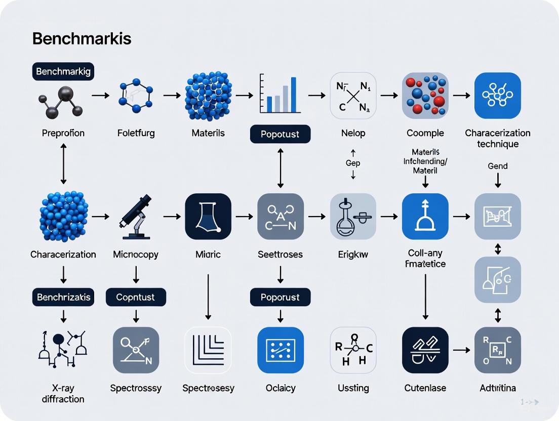
The precise characterization of materials is fundamental to advancements in drug development, materials science, and numerous other scientific fields. Understanding a material's structure, composition, and properties is crucial for linking its atomic and microscopic features to its macroscopic performance. This guide provides a comparative overview of four cornerstone categories of materials characterization techniques: Microscopy, Spectroscopy, Diffraction, and Thermal Analysis. Framed within a broader thesis on benchmarking these methods, this document objectively compares their performance, applications, and limitations, supported by experimental data and standardized protocols. For researchers and scientists, this serves as a strategic toolkit for selecting the optimal technique for specific analytical challenges.
Technique Categories and Comparative Benchmarking
The following section defines each technique category and provides a direct, data-driven comparison of their capabilities, resolutions, and typical applications.
Category Definitions
- Microscopy: Techniques that produce magnified images to visualize a material's surface or internal structure. They provide spatial information about features ranging from the millimeter scale down to the atomic level. Examples include Scanning Electron Microscopy (SEM) and Atomic Force Microscopy (AFM) [6].
- Spectroscopy: Techniques that probe the interaction between matter and electromagnetic radiation or other energy sources to determine a material's chemical composition, bonding, and electronic structure. Examples include Fourier Transform Infrared (FTIR) spectroscopy and X-ray Photoelectron Spectroscopy (XPS) [7] [8].
- Diffraction: Techniques that use the constructive and destructive interference of waves (like X-rays) scattered by a material to determine its long-range crystalline structure, including phase identification, lattice parameters, and crystal defects. The primary example is X-ray Diffraction (XRD) [9].
- Thermal Analysis: Techniques that measure a material's physical and chemical properties as a function of temperature. They provide information on phase transitions, thermal stability, and composition. Examples include Differential Scanning Calorimetry (DSC) and Thermogravimetric Analysis (TGA) [10] [4].
Performance Data Table
The table below summarizes the key performance metrics and applications of representative techniques from each category, enabling direct comparison.
Table 1: Comparative overview of major materials characterization techniques.
| Technique Category | Example Techniques | Key Measured Parameters | Spatial Resolution | Primary Applications |
|---|---|---|---|---|
| Microscopy | SEM [6], TEM [6], AFM [6] | Surface topography, elemental composition (with EDS), crystal structure (TEM) | SEM: ~0.5 nm (HIM) [6]TEM: Sub-Ångström [6]AFM: Atomic [6] | Morphology analysis, defect observation, chemical mapping |
| Spectroscopy | FTIR [11], XPS [7], Raman [9] | Vibrational modes (FTIR, Raman), elemental & chemical state (XPS) | ~10 μm (conventional FTIR) to sub-μm (Raman microscopy) [9] [12] | Chemical identification, functional group analysis, surface chemistry |
| Diffraction | XRD [9], SAXS [4] | Crystal phase, lattice parameters, crystallite size, preferred orientation | Bulk technique; crystallite size detection limit ~1-10 nm [9] | Polymorphism identification, crystallinity quantification, crystal structure solving |
| Thermal Analysis | DSC [10], TGA [11], TMA [10] | Enthalpy changes (DSC), mass loss (TGA), dimensional change (TMA) | Bulk technique (milligram-scale samples) | Melting point, glass transition, thermal stability, composition |
Experimental Protocols for Integrated Characterization
To illustrate how these techniques are applied in practice, the following is a detailed methodology from a published study analyzing natural fibers. This protocol demonstrates a multi-technique approach to fully characterize material properties [11].
Sample Preparation: Tinospora Cordifolia Fiber
- Materials and Extraction: Stems of the Tinospora cordifolia plant were soaked in water for 10 days to enable retting. Fibers were then manually separated, cleaned with distilled water, and dried completely [11].
- Chemical Treatment: A subset of the raw fibers was treated with a 3% (w/v) sodium hydroxide (NaOH) solution for 90 minutes to modify the surface chemistry and morphology [11].
Multi-technique Analysis Workflow
The characterization of the fibers involved a sequential, complementary workflow to assess physical, morphological, structural, and thermal properties.
Figure 1: Experimental workflow for comprehensive fiber analysis.
- Physical Characterization: Fiber diameter was measured using an optical microscope, with at least five measurements per fiber from five different fibers. Density was determined using a pycnometer with distilled water [11].
- Morphological Analysis (Microscopy): The surface morphology of raw and treated fibers was examined using a High-Resolution Field Emission Scanning Electron Microscope (HR-FESEM). Samples were coated with gold prior to imaging to enhance conductivity [11].
- Structural Analysis (Diffraction and Spectroscopy):
- XRD: Patterns were collected using a Rigaku Miniflex 600 with CuKα radiation (λ = 0.154 nm) over a 2θ range of 10° to 80°. The Crystallinity Index (C.I.) was calculated using the Segal method, and Crystallite Size (C.S.) was determined using the Scherrer equation [11].
- FTIR Spectroscopy: Spectra were captured in transmittance mode from 4000 to 400 cm⁻¹ using a Shimadzu spectrometer to identify functional groups [11].
- Mechanical Testing: Single-fiber tensile tests were conducted according to ASTM D3379 standard using a Zwick Roell machine with a 50 mm gauge length and a crosshead speed of 8 mm/min [11].
- Thermal Analysis (Thermal Analysis): Thermal stability was assessed via Thermogravimetric Analysis (TGA) using a PerkinElmer instrument. Powdered fiber samples (~6 mg) were heated from room temperature to 800°C at a rate of 10°C per minute under a nitrogen atmosphere [11].
Research Reagent Solutions
The following table lists key reagents, materials, and instruments used in the featured experimental protocol, along with their critical functions [11].
Table 2: Essential research reagents and materials for fiber characterization.
| Item Name | Function / Application | Technical Specification Example |
|---|---|---|
| Sodium Hydroxide (NaOH) | Alkali treatment to remove hemicellulose, lignin, and wax from fiber surfaces. | 3% (w/v) solution, 90 min immersion [11]. |
| Distilled Water | Fiber washing and density measurement medium. | Used as immersion liquid in pycnometer method [11]. |
| Gold Coating | Conductive layer for high-quality SEM imaging. | Applied to fiber samples prior to HR-FESEM examination [11]. |
| Nitrogen Gas | Inert atmosphere for thermal analysis. | TGA purge gas, 20 mL/min flow rate [11]. |
| Rigaku Miniflex 600 | X-ray Diffractometer for crystallinity analysis. | CuKα radiation source (λ = 0.154 nm) [11]. |
| Shimadzu Spectrometer | FTIR for functional group analysis. | Transmittance mode, 400–4000 cm⁻¹ range [11]. |
Integrated Workflows and Data Correlation
The true power of modern materials characterization lies in the correlation of data from multiple techniques. No single method can provide a complete picture; instead, they offer complementary insights.
The Synergy of Techniques
A combined approach is essential for solving complex analytical problems. For instance, while thermal analysis (DSC) can detect a phase transition, it cannot reveal the structural changes causing it. This requires a diffraction technique like XRD. Similarly, spectroscopy (XPS) can identify surface chemical composition, while microscopy (SEM) can visualize the morphology of that same surface [10] [9].
Table 3: Resolving research questions through multi-technique approaches.
| Research Question | Recommended Technique Combination | Correlated Data Output |
|---|---|---|
| Polymorph Identification & Purity | XRD [9] + DSC [10] + Raman Microscopy [9] | XRD confirms crystal structure, DSC measures transition enthalpies/temperatures, and Raman microscopy maps phase distribution. |
| Surface Contamination & Morphology | XPS [7] + SEM/EDS [6] | XPS identifies elemental composition and chemical states of contaminants, while SEM visualizes their location and morphology. |
| Fiber Reinforced Composite Analysis | SEM [11] + FTIR [11] + Tensile Test + TGA [11] | SEM shows fiber-matrix adhesion, FTIR confirms chemical modification, tensile tests mechanical properties, and TGA assesses thermal stability. |
Future Directions: Integration and Automation
The field of materials characterization is rapidly evolving. Two key trends are shaping its future:
- Hybrid and In-situ Techniques: There is a growing emphasis on combining traditional thermal analysis with in-situ structural probes. This allows researchers to observe structural evolution directly as a function of temperature, greatly aiding the understanding of structure-property relationships [10]. Furthermore, techniques are being adapted for small dimensions like thin films with high resolution [10].
- AI and Machine Learning Integration: The explosion of complex, multi-modal data has driven the integration of Artificial Intelligence (AI) and Machine Learning (ML) into characterization workflows. These tools enable automated feature recognition, anomaly detection, and even real-time experimental steering, accelerating insight extraction and materials design [6]. Benchmark datasets like MatQnA are being developed to evaluate AI capabilities in interpreting materials characterization data [7].
Microscopy, Spectroscopy, Diffraction, and Thermal Analysis form an indispensable toolkit for researchers. Each category offers unique and powerful capabilities, from visualizing atomic structures to quantifying thermal transitions. As demonstrated through the integrated experimental workflow, the most profound insights are often gained not from a single technique, but from the strategic combination of multiple methods. The ongoing trends of hybrid instrumentation, in-situ analysis, and AI-driven data processing promise to further enhance the power and throughput of these techniques, solidifying their critical role in the future of materials science and drug development.
In materials research, where conclusions drawn from data direct multi-million dollar R&D decisions, the ability to trust one's data is not just convenient—it is foundational. A lack of rigorous reproducibility and validation poses a significant hurdle for scientific development, a challenge acutely felt in fields encompassing diverse experimental and theoretical approaches like materials science [13]. Benchmarking, the systematic process of comparing computational methods and analytical techniques using well-characterized reference data, has emerged as the critical discipline for overcoming these challenges. It provides the framework to quantify performance, validate claims, and ultimately, ensure that scientific conclusions are built upon a reliable and reproducible foundation. This guide objectively compares benchmarking methodologies and platforms, providing researchers with the experimental data and protocols necessary to anchor their materials characterization research in verifiable accuracy.
The Benchmarking Landscape in Materials Science
The drive for reproducibility has led to the creation of several community-driven platforms and datasets specifically designed for materials research. These initiatives provide standardized tasks and metrics to impartially evaluate everything from AI models to electronic structure methods.
Table 1: Overview of Major Materials Science Benchmarking Platforms
| Platform/Dataset | Primary Focus | Key Metrics | Data Modalities | Notable Scale |
|---|---|---|---|---|
| MatQnA [14] [7] | Evaluating Multi-modal LLMs on materials characterization | Accuracy on objective (multiple-choice) and subjective questions | Spectra (XPS, XRD), microscopy images (SEM, TEM), text | 10 characterization techniques, 3,800+ questions |
| JARVIS-Leaderboard [13] | Integrated benchmarking of diverse materials design methods | Performance scores specific to property prediction tasks | Atomic structures, atomistic images, spectra, text | 1,281 contributions to 274 benchmarks, 152 methods |
| Specialized Benchmarks (e.g., for battery diagnostics [15]) | Comparing optimization algorithms for specific analysis tasks | Parameter estimation quality, computational cost, stability | Voltage/capacity curves from cycling experiments | Case-specific (e.g., 309 battery cycles) |
Preliminary evaluations on the MatQnA dataset reveal that the most advanced multi-modal AI models (e.g., GPT-4.1, Claude 4, Gemini 2.5) are already achieving nearly 90% accuracy on objective questions involving the interpretation of materials data [14] [7]. This performance, broken down by technique, demonstrates the varying levels of model proficiency across different characterization methods.
Table 2: Performance of Multi-modal LLMs on MatQnA Objective Questions (Accuracy %)
| Characterization Technique | GPT-4.1 | Claude 4 | Gemini 2.5 | Doubao Vision Pro |
|---|---|---|---|---|
| X-ray Diffraction (XRD) | 91.5 | 89.8 | 88.3 | 90.1 |
| Scanning Electron Microscopy (SEM) | 87.2 | 85.5 | 84.0 | 86.8 |
| Transmission Electron Microscopy (TEM) | 85.1 | 82.4 | 83.7 | 84.5 |
| X-ray Photoelectron Spectroscopy (XPS) | 83.3 | 80.9 | 81.5 | 82.0 |
For lower-level computations, the JARVIS-Leaderboard facilitates extensive comparisons. For instance, it hosts benchmarks for foundational properties like the formation energy of crystals, where different AI and electronic structure methods can be directly compared. A hypothetical snapshot of such a benchmark might show AI models like ALIGNN achieving Mean Absolute Errors (MAE) below 0.05 eV/atom on a test set of known crystals, while various DFT codes (VASP, Quantum ESPRESSO) might show MAEs between 0.03-0.15 eV/atom when compared to high-fidelity experimental or quantum Monte Carlo reference data [13].
Experimental Benchmarking Protocols
Adhering to rigorous methodology is what separates a robust benchmark from a simple comparison. The following protocols, synthesized from best practices in computational science [16] and applied materials research [15], provide a template for designing a conclusive benchmarking experiment.
Protocol 1: Benchmarking AI Models for Materials Data Interpretation
This protocol is designed for evaluating the performance of multi-modal AI models in interpreting materials characterization data, such as spectra and micrographs.
- Objective Definition: Clearly state the model's task (e.g., "Identify the crystalline phases present from this XRD pattern" or "Classify the type of defect in this SEM image").
- Benchmark Dataset Selection: Utilize a curated, high-quality dataset with established ground truth. MatQnA is an example for this purpose [7]. The dataset should be partitioned into training/validation/test sets, ensuring the test set is held out from the model during training.
- Model Selection & Training: Select a representative set of models to compare (e.g., state-of-the-art, widely used, and a simple baseline model). Train each model on the training set, using the validation set for hyperparameter tuning.
- Performance Evaluation: Execute the trained models on the unseen test set. Calculate quantitative metrics relevant to the task:
- For classification: Accuracy, F1-Score, Area Under the ROC Curve (AUC).
- For regression/quantification: Mean Absolute Error (MAE), Root Mean Square Error (RMSE).
- Results Analysis & Reporting: Compile results in a clear table (see Table 2). Perform statistical significance testing on performance differences. Include qualitative examples of success and failure cases to illustrate model behavior.
Protocol 2: Benchmarking Optimization Algorithms for Parameter Estimation
This protocol is applicable for comparing optimization methods used to extract quantitative parameters from experimental data, such as in battery aging diagnostics [15].
- Objective Definition: Define the parameter estimation problem (e.g., "Extract the loss of active material (LAM) parameters from a differential voltage analysis (DVA) curve").
- Data & Ground Truth Preparation: Use a dataset where parameters have been reliably determined via a high-fidelity method (e.g., post-mortem analysis for batteries) or synthetic data with known true parameters.
- Algorithm Selection & Setup: Choose algorithms with different approaches (e.g., Gradient Descent for local, fast convergence vs. Bayesian Optimization for global, robust search). Implement them with their respective critical parameters:
- Gradient Descent: Learning rate, number of iterations, convergence tolerance.
- Bayesian Optimization: Acquisition function, number of initial points, iteration budget.
- Performance Evaluation: Run each algorithm multiple times to account for stochasticity. Record:
- Result Quality: Error between estimated and true parameters (MAE).
- Computational Cost: Average runtime and number of function evaluations.
- Stability/Robustness: Standard deviation of results across multiple runs.
- Results Analysis & Reporting: Summarize the trade-offs. For example, a benchmark might find that Gradient Descent is 5x faster but exhibits higher variance, while Bayesian Optimization is more stable but computationally intensive [15]. This provides a data-driven basis for algorithm selection.
Diagram 1: Generic benchmarking workflow.
The Scientist's Toolkit: Essential Research Reagent Solutions
Beyond software, benchmarking relies on a suite of essential resources. The table below details key "reagent solutions" for conducting rigorous benchmarking in computational materials science.
Table 3: Essential Reagents for Computational Benchmarking
| Tool/Resource Name | Category | Primary Function in Benchmarking |
|---|---|---|
| MatQnA Dataset [14] [7] | Benchmark Dataset | Provides standardized, multi-modal questions and answers to evaluate AI performance on materials characterization tasks. |
| JARVIS-Leaderboard [13] | Benchmarking Platform | An integrated, community-driven platform to submit, compare, and track performance of various AI, electronic structure, and force-field methods. |
| Reference Experimental Datasets (e.g., battery cycling data [15]) | Ground Truth Data | Serves as the objective, high-fidelity standard against which the accuracy of computational methods is measured. |
| R & Python (scikit-learn, PyTorch) [17] | Statistical & ML Programming | Provides the environment and libraries for implementing, running, and evaluating custom methods and analyses. |
| Bayesian Optimization & Gradient Descent Algorithms [15] | Optimization Methods | Core algorithms for parameter estimation and inverse design problems; their performance is often the subject of benchmarks. |
Benchmarking is the cornerstone of reliable and reproducible materials research. By leveraging established platforms like MatQnA and JARVIS-Leaderboard, and adhering to rigorous experimental protocols, scientists can move beyond anecdotal evidence and make informed, data-driven decisions about their analytical tools. The quantitative comparisons and detailed methodologies provided here offer a pathway to not only validate existing methods but also to identify the critical gaps and challenges that will drive future methodological innovations. In the high-stakes field of materials characterization, a commitment to rigorous benchmarking is synonymous with a commitment to scientific truth.
The field of materials science is undergoing a significant transformation, driven by the integration of artificial intelligence (AI) and large language models (LLMs) into scientific research workflows. However, the capabilities of AI models in highly specialized domains like materials characterization and analysis have not been systematically or sufficiently validated [14]. This gap represents a critical hurdle for the reliable application of multi-modal AI in scientific discovery and laboratory practice.
MatQnA emerges as the first multi-modal benchmark dataset specifically designed to address this challenge by providing a standardized framework for evaluating AI performance in interpreting experimental materials data [14] [18]. Derived from over 400 peer-reviewed journal articles and expert case studies, MatQnA enables rigorous assessment of AI systems in supporting materials research workflows, from property prediction to materials discovery [18]. This benchmark represents a crucial step toward establishing metrology for AI in scientific domains, joining other notable benchmarking efforts in materials informatics such as Matbench [19] and JARVIS-Leaderboard [13].
MatQnA Dataset Design and Scope
Core Design Objectives
MatQnA was constructed to fill a critical gap in AI benchmarking, with several clearly defined design objectives. The dataset specifically targets comprehensive validation of LLMs in the specialized domain of materials characterization, focusing on deeper scientific reasoning associated with experimental data interpretation [18]. Its primary aim is to evaluate model performance in real-world materials scenarios, requiring the understanding of technical concepts and the integration of image and text information [18].
The questions are strategically designed around experimental figures, spectral patterns, microscopy images, and domain-specific data tables, reflecting the complexity encountered in scientific practice rather than simplified theoretical exercises [18]. This approach ensures that the benchmark assesses practical AI capabilities relevant to materials researchers and characterization specialists.
Covered Characterization Techniques
MatQnA encompasses ten major characterization methods central to materials science, each presenting unique multi-modal challenges for AI interpretation [14] [18]. The comprehensive coverage ensures broad applicability across different subdisciplines of materials research.
Table: Materials Characterization Techniques in MatQnA
| Technique | Key Analytical Focus | Modality |
|---|---|---|
| XPS (X-ray Photoelectron Spectroscopy) | Chemical state, element, peak assignment | Image, Text |
| XRD (X-ray Diffraction) | Crystal structure, phase, grain sizing | Image, Text |
| SEM (Scanning Electron Microscopy) | Surface morphology, defects | Image |
| TEM (Transmission Electron Microscopy) | Internal lattice, microstructure | Image |
| AFM (Atomic Force Microscopy) | 3D topography, roughness | Image |
| DSC (Differential Scanning Calorimetry) | Thermal transitions, enthalpy | Chart |
| TGA (Thermogravimetric Analysis) | Decomposition, stability | Chart |
| FTIR (Fourier-Transform Infrared Spectroscopy) | Bonds, vibrational modes | Spectrum |
| Raman (Raman Spectroscopy) | Molecular vibration, phase composition | Spectrum |
| XAFS (X-ray Absorption Fine Structure) | Atomic environment, oxidation states | Spectrum |
Quantitative analysis tasks commonly appear throughout the dataset, such as using the Scherrer equation for XRD grain size estimation: $L = \frac{K\lambda}{\beta \cos \theta}$, where $L$ is crystallite size, $\lambda$ the X-ray wavelength, $\beta$ peak width, and $\theta$ the Bragg angle [18]. Each technique's section contains domain-relevant figures paired with structured questions that test both fundamental understanding and practical interpretation skills.
Dataset Construction Methodology
The MatQnA dataset was assembled through a sophisticated hybrid methodology combining automated LLM-based question generation with expert human validation, ensuring both scalability and scientific accuracy [18].
Source Extraction and Processing
The construction process began with raw data extraction primarily from PDFs of journal articles and domain case reports, preprocessed using PDF Craft to isolate relevant text, images, and figure captions [18]. This initial phase ensured that the dataset was grounded in authentic scientific literature and represented real-world characterization challenges rather than artificial examples created solely for benchmarking purposes.
Automated QA Generation and Validation
Structured prompt templates and OpenAI's GPT-4.1 API were employed to draft multi-format questions, including both objective and open-ended types [18]. The process incorporated automatic coreference handling and context enforcement to ensure clarity, particularly for image-based queries where contextual information is crucial for accurate interpretation [18].
Domain experts then performed rigorous review, filtering, and correction of the generated QA pairs for terminological precision and logical relevance [18]. This human-in-the-loop validation employed regex-based methods to enforce answer self-containment, ensuring that responses could be evaluated objectively without requiring additional contextual knowledge not present in the questions themselves [14] [18].
Final Dataset Structure
The finalized dataset organization by characterization technique resulted in approximately 5,000 QA pairs (2,749 subjective and 2,219 objective) stored in Parquet format [18]. Each entry is explicitly linked to its associated technique, enabling both comprehensive evaluation and technique-specific performance analysis. This structured organization facilitates targeted benchmarking for specific application domains within materials characterization.
Question Formats and Evaluation Protocols
Question Categories
MatQnA's QA pairs are systematically divided into two main categories, each serving distinct evaluation purposes:
- Multiple-Choice Questions (MCQs): Objective, closed-form items designed for unambiguous grading, focusing on factual recognition, calculation, or discrete judgment from presented experimental data [18].
- Subjective Questions: Open-ended prompts requiring detailed explanation, justification, or synthesis, emphasizing models' ability to express scientific reasoning and communicate technical concepts [18].
Both formats are strategically designed to diagnose model competence across multiple dimensions, including image interpretation, quantitative analysis, and domain-specific nomenclature mastery.
Evaluation Procedures
Scoring protocols for objective questions are standardized to ensure consistent evaluation across different models and research groups [18]. For subjective items, expert rubric review provides the evaluation framework, assessing the quality, accuracy, and completeness of model-generated explanations [18].
This dual approach enables comprehensive assessment of both factual knowledge recall and deeper scientific reasoning capabilities, providing a more complete picture of model performance than single-format benchmarks could achieve.
Performance Comparison of Multi-modal AI Models
Preliminary evaluation results on MatQnA reveal that state-of-the-art multi-modal LLMs demonstrate strong proficiency in materials data interpretation, with nearly 90% accuracy achieved by leading models on objective questions [14] [18].
Table: Model Performance on MatQnA Objective Questions
| Model | Overall Accuracy | Strengths | Limitations |
|---|---|---|---|
| GPT-4.1 | 89.8% | Strong overall performance across techniques | Spatial reasoning challenges |
| Claude Sonnet 4 | ~89% | High accuracy on spectral analysis | Slightly lower on microscopy |
| Gemini 2.5 | ~88% | Competitive across multiple modalities | Inconsistencies in quantitative tasks |
| Doubao Vision Pro 32K | ~87% | Solid performance on Chinese-language materials | Slightly lower on Western literature |
The evaluation encompassed multiple state-of-the-art multi-modal models including GPT-4.1, Claude 4, Gemini 2.5, and Doubao Vision Pro 32K [14] [18]. Heatmap analyses across 31 subcategories confirmed systematic strengths and weaknesses, providing detailed insights beyond aggregate performance metrics [18].
Technique-Specific Performance Variations
Model performance varies significantly across different characterization techniques, revealing important patterns about current AI capabilities and limitations:
- Highest Performance: Spectroscopic characterization methods (FTIR, Raman) achieve accuracy exceeding 95%, suggesting that spectral pattern recognition aligns well with current model capabilities [18].
- Lowest Performance: Techniques requiring spatial reasoning or 3D topology analysis (e.g., AFM) show reduced performance (83.9%), indicating areas where architectural improvements are needed [18].
- Moderate Performance: Methods combining image interpretation with quantitative analysis (XRD, XPS, TEM) typically fall in the 85-92% range, balancing visual and analytical challenges [18].
These variations suggest that while current models are highly adept at standard data interpretation, there exist specific modalities requiring further algorithmic innovation, particularly those involving spatial reasoning and complex topological relationships.
Essential Research Reagent Solutions
The effective implementation and utilization of benchmarks like MatQnA require specific computational tools and resources that constitute the essential "research reagents" for AI-driven materials science.
Table: Essential Research Reagents for AI Materials Characterization
| Tool/Resource | Function | Application in MatQnA |
|---|---|---|
| Multi-modal LLMs (GPT-4.1, Claude, etc.) | Core inference engines for benchmark evaluation | Primary models being evaluated on interpretation tasks |
| MatQnA Dataset | Benchmarking standard for materials characterization | Central evaluation corpus containing 5,000 QA pairs |
| Hugging Face Platform | Dataset hosting and distribution | Public access point for MatQnA dataset |
| PDF Craft | PDF text and image extraction | Preprocessing of source documents during dataset creation |
| Matminer Featurization Library | Materials-specific feature generation | Reference for traditional ML approaches in materials science |
| JARVIS-Leaderboard | Comprehensive benchmarking platform | Context for MatQnA within broader materials AI ecosystem |
These tools collectively enable researchers to not only evaluate existing models but also to develop new approaches and contribute to the growing ecosystem of AI-driven materials characterization.
Implications for Materials Characterization Research
Scientific and Practical Impact
MatQnA provides a foundational resource for diverse applications across materials science research and development. For benchmarking and model selection, it offers a rigorous, standardized foundation for evaluating LLMs in materials science, enabling informed decisions about model deployment for specific characterization tasks [18].
Regarding workflow integration, the benchmark enables AI-assisted materials discovery, property prediction, and experimental support by establishing reliable performance baselines [18]. This can significantly accelerate research cycles and reduce dependency on purely manual data interpretation. For domain-specific model development, MatQnA facilitates targeted fine-tuning and robust analysis of multi-modal AI systems, guiding architectural improvements toward areas of current weakness [18].
The demonstrated feasibility of extending LLM-based evaluation frameworks to specialized scientific fields suggests potential for similar benchmarks in other domains, promoting interdisciplinary methodological exchange [18].
Relationship to Broader Benchmarking Ecosystem
MatQnA occupies a unique position within the expanding landscape of materials informatics benchmarks. While platforms like Matbench focus on structure-property predictions [19] and JARVIS-Leaderboard provides comprehensive coverage across multiple computational approaches [13], MatQnA specifically addresses the critical gap in experimental data interpretation.
This specialization makes it complementary to existing resources rather than competitive, together forming a more complete evaluation framework for AI in materials science. The nearly 90% accuracy achieved by leading models on MatQnA's objective questions [14] suggests that AI systems are approaching human-level performance on certain characterization tasks, potentially enabling their practical deployment in research workflows.
Access and Future Directions
MatQnA is freely available to the research community through the Hugging Face repository at https://huggingface.co/datasets/richardhzgg/matQnA [18]. Researchers are encouraged to utilize, evaluate, and iteratively improve the dataset, contributing to its evolution as a community resource.
The presence of robust validation and comprehensive coverage positions MatQnA as a reference standard for future work in multi-modal AI benchmarking within scientific domains [18]. As models continue to advance, the benchmark will likely expand to include more complex reasoning tasks, additional characterization techniques, and more sophisticated evaluation metrics that better capture scientific understanding beyond factual recall.
The demonstrated strong performance of current models suggests a promising trajectory toward AI systems that can genuinely assist and augment human expertise in materials characterization, potentially transforming how experimental data is analyzed and interpreted across the materials research community.
In the field of benchmarking materials characterization techniques, researchers face significant challenges when working with real-world data (RWD). The inherent characteristics of such data—including sparsity, integration of multiple sources, and various biases—directly impact the validity, reliability, and generalizability of research findings. As materials science increasingly relies on data-driven approaches, understanding and addressing these challenges becomes paramount for advancing the field. This guide objectively compares methodologies for handling RWD complexities, providing experimental data and protocols to equip researchers with practical solutions for robust materials characterization research.
The fundamental issue with RWD lies in its observational nature; unlike data generated in controlled laboratory settings, RWD is often collected for administrative or clinical purposes, leading to unique structural challenges. Among these, data sparsity presents a primary obstacle, particularly in studies involving high-dimensional feature spaces. Furthermore, the integration of multiple data sources introduces heterogeneity, while selection bias and other systematic errors can compromise analytical validity if not properly addressed. This guide systematically explores these interconnected challenges and provides evidence-based approaches for mitigating their effects in materials characterization research.
Understanding and Addressing Data Sparsity
Defining Sparse Data in Characterization Research
Sparse data refers to datasets predominantly composed of zeroes or near-zero values, creating what can be visualized as a "desert of data" with only scattered oases of meaningful information [20]. In materials characterization, this occurs frequently in scenarios such as:
- High-Throughput Experimental Data: Where most measured features show negligible activity or response across most experimental conditions
- Multi-technique Characterization: When integrating data from multiple analytical techniques where each technique captures different material aspects
- Composition-Space Mapping: Where only specific regions of a compositional space have been experimentally explored
Mathematically, sparse data can be represented as a matrix where only a handful of elements contain non-zero values. For example, a 4×4 matrix with only two non-zero entries exemplifies classic sparse structure [20]. The key challenge lies in extracting meaningful patterns from such underpopulated data structures while maintaining statistical rigor.
Comparative Analysis: Sparse vs. Dense Data
The structural differences between sparse and dense data significantly impact computational strategies in materials informatics. The table below summarizes key distinctions:
Table 1: Characteristics of Sparse vs. Dense Data in Materials Research
| Characteristic | Sparse Data | Dense Data |
|---|---|---|
| Memory Efficiency | High (stores only non-zero values + positions) | Low (stores all values regardless of content) |
| Computational Efficiency | Situation-dependent (fast with optimized algorithms) | Generally consistent (benefits from contiguous memory) |
| Algorithm Suitability | Naive Bayes, L1-regularized models, Random Forests | Deep Learning (CNNs), distance-based algorithms |
| Real-World Examples | User-item interactions in recommendation systems | Image pixels in microstructure analysis |
| Storage Formats | Compressed Sparse Row (CSR), Compressed Sparse Column (CSC) | Standard arrays, dense matrices |
Handling Sparse Data: Experimental Protocols
Research indicates several effective methodologies for addressing sparsity in materials characterization data:
Data Preprocessing and Thresholding Initial assessment should determine whether zero values represent true negatives or missing data. For legitimate zero values, thresholding techniques can remove features or samples with excessive zeros (e.g., >99% sparse), effectively reducing noise and computational burden [20]. Implementation protocols include:
- Sparsity Distribution Analysis: Calculate sparsity percentage for each feature and sample
- Strategic Thresholding: Remove features/samples exceeding predetermined sparsity thresholds (typically 90-99%)
- Imputation Validation: When imputing missing values, validate methods (mean, median, model-based) against known subsets
Algorithm Selection for Sparse Data Certain machine learning algorithms naturally accommodate sparse data structures [20]:
- Naive Bayes: Effectively handles high-dimensional sparse features by calculating probabilities based on present features
- L1-Regularized Models (Lasso Regression): Promote sparsity by zeroing out unimportant coefficients
- Tree-Based Methods (Random Forests): Insensitive to data sparsity patterns
- Scikit-learn Implementation: Many algorithms in Scikit-learn offer built-in sparse matrix support
Dimensionality Reduction Techniques When raw sparsity impedes analysis, dimensionality reduction methods can concentrate meaningful information:
- Principal Component Analysis (PCA): Transforms sparse features into denser principal components
- Singular Value Decomposition (SVD): Particularly effective for sparse matrix factorization
- Non-negative Matrix Factorization: Constrained approach suitable for physical material properties
Diagram: Workflow for Handling Sparse Data in Materials Characterization
Benchmarking Framework for Multi-Source Data
Integrating data from multiple characterization techniques or laboratories requires systematic benchmarking approaches. Recent research demonstrates that establishing interoperable digital platforms enables real-world time data assessment and automated analysis [21]. The core challenges in multi-source data integration include:
- Heterogeneous Data Structures: Different formats, resolutions, and measurement principles across techniques
- Semantic Harmonization: Consistent meaning and interpretation of measured parameters
- Temporal Alignment: Synchronizing data collected at different timepoints or frequencies
- Scale Compatibility: Reconciling measurements across different spatial and temporal scales
The benchmarking process involves comparing methodologies across different analytical centers or techniques to identify optimal practices and establish quality standards [21]. This approach aligns with value-based healthcare principles where the ratio of quality to cost defines value, translated to materials science as the ratio of information content to analytical investment [21].
Experimental Protocol: Interoperable Platform Implementation
Based on sarcoma research benchmarking, the following protocol facilitates multi-source data integration:
Platform Architecture
- Front-End Components: Data entry interfaces and real-time visualization tools
- Back-End Infrastructure: SQL database structures with statistical analysis programming (e.g., R)
- API Data Exporters: Tools for seamless data transfer between laboratory information systems
- Cloud Integration: Secure data storage and computation capabilities [21]
Harmonization Methodology
- Structured Data Frameworks: Establish standardized data models for specific characterization domains
- Metadata Standards: Consistent documentation of experimental conditions and parameters
- Cross-Validation Protocols: Systematic comparison of measurements across techniques
- Federated Learning Approaches: Enable collaborative model training without centralizing sensitive data [21]
Performance Estimation with Limited External Data
A groundbreaking method enables estimation of model performance on external data sources using only summary statistics, significantly reducing the barriers to multi-source validation [22]. The experimental protocol involves:
Table 2: Weighting Method Comparison for Multi-Source Data Integration
| Method | External Data Requirements | Implementation Complexity | Best Use Cases |
|---|---|---|---|
| Pseudolikelihood Estimating Equations | Individual-level data from probability sample | High | When representative reference data available |
| Beta Regression GLM | Individual-level external data | Medium | Selection probability estimation |
| Poststratification | Summary-level population data | Low | When population margins known |
| Raking/Calibration | Summary-level data | Low-Medium | Efficient approximation with known demographics |
Implementation Steps:
- Internal Model Training: Develop predictive models using accessible internal data sources
- External Statistics Collection: Gather limited descriptive statistics from target external sources
- Weight Optimization: Find weights that induce internal weighted statistics matching external characteristics
- Performance Estimation: Compute metrics using weighted internal labels and predictions [22]
Validation Results: Recent benchmarking demonstrated accurate performance estimations across multiple data sources with 95th error percentiles for AUROC at 0.03, calibration-in-the-large at 0.08, and Brier score at 0.0002 [22]. This method enables researchers to assess model transportability without direct access to external unit-level data.
Addressing Selection Bias in Real-World Data
Understanding Selection Mechanisms
Selection bias represents a critical challenge in observational data analysis, including materials characterization research. In administrative healthcare data, selection mechanisms vary widely based on "Who is in my study sample?" [23]. Analogous issues appear in materials science where:
- Instrumentation Bias: Certain measurement techniques favor specific material classes or properties
- Publication Bias: Well-characterized materials are overrepresented in literature
- Laboratory Bias: Specific synthesis or processing methods dominate particular research groups
The "curse of large n" or "big data paradox" phenomenon highlights that with vast sample sizes leading to small standard errors, biases become increasingly problematic as they don't diminish with increasing sample size [23]. This necessitates updated statistical thinking focused on bias reduction rather than variance reduction.
Framework for Analyzing Selection Bias
Directed Acyclic Graphs (DAGs) provide an analytical framework for understanding how different sources of selection bias affect estimates of association between variables [23]. The framework enables researchers to:
- Identify Selection Mechanisms: Map the pathways through which selection occurs
- Assess Bias Magnitude: Estimate the extent of distortion in association parameters
- Design Adjustment Strategies: Develop targeted methods to reduce selection bias
Diagram: Selection Bias Analysis Framework Using Directed Acyclic Graphs
Weighting Methods for Bias Reduction
Four weighting approaches have demonstrated effectiveness in reducing selection bias in real-world data:
Inverse Probability Weighted (IPW) Logistic Regression This method constructs weights to account for unequal selection probabilities [23]. Implementation involves:
- Selection Model Specification: Identify variables influencing selection into the sample
- Weight Calculation: Estimate inverse probabilities of selection
- Weighted Analysis: Apply weights in association analyses
- Variance Adjustment: Use robust variance estimators to account for weighting
Comparison of Weighting Approaches:
Table 3: Weighting Methods for Selection Bias Reduction
| Method | Data Requirements | Key Advantages | Limitations |
|---|---|---|---|
| IPW with Known Probabilities | Known selection probabilities | Unbiased if model correct | Rarely known in practice |
| Pseudolikelihood Estimation | Individual-level external reference data | Efficient estimation | Requires representative external data |
| Beta Regression GLM | Individual-level external data | Flexible probability modeling | Computationally intensive |
| Poststratification/Raking | Summary-level population data | Minimal external data needs | Assumes representative strata |
Variance Formulae: Each weighting method requires specific variance estimation techniques to ensure valid inference [23]. These typically involve Taylor series linearization or resampling methods to account for the weighting complexity.
Experimental Protocols for Benchmarking Studies
Comprehensive Benchmarking Framework
Establishing robust benchmarking protocols enables meaningful comparison of characterization techniques across different data challenges. Based on healthcare research, a comprehensive framework includes:
Primary Objectives:
- Comparison of Independent Centers: Analyze demographics and basic protocols across multiple laboratories or characterization centers
- Platform Establishment: Implement interoperable digital platforms for standardized data assessment
- Quality Metric Definition: Define outcome and quality indicators for consistent benchmarking [21]
Implementation Protocol:
- Cohort Definition: Establish clear inclusion criteria for materials or samples
- Multi-Center Recruitment: Engage multiple characterization laboratories or techniques
- Standardized Data Collection: Implement consistent data elements across participants
- Blinded Analysis: Prevent analytical bias through blinded assessment procedures
- Statistical Harmonization: Apply appropriate methods for cross-center comparison
Case Study: Sarcoma Care Benchmarking Protocol
A recent study on sarcoma care provides a transferable model for materials characterization benchmarking [21]:
Study Population:
- 983 patients across two independent multidisciplinary teams
- Prospective collection over 15 months
- Consecutive inclusion of all eligible samples
- Reference review of all relevant characterization data
Platform Implementation:
- Sarconnector Digital Platform: Interoperable system with front-end (data entry, visualization) and back-end (SQL database, R programming) components
- Real-World Time Data Assessment: Continuous data collection throughout characterization workflow
- Automated Analysis: Standardized computational pipelines for consistent evaluation
Outcome Measures:
- Process Metrics: First-time presentations, follow-up presentations, primary analyses
- Technical Parameters: Experimental conditions, measurement specifications
- Quality Indicators: Reproducibility measures, inter-technique concordance
Sample Size Considerations in Benchmarking
The accuracy of benchmarking analyses depends on appropriate sample sizes for both internal and external datasets [22]. Experimental evidence indicates:
Internal Sample Size Effects:
- Algorithms frequently fail to converge with samples below 1,000 units
- Variance and upper quartiles increase significantly with smaller samples
- Error convergence improves with larger internal samples
External Sample Size Effects:
- Impact less pronounced than internal sample size
- Stratified sampling maintaining outcome prevalence improves performance
- Minimum external sample size depends on outcome rarity and diversity
Recommended Practice: For reliable benchmarking, internal sample sizes should exceed 2,000 units whenever possible, with proportional representation of key subgroups or material classes.
The Scientist's Toolkit: Essential Research Reagents and Solutions
Table 4: Essential Research Reagents and Computational Tools for Data Challenges
| Tool/Category | Specific Examples | Function/Purpose | Implementation Considerations |
|---|---|---|---|
| Sparse Data Algorithms | Scikit-learn sparse matrix support, Naive Bayes, L1-regularized models | Efficient handling of predominantly zero data | Memory optimization, algorithm-specific preprocessing |
| Bias Reduction Methods | Inverse probability weighting, poststratification, calibration | Mitigate selection bias in observational data | External reference data requirements, variance estimation |
| Multi-Source Integration Platforms | Sarconnector, OHDSI tools, interoperable digital platforms | Harmonize data from disparate sources | API development, cloud integration, standardized data models |
| Benchmarking Frameworks | Real-world time data assessment, automated analysis pipelines | Standardized comparison of techniques/methodologies | Quality indicator definition, statistical harmonization |
| Performance Estimation Tools | Weight optimization algorithms, statistical characteristic analysis | Estimate external performance without unit-level data | Feature importance consideration, convergence validation |
Addressing sparsity, multiple sources, and bias in real-world data requires methodical approaches and specialized tools. The experimental protocols and comparative analyses presented demonstrate that through strategic algorithm selection, weighting methods, and benchmarking frameworks, researchers can extract reliable insights from complex materials characterization data.
Future directions in this field include increased automation of bias detection and correction, development of more sophisticated federated learning approaches for multi-source data integration, and establishment of domain-specific standards for data quality assessment. As materials characterization continues to generate increasingly large and complex datasets, the methodologies outlined in this guide will become essential components of the materials informatics toolkit.
The integration of real-world data from multiple sources, when properly handled for sparsity and bias, offers unprecedented opportunities for accelerating materials discovery and characterization. By implementing the protocols and comparisons presented, researchers can advance the rigor and reproducibility of materials research while leveraging the rich information contained in diverse, real-world datasets.
Technique Deep Dive: Operational Methods and Specific Applications in Research
X-ray Photoelectron Spectroscopy (XPS) is a powerful surface-sensitive analytical technique that provides quantitative information on the elemental composition, chemical state, and electronic structure of the top 1–10 nm of a material surface [24] [25]. This guide objectively benchmarks XPS against other surface analysis techniques, detailing its operational principles, capabilities, and limitations within the context of benchmarking materials characterization.
Fundamental Principles and Instrumentation
XPS is based on the photoelectric effect. When a material is irradiated with X-rays, photons are absorbed by atoms, ejecting core-level electrons known as photoelectrons. The kinetic energy of these ejected electrons is measured by the spectrometer, and the electron binding energy is calculated using the equation:
Ebinding = Ephoton - (E_kinetic + ϕ), where E_photon is the energy of the incident X-ray, E_kinetic is the measured kinetic energy of the electron, and ϕ is the work function of the spectrometer [25]. This binding energy is a unique fingerprint for each element and its chemical state, as it is influenced by the local chemical environment.
A typical XPS instrument requires an ultra-high vacuum (UHV) environment (typically below 10⁻⁷ Pa) to allow the ejected photoelectrons to travel to the detector without colliding with gas molecules [25]. Key components include an X-ray source (commonly Al Kα or Mg Kα), a hemispherical electron energy analyzer, and an electron detection system. Modern systems often include complementary capabilities such as ultraviolet photoelectron spectroscopy (UPS), ion scattering spectroscopy (ISS), and gas cluster ion sources for depth profiling of organic materials [26].
Experimental Workflow
The following diagram illustrates the generalized workflow for conducting an XPS analysis, from sample preparation to data interpretation.
Detailed Methodologies for Key Experiments
Sample Preparation and Analysis Protocol (Based on a Thin Film Study) [27]:
- Sample Fabrication: Copper oxide (CuO) thin films were deposited via reactive magnetron sputtering onto silicon and glass substrates. Key parameters included a discharge power of 50 W and an oxygen atmosphere at a pressure of 1.5 × 10⁻² mbar.
- Experimental Treatment: Films were doped by implanting Cr⁺ ions at energies of 10-15 keV with varying doses (e.g., 1×10¹⁴ cm⁻² to 5×10¹⁶ cm⁻²). Post-implantation annealing was performed in air at 400°C for 6 hours.
- XPS Data Acquisition:
- Instrument Settings: A monochromatic Al Kα X-ray source (1486.6 eV) operating at 250 W (12.5 kV, 20 mA) was used.
- Analysis: A hemispherical analyzer operated at a pass energy of 20 eV in fixed transmission mode. Charge compensation was achieved using a flood gun (1 V, 0.1 mA).
- Data Processing: Binding energy calibration was performed by referencing the C 1s contamination peak to 286.4 eV. Spectra were fitted using Voigt functions, with Shirley or Tougaard backgrounds applied to account for inelastically scattered electrons [28] [27].
Overlayer Thickness Determination [28]:
- For planar thin films, XPS can determine overlayer thickness non-destructively. The Strohmeier equation is one approach, utilizing the intensity ratio of the overlayer and substrate signals:
d = λ * cos(θ) * ln(I_substrate / (N_substrate * λ_substrate) / (I_overlayer / (N_overlayer * λ_overlayer)) + 1), wheredis the thickness,λis the inelastic mean free path,θis the emission angle,Iis the measured intensity, andNis the atomic volume density.
Depth Profiling [26]:
- To analyze composition as a function of depth, XPS is combined with sputter depth profiling. This involves alternating between ion beam etching (e.g., using Ar⁺ ions at 3 keV) to remove material and XPS analysis to characterize the newly exposed surface. For organic or sensitive materials, gas cluster ion beams are used to reduce damage [26].
Performance Benchmarking Against Other Techniques
The table below provides a quantitative comparison of XPS with other common surface and depth profiling techniques.
Table 1: Comparison of XPS with Other Surface and Depth Analysis Techniques [29]
| Technique | Information Depth | Detection Limits | Chemical State Information | Lateral Resolution | Vacuum Requirement | Key Applications & Notes |
|---|---|---|---|---|---|---|
| XPS (X-ray Photoelectron Spectroscopy) | Top 5-10 nm (<10 nm) [24] [25] | 0.1-1.0 at% (1000-10000 ppm); can reach ppm with long acquisition [25] | Yes, excellent for all elements except H and He [25] | ~10-200 μm; can be <200 nm with imaging systems [25] | Ultra-High Vacuum (UHV) [25] | Surface chemical composition, empirical formula, oxidation states. |
| GDOES (Glow Discharge Optical Emission Spectroscopy) | Sputtering depth; can profile many μm | ppm range [29] | Limited | No lateral resolution; signal averaged over mm [29] | Low vacuum (a few Torr) [29] | Fast depth profiling of thin/thick films; minimal matrix effects; no UHV needed. |
| SIMS (Secondary Ion Mass Spectrometry) | ~10 monolayers [29] | ppb-ppm (excellent sensitivity) [29] | Limited, complex to interpret | High (can be nm-scale) | High Vacuum (<10⁻⁷ Torr) [29] | Ultra-trace elemental and isotopic analysis; high detection efficiency. |
| SEM (Scanning Electron Microscopy) | Varies with beam energy (μm scale) | Not quantitative for composition | No, primarily elemental (with EDS) | High (nm-scale) | High Vacuum | Topography and elemental mapping; often used complementarily with XPS. |
Key Performance Differentiators
- Chemical State Specificity: XPS is unparalleled among the techniques listed for providing direct chemical bonding information. For example, it can distinguish between silicon (Si), silicon dioxide (SiO₂), and silicon in a polymer [30].
- Surface Sensitivity vs. Profiling Depth: While XPS is restricted to the top ~10 nm, GDOES offers much faster and deeper profiling, capable of analyzing layers several micrometers thick in a short time [29].
- Sample Requirements: XPS and SIMS require UHV and can analyze both conductors and insulators (with charge compensation). In contrast, GDOES does not require UHV or charge compensation for insulating samples [29].
The Scientist's Toolkit: Essential Research Reagents and Materials
Table 2: Key Research Reagents and Materials for XPS Analysis
| Item / Material | Function / Relevance in XPS Experiments |
|---|---|
| Monochromatic Al Kα X-ray Source | Standard laboratory source for exciting photoelectrons; provides high-energy-resolution spectra [25]. |
| Argon Gas Cluster Ion Source | Used for sputter depth profiling of soft materials (e.g., polymers, organics) with minimal chemical damage [26]. |
| Charge Neutralization Flood Gun | Essential for analyzing insulating samples (e.g., polymers, ceramics) to prevent surface charging that distorts spectral data [26] [27]. |
| Hemispherical Electron Analyzer | The core component that measures the kinetic energy of photoelectrons with high resolution [27]. |
| Reference Materials | Certified standard samples are crucial for instrument calibration and ensuring quantitative accuracy [31]. |
XPS stands as a cornerstone technique for surface chemical analysis, offering unrivaled quantitative capabilities and chemical state information from the topmost atomic layers. Its strengths are complementary to other techniques like GDOES (for fast, deep profiling) and SIMS (for ultra-trace detection). A comprehensive benchmarking approach reveals that the choice of technique is dictated by the specific analytical question, whether it requires extreme surface sensitivity, detailed chemical bonding information, rapid depth analysis, or the highest elemental sensitivity. A multi-technique strategy, leveraging the strengths of each method, often provides the most complete understanding of a material's surface properties.
X-ray Diffraction (XRD) stands as a cornerstone technique in materials characterization, providing unparalleled insights into the atomic and molecular structure of crystalline materials. This guide objectively compares the performance of primary XRD analysis methods, supported by experimental data, to benchmark their efficacy in crystal structure identification and phase analysis within research and development.
Fundamental Principles of XRD Analysis
X-ray diffraction is a powerful non-destructive analytical technique that evaluates crystalline materials by measuring the diffraction patterns produced when X-rays interact with a crystal lattice [32]. When a beam of X-rays strikes a crystalline sample, it is scattered by the electrons of the atoms. If the scattered waves are in phase, they constructively interfere, creating a unique diffraction pattern that serves as a fingerprint for that specific crystalline material [33]. This pattern provides comprehensive structural information, including crystal structure, phase composition, lattice parameters, crystallite size, and strain [32].
The fundamental principle governing XRD is Bragg's Law, expressed mathematically as nλ = 2d sin θ, where n is an integer representing the diffraction order, λ is the wavelength of the X-ray beam, d is the interplanar spacing between crystal planes, and θ is the Bragg angle between the incident X-ray beam and the crystal plane [34] [32]. This relationship enables the calculation of interplanar spacings from measured diffraction angles, forming the basis for all structural determinations via XRD. The technique's versatility allows for analysis of diverse sample types, including powders, polycrystalline solids, suspensions, and thin films, using optimized measurement geometries such as reflection, transmission, or grazing incidence setups [35].
Comparative Analysis of XRD Phase Identification Methods
The identification of crystalline phases in an unknown sample is a primary application of X-ray powder diffraction. Several analytical methods have been developed, each with distinct strengths, limitations, and optimal use cases, particularly regarding their accuracy in handling different material types.
Key Quantitative XRD Methods and Their Performance
A 2023 comparative study systematically investigated the accuracy and applicability of three mainstream quantitative mineral analysis methods: the Rietveld method, the Full Pattern Summation (FPS) method, and the Reference Intensity Ratio (RIR) method [36]. The study used artificially mixed samples, some containing clay minerals and others free of them, to evaluate performance. The results, which are critical for benchmarking, are summarized in the table below.
Table 1: Comparison of Quantitative XRD Analysis Methods Based on a 2023 Study
| Method | Key Principle | Reported Accuracy (Clay-Free Samples) | Reported Accuracy (Clay-Containing Samples) | Key Strengths | Major Limitations |
|---|---|---|---|---|---|
| Rietveld Method | Refines a calculated pattern to match the observed pattern using crystal structure models [36]. | High analytical accuracy [36] | Significant accuracy differences; struggles with disordered or unknown structures [36]. | High accuracy for non-clay samples; can refine microstructural parameters [36]. | Requires a predefined crystal structure model; fails with disordered or unknown structures [36]. |
| Full Pattern Summation (FPS) | The observed pattern is the sum of signals from individual phases based on a reference library [36]. | High analytical accuracy [36] | Wide applicability; more appropriate for sediments [36]. | Does not require crystal structure models, only reference patterns; wide applicability [36]. | Accuracy dependent on the quality and completeness of the reference library. |
| Reference Intensity Ratio (RIR) | Uses the intensity of a single peak and a reference value to quantify phases [36]. | Lower analytical accuracy [36] | Lower analytical accuracy [36]. | Handy and simple approach [36]. | Lower overall analytical accuracy [36]. |
The findings indicate that for samples free from clay minerals, the analytical accuracy of all three methods is fundamentally consistent. However, for samples containing clay minerals—which often have disordered structures—significant differences in accuracy emerge [36]. The FPS method demonstrated the widest applicability for complex samples like sediments, whereas the Rietveld method, while highly accurate for well-crystallized phases, is limited by its dependence on known crystal structure models [36].
Database-Dependent vs. Database-Independent Approaches
Traditionally, phase identification is accomplished by comparing a measured diffraction pattern with hundreds of thousands of entries in international databases like the Powder Diffraction File (PDF) or the Crystallography Open Database (COD) [33] [37]. This search-match process is highly effective for identifying known phases but falls short when analyzing novel materials with patterns not present in databases.
To address this limitation, advanced database-independent approaches have been developed. These methods invert the process by directly creating crystal structures that reproduce a target XRD pattern. One such scheme, named Evolv&Morph, employs an evolutionary algorithm and crystal morphing to generate structures whose simulated patterns maximize similarity to the target pattern, successfully achieving cosine similarities over 96% for experimentally measured patterns [38]. Another global search method integrated into the CALYPSO software automates structure searching by using the dissimilarity between simulated and experimental patterns as a fitness function, requiring no initial structural assumptions [39].
Experimental Protocols for XRD Analysis
A reliable XRD analysis hinges on meticulous sample preparation and a well-defined measurement protocol. The following workflow outlines the standard procedure for powder diffraction analysis.
Detailed Methodology for Quantitative Mineral Analysis
The following protocol is adapted from a 2023 comparative study published in Minerals [36]:
- Sample Preparation: High-purity minerals are ground into powders of less than 45 µm (325 mesh). This fine grain size is essential to minimize micro-absorption effects, ensure reproducible peak intensities, and reduce preferred orientation. For artificial mixtures, phases are weighed with a high-precision balance (e.g., with an accuracy of 1/100,000) and homogenized by hand in an agate mortar for at least 30 minutes [36].
- Instrument Configuration and Data Collection: Measurements are performed using a diffractometer with Cu Kα radiation (λ = 1.5418 Å). A standard configuration includes divergence and scattering slits of 1°. The sample is typically scanned from 3° to 70° (2θ) with a step size of 0.0167° and a scan speed of 2°/min, under constant temperature and humidity conditions [36].
- Data Analysis: The collected pattern is analyzed using specialized software. For the Rietveld method, refinement parameters can include scale factors, zero-shift, background polynomial coefficients, unit cell parameters, half-width parameters, and preferred orientation [36]. The quality of the fit is assessed using standard agreement indices (Rp, Rwp, Rexp) and the goodness-of-fit (GOF) index.
Essential Research Reagents and Materials
Table 2: Key Research Reagent Solutions and Materials for XRD Experiments
| Item Name | Function / Role in Experiment |
|---|---|
| High-Purity Crystalline Standards | Used for creating reference patterns, calibrating the instrument, and validating quantitative methods [36]. |
| Corundum (Al₂O₃) or Quartz | Often used as an inert standard matrix for Limit of Detection (LOD) tests or as an internal standard in quantitative analysis [36]. |
| Soller Slits | Optical components that control the divergence of the X-ray beam, reducing axial divergence and improving peak resolution [32]. |
| Crystal Monochromator | Filters the X-ray beam to ensure it is monochromatic (single wavelength), which is crucial for accurate d-spacing calculations via Bragg's Law [32]. |
| International Databases (ICDD, ICSD, COD) | Provide the reference fingerprint patterns and crystal structure models essential for phase identification via search-match and for Rietveld refinement [37] [36]. |
Emerging Trends and Advanced Applications
The field of XRD analysis is being transformed by the integration of machine learning (ML) and advanced computational methods. These approaches are particularly powerful for tackling the "inverse problem" of determining crystal structures directly from powder XRD data, a process that is inherently challenging due to the compression of three-dimensional structural information into a one-dimensional pattern [37] [39].
Machine Learning and Open Datasets
A significant advancement is the creation of large, public benchmarks like the Simulated Powder X-ray Diffraction Open Database (SIMPOD), which contains nearly 470,000 crystal structures and their simulated powder XRD patterns [37]. Such datasets facilitate the training of ML models for tasks like space group and crystal parameter prediction. Initial experiments with models like DenseNet and Swin Transformer V2 on the SIMPOD dataset have achieved prediction accuracies of over 45% for space groups, demonstrating the potential of computer vision models applied to transformed diffraction images [37].
Autonomous Crystal Structure Creation
Beyond prediction, novel combinatorial inverse design methods are now capable of creating crystal structures that reproduce a given XRD pattern without using a database. The Evolv&Morph scheme, for instance, combines an evolutionary algorithm with crystal morphing, guided by Bayesian optimization, to maximize the similarity between target and created XRD patterns [38]. This method has successfully created structures with the same XRD pattern as the target (cosine similarity of 99% for simulated targets and >96% for experimental powder patterns), offering a powerful tool for identifying unknown crystalline phases [38].
These advanced methods represent a paradigm shift from database-reliant identification to direct computational structure solution, significantly expanding the scope of XRD for discovering and characterizing novel materials.
This guide provides an objective comparison of Scanning Electron Microscopy (SEM) and Transmission Electron Microscopy (TEM), two cornerstone techniques for microstructural analysis. Framed within a broader thesis on benchmarking materials characterization methods, this article equips researchers with the data to select the appropriate technique based on specific experimental needs, spanning materials science, life sciences, and drug development.
Electron microscopy (EM) has revolutionized our ability to visualize and analyze materials at micro- to atomic-scale resolutions, far beyond the limits of optical microscopy. By using a beam of accelerated electrons as the illumination source, EM techniques provide invaluable insights into the surface and internal structure of samples. The two most common types of electron microscopes are the Scanning Electron Microscope (SEM) and the Transmission Electron Microscope (TEM) [40] [41].
While both techniques operate in a high-vacuum environment and use electromagnetic lenses to control the electron beam, their fundamental principles and the type of information they yield are distinctly different [41]. SEM is primarily used for detailed surface imaging and topographical analysis, creating a three-dimensional-like image. In contrast, TEM transmits electrons through an ultrathin specimen to project a two-dimensional image of its internal structure, including details like crystal structure and defects [40] [42]. The choice between SEM and TEM is critical and depends on the specific analytical requirements, sample properties, and available resources. This guide provides a detailed, data-driven comparison to inform this decision-making process.
Comparative Analysis: SEM vs. TEM
The following sections break down the operational characteristics, performance, and practical considerations of SEM and TEM to highlight their respective strengths and weaknesses.
Working Principles and Information Obtained
The core difference lies in the electron-sample interaction and the signal detected.
- Scanning Electron Microscopy (SEM): An SEM uses a fine, focused electron beam that scans the sample's surface in a raster pattern. The image is constructed by detecting secondary electrons (SE) or backscattered electrons (BSE) emitted from the sample's surface. SE imaging is highly sensitive to surface topography, while BSE imaging provides contrast based on atomic number differences, making it useful for compositional analysis. The result is a 3D-like image of the surface [40] [41] [42].
- Transmission Electron Microscopy (TEM): A TEM uses a broad beam of electrons that is transmitted through an ultrathin sample. The image is formed by the electrons that pass through the specimen, with contrast arising from variations in the sample's density and atomic structure. This provides information on the internal structure of the sample, such as morphology, crystallography, and stress state, in the form of a 2D projection image [40] [41].
The following workflow diagrams illustrate the fundamental operational differences between the two techniques.
Diagram 1: Simplified SEM workflow. The beam is scanned across the sample surface, and emitted electrons are detected.
Diagram 2: Simplified TEM workflow. The broad beam is transmitted through the sample to project an internal structure image.
Performance Metrics and Technical Specifications
The differing principles of SEM and TEM lead to significant variations in their performance capabilities, particularly in resolution, magnification, and sample requirements. TEM generally offers superior resolution and magnification, but with much more stringent sample preparation demands.
Table 1: Key performance and operational differences between SEM and TEM.
| Characteristic | Scanning Electron Microscopy (SEM) | Transmission Electron Microscopy (TEM) |
|---|---|---|
| Primary Information | Surface topography, composition (via BSE/EDS) [41] [42] | Internal structure, crystallography, morphology [40] [41] |
| Image Dimensionality | 3D-like [40] | 2D projection [40] |
| Maximum Practical Resolution | ~0.5 nm [41] | < 50 pm (aberration-corrected) [41] |
| Maximum Magnification | Up to 1-2 million times [40] [42] | More than 50 million times [40] [42] |
| Sample Thickness | Any (limited by chamber size) [42] | Ultrathin, typically < 150 nm [40] [41] |
| Optimal Spatial Resolution | ~0.5 nm [41] | < 50 pm (aberration-corrected) [41] |
Experimental and Practical Considerations
Beyond performance, practical aspects like cost, ease of use, and sample preparation complexity are critical for technique selection.
Table 2: Practical considerations for selecting between SEM and TEM.
| Consideration | Scanning Electron Microscopy (SEM) | Transmission Electron Microscopy (TEM) |
|---|---|---|
| Sample Preparation | Relatively simple; mounting and conductive coating [40] [42] | Complex and labor-intensive; requires ultrathin sectioning (e.g., FIB, microtomy) [40] [41] |
| Operational Cost | Less expensive [40] [42] | More expensive (instrument and maintenance) [40] [42] |
| Ease of Use | Easier to operate [41] | Requires intensive training [41] |
| Field of View (FOV) | Large [41] | Limited [41] |
| Analytical Add-ons | Energy-Dispersive X-ray Spectroscopy (EDS) for elemental analysis [41] [42] | EDS and Electron Energy Loss Spectroscopy (EELS) for elemental/chemical analysis [41] [42] |
Experimental Protocols for Benchmarking
To ensure reproducible and high-quality results, standardized protocols for sample preparation and data acquisition are essential. The following methodologies are considered best practices in the field.
Sample Preparation Protocols
SEM Sample Preparation (for non-conductive materials):
- Mounting: Securely mount the sample on an aluminum stub using conductive double-sided carbon tape or silver glue [42].
- Drying: For conventional high-vacuum SEM, ensure the sample is completely dry. Modern techniques like cryo-SEM or environmental SEM can handle wet samples [42].
- Coating: Sputter-coat the sample with a thin layer (a few nanometers) of a conductive material like gold, gold-palladium, or carbon to prevent charge buildup, which causes imaging artifacts [42].
TEM Sample Preparation (general protocol for solid materials):
- Primary Preparation: Techniques vary by sample. For bulk materials, this often involves mechanical polishing (e.g., with diamond lapping films) or electropolishing to create an electron-transparent region [41].
- Final Thinning: Use Focused Ion Beam (FIB) milling or ultramicrotomy to achieve a final thin section below 100 nm [41] [42]. FIB is particularly powerful for site-specific preparation.
- Mounting: Carefully place the prepared thin sample on a dedicated TEM grid, typically made of copper or gold [41].
Data Acquisition and Analysis Protocols
SEM Imaging Protocol:
- Setup: Insert the prepared sample into the chamber and evacuate to high vacuum.
- Alignment: Align the electron beam and select an acceleration voltage (typically 1-30 kV) [41].
- Imaging: Select a region of interest and begin scanning with a low magnification. Adjust the working distance, stigmation, and contrast/brightness for optimal image quality.
- Analysis: For elemental composition, switch to the EDS detector, select multiple points or areas for analysis, and collect spectra. Use standardless or standards-based quantification software for analysis [42].
TEM Imaging and Diffraction Protocol:
- Setup: Insert the grid into the holder and load it into the microscope. The instrument requires precise alignment, often performed automatically by modern systems.
- Imaging: Use an acceleration voltage of 60-300 kV [41]. Start with a low magnification to locate a region of interest. Increase magnification as needed. Defocus slightly can enhance contrast for certain samples (phase contrast).
- Diffraction: Switch to diffraction mode to obtain a selected area electron diffraction (SAED) pattern, which provides crystallographic information.
- Spectroscopy: For chemical analysis, EELS can be performed in STEM (Scanning TEM) mode, where the beam is finely focused and scanned across the sample [41].
Essential Research Reagent Solutions
Successful electron microscopy analysis relies on a suite of specialized reagents and materials. The following table details key items used in standard experimental workflows.
Table 3: Key reagents and materials for electron microscopy experiments.
| Item | Function/Application |
|---|---|
| Conductive Tape/Glue | Used to mount samples onto SEM stubs to ensure electrical conductivity and prevent charging [42]. |
| Sputter Coater | A device used to deposit an ultra-thin layer of conductive metal (e.g., Au, Pt, C) onto non-conductive SEM samples [42]. |
| TEM Grids | Small (3.05 mm diameter) meshes (e.g., Cu, Au, Ni) that support the ultrathin sample within the TEM column [41]. |
| Focused Ion Beam (FIB) Instrument | A dual-beam instrument (SEM-FIB) used for precise site-specific milling, cross-sectioning, and final thinning of TEM samples [41] [42]. |
| Ultramicrotome | An instrument used to slice thin (50-150 nm) sections of embedded soft materials (e.g., polymers, biological tissues) for TEM analysis. |
| Energy-Dispersive X-ray Spectroscopy (EDS) Detector | An accessory attached to SEM or TEM that detects characteristic X-rays from the sample to perform qualitative and quantitative elemental analysis [41] [42]. |
Emerging Trends and Applications in Advanced Research
The field of electron microscopy is dynamically evolving, driven by technological innovations that expand its application in cutting-edge research.
- Automation and AI Integration: A major trend is the automation of image acquisition and analysis. Machine learning, particularly convolutional neural networks (CNNs), is being applied to automate the segmentation and quantification of microstructural features from large EM datasets, significantly increasing throughput and consistency [43] [44]. Initiatives like those at NIST are creating benchmark datasets to standardize and improve trust in these ML models [45].
- The Rise of Cryo-Electron Microscopy (Cryo-EM): In the life sciences, cryo-EM has revolutionized structural biology. It involves rapidly freezing samples in vitreous ice to preserve their native state, allowing for high-resolution 3D structure determination of biomolecules. This is particularly impactful for membrane proteins and large complexes that are difficult to crystallize [46]. Cryo-EM is now a mainstream tool for structure-based drug design (SBDD), enabling the development of therapeutics targeting G protein-coupled receptors (GPCRs) and other challenging targets [46] [47].
- Hybrid Techniques: Scanning Transmission Electron Microscopy (STEM): Modern TEMs can often operate in STEM mode. Here, the instrument functions like an SEM by scanning a focused beam, but it detects the transmitted electrons (like a TEM). This hybrid mode offers superior resolution for inner-structure analysis and is ideal for performing techniques like EELS [41].
- Market and Regional Growth: The electron microscope market is experiencing robust growth, with a forecasted CAGR of 8.5% from 2025-2029 [48]. This is driven by demand from semiconductors, life sciences, and nanotechnology. The Asia-Pacific region is estimated to contribute 54% of this growth, reflecting increased investment in R&D and an expanding manufacturing sector [49] [48].
Thermal analysis is a critical component in the field of materials characterization, providing essential data on how material properties transform in response to temperature changes [50]. Within this domain, Differential Scanning Calorimetry (DSC) and Thermogravimetric Analysis (TGA) stand as two of the most widely employed techniques across industries ranging from pharmaceuticals and polymers to advanced materials development [51]. These techniques offer unique yet complementary insights into material behavior under thermal stress, enabling researchers to make informed decisions in product development, quality control, and failure analysis. The fundamental distinction between these methods lies in their measurement focus: DSC precisely monitors heat flow associated with thermal transitions, while TGA tracks mass changes indicative of compositional changes and thermal stability [51] [52]. This comparative guide examines the principles, applications, and experimental protocols for both techniques within the context of benchmarking materials characterization methodologies, providing researchers with a framework for selecting the appropriate analytical approach based on specific material properties of interest.
Fundamental Principles and Measurement Focus
Differential Scanning Calorimetry (DSC) - Measuring Energy Transitions
DSC operates on the principle of measuring the heat flow difference between a sample and an inert reference material as they undergo identical temperature programs [51] [53]. The technique detects energy changes associated with thermal transitions in materials, providing quantitative data on endothermic (heat-absorbing) and exothermic (heat-releasing) processes [54]. Two primary DSC designs are commercially available: heat-flux DSC (hf-DSC), which measures the temperature difference between the sample and reference to determine heat flow, and power-compensation DSC (pc-DSC), which maintains both at the same temperature by supplying differential power and measures the energy required to maintain this thermal equilibrium [53]. Modern DSC instruments exhibit remarkable sensitivity, capable of detecting heat changes as minute as approximately 0.1 microWatts, enabling identification of subtle thermal events including glass transitions, melting, crystallization, and curing reactions [54]. The temperature range for most DSC instruments typically spans from -170°C to 600°C, with heating rates programmable from 0.1°C to 200°C per minute, accommodating a wide variety of materials and experimental conditions [50].
Thermogravimetric Analysis (TGA) - Tracking Mass Changes
TGA functions by continuously monitoring the mass of a sample as it is subjected to a controlled temperature program within a specific atmosphere [51] [55]. The sample is placed in a crucible suspended from a highly sensitive microbalance into a temperature-controlled furnace, with mass measurements recorded as a function of temperature or time [51]. This technique excels at detecting processes that involve mass changes, including dehydration, desolvation, decomposition, and oxidation [52]. Modern TGA instruments can operate from room temperature to exceeding 1000°C, with heating rates similar to DSC (0.1°C–200°C/min), and can utilize various atmospheric conditions including inert gases like nitrogen or reactive environments like air or oxygen to study different thermal processes [51] [50]. The microbalances employed in TGA provide exceptional sensitivity, capable of detecting mass changes as small as 0.1 micrograms, allowing for precise quantification of compositional components in complex materials [54]. The fundamental output is a thermogram plotting mass percentage against temperature, from which decomposition temperatures, residual ash content, and moisture levels can be accurately determined [51].
Core Differences in Measurement Objectives
The table below summarizes the fundamental differences in what each technique measures:
Table 1: Core Measurement Principles of DSC and TGA
| Feature | Differential Scanning Calorimetry (DSC) | Thermogravimetric Analysis (TGA) |
|---|---|---|
| Primary Measurement | Heat flow into or out of the sample [51] [52] | Mass change of the sample [51] [55] |
| Typical Output Unit | milliwatts (mW) [52] | milligrams (mg) or mass percentage [52] |
| Nature of Data | Quantitative enthalpy data [54] | Quantitative mass loss/gain data [51] |
| Detectable Events | Melting, crystallization, glass transitions, curing, oxidation [51] [50] | Decomposition, evaporation, sublimation, oxidation, moisture loss [51] [50] |
Diagram 1: Technique Selection Workflow - This flowchart guides researchers in selecting the appropriate thermal analysis technique based on the material properties of interest.
Comparative Technical Specifications and Applications
Side-by-Side Technical Comparison
The selection between DSC and TGA depends heavily on the specific analytical requirements and material properties under investigation. The following table provides a detailed comparison of technical specifications and ideal applications for each technique:
Table 2: Technical Specifications and Application Profiles of DSC and TGA
| Feature | Differential Scanning Calorimetry (DSC) | Thermogravimetric Analysis (TGA) |
|---|---|---|
| Primary Measurement | Heat flow [51] [52] | Mass change [51] [52] |
| Temperature Range | Typically -170 °C to 600 °C [50] | Ambient temperature to >1000 °C [51] [50] |
| Sample Mass | 1–10 mg [51] [52] | 1–20 mg, typically 5–30 mg [51] [52] |
| Key Measured Parameters | Melting point (Tm), crystallization point (Tc), glass transition (Tg), enthalpy (ΔH), heat capacity (Cp) [51] [50] | Decomposition temperature, moisture content, filler content, ash content, thermal stability [51] [52] |
| Polymer Science | Glass transition temperature, melting behavior, crystallinity degree, curing kinetics [51] [52] | Polymer decomposition temperature, filler content (e.g., carbon-black), thermal stability [51] [54] |
| Pharmaceuticals | Polymorphism, drug purity, melting point, excipient compatibility [51] [52] | Moisture/solvent content, loss on drying, formulation stability [51] [52] |
| Other Industries | Food science (fat crystallization), biomaterials (protein denaturation) [51] [50] | Energy (biomass decomposition), construction materials [51] [50] |
Complementary Applications in Material Characterization
While each technique has its distinct strengths, their combined use provides a comprehensive thermal profile that is particularly powerful for complex materials:
- Polymer Degradation Studies: TGA quantitatively determines the decomposition temperature and filler content (e.g., quantifying carbon-black in rubber), while DSC concurrently evaluates curing efficiency and thermal transitions in the same material batch. Research indicates that combining DSC and TGA improves polymer degradation modeling accuracy by 19–23% compared to single-method approaches [54].
- Pharmaceutical Development: DSC is indispensable for detecting polymorphic forms and assessing drug purity through melting point depression analysis, while TGA provides critical data on hydration/solvation states, residual solvent levels, and overall formulation stability under thermal stress [51] [53].
- Advanced Material Systems: In characterizing biochar, TGA reveals strong thermal stability with residual masses of 42-47% at 1000°C, while DSC analysis demonstrates distinct energy retention behavior between different biomass sources [56]. This combined approach validates such materials as carbon-rich alternatives for high-temperature industrial applications.
Experimental Protocols and Methodologies
Standard DSC Experimental Protocol
A rigorous DSC experiment requires careful attention to sample preparation, instrument calibration, and experimental parameters to ensure reliable and reproducible results:
- Sample Preparation: Weigh 1-10 mg of representative sample using a precision microbalance [51] [54]. For solids, ensure a consistent particle size and pack samples uniformly into standard aluminum crucibles. Hermetically sealed pans are required for volatile samples to prevent solvent loss during analysis [53].
- Instrument Calibration: Calibrate the DSC instrument for temperature and enthalpy using high-purity reference standards such as indium (melting point 156.6°C, ΔH = 28.45 J/g) or zinc [54] [53]. Perform baseline correction with empty pans to account for instrumental artifacts.
- Experimental Parameters: Set the desired temperature range and heating rate (typically 5-20°C/min for screening) [50]. Select appropriate purge gas (nitrogen for inert atmosphere, oxygen or air for oxidative studies) at a flow rate of 50 mL/min [50]. Include a suitable reference material (empty pan or inert substance like alumina) in the simultaneous measurement [51].
- Data Collection and Analysis: Run the temperature program, recording heat flow as a function of temperature. Identify thermal events by analyzing endothermic and exothermic peaks in the resulting thermogram. Integrate peak areas to determine transition enthalpies and extrapolate onset temperatures for phase transitions [53].
Standard TGA Experimental Protocol
TGA methodology must control for various factors that influence mass loss measurements, including atmosphere, sample mass, and heating rate:
- Sample Preparation: Weigh 5-30 mg of sample into an open alumina or platinum crucible [52] [55]. Distribute the sample evenly in a thin layer to minimize thermal gradients and ensure consistent gas flow throughout the sample [55].
- Instrument Calibration: Calibrate the microbalance and temperature sensor using certified reference materials. Magnetic standards with known Curie points or high-purity metals like nickel are often used for temperature calibration [55].
- Experimental Parameters: Program the temperature regime, selecting heating rates based on analysis goals (2-80 K/min have been studied, with lower rates providing better resolution of overlapping processes) [55]. Carefully select and control the atmosphere—inert (nitrogen, argon) for pyrolysis studies or oxidative (air, oxygen) for combustion behavior [55] [50]. A 2024 study demonstrated that different atmospheres (inert vs. synthetic air) can cause differences up to 75°C in onset temperature for materials like PMMA [55].
- Data Collection and Analysis: Execute the temperature program while continuously recording mass. Plot mass (or mass percentage) versus temperature. Calculate percentage mass losses for different decomposition steps and determine derivative (DTG) curves to pinpoint specific decomposition temperatures where mass loss rates are maximized [55].
The Researcher's Toolkit: Essential Materials and Reagents
Table 3: Essential Research Reagents and Materials for Thermal Analysis
| Item | Function/Brief Description |
|---|---|
| High-Purity Calibration Standards | Metals like Indium, Zinc, and Tin with certified melting points and enthalpies for precise temperature and energy calibration of DSC instruments [53]. |
| Reference Materials | Chemically inert materials such as powdered alumina or empty crucibles used as references to establish baseline heat flow or mass [51]. |
| Analysis Crucibles | Sample containers made from materials like aluminum (DSC), alumina, or platinum (TGA), available in various configurations (sealed, vented) for different sample types [51] [55]. |
| High-Purity Gases | Ultra-pure Nitrogen (for inert atmospheres), Air, or Oxygen (for oxidative atmospheres) to control the chemical environment during analysis [51] [55]. |
| Precision Microbalance | Highly sensitive balance capable of measuring microgram-level mass changes, essential for both sample preparation and integral to TGA instrumentation [51]. |
Complementary Techniques and Emerging Trends
Evolved Gas Analysis (EGA) and Advanced Hyphenated Systems
A significant limitation of standalone TGA is its inability to identify the specific gases evolved during decomposition. This challenge is addressed through Evolved Gas Analysis (EGA), where TGA is coupled with analytical techniques such as Fourier-Transform Infrared Spectroscopy (FTIR) or Mass Spectrometry (MS) [51] [50]. These hyphenated systems (TGA-FTIR, TGA-MS) enable simultaneous monitoring of mass loss and identification of the gaseous decomposition products in real-time, providing profound insights into decomposition mechanisms and pathways [50]. For instance, EGA can distinguish between the release of water vapor, carbon dioxide, carbon monoxide, or organic fragments during thermal decomposition, information crucial for understanding material stability and optimizing processing conditions.
Integrated TGA-DSC for Simultaneous Thermal Analysis
A prominent trend in thermal analysis is the development and adoption of integrated TGA-DSC instruments, which measure both mass change and heat flow on the same sample simultaneously [52] [54]. This approach provides several key advantages:
- Enhanced Data Correlation: It eliminates uncertainties caused by sample heterogeneity or positional effects when running separate DSC and TGA experiments, allowing direct correlation of mass loss events with endothermic or exothermic peaks [54].
- Improved Efficiency: Simultaneous analysis reduces total experimental time by approximately 35-40% and requires only a single sample, increasing throughput and efficiency [54].
- Superior for Complex Processes: For multi-stage processes like epoxy resin curing or complex degradation, the simultaneous measurement provides a more coherent and interpretable dataset. A 2023 study noted that approximately 70% of researchers reported improved data consistency using integrated systems compared to separate sequential analyses [54].
Diagram 2: Complementary Analysis Workflow - This diagram illustrates how TGA, DSC, and Evolved Gas Analysis can be integrated for a comprehensive thermal characterization of a material.
DSC and TGA represent foundational pillars in the thermal analysis landscape, each providing distinct yet highly complementary information about material behavior. DSC excels in characterizing energy-associated transitions such as melting, crystallization, and glass transitions, while TGA provides definitive data on thermal stability, composition, and decomposition profiles. The choice between these techniques is not a matter of superiority but rather of analytical objective—researchers should select DSC for heat flow events and TGA for mass change events. For the most comprehensive understanding of complex materials, the synergistic application of both techniques, particularly through emerging integrated TGA-DSC systems, offers an unparalleled approach to thermal characterization. This comparative analysis provides researchers and drug development professionals with a definitive framework for selecting and implementing these powerful techniques within their materials characterization benchmarking programs, ultimately supporting robust product development, quality assurance, and regulatory compliance across diverse industrial and research sectors.
In the field of materials characterization, the ability to precisely determine the chemical identity of a substance is foundational. Fourier-Transform Infrared (FTIR) spectroscopy, Raman spectroscopy, and Inductively Coupled Plasma Mass Spectrometry (ICP-MS) represent three cornerstone techniques that provide complementary molecular and elemental information. FTIR and Raman are vibrational spectroscopy techniques that probe molecular bonds and functional groups, while ICP-MS is a powerful elemental mass spectrometry technique capable of detecting trace metals and non-metals at ultra-low concentrations. This guide provides an objective comparison of these techniques, underpinned by experimental data and contextualized within the broader framework of benchmarking materials characterization research for scientific and industrial applications.
Technique Fundamentals and Comparative Specifications
The core principle of FTIR spectroscopy involves passing infrared light through a sample and measuring the absorption of specific wavelengths that correspond to the vibrational frequencies of molecular bonds, creating a fingerprint for functional groups and chemical structures [57]. Raman spectroscopy, in contrast, relies on the inelastic scattering of monochromatic light, typically from a laser, measuring the energy shifts corresponding to molecular vibrations and rotational modes [57]. ICP-MS operates on a fundamentally different principle, where a sample is atomized and ionized in a high-temperature argon plasma, and the resulting ions are separated and quantified based on their mass-to-charge ratio, providing exceptional sensitivity for elemental analysis [58] [59].
Table 1: Fundamental Characteristics and Analytical Capabilities
| Parameter | FTIR Spectroscopy | Raman Spectroscopy | ICP-MS |
|---|---|---|---|
| Underlying Principle | Absorption of infrared light | Inelastic scattering of light | Ionization in plasma & mass spectrometry |
| Primary Information | Molecular functional groups, chemical bonds | Molecular vibrations, crystal structure, phases | Elemental composition, isotope ratios |
| Spatial Resolution | ~10-50 μm (micro-FTIR) [58] | ~1 μm (micro-Raman) [58] | N/A (bulk analysis) |
| Detection Limits | ~1% for major components | ~0.1-1% for major components | ppt (pg/L) to ppq (fg/L) levels [58] |
| Quantification | Yes (with calibration) | Yes (with calibration) | Yes (highly quantitative) |
| Sample Destruction | Non-destructive | Non-destructive | Destructive (sample digestion/ionization) |
| Key Strength | Identification of organic functional groups | Analysis of aqueous solutions, carbon materials | Ultra-trace multi-element analysis |
| Primary Limitation | Water interference, poor for metals | Fluorescence interference, low signal | Requires sample dissolution for solids |
Table 2: Applications in Specific Research Contexts
| Application Context | FTIR Performance | Raman Performance | ICP-MS Performance |
|---|---|---|---|
| Polymer Identification | Excellent for most polymers [60] | Excellent, especially for C-C bonds [60] | Not applicable |
| Microplastic Analysis | Good for particles >20 μm, detects eco-corona [60] | Good for particles >1 μm [60] | Single-particle mode detects particles down to ~1.2 μm via carbon [61] |
| Pharmaceutical Solids | Excellent for polymorph screening | Excellent for polymorph tracking [62] | Detects elemental impurities per USP/ICH guidelines [63] |
| Biological Tissues | Limited by water signal | Good for low-water components | Excellent for trace metal mapping (LA-ICP-MS) [58] |
| Liquid Process Monitoring | Requires flow cells, can monitor organics [64] | Excellent for in-line monitoring, even through glass [64] | Requires automated sampling, measures trace metals |
Experimental Protocols and Workflows
Direct Microplastic Analysis in Human Milk via FTIR and Raman
A recent feasibility study demonstrated a protocol for the direct analysis of microplastics in complex biological matrices without purification, which could otherwise damage particles or alter the matrix [60].
- Objective: To qualitatively screen for common microplastics like polyethylene (PE) and polystyrene (PS) in human milk samples.
- Sample Preparation: Milk samples were analyzed directly without any chemical pre-treatment or purification to preserve the native state of both the microplastics and the biological matrix [60].
- Instrumentation & Data Acquisition:
- FTIR: Spectra were collected in mapping mode to scan large areas of the sample. This technique was particularly noted for its ability to detect the "eco-corona," a layer of biomolecules that forms on particles in biological environments [60].
- Raman: Spectra were acquired using a 532 nm laser. The study highlighted the complementary nature of Raman and FTIR for qualitative measurement in this complex matrix [60].
- Data Analysis: Collected spectra were compared against reference spectral libraries for polymers like PE and PS to confirm their presence.
- Key Findings: The combined approach allows for rapid preliminary screening of numerous samples. However, it is not suitable for quantitative analysis or for detecting very small (low-size fraction) microplastic particles [60].
Single-Particle ICP-TOFMS for Microplastic Analysis
Single-particle ICP-MS (SP-ICP-MS) represents an advanced approach for characterizing nano- and micro-scale particles. A 2025 technical note detailed a method for detecting secondary polystyrene (PS) and polyvinyl chloride (PVC) particles [61].
- Objective: To simultaneously analyze carbon and chlorine in individual microplastic particles for polymer discrimination and sizing.
- Sample Preparation: Artificially aged PS and PVC particles were suspended in ultrapure water. The suspension was sonicated briefly prior to analysis and agitated during aspiration to prevent sedimentation [61].
- Instrumentation & Data Acquisition: An ICP-TOFMS (time-of-flight mass spectrometer) was used. The method was optimized with a hydrogen-collision/reaction cell to monitor Chlorine as
35ClH2+and Carbon asC+. This allowed for the simultaneous detection of both elements within a single particle event [61]. - Data Analysis: Particles were discriminated based on their elemental signature: PS particles contained only carbon (
C+), while PVC particles showed a coincident detection of both carbon and chlorine (C+and35ClH2+). Particle size was calculated from the carbon mass. - Key Findings & Limitations:
- Detection Thresholds: Critical size thresholds were determined to be 1.2 μm (0.8 pg) for PS and 1.3-1.4 μm (0.8-0.9 pg) for PVC [61].
- Transport Limitation: Particles larger than approximately 10 μm were poorly transported by the nebulization system, effectively limiting the analysis to smaller microparticles [61].
PAT-Integrated FT-IR and Raman in Lithium Recycling
A 2025 study showcased the application of these spectroscopies as Process Analytical Technology (PAT) for real-time monitoring in a hydrometallurgical process [64].
- Objective: To monitor the concentration of extractants, degree of saponification, and metal-ion complexes in the organic phase during lithium purification.
- Sample Preparation: A series of solutions covering the expected operational window were prepared for model development.
- Instrumentation & Data Acquisition: Both FT-IR and Raman spectrometers were integrated directly into the continuous process stream for in-line measurements.
- Data Analysis: Partial least squares (PLS) regression models were generated from the spectral data. These models achieved a minimum coefficient of regression (R²) of 0.95, enabling accurate quantitative prediction of process parameters [64].
- Key Findings: The implementation of this PAT framework was estimated to reduce chemical costs by 15% and the global warming potential (GWP) by 20%, with a return on investment in under 0.4 years [64].
Workflow Visualization
The following diagram illustrates the logical decision pathway for selecting and applying FTIR, Raman, and ICP-MS based on the analytical question and sample properties.
Essential Research Reagent Solutions
The experimental protocols described rely on specific reagents and materials for optimal performance.
Table 3: Key Reagents and Materials for Spectroscopy
| Reagent/Material | Function/Application | Experimental Context |
|---|---|---|
| TTA/TOPO Synergistic System | Organic phase extractants for lithium-ion complexation. | PAT-integrated monitoring in hydrometallurgy [64]. |
| Polyethylene (PE) & Polystyrene (PS) Particles | Reference microplastic materials for calibration and identification. | Direct spectroscopic analysis in human milk [60]. |
| Artificially Aged Bulk Plastics | Source of secondary microplastics with environmentally relevant properties. | Single-particle ICP-TOFMS analysis [61]. |
| Silicon (Si) Filters (1 μm pore size) | Substrate for filtering and concentrating particulate samples. | Sample preparation for Raman and SEM-EDX analysis of microplastics [61]. |
| Sodium Carbonate (Na₂CO₃) | Provides a consistent and low-volatility source of carbon for calibration. | Used as a carbon standard for ICP-MS quantification [61]. |
| Ultra-pure Water & Nitric Acid | Diluent and digesting acid for trace metal analysis. | Essential for preparing samples and standards in ICP-MS to prevent contamination [61]. |
FTIR, Raman, and ICP-MS are not competing techniques but rather complementary pillars of a modern analytical laboratory. The choice between them is unequivocally dictated by the analytical question: FTIR and Raman for molecular structure and functional group identification, with the decision between them often hinging on sample properties like water content; and ICP-MS for unparalleled sensitivity in elemental and isotopic analysis. For the most complex characterization challenges, such as understanding the fate of microplastics in the environment or optimizing industrial chemical processes, an integrated approach that combines the molecular intelligence of spectroscopy with the elemental power of ICP-MS provides the most comprehensive insights. Benchmarking these techniques against standardized protocols ensures reliable, comparable data, driving innovation in scientific research and industrial application.
In the fields of drug development and advanced alloy manufacturing, the accurate identification and characterization of materials are not merely beneficial—they are a fundamental requirement for safety, efficacy, and performance. Benchmarking, the systematic process of comparing methods and outcomes against standards, provides the necessary framework to ensure confidence in these characterizations. This guide objectively compares the performance of various characterization techniques across two critical domains: drug substance identification regulated by agencies like the U.S. Food and Drug Administration (FDA) and alloy quality control in industrial manufacturing. The drive for robust benchmarks is supported by initiatives like the JARVIS-Leaderboard, an open-source, community-driven platform designed to rectify the lack of rigorous reproducibility and validation in materials science [13]. By integrating experimental data, detailed protocols, and comparative analysis, this guide serves as a definitive resource for researchers and professionals dedicated to achieving precision and reliability in their material characterization workflows.
Foundational Concepts and Terminology
This section establishes the core concepts that form the basis of material characterization in the discussed application scenarios.
- Investigational New Drug (IND) Application: A request for FDA authorization to administer an investigational drug to humans in clinical trials. It must include data from comprehensive preclinical safety assessments, and its Chemistry, Manufacturing, and Controls (CMC) section is critical for drug substance identification [65].
- Chemistry, Manufacturing, and Controls (CMC): A key section of an IND application that outlines how a sponsor ensures the proper identification, quality, purity, stability, and strength of a drug candidate. It details the manufacturing process and the analytical methods used to characterize the drug substance [65].
- Quality Control (QC) in Manufacturing: A systematic process to ensure products are manufactured to specified requirements and standards. In alloy production, this involves checks at all stages, from raw material inspection to final product testing, to ensure properties like strength, corrosion resistance, and dimensional accuracy [66] [67].
- Interlaboratory Comparison (ILC): A technique used to benchmark analytical methods by comparing the results of the same test conducted by multiple laboratories. It is a cornerstone for validating methods like those for nanomaterial characterization [68].
- JARVIS-Leaderboard: A comprehensive benchmarking platform for materials design methods, covering categories from artificial intelligence and electronic structure to experiments. It addresses the need for large-scale, reproducible, and transparent scientific development [13].
Benchmarking in Drug Substance Identification
The path to clinical trials for a new drug requires rigorous characterization and benchmarking of the substance itself, a process strictly governed by regulatory frameworks.
The IND Application and CMC Requirements
A foundational element of drug development is the Investigational New Drug (IND) application submitted to the FDA. Before any human clinical trials can begin, the FDA must review the IND to ensure subjects are not exposed to unreasonable risk [65]. The Chemistry, Manufacturing, and Controls (CMC) section of the IND is the primary vehicle for drug substance identification. It is responsible for:
- Defining the Drug Substance: Detailed description of the drug's physical, chemical, or biological characteristics.
- Manufacturing Process: Outlining the method and process used to produce the active pharmaceutical ingredient (API).
- Analytical Methods: Defining the techniques used to ensure the drug's identity, strength, quality, and purity [65].
A robust CMC strategy is not created in a vacuum. Sponsors are strongly encouraged to hold a pre-IND meeting with the FDA. This meeting is a crucial benchmarking exercise where the sponsor's development plan, including the CMC strategy and key nonclinical data, is presented for feedback. Best practices include preparing a comprehensive briefing package and developing specific, targeted questions for the agency to ensure alignment before submission [65].
Key Characterization Techniques and Workflow
Characterizing a drug substance involves a suite of analytical techniques to confirm its identity and purity. The following workflow outlines the logical process from sample preparation to final assessment, which underpins the protocols in subsequent sections.
Experimental Protocol: Drug Substance Purity and Impurity Analysis
1.0 Purpose To identify and quantify the purity of the drug substance and profile its impurities using chromatographic and spectroscopic methods.
2.0 Scope This procedure applies to the analysis of the active pharmaceutical ingredient (API) during development and before release for GLP-compliant toxicology studies.
3.0 Methodology
- 3.1 Sample Preparation: Precisely weigh 10 mg of the drug substance and dissolve in a suitable volumetric flask with the appropriate mobile phase to achieve a known concentration (e.g., 1 mg/mL). Filter the solution through a 0.45 µm membrane filter.
- 3.2 High-Performance Liquid Chromatography (HPLC) Analysis
- Column: C18, 4.6 x 150 mm, 3.5 µm.
- Mobile Phase: Gradient elution with a mixture of solvent A (0.1% formic acid in water) and solvent B (0.1% formic acid in acetonitrile).
- Flow Rate: 1.0 mL/min.
- Detection: Ultraviolet (UV) Diode Array Detector (DAD), scanning from 200 nm to 400 nm.
- Injection Volume: 10 µL.
- 3.3 Mass Spectrometry (MS) Coupling: The HPLC system is coupled to a Mass Spectrometer for impurity identification. Operate in positive electrospray ionization (ESI+) mode.
4.0 Data Analysis
- Purity Calculation: The area percentage of the main peak in the chromatogram at the specified wavelength is reported as the drug substance purity.
- Impurity Identification: Any peak exceeding the reporting threshold (e.g., 0.1% area) is identified based on its retention time and mass spectral data. The impurity is quantified against a qualified reference standard.
5.0 Benchmarking & Validation The method should be validated per ICH guidelines for parameters including specificity, accuracy, precision, and linearity. Participation in interlaboratory comparisons (ILCs) can benchmark this method's performance against other laboratories [68].
Benchmarking in Alloy Quality Control
In industrial manufacturing, the performance and safety of final products are directly dependent on the quality and consistency of the alloys used.
The Imperative of Quality Control
Quality control in alloy manufacturing is a systematic process to ensure products meet specified requirements for mechanical properties, chemical composition, and dimensional accuracy [66]. Its importance is multifaceted:
- Achieving Zero Defects: QC checks at production stages help catch problems early, reducing rejected items and rework costs [66].
- Ensuring Consistency: In industries like aerospace and automotive, consistent material performance is crucial for safety and reliability [66] [67].
- Meeting Standards: Alloy products must conform to international standards such as ISO 9001 (Quality Management Systems) and ASTM standards for material properties [66].
The process is comprehensive, spanning from raw material inspection, where the chemical composition of each element is verified, to final inspection, which includes mechanical tests, chemical analysis, and visual checks for surface imperfections [67]. For instance, in aluminium production, critical control points include melting and casting (to prevent inclusions or porosity), extrusion and rolling (to ensure proper dimensions), and heat treatment (to achieve desired mechanical properties like hardness and toughness) [66].
Key Characterization Techniques and Workflow
The quality control of alloys relies on a multi-stage process where both chemical and mechanical properties are rigorously tested. The workflow below illustrates the integrated system from raw material to certified product.
Experimental Protocol: Metallographic Analysis of an Aluminium-Silicon (Al-Si) Alloy
1.0 Purpose To prepare a metallographic sample of an Al-Si alloy (e.g., A356) for microstructural analysis to evaluate parameters such as silicon particle size, shape, and distribution, and to identify any defects like porosity or inclusions.
2.0 Scope This procedure is used for quality control of cast aluminium alloys in applications such as automotive or aerospace components.
3.0 Methodology
- 3.1 Sectioning: Cut a representative sample from the casting using an abrasive cutter with adequate coolant to prevent microstructural alterations.
- 3.2 Mounting: Mount the sample in a thermosetting resin (e.g., phenolic resin) to facilitate handling during subsequent steps.
- 3.3 Grinding and Polishing:
- Grind the sample sequentially using silicon carbide (SiC) paper of increasing fineness (e.g., 180, 320, 600, 1200 grit).
- Follow with polishing using diamond suspensions (e.g., 9 µm, 3 µm, and 1 µm) on a suitable polishing cloth.
- Clean and dry the sample thoroughly after each step.
- 3.4 Etching: Immerse the polished sample in an etchant (e.g., 0.5% Hydrofluoric (HF) acid in water) for 10-15 seconds. Rinse immediately with ethanol and dry.
- 3.5 Microscopy: Examine the etched sample under an optical microscope or Scanning Electron Microscope (SEM). Acquire images at various magnifications (e.g., 100x, 200x, 500x).
4.0 Data Analysis
- Microstructure Evaluation: Assess the morphology (shape) and distribution of the silicon particles in the aluminium matrix.
- Defect Analysis: Identify and document the presence, type, and severity of any microstructural defects, such as porosity, oxide films, or intermetallic compounds.
- Image Analysis: Use software to quantitatively measure features like the average silicon particle size or the percentage area of porosity.
5.0 Benchmarking & Validation The results should be compared against acceptance criteria defined in material specifications (e.g., ASTM E3 or ISO 17025). Benchmarking the performance of different laboratories or analysis software can be achieved through Interlaboratory Comparisons (ILCs), similar to those conducted for nanomaterial characterization [68].
Comparative Analysis of Technique Performance
This section provides a direct, data-driven comparison of characterization techniques, highlighting their performance in key, benchmarked tasks.
Table 1: Benchmarking Performance of Nanomaterial Characterization Techniques via Interlaboratory Comparisons (ILCs)
| Technique | Application Scenario | Benchmark Sample | Consensus Value (Mean Size) | Interlaboratory Variation (Robust Std. Dev.) | Key Performance Insight |
|---|---|---|---|---|---|
| Particle Tracking Analysis (PTA) [68] | Size of nanoparticles in suspension | 60 nm Au NPs in water | 62 nm | 2.3 nm | Excellent performance for well-defined particles in simple matrices. |
| Single Particle ICP-MS (spICP-MS) [68] | Size & concentration of nanoparticles | 60 nm Au NPs in water | 61 nm | 4.9 nm | Good size determination; particle concentration analysis is more challenging. |
| spICP-MS & TEM/SEM [68] | Identifying nanomaterials in complex products | TiO2 in Sunscreen Lotion | N/A (Pass/Fail vs. EU NM definition) | N/A | Techniques successfully identified TiO2 as a nanomaterial per regulatory definition. |
| PTA, spICP-MS & TEM/SEM [68] | Identifying nanomaterials in complex products | TiO2 in Toothpaste | N/A (Pass/Fail vs. EU NM definition) | N/A | Orthogonal techniques agreed that TiO2 did not fit the EU definition of a nanomaterial. |
Table 2: Comparison of Material Characterization Modalities Across Domains
| Characterization Modality | Primary Application in Drug Substance ID | Primary Application in Alloy QC | Key Benchmarking Metric | Inherent Challenges |
|---|---|---|---|---|
| Chromatography & Spectroscopy | Identity, purity, and impurity profiling of the API [65]. | Verification of alloying element composition (e.g., OES). | Accuracy, precision, and detection limits, validated per ICH/FDA/ISO guidelines. | Method development for complex molecules; analysis of trace elements in complex matrices. |
| Microscopy & Image Analysis | Limited use (e.g., particle size of powdered API). | Microstructural analysis (grain size, phase distribution, defects) [69]. | Resolution, quantitative analysis capability, and reproducibility (e.g., via ILCs) [68]. | Qualitative to quantitative transition; analysis of complex, multi-phase structures. |
| AI & Data-Driven Analysis | Accelerated analysis of spectral data; predictive modeling for CMC. | Autonomous phase mapping from XRD data; image analysis for defect detection [69]. | Prediction accuracy and robustness on benchmarked datasets like JARVIS-Leaderboard [13]. | Requires large, high-quality datasets; model interpretability and generalization. |
The Scientist's Toolkit: Essential Research Reagents and Materials
A successful characterization process relies on a set of essential materials and reagents. The following table details key items used in the experimental protocols featured in this guide.
Table 3: Essential Research Reagents and Materials for Characterization
| Item Name | Function / Application | Example / Specification |
|---|---|---|
| High-Performance Liquid Chromatography (HPLC) System | Separates, identifies, and quantifies each component in a mixture. Used for drug substance purity and impurity analysis. | System with C18 column, UV/DAD detector, and gradient pump. |
| Mass Spectrometer (MS) | Identifies molecules based on their mass-to-charge ratio. Coupled with HPLC for definitive impurity identification. | System with electrospray ionization (ESI) source. |
| Scanning Electron Microscope (SEM) | Provides high-resolution imaging of a sample's surface topography and composition. Used for microstructural analysis of alloys. | Instrument capable of secondary electron (SE) and backscattered electron (BSE) imaging. |
| Single Particle ICP-MS (spICP-MS) | Determines the size and number concentration of nanoparticles in a suspension. Benchmarked via ILCs [68]. | ICP-MS with high-speed data acquisition software. |
| Particle Tracking Analysis (PTA) | Measures the hydrodynamic size and concentration of nanoparticles in liquid by tracking Brownian motion. | Instrument with laser illumination and digital camera. |
| Reference Standards | Calibrates instruments and validates methods to ensure accuracy and traceability. | Certified Reference Materials (CRMs) for drug impurities or elemental analysis. |
| Metallographic Consumables | For preparing alloy samples for microscopic examination. | SiC papers (various grits), diamond polishing suspensions, and etching reagents (e.g., HF for Al-Si). |
| AI/ML Modeling Framework | For developing autonomous analysis pipelines and predictive models for materials properties. | Frameworks benchmarked on platforms like JARVIS-Leaderboard [13]. |
The rigorous benchmarking of materials characterization techniques is a universal imperative, bridging the distinct yet equally high-stakes domains of pharmaceutical development and industrial alloy production. As demonstrated, frameworks like the JARVIS-Leaderboard for computational methods and Interlaboratory Comparisons (ILCs) for analytical techniques are vital for establishing reproducibility, validating methods, and driving innovation [13] [68]. The future of this field is inextricably linked to the adoption of AI and autonomous workflows, which promise to enhance the speed, robustness, and quantitative power of characterization from drug substance analysis to alloy quality control [69]. By adhering to the detailed protocols, performance benchmarks, and toolkit recommendations outlined in this guide, researchers and professionals can ensure their work meets the highest standards of scientific rigor and contributes to the development of safe, effective, and reliable products.
Overcoming Analytical Challenges: Artifacts, Limitations, and Best Practices
Common Data Collection Artifacts and Sample Preparation Pitfalls
In the field of materials characterization, the integrity of research conclusions is fundamentally dependent on the quality of data collection and sample preparation. As benchmarking efforts like the JARVIS-Leaderboard reveal, lack of rigorous reproducibility and validation remains a significant hurdle for scientific development across many fields, with more than 70% of research works shown to be non-reproducible in some areas [13]. This comprehensive guide examines common artifacts introduced during data collection and sample preparation while providing objective comparisons of characterization methodologies. Understanding these pitfalls is essential for researchers, scientists, and drug development professionals seeking to generate reliable, reproducible data in materials characterization research, particularly within the critical context of benchmarking studies where methodological consistency determines the validity of cross-comparisons.
Common Data Collection Artifacts
Data collection artifacts systematically introduce errors that compromise data integrity, leading to inaccurate scientific conclusions and flawed benchmarking outcomes. These artifacts manifest differently across characterization techniques but share common origins in procedural shortcomings.
Preanalytical Errors in Sample Collection and Handling
The preanalytical phase—encompassing sample collection, preparation, and transportation—represents a critical vulnerability point where numerous artifacts can be introduced without proper protocols [70].
Inappropriate Blood Drawing Techniques: Drawing blood from small veins with small-gauge needles and multiple sticks may lead to excessive blood turbulence, hemolysis, and spurious activation of the coagulation system. Small (even microscopic) blood clots and platelet aggregations may lead to false results such as anemia, thrombocytopenia, and inaccurate coagulation test results [70].
Improper Fasting Procedures: Ingestion of food can have a considerable influence on the composition of blood, plasma, and serum. For minimum database, including CBC and a biochemistry panel, a fasting period of at least 12 hours is recommended for most mammalian species, although some clinicians prefer a shorter period of 6 hours [70].
Collection Tube Selection Errors: Various anticoagulants define different tubes and dictate their use. Blood collected into a tube that contains one additive (e.g., heparin) cannot be used for tests requiring a different additive (e.g., EDTA). Hemolysis can be caused by improper collection, handling, and storage of blood samples [70].
Transportation and Timing Issues: Time elapsing between sample collection and sample processing should be minimized. During transportation, samples should be kept in a cooler, protected from extreme temperatures and vibrations. Serum and plasma should be separated from cells as soon as possible, preferably within 2 hours of collection [70].
Instrumentation and Workflow Artifacts
Modern materials characterization faces significant artifacts stemming from inappropriate tool selection and workflow design, particularly when using general-purpose tools for specialized clinical or research applications [71].
Use of General-Purpose Tools for Data Collection: Many research teams begin studies using general-purpose tools like spreadsheets or basic document management options. While convenient initially, these tools are not designed to meet regulatory requirements, especially those around validation. According to ISO 14155:2020 (section 7.8.3), any electronic system used for clinical activities must be validated to evaluate "the authenticity, accuracy, reliability, and consistent intended performance of the data system"—a difficult, sometimes impossible task for general-purpose tools [71].
Inadequate Data Access Controls: Auditors frequently identify issues with user access management in electronic data capture (EDC) systems. Teams often grant study access to personnel without strictly managing user roles and permissions. Over time, as employees leave the company or change roles, their access remains active, creating compliance risks [71].
Closed System Limitations: Using closed systems that make it difficult or impossible to bring data in or out creates massive hurdles for research teams. When teams use multiple disconnected systems, they must manually export and merge data, which is both highly inefficient and creates enormous opportunity for human error [71].
Table 1: Common Data Collection Artifacts and Their Impacts
| Artifact Category | Specific Examples | Impact on Data Quality | Common Characterization Techniques Affected |
|---|---|---|---|
| Sample Collection | Inadequate fasting, improper blood drawing techniques, wrong collection tubes | Altered composition, hemolysis, microclots | Spectroscopy, chemical analysis, biological assays |
| Sample Handling | Improper mixing, delayed processing, extreme temperatures during transport | Settling of components, degradation, precipitation | Hematology, cytometry, thermal analysis |
| Instrumentation | Unvalidated software, poor calibration, inadequate controls | Data integrity issues, compliance failures, measurement drift | All techniques, particularly automated systems |
| Workflow Design | Poorly designed protocols, mismatched tools for complex studies | Workflow friction, user errors, protocol deviations | Multi-step characterization, multi-site studies |
Sample Preparation Pitfalls
Sample preparation represents the foundational stage where improper techniques introduce systematic errors that propagate through all subsequent characterization steps, compromising data reliability and benchmarking validity.
Physical and Structural Alterations
Inappropriate sample handling during preparation induces physical changes that misrepresent the material's true characteristics, particularly affecting microstructural analysis.
Inadequate Mixing Procedures: With blood samples, erythrocytes tend to settle if the anticoagulated blood is left standing in the tube rack, especially when pronounced rouleaux formation is present. Falsely decreased packed cell volume (PCV) occurs if the microhematocrit tube is filled with blood taken from the upper portion of the anticoagulated blood tube, while falsely increased PCV would occur if the sample is taken from the bottom [70].
Contamination Issues: During sampling, contamination of the sample with the contents of neighboring tissues or organs should be avoided. For instance, when collecting urine by cystocentesis, the sample can be contaminated with blood from the puncture. Contamination of the sample with hair, dirt, or feces will invalidate culture and cytology results [70].
Structural Damages: Drawing blood from small veins with small-gauge needles and multiple sticks may lead to excessive blood turbulence, hemolysis, and spurious activation of the coagulation system [70].
Methodological Limitations
The selection of inappropriate characterization methodologies for specific material systems generates fundamental limitations in data interpretation, particularly evident in historical materials analysis where multi-technique approaches are essential [72].
Limited Holistic Analysis: Challenges persist in historical materials characterization, including limited holistic analysis of material properties and a lack of clear guidance for choosing characterization methods, which hinder scientific restoration and conservation efforts [72].
Inadequate Technique Selection: Different characterization techniques provide complementary information, yet researchers often rely on single techniques. For example, in historical structure analysis, the combined use of physical, chemical, mechanical, and visualization techniques yields more consistent and reliable results [72].
Over-reliance on Qualitative Assessment: Particularly in atomistic image analysis, a significant challenge exists in moving from qualitative to quantitative assessment, limiting the rigor of benchmarking efforts [13].
Table 2: Sample Preparation Pitfalls Across Characterization Techniques
| Characterization Technique | Common Sample Prep Pitfalls | Consequences | Recommended Mitigation Strategies |
|---|---|---|---|
| Scanning Electron Microscopy (SEM) | Improper coating, charging effects, inadequate drying | Image artifacts, misinterpretation of surface features | Optimal coating thickness, proper grounding, critical point drying |
| X-ray Diffraction (XRD) | Preferred orientation, sample displacement, inappropriate thickness | Peak intensity variations, position shifts, phase misidentification | Sample spinning, back-loaded preparation, optimal sample quantity |
| Thermal Analysis (TGA/DSC) | Incorrect sample mass, poor contact, inappropriate heating rates | Thermal lag, inaccurate transitions, poor resolution | Optimized mass, good pan contact, matched heating rates to phenomena |
| Spectroscopy (FTIR, Raman) | Fluorescence, burning, inadequate penetration, contamination | Spectral artifacts, peak masking, signal saturation | Laser power optimization, appropriate wavelength, clean handling |
Benchmarking Materials Characterization Techniques
Rigorous benchmarking of characterization methodologies is essential for advancing materials research, enabling researchers to select appropriate techniques, validate findings, and establish reproducible protocols across laboratories.
Established Benchmarking Platforms
The materials science community has developed comprehensive benchmarking platforms to address reproducibility challenges and enable systematic method comparisons across diverse characterization modalities.
JARVIS-Leaderboard: This open-source, community-driven platform facilitates benchmarking and enhances reproducibility across multiple materials design categories: Artificial Intelligence (AI), Electronic Structure (ES), Force-fields (FF), Quantum Computation (QC), and Experiments (EXP). The platform allows users to set up benchmarks with custom tasks and enables contributions in the form of dataset, code, and meta-data submissions. As of the latest reporting, there are 1281 contributions to 274 benchmarks using 152 methods with more than 8 million data points [13].
MatQnA Benchmark Dataset: Specifically designed for evaluating multi-modal Large Language Models in materials characterization and analysis, this dataset comprises 4,968 questions (2,749 subjective and 2,219 objective items) across ten mainstream material characterization techniques: XPS, XRD, SEM, TEM, AFM, DSC, TGA, FTIR, Raman Spectroscopy, and XAFS. The dataset is constructed through a hybrid approach combining LLM-assisted generation with human-in-the-loop validation [73].
Method-Specific Benchmarks: Multiple specialized benchmarking efforts exist for individual characterization methods, including MatBench for machine-learned structure-based property predictions, the Lejaeghere et al. benchmark for electronic structure methods, and various phase-field benchmarks by Wheeler et al. [13].
Performance Comparison Across Characterization Methods
Objective performance assessment reveals significant variations in accuracy, reproducibility, and implementation requirements across characterization methodologies, informing appropriate technique selection for specific research needs.
Table 3: Performance Comparison of Characterization Techniques Based on Benchmarking Data
| Characterization Method | Overall Accuracy | Reproducibility Score | Computational Cost | Experimental Complexity | Key Strengths |
|---|---|---|---|---|---|
| FTIR | >90% [73] | High | Low | Moderate | Functional group identification, chemical bonding |
| Raman Spectroscopy | >90% [73] | High | Low | Moderate | Molecular vibrations, crystal structure |
| XRD | 86.3-89.8% [73] | High | Moderate | Moderate | Crystal structure, phase identification |
| TGA | >90% [73] | High | Low | Moderate | Thermal stability, decomposition behavior |
| AFM | 79.7-84.7% [73] | Moderate | High | High | Surface topography, mechanical properties |
| SEM | 86.3-89.8% [73] | High | Moderate | High | Surface morphology, microstructural analysis |
Experimental Protocols for Reliable Characterization
Standardized experimental protocols are essential for minimizing artifacts and enabling reproducible materials characterization, particularly in benchmarking contexts where methodological consistency determines cross-comparison validity.
Protocol for Multi-technique Historical Materials Characterization
The analysis of historical building materials requires an integrated approach combining multiple characterization techniques to overcome the limitations of individual methods [72].
Sample Selection and Documentation: Select representative samples from structurally significant but minimally visible locations. Document sampling locations with photographs and precise descriptions of context and relationship to building elements.
Physical Property Analysis:
- Perform Mercury Intrusion Porosimetry (MIP) to determine porosity, pore structure, and water permeability. For historical mortars, identify two main pore size distributions typically ranging between 0.01-1 μm and 1-10 μm [72].
- Conduct ultrasonic pulse velocity (UPV) measurements to correlate wave speed with material quality and deterioration state.
Thermal Analysis:
- Execute Thermogravimetric Analysis (TGA) to assess thermal resistance and mass loss behavior. Note that calcite in historical mortars typically decomposes at 600-900°C with 20-40% mass loss due to CO₂ emission [72].
- Perform Differential Thermal Analysis (DTA) to identify thermal transitions and mineral transformations.
Chemical Composition Analysis:
- Utilize X-ray Diffraction (XRD) for mineral composition identification, with calcite and quartz as main minerals in most historical mortars [72].
- Apply X-ray Fluorescence (XRF) for elemental content analysis, particularly SiO₂ and CaO ratios.
- Implement Fourier Transform Infrared Spectroscopy (FTIR) and Raman Spectrometry for molecular structure identification.
Microstructural Visualization:
- Conduct Scanning Electron Microscopy with Energy Dispersive X-ray Spectroscopy (SEM-EDS) for microstructural and elemental distribution analysis.
- Perform Optical Microscopy (OM) under polarized light for mineral observation and identification.
Protocol for AI-Assisted Materials Image Analysis
The integration of artificial intelligence with materials characterization requires specialized protocols to ensure training data quality and model reliability [69].
Data Acquisition and Preprocessing:
- Collect images from multiple sources, including peer-reviewed journal articles and expert case studies.
- Parse PDF documents using deep learning-based multimodal PDF parsers that extract text, images, and document structures into flexible outputs like Markdown.
- Filter irrelevant content using keyword-based retrieval systems and text-image indexes aligned with document structure.
Benchmark Data Synthesis:
- Feed relevant text fragments and corresponding images for specific characterization techniques into AI systems like GPT-4 API.
- Generate structured question-answer pairs using predefined prompt templates, encompassing both multiple-choice and subjective questions.
- Limit each unit to a maximum of five questions to ensure scientific relevance and quality.
Post-Processing and Validation:
- Implement coreference resolution using regex-based normalization to detect and resolve ambiguous references within questions.
- Apply self-containment enforcement through image non-nullity checks to ensure each QA item incorporates adequate multimodal context.
- Conduct human validation through a two-stage filtering mechanism with materials science experts to ensure terminological correctness, logical coherence, and domain relevance.
Visualization of Characterization Workflows
Effective materials characterization requires well-defined workflows that integrate multiple techniques while maintaining provenance tracking and metadata management for reproducibility.
Sample Preparation and Data Collection Workflow
Sample Preparation and Data Collection Workflow
Benchmarking Process for Characterization Methods
Characterization Method Benchmarking Process
The Scientist's Toolkit: Essential Research Reagents and Materials
Successful materials characterization requires carefully selected reagents, reference materials, and instrumentation calibrated to specific research needs and benchmarking requirements.
Table 4: Essential Research Reagents and Materials for Materials Characterization
| Reagent/Material | Function/Application | Key Considerations | Common Techniques |
|---|---|---|---|
| Reference Standards | Calibration and validation of instruments | Certified reference materials with known properties | All quantitative techniques |
| Sample Preparation Kits | Consistent sample preparation across laboratories | Standardized protocols, lot-to-lot consistency | SEM, TEM, XRD, Spectroscopy |
| Specialized Anticoagulants | Blood sample preservation for biological materials | Tube type matching to analysis (EDTA, heparin, citrate) | Hematology, biochemical analysis |
| Calibration Materials | Instrument performance verification | SI-traceable certifications, appropriate matrix matching | Spectroscopy, chromatography |
| Sample Mounting Media | Sample stabilization for analysis | Compatibility with analysis technique, minimal interference | Microscopy, surface analysis |
| Data Management Software | Metadata capture and provenance tracking | Compliance with FAIR principles, interoperability | All techniques, particularly benchmarking |
The rigorous benchmarking of materials characterization techniques reveals critical dependencies on proper data collection practices and sample preparation methodologies. As demonstrated by platforms like JARVIS-Leaderboard and MatQnA, consistent protocols and comprehensive metadata management are essential for generating reproducible, reliable characterization data. The integration of AI-assisted analysis methods with traditional characterization techniques offers promising pathways for overcoming existing limitations in quantitative materials analysis, particularly for complex datasets requiring multimodal interpretation. By adhering to standardized protocols, implementing appropriate controls, and participating in community benchmarking efforts, researchers can significantly reduce artifacts and pitfalls while advancing the overall rigor and reproducibility of materials characterization science.
In materials characterization, the selection of an appropriate testing technique is paramount and is primarily governed by the fundamental trade-off between the comprehensive material properties obtained from Destructive Testing (DT) and the component-preserving nature of Nondestructive Testing (NDT). This choice is further refined by specific technical parameters, most notably penetration depth and detection limits, which define the capability boundaries of each method. This guide provides an objective comparison for researchers and drug development professionals, framing these techniques within a benchmarking context to support informed decision-making for material and component analysis. The integrity of critical components in sectors such as aerospace, pharmaceuticals, and automotive manufacturing hinges on a precise understanding of these limitations [74] [75].
Core Principles: Destructive vs. Non-Destructive Testing
Fundamental Definitions and Objectives
Nondestructive Testing (NDT) encompasses a suite of analysis techniques used to evaluate the properties of a material, component, or system without causing damage. The primary objective is to inspect for defects, verify quality, and assess integrity while allowing the part to remain in service [74] [76]. Common methods include Ultrasonic Testing, Radiographic Testing, and Visual Testing.
Destructive Testing (DT), in contrast, comprises tests carried out to a component's failure. The aim is to understand its structural performance, material properties, and precise failure modes under controlled conditions. These methods, such as tensile and impact tests, provide definitive data on a material's limits but render the specimen unusable [77] [78].
Comparative Analysis: Advantages and Limitations
The choice between NDT and DT involves balancing multiple factors, as outlined in the table below.
Table 1: High-level comparison between Destructive and Non-Destructive Testing
| Factor | Destructive Testing (DT) | Nondestructive Testing (NDT) |
|---|---|---|
| Sample Integrity | Specimen is destroyed and cannot be used [74] [78]. | Specimen is preserved and remains fit for use [74] [79]. |
| Primary Objective | Determine fundamental material properties & failure mechanisms [74] [77]. | Detect flaws, ensure quality, and verify integrity in-service [74] [76]. |
| Cost Implications | Higher due to cost of destroyed samples and replacement [74] [78]. | Generally more cost-effective; no sample loss [74] [79]. |
| Industry Application | R&D, material characterization, failure analysis, prototype validation [74] [80]. | In-service inspection, quality control, preventive maintenance [74] [80]. |
| Key Limitation | Resource waste, high cost, time-consuming, only sample-based [78]. | Requires skilled operators, limited detection for some internal flaws, equipment can be costly [79]. |
Quantitative Benchmarking of NDT Techniques
Penetration Depth and Detection Capabilities
A critical parameter in technique benchmarking is the ability to detect and characterize defects at various scales and depths. The following table synthesizes experimental data on the detection capabilities of common NDT methods.
Table 2: Technique-specific limitations in flaw detection and penetration
| Testing Method | Typical Penetration Depth / Material | Detection Limits (Flaw Size) | Primary Flaw Types Detected |
|---|---|---|---|
| Ultrasonic Testing (UT) | High (e.g., several meters in metals) [81]. | Capable of detecting small internal flaws; precise sizing and location [77] [81]. | Internal voids, inclusions, delaminations, and thickness variations [76]. |
| Radiographic Testing (RT) | High (dependent on material density and radiation energy) [81]. | Reliable detection of small-scale defects; provides 2D image for analysis [75] [81]. | Internal voids, porosity, inclusions, and assembly issues [76] [81]. |
| Eddy Current Testing (ET) | Limited to surface and near-surface (a few mm) [81]. | High sensitivity for fine, surface-breaking cracks [76] [81]. | Surface cracks, pitting, corrosion, and coating thickness [76] [81]. |
| Magnetic Particle (MP) | Surface and near-surface in ferromagnetic materials [76]. | High sensitivity to surface-breaking and slightly subsurface defects [80] [76]. | Cracks, laps, seams, and other linear discontinuities [74] [76]. |
| Liquid Penetrant (PT) | Surface-breaking defects only [76]. | Highly sensitive to fine, surface-breaking defects [80] [76]. | Cracks, porosity, leaks in non-porous materials [77] [76]. |
Advanced and Emerging NDT Capabilities
Research continues to push the boundaries of detection. Advanced techniques like time-of-flight diffraction (TOFD) and phased array ultrasonics offer improved accuracy for defect sizing [74] [75]. Furthermore, the integration of NDT with machine learning for signal processing is proving to enhance the reliable characterization of small-scale defects with dimensions below 100 µm, which is crucial for the structural safety of critical components [75]. Studies using Spatial Offset Raman Spectroscopy (SORS) have demonstrated the ability to probe biochemical composition through turbid layers up to 3 mm thick, showing promise for subsurface detection in biomedical applications [82].
Experimental Protocols for Key Characterization Studies
Protocol 1: Quantifying Penetration Depth in UV/Vis Spectroscopy
Objective: To characterize the penetration depth and effective sample size of UV/Vis radiation for pharmaceutical tablet analysis, supporting its use in Real-Time Release Testing (RTRT) [83].
Materials & Methods:
- Tablet Preparation: Bilayer tablets are compressed using a hydraulic press. The lower layer contains a marker material (e.g., titanium dioxide in a Microcrystalline Cellulose matrix), while the upper layer consists of the material under study (e.g., MCC, lactose, or a blend with an API like theophylline). The thickness of the upper layer is incrementally increased [83].
- Data Acquisition: Spectra across a range (e.g., 224 to 820 nm) are recorded using an orthogonally aligned UV/Vis probe for each tablet with a different upper-layer thickness [83].
- Data Analysis: The penetration depth is determined experimentally by identifying the maximum upper-layer thickness at which the signal from the lower-layer marker can still be detected. Theoretically, the Kubelka-Munk model is applied to calculate the maximum penetration depth based on the scattering and absorption coefficients of the material. The effective sampling volume is then derived from these depth values [83].
Figure 1: UV/Vis penetration depth experimental workflow.
Protocol 2: Establishing Depth Sensing in Spatial Offset Raman Spectroscopy (SORS)
Objective: To develop an experimental method that correlates spatial offset (Δs) in SORS with the sampling depth in turbid media, relevant for biomedical applications like tumor margin assessment [82].
Materials & Methods:
- Phantom Fabrication: Solid bilayer optical phantoms are produced. The bottom layer is a material with a distinct Raman signature (e.g., Nylon). The top layer is a turbid material (e.g., Poly(dimethylsiloxane) - PDMS) whose thickness (0.5 to 3 mm) and optical properties (absorption µa, reduced scattering µs') are precisely controlled by adding agents like Indian Ink and Titanium Dioxide (TiO2) [82].
- Imaging & Data Collection: A hyperspectral line-scanning Raman system is used. For each phantom, SORS measurements are taken at multiple, incrementally increasing spatial offsets (Δs) between the excitation and collection lines [82].
- Data Analysis: A quantitative metric, the Spectral Angle Mapper (SAM), is used to compare each SORS measurement to reference spectra of the pure top and bottom layer materials. The resulting data is used to create correlation curves that predict the sensing depth for any given spatial offset and top-layer optical properties [82].
Figure 2: SORS depth-sensing characterization workflow.
The Scientist's Toolkit: Essential Research Reagent Solutions
The following table details key materials and reagents used in the featured experimental protocols, highlighting their critical function in materials characterization research.
Table 3: Key research reagents and materials for characterization experiments
| Reagent/Material | Function in Experiment | Application Context |
|---|---|---|
| Microcrystalline Cellulose (MCC) | A common excipient used as a matrix for producing compacts and tablets for analysis. [83] | Pharmaceutical material characterization, UV/Vis penetration studies. [83] |
| Titanium Dioxide (TiO₂) | Used as a scattering agent to simulate light diffusion and control the reduced scattering coefficient (μs') in optical phantoms. [83] [82] | Biomedical optics, phantom studies for UV/Vis and SORS calibration. [83] [82] |
| Poly(dimethylsiloxane) (PDMS) | A silicone-based polymer used to create solid, Raman-inactive top layers in bilayer phantoms. Its optical properties are easily tunable. [82] | SORS depth-sensing experiments, calibration of optical systems. [82] |
| Indian Ink | Serves as an absorption agent to control the absorption coefficient (μa) in solid optical phantoms. [82] | Simulating light absorption in biological tissues for SORS and other optical techniques. [82] |
| Nylon | A material with a strong, distinctive Raman spectrum, used as the bottom, target layer in bilayer SORS phantoms. [82] | Providing a reference signal for depth-sensitive SORS measurements. [82] |
| Theophylline | A model Active Pharmaceutical Ingredient (API) used in tablet formulation to study API distribution and detection. [83] | Pharmaceutical development, method validation for RTRT. [83] |
The benchmarking of materials characterization techniques clearly demonstrates that there is no universal solution. The choice between destructive and non-destructive methods, and the selection of a specific NDT technique, must be driven by the research or quality control objective. Destructive testing remains the authoritative method for establishing fundamental material properties and failure limits. Nondestructive testing offers a powerful, cost-effective toolkit for in-situ and in-service evaluation, with each method defined by its specific limitations in penetration depth, detection sensitivity, and applicability.
Emerging trends point towards the combination of multiple NDT methods and the integration of machine learning to overcome individual technique limitations, enhancing the detection and characterization of small-scale defects. For researchers, a rigorous understanding of these technique-specific parameters, as quantified through standardized experimental protocols, is fundamental to ensuring the safety, reliability, and quality of materials and components across advanced industries.
In materials science and drug development, the relationship between a material's structure, its processing history, and its resulting properties is fundamental. Materials characterization provides the critical data needed to unravel these relationships, guiding everything from fundamental research to the development of life-saving therapeutics. However, with a vast arsenal of analytical techniques available, selecting the most appropriate method presents a significant challenge. An ill-suited technique can lead to misinterpretation, wasted resources, and project delays. This guide provides a structured framework for technique selection, offering objective comparisons and experimental data to empower researchers in making informed decisions that accelerate innovation.
Comparative Analysis of Core Characterization Techniques
The following table summarizes the operational principles, key applications, and limitations of major characterization techniques, providing a foundation for the selection process.
Table 1: Overview of Major Materials Characterization Techniques
| Technique | Acronym | Primary Information | Typical Applications | Key Limitations |
|---|---|---|---|---|
| Optical Emission Spectrometry [84] | OES | Elemental composition (bulk) | Quality control of metallic materials; alloy analysis [84] | Destructive testing; complex sample preparation; requires specific sample geometry [84] |
| X-ray Photoelectron Spectroscopy [4] [85] | XPS | Elemental composition, chemical and electronic state of surfaces (top 1-10 nm) | Analysis of surface chemistry, coatings, contamination [85] | Ultra-high vacuum required; limited sampling depth; can be sensitive to sample charging |
| X-ray Diffraction [4] [85] | XRD | Crystalline phase identification, crystal structure, preferred orientation | Phase analysis of crystalline materials, stress measurement, identification of unknown powders [85] | Not suitable for amorphous materials; requires a sufficiently crystalline sample |
| Scanning Electron Microscopy [4] [85] | SEM | High-resolution surface topography and morphology | Imaging of micro/nanostructures, fracture surfaces, particle morphology [85] | Requires conductive samples (or coating); high vacuum typically needed; primarily surface information |
| Transmission Electron Microscopy [4] [85] | TEM | Nanoscale morphology, crystal structure, and composition | Atomic-scale imaging, defect analysis, nanoparticle characterization [85] | Extremely complex sample preparation (very thin specimens); time-consuming analysis; high cost |
| Fourier-Transform Infrared Spectroscopy [85] | FTIR | Molecular bonding and functional groups | Identification of organic compounds, polymer analysis, coating chemistry [85] | Can be difficult to interpret for complex mixtures; interference from water vapor |
| Differential Scanning Calorimetry [4] | DSC | Thermal transitions (melting point, glass transition, crystallization) | Polymer characterization, drug polymorphism, purity analysis [4] | Provides information on transitions but not their chemical nature; requires complementary techniques |
| Atomic Force Microscopy [4] | AFM | Surface topography and mechanical properties in 3D | Nanoscale imaging of any solid surface, measurement of adhesion, modulus [4] | Slow scan speeds; small scan area; data interpretation can be complex for mechanical properties |
Quantitative Performance Comparison
Beyond principles and applications, direct performance comparison based on metrics like accuracy, detection limits, and operational requirements is crucial for selection.
Table 2: Quantitative Comparison of Elemental Analysis Techniques
| Method | Reported Accuracy | Detection Limit | Sample Preparation | Primary Application Area [84] |
|---|---|---|---|---|
| Optical Emission Spectrometry (OES) | High [84] | Low [84] | Complex [84] | Metal analysis [84] |
| X-ray Fluorescence (XRF) | Medium [84] | Medium [84] | Less Complex [84] | Versatile [84] |
| Energy Dispersive X-ray Spectroscopy (EDX) | High [84] | Low [84] | Less Complex [84] | Surface analysis [84] |
Experimental Protocols: A Multi-Technique Workflow in Action
To illustrate how multiple techniques are integrated to solve a complex problem, consider the following case study from recent literature on the synthesis of a biomedical material.
Objective: To successfully synthesize and characterize hydroxyapatite (HA) from eggshells and investigate the impact of sintering temperature on its mechanical and antibacterial properties.
Experimental Workflow:
The characterization process followed a logical sequence, as visualized in the workflow below.
Detailed Methodologies:
Material Synthesis and Preparation:
Phase Content and Crystallinity Analysis (XRD):
- Technique: X-ray Diffraction (XRD).
- Protocol: The phase content and crystallinity of the sintered E-HA samples were analyzed using XRD. The resulting diffraction patterns were compared to standard reference patterns to confirm the formation of the hydroxyapatite phase and to assess changes in crystallinity as a function of sintering temperature [85].
Microstructural Analysis (FESEM):
- Technique: Field Emission Scanning Electron Microscopy (FESEM).
- Protocol: The microstructure of the sintered E-HA samples was observed using FESEM. This provided high-resolution images of grain size, porosity, and overall surface morphology, which are critical for explaining the measured mechanical properties [85].
Mechanical Properties Evaluation:
- Protocol: The mechanical properties, including hardness, compressive strength, and fracture toughness, of the sintered samples were measured. These properties were correlated with the observed microstructure and crystallinity from FESEM and XRD to understand the effect of sintering temperature [85].
Functional Biological Testing:
- Protocol: Bacterial culture experiments were conducted to evaluate the antibacterial efficacy of the sintered E-HA against Streptococcus mutans. This functional test validated the material's potential for real-world biomedical applications [85].
The Scientist's Toolkit: Essential Research Reagents and Materials
The following table details key reagents and materials commonly used in the synthesis and characterization of advanced materials, as exemplified in the case studies above.
Table 3: Key Research Reagents and Materials for Synthesis and Characterization
| Item | Function/Description | Example Use Case |
|---|---|---|
| Gemini Surfactants | Diester gemini surfactants used as pore templates in sol-gel synthesis. | Creating mesoporous silica sieves for environmental applications like water remediation [85]. |
| Glycine Nitrate Process | A solution combustion synthesis method for producing fine-grained, homogeneous oxide powders. | Synthesis of Co0.9R0.1MoO4 molybdenum-based ceramic nanostructured materials [85]. |
| CuNi2Si Alloy | A copper-nickel-silicon alloy system known for age-hardening behavior. | Used as a model system for optimizing mechanical and electrical properties via aging parameters [85]. |
| Dielectric Barrier Discharge Reactor | A device for generating non-thermal plasma at atmospheric pressure for surface treatment. | Used to deposit and optimize functional coatings on fluoropolymer substrates [85]. |
| CETSA (Cellular Thermal Shift Assay) | A method for validating direct drug-target engagement in intact cells and tissues. | Quantifying drug-target engagement of DPP9 in rat tissue for drug discovery [86]. |
Emerging Frontiers: AI and Automation in Characterization
The field of materials characterization is undergoing a transformation driven by artificial intelligence (AI) and automation.
- AI in Data Interpretation: Multimodal Large Language Models (MLLMs) are being benchmarked for their ability to understand materials characterization data. While models like GPT-4.1 and Gemini 2.5 show promise, achieving nearly 90% accuracy on objective questions, a significant performance gap remains compared to human experts when dealing with complex, expert-level image understanding tasks [14] [87] [88].
- Automation in Workflows: In drug discovery, AI and high-throughput experimentation (HTE) are compressing traditional "hit-to-lead" timelines from months to weeks. Deep graph networks can generate thousands of virtual analogs for rapid potency improvement, representing a shift towards data-driven, automated optimization cycles [86].
Selecting the right characterization technique is not a one-size-fits-all process but a strategic decision based on the specific material, the property of interest, and the scale of the investigation. A multi-modal approach, as demonstrated in the hydroxyapatite case study, is often essential to build a comprehensive understanding. By leveraging comparative performance data, established experimental protocols, and an awareness of emerging AI tools, researchers can develop robust problem-solving strategies. This disciplined approach to materials characterization ensures reliable data, reduces misinterpretation, and ultimately accelerates the development of new materials and therapeutics.
Optimizing Instrument Resolution and Data Interpretation Workflows
Table of Contents
- Introduction
- Defining Core Performance Metrics
- Quantitative Comparison of Characterization Techniques
- Experimental Protocol for Measuring Experimental Resolution
- Workflow for Data Integrity in Materials Characterization
- The Scientist's Toolkit: Essential Research Reagent Solutions
- Conclusion
In the field of materials science and drug development, the reliability of research conclusions is fundamentally tied to the quality of experimental data. Optimizing instrument resolution and establishing robust data interpretation workflows are therefore critical for accurate benchmarking of material properties [2]. This guide provides a structured, data-driven approach to comparing analytical techniques, grounded in the principles of measurement science. It presents standardized protocols for assessing instrument performance and outlines engineered workflows designed to enhance data integrity from acquisition to interpretation, providing researchers with a framework for rigorous materials characterization [89].
Defining Core Performance Metrics
To objectively compare instruments, a clear understanding of key performance metrics is essential. Accuracy, precision, and resolution are often conflated but represent distinct concepts [90].
- Accuracy refers to how close a measured value is to the true value. It is influenced by systematic errors and is often verified through calibration with traceable standards [91] [90].
- Precision describes the consistency of repeated measurements under unchanged conditions. High precision means low random error and high repeatability [91].
- Resolution is the smallest change in a measured quantity that an instrument can detect. It is a fundamental property of the digital-to-analog converter and does not, on its own, define accuracy [90]. A high-resolution instrument may still be inaccurate.
A critical concept for evaluating quantitative performance is Experimental Resolution, defined as the minimum concentration gradient or change that an analytical method can reliably detect within a certain range. It provides a crucial index for evaluating the reliability of methods in a laboratory setting [92].
Quantitative Comparison of Characterization Techniques
The following tables summarize key performance indicators for a range of common materials characterization techniques, with data on experimental resolution informed by standardized testing methodologies [92].
Table 1: Performance Comparison of Microstructural and Surface Analysis Techniques
| Technique | Typical Experimental Resolution (Concentration Gradient) | Lateral Resolution | Information Depth | Primary Applications |
|---|---|---|---|---|
| X-ray Photoelectron Spectroscopy (XPS) | Not Specified in Results | 3 - 10 µm | 1 - 10 nm | Surface chemical composition, elemental oxidation states [4] [14] |
| Scanning Electron Microscopy (SEM) | Not Specified in Results | 1 nm (high-vacuum) | 1 µm | Topography, microstructure, elemental mapping (with EDS) [4] |
| Transmission Electron Microscopy (TEM) | Not Specified in Results | 0.1 - 0.2 nm | Electron transparent sample (< 100 nm) | Atomic-scale structure, crystal defects, nanomaterial analysis [4] [14] |
| Atomic Force Microscopy (AFM) | Not Specified in Results | 0.1 - 1 nm (lateral) | Surface topography | 3D surface topography, nanomechanical properties [4] |
Table 2: Performance Comparison of Bulk and Spectroscopic Techniques
| Technique | Typical Experimental Resolution (Concentration Gradient) | Key Performance Metric | Primary Applications |
|---|---|---|---|
| X-ray Diffraction (XRD) | Not Specified in Results | Angular Resolution (e.g., 0.01° 2θ) | Crystalline phase identification, crystal structure, residual stress [4] [14] |
| Differential Scanning Calorimetry (DSC) | Not Specified in Results | Temperature Resolution (e.g., < 0.1°C) | Phase transitions, glass transition temperature, melting point, curing kinetics [4] |
| Clinical Biochemical Analyzer | 10% (some indices 1%) [92] | Minimum detectable concentration change | Quantitative analysis of biomolecules (proteins, metabolites) in serum [92] |
| Enzyme-Linked Immunosorbent Assay (ELISA) | 25% (manual method) [92] | Minimum detectable concentration change | Detection and quantification of specific proteins or antibodies [92] |
| qPCR | 10% [92] | Minimum detectable concentration change | Gene expression analysis, pathogen detection [92] |
Experimental Protocol for Measuring Experimental Resolution
This protocol, adapted from clinical laboratory medicine, provides a standardized method for determining the experimental resolution of quantitative instruments, which is foundational for the comparisons in Table 2 [92].
1. Principle The experimental resolution is determined by preparing a series of samples diluted in equal proportions, measuring them, and identifying the smallest concentration gradient for which the measured values show a statistically significant linear correlation with the relative concentration. A smaller resolution value indicates higher analytical performance [92].
2. Reagents and Equipment
- Standard or sample material (e.g., serum, a standard solution)
- Diluent (e.g., normal saline, buffer solution)
- Precision pipettes and tubes or volumetric glassware
- Instrument/analyzer to be evaluated (e.g., biochemical analyzer, qPCR system) [92]
3. Procedure
- Sample Preparation: Create a series of equal-proportion dilutions.
- For a 10% gradient: Add 1440 µl of serum to 160 µl of saline (Tube 1). Mix. Transfer 720 µl from Tube 1 to 80 µl of saline (Tube 2). Continue this serial dilution to create relative concentrations of 1000%, 900%, 810%, 729%, and 656% [92].
- For a 1% gradient: Use larger volumes and more precise volumetric equipment (e.g., burettes) to create concentrations of 1000%, 990%, 980%, 970%, and 961% [92].
- Measurement: Analyze all diluted samples using the instrument under evaluation. Repeat the measurement series to ensure reproducibility.
- Data Analysis:
- Plot the measured value for each analyte against its relative concentration.
- Perform linear correlation analysis. A dilution series is considered accurate and linear if the correlation coefficient is significant (e.g., p ≤ 0.01).
- The minimum concentration gradient (e.g., 10%, 1%) that yields a significant linear correlation is defined as the experimental resolution for that method and analyte [92].
Workflow for Data Integrity in Materials Characterization
A well-engineered data workflow is crucial for transforming raw instrument data into reliable, interpretable information. The following diagram and description outline a robust, optimized workflow.
Diagram 1: Optimized Data Engineering Workflow for Materials Characterization. The workflow progresses through six core stages (yellow to blue), supported by continuous optimization and automation practices (pink layer).
- Data Ingestion: Gathering raw data from various instruments (e.g., SEM, XRD, DSC) into a centralized system. Optimization involves using automated tools for both batch and real-time ingestion to reduce manual effort and improve reliability [89].
- Data Storage: Securely saving collected data in appropriate repositories (e.g., databases, data lakes). Key considerations include data security, access control, and choosing scalable solutions to handle growing data volumes [89].
- Data Integration: Combining data from different sources and techniques to provide a unified view. This step is critical for breaking down data silos and enabling multi-modal analysis, such as correlating SEM images with XRD patterns [89] [93].
- Data Transformation: Converting raw data into a structured, analyzable format. This includes cleaning, normalizing, and applying algorithms (e.g., peak fitting in XRD). Maintaining data lineage during this step is essential for transparency and reproducibility [89].
- Data Quality & Governance: Ensuring data is accurate, consistent, and secure. This involves implementing validation checks, regular audits, and governance policies. Appointing data stewards to oversee quality is a recommended best practice [89] [93].
- Data Presentation: Delivering processed data in a comprehensible format for end-users through visualization tools and dashboards. Effective presentation turns complex datasets into intuitive representations for swift decision-making [89].
Optimization Strategies Integrated into the Workflow:
- Break into Modular Components: Dividing the workflow into smaller, manageable tasks simplifies development, maintenance, and troubleshooting [89].
- Implement Error Handling & Logging: Robust mechanisms ensure workflow resilience by managing unexpected errors and providing records for diagnosis [89].
- Utilize Version Control: Systems like Git manage and track changes to analysis scripts and workflows, improving collaboration and reproducibility [89].
- Implement Automated Testing: Systematically validating workflow functionality prevents flawed processes from advancing and maintains high data quality [89].
- Track KPIs & Metrics: Monitoring metrics like data processing time and error rates helps quantify workflow performance and guides continuous improvement [89] [93].
The Scientist's Toolkit: Essential Research Reagent Solutions
The following table details key reagents and materials used in the experimental protocol for measuring resolution and in general materials characterization.
Table 3: Essential Reagents and Materials for Characterization Experiments
| Item | Function/Brief Explanation |
|---|---|
| Standard Reference Materials (SRMs) | Certified materials with known properties used for instrument calibration to ensure measurement accuracy and traceability to national standards [90]. |
| Precision Diluent (e.g., Normal Saline, Buffer) | A chemically inert solution used for the serial dilution of samples to create precise concentration gradients for determining experimental resolution [92]. |
| High-Purity Solvents (Toluene, Benzene for GC) | Used as standards and solvents in techniques like Gas Chromatography to calibrate instruments and prepare samples [92]. |
| Certified Calibration Standards | Standards for techniques like XPS and AES, which are used to calibrate the binding energy scale of the spectrometer, crucial for accurate peak assignment. |
| Silicon Wafer (e.g., for AFM/XRR) | A substrate with an atomically flat surface, used for calibrating the vertical (Z) scale of Atomic Force Microscopes and the alignment of X-ray Reflectometers. |
| Latex Beads or Grating (for SEM/TEM) | Nanoparticles or patterned gratings with known size and spacing, used to verify and calibrate the magnification and spatial resolution of electron microscopes. |
| Alumina or Silicon Carbide Powder (for XRD) | Crystalline powders with well-defined peak positions, used to calibrate the diffraction angle (2θ) scale in X-ray Diffractometers. |
This guide establishes a framework for benchmarking materials characterization techniques through the lens of instrument resolution and data workflow integrity. The comparative performance data and the standardized protocol for measuring experimental resolution provide a foundation for objective instrument evaluation. Furthermore, the implementation of an optimized, automated data engineering workflow is not merely an IT concern but a critical scientific practice. It ensures that the high-resolution data generated by sophisticated instruments is transformed into reliable, interpretable, and actionable knowledge, thereby accelerating research and development in materials science and drug discovery.
Accurate elemental analysis is a cornerstone of materials characterization, playing a critical role in quality control, failure analysis, and research and development across industries such as metallurgy, pharmaceuticals, and environmental science [94]. The selection of an appropriate analytical technique is paramount, as it directly impacts the reliability of data used for material certification, process optimization, and safety compliance. This guide provides an objective comparison of three widely used techniques: Optical Emission Spectrometry (OES), X-ray Fluorescence (XRF), and Energy Dispersive X-ray Spectroscopy (EDX). Framed within a broader thesis on benchmarking materials characterization techniques, this article is designed to assist researchers, scientists, and drug development professionals in making informed, application-driven decisions. We synthesize experimental data and procedural details to highlight the distinct operational profiles, capabilities, and limitations of OES, XRF, and EDX, emphasizing their complementary roles in a comprehensive analytical strategy.
The fundamental operating principles of each technique dictate its specific strengths and ideal application scenarios.
Optical Emission Spectrometry (OES) utilizes a high-energy electrical spark to vaporize and excite a small amount of material from the sample surface. The characteristic light emitted by the excited atoms is then separated into a spectrum and analyzed to determine elemental composition. This spark is destructive, leaving a small burn mark on the sample [95] [96].
X-ray Fluorescence (XRF) operates by irradiating the sample with primary X-rays. This exposure causes atoms in the sample to become excited and emit secondary (fluorescent) X-rays that are characteristic of each element. An energy-dispersive (EDXRF) or wavelength-dispersive (WDXRF) detector then measures these emissions for qualitative and quantitative analysis. XRF is non-destructive and requires minimal sample preparation [94] [95].
Energy Dispersive X-ray Spectroscopy (EDX) is typically coupled with a Scanning Electron Microscope (SEM). A focused electron beam interacts with the sample's surface, generating characteristic X-rays that are captured by a detector. While EDX shares some detection principles with XRF, its use of an electron beam allows for extremely high spatial resolution, enabling elemental analysis of microscopic features. It is generally considered non-destructive for most solid samples [94] [84].
Table 1: Key Performance Indicators for OES, XRF, and EDX
| Performance Indicator | OES | XRF | EDX |
|---|---|---|---|
| Detection Limits | Very Low (ppm) [84] | Medium to Low [84] | Low (~0.1-0.5 wt%) [94] |
| Light Element Analysis (C, S, P) | Excellent [95] [96] | Limited to Poor [95] [96] | Limited [94] |
| Analysis Penetration/Volume | Bulk (tens of µm) | Bulk (µm to mm) [94] | Surface (µm) [94] [84] |
| Spatial Resolution | Low (mm) | Low (mm) | Very High (µm to nm) [94] |
| Analysis Speed | Very Rapid (seconds) [95] | Rapid (seconds to minutes) [94] | Slower (minutes per point/area) |
| Destructive to Sample | Yes (leaves micro-burn) [95] | No [94] [95] | Typically No [84] |
Table 2: Operational and Application Comparison
| Aspect | OES | XRF | EDX |
|---|---|---|---|
| Sample Requirements | Electrically conductive solids; requires flat, clean surface [95] | Solids, powders, liquids; minimal preparation [94] [95] | Solid, vacuum-compatible; often requires conductive coating [94] |
| Primary Application Focus | High-precision metallurgy, grade identification [95] | Alloy ID, scrap sorting, quality control [94] [95] | Surface morphology, micro-scale inclusion/defect analysis [94] [84] |
| Typical Analytical Environment | Controlled lab or production floor; requires argon gas [95] [96] | Lab and field (handheld); air, helium, or vacuum [94] [96] | Laboratory; high-vacuum chamber [94] |
| Key Advantage | Accurate quantification of light elements in metals | Non-destructive, portable, and versatile | Exceptional spatial resolution and morphological correlation |
Experimental Protocols and Representative Data
To provide context for the comparisons, this section outlines typical experimental methodologies and presents quantitative data from comparative studies.
Experimental Workflow for Technique Comparison
A standardized approach is crucial for a fair comparison. The following workflow, common in benchmarking studies, involves analyzing well-characterized or certified reference materials (CRAs) with each technique [97].
Detailed Methodologies
1. Sample Preparation:
- OES: Requires significant preparation. The sample surface must be cleaned and ground flat using a lathe or abrasive paper to ensure consistent electrical contact and spark discharge. This step is critical for accuracy and is a primary source of its destructive nature [95] [96].
- XRF: Requires minimal preparation. Samples can often be analyzed "as-is." For loose powders or liquids, they are simply placed in a specialized sample cup sealed with a thin polypropylene film [94] [98].
- EDX: Sample preparation can be complex. Materials must be compatible with a high-vacuum environment. Non-conductive samples often require a thin conductive coating (e.g., gold or carbon) to prevent charging effects that interfere with the electron beam [94].
2. Instrumental Analysis & Data Acquisition:
- OES Analysis: The sample is grounded and placed in the spectrometer. An electrode approaches the surface, and a high-voltage spark is generated, vaporizing and exciting the material. The emitted light is guided through optical fibers to diffraction gratings for separation and detection. A single measurement typically takes 10-15 seconds [97] [95].
- XRF Analysis: The sample is irradiated by an X-ray tube. The resulting fluorescent X-rays are detected. In Energy-Dispersive XRF (EDXRF), a silicon drift detector (SDD) resolves the energies of the incoming photons. In Wavelength-Dispersive XRF (WDXRF), crystals are used to diffract specific wavelengths onto detectors. Measurement times range from 30 seconds to several minutes [94] [97].
- EDX Analysis: The sample is placed inside the SEM vacuum chamber. A focused electron beam is rastered across the area of interest. The emitted X-rays are collected by an EDX detector, and a spectrum is generated for the selected point or area. Creating elemental maps can take several minutes to hours depending on the area and resolution [94].
Supporting Experimental Data
A comparative study on household alloy materials provides direct quantitative data on the detection capabilities of XRF and EDX. The study analyzed 15 different alloy samples using both techniques and employed statistical analysis (paired t-tests and Bland-Altman analysis) to evaluate performance [94].
Table 3: Comparative Detection Performance: XRF vs. SEM-EDX on Household Alloys [94]
| Metric | XRF | SEM-EDX |
|---|---|---|
| Mean Number of Elements Detected per Sample | 7.33 | 2.87 |
| Total Elements Detected Across All Samples | 110 | 43 |
| Statistical Significance (p-value) | p < 0.05 | |
| Key Strengths | Bulk analysis, detection of trace and major components [94] | Surface-specific analysis, high spatial resolution [94] |
Another study comparing techniques for commercial alloy characterization validated Spark-OES as a highly accurate method for determining the composition of metal alloys, making it a suitable benchmark for evaluating other techniques like XRF and LIBS [97].
The Scientist's Toolkit: Essential Research Reagent Solutions
Successful elemental analysis relies on more than just the core instrument. The following table details key materials and reagents required for the operation and calibration of these techniques.
Table 4: Essential Materials and Reagents for Elemental Analysis
| Item | Function/Brief Explanation | Primary Technique |
|---|---|---|
| Certified Reference Materials (CRMs) | Calibration standards with known, certified compositions; essential for quantitative accuracy and method validation. | OES, XRF, EDX |
| Argon Gas | Inert gas used to create a controlled atmosphere during spark discharge, preventing oxidation and ensuring a clean spectral signal. | OES [96] |
| Sample Cups & Polypropylene Film | Disposable containers and thin, X-ray transparent films used to hold powdered or liquid samples for analysis. | XRF [98] |
| Conductive Coatings (Carbon, Gold) | A thin layer sputter-coated onto non-conductive samples to prevent surface charging under the electron beam. | EDX |
| Abrasive Disks & Grinding Stones | Used for sample preparation to create a flat, clean, and representative surface for analysis, crucial for OES accuracy. | OES |
| Calibration Check Samples | Independent standards used to verify the ongoing accuracy and performance of the instrument after initial calibration. | OES, XRF, EDX |
| Silicon Drift Detector (SDD) | A key component in modern EDXRF and EDX systems that detects and resolves the energy of incoming X-rays with high speed and resolution. | XRF (EDXRF), EDX [97] [99] |
Analysis Flowchart: Selecting the Right Technique
The choice between OES, XRF, and EDX is driven by the specific analytical question. The following decision tree guides this selection based on key criteria.
OES, XRF, and EDX are not universally interchangeable but are highly complementary tools within a materials characterization laboratory. OES remains the definitive choice for the precise, quantitative analysis of metallic samples, especially when light element concentrations are critical. XRF offers unparalleled versatility and speed for non-destructive testing, material identification, and sorting across a vast range of sample types and forms. EDX provides a unique capability to correlate elemental composition with microstructural morphology, making it indispensable for failure analysis and R&D.
The optimal technique is dictated by a clear understanding of the analytical requirements: the necessity for non-destructiveness, the required detection limits, the importance of light elements, and the scale of the features of interest. By leveraging their synergistic strengths, researchers and industry professionals can construct a robust analytical strategy to ensure material quality, safety, and performance.
Establishing Confidence: Validation Protocols and Cross-Technique Performance Comparison
Designing Robust Benchmarking Protocols for Computational and Experimental Methods
Benchmarking is a cornerstone of scientific advancement, providing a systematic framework for validating new methodologies, guiding tool selection, and establishing trust in research outcomes. In fields ranging from drug discovery to single-cell genomics, robust benchmarking is essential for translating computational and experimental innovations into reliable practices [100] [101]. The absence of rigorous, standardized comparisons can lead to the proliferation of methods whose real-world performance is not well characterized, ultimately hindering scientific progress [101] [102]. This guide synthesizes current best practices and provides a structured approach for designing benchmarking protocols that yield actionable, reproducible, and generalizable insights for researchers and drug development professionals.
The Critical Role of Benchmarking in Modern Research
Benchmarking serves multiple critical functions in the research ecosystem. It assists in (i) designing and refining computational pipelines; (ii) estimating the likelihood of success in practical predictions; and (iii) choosing the most suitable pipeline for a specific scenario [100]. As noted by Nature Biomedical Engineering, benchmarking data is what distinguishes a good paper from a great one that clearly warrants further consideration; it is a sign of a healthy research ecosystem with continuous innovation [101].
The challenges are particularly acute in fast-moving fields. For instance, in single-cell RNA sequencing alone, over 1,500 computational tools have been recorded, creating an overwhelming challenge for scientists in selecting appropriate methods [103]. Similarly, in computational drug discovery, the proliferation of data sources and the limited availability of guidance on benchmarking have resulted in numerous different benchmarking practices across publications [100]. Effective benchmarking cuts through this complexity by providing empirical evidence of performance under controlled conditions.
Quantitative Benchmarking Performance Across Domains
Performance benchmarks vary significantly across domains, reflecting differing methodological approaches and evaluation criteria. The following tables summarize key quantitative findings from recent large-scale benchmarking efforts.
Table 1: Benchmarking Performance in Computational Drug Discovery
| Platform / Metric | Data Source | Performance Result | Key Correlations |
|---|---|---|---|
| CANDO (Drug-Indication Prediction) [100] | Comparative Toxicogenomics Database (CTD) | 7.4% of known drugs ranked in top 10 | Weak positive correlation (>0.3) with number of drugs per indication |
| CANDO (Drug-Indication Prediction) [100] | Therapeutic Targets Database (TTD) | 12.1% of known drugs ranked in top 10 | Moderate correlation (>0.5) with intra-indication chemical similarity |
| General Drug Discovery Platforms [100] | Multiple (CTD, TTD, DrugBank, etc.) | High variability in benchmarking outcomes | Performance heavily dependent on chosen ground truth and data splitting |
Table 2: Benchmarking Outcomes in Genomics and Single-Cell Analysis
| Field / Study | Number of Methods/Datasets Benchmarked | Primary Performance Metrics | Key Finding |
|---|---|---|---|
| Expression Forecasting (PEREGGRN) [102] | 11 large-scale perturbation datasets; 9 regression methods | Mean Absolute Error (MAE), Mean Squared Error (MSE), Spearman correlation | Uncommon for complex methods to outperform simple baselines |
| Single-Cell Bioinformatics [103] | 282 papers reviewed (130 benchmark-only) | Accuracy, Scalability, Stability, Downstream Analysis Quality | Exponential growth in tools presents a major selection challenge |
| Color Texture Classification (T1K+ Database) [104] | 1,129 texture classes; 6,003 total images | Classification Accuracy, Retrieval Precision | Enables fine-grained and coarse-grained classification scenarios |
Foundational Principles of Robust Benchmarking
Pre-Benchmarking Planning
Successful benchmarking begins with careful planning and clear definitions. Before collecting data, researchers should solidify their definition of benchmarking, select appropriate metrics, and create a flexible platform for storage and analysis [105]. A well-defined objective is crucial; this includes identifying specific areas for improvement and setting clear, measurable goals and targets [106]. The scope of analysis must be determined to ensure focus and avoid wasting resources on irrelevant data [106].
Data Sourcing and Ground Truth Establishment
The foundation of any benchmark is the data against which methods are evaluated. Key considerations include:
- Ground Truth Mapping: Most benchmarking protocols start with a ground truth mapping, though numerous "ground truths" are currently in use across different studies [100]. In drug discovery, this often involves using established sources like the Comparative Toxicogenomics Database (CTD) or Therapeutic Targets Database (TTD) [100].
- Dataset Composition and Control: High-quality, controlled datasets are essential. The T1K+ texture database, for instance, was specifically acquired to include only images representing textures, excluding scenes or objects, allowing for clearer interpretation of results [104].
- Data Splitting Strategies: The method of splitting data into training and testing sets significantly impacts benchmark validity. For evaluating predictions about unseen interventions, a non-standard data split where no perturbation condition occurs in both training and test sets is crucial [102].
Experimental Design and Protocol
A robust experimental protocol ensures that benchmarking results are meaningful and reproducible.
Protocol 1: Benchmarking Computational Drug Discovery Platforms This protocol is adapted from recent practices in benchmarking the CANDO platform [100].
- Data Compilation: Compile a ground truth set of known drug-indication associations from databases such as CTD and TTD.
- Data Splitting: Implement k-fold cross-validation or temporal splits based on drug approval dates to separate training and testing data.
- Method Application: Run the platform to generate ranked lists of candidate compounds for each indication.
- Performance Calculation: Calculate the percentage of known drugs recovered in the top 10 ranked compounds. Compute additional metrics including recall, precision, area under the receiver-operating characteristic curve (AUROC), and area under the precision-recall curve (AUPR).
- Correlation Analysis: Assess correlations between performance and factors like the number of drugs associated with an indication and intra-indication chemical similarity.
Protocol 2: Benchmarking Expression Forecasting Methods This protocol is based on the PEREGGRN framework for evaluating gene expression forecasting methods [102].
- Dataset Curation: Assemble multiple large-scale perturbation transcriptomics datasets (PEREGGRN uses 11). Apply quality control to remove samples where targeted transcripts do not change as expected.
- Data Splitting: Allocate distinct perturbation conditions to training and test sets. Ensure no perturbation condition appears in both sets.
- Baseline Establishment: Implement simple baseline predictors (e.g., mean or median dummy predictors).
- Model Training & Prediction: Train models on control samples and non-perturbed genes. To predict outcomes for a perturbation, set the perturbed gene's expression to 0 (for knockout) or its observed value post-intervention, then predict all other genes.
- Multi-Metric Evaluation: Evaluate predictions using a suite of metrics: Mean Absolute Error (MAE), Mean Squared Error (MSE), Spearman correlation, and proportion of genes with correctly predicted direction of change. Also evaluate accuracy on the top 100 most differentially expressed genes and, for reprogramming studies, cell type classification accuracy.
Performance Metrics and Evaluation
Choosing the right metrics is vital for correct interpretation. Commonly used metrics include AUROC and AUPR, though their relevance to drug discovery has been questioned [100]. More interpretable metrics like recall, precision, and accuracy at specific thresholds are also frequently reported [100]. In expression forecasting, no single metric is universally best; different metrics (MAE, MSE, performance on top DEGs) can lead to substantially different conclusions, and the optimal choice depends on biological assumptions [102].
Visualization of Benchmarking Workflows
Generalized Benchmarking Process
Computational Method Evaluation Framework
Table 3: Key Reagents and Databases for Benchmarking Studies
| Resource Name | Type / Category | Primary Function in Benchmarking |
|---|---|---|
| Comparative Toxicogenomics Database (CTD) [100] | Database | Provides curated drug-indication associations for ground truth in drug discovery benchmarking. |
| Therapeutic Targets Database (TTD) [100] | Database | Offers alternative drug-indication mappings for comparative performance assessment. |
| T1K+ Database [104] | Image Database | Provides 1,129 texture classes for benchmarking color texture classification and retrieval methods. |
| PEREGGRN Datasets [102] | Genomics Dataset | Collection of 11 perturbation transcriptomics datasets for evaluating expression forecasting methods. |
| DrugBank [100] | Database | Source of comprehensive drug and drug target information for pharmaceutical benchmarking. |
| MySQL [105] | Software / Database Management | Relational database system for centralized storage and management of benchmarking data. |
| Microsoft Power BI / Tableau [105] | Software / Data Visualization | Tools for creating interactive dashboards and reports from benchmarking results. |
| Revit BIM Software [105] | Software / Spatial Analysis | Used for pulling space metrics directly from architectural models in facility design benchmarking. |
Robust benchmarking is not a one-time activity but a continuous process that requires careful planning, execution, and iteration. By defining clear objectives, selecting appropriate data sources and partners, implementing rigorous experimental protocols, and using multiple relevant performance metrics, researchers can generate reliable evidence to guide method selection and improvement. As the volume and complexity of computational and experimental methods continue to grow, the role of rigorous benchmarking will only become more critical in ensuring scientific progress remains grounded in reproducible and comparable results. The frameworks and protocols outlined in this guide provide a foundation for developing benchmarking studies that yield meaningful, actionable insights for the scientific community.
In the field of materials science, rigorous benchmarking is fundamental for scientific development and methodological validation. The lack of rigorous reproducibility and validation presents significant hurdles across many scientific fields, and materials science encompasses a particularly wide variety of experimental and theoretical approaches that require careful benchmarking [13]. Benchmarking, defined as a data-driven process that integrates planning variables, operations, and human behavior to optimize outcomes, provides a framework for systematic comparison [105]. In materials characterization, this involves assessing techniques against standardized metrics to determine their performance boundaries, reliability, and suitability for specific applications.
The JARVIS-Leaderboard project, an open-source community-driven platform, highlights the importance of large-scale benchmarking for materials design methods. This initiative covers multiple categories including Artificial Intelligence (AI), Electronic Structure (ES), Force-fields (FF), Quantum Computation (QC), and Experiments (EXP), addressing the critical need for reproducibility and method validation across the materials science community [13]. Such benchmarking efforts enable researchers to identify state-of-the-art methods, add contributions to existing benchmarks, establish new benchmarks, and compare novel approaches against established ones, ultimately driving the field forward through transparent, unbiased scientific development.
Core Evaluation Metrics Explained
The evaluation of any characterization technique rests on understanding its core performance metrics. These quantitative measures—accuracy, precision, detection limits, and uncertainty—provide the fundamental language for comparing methodological capabilities and limitations.
Accuracy and Precision
Accuracy refers to the closeness of agreement between a measured value and the true value of the measurand. It indicates how correct a measurement is and is often established through comparison with certified reference materials (CRMs) or primary methods. Precision, in contrast, refers to the closeness of agreement between independent measurements obtained under stipulated conditions. It describes the reproducibility of measurements but does not imply accuracy—a method can be precise (repeatable) yet inaccurate if measurements consistently deviate from the true value in the same direction [107] [108].
In analytical practice, precision is typically expressed quantitatively using measures of dispersion such as standard deviation, variance, or coefficient of variation (relative standard deviation). For example, in X-ray fluorescence (XRF) analysis of geological samples, measurement precision is determined through replicate analysis of diverse reference materials, with results fit versus concentration with power functions to establish precision profiles across expected concentration ranges [108].
Detection and Quantification Limits
The Limit of Detection (LoD) is the smallest solute concentration that an analytical system can reliably distinguish from a blank sample (one without analyte). Following IUPAC definition, it represents the minimum amount of analyte that can be detected, but not necessarily quantified, under stated experimental conditions. The Limit of Quantification (LoQ), alternatively, is the minimum amount of analyte that can be determined with acceptable accuracy and precision [109].
The LoD is formally estimated through the relation involving the blank signal (yB), its standard deviation (sB), and the analytical sensitivity (a, the slope of the calibration curve): CLoD = ksB/a, where k is a numerical factor chosen according to the desired confidence level, typically 3 (corresponding to approximately 99% confidence) [109]. It's crucial to distinguish these from the Limit of Determination, defined as the concentration where measurement uncertainty reaches 50% at 95% confidence, with concentrations at half this limit considered 100% uncertain [108].
Measurement Uncertainty
Measurement uncertainty is a parameter associated with the result of a measurement that characterizes the dispersion of values that could reasonably be attributed to the measurand. Every measurement contains uncertainty, and it's impossible to measure a "true" value using any analytical method [108]. Uncertainty assessment is vital because it directly affects data interpretation and decisions regarding method suitability for intended purposes.
Two primary approaches exist for uncertainty assessment: the "GUM" approach (Guide to the Expression of Uncertainty in Measurement) evaluates the uncertainty of each step in a method, while the alternative "Nordtest" method assesses uncertainty of the overall measurement procedure rather than each individual step [108]. The Nordtest method, for instance, incorporates four components: measurement precision, uncertainty in determination of reference material values, uncertainty in reference material values themselves, and uncertainty due to instrumental drift. The combined uncertainty (u) is calculated as the square root of the sum of squared one-sigma uncertainties, with total uncertainty (U) at 95% confidence being 2u [108].
Table 1: Key Metrics for Evaluating Analytical Techniques
| Metric | Definition | Typical Expression | Significance |
|---|---|---|---|
| Accuracy | Closeness to true value [107] | Comparison to CRM or reference value [107] | Measures correctness, establishes validity |
| Precision | Closeness between repeated measurements [108] | Standard deviation, % RSD [108] | Measures reproducibility, not accuracy |
| Limit of Detection (LoD) | Minimum detectable concentration [109] | CLoD = 3sB/a (k=3) [109] | Defines detection capability |
| Limit of Quantification (LoQ) | Minimum quantifiable concentration [109] | CLoQ = 10sB/a (typically) [109] | Defines reliable quantification boundary |
| Measurement Uncertainty | Dispersion of plausible values [108] | Expanded uncertainty U (95% confidence) [108] | Quantifies reliability of reported result |
Experimental Protocols for Metric Determination
Establishing standardized experimental protocols is essential for consistent determination of evaluation metrics across different laboratories and techniques.
Protocol for Detection Limit and Uncertainty Determination
The determination of detection limits follows a systematic procedure beginning with blank measurement. Multiple measurements (typically n ≥ 10) are performed on a blank sample (without analyte) to establish the mean signal (yB) and standard deviation (sB) of the blank [109]. A calibration curve is then constructed across a relevant concentration range using certified reference materials, with the slope (a) representing the analytical sensitivity. The critical value (yC) is calculated as yB + ksB, where k is chosen based on acceptable false-positive probability (α), with k=3 corresponding to approximately 99% confidence being common practice. The detection limit (yLoD) is then established as the signal level that provides acceptable probabilities for both false positives (α) and false negatives (β), with the concentration LoD calculated as (yLoD - yB)/a [109].
For uncertainty estimation using the Nordtest method, the protocol involves: (1) determining measurement precision through replicate analysis of diverse samples across the concentration range; (2) assessing uncertainty in reference material value determination by measuring certified reference materials as unknowns; (3) establishing uncertainty in the reference materials themselves from certificate values or literature compilations; and (4) evaluating instrument drift through periodic measurement of control materials. The combined standard uncertainty is the square root of the sum of squares of these components, with expansion to 95% confidence level using a coverage factor of 2 [108].
Case Study: Cadmium Calibration Solutions Characterization
A comprehensive comparison of characterization approaches was conducted by National Metrology Institutes of Türkiye (TÜBİTAK-UME) and Colombia (INM(CO)) for cadmium calibration solutions. Each institute prepared cadmium solutions at a nominal mass fraction of 1 g kg⁻¹ using independent cadmium sources and characterized both their own solution and the other's solution [107].
TÜBİTAK-UME employed a Primary Difference Method (PDM), which involved determining cadmium purity by quantifying all possible impurities (73 elements) using a combination of high-resolution inductively coupled plasma mass spectrometry (HR-ICP-MS), inductively coupled plasma optical emission spectrometry (ICP-OES), and carrier gas hot extraction (CGHE). The quantified impurities were subtracted from 100% to establish metal purity, which was then used for gravimetric preparation of their CRM. They also used high-performance ICP-OES to confirm the gravimetric value [107].
INM(CO) used a Classical Primary Method (CPM) based on direct assaying of cadmium in the solutions using gravimetric complexometric titration with EDTA. The EDTA salt was previously characterized by titrimetry to establish its purity [107].
Despite these fundamentally different measurement approaches and independent metrological traceability paths to the SI, the results exhibited excellent agreement within stated uncertainties, demonstrating the robustness of both methodologies and the importance of rigorous protocol implementation [107].
Table 2: Comparative Methodologies for High-Accuracy Characterization
| Aspect | TÜBİTAK-UME (Turkey) Approach | INM(CO) (Colombia) Approach |
|---|---|---|
| Method Type | Primary Difference Method (PDM) [107] | Classical Primary Method (CPM) [107] |
| Core Principle | Indirect purity via impurity subtraction [107] | Direct elemental assay [107] |
| Primary Technique | Impurity assessment (HR-ICP-MS, ICP-OES, CGHE) [107] | Gravimetric complexometric titration [107] |
| Traceability Path | Certified high-purity metal → gravimetry [107] | Characterized EDTA → titrimetry [107] |
| Key Instruments | HR-ICP-MS, ICP-OES, CGHE [107] | Analytical balance, titration apparatus [107] |
| Uncertainty Sources | Impurity quantification, gravimetry, ICP-OES confirmation [107] | Titrant characterization, gravimetry, endpoint detection [107] |
The Scientist's Toolkit: Essential Research Reagent Solutions
The implementation of rigorous characterization protocols requires specific high-quality materials and reagents. The following table details essential research reagent solutions and their functions in analytical characterization.
Table 3: Essential Research Reagent Solutions for Materials Characterization
| Reagent/Material | Function & Importance | Application Example |
|---|---|---|
| Certified Reference Materials (CRMs) | Provide traceability to SI units, method validation, accuracy control [107] | Purity determination, calibration curve establishment [107] |
| Monoelemental Calibration Solutions | Primary calibrants for elemental analysis, link results to SI [107] | ICP-MS, ICP-OES calibration [107] |
| High-Purity Metals | Primary standards for solution preparation via gravimetry [107] | CRM production, method development [107] |
| Ultrapure Acids | Sample digestion and solution stabilization without introducing contaminants [107] | ICP-MS sample preparation, cleaning procedures [107] |
| Characterized Complexometric Titrants | Primary standards for direct assay methods [107] | Titrimetric determination of metal ions [107] |
| Certified Matrix-Matched Materials | Quality control for specific sample types, accounting for matrix effects [108] | Method validation for complex samples [108] |
Comparative Analysis of Characterization Techniques
Different characterization techniques offer varying capabilities in accuracy, detection limits, and applicable uncertainty estimation approaches. The selection of an appropriate technique depends on the specific analytical requirements, available resources, and required measurement certainty.
The comparative case study of cadmium solution characterization demonstrates that fundamentally different methodological approaches can yield metrologically equivalent results when properly executed. The PDM approach employed by TÜBİTAK-UME involved comprehensive impurity assessment, while the CPM approach used by INM(CO) relied on direct assay via titrimetry [107]. Both pathways demonstrated high accuracy and established proper metrological traceability, albeit through different routes. This highlights that methodological diversity, when coupled with rigorous validation, can enhance robustness in materials characterization rather than creating inconsistency.
For detection capability assessment, the comparison reveals that while the 3σ approach for LoD determination is widely practiced, understanding its statistical implications regarding false-positive (α) and false-negative (β) error probabilities is crucial for proper application [109]. The distinction between detection limits and determination limits further refines this understanding, with the latter representing the concentration where uncertainty reaches 50% at 95% confidence [108]. This nuanced differentiation helps prevent overinterpretation of data near methodological detection boundaries.
Uncertainty estimation approaches similarly present alternatives with the comprehensive but resource-intensive GUM method versus the more practical Nordtest approach that focuses on overall method performance [108]. The Nordtest method specifically incorporates precision, reference material determination uncertainty, reference material uncertainty itself, and instrumental drift (when significant), providing a balanced approach that captures major uncertainty contributors without excessive analytical overhead [108].
The rigorous evaluation of materials characterization techniques through standardized metrics—accuracy, precision, detection limits, and uncertainty estimation—forms the foundation of reliable materials research. The establishment of comprehensive benchmarking frameworks like JARVIS-Leaderboard represents a critical step toward enhanced reproducibility and method validation across the materials science community [13]. The comparative analysis of cadmium characterization methodologies demonstrates that diverse technical approaches, when implemented with metrological rigor, can produce equivalent results, thereby strengthening confidence in analytical measurements.
As characterization techniques continue to evolve with increasing complexity and capability, the consistent application of these evaluation metrics will remain essential for assessing methodological performance, enabling valid comparisons across laboratories and techniques, and ultimately ensuring that analytical data remains fit-for-purpose across diverse applications in materials research and development. The integration of both quantitative metrics and qualitative understanding through hybrid benchmarking strategies provides the most comprehensive approach to materials characterization evaluation [110].
The adoption of artificial intelligence (AI) and data-driven models in drug discovery represents a paradigm shift from traditional, labor-intensive workflows to computationally-driven approaches that can dramatically compress development timelines and reduce costs [111] [112]. However, the rapid proliferation of these methods has created an urgent need for standardized benchmarking frameworks that can objectively evaluate model performance, ensure reproducibility, and guide translational applications from basic research to clinical impact [113]. Without rigorous benchmarks, claims of model superiority remain anecdotal, hindering scientific progress and reliable implementation in pharmaceutical development.
The CARA (Compound Activity benchmark for Real-world Applications) benchmark addresses this critical gap by providing a comprehensive evaluation framework specifically designed for real-world drug discovery applications [114] [113]. Unlike earlier benchmarks that often failed to capture the complexity and data characteristics of actual pharmaceutical workflows, CARA introduces assay-level organization, distinguishes between different discovery stages, and implements appropriate train-test splitting schemes to prevent data leakage and overoptimistic performance estimates [113]. This review examines CARA's architecture, experimental protocols, and performance metrics while contextualizing its contributions alongside other emerging benchmarking initiatives in the field.
CARA Benchmark Architecture and Design Principles
Foundational Design and Assay Organization
CARA's architecture fundamentally addresses key limitations in previous compound activity prediction benchmarks through several innovative design principles. The benchmark is constructed from large-scale, high-quality, real-world compound activity data measured through wet-lab experiments and collected from the ChEMBL database [114] [113]. These data are organized into assays—collections of samples sharing the same protein target and measurement conditions but involving different compounds—with each assay representing a specific case in the drug discovery process [113].
This assay-level organization is particularly significant because it mirrors real-world research contexts where data originate from multiple sources with different experimental protocols [113]. CARA carefully distinguishes between two fundamental assay types based on compound distribution patterns: Virtual Screening (VS) assays containing compounds with diffused, widespread patterns characteristic of diverse chemical libraries, and Lead Optimization (LO) assays featuring aggregated, concentrated compound patterns indicative of congeneric series [113]. This critical differentiation enables task-specific evaluation that reflects the distinct goals of different drug discovery stages.
Task Definitions and Data Splitting Schemes
CARA defines six distinct benchmarking tasks combining two task types (VS and LO) with three target types (All, Kinase, and GPCR), resulting in VS-All, VS-Kinase, VS-GPCR, LO-All, LO-Kinase, and LO-GPCR evaluations [114]. The benchmark employs carefully designed train-test splitting schemes conducted at the assay level to prevent data leakage and ensure realistic performance estimation [114] [113]. For VS tasks, CARA uses new-protein splitting where protein targets in test assays are completely unseen during training, simulating the realistic challenge of predicting activities for novel targets [114]. For LO tasks, new-assay splitting ensures that congeneric compounds in test assays were not seen during training, reflecting the lead optimization scenario where researchers design novel analogous compounds [114].
The benchmark further incorporates two learning scenarios: zero-shot (ZS) where no task-related data are available, and few-shot (FS) where limited samples from test assays can be used for training or fine-tuning [114] [113]. This distinction acknowledges the varied data availability scenarios encountered in practical drug discovery settings and enables evaluation of model adaptability and data efficiency.
Dataset Composition and Statistical Properties
The CARA benchmark provides substantial dataset sizes across its various tasks, ensuring robust statistical evaluation as detailed in the table below.
Table 1: Statistical Overview of CARA Benchmark Tasks
| Task | #Assays | #Proteins | #Compounds | #Samples | #Training Assays | #Test Assays |
|---|---|---|---|---|---|---|
| VS-All | 12,029 | 2,242 | 317,855 | 1,237,256 | 9,408 | 100 |
| VS-Kinase | 2,733 | 434 | 25,943 | 84,605 | 1,459 | 58 |
| VS-GPCR | 2,256 | 268 | 41,352 | 70,179 | 1,584 | 18 |
| LO-All | 81,187 | 4,456 | 625,099 | 1,187,136 | 81,033 | 100 |
| LO-Kinase | 11,276 | 487 | 111,279 | 200,800 | 11,220 | 54 |
| LO-GPCR | 22,917 | 579 | 161,263 | 321,904 | 22,872 | 43 |
This substantial data volume, particularly the inclusion of over 1.2 million samples for VS-All and LO-All tasks, provides the statistical power necessary for rigorous evaluation of data-driven models while reflecting the real-world data landscape in pharmaceutical research [114].
Experimental Protocols and Evaluation Metrics
Task-Specific Evaluation Metrics
CARA employs distinct evaluation metrics for VS and LO tasks, reflecting their different objectives in actual drug discovery workflows. For VS tasks, which focus on identifying active compounds from large chemical libraries, CARA primarily uses enrichment factors (EF) that measure the concentration of true active compounds at the top of a ranked list [114] [113]. Key metrics include EF@1% and EF@5%, representing enrichment factors at the top 1% and 5% of ranked compounds, respectively [114]. Additionally, success rates (SR@1% and SR@5%) measure the percentage of assays where at least one true hit compound is ranked within the specified top percentage [114].
For LO tasks, where accurate ranking of activity among similar compounds is crucial, CARA employs correlation coefficients including Pearson's correlation coefficient (PCC) and Spearman's correlation coefficient (SCC) to evaluate how well model predictions preserve the ordinal relationships between compounds [114]. The benchmark also reports SR@0.5, representing the success rate of achieving PCC > 0.5 across test assays [114]. This metric differentiation acknowledges that VS prioritizes early enrichment while LO requires accurate relative activity prediction across structurally similar compounds.
Benchmarking Workflow and Experimental Protocol
The experimental protocol for evaluating models on CARA follows a structured workflow that ensures consistent and reproducible assessment across different methods and research groups.
Diagram 1: CARA Benchmarking Workflow illustrates the standardized evaluation procedure, beginning with task selection and proceeding through appropriate data splitting, model training, prediction generation, and task-specific performance assessment.
Implementation of CARA benchmarking requires specific computational dependencies including Python 3.7.11, PyTorch 1.12.1, RDKit 2020.09.1.0, and related scientific computing libraries [114]. The benchmark provides code for general training, pre-training, meta-training, and corresponding testing procedures, supporting comprehensive evaluation of both standard and specialized learning approaches [114].
Comparative Performance Analysis of State-of-the-Art Models
Virtual Screening Task Performance
The CARA benchmark enables direct comparison of state-of-the-art compound activity prediction methods on standardized tasks under consistent evaluation conditions. The table below summarizes the performance of leading models on the VS-All task under the zero-shot scenario, demonstrating substantial variation in model capabilities.
Table 2: Virtual Screening (VS-All) Task Performance under Zero-Shot Scenario
| Method | EF@1% | SR@1% (%) | EF@5% | SR@5% (%) |
|---|---|---|---|---|
| DeepConvDTI | 9.48 ± 1.22 | 39.40 ± 2.73 | 3.22 ± 0.24 | 81.60 ± 2.87 |
| DeepDTA | 8.76 ± 1.56 | 36.00 ± 3.52 | 3.37 ± 0.43 | 83.40 ± 2.87 |
| DeepCPI | 7.73 ± 0.34 | 31.80 ± 1.94 | 2.95 ± 0.22 | 78.60 ± 2.65 |
| MONN | 7.08 ± 0.64 | 33.00 ± 2.68 | 2.70 ± 0.47 | 76.00 ± 4.15 |
| Tsubaki | 6.09 ± 1.30 | 30.60 ± 2.80 | 2.53 ± 0.14 | 79.20 ± 2.86 |
| TransformerCPI | 5.61 ± 0.65 | 28.20 ± 3.06 | 2.46 ± 0.29 | 78.00 ± 2.53 |
| MolTrans | 5.61 ± 0.90 | 29.60 ± 2.80 | 2.20 ± 0.13 | 74.00 ± 1.79 |
| GraphDTA | 4.70 ± 0.88 | 24.40 ± 1.96 | 1.88 ± 0.21 | 70.80 ± 4.07 |
Performance analysis reveals that DeepConvDTI achieves the highest EF@1% score of 9.48 ± 1.22, indicating superior capability in enriching active compounds at the very top of ranked lists [114]. However, DeepDTA shows competitive performance with the highest EF@5% of 3.37 ± 0.43 and SR@5% of 83.40 ± 2.87%, suggesting potential advantages in broader early enrichment [114]. The substantial performance gaps between methods—with top-performing DeepConvDTI achieving approximately double the EF@1% of lower-ranked GraphDTA—highlight the critical importance of model architecture selection for virtual screening applications.
Lead Optimization Task Performance
For lead optimization tasks, which demand accurate prediction of activity relationships among structurally similar compounds, correlation-based metrics tell a different performance story.
Table 3: Lead Optimization (LO-All) Task Performance under Zero-Shot Scenario
| Method | SCC | PCC | SR@0.5 (%) |
|---|---|---|---|
| DeepConvDTI | 0.30 ± 0.01 | 0.31 ± 0.01 | 26.60 ± 2.15 |
| DeepDTA | 0.28 ± 0.01 | 0.30 ± 0.01 | 22.40 ± 1.36 |
| DeepCPI | 0.24 ± 0.01 | 0.25 ± 0.01 | 16.00 ± 0.63 |
| MONN | 0.25 ± 0.01 | 0.27 ± 0.01 | 15.40 ± 2.24 |
| Tsubaki | 0.19 ± 0.02 | 0.19 ± 0.01 | 9.40 ± 1.62 |
| TransformerCPI | 0.19 ± 0.01 | 0.19 ± 0.02 | 8.00 |
In the LO-All task, DeepConvDTI again leads with SCC of 0.30 ± 0.01 and PCC of 0.31 ± 0.01, followed closely by DeepDTA [114]. However, the absolute correlation values across all models remain relatively modest, with even the top performer achieving only approximately 0.3 correlation, highlighting the fundamental challenge of predicting precise activity relationships for congeneric series in zero-shot settings [114]. The SR@0.5 metric further emphasizes this difficulty, with the best model succeeding in only 26.6% of test assays [114]. These results suggest that current models have significant room for improvement in lead optimization applications, particularly for unseen compound series.
CARA in Context: Comparison with Alternative Benchmarking Frameworks
While CARA provides comprehensive coverage for compound activity prediction, other benchmarking approaches address complementary aspects of the AI drug discovery landscape. The DO Challenge benchmark focuses on evaluating autonomous AI agent systems in virtual screening scenarios, assessing capabilities beyond mere predictive accuracy to include strategic planning, resource management, and code development [115]. In the 2025 DO Challenge, the top AI agent system (Deep Thought) achieved 33.5% overlap with true top compounds under time-constrained conditions, nearly matching the top human expert solution at 33.6% but significantly trailing expert solutions (77.8%) in time-unrestricted evaluations [115].
For large language model (LLM) applications in life sciences, benchmarks like BLUE (Biomedical Language Understanding Evaluation) and BLURB (Biomedical Language Understanding and Reasoning Benchmark) provide standardized evaluation for biomedical natural language processing tasks [116]. These benchmarks assess capabilities in named entity recognition (NER), relation extraction, document classification, and question-answering using metrics such as F1 scores and accuracy [116]. Domain-specific models like BioALBERT achieve F1 scores of 85-90% on biomedical NER tasks, outperforming general-purpose LLMs and highlighting the continued value of specialized model development for domain-specific applications [116].
The ATOM Modeling PipeLine (AMPL) offers another complementary approach, providing an end-to-end modular software pipeline for building and sharing machine learning models that predict pharmaceutically-relevant parameters [117]. AMPL benchmarking studies have yielded important insights, including that traditional molecular fingerprints underperform newer feature representation methods, and that dataset size directly correlates with prediction performance—highlighting the need for expanded public data resources [117].
Implementation Guidance: Research Reagent Solutions for CARA Benchmarking
Successful implementation of CARA benchmarking requires specific computational tools and resources that constitute the essential "research reagent solutions" for this domain.
Table 4: Essential Research Reagent Solutions for CARA Benchmark Implementation
| Resource Category | Specific Tools/Solutions | Function in Benchmarking |
|---|---|---|
| Core Dependencies | Python 3.7.11, PyTorch 1.12.1, RDKit 2020.09.1.0 | Foundation for model implementation, training, and molecular processing |
| Featurization Methods | Extended Connectivity Fingerprints (ECFP), Graph Convolution Latent Vectors, Mordred Descriptors | Molecular representation for machine learning |
| Specialized Models | DeepConvDTI, DeepDTA, GraphDTA, TransformerCPI | Reference implementations for performance comparison |
| Evaluation Metrics | Enrichment Factors (EF@1%, EF@5%), Success Rates (SR@1%, SR@5%), Pearson/Spearman Correlation | Standardized performance quantification |
| Data Resources | ChEMBL-derived CARA datasets, Assay-specific splits | Curated benchmark data with appropriate splitting schemes |
These computational reagents represent the essential toolkit for researchers implementing CARA benchmarks, with proper selection and configuration significantly impacting resulting performance metrics and reproducibility.
The CARA benchmark represents a significant advancement in standardized evaluation for AI-driven drug discovery, addressing critical limitations of previous benchmarks through its assay-level organization, appropriate task differentiation, and real-world data splitting schemes. Performance analysis on CARA reveals substantial variation among state-of-the-art methods, with DeepConvDTI and DeepDTA consistently leading across both virtual screening and lead optimization tasks, but with absolute performance levels indicating significant room for improvement, particularly in lead optimization scenarios [114].
Future benchmarking efforts should expand to incorporate more diverse data types, including structural information, ADMET properties, and clinical outcomes data to enable more comprehensive model evaluation across the entire drug development pipeline [118] [119]. Additionally, as AI agent systems become more sophisticated, benchmarks evaluating autonomous decision-making and strategic planning capabilities—like the DO Challenge—will become increasingly important for assessing end-to-end drug discovery systems [115]. The progression from static benchmarks to dynamic, challenge-based evaluations represents an important evolution in how the field measures and advances AI capabilities in drug discovery.
As the field continues to evolve, benchmarks like CARA provide the essential foundation for objective comparison, method selection, and progress tracking, ultimately accelerating the development of more effective AI-driven approaches to drug discovery and their successful translation to clinical applications [111] [118]. Through continued refinement and expansion of these evaluation frameworks, the research community can ensure that AI methodologies deliver on their promise to transform pharmaceutical development and patient care.
The accelerated design and characterization of advanced materials hinges on the selection of appropriate analytical techniques. In materials science, a field encompassing a vast array of experimental and theoretical approaches, the lack of rigorous reproducibility and validation presents a significant hurdle for scientific development and method selection [13]. A comprehensive comparison and benchmarking on an integrated platform with multiple data modalities is therefore essential [13]. This guide provides an objective comparative framework for several prominent materials characterization techniques, evaluating their accuracy, application areas, and underlying experimental protocols. By framing this comparison within the broader context of benchmarking research, we aim to support researchers, scientists, and drug development professionals in making informed decisions based on consolidated performance data.
Comparative Analysis of Characterization Techniques
The selection of a characterization technique is a trade-off between factors such as analytical accuracy, detection limits, sample preparation requirements, and the specific information required. The following section provides a structured comparison of various methods.
Table 1: Comparison of Elemental and Chemical Composition Techniques
| Technique | Full Name | Typical Accuracy | Detection Limit | Sample Preparation Complexity | Primary Application Areas |
|---|---|---|---|---|---|
| OES [84] | Optical Emission Spectrometry | High | Low (e.g., ppm-ppb) | Complex, requires suitable geometry | Bulk metal analysis, quality control of alloys |
| XRF [84] | X-ray Fluorescence Analysis | Medium | Medium | Less complex, minimal | Geology (minerals), environmental samples (pollutants), versatile applications |
| EDX [84] | Energy Dispersive X-ray Spectroscopy | High | Low | Less complex (non-destructive for small samples) | Surface and near-surface composition, particle and residue analysis (e.g., corrosion) |
| spICP-MS [68] | Single Particle Inductively Coupled Plasma Mass Spectrometry | High for size, Variable for concentration [68] | Very Low (ppt-ppq for particles) | Complex, requires dilution and calibration | Nanoparticle size and number concentration in complex matrices (e.g., cosmetics, food) |
| PTA [68] | Particle Tracking Analysis | Good for pristine NPs [68] | N/A (size technique) | Medium, requires liquid suspension | Hydrodynamic size and concentration of nanoparticles in simple liquids and complex formulations |
Table 2: Comparison of Material Structure and Morphology Techniques
| Technique | Full Name | Information Provided | Spatial Resolution | Key Application Areas |
|---|---|---|---|---|
| XRD [85] | X-ray Diffraction | Crystallinity, crystal structure, phase content | Macroscopic / Averaged | Phase identification, analysis of sintered materials [85], microstructure |
| SEM [120] [85] | Scanning Electron Microscopy | Surface morphology, topography, microstructure | Nanometer scale | High-resolution surface imaging, analysis of coatings, fibrous materials |
| TEM [120] [68] | Transmission Electron Microscopy | Internal structure, crystallinity (with diffraction), single-particle properties | Atomic scale | Nanomaterial structure, defect analysis, particle size and shape |
| AFM [120] [73] | Atomic Force Microscopy | Surface topography, 3D visualization, roughness | Atomic scale | Surface roughness, nanoscale features, challenging for complex spatial reasoning [73] |
| FIB-SEM [85] | Focused Ion Beam-Scanning Electron Microscopy | 3D microstructure tomography | Nanometer scale | 3D reconstruction of microstructures in batteries, solar cells, alloys |
Experimental Protocols for Benchmarking
Robust benchmarking requires standardized and detailed experimental methodologies. The following protocols are adapted from interlaboratory comparisons and high-accuracy metrological studies.
Protocol for Nanoparticle Size and Concentration Benchmarking
This protocol is derived from interlaboratory comparisons (ILCs) for techniques like spICP-MS and PTA, which are critical for characterizing nanomaterials in regulated products [68].
- Sample Preparation: A pristine, well-characterized nanoparticle suspension (e.g., 60 nm Au nanoparticles) is used as a primary reference material. For complex matrices, a known amount of these nanoparticles may be spiked into a representative sample, such as sunscreen lotion or toothpaste [68].
- Instrument Calibration:
- For spICP-MS: The instrument is calibrated for particle size using ionic standard solutions of the target element. The transport efficiency, a critical parameter, must be determined using a reference nanomaterial of known size and concentration [68].
- For PTA: The instrument is calibrated using monodisperse latex or silica nanoparticles of a known size (e.g., 100 nm) to ensure the accuracy of the hydrodynamic diameter measurement [68].
- Data Acquisition and Analysis:
- spICP-MS: The nanoparticle suspension is introduced at a sufficiently low concentration (~10^5 - 10^7 particles/mL). The instrument operates with a high time resolution (e.g., 100 µs) to detect discrete particle events. Particle size is calculated from the pulse intensity, and concentration is determined from the particle count rate [68].
- PTA: The sample is loaded into a chamber and illuminated by a laser. A camera records the Brownian motion of individual particles. The hydrodynamic diameter is calculated from the diffusion coefficient via the Stokes-Einstein equation [68].
- Benchmarking Metric: The key performance metrics are the consensus value for the median particle size and the robust standard deviation across multiple laboratories. For concentration, the consensus value and a fit-for-purpose criterion (e.g., ±20%) are used [68].
Protocol for High-Accuracy Elemental Mass Fraction Determination
This protocol outlines the primary methods used by National Metrology Institutes (NMIs) to certify monoelemental calibration solutions, which are the foundation for traceable elemental analysis [121].
- Primary Difference Method (PDM - TÜBİTAK-UME):
- Impurity Assessment: A high-purity cadmium metal standard is analyzed using a combination of techniques, including High-Resolution ICP-MS (HR-ICP-MS), ICP-OES, and Carrier Gas Hot Extraction (CGHE). The goal is to quantify all possible elemental impurities (e.g., 73 elements). The purity is calculated by subtracting the total impurity mass fraction from 100% [121].
- Gravimetric Preparation: The certified high-purity metal is dissolved in ultrapure nitric acid and diluted with high-purity water under full gravimetric control to produce a calibration solution with a known mass fraction (e.g., 1 g/kg) [121].
- Verification by HP-ICP-OES: The gravimetrically prepared solution is analyzed using High-Performance ICP-OES calibrated with the primary metal standard to verify the assigned mass fraction value [121].
- Classical Primary Method (CPM - INM(CO)):
- Direct Assay by Titrimetry: The cadmium mass fraction in the calibration solution is directly determined by gravimetric complexometric titration with Ethylenediaminetetraacetic acid (EDTA). The EDTA titrant itself is first characterized to ensure accuracy [121].
- Benchmarking Metric: The agreement between the mass fraction values and their associated uncertainties, as determined by these two fundamentally independent metrological pathways, serves as the benchmark for reliability and accuracy [121].
Benchmarking Workflow for Materials Characterization Techniques
The following diagram illustrates the general workflow for establishing a benchmark, from sample selection to method validation, integrating the protocols described above.
Diagram 1: Benchmarking Workflow
Benchmarking AI and Large Language Models in Materials Science
The emergence of artificial intelligence (AI) and large language models (LLMs) has introduced new tools and new benchmarking challenges in materials characterization.
Benchmarking LLMs for Materials Synthesis Knowledge
The ALDbench benchmark evaluates the capability of LLMs, like GPT-4o, in the specialized domain of Atomic Layer Deposition (ALD) [122].
- Experimental Protocol: Domain experts curate a set of open-ended questions with varying difficulty and specificity. Model responses are independently evaluated by human experts against four criteria: overall quality, specificity, relevance, and accuracy, using a 1-5 Likert scale [122].
- Results: On a composite quality score, a leading LLM achieved a passing grade of 3.7/5. However, statistically significant correlations were found between question difficulty and response quality, and 36% of questions received at least one below-average score, highlighting limitations with complex, expert-level queries [122].
Benchmarking Multi-Modal AI for Characterization Data Analysis
The MatQnA dataset is the first multi-modal benchmark designed to evaluate LLMs on materials characterization data, combining text with images from techniques like XPS, XRD, SEM, and TEM [73].
- Experimental Protocol: The dataset is constructed by parsing peer-reviewed articles, followed by LLM-assisted generation of question-answer pairs. A critical human-validation step by materials experts ensures scientific rigor, correctness, and logical coherence [73].
- Results: Preliminary evaluations of models like GPT-4V and Claude Sonnet 4 show high overall accuracy (~87-90%). However, performance varies significantly by technique; for example, AFM—which requires complex 3D spatial reasoning—was the most challenging category, while FTIR and TGA were high-performance categories [73].
Essential Research Reagent Solutions
The following table details key reagents and materials used in the high-accuracy experimental protocols cited in this guide.
Table 3: Key Reagents and Materials for Characterization Experiments
| Reagent / Material | Function in Experiment | Example Use Case |
|---|---|---|
| High-Purity Metal Standards [121] | Serves as the primary reference material with defined purity for gravimetric preparation of calibration solutions. | Certification of cadmium (Cd) monoelemental calibration solution [121]. |
| Monoelemental Calibration CRMs [121] | Provide traceable calibration for instrumental techniques, linking results to the International System of Units (SI). | Calibration of HP-ICP-OES for mass fraction verification [121]. |
| Certified Nanoparticle Suspensions [68] | Act as a reference material for method development and validation of nanoparticle size and concentration measurements. | Interlaboratory comparison of spICP-MS and PTA using 60 nm Au nanoparticles [68]. |
| Ultrapure Nitric Acid [121] | Used to dissolve metal standards and prepare stable calibration solutions without introducing elemental contaminants. | Preparation of Cd calibration solution in a 2% HNO₃ matrix [121]. |
| EDTA Titrant [121] | A complexometric titrant used in classical primary methods for the direct assay of metal ions in solution. | Gravimetric titration of cadmium for direct mass fraction determination [121]. |
A rigorous comparative framework is indispensable for navigating the complex landscape of materials characterization techniques. As demonstrated, benchmarks for analytical methods—from spICP-MS and titrimetry to emerging AI tools—rely on standardized protocols, interlaboratory comparisons, and expert validation. The consistent finding is that while many techniques perform excellently in their domain of applicability, their accuracy and reliability are highly dependent on the sample matrix, the specific question being asked, and the implementation of validated protocols. Initiatives like JARVIS-Leaderboard [13], ALDbench [122], and MatQnA [73] are critical for providing the community with the transparent, reproducible, and unbiased data needed to drive method selection, development, and ultimately, scientific progress in materials science and drug development.
NIST Standards and Benchmarking Challenges in Additive Manufacturing
The Additive Manufacturing Benchmark Test Series (AM Bench) is a NIST-led initiative designed to provide a rigorous foundation for validating computational models in additive manufacturing (AM). By producing highly controlled benchmark measurements, AM Bench addresses a critical community need for reliable data to test simulations across the full range of industrially relevant AM processes and materials [123] [124]. The program's mission is to "promote US innovation and industrial competitiveness in AM by providing open and accessible benchmark measurement data for guiding and validating predictive AM simulations" [123].
As a transformative manufacturing technology, AM produces microstructures with steep compositional gradients and unexpected phases that challenge traditional qualification approaches [125] [124]. The extreme thermal conditions during AM processes create materials that often don't respond to conventional heat treatments based on equilibrium phase diagrams. This technological gap has heightened the need for traceable standards and benchmark tests that enable modelers to validate their simulations against rigorous experimental data [125]. AM Bench fulfills this need through a continuing series of benchmark measurements, challenge problems for the modeling community, and an international conference series, creating a vital resource for researchers characterizing AM materials [123] [124].
Comparative Analysis of AM Bench Challenges Across Test Cycles
Evolution of Benchmark Measurements from 2018 to 2025
AM Bench operates on a nominal three-year cycle, with completed rounds in 2018 and 2022, and the next round scheduled for 2025 [123]. Each cycle has expanded the scope and complexity of benchmark measurements, reflecting the evolving needs of the AM research community. The 2018 benchmarks established foundational measurements for both metals and polymers, focusing on laser powder bed fusion (LPBF) of nickel-based superalloy 625 and 15-5 stainless steel for metals, and material extrusion (MatEx) of polycarbonate and selective laser sintering (SLS) of polyamide 12 for polymers [123] [126].
The 2022 benchmarks built upon this foundation with five sets of metals benchmarks and two sets of polymers benchmarks, including follow-on mechanical performance and microstructure measurements for the 2018 LPBF studies using nickel-based superalloy 625 [123] [127]. A significant innovation in 2022 was the introduction of asynchronous benchmarks that are not tied to the regular three-year test cycle, providing increased flexibility in responding to community needs [123]. The 2025 benchmarks continue this expansion with more complex challenge problems that require participants to predict increasingly sophisticated material responses and properties [128] [129].
Table 1: Comparison of AM Benchmark Test Cycles
| Test Cycle | Metals Focus | Polymers Focus | Key Innovations |
|---|---|---|---|
| AM Bench 2018 | LPBF of IN625 and 15-5 stainless steel; individual laser traces on bare plates | Material extrusion of polycarbonate; SLS of polyamide 12 | Establishment of foundational benchmark protocols; first challenge problems |
| AM Bench 2022 | LPBF of IN718; follow-on studies for IN625; asynchronous benchmarks for Ti and Al alloys | Material extrusion of polycarbonate; vat photopolymerization | Introduction of asynchronous benchmarks; expanded data management systems |
| AM Bench 2025 | LPBF of IN625 with varied feedstock; DED of IN718; fatigue tests of Ti-6Al-4V; phase transformations in Fe-Cr-Ni alloys | Vat photopolymerization cure depth with varying resin composition | Extended submission timeline; increased complexity of challenge problems |
Detailed Comparison of 2025 Metal AM Challenge Problems
The AM Bench 2025 challenge problems represent the most comprehensive set of benchmarks to date, with eight distinct sets of metals benchmarks covering a wide range of AM processes and measurement types [128] [129]. These challenges require participants to predict outcomes based on provided calibration data, with submissions due by August 29, 2025 [128]. The benchmarks are designed to test modeling capabilities across different length scales and phenomena, from melt pool geometry to mechanical performance.
Table 2: AM Bench 2025 Metals Benchmark Challenge Problems
| Benchmark ID | AM Process | Material | Key Challenge Measurements | Provided Calibration Data |
|---|---|---|---|---|
| AMB2025-01 | Laser powder bed fusion | Nickel-based superalloy 625 with varied feedstock | Precipitate volume fractions after heat treatment; solidification cell size; segregated mass fractions | As-deposited microstructure data; powder feedstock chemistries |
| AMB2025-02 | Laser powder bed fusion | IN718 (follow-on from AMB2022-01) | Average tensile properties of specimens extracted from as-built parts | 3D serial sectioning EBSD data; all processing parameters from AMB2022-01 |
| AMB2025-03 | Laser powder bed fusion | Ti-6Al-4V | S-N curves for high-cycle rotating bending fatigue; specimen-specific fatigue strength and crack initiation locations | Build parameters; powder characteristics; residual stress measurements; microstructure data |
| AMB2025-04 | Laser hot-wire directed energy deposition | Nickel-based superalloy 718 | Residual stress/strain components; baseplate deflection; temperature history; grain-size distributions | Laser calibration data; material composition; G-code; thermocouple data |
| AMB2025-05 | Laser hot-wire DED (single beads and walls) | Alloy 718 | Melt pool geometry; grain-size distributions; single-track surface topography | Process parameters; track path information; material composition |
| AMB2025-06 | Laser tracks (pads) on bare plates | Alloy 718 | Melt pool geometry; surface topography; time above melting temperature | Laser power profile; scan strategy; powder characteristics |
| AMB2025-07 | Laser tracks (pads) with varied turnaround time | Alloy 718 | Cooling rates; time above melting; melt pool geometry | Laser parameters; track path information; single-track calibration data |
| AMB2025-08 | Single laser tracks | Fe-Cr-Ni alloys with varying composition | Phase transformation sequence and kinetics during solidification | Laser calibration data; material composition; sample dimensions |
The 2025 challenge problems demonstrate several advances over previous cycles, including more sophisticated heat treatment predictions (AMB2025-01), fatigue behavior forecasting (AMB2025-03), and phase transformation kinetics (AMB2025-08) [128] [129]. Notably, many challenges focus on location-specific predictions rather than bulk properties, requiring models to capture local variations in microstructure and properties. This reflects the growing recognition that AM components exhibit significant spatial variations that must be accounted for in predictive models.
Experimental Protocols and Methodologies in AM Bench
Standardized Measurement Approaches for AM Characterization
AM Bench employs rigorously controlled measurement protocols to ensure data quality and comparability across different modeling submissions. The experimental methodologies are carefully designed to provide comprehensive characterization across multiple length scales and phenomena, from in-process monitoring to post-build analysis.
For microstructure characterization, AM Bench employs a combination of scanning electron microscopy (SEM), electron backscatter diffraction (EBSD), and x-ray computed tomography (XCT) to quantify grain size, morphology, texture, and pore distribution [129]. For example, in AMB2025-03 (PBF-LB Ti-6Al-4V), participants receive 2D grain size and morphology data from SEM, crystallographic texture from EBSD, and pore size/spatial distribution from XCT [129]. These complementary techniques provide a comprehensive picture of microstructure across multiple length scales.
Mechanical testing follows established standards such as ASTM E8 for quasi-static tensile tests and ISO 1143 for high-cycle rotating bending fatigue tests [129]. In AMB2025-02, eight continuum-but-miniature tensile specimens are excised from the same size legs of an original AMB2022-01 specimen and tested according to ASTM E8, ensuring consistent and comparable results [129]. Similarly, AMB2025-03 employs approximately 25 specimens per condition tested in high-cycle 4-point rotating bending fatigue (R = -1) according to ISO 1143 [129].
In-situ monitoring techniques include thermocouples for temperature history (AMB2025-04) [128] and high-speed thermography for surface temperature data (AMB2025-06 and AMB2025-07) [129]. For residual stress measurements, AM Bench employs multiple complementary techniques including neutron diffraction, synchrotron X-ray diffraction, and the contour method [130] [126]. This multi-technique approach provides robust validation data while highlighting the uncertainties and limitations of each individual method.
Workflow for Benchmark Development and Data Dissemination
The following diagram illustrates the comprehensive workflow for AM Benchmark development, measurement, and data dissemination:
Successful participation in AM Bench challenges requires familiarity with a suite of experimental data and computational resources. The following table details key resources available to researchers:
Table 3: Essential Research Resources for AM Bench Participation
| Resource Category | Specific Tools/Data | Function in AM Research | Access Method |
|---|---|---|---|
| Data Repositories | NIST Public Data Repository (PDR) | Primary access to all public AM Bench measurement data with persistent DOIs | data.nist.gov |
| Metadata Catalogs | Configurable Data Curation System (CDCS) | Searchable curation system for structured data and metadata using XML templates | ambench2022.nist.gov |
| Analysis Platforms | SciServer with Jupyter notebooks | Server-side processing of large datasets (>1 TB) without downloading | sciserver.org |
| Code Repositories | AM Bench GitHub | Sharing codes, algorithms, and processing strategies for AM Bench data | github.com/usnistgov/ambench |
| Reference Publications | Integrating Materials and Manufacturing Innovation (IMMI) | Traditional journal publications with full methodological details | Springer Link |
| Conference Proceedings | AM Bench Conference presentations | Forum for discussing results, methodologies, and community needs | TMS/NIST organized |
Data Management and Access Frameworks
AM Bench provides multiple sophisticated systems for accessing, searching, and analyzing benchmark data. The data management infrastructure has been completely redesigned for AM Bench 2022, with significant improvements over the 2018 systems [130]. The primary data access pathway is through the NIST Public Data Repository (PDR), which provides a user-friendly discovery and exploration tool for all public AM Bench datasets [130]. Each dataset includes a Digital Object Identifier (DOI) for persistent citation and tracking.
For complex analyses of large datasets, AM Bench provides SciServer, a free analysis platform operated by the Institute for Data Intensive Engineering and Science at Johns Hopkins University [130]. This platform allows researchers to process large datasets (>1 TB) directly on the server using Jupyter notebooks, eliminating the need to download massive datasets locally. The platform includes pre-installed software packages specifically configured for AM Bench data analysis [130].
The AM Bench Measurement Catalog provides sophisticated search capabilities for both data and metadata through the NIST Configurable Data Curation System (CDCS) [130]. This system transforms unstructured data into a structured format based on Extensible Markup Language (XML) with custom XML Schema templates specifically designed for AM Bench data. This structured approach ensures that critical metadata describing measurement instruments, configurations, calibrations, sample details, and analysis methods are preserved and searchable [130].
Impact on Qualification and Certification Frameworks
The rigorous benchmark data provided by AM Bench plays a crucial role in advancing the use of computational materials (CM) in formal qualification and certification (Q&C) processes, particularly for safety-critical applications in aerospace and healthcare [124]. The Computational Materials for Qualification and Certification (CM4QC) steering group, a collaboration of aviation-focused companies, government agencies, and universities, has identified model validation as a key requirement for incorporating CM approaches into Q&C frameworks [124].
Traditional qualification of AM components relies heavily on extensive coupon testing, which may not capture the spatial variations in microstructure and properties within actual components [124]. The location-specific benchmark data provided by AM Bench enables the development and validation of computational models that can predict these spatial variations, potentially reducing the need for exhaustive physical testing [124]. This is particularly important for the aviation industry, where the Federal Aviation Administration (FAA) and other certifying agencies require rigorous demonstration of part reliability [124].
The confluence of AM Bench, CM4QC, and standards organizations represents a fundamental shift in how AM components may be qualified and certified in the future [124]. By providing rigorously controlled benchmark data across the complete process-structure-properties-performance spectrum, AM Bench enables the development of validated computational tools that can account for the complex relationships between AM processing conditions and resulting material properties [124]. This approach has the potential to significantly reduce the time and cost required for Q&C while maintaining the rigorous safety standards required for critical applications.
AM Bench continues to evolve in response to community needs, with the 2025 cycle incorporating several significant enhancements over previous cycles. Based on feedback from AM Bench 2022 participants, the timeline for the 2025 challenge problems has been significantly extended, with short descriptions released in September 2024, detailed descriptions in March 2025, and submission deadlines in August 2025 [123] [129]. This extended timeframe allows modelers more opportunity to develop and refine their approaches.
The 2025 benchmarks also expand into new AM processes, particularly directed energy deposition (DED) with laser hot-wire approaches (AMB2025-04 and AMB2025-05) [128] [129]. This expansion beyond the powder bed fusion focus of earlier benchmarks reflects the growing industrial adoption of DED for larger components and repair applications. Additionally, the increased focus on fatigue performance (AMB2025-03) and phase transformation kinetics (AMB2025-08) addresses critical gaps in predicting the in-service performance of AM components [128].
Looking forward, AM Bench is developing more sophisticated asynchronous benchmarks that are not tied to the regular three-year cycle, providing increased responsiveness to emerging community needs [123]. The data management systems continue to evolve, with ongoing development of the AM Bench GitHub repository for code sharing and enhanced versioning systems for tracking updates to datasets and metadata [130] [127].
In conclusion, AM Bench provides an essential foundation for advancing materials characterization in additive manufacturing through rigorously controlled benchmarks, comprehensive data management, and community engagement. By enabling the development and validation of computational models across multiple length scales and material systems, AM Bench supports the growing integration of computational materials approaches into industrial qualification and certification processes. The continued evolution of AM Bench ensures that it will remain a critical resource for researchers and engineers working to advance the science and application of additive manufacturing technologies.
Assessing Model Generalizability and Performance in Real-World Scenarios
The adoption of artificial intelligence (AI) and machine learning (ML) in materials characterization promises to revolutionize the pace and precision of materials design and drug development. However, the true value of these models lies not just in their performance on benchmark datasets, but in their ability to maintain this performance when applied to real-world data. This guide objectively compares the performance of leading multi-modal AI models across various materials characterization tasks, providing researchers and scientists with a clear framework for evaluating model generalizability. By synthesizing data from recent large-scale benchmarks, we situate these findings within the broader thesis of rigorous, reproducible materials informatics, a field increasingly dependent on platforms like JARVIS-Leaderboard to validate method performance across computational and experimental modalities [13].
Performance Comparison of Leading Multi-Modal AI Models
A critical assessment of model generalizability requires a standardized benchmark. The MatQnA dataset, the first multi-modal benchmark specifically for material characterization techniques, provides a platform for such a comparison. It encompasses ten mainstream characterization methods, including X-ray Photoelectron Spectroscopy (XPS), X-ray Diffraction (XRD), Scanning Electron Microscopy (SEM), and Transmission Electron Microscopy (TEM) [14].
The table below summarizes the performance of state-of-the-art multi-modal models on objective questions within the MatQnA benchmark, offering a direct comparison of their proficiency in materials data interpretation and analysis.
Table 1: Performance of multi-modal AI models on the MatQnA benchmark for materials characterization
| Model Name | Reported Accuracy on MatQnA Objective Questions | Key Characterization Techniques Addressed |
|---|---|---|
| GPT-4.1 | ~90% | XPS, XRD, SEM, TEM, and others |
| Claude 4 | ~90% | XPS, XRD, SEM, TEM, and others |
| Gemini 2.5 | ~90% | XPS, XRD, SEM, TEM, and others |
| Doubao Vision Pro 32K | ~90% | XPS, XRD, SEM, TEM, and others |
Source: Data derived from MatQnA benchmark evaluation [14].
Preliminary results indicate that the most advanced models have achieved a high level of performance, nearing 90% accuracy on objective questions. This demonstrates their strong potential for application in materials characterization and analysis [14]. It is crucial to note that this performance is achieved on a specific benchmark, and generalizability to novel, real-world data from individual laboratories remains a separate and vital consideration, a challenge that frameworks like JARVIS-Leaderboard aim to address by providing a broader range of datasets and tasks [13].
Experimental Protocols for Benchmarking Generalizability
To ensure fair and reproducible comparisons, benchmarks must employ rigorous and transparent experimental protocols. The following sections detail the methodologies used in the key studies cited in this guide.
The MatQnA Benchmark Construction and Evaluation
The MatQnA dataset was constructed to systematically validate AI capabilities in the specialized field of materials characterization [14].
- Dataset Curation: A hybrid approach combining large language models (LLMs) with human-in-the-loop validation was used to construct high-quality question-answer pairs. This ensures both scale and expert-level accuracy.
- Question Design: The dataset integrates both multiple-choice (objective) and subjective questions, testing a model's ability in both quantitative data interpretation and qualitative analysis.
- Model Evaluation: A standardized testing protocol is applied across all models, using the same set of questions and evaluation metrics (e.g., accuracy) to ensure a fair comparison of performance on materials data interpretation tasks.
The TrialTranslator Framework for Real-World Validation
While developed for oncology, the TrialTranslator framework provides a robust methodological template for assessing generalizability that is highly relevant to materials science [131]. This two-step process emulates controlled trials in real-world populations.
- Step I: Prognostic Model Development: Machine learning models (e.g., Gradient Boosting Machine (GBM), Random Survival Forest) are trained to predict a key outcome, such as material failure or property degradation, from initial characterization data. The top-performing model is selected for the next step.
- Step II: Trial Emulation: This step involves three parts:
- Eligibility Matching: Real-world data samples that meet the key criteria of the original "trial" (e.g., a benchmark study) are identified.
- Prognostic Phenotyping: The trained model stratifies the real-world samples into distinct risk groups (e.g., low, medium, high) based on their predicted outcome.
- Survival Analysis: The performance or "treatment effect" of a model or material is assessed within each risk group using statistical methods, comparing results to the original benchmark to identify variations in generalizability [131].
JARVIS-Leaderboard Contribution and Validation
The JARVIS-Leaderboard provides an open-source, community-driven platform for comprehensive benchmarking across multiple categories, including AI, Electronic Structure, and Force-fields [13].
- Contribution Protocol: To submit a model, contributors must provide code, dataset, and metadata. Reproducibility is enhanced by encouraging peer-reviewed DOIs, run scripts, and detailed records of software versions and hardware used.
- Validation Scope: The platform validates models across a wide array of data modalities (atomic structures, atomistic images, spectra, text) and properties, preventing overfitting to a single data source and giving a better overview of true model performance and robustness [13].
Workflow Diagram for Generalizability Assessment
The following diagram illustrates a synthesized workflow for developing and assessing the generalizability of AI models in materials characterization, integrating principles from the cited experimental protocols.
Successful AI-driven materials characterization relies on both computational tools and physical data. The following table details key resources, their functions, and relevance to generalizability.
Table 2: Key research reagents and resources for AI-driven materials characterization
| Tool/Resource Name | Type | Primary Function in Research | Role in Generalizability |
|---|---|---|---|
| MatQnA Dataset [14] | Benchmark Data | Provides standardized Q&A pairs across 10 characterization techniques to train and test multi-modal AI models. | Serves as the initial benchmark for evaluating baseline model performance before real-world testing. |
| JARVIS-Leaderboard [13] | Benchmarking Platform | An open-source platform for comparing model performance across AI, electronic structure, and force-field methods. | Mitigates overfitting by testing models on a broad range of tasks and data sources beyond a single repository. |
| CAMEO (Closed-Loop Autonomous System) [69] | AI Algorithm | Autonomously and simultaneously maps crystal structure and material properties using synchrotron X-ray diffraction. | Demonstrates an autonomous workflow that can continuously learn and adapt from new data, improving its own generalizability. |
| TrialTranslator Framework [131] | Methodological Framework | A machine learning framework to systematically evaluate the generalizability of results from controlled trials to real-world patients. | Provides a methodological template for stratifying real-world data to quantitatively assess performance variation across different sub-populations. |
| X-ray Diffraction (XRD) | Characterization Technique | Determines the crystal structure and phase of solid-state materials, a fundamental property. | A core data modality; variations in XRD data quality and sample preparation are key challenges for model generalization. |
| Scanning Electron Microscopy (SEM) | Characterization Technique | Provides high-resolution images of material surface morphology and microstructure. | AI models for image analysis must generalize across different microscope settings, sample preparations, and noise profiles. |
The benchmarking data reveals that leading multi-modal AI models have achieved impressive accuracy on standardized materials characterization tasks. However, this high performance on a benchmark is the starting point, not the finish line. True generalizability requires rigorous validation against real-world data heterogeneity, a process guided by frameworks like TrialTranslator and enabled by community-driven platforms like JARVIS-Leaderboard. For researchers in drug development and materials science, the path forward involves not just selecting the highest-performing model from a leaderboard, but actively engaging in a continuous cycle of testing, stratification, and refinement to ensure these powerful tools deliver robust and reliable performance in everyday practice.
Conclusion
Benchmarking is not a peripheral activity but a central pillar of rigorous materials science and drug development. A systematic approach to benchmarking, as outlined across foundational principles, methodological applications, troubleshooting, and validation, is essential for generating reliable, interpretable, and comparable data. The emergence of specialized benchmarks like MatQnA for multi-modal analysis and CARA for compound activity prediction signals a maturation of the field, enabling more accurate evaluation of both traditional and AI-driven methods. Future progress hinges on developing even more sophisticated, domain-specific benchmarks, improving model performance on activity cliffs and uncertainty estimation in drug discovery, and fostering greater adoption of standardized protocols. This will ultimately accelerate the translation of material characterization data into successful clinical applications and innovative therapeutic solutions.
References
- 1. Overview of the characterization technique groups for ... [https://pubs.rsc.org/en/content/articlehtml/2025/cc/d5cc04745e]
- 2. Materials Characterization | Journal [https://www.sciencedirect.com/journal/materials-characterization]
- 3. "Materials Characterization: Can Artificial Intelligence Be Used ... [https://scholarworks.boisestate.edu/cs_facpubs/399/]
- 4. Advanced Materials Characterization 2025 [https://mrl.illinois.edu/events/conferences-and-workshops/advanced-materials-characterization-2025]
- 5. Development of Material Characterization Techniques using ... [https://repository.rit.edu/theses/8875/]
- 6. Atomic-scale characterization: a review of advances in ... [https://jmscomposites.springeropen.com/articles/10.1186/s42252-025-00073-x]
- 7. MatQnA: A Benchmark Dataset for Multi-modal Large ... [https://arxiv.org/html/2509.11335v1]
- 8. 2025 Review of Spectroscopic Instrumentation [https://www.spectroscopyonline.com/view/2025-review-of-spectroscopic-instrumentation]
- 9. Advanced Approaches to Effective Solid-state Analysis : X-Ray... [https://www.americanpharmaceuticalreview.com/Featured-Articles/37105-Advanced-Approaches-to-Effective-Solid-state-Analysis-X-Ray-Diffraction-Vibrational-Spectroscopy-and-Solid-state-NMR/]
- 10. Thermal analysis: the next two decades [https://www.sciencedirect.com/science/article/abs/pii/S0040603100004378]
- 11. Comparative analysis of physical, morphological, tensile ... [https://www.nature.com/articles/s41598-025-03627-y]
- 12. Laser Direct Infrared Spectroscopy: A cutting-edge ... [https://www.sciencedirect.com/science/article/pii/S0039914024016667]
- 13. JARVIS-Leaderboard: a large scale benchmark of ... [https://www.nature.com/articles/s41524-024-01259-w]
- 14. MatQnA: A Benchmark Dataset for Multi-modal Large ... [https://arxiv.org/abs/2509.11335]
- 15. Benchmarking optimization methods for materials research [https://www.sciencedirect.com/science/article/abs/pii/S0378775325018853]
- 16. Essential guidelines for computational method benchmarking [https://pmc.ncbi.nlm.nih.gov/articles/PMC6584985/]
- 17. Quantitative Data Analysis. A Complete Guide [2025] [https://www.6sigma.us/six-sigma-in-focus/quantitative-data-analysis/]
- 18. MatQnA: Multi-Modal Benchmark Dataset [https://www.emergentmind.com/topics/matqna-dataset]
- 19. the Matbench test set and Automatminer reference algorithm [https://www.nature.com/articles/s41524-020-00406-3]
- 20. Sparse Data vs Dense Data - Biased-Algorithms [https://medium.com/biased-algorithms/sparse-data-vs-dense-data-af8d66f931b7]
- 21. Unlocking the Power of Benchmarking: Real-World-Time ... [https://pmc.ncbi.nlm.nih.gov/articles/PMC10486409/]
- 22. Extensive benchmarking of a method that estimates ... [https://www.nature.com/articles/s41746-024-01414-z]
- 23. A framework for understanding selection bias in real-world ... [https://pmc.ncbi.nlm.nih.gov/articles/PMC11393555/]
- 24. Review on surface-characterization applications of X-ray ... [https://www.sciencedirect.com/science/article/pii/S2666523922001222]
- 25. X-ray photoelectron spectroscopy [https://en.wikipedia.org/wiki/X-ray_photoelectron_spectroscopy]
- 26. X-Ray Photoelectron Spectroscopy | Learning Center [https://www.thermofisher.com/us/en/home/materials-science/learning-center/surface-analysis.html]
- 27. XPS study and electronic structure of non-doped and Cr+ ... [https://www.nature.com/articles/s41598-025-08421-4]
- 28. XPS-based multi-technique analyses for comprehensive ... [https://pubs.rsc.org/en/content/articlehtml/2021/qm/d1qm00969a]
- 29. Comparison with Other Techniques: Surface Analysis [https://www.horiba.com/usa/scientific/technologies/glow-discharge-optical-emission-spectroscopy/comparison-with-other-techniques-surface-analysis/]
- 30. Comprehensive Guide to Understanding X-ray ... [https://www.brighton-science.com/blog/using-an-xps-to-understand-surfaces-critical-to-adhesion]
- 31. Practical Guides for X-Ray Photoelectron Spectroscopy ... [https://pmc.ncbi.nlm.nih.gov/articles/PMC6774202/]
- 32. XRD Explained: Principles, Analysis & Applications [https://www.technologynetworks.com/analysis/articles/x-ray-diffraction-xrd-xrd-principle-xrd-analysis-and-applications-404260]
- 33. Identification of crystalline materials with X-Ray Diffraction [https://forcetechnology.com/en/articles/identification-crystalline-materials-x-ray-diffraction]
- 34. X-Ray Diffraction - an overview [https://www.sciencedirect.com/topics/materials-science/x-ray-diffraction]
- 35. XRD Phase Identification [https://www.malvernpanalytical.com/en/products/measurement-type/phase-identification]
- 36. Comparison of Quantitative X-ray Diffraction Mineral ... [https://www.mdpi.com/2075-163X/13/4/566]
- 37. A new benchmark for machine learning applied to powder ... [https://pmc.ncbi.nlm.nih.gov/articles/PMC12246473/]
- 38. Creation of crystal structure reproducing X-ray diffraction ... [https://www.nature.com/articles/s41524-023-01096-3]
- 39. X-ray diffraction data-assisted structure searches [https://www.sciencedirect.com/science/article/abs/pii/S0010465516303617]
- 40. SEM vs TEM: Microscopy Compared [https://www.technologynetworks.com/analysis/articles/sem-vs-tem-331262]
- 41. Electron Microscopy | TEM vs SEM [https://www.thermofisher.com/us/en/home/materials-science/learning-center/applications/sem-tem-difference.html]
- 42. SEM vs TEM: Electron Microscopy Technique Comparison [https://measurlabs.com/blog/sem-vs-tem-comparison-of-electron-microscopy-techniques/]
- 43. A new paradigm in electron microscopy [https://www.sciencedirect.com/science/article/pii/S2590049824000055]
- 44. Generalizable image segmentation for microstructure ... [https://www.nature.com/articles/s41524-025-01801-4]
- 45. Creating Benchmark Datasets for Electron Microscopy ... [https://www.nist.gov/programs-projects/creating-benchmark-datasets-electron-microscopy-based-machine-learning-models]
- 46. Cryo-electron microscopy-based drug design - PMC [https://pmc.ncbi.nlm.nih.gov/articles/PMC10945328/]
- 47. Applications and prospects of cryo-EM in drug discovery [https://mmrjournal.biomedcentral.com/articles/10.1186/s40779-023-00446-y]
- 48. Electron Microscope Market Size 2025-2029 [https://www.technavio.com/report/electron-microscope-market-industry-analysis]
- 49. Electron Microscope Analysis Test [https://www.archivemarketresearch.com/reports/electron-microscope-analysis-test-352687]
- 50. Thermal Analysis Techniques for Material Characterization [https://measurlabs.com/blog/thermal-analysis-technique-comparison/]
- 51. Thermogravimetric Analysis (TGA) vs Differential Scanning ... [https://www.labmanager.com/thermogravimetric-analysis-tga-vs-differential-scanning-calorimetry-dsc-comparing-thermal-analysis-techniques-33678]
- 52. DSC vs TGA: A Simple Comparison Guide [https://resolvemass.ca/dsc-vs-tga-a-simple-comparison-guide/]
- 53. Thermal Analysis – A Review of Techniques and ... [https://www.americanpharmaceuticalreview.com/Featured-Articles/36776-Thermal-Analysis-A-Review-of-Techniques-and-Applications-in-the-Pharmaceutical-Sciences/]
- 54. Differential Scanning Calorimeter vs. Other Thermal Analysis ... [https://www.skztester.com/blog/differential-scanning-calorimeter-vs-other-thermal-analysis-methods]
- 55. The influence of experimental conditions on the mass loss ... [https://www.sciencedirect.com/science/article/pii/S0379711223003478]
- 56. Comparative thermal and structural analysis of biochar ... [https://www.sciencedirect.com/science/article/pii/S2590123025024673]
- 57. Advanced Materials Characterization: Driving Scientific ... [https://www.labmanager.com/advanced-materials-characterization-driving-scientific-and-industrial-advancement-34314]
- 58. Modern Bioimaging Techniques for Elemental Tissue ... [https://pmc.ncbi.nlm.nih.gov/articles/PMC12251343/]
- 59. Atomic spectrometry update: review of advances in elemental ... [https://pubs.rsc.org/en/content/articlehtml/2025/ja/d5ja90023a]
- 60. Feasibility of Raman and FTIR spectroscopy for direct ... [https://www.sciencedirect.com/science/article/pii/S0147651325004956]
- 61. Single particle ICP-TOFMS for the detection of micro-scale ... [https://pubs.rsc.org/en/content/articlehtml/2025/ja/d5ja00213c]
- 62. PMA05 - SciX 2025 [https://scix2025.eventscribe.net/ajaxcalls/SessionInfo.asp?PresentationID=1669559]
- 63. Application Notes: Mass Spectrometry [https://www.spectroscopyonline.com/application-notes/mass-spectrometry]
- 64. Process Analytical Technology-Integrated FT-IR and ... [https://pmc.ncbi.nlm.nih.gov/articles/PMC12573149/]
- 65. Preparing an IND Application: 5 Best Practices from the Field [https://www.thefdagroup.com/blog/investigational-new-drug]
- 66. Quality Control in Aluminium Production: Achieving ... [https://jmaluminium.com/quality-control-aluminium-production/]
- 67. Quality Control Measures in Alloy Manufacturing [https://www.wieland-diversified.com/blog/quality-control-measures-in-alloy-manufacturing/]
- 68. Benchmarking the ACEnano Toolbox for Characterisation ... [https://pmc.ncbi.nlm.nih.gov/articles/PMC8433974/]
- 69. AI-Based Materials Characterization Techniques and ... [https://www.nist.gov/mml/mmsd/data-and-ai-driven-materials-science-group/ai-based-materials-characterization-techniques]
- 70. Common Laboratory Artifacts Caused by Inappropriate ... [https://www.sciencedirect.com/science/article/abs/pii/S1938973611000250]
- 71. The 5 most common clinical data pitfalls in 2025 (and how ... [https://www.greenlight.guru/blog/common-clinical-data-pitfalls]
- 72. Materials Characterization of Historical Structures: A Review [https://www.newswise.com/articles/materials-characterization-of-historical-structures-a-review]
- 73. [Literature Review] MatQnA: A Benchmark Dataset for Multi ... [https://www.themoonlight.io/en/review/matqna-a-benchmark-dataset-for-multi-modal-large-language-models-in-materials-characterization-and-analysis]
- 74. Test NDT: Comparison of Nondestructive Vs. Destructive [https://circlesafe.com/test-ndt/]
- 75. Review of conventional and advanced non-destructive ... [https://www.sciencedirect.com/science/article/pii/S0079642523000877]
- 76. A Complete Guide to Non-Destructive Testing - NTS Unitek [https://www.unitek-kiwa.com/blog/non-destructive-testing-guide/]
- 77. What Are Destructive and Nondestructive Testing? [https://dewesoft.com/blog/what-are-destructive-and-nondestructive-testing]
- 78. Destructive vs Non‑Destructive Testing: Types & Examples [https://ndtblog-us.fujifilm.com/blog/destructive-versus-non-destructive-testing/]
- 79. Evaluating the Benefits and Limitations of Nondestructive ... [https://www.manufacturingtechnologyinsights.com/news/evaluating-the-benefits-and-limitations-of-nondestructive-testing-nwid-4730.html]
- 80. Non-destructive testing vs. destructive testing methods [https://euroq.eu/en/news/non-destructive-testing-vs-destructive-testing-methods-a-comparison-of-material-testing-methods/]
- 81. Non‑Destructive Testing in 2025: Choosing the Right Tool ... [https://www.linkedin.com/pulse/nondestructive-testing-2025-choosing-right-tool-flaw-detection-ekici-czcdf]
- 82. Experimental method to assess depth sensing limits of ... [https://pmc.ncbi.nlm.nih.gov/articles/PMC12447185/]
- 83. Penetration depth and effective sample size ... [https://www.sciencedirect.com/science/article/pii/S0022354925003417]
- 84. Comparison of analytical methods in materials science [https://www.cleancontrolling.com/en/news/newsdetails/comparison-of-analytical-methods-in-materials-science]
- 85. The 15th Anniversary of Materials—Recent Advances in ... [https://www.mdpi.com/1996-1944/18/16/3767]
- 86. Top 5 Drug Discovery Trends 2025 Driving Breakthroughs [https://www.pelagobio.com/cetsa-drug-discovery-resources/blog/drug-discovery-trends-2025/]
- 87. Can Multimodal LLMs See Materials Clearly? A ... [https://aclanthology.org/2025.findings-emnlp.235/]
- 88. Can Multimodal LLMs See Materials Clearly? A ... [https://ui.adsabs.harvard.edu/abs/2025arXiv250909307L/abstract]
- 89. A 6-Step Data Engineering Workflow and 6 Ways to ... [https://dagster.io/guides/a-6-step-data-engineering-workflow-and-6-ways-to-optimize-it]
- 90. Data Acquisition: Speed, Accuracy, and Resolution Specs [https://www.fluke.com/en-us/learn/blog/data-acquisition-and-test-equipment/data-acquisition-speed-accuracy-resolution-specs?srsltid=AfmBOoqytwSOc3Ow0NICHkmZeHSqt9IRzgHKpae4kT_wDCuTNjjcL1LQ]
- 91. Measurement accuracy, precision and resolution [https://www.stablemicrosystems.com/blog/2025/understanding-how-a-texture-analyser-works-measurement-accuracy-precision-and-resolution/]
- 92. A method for measuring the experimental resolution ... [https://pmc.ncbi.nlm.nih.gov/articles/PMC8649380/]
- 93. Data-Driven Workflow Optimization: Best Practices [https://www.leadboxer.com/learn/data-driven-workflow-optimization-best-practices]
- 94. Exploring the elemental detection on portable XRF vs SEM ... [https://www.sciencedirect.com/science/article/abs/pii/S0026265X25010215]
- 95. XRF vs OES: Which is Right for You? [https://qualitest.ae/blog/xrf-vs-oes/]
- 96. PMI - COMPARING TWO TEST METHODS: XRF & OES [https://tecotec.com.vn/en/news/pmi-comparing-two-test-methods-xrf-oes]
- 97. Comparison of Elemental Analysis Techniques for the ... [https://www.mdpi.com/2075-4701/11/5/736]
- 98. EDXRF Analysis of Sulfur and Trace Element Content in ... [https://www.spectroscopyonline.com/view/edxrf-analysis-sulfur-and-trace-element-content-fames]
- 99. News [https://www.shimadzu.com/news/2025/o1oz8ool_9_nblhw.html]
- 100. Strategies for robust, accurate, and generalizable ... [https://pmc.ncbi.nlm.nih.gov/articles/PMC12607264/]
- 101. Benchmarking matters | Nature Biomedical Engineering [https://www.nature.com/articles/s41551-025-01572-9]
- 102. A comparison of computational methods for expression ... [https://genomebiology.biomedcentral.com/articles/10.1186/s13059-025-03840-y]
- 103. The current landscape and emerging challenges of ... [https://pmc.ncbi.nlm.nih.gov/articles/PMC12495992/]
- 104. T1K+: A Database for Benchmarking Color Texture ... [https://pmc.ncbi.nlm.nih.gov/articles/PMC7867336/]
- 105. Benchmarking Data Can Drive Both Quantitative and ... [https://www.tradelineinc.com/reports/2022-9/benchmarking-data-can-drive-both-quantitative-and-qualitative-space-decisions]
- 106. A Step-by-Step Guide to Conducting Effective ... [https://www.comparables.ai/articles/step-by-step-guide-to-conducting-benchmarking-analysis]
- 107. Comparison of different characterization approaches for ... [https://pmc.ncbi.nlm.nih.gov/articles/PMC12003560/]
- 108. Measurement Uncertainty - Hamilton Analytical Lab [https://www.hamilton.edu/academics/analytical-lab/measurement-uncertainty]
- 109. On the Determination of Uncertainty and Limit of Detection in ... [https://pmc.ncbi.nlm.nih.gov/articles/PMC6068557/]
- 110. Benchmarking Methodologies: Quantitative vs Qualitative ... [https://apm-pfq.co.uk/benchmarking-methodologies-quantitative-vs-qualitative-approaches]
- 111. Leading AI-Driven Drug Discovery Platforms [https://www.sciencedirect.com/science/article/abs/pii/S0031699725075118]
- 112. What is data-driven drug discovery and how ... [https://neovarsity.org/blogs/data-driven-drug-discovery-transforming-traditional-approaches]
- 113. Benchmarking compound activity prediction for real-world drug discovery applications | Communications Chemistry [https://www.nature.com/articles/s42004-024-01204-4]
- 114. CARA | Benchmarking compound activity prediction for real-world drug discovery applications [https://tiantz17.github.io/CARA/]
- 115. Benchmarking and Development of AI-Based Agentic ... [https://deeporigin.com/blog/benchmarking-and-development-of-ai-based-agentic-systems-for-autonomous-drug-discovery]
- 116. LLM Benchmarks in Life Sciences: Comprehensive Overview [https://intuitionlabs.ai/articles/large-language-model-benchmarks-life-sciences-overview]
- 117. AMPL: A Data-Driven Modeling Pipeline for Drug Discovery [https://pmc.ncbi.nlm.nih.gov/articles/PMC7189366/]
- 118. From Lab to Clinic: How Artificial Intelligence (AI) Is ... [https://pmc.ncbi.nlm.nih.gov/articles/PMC12298131/]
- 119. A Data-Driven Approach to Predicting Successes and ... [https://www.sciencedirect.com/science/article/pii/S2451945616302914]
- 120. Materials Characterization Technique - an overview [https://www.sciencedirect.com/topics/materials-science/materials-characterization-technique]
- 121. Comparison of different characterization approaches for ... [https://link.springer.com/article/10.1007/s00216-025-05731-4]
- 122. Benchmarking large language models for materials synthesis [https://arxiv.org/html/2412.10477v1]
- 123. Additive Manufacturing Benchmark Test Series (AM-Bench) [https://www.nist.gov/ambench]
- 124. Providing a Rigorous Benchmark Measurement ... [https://link.springer.com/article/10.1007/s11837-024-06388-7]
- 125. A Preview of the NIST/TMS Additive Manufacturing ... [https://pmc.ncbi.nlm.nih.gov/articles/PMC6459205/]
- 126. Benchmark Test Data | NIST [https://www.nist.gov/ambench/benchmark-test-data]
- 127. AMBench 2022 [https://ambench2022.nist.gov/]
- 128. AM Bench 2025 Measurements and Challenge Problems [https://www.nist.gov/ambench/am-bench-2025-measurements-and-challenge-problems]
- 129. Short Descriptions of 2025 Benchmarks | NIST [https://www.nist.gov/ambench/short-descriptions-2025-benchmarks]
- 130. AM Bench Data Management Systems | NIST [https://www.nist.gov/ambench/am-bench-data-management-systems]
- 131. Evaluating generalizability of oncology trial results to real- ... [https://www.nature.com/articles/s41591-024-03352-5]